Biosynthesis of archaeal membrane ether lipids - rug.nl · Jain etal. Biosynthesis of archaeal...
17
University of Groningen Biosynthesis of archaeal membrane ether lipids Jain, Samta; Caforio, Antonella; Driessen, Arnold J. M. Published in: Frontiers in Microbiology DOI: 10.3389/fmicb.2014.00641 IMPORTANT NOTE: You are advised to consult the publisher's version (publisher's PDF) if you wish to cite from it. Please check the document version below. Document Version Publisher's PDF, also known as Version of record Publication date: 2014 Link to publication in University of Groningen/UMCG research database Citation for published version (APA): Jain, S., Caforio, A., & Driessen, A. J. M. (2014). Biosynthesis of archaeal membrane ether lipids. Frontiers in Microbiology, 5, [641]. https://doi.org/10.3389/fmicb.2014.00641 Copyright Other than for strictly personal use, it is not permitted to download or to forward/distribute the text or part of it without the consent of the author(s) and/or copyright holder(s), unless the work is under an open content license (like Creative Commons). Take-down policy If you believe that this document breaches copyright please contact us providing details, and we will remove access to the work immediately and investigate your claim. Downloaded from the University of Groningen/UMCG research database (Pure): http://www.rug.nl/research/portal. For technical reasons the number of authors shown on this cover page is limited to 10 maximum. Download date: 13-08-2019
Transcript of Biosynthesis of archaeal membrane ether lipids - rug.nl · Jain etal. Biosynthesis of archaeal...

University of Groningen
Biosynthesis of archaeal membrane ether lipidsJain, Samta; Caforio, Antonella; Driessen, Arnold J. M.
Published in:Frontiers in Microbiology
DOI:10.3389/fmicb.2014.00641
IMPORTANT NOTE: You are advised to consult the publisher's version (publisher's PDF) if you wish to cite fromit. Please check the document version below.
Document VersionPublisher's PDF, also known as Version of record
Publication date:2014
Link to publication in University of Groningen/UMCG research database
Citation for published version (APA):Jain, S., Caforio, A., & Driessen, A. J. M. (2014). Biosynthesis of archaeal membrane ether lipids. Frontiersin Microbiology, 5, [641]. https://doi.org/10.3389/fmicb.2014.00641
CopyrightOther than for strictly personal use, it is not permitted to download or to forward/distribute the text or part of it without the consent of theauthor(s) and/or copyright holder(s), unless the work is under an open content license (like Creative Commons).
Take-down policyIf you believe that this document breaches copyright please contact us providing details, and we will remove access to the work immediatelyand investigate your claim.
Downloaded from the University of Groningen/UMCG research database (Pure): http://www.rug.nl/research/portal. For technical reasons thenumber of authors shown on this cover page is limited to 10 maximum.
Download date: 13-08-2019

REVIEW ARTICLEpublished: 26 November 2014
doi: 10.3389/fmicb.2014.00641
Biosynthesis of archaeal membrane ether lipidsSamta Jain1,2 †, Antonella Caforio1,2 and Arnold J. M. Driessen1,2*
1 Department of Molecular Microbiology, Groningen Biomolecular Sciences and Biotechnology Institute, University of Groningen, Groningen, Netherlands2 The Zernike Institute for Advanced Materials, University of Groningen, Groningen, Netherlands
Edited by:
Sonja-Verena Albers, University ofFreiburg, Germany
Reviewed by:
Dong-Woo Lee, Kyungpook NationalUniversity, South KoreaJerry Eichler, Ben Gurion University ofthe Negev, Israel
*Correspondence:
Arnold J. M. Driessen, Department ofMolecular Microbiology, GroningenBiomolecular Sciences andBiotechnology Institute –The ZernikeInstitute for Advanced Materials,University of Groningen, Nijenborgh 7,9747 AG Groningen, Netherlandse-mail: [email protected]†Present address:
Samta Jain, Department of Medicine,Section of Infectious Diseases,Boston University School of Medicine,02118 Boston, MA, USA
A vital function of the cell membrane in all living organism is to maintain the membranepermeability barrier and fluidity. The composition of the phospholipid bilayer is distinct inarchaea when compared to bacteria and eukarya. In archaea, isoprenoid hydrocarbon sidechains are linked via an ether bond to the sn-glycerol-1-phosphate backbone. In bacteriaand eukarya on the other hand, fatty acid side chains are linked via an ester bond to thesn-glycerol-3-phosphate backbone. The polar head groups are globally shared in the threedomains of life.The unique membrane lipids of archaea have been implicated not only in thesurvival and adaptation of the organisms to extreme environments but also to form the basisof the membrane composition of the last universal common ancestor (LUCA). In nature,a diverse range of archaeal lipids is found, the most common are the diether (or archaeol)and the tetraether (or caldarchaeol) lipids that form a monolayer. Variations in chain length,cyclization and other modifications lead to diversification of these lipids. The biosynthesisof these lipids is not yet well understood however progress in the last decade has ledto a comprehensive understanding of the biosynthesis of archaeol. This review describesthe current knowledge of the biosynthetic pathway of archaeal ether lipids; insights onthe stability and robustness of archaeal lipid membranes; and evolutionary aspects ofthe lipid divide and the LUCA. It examines recent advances made in the field of pathwayreconstruction in bacteria.
Keywords: archaea, ether lipids, isoprenoids, biosynthesis, lipid divide
INTRODUCTIONThe “Woesian Revolution” in 1977 defined the three domains oflife as the Eukarya, the Bacteria and the Archaea (Woese and Fox,1977). The archaeal membrane lipid composition is one of themost remarkable feature distinguishing Archaea from Bacteriaand Eukarya where the hydrocarbon chain consists of isoprenoidmoieties which are ether linked to the enantiomeric glycerolbackbone, glycerol-1-phosphate (G1P) in comparison to glycerol-3-phosphate (G3P) of bacteria and eukarya that is ester linked tothe fatty acid derived hydrocarbon chain. Polar head groups onthe other hand are common in all three domains of life. Otherthan this core archaeal diether lipid structure, a bipolar tetraetherlipid structure is also prevalent in many archaea that span theentire archaeal membrane forming a monolayer (Koga and Morii,2007). It should be stressed that ether-linked lipids are not uniqueto archaea per se, but are also found in Bacteria and Eukarya,although not ubiquitously distributed and usually only a minorcomponent of the lipid membrane.
The stereo specificity of archaeal lipids and their unique struc-ture was hypothesized to be chemically more stable thereby ren-dering the organism with the ability to resist and thrive in extremeenvironmental conditions (Koga, 2012). However, archaea are alsofound in mesophilic and neutrophilic environment where such astructural role of ether lipids is still not postulated. At the sametime, the distinguishing lipid structures have formed the basisto the evolutionary studies describing archaeal and bacterial dif-ferentiation. Several models hypothesizing the early evolution ofarchaeal and bacterial phospholipid biosynthesis were proposed
to answer intriguing questions about the nature of the ancestralmembrane lipid composition (Lombard et al., 2012b). Under-standing the archaeal lipid biosynthetic pathway is crucial to theabove studies.
Decades of studies on the biosynthesis of archaeal lipids haveadvanced our knowledge on the major enzymatic processes butthe pathway is, however still not completely understood. Sev-eral enzymes of the pathway have been studied and characterizedbiochemically but there are also gaps in our understanding ofthe archaeal lipid biosynthetic pathway and little is known aboutits regulation. With more genome sequences becoming avail-able, advanced phylogenetic studies have been performed recently(Boucher et al., 2004; Daiyasu et al., 2005; Lombard et al., 2012a;Villanueva et al., 2014) and this helped to more precisely defineits evolution. This review will focus on existing knowledge andrecent studies on the enzymes of the pathway, the physicochem-ical properties of archaeal lipids, and the theories on the lipiddivide.
BIOSYNTHESIS OF ARCHAEAL MEMBRANE LIPIDSISOPRENOID BUILDING BLOCKS AND CHAIN ELONGATIONIsoprenoids are ubiquitous to all three domains of life. They arestructurally diverse, forming more than 30,000 different com-pounds in nature ranging from steroids, quinones, carotenoids,and membrane lipids. The building blocks of isoprenoids areuniversal carbon five subunits called isopentenyl pyrophosphate(IPP) and dimethylallyl pyrophosphate (DMAPP) that are iso-mers. The biosynthetic pathway leading to the synthesis of IPP
www.frontiersin.org November 2014 | Volume 5 | Article 641 | 1

Jain et al. Biosynthesis of archaeal membrane ether lipids
and DMAPP vary in different organisms (reviewed in Lombardand Moreira, 2011; Matsumi et al., 2011). To date, three pathwayshave been reported – 2-C-methyl-D-erythritol 4-phosphate/1-deoxy-D-xylulose 5-phosphate pathway (MEP/DOXP pathway)and two mevalonate (MVA) pathways. The MEP pathway genesshare no homology to genes of the MVA pathway where pyru-vate and glyceraldehyde-3-phosphate molecules are condensedtogether to form 1-deoxy-D-xylulose-5-phosphate (DXP) which issubsequently converted to IPP and DMAPP by five enzymes (Xueand Ahring, 2011; Figure 1). The MEP pathway is most common inbacteria although some Firmicutes possess the MVA pathway. TheMVA pathway consists of seven enzymatic reactions where twoacetyl-CoA molecules are condensed to form acetoacetyl-CoA,which is further condensed to form 3-hydroxy-3-methylglutarylCoA (HMG-CoA). HMG-CoA undergoes phosphorylation anddecarboxylation to form IPP via the formation of MVA (Figure 1).The classical MVA pathway is common to eukaryotes while someplants and photosynthetic eukaryotes possessing the MEP pathwayin addition (Lombard and Moreira, 2011).
Interestingly, homologs of the last three enzymes of the classicalMVA pathway, i.e., phosphomevalonate kinase, diphosphomeval-onate decarboxylase and isopentenyl diphosphate isomerase couldnot be found in the majority of archaea (except in Sulfolobales thathave classical MVA pathway; Boucher et al., 2004). This search ledto the discovery of the alternate MVA pathway that differs fromthe classical one in the last three steps (Figure 1). The enzymeisopentenyl kinase (IPK) was first discovered in Methanocaldococ-cus jannaschii and found to be conserved in archaea (Grochowskiet al., 2006). Its structure was determined (Dellas and Noel, 2010)and IPK enzymes from Methanothermobacter thermautotrophicusand Thermoplasma acidophilum were characterized biochemically(Chen and Poulter, 2010). In the alternate MVA pathway, phos-phomelavonate is decarboxylated to isopentenyl phosphate by adecarboxylase (enzyme yet to be identified in archaea), which issubsequently phosphorylated to IPP by IPK. Furthermore, insteadof the typical IDI1 isomerase that performs the last step of theclassical MVA pathway, archaea have IDI2 which is not homol-ogous to IDI1 but that performs the same reaction (Matsumiet al., 2011). Interestingly, a decarboxylase enzyme that convertsphosphomelavonate to isopentenyl phosphate was found in greennon-sulfur bacteria Roseiflexus castenholzii along with the presenceof IPK enzymes indicating the existence of alternate MVA pathwayin organisms other than archaea (Dellas et al., 2013). In general,IPP and DMAPP are synthesized by the MEP pathway in most ofthe bacteria and by two MVA pathways in eukarya and archaea.The classical MVA pathway of eukaryotes and the alternate MVApathway of archaea share four of their seven steps.
The isoprenoid building blocks IPP and DMAPP undergosequential condensation reactions where DMAPP acts as the firstallylic acceptor of IPP leading to the formation of a carbon10 (C10) compound termed geranyl diphosphate (GPP). Fur-ther condensation reactions proceed with the addition of IPPmolecules where the chain length increases each time by a C5unit forming farnesyl (C15), geranylgeranyl (C20), farnesylgeranyl(C25) diphosphate etc. This reaction of chain elongation is cat-alyzed by enzymes belonging to the family of prenyl transferasesthat are common to all three domains of life (Wang and Ohnuma,
1999; Vandermoten et al., 2009). Depending on the length andgeometry of the final molecule, prenyl transferases can have sev-eral members in its family. The geometry of the molecule couldbe cis or trans and the chain length of the trans form generallyranges from C10 (e.g., monoterpenes) to C50 (e.g., CoenzymeQ10) and even longer for the cis forms. The chain length foundin archaeal membrane lipids is always in the trans form and com-posed mostly of C20 [geranylgeranyl diphosphate (GGPP)] or C25(farnesylgeranyl diphosphate). Tetraethers are composed of a C40chain length, the synthesis of which is still unknown (discussedbelow). The archaeal prenyltransferase enzymes GGPP synthaseand farnesylgeranyl diphosphate synthase synthesize specificallyC20 or C25 product chain lengths, respectively (Ohnuma et al.,1994; Tachibana et al., 2000; Hemmi et al., 2002; Lai et al., 2009).They belong to short chain trans prenyl transferases family (thatcatalyze reactions ranging from C10–C25). Interestingly, a bifunc-tional prenyltransferase that catalyzes the synthesis of both C15and C20 isoprenoids has been characterized from Thermococcuskodakaraensis (Fujiwara et al., 2004) and Methanobacterium ther-moautotrophicum and is considered to be an ancient enzyme (Chenand Poulter, 1993). Multiple sequence alignment of homologuesof the family display high sequence similarity with seven conservedregions where region two and seven contain the highly conservedaspartate rich sequences called first aspartate-rich motif (FARM)and second aspartate-rich motif (SARM) domains, respectively.Numerous mutagenesis and structural studies including severalmembers of the family show that the region within the aspar-tate rich domains are involved in the binding and catalysis of thesubstrate while the regions flanking these domains are the majordeterminants of the chain length as they contribute to the size ofthe active site hydrophobic pocket. For example, GGPP synthasefrom Sulfolobus acidocaldarius could be mutated (at Phe-77 whichis fifth amino acid upstream of FARM) to catalyze longer chainlength (C30–C50) products (Ohnuma et al., 1996) and farnesylpyrophosphate (FPP) synthase of Escherichia coli could be mutated(at Tyr-79, also 5th amino acid upstream of FARM) to create GGPPsynthase (Lee et al., 2005). Physical factors have also been shownto influence the chain length of the product, e.g., the bifunctionalenzyme farnesyl diphosphate/GGPP synthase of Thermococcuskodakaraensis shows an increase in the FPP/GGPP ratio with thereaction temperature (Fujiwara et al., 2004).
GLYCEROL-1-PHOSPHATE BACKBONEThe glycerophosphate backbone of archaea has an opposite stere-oconfiguration than those of bacteria and eukarya. The archaealenzyme responsible is G1P dehydrogenase that shares homol-ogy with alcohol and glycerol dehydrogenases but no homologyto the bacterial/eukaryal G3P dehydrogenase. They belong totwo separate families. However, both catalyze the reduction ofdihydroxyacetone phosphate (DHAP) using nicotinamide adeninedinucleotide hydrogen (NADH) or nicotinamide adenine dinu-cleotide phosphate hydrogen (NADPH) as substrate (Figure 2;Table 1). G1P dehydrogenase uses Zn2+ for metal ion interac-tion in its active site (Han and Ishikawa, 2005) and transfersthe pro-R hydrogen of NADH in contrast to G3P dehydroge-nase that transfers the pro-S hydrogen (Koga et al., 2003); bothenzymes bind the nicotinamide ring in an opposite orientation.
Frontiers in Microbiology | Microbial Physiology and Metabolism November 2014 | Volume 5 | Article 641 | 2

Jain et al. Biosynthesis of archaeal membrane ether lipids
FIGURE 1 | Biosynthesis of Isoprenoid building blocks. The threepathways leading to the synthesis of IPP and DMAPP – MEP/DOXPpathway, Mevalonate and the alternate mevalonate pathway (in red), whichis prevalent in archaea are shown. Mevalonate and the alternate mevalonatepathways share four of their seven enzymatic steps. The enzyme acronymsare written in bold. DXS, 1-Deoxy-d-xylulose 5-phosphate synthase; DXR,1-Deoxy-d-xylulose 5-phosphate reductase; CMS, 2-C-methyl-d-erythriol4-phosphate cytidyltransferase; CMK, 4-diphosphocytidyl-2-C-methyl-
d-erythritol kinase; MCS, 2-C-methyl-d-erythritol 2,4-cyclodiphosphatesynthase; HDS, (E)-4-Hydroxy-3-methyl-but-2-enyl pyrophosphate synthase;HDR, (E)-4-Hydroxy-3-methyl-but-2-enyl pyrophosphate reductase; IDI,Isopentenyl diphosphate isomerase; AACT, acetoacetyl-CoA thiolase; HMGS,3-hydroxy-3-methylglutaryl-CoA synthase; HMGR, 3-hydroxy-3-methylglutaryl-CoA reductase; MVK, mevalonate kinase; PMK,phosphomevalonate kinase; MDC, mevalonate-5-decarboxylase; IPK,isopentenyl phosphate kinase.
G1P dehydrogenase is conserved in archaea. The enzyme has beenpurified and characterized from Methanothermobacter thermoau-totrophicus as an octamer (Nishihara and Koga, 1997), Aeropyrumpernix as a homodimer (Han et al., 2002) and from Sulfolobus toko-daii (Koga et al., 2006). Its activity has been accessed in cell freehomogenates of several archaea (Koga and Morii, 2007; Lai et al.,2009).
Until recently it was thought that the stereo specificity is thehallmark of the ‘lipid divide’ where the G1P backbone is exclu-sively attributed to archaea. This was challenged by the discoveryand characterization of the bacterial G1P dehydrogenase homologof Bacillus subtilis which is annotated as ‘AraM’ (Guldan et al.,2008). It is also found in other related Gram positive and neg-ative bacteria. Similar to the G1P dehydrogenase of Aeropyrum
www.frontiersin.org November 2014 | Volume 5 | Article 641 | 3

Jain et al. Biosynthesis of archaeal membrane ether lipids
FIGURE 2 | Enzymatic pathway of archaeal lipid biosynthesis. Thesoluble enzymes of the pathway are colored in blue and themembrane proteins in green. The biosynthetic steps leading to theformation of tetraether lipids is unknown. Archaetidylserine (AS) andsaturated AS (sAS) are depicted as an example of polar head
group modification. Isopentenyl pyrophosphate (IPP) and dimethylallylpyrophosphate (DMAPP) are isomers. GGPP, geranylgeranyldiphosphate; G1P, sn-glycerol-1-phosphate; GGGP, 3-O-geranylgeranyl-sn-glyceryl-1-phosphate; DGGGP, 2,3-bis-O- geranylgeranyl-sn-glyceryl-1-phosphate.
pernix, AraM forms a homodimer and performs G1P dehydroge-nase activity. However, the two enzymes have different catalyticefficiencies and AraM is Ni2+ ion dependent (Han et al., 2002;Guldan et al., 2008). Remarkably, the G1P molecule eventuallybecomes part of an archaea type ether lipid heptaprenylglycerylphosphate in B. subtilis, the function of which is still unknown(Guldan et al., 2011).
ETHER LINKAGESThe first and second ether bonds between G1P and GGPP is cat-alyzed by the enzyme geranylgeranylglycerly diphosphate (GGGP)synthase and di-0- geranylgeranylglycerly diphosphate (DGGGP)synthase respectively. GGGP synthase is a conserved enzymefound in all archaea except Nanoarchaeota, which is a sym-biont and possesses no genes of the lipid biosynthesis pathway
Frontiers in Microbiology | Microbial Physiology and Metabolism November 2014 | Volume 5 | Article 641 | 4
Jain et al. Biosynthesis of archaeal membrane ether lipids
Table 1 | Summary of kinetic parameters derived for enzymes of archaeal lipid biosynthetic pathway.
Name Organism Reaction Kinetic parameters Reference
GGPP synthase Methanobacterium
thermoformicicum
IPP + FPP → GGPP (60◦C) Km IPP = 51.4 μM
Km FPP = 28 μM
Vmax = 437.9 nmol/min/mg
Tachibana et al. (1993)
FPP/GGPP synthase Thermococcus
kodakaraensis
IPP + DMAPP → GPP
IPP + GPP → FPP
IPP + FPP → GGPP (70◦C)
IPP + DMAPP → GPP
IPP + GPP → FPP
IPP + FPP → GGPP (90◦C)
Km IPP = 23 μM, DMAPP = 9.5 μM
Km IPP = 22 μM, GPP = 2.2 μM
Km IPP = 3 μM, FPP = 1.7 μM
Km IPP = 79 μM, DMAPP = 13 μM
Km IPP = 31 μM, GPP = 3 μM
Km IPP = 16 μM, FPP = 0.81 μM
Fujiwara et al. (2004)
G1P dehydrogenase Aeropyrum pernix DHAP+NADH → G1P (65◦C) Km DHAP = 0.46 mM,
kcat (min−1) = 154.25
Km NADH = 0.032 mM,
kcat (min −1) = 143.96
Han et al. (2002)
GGGP synthase Methanobacterium
thermoautotroph-
icum
G1P + GGPP → GGGP
(55◦C)
Km G1P = 13.5 μM
Km GGPP = 0.51 μM
kcat (s−1) = 0.34
Soderberg et al. (2001)
Thermoplasma
acidophilum
G1P+GGPP → GGGP (55◦C) Km G1P = 21.2 μM
Km GGPP = 75 nM
kcat (s−1) = 6.1,
Vmax = 13.5 μmol/min/mg
Nemoto (2003)
CDP-archaeol synthase Archaeoglobus
fulgidus
DGGGP + CTP → CDP
archaeol (65◦C)
Km DGGGP = 0.12 mM
kcat (s−1) = 0.55
Jain et al. (2014)
(Podar et al., 2013). It is also found in some bacteria where thepolyprenyl diphosphate substrate chain length could vary, e.g.,PcrB of Bacillus subtilis which is a heptaprenyl diphosphate syn-thase (Ren et al., 2013). GGGP synthase is a crucial enzyme inthe biosynthetic pathway of phospholipid metabolism in archaeaas it brings together the three important characteristic featuresof the archaeal lipid structure – stereoisomeric G1P glycerol back-bone and isoprenoid GGPP side chain linking them together via anether bond (Figure 2; Table 1). Phylogenetic analysis of the GGGPsynthase enzymes distinguishes it into two families, group I andgroup II, both comprising of archaeal and bacterial sequences. Sev-eral enzymes from both the groups have been characterized anda recent study performed the biochemical analysis of 17 membersof GGGP synthase family (Peterhoff et al., 2014). The enzymes ofgroup I form dimers (except the monomeric GGGP synthase ofHalobacterium salinarum) and the group II enzymes are dimericor hexameric in nature. Both the groups are further subdividedinto Ia, Ib, IIa, and IIb with a and b corresponding archaea andbacteria, respectively. Crystal structures of enzymes from all thefour groups have been solved. The first crystal structure from thegroup I GGGP synthase of Archaeoglobus fulgidus displays a modi-fied triose phosphate isomerase (TIM)-barrel structure (Payandehet al., 2006). It forms a dimer bound to the G1P substrate with acentral eight-stranded parallel β-barrel and a hydrophobic coresurrounded by α-helices (Figure 3A). Helix-3 is replaced by a‘strand’ which is a novel TIM-barrel modification not observed
previously. The substrate GGPP binds to the deep cleft traversingthe top of the β-barrel. There is a ‘plug’ at the bottom of the barreland the active site lies at the C-terminal end. The G1P molecule sitsnear the top inner rim of the barrel and the phosphate group bindsto the standard phosphate-binding motif of the TIM-barrel. G1Pforms 14 hydrogen bonds within the active site. The (βα)8-barrelfold is found in all the other structures of the GGGP synthases aswell with the active site at the C-terminus. The crystal structureof group II archaeal hexameric GGGP synthase of Methanoth-ermobacter thermoautrophicus displays a combination of threedimers that resemble the group I dimer (Figures 3B,C). In groupII, however, the plug of the barrel is longer than in group I andthere are ‘limiter residues’ that restrict the length of hydrophobicpocket to accommodate the polyprenyl diphosphates of a specificlength. Interestingly, an aromatic anchor residue is responsible forthe hexameric configuration of the enzyme, mutation of whichcauses it to dimerize without any loss of activity (Peterhoff et al.,2014).
The intrinsic membrane protein DGGGP synthase catalyzes theformation of the second ether bond between the substrate GGGPand GGPP to form DGGGP (Figure 2). It belongs to the familyof ubiquinone-biosynthetic (UbiA) prenyltransferases, the mem-bers of which are responsible for the biosynthesis of respiratoryquinones, chlorophyll, heme etc. by transferring a prenyl groupto the acceptors that generally have hydrophobic ring structures.DGGGP synthase is divergent among archaea and could not be
www.frontiersin.org November 2014 | Volume 5 | Article 641 | 5

Jain et al. Biosynthesis of archaeal membrane ether lipids
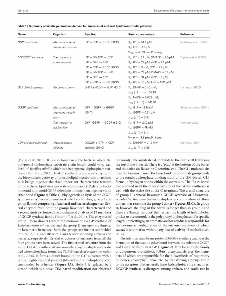
FIGURE 3 | Structure of 3-O-geranylgeranyl-sn-glyceryl-1-phosphate
synthase (GGGP synthase). (A) Crystal structure of group I dimericGGGP synthase from Archaeoglobus fulgidus solved at 2.0 Å resolution(PDB: 2F6X) has a novel TIM-barrel modification (Payandeh et al., 2006).Sn-glycerol-1-phosphate (G1P) and a cryoprotectant (MPD) used in theexperiment are colored as red and yellow spheres respectively. Thecavity for MPD likely represents the binding site for the secondsubstrate geranylgeranyl diphosphate (GGPP). (B) Crystal structure ofgroup II GGGP synthase from Methanothermobacter
thermoautotrophicus that forms a hexamer as a combination of threedimers was solved at 2.8 Å resolution (PDB: 4MM1; Peterhoff et al.,2014). G1P is displayed as red spheres. (C) A dimer from (B) isrepresented to compare with the dimeric group I GGGP synthase of(A). The G1P and triethelene glycol (PGE) are colored as red and yellowspheres respectively. The hexameric subunits are rotated to each otherunlike the dimers in (A). The anchoring of G1P in group I and II takesplace by the standard phosphate binding motif but their G1P bindingpocket are different.
identified in the genomes of Thaumarchaeota (Villanueva et al.,2014).
Unlike other enzymes of the pathway, DGGGP synthase has notbeen well characterized probably due to technical limitations withoverexpression of the membrane protein. The DGGGP synthaseactivity was first found in the membrane fraction of Methanother-mobacter marburgensis (Zhang and Poulter, 1993). Later the genewas identified in the genome of Sulfolobus solfataricus as UbiA-2, cloned in E. coli and purified to study the Mg2+ dependentenzymatic activity using radiolabeled substrates and mass spec-trometry (Hemmi et al., 2004). The ratio of the substrates utilizedin the reaction was found to be 1:1.1 in a double labeling exper-iment using [3H]GGPP and [14C]GGGP, respectively. Specificityfor GGPP and GGGP was also measured by substituting themwith different prenyl substrates, of which none of them were usedin the reaction by DGGGP synthase. In another study, DGGGPsynthase was shown to accept both the S and R form of GGGPshowing that unlike GGGP synthase, it is enantio unselective(Zhang et al., 2006). DGGGP synthase activity of Archaeoglobusfulgidus (Lai et al., 2009) and Methanosarcina acetivorans (Yokoiet al., 2012) was also observed in E. coli when the correspondinggenes were expressed along with four previous enzymes of thepathway. However, the expression level of the enzyme was eithertoo low to detect (Lai et al., 2009) or not investigated (Yokoi et al.,2012). In a later study, a higher expression level of DGGGP syn-thase of Archaeoglobus fulgidus was obtained in E. coli by changing
the ribosome-binding site and the activity of purified DGGGPsynthase was monitored (Jain et al., 2014).
CDP ARCHAEOL FORMATIONThe next step in the archaeal lipid biosynthetic pathway is theactivation of DGGGP by cytidine triphosphate (CTP) to form thesubstrate for polar head group attachment called cytidine diphos-phate (CDP)-archaeol (Figure 2; Table 1). The reaction is broughtabout by the enzyme CDP-archaeol synthase (CarS), the activity ofwhich was first studied in the membrane fraction of Methanother-mobacter thermoautotrophicus (Morii et al., 2000). Using varioussynthetic substrate analog, the activity was found to be specificfor unsaturated archaetidic acid with geranylgeranyl chains anddid not depend on the stereo specificity or ether/ester bond of thesubstrate. Minute amount of CDP-archaeol were also detected ingrowing cells labeled with inorganic 32P. The gene responsible forthis activity was only identified in a recent study (Jain et al., 2014).The enzyme CarS is conserved among archaea (except Nanoar-chaeota). However, like the enzyme DGGGP synthase, it couldnot be identified in the families of Thaumarchaeota.
Interestingly, an analogous reaction is found in the bacterialphospholipid biosynthetic pathway where phosphatidic acid isactivated by CTP to form CDP diacylglycerol by the enzyme CDPdiacylglycerol synthase (CdsA). Although the sequence similaritybetween CdsA and CarS is very low, hydropathy profile alignmentof the two families shows similarity in their secondary structure
Frontiers in Microbiology | Microbial Physiology and Metabolism November 2014 | Volume 5 | Article 641 | 6

Jain et al. Biosynthesis of archaeal membrane ether lipids
with overlapping transmembrane segments and cytoplasmic loopregions residing in the C-terminus half. CarS from Archaeoglobusfulgidus was expressed and purified from E. coli. Similar to CdsA,CarS activity was found to be dependent on Mg2+, both acceptsCTP and deoxycytidine triphosphate (dCTP) as substrates anddoes not utilize adenosine triphosphate (ATP), guanosine triphos-phate (GTP), or thymidine triphosphate (TTP) nucleotides in thereaction using substrate DGGGP. However, the two enzymes dis-played distinct activity with respect to the lipid substrate specificitywhere CarS only accepts unsaturated archaetidic acid with ger-anylgeranyl chains, while CdsA takes phosphatidic acid (Jain et al.,2014).
POLAR HEAD GROUP ATTACHMENTThe polar head groups serine, ethanolamine, glycerol and myo-inositol are found in the phospholipids in all three domainsof life. The enzymes that catalyze the replacement of thecytidine monophosphate (CMP) entity of CDP-archaeol or CDP-diacylglycerol with a polar head group are homologous andbelong to CDP-alcohol phosphatidyltransferase family (Koga,2011). Archaetidylserine (AS) synthase catalyzes the formationof AS from CDP-archaeol and L-serine (Figure 2) and is homol-ogous to bacterial phosphatidylserine (PS) synthase. The enzymecan be classified into two subclasses. Subclass I includes enzymesdistributed in Gram-negative bacteria, such as E. coli while sub-class II enzymes are widespread among Gram-positive bacteria(B. subtilis), yeast and archaea. Studies using cell free extractsof Methanothermobacter thermautotrophicus, B. subtilis, and E.coli showed that both the AS and PS synthase from Methan-othermobacter thermautotrophicus and B. subtilis have a broadsubstrate specificity and can accept lipid derivatives from archaeaor bacteria. On the other hand, the E. coli PS synthase wasspecific for bacterial lipid derivatives only (Morii and Koga,2003).
Archaetidylinositol phosphate (AI) synthase catalyzes the reac-tion where precursors L-myo-inositol-1-phosphate and CDP-archaeol are converted to AI phosphate as an intermediate which isfurther dephosphorylated to AI (Morii et al., 2009). This reactionis similar to the bacterial phosphatidylinositol phosphate (PI) syn-thase. Similar to AS and PS synthase, the AI and PI synthase showa broad substrate specificity accepting both, archaeal and bacte-rial lipid derivatives as substrates (Morii et al., 2014). Enzymeshomologous to PS decarboxylase and phosphatidylglycerol (PG)synthase have been identified in archaea as AS decarboxylaseand archaetidylglycerol (AG) synthase but not yet characterizedbiochemically (Daiyasu et al., 2005).
SATURATION OF DOUBLE BONDSThe mature phospholipids of archaea exist in their fully saturatedform. The archaeal enzyme digeranylgeranylglycerophospholipidreductase catalyzes the hydrogenation or saturation of the ger-anylgeranyl chains of unsaturated archaetidic acid (DGGGP) in astereospecific manner (Xu et al., 2010). It belongs to the geranyl-geranyl reductase (GGR) family that includes GGR from plant andprokaryotes that are mainly involved in photosynthesis. Prenylreductases other than GGRs are also found in all three domains oflife and these enzymes catalyze the complete or partial reduction
of isoprenoid compounds like respiratory quinones, tocopherol,dolichol, and other polyprenols (Ogawa et al., 2014).
The structures of the archaeal GGR monomer from Thermo-plasma acidophilum (Xu et al., 2010) and Sulfolobus acidocaldarius(Sasaki et al., 2011) show that they belong to p-hydroxybenzoatehydroxylase (PHBH) superfamily of flavoproteins (Figure 4A).The GGR from the thermophilic archaea Thermoplasma aci-dophilum was crystalized in complex with flavin adenine din-ucleotide (FAD) where FAD adopts the close confirmation thatpossibly changes with the binding of the substrate, like in othermembers of the PHBH family. The reduction of FAD is broughtabout by either NADH or other reducing agents. Since the proteinwas overexpressed in E. coli, a surrogate lipid-like ligand assignedas phosphatidylglycerol (PGX) was found in the active site formingan imperfect fit to the substrate binding pocket. The lipid bindingcavity of GGR is R shaped having two tunnels where the largertunnel B is more permissive than the smaller tunnel A which isrestricted in shape (Figure 4B). The S. acidocaldarius GGR is struc-turally similar to GGR from Thermoplasma acidophilum in FADbinding and the catalytic region (Figures 4C,D) but not in theC-terminal domain which is longer in S. acidocaldarius GGR. Theconserved sequence motif (YxWxFPx7-8GxG) lies in the large cav-ity of the catalytic domain and is thought to keep the substrate inposition for the reduction reaction as also indicated by mutationalstudies. Although the enzymes reduce GGGP, they also reduce thedouble bonds of related compounds like GGGP and GGPP (Sasakiet al., 2011). Another study where the Methanosarcina acetivoransGGR was expressed in E. coli along with four previous genes of thearchaeal lipid biosynthetic pathway, the DGGGP derivative witha fully saturated isoprenoid chain could be obtained (Isobe et al.,2014). Interestingly, the saturation only took place when GGR wascoexpressed with a ferredoxin gene found upstream of GGR inthe genome of Methanosarcina acetivorans, the ferredoxin possiblyfunctioning as a specific electron donor. However, no ferredoxincoexpression was required when the Methanosarcina acetivoransGGR was replaced by S. acidocaldarius GGR in the same study.Also, the conservation of the ferredoxin gene upstream of GGR inother archaea was not analyzed.
It is not known at what step of the biosynthetic pathway, hydro-genation takes place. However, since that CarS is specific for theunsaturated substrate, saturation probably takes place after the for-mation of CDP-archaeol. Although the enzyme AS synthase canaccept both saturated and unsaturated substrates for catalysis, thedetection of unsaturated AS in the cells of Methanothermobacterthermautotrophicus suggests that hydrogenation may already takeplace after the polar head groups are attached (Koga and Morii,2007).
TETRAETHER FORMATIONTetraether (caldarchaeol) lipid structure with varying number(0–8) of cyclopentane moieties are widespread among archaeaand a dominating membrane lipid structure in Crenarchaeotaand Thaumarchaeota. Euryarchaeota synthesize archaeol or botharchaeol and caldarchaeols. On the other hand, Thaumarchaeotahave characteristic tetraether lipids with four cyclopentane moi-eties and a cyclohexane moiety (Villanueva et al., 2014). One ofthe most intriguing steps in the archaeal biosynthetic pathway is
www.frontiersin.org November 2014 | Volume 5 | Article 641 | 7

Jain et al. Biosynthesis of archaeal membrane ether lipids
FIGURE 4 | Structure of geranylgeranyl reductase (GGR). (A) The crystalstructure of Sulfolobus acidocaldarius GGR at 1.85 Å resolution (PDB: 3ATQ)is shown (Sasaki et al., 2011) in complex with FAD (yellow). It is similar to thestructure of GGR from Thermoplasma acidophilum (PDB: 3OZ2; Xu et al.,2010) sharing the FAD binding and the catalytic domain. Both the enzymeswere crystallized where a lipid molecule (red) originating from the host
organism was found in the substrate binding site. (B) The lipid binding cavityof GGR is R shaped having two tunnels where the larger tunnel B is morepermissive and the smaller tunnel A is restricted in shape. One edge islocated at the branching point of the tunnel. (C,D) Comparison of the ‘nearperfect’ placement of the hydrocarbons of lipid moieties on the re face of theFAD from S. acidocaldarius GGR (C) andThermoplasma acidophilum GGR (D).
the tetraether formation. In vivo studies suggested that tetraethersare formed from saturated diethers via head to head condensationreaction. Pulse chase and labeling experiment of Thermoplasmaacidophilum cells with [14C]-MVA showed that the label firstincorporates into the archaeol until saturation and only theninto caldarchaeol. When an inhibitor of tetraether lipid synthe-sis (terbinafine) is used, pulse labeling leads to the accumulationof diethers and this phenomenon can be reversed by removal of theinhibitor (Nemoto et al., 2003). However, in another study, radi-olabelled archaeol was not incorporated into the tetraethers ofMethanospirillum hungatei cells and the presence of double bondswas necessary for the incorporation of labeled DGGGP into thetetraethers of Methanobacterium thermoautotrophicus cells (Poul-ter et al., 1988; Eguchi et al., 2000). The enzyme responsible for theformation of the presumed and unusual C–C bond for tetraethershas not been identified and there is no in vitro data to supportthis hypothesis (Koga and Morii, 2007). Recently, an alterna-tive pathway for tetraether and cyclopentane ring formation washypothesized (Villanueva et al., 2014). A multiple lock and keymechanism was proposed owing to the ‘greater functional plastic-ity’ of the enzymes IPP synthase, GGGP synthase, and DGGGPsynthase so that they can accommodate prenyl substrates with aring structure and chain length longer than C20. The cyclopentanerings would be formed early in the pathway before attachment ofthe glycerol moiety and the C20 geranyl molecules would coupletogether via a tail-to-tail mechanism to form the C40 phytoenechain by phytoene synthase, an enzyme that is wide spread inarchaea. However, both possibilities still need to be experimentallydemonstrated.
PHYSICOCHEMICAL PROPERTIES OF ARCHAEAL LIPIDSARCHAEAL MEMBRANE LIPID COMPOSITION – RESPONSE TO STRESSWithin the archaeal lipids there is a great diversity varyingin length, composition and configuration of the side chains(Figure 5). The most common archaeal core lipid is sn-2,3-diphytanylglycerol diether, generally called archaeol, which canundergo several modifications including hydroxylation and con-densation. Pioneering studies on archaeal membrane lipid compo-sition and biosynthesis have been performed in early eighties andnineties especially in halophiles using in vivo labeling experimentsby Kates and colleagues (Kates, 1992, 1993; Kamekura and Kates,1999) and reviewed in detail (Koga and Morii, 2007). Amongothers, the studies showed that halophiles are mainly character-ized by the phospholipid known as phosphatidylglycerol methylphosphate (PGP-Me) along with sulfated and desulfated archae-ols (Kates, 1993). Archaeol bearing elongated hydrocarbon chains(C20–C25) are found in some methanogens and halobacteri-ales (Figure 5A; Koga et al., 1993; Kamekura and Kates, 1999).A seemingly head-to-head condensation of two diether lipidmolecules is one of the most frequent and functionally impor-tant structural variations that leads to a glycerol-dialkyglyceroltetraether lipid, known as caldarcheol. It should be stressed, how-ever, that the enzymatic mechanism resulting in this lipid speciesis entirely unresolved. This core lipid is the most widespreadin Archaea, characterized by different modifications depend-ing on the archaeal species. It is in particular abundant amongthe phyla Euryarcheaota, Creanarchaeota, and Thaumarchaetoa.Up to 8 cyclopentane moieties can be found in lipids of theThermoplasmatales (Figure 5B) and in the Euryarchaeota phylum,
Frontiers in Microbiology | Microbial Physiology and Metabolism November 2014 | Volume 5 | Article 641 | 8

Jain et al. Biosynthesis of archaeal membrane ether lipids
FIGURE 5 | Structures of archaeal lipids. (A) Diether lipids (archaeol) withmodifications in chain length and with macrocyclic ring structure are mostlyfound in Euryarchaeota. (B) Tetraether lipids (caldarchaeol) can contain up to 8
cyclopentane ring moieties. (C) Crenarchaeol lipid structure withcyclopentane and cyclohexane rings found only in Thaumarchaeota.(D) Calditol derivative of tetraether lipid found in Sulfolobus spp.
in general (Macalady et al., 2004; UDA et al., 2004; Schoutenet al., 2013). Interestingly, the presence of cyclopentane andcyclohexane ring is a distinct feature of Thaumarchaeota lead-ing to a structure known as creanarcheol (Figure 5C; Pitcheret al., 2010; Damsté et al., 2012). In some thermoacidophilesand methanogens, a polar head group called nonitol is foundwhich is composed of nine-carbon chain. Recent studies haverevealed that the 9-carbon nonitol structure is often found as apolyhydroxylated cyclopentanic form called calditol (Koga andMorii, 2005). Therefore, these structure are now known asglycerol-dialkyl-calditol-tetraether (Figure 5D) and are the majorcomponent of the membrane of Sulfolobales species (Sugai et al.,1995; Untersteller et al., 1999; Gambacorta et al., 2002). Thepresence of tetraether lipids and the ratio of diether/tetraetherlipids vary depending on the archaeal species and also upongrowth conditions (Gambacorta et al., 1995). Likewise, there
is also a wide diversity of polar lipids in archaea includingphospholipids, glycolipids, phosphoglycolipids, sulpholipids, andaminolipids (Koga and Morii, 2005). The occurrence of dif-ferent polar head groups depends on the archaeal family andcan be used as unique taxonomic marker (Ulrih et al., 2009).Aminolipids, for instance, are prevalent in methanogens and arecompletely absent in halophiles and thermophiles (Gambacortaet al., 1995).
Bacteria and Eukarya use several mechanisms to maintain themembrane fluidity over a range of temperatures such the regula-tion of fatty acid composition adapting the degree of branching,saturation and chain length (Zhang and Rock, 2008). The home-oviscous adaption theory states that the lipid compositions inthe membrane varies in response to environmental stresses inorder to preserve a proper membrane fluidity (Oger and Cario,2013). However, the exact changes in fatty acid composition in
www.frontiersin.org November 2014 | Volume 5 | Article 641 | 9

Jain et al. Biosynthesis of archaeal membrane ether lipids
the membrane upon, for instance, a temperature shift differs fromspecies to species. In E. coli the degree of fatty acid unsaturationincreases along with a lowering of the temperature while someBacillus species increase the amount of iso-fatty acids with thegrowth temperatures. The membrane also has to maintain its per-meability barrier and in general it is believed that at the growthtemperature, the lipid bilayer is a liquid crystalline state. The phasetransition temperature at which the membrane is transferred fromthe crystalline into the liquid state is considered as a very impor-tant characteristic, and depends on the length of the hydrocarbonchains, the degree of saturation and the position of methyl groups;in Bacteria, the transition temperature ranges between −20 toup to 65◦C compared to archaea where the range is much widerbetween even up to 100◦C, a temperature where some archaeagrow (Koga, 2012).
Archaea use different mechanisms to maintain the liquid crys-talline phase over the entire growth temperature range. One con-trol mechanism has been reported in the archaeon Methanococcusjannaschii, in which case the membrane properties are finelytuned by varying the ratio of diether, macrocyclic diether, andtetraether lipid (Sprott et al., 1991). In contrast, at higher tem-peratures, hyperthermophilic archaea may incorporate a higherdegree of cyclopentane rings in their isoprenoid chain that increasethe transition temperature. The presence of such lipid structuresin archaea is an indication of a need to preserve the membranefunction at the hostile environmental conditions. In particular,the presence of tetraether lipids and the chemically stable etherbonds are major adaptions (Jacquemet et al., 2009). The latterconfers resistance to phospholipases attack from other organ-isms. Despite the general thought that the isoprenoid chains ofthe ether lipid are involved in thermal resistance, it has beenshown that they are not absolutely required for tolerance to hightemperature. Archaeol and caldarcheol were also found in thethermophilic methanoarchaeota (65◦C) and in mesophilic species(37◦C; Koga and Nakano, 2008). This suggests that the remark-able properties of the membranes of hyperthermophiles dependnot exclusively on the tetraether composition of their lipids butthat other aspects are involved as well. When fully stretched, thetetraether lipids span the entire membrane thickness leading toa monolayer, which is believed to stiffen the membrane in thepresence of high growth temperatures (Kates, 1992; Jacquemetet al., 2009). This may also protect the membrane from possiblelysis at high temperatures (Jacquemet et al., 2009). The presenceof cyclic structures, in particular cyclopentene rings (De Rosaet al., 1980), is a hallmark for high growth temperatures andcauses an increased membrane packing and thus a reduction inmembrane fluidity (Benvegnu et al., 2008). However, also thecharacteristics of the polar head groups may influence membranefluidity since the proper balance of negative and positive chargesat the membrane surface is essential for its functioning. There-fore, varying the proportion of different polar head groups mightbe another way to response to external stresses (Oger and Cario,2013). Furthermore, modification of the polar headgroups withcarbohydrates increases hydrogen bonding between the lipids andthus will influence the stability of the membranes. In halophiles,the presence of glycerol methylphosphate attached to the archaeolmoiety, contributes to the low membrane permeability under
high salt concentration (Tenchov et al., 2006). A further exampleis cold adaption in the psychrophile archaeon Methanococcoidesburtonii, where an increase in the degree of unsaturation in theisoprenoid chains allow growth at low temperatures as they exist inglacial environments (Russell and Nichols, 1999; Oger and Cario,2013).
IN VITRO BASED STUDIES ON THE STABILITY OF ARCHAEAL LIPIDSTemperature impacts the membrane properties, influencing theion permeability and phase behavior. It is believed that the uniquemembrane organization of archaeal tetraether lipid in a mono-layer structure along with the presence of ether bonds renderssuch membranes, thermal resistance. Therefore, many studieshave been conducted to understand the higher thermal stabilityof archaeal membrane lipids compared to the bacterial phospho-lipids. By comparing liposomes made of a polar lipid fractionfrom S. acidocaldarius and liposomes prepared from a bacte-rial lipid (POPC) or a synthetic lipid with a phytanyl chain(DPhPC), the importance of the methyl branched isoprenoidchain in membrane stability has been examined (Chang, 1994).When incubated at 100◦C for approximately half hour, archaealliposomes showed a very low ion leakage compared to POPC andDPhPC liposomes that collapse after a few minutes incubationat that temperature. Likewise, only very slow release (8–10%) ofthe fluorophore carboxyfluorescin (CF) was observed with lipo-somes composed of S. acidocaldarius lipids compared to E. coliliposomes (50%) over a time interval of 62 days, while lipo-somes composed of a lipid extract from the thermophile bacteriumB. stearothermophilus showed an intermediate stability (Elferinket al., 1994).
The extremely high heat tolerance of archaeal liposomes openopportunities for biotechnological applications with the abilityto be stable even after several autoclaving cycles, which mightbe exploited for biomedical uses. Autoclaving is a common andeffective method for decontamination and the possibility to auto-clave the archaeosomes vesicle for the drug delivery gives newprospects for the liposomes formulation. Several studies have beenperformed regarding the ability of archaeosomes to enhance theimmune response when used as novel vaccine and drug deliverysystem (Sprott et al., 1997; Patel and Sprott, 1999; Patel et al., 2000).The superior adjuvant activity of archaeosomes compared to theliposomes evokes sufficient immunostimulation and sustainedimmune response against cancer or specific infectious diseases.Archaeosomes confer higher stability to the vesicles enhancing thefusion with the immune cells (Krishnan and Sprott, 2008). Theseproperties are dependent on the type of polar head groups anddegrees of glycosylation on archaeal lipids (Dicaire et al., 2010;Whitfield et al., 2010). An enhanced cytotoxic cell response wasin fact observed in archaeosomes enriched with archaeatidylserineand archaeatidylethanomine due to their fusogenic nature (Dicaireet al., 2010; Sprott et al., 2012). The effective archaeosomes stabil-ity against autoclaving was tested showing a remarkable strengthagainst 2–3 cycles of autoclaving at pH 4.0–10.0 (Brown et al.,2009). Besides the high heat tolerance, archaeal lipids are known tobe resistant to conditions of extremely low pH and their low protonpermeability contributes to maintaining a constant intracellularpH. Monolayer liposomes reconstituted from the lipid fraction of
Frontiers in Microbiology | Microbial Physiology and Metabolism November 2014 | Volume 5 | Article 641 | 10

Jain et al. Biosynthesis of archaeal membrane ether lipids
S. acidocaldarius indeed exhibited very low proton permeabilityeven at higher temperatures (Elferink et al., 1994; Komatsu andChong, 1998). However, at acidic conditions the archaeosomesappeared less resistant to autoclaving possibly because of a higherprotonation of the polar head groups, influencing hydrogen-bonds formation among the lipids. Liposomes containing longsugar chains linked to the phospholipids show much lower protonpermeability than liposomes composed of lipids with only onesugar unit. The glycolipids amount in the polar lipid fraction ofThermoplasma acidophilum increases at lower pH and this seemsa general mechanism for acidophiles against the chemically unsta-ble conditions (Shimada et al., 2008). The low permeability ofsuch liposomes can be exploited for drug-delivery with the addedvalue of a high resistance against phospholipase A2, B and pancre-atic lipase (Choquet et al., 1994). Due to their high pH tolerancethey can easily pass the gastro-intestinal tract without damage.Overall, these studies confirm that the presence of tetraethers inarchaeal membranes confers these membranes with a remark-able stability against high temperatures, low pH, and high salt.The degree of hydrocarbon chain saturation, the features of thepolar head groups and the presence of cyclopentane ring (Dannen-muller et al., 2000) appear of secondary importance in providingstability.
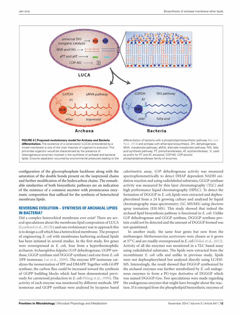
THE LIPID DIVIDEPROSPECT OF LUCA WITH MIXED MEMBRANE LIPID COMPOSITIONDuring the last decades many theories have been proposed aboutthe origin of life and how the differentiation between the threedomains of life occurred. All of these theories accept the existenceof the last universal common ancestor (LUCA), also known asLUCAS or cenancestor from which organisms have diverged. Par-ticular attention has been given on the membrane composition ofLUCA. Isoprenoids are involved in a wide range of functions, andfound both in archaea and bacteria. Whereas fatty acid metabolismis also widely distributed, fatty acid biosynthesis seems underde-veloped in archaea and in some organisms even absent. Therefore,one of the hypothesis is that early life forms were dependent on thepresence of membrane lipids with isoprenoid hydrocarbon core.However, the most divergent feature that is at the base of the LipidDivide is the glycerophosphate backbone. Archaea contains G1Pas glycerophosphate moiety while bacteria depend on G3P. Thesetwo compounds are synthetized by two different enzymes that arenot evolutionary correlated (Koga et al., 1998).
Thus, different hypothesis were suggested to understand theprocess that brought the ancestor to archaeal, bacterial and eukary-otic organisms. According to Koga et al. (1998) the evolution ofthe two phospholipid pathways occurred independently leading tothe simultaneous appearance of bacterial and archaeal enzymes.This contrast the theory of Martin and Russell (2003), accordingto whom the two different organisms evolved from an ances-tor characterized by iron monosulphide compartment. Anotherhypothesis (Wächtershäuser, 2003) proposed a three stage pro-cess from the pre-cell to the eukaryotic cell. It was suggested thatthere was a cenancestor with a chemically derived heterochiralmembrane containing both the enantiomeric forms of the glyc-erophosphate backbone, which slowly diverged into a more stablehomochiral membrane organism leading to the differentiation
between Bacteria and Archaea. This idea implies that such het-erochiral mixed membranes are intrinsically unstable leading tothe emergence of chiral selective enzymes and a divide betweenbacteria and archaea. However, hybrid membranes were testedfor their stability using a mixture of egg phosphatidylcholine andextracted lipid from Sulfolobus solfataricus (Fan et al., 1995) anda higher stability compared to liposomes reconstituted of onlyarchaeal lipid was observed. Moreover a heterochiral membraneconsisting of bacterial G3P lipids and archaeal G1P lipids wasanalyzed (Shimada and Yamagishi, 2011) and the heterochiralmembranes were found to be more stable at higher temperaturescompared to liposomes prepared from only the bacterial lipids.Thus, based on these studies, the hypothesis of the existence ofan ancestor with both the G1PDH and G3PDH would be pos-sible but there must have been other factors that have driven thesegregation into the two different domains. The theory of the coex-istence of both the enzymes for the glycerophosphate enantiomersproductions can be also extended to the other enzymes involvedeither in the synthesis of isoprenoid and fatty acid based phos-pholipids (Figure 6). In LUCA, four different membrane lipidsmay be obtained by the combination of the two glycerophos-phate backbones with either isoprenoid or fatty acid hydrocarbonchains (Koga,2011). Environmental pressure and the need to adaptto extreme conditions may have induced archaea to evolve theirmembranes (Valentine, 2007; Lombard et al., 2012b). In particular,the use of different hydrocarbon chains in response to the growthenvironment may have induced the segregation in Archaea andBacteria and resulted in homochiral membrane formation (Koga,2014).
DIFFERENTIATION OF MEMBRANE LIPIDS IN ARCHAEA AND BACTERIAThe structural variability found in the membrane lipid composi-tion of archaea and bacteria reflects differences and similaritiesin the respective biosynthetic pathways. When compared withthe well-characterized bacterial ester-lipid biosynthetic pathway,several similarities with the archaeal ether-lipid biosynthesis areevident.
First, the sequence of reactions that yield the final membranelipid from the building blocks is basically the same even thoughsome enzymes involved in these reactions are equipped with spe-cific features to the lipid of the two different domains. Second,the glycerophosphate backbone in both cases is synthetized by areduction of DHAP at the 2-OH using NADH as cofactor whilethe two hydrocarbon chains are linked to the same position onthe glycerol moiety. Third, the polar head attachment takes placevia a CDP-activated intermediate (Koga, 2014). In particular thepeculiar features that distinguish archaeal lipids from bacterialones occur in the first half of the biosynthetic pathway while thelast stages, which involve the replacement of the CDP GROUPwith one of the polar groups, is essentially similar in these twodomains of life. For the latter reactions, the enzymes belong to thesame superfamily and share sequence similarity (Daiyasu et al.,2005; Koga, 2011). On the other hand, the isoprenoid buildingblocks synthesis differs in Bacteria and in Archaea since it takesplace via two different pathway, the DOXP and the alternate MVApathway, respectively (Koga and Morii, 2007). The other strik-ing difference involved in the lipid divide is the enantiomeric
www.frontiersin.org November 2014 | Volume 5 | Article 641 | 11
Jain et al. Biosynthesis of archaeal membrane ether lipids
FIGURE 6 | Proposed evolutionary model for Archaea and Bacteria
differentiation. The existence of a cenancestor (LUCA) embordered by amixed membrane is one of the main theories of organism’s evolution. Thisprimordial organism would be characterized by the presence ofheterogeneous enzymes involved in the synthesis of archaeal and bacteriallipids. Enzyme separation occurred by environmental pressures leading to the
differentiation of bacteria with a phospholipid biosynthetic pathway (Yao andRock, 2013) and archaea with ether-lipid biosynthesis. DH, dehydrogenase;MVA, mevalonate pathway; aMVA, alternate mevalonate pathway; FAS, fattyacid synthesis pathway; PT, prenyltransferases; AT, acyltransferases; ‘a’ usedas prefix for PT and AT, ancestral; CDP-AD, CDP-alcoholphosphatidyltransferase family of enzymes.
configuration of the glycerophosphate backbone along with thesaturation of the double bonds present on the isoprenoid chainsand further modification of the hydrocarbon chains. The remark-able similarities of both biosynthetic pathways are an indicationof the existence of a common ancestor with promiscuous enzy-matic composition that sufficed for the synthesis of heterochiralmembrane lipids.
REVERSING EVOLUTION – SYNTHESIS OF ARCHAEAL LIPIDSIN BACTERIA?Did a complex heterochiral membrane ever exist? There are sev-eral speculations about the membrane lipid composition of LUCA(Lombard et al., 2012b) and one evolutionary way to approach thisis to design a cell which has a heterochiral membrane. The prospectof engineering E. coli with membranes harboring archaeal lipidshas been initiated in several studies. In the first study, five geneswere overexpressed in E. coli, four from a hyperthermophilicarchaeon Archaeoglobus fulgidus (G1P dehydrogenase, GGPP syn-thase, GGGP synthase and DGGGP synthase) and one from E. coli(IPP isomerase; Lai et al., 2009). The enzyme IPP isomerase cat-alyzes the isomerization of IPP and DMAPP. Together with GGPPsynthase, the carbon flux could be increased toward the synthesisof GGPP building blocks which had been demonstrated previ-ously for carotenoid production in E. coli (Wang et al., 1999). Theactivity of each enzyme was monitored by different methods. IPPisomerase and GGPP synthase were analyzed by lycopene based
calorimetric assay, G1P dehydrogenase activity was measuredspectrophotometrically to detect DHAP dependent NADH oxi-dation reaction and using radiolabeled substrates, GGGP synthaseactivity was measured by thin layer chromatography (TLC) andhigh performance liquid chromatography (HPLC). To detect theformation of DGGGP in E. coli, lipids were extracted and dephos-phorylated from a 24 h growing culture and analyzed by liquidchromatography mass spectrometry (LC-MS/MS) using electronspray ionisation (ESI-MS). This study showed that indeed thearchaeal lipid biosynthesis pathway is functional in E. coli. UnlikeG1P dehydrogenase and GGGP synthase, DGGGP synthase pro-tein could not be detected and the amount of DGGGP formed wasnot quantitated.
In another study, the same four genes but now from themethanogen Methanosarcina acetivorans were chosen as it growsat 37◦C and are readily overexpressed in E. coli (Yokoi et al., 2012).Activity of all the enzymes was monitored in a TLC based assayusing radiolabeled substrates. The lipids were extracted from therecombinant E. coli cells and unlike in previous study, lipidswere not dephosphorylated but analyzed directly using LC/ESI-MS. Interestingly, the result showed that DGGGP synthesized bythe archaeal enzymes was further metabolized by E. coli endoge-nous enzymes to form a PG-type derivative of DGGGP whichwas named DGGGP-Gro. Two speculations were made regardingthe endogenous enzymes that might have brought about the reac-tion. If it emerged from the phospholipid biosynthetic enzymes of
Frontiers in Microbiology | Microbial Physiology and Metabolism November 2014 | Volume 5 | Article 641 | 12

Jain et al. Biosynthesis of archaeal membrane ether lipids
E. coli, it would require three enzymes to recognize and accept thearchaeal substrate, namely CdsA, sn-G3P transferase and the phos-phatase. However, addition of CTP to their in vitro TLC based assaydid not increase the amount of product formation and no otherpolar head group attachment was observed. The other possibilityis that the sn-1-phosphoglycerol group from the osmoregulatedperiplasmic glucans was transferred to digeranylgeranylglycerol(DGGGOH) directly by the phosphoglyceroltransferase system.The estimated amount of total archaeal membrane lipids extractedfrom E. coli cells in this study was only 60 μg/g wet cells and atthese levels it is not possible to study the influence of archaeallipids on the physical properties of the cytoplasmic membrane. Ina follow up study, GGR and ferredoxin (see section saturation ofdouble bonds) were introduced along with the other genes fromMethanosarcina acetivorans and expressed in E. coli. The formationof DGGGP-Gro was reduced yielding mostly saturated archaetidicacid in E. coli (Isobe et al., 2014).
The archaeal lipids have also been synthesized in vitro usinga set of five purified enzymes, two from bacteria and three fromarchaea. A mutant of FPP synthase of E. coli was used that wasshown previously to synthesize GGPP, G1P dehydrogenase wasfrom B. subtilis, GGGP synthase from methanogen Methanococcusmaripaludis, DGGGP synthase and CarS from the hyperther-mophilic Archaeoglobus fulgidus. All enzymes were purified, andby using substrates IPP, FPP, DHAP, and NADH, the enzymeswere shown to be able to synthesize CDP-archaeol in the presenceof CTP, Mg2+ and detergent at 37◦C (Jain et al., 2014). The fea-sibility of synthesizing archaeal lipids in E. coli and in vitro arepromising first steps toward deciphering the biosynthetic pathwayfurther and eventually understanding the properties of a cell witha heterochiral membrane lipid composition.
FUTURE CHALLENGESAlthough during the last decade, many of the intimate features ofthe archaeal lipid biosynthesis pathway have been resolved, thereare still several important questions that need to be answered.Understanding the mechanism of tetraether formation and iden-tifying the enzyme(s) involved in the reaction requires thoroughinvestigations. In vitro analysis of such a reaction(s) would be greatadvancement in the field. Various other derivatives of diether andtetraether lipids like cyclopentane and macrocyclic ring forma-tion, glycosylation and formation of crenarchaeol are also not wellunderstood. The pathway refractory of archaeal lipid biosynthesisin E. coli is currently incomplete where the amount of archaeallipids formed in comparison to the E. coli lipids is very low.Challenging aspect is to modulate and suppress the endogenouspathway and integrate the archaeal lipid biosynthesis pathway inthe genome of the host yielding the exclusive formation of theselipid species. Further structural and biochemical analysis of theenzymes of the pathway from different families of archaea wouldprogress the field and bring it in par with the understanding ofbacterial phospholipid biosynthesis. Also, regulation of the phos-pholipid metabolism is poorly understood and could be enhancedthrough the use of genetic studies, which now became feasiblebecause of the rapid developments in genetic toolbox in archaea.However, to study essential genes using these techniques is still achallenge.
Recent studies have shown that in spite of the uniqueness ofthe archaeal membrane lipid structure, they are not as distinct aspreviously thought. The presence of fatty acids and isoprenoidsin the three domains of life and the common mode of polar headgroup attachment in bacteria and archaea, and the presence ofhomologues of archaeal G1P dehydrogenase and GGGP synthasein bacteria are a few of the similarities. Other question that stillremains unanswered is the exact reason why G1P is ether linkedto the isoprenoid hydrocarbon chains and G3P is linked via esterbonds to the fatty acid chains; are there still organisms with suchmixed membranes? In order to answer such questions, more bio-chemical and functional investigation are needed on the archaeallipid biosynthetic pathway along with a deep phylogenetic analysis.
ACKNOWLEDGMENTSSamta Jain and Antonella Caforio are supported by the researchprogram of the biobased ecologically balanced sustainable indus-trial chemistry (BE-BASIC). We thank Ilja Kusters, John van derOost, Melvin Siliakus and Servè Kengen for fruitful discussions.
REFERENCESBenvegnu, T., Lemiègre, L., and Cammas-Marion, S. (2008). Archaeal lipids: inno-
vative materials for biotechnological applications. Eur. J. Org. Chem. 2008,4725–4744. doi: 10.1002/ejoc.200800452
Boucher, Y., Kamekura, M., and Doolittle, W. F. (2004). Origins and evolutionof isoprenoid lipid biosynthesis in archaea. Mol. Microbiol. 52, 515–527. doi:10.1111/j.1365-2958.2004.03992.x
Brown, D. A., Venegas, B., Cooke, P. H., English, V., and Chong, P. L.-G. (2009).Bipolar tetraether archaeosomes exhibit unusual stability against autoclaving asstudied by dynamic light scattering and electron microscopy. Chem. Phys. Lipids159, 95–103. doi: 10.1016/j.chemphyslip.2009.03.004
Chang, E. L. (1994). Unusual thermal stability of liposomes made from bipo-lar tetraether lipids. Biochem. Biophys. Res. Commun. 202, 673–679. doi:10.1006/bbrc.1994.1983
Chen, A., and Poulter, C. D. (1993). Purification and characterization of farnesyldiphosphate/geranylgeranyl diphosphate synthase. A thermostable bifunctionalenzyme from Methanobacterium thermoautotrophicum. J. Biol. Chem. 268, 11002–11007.
Chen, M., and Poulter, C. D. (2010). Characterization of thermophilic archaealisopentenyl phosphate kinases. Biochemistry 49, 207–217. doi: 10.1021/bi9017957
Choquet, C. G., Patel, G. B., Sprott, G. D., and Beveridge, T. J. (1994). Stabilityof pressure-extruded liposomes made from archaeobacterial ether lipids. Appl.Microbiol. Biotechnol. 42, 375–384. doi: 10.1007/BF00902745
Daiyasu, H., Kuma, K.-I., Yokoi, T., Morii, H., Koga, Y., and Toh, H. (2005). A studyof archaeal enzymes involved in polar lipid synthesis linking amino acid sequenceinformation, genomic contexts and lipid composition. Archaea 1, 399–410. doi:10.1155/2005/452563
Damsté, J. S. S., Rijpstra, W. I. C., Hopmans, E. C., Jung, M.-Y., Kim, J.-G., Rhee,S.-K., et al. (2012). Intact polar and core glycerol dibiphytanyl glycerol tetraetherlipids of group I.1a and I.1b thaumarchaeota in soil. Appl. Environ. Microbiol. 78,6866–6874. doi: 10.1128/AEM.01681-12
Dannenmuller, O., Arakawa, K., Eguchi, T., Kakinuma, K., Blanc, S.,Albrecht, A. M., et al. (2000). Membrane properties of archaeal macro-cyclic diether phospholipids. Chemistry 6, 645–654. doi: 10.1002/(SICI)1521-3765(20000218)6:4 < 645::AID-CHEM645 > 3.0.CO;2-A
Dellas, N., and Noel, J. P. (2010). Mutation of archaeal isopentenyl phosphate kinasehighlights mechanism and guides phosphorylation of additional isoprenoidmonophosphates. ACS Chem. Biol. 5, 589–601. doi: 10.1021/cb1000313
Dellas, N., Thomas, S. T., Manning, G., and Noel, J. P. (2013). Discovery of ametabolic alternative to the classical mevalonate pathway. Elife 2, e00672. doi:10.7554/eLife.00672
De Rosa, M., Esposito, E., Gambacorta, A., Nicolaus, B., and Bu’Lock, J. D. (1980).Effects of temperature on ether lipid composition of Caldariella acidophila.Phytochemistry 19, 827–831. doi: 10.1016/0031-9422(80)85120-X
www.frontiersin.org November 2014 | Volume 5 | Article 641 | 13

Jain et al. Biosynthesis of archaeal membrane ether lipids
Dicaire, C. J., Yu, S. H., Whitfield, D. M., and Sprott, G. D. (2010).Isopranoid- and dipalmitoyl-aminophospholipid adjuvants impact differentlyon longevity of CTL immune responses. J. Liposome Res. 20, 304–314. doi:10.3109/08982100903544151
Eguchi, T., Takyo, H., Morita, M., Kakinuma, K., and Koga, Y. (2000). Unusualdouble-bond migration as a plausible key reaction in the biosynthesis of theisoprenoidal membrane lipids of methanogenic archaea. Chem. Commun. 2000,1545–1546. doi: 10.1039/b003948i
Elferink, M. G. L., de Wit, J. G., Driessen, A. J. M., and Konings, W. N. (1994).Stability and proton-permeability of liposomes composed of archaeal tetraetherlipids. Biochim. Biophys. Acta Biomembr. 1193, 247–254. doi: 10.1016/0005-2736(94)90160-0
Fan, Q., Relini, A., Cassinadri, D., Gambacorta, A., and Gliozzi, A. (1995).Stability against temperature and external agents of vesicles composed ofarchael bolaform lipids and egg PC. Biochim. Biophys. Acta 1240, 83–88. doi:10.1016/0005-2736(95)00157-X
Fujiwara, S., Yamanaka, A., Hirooka, K., Kobayashi, A., Imanaka,T., and Fukusaki, E.-I. (2004). Temperature-dependent modulation offarnesyl diphosphate/geranylgeranyl diphosphate synthase from hyperther-mophilic archaea. Biochem. Biophys. Res. Commun. 325, 1066–1074. doi:10.1016/j.bbrc.2004.10.129
Gambacorta, A., Caracciolo, G., Trabasso, D., Izzo, I., Spinella, A., and Sodano,G. (2002). Biosynthesis of calditol, the cyclopentanoid containing moiety of themembrane lipids of the archaeon Sulfolobus solfataricus. Tetrahedron Lett. 43,451–453. doi: 10.1016/S0040-4039(01)02187-6
Gambacorta, A., Gliozzi, A., and De Rosa, M. (1995). Archaeal lipids and theirbiotechnological applications. World J. Microbiol. Biotechnol. 11, 115–131. doi:10.1007/BF00339140
Grochowski, L. L., Xu, H., and White, R. H. (2006). Methanocaldococcus jannaschiiuses a modified mevalonate pathway for biosynthesis of isopentenyl diphosphate.J. Bacteriol. 188, 3192–3198. doi: 10.1128/JB.188.9.3192-3198.2006
Guldan, H., Matysik, F.-M., Bocola, M., Sterner, R., and Babinger, P. (2011).Functional assignment of an enzyme that catalyzes the synthesis of an archaea-type ether lipid in bacteria. Angew. Chem. Int. Ed. Engl. 50, 8188–8191. doi:10.1002/anie.201101832
Guldan, H., Sterner, R., and Babinger, P. (2008). Identification and characterizationof a bacterial glycerol-1-phosphate dehydrogenase: Ni2+-dependent AraM fromBacillus subtilis. Biochemistry 47, 7376–7384. doi: 10.1021/bi8005779
Han, J.-S., and Ishikawa, K. (2005). Active site of Zn(2+)-dependent sn-glycerol-1-phosphate dehydrogenase from Aeropyrum pernix K1. Archaea 1, 311–317. doi:10.1155/2005/257264
Han, J.-S., Kosugi, Y., Ishida, H., and Ishikawa, K. (2002). Kinetic study ofsn-glycerol-1-phosphate dehydrogenase from the aerobic hyperthermophilicarchaeon, Aeropyrum pernix K1. Eur. J. Biochem. 269, 969–976. doi:10.1046/j.0014-2956.2001.02731.x
Hemmi, H., Ikejiri, S., Yamashita, S., and Nishino, T. (2002). Novel medium-chainprenyl diphosphate synthase from the thermoacidophilic archaeon Sulfolobussolfataricus. J. Bacteriol. 184, 615–620. doi: 10.1128/JB.184.3.615-620.2002
Hemmi, H., Shibuya, K., Takahashi, Y., Nakayama, T., and Nishino, T. (2004).(S)-2,3-Di-O-geranylgeranylglyceryl phosphate synthase from the thermoaci-dophilic archaeon Sulfolobus solfataricus. Molecular cloning and characteri-zation of a membrane-intrinsic prenyltransferase involved in the biosynthe-sis of archaeal ether-linked memb. J. Biol. Chem. 279, 50197–50203. doi:10.1074/jbc.M409207200
Isobe, K., Ogawa, T., Hirose, K., Yokoi, T., Yoshimura, T., and Hemmi, H. (2014).Geranylgeranyl reductase and ferredoxin from Methanosarcina acetivorans arerequired for the synthesis of fully reduced archaeal membrane lipid in Escherichiacoli cells. J. Bacteriol. 196, 417–423. doi: 10.1128/JB.00927-13
Jacquemet, A., Barbeau, J., Lemiègre, L., and Benvegnu, T. (2009). Archaealtetraether bipolar lipids: structures, functions and applications. Biochimie 91,711–717. doi: 10.1016/j.biochi.2009.01.006
Jain, S., Caforio, A., Fodran, P., Lolkema, J. S., Minnaard, A. J., and Driessen,A. J. M. (2014). Identification of CDP-archaeol synthase, a missing linkof ether lipid biosynthesis in archaea. Chem. Biol. 21, 1392–1401. doi:10.1016/j.chembiol.2014.07.022
Kamekura, M., and Kates, M. (1999). Structural diversity of membrane lipids inmembers of Halobacteriaceae. Biosci. Biotechnol. Biochem. 63, 969–972. doi:10.1271/bbb.63.969
Kates, M. (1992). Archaebacterial lipids: structure, biosynthesis and function.Biochem. Soc. Symp. 58, 51–72.
Kates, M. (1993). Biology of halophilic bacteria, Part II. Membrane lipids of extremehalophiles: biosynthesis, function and evolutionary significance. Experientia 49,1027–1036. doi: 10.1007/BF01929909
Koga, Y. (2011). Early evolution of membrane lipids: how did the lipid divide occur?J. Mol. Evol. 72, 274–282. doi: 10.1007/s00239-011-9428-5
Koga, Y. (2012). Thermal adaptation of the archaeal and bacterial lipid membranes.Archaea 2012, 789652. doi: 10.1155/2012/789652
Koga, Y. (2014). From promiscuity to the lipid divide: on the evolution of dis-tinct membranes in archaea and bacteria. J. Mol. Evol. 78, 234–242. doi:10.1007/s00239-014-9613-4
Koga, Y., Kyuragi, T., Nishihara, M., and Sone, N. (1998). Did archaeal and bacte-rial cells arise independently from noncellular Precursors? A hypothesis statingthat the advent of membrane phospholipid with enantiomeric glycerophosphatebackbones caused the separation of the two lines of descent. J. Mol. Evol. 47, 631.doi: 10.1007/PL00006419
Koga, Y., and Morii, H. (2005). Recent advances in structural research on ether lipidsfrom archaea including comparative and physiological aspects. Biosci. Biotechnol.Biochem. 69, 2019–2034. doi: 10.1271/bbb.69.2019
Koga, Y., and Morii, H. (2007). Biosynthesis of ether-type polar lipids in archaeaand evolutionary considerations. Microbiol. Mol. Biol. Rev. 71, 97–120. doi:10.1128/MMBR.00033-06
Koga, Y., and Nakano, M. (2008). A dendrogram of archaea based on lipid com-ponent parts composition and its relationship to rRNA phylogeny. Syst. Appl.Microbiol. 31, 169–182. doi: 10.1016/j.syapm.2008.02.005
Koga, Y., Nishihara, M., Morii, H., and Akagawa-Matsushita, M. (1993). Etherpolar lipids of methanogenic bacteria: structures, comparative aspects, andbiosyntheses. Microbiol. Rev. 57, 164–182.
Koga, Y., Ohga, M., Tsujimura, M., Morii, H., and Kawarabayasi, Y. (2006).Identification of sn-glycerol-1-phosphate dehydrogenase activity from genomicinformation on a hyperthermophilic archaeon, Sulfolobus tokodaii strain 7. Biosci.Biotechnol. Biochem. 70, 282–285. doi: 10.1271/bbb.70.282
Koga, Y., Sone, N., Noguchi, S., and Morii, H. (2003). Transfer of pro-R hydrogenfrom NADH to dihydroxyacetonephosphate by sn-glycerol-1-phosphate dehy-drogenase from the archaeon Methanothermobacter thermautotrophicus. Biosci.Biotechnol. Biochem. 67, 1605–1608. doi: 10.1271/bbb.67.1605
Komatsu, H., and Chong, P. L. (1998). Low permeability of liposomal membranescomposed of bipolar tetraether lipids from thermoacidophilic archaebacteriumSulfolobus acidocaldarius. Biochemistry 37, 107–115. doi: 10.1021/bi972163e
Krishnan, L., and Sprott, G. D. (2008). Archaeosome adjuvants: immunolog-ical capabilities and mechanism(s) of action. Vaccine 26, 2043–2055. doi:10.1016/j.vaccine.2008.02.026
Lai, D., Lluncor, B., Schröder, I., Gunsalus, R. P., Liao, J. C., and Monbouquette,H. G. (2009). Reconstruction of the archaeal isoprenoid ether lipid biosynthesispathway in Escherichia coli through digeranylgeranylglyceryl phosphate. Metab.Eng. 11, 184–191. doi: 10.1016/j.ymben.2009.01.008
Lee, P. C., Petri, R., Mijts, B. N., Watts, K. T., and Schmidt-Dannert, C. (2005).Directed evolution of Escherichia coli farnesyl diphosphate synthase (IspA) revealsnovel structural determinants of chain length specificity. Metab. Eng. 7, 18–26.doi: 10.1016/j.ymben.2004.05.003
Lombard, J., López-garcía, P., and Moreira, D. (2012a). Phylogenomic inves-tigation of phospholipid synthesis in archaea. Archaea 2012, 630910. doi:10.1155/2012/630910
Lombard, J., López-García, P., and Moreira, D. (2012b). The early evolution of lipidmembranes and the three domains of life. Nat. Rev. Microbiol. 10, 507–515. doi:10.1038/nrmicro2815
Lombard, J., and Moreira, D. (2011). Origins and early evolution of the mevalonatepathway of isoprenoid biosynthesis in the three domains of life. Mol. Biol. Evol.28, 87–99. doi: 10.1093/molbev/msq177
Macalady, J. L., Vestling, M. M., Baumler, D., Boekelheide, N., Kaspar, C. W., andBanfield, J. F. (2004). Tetraether-linked membrane monolayers in Ferroplasmaspp: a key to survival in acid. Extremophiles 8, 411–419. doi: 10.1007/s00792-004-0404-5
Martin, W., and Russell, M. J. (2003). On the origins of cells: a hypothesis forthe evolutionary transitions from abiotic geochemistry to chemoautotrophicprokaryotes, and from prokaryotes to nucleated cells. Philos. Trans. R. Soc. Lond.B Biol. Sci. 358, 59–83; discussion 83–85. doi: 10.1098/rstb.2002.1183
Frontiers in Microbiology | Microbial Physiology and Metabolism November 2014 | Volume 5 | Article 641 | 14

Jain et al. Biosynthesis of archaeal membrane ether lipids
Matsumi, R., Atomi, H., Driessen, A. J. M., and van der Oost, J. (2011). Iso-prenoid biosynthesis in archaea–biochemical and evolutionary implications. Res.Microbiol. 162, 39–52. doi: 10.1016/j.resmic.2010.10.003
Morii, H., Kiyonari, S., Ishino, Y., and Koga, Y. (2009). A novel biosyntheticpathway of archaetidyl-myo-inositol via archaetidyl-myo-inositol phosphatefrom CDP-archaeol and D-glucose 6-phosphate in methanoarchaeon Methan-othermobacter thermautotrophicus cells. J. Biol. Chem. 284, 30766–30774. doi:10.1074/jbc.M109.034652
Morii, H., and Koga, Y. (2003). CDP-2,3-Di-O-geranylgeranyl-sn-glycerol:L-serine O-archaetidyltransferase (archaetidylserine synthase) in the methanogenicarchaeon Methanothermobacter thermautotrophicus. J. Bacteriol. 185, 1181–1189.doi: 10.1128/JB.185.4.1181-1189.2003
Morii, H., Nishihara, M., and Koga, Y. (2000). CTP:2,3-di-O-geranylgeranyl-sn-glycero-1-phosphate cytidyltransferase in the methanogenic archaeon Methan-othermobacter thermoautotrophicus. J. Biol. Chem. 275, 36568–36574. doi:10.1074/jbc.M005925200
Morii, H., Ogawa, M., Fukuda, K., and Taniguchi, H. (2014). Ubiquitous dis-tribution of phosphatidylinositol phosphate synthase and archaetidylinositolphosphate synthase in bacteria and archaea, which contain inositol phospholipid.Biochem. Biophys. Res. Commun. 443, 86–90. doi: 10.1016/j.bbrc.2013.11.054
Nemoto, N. (2003). Purification and characterization of geranylgeranylglyc-eryl phosphate synthase from a thermoacidophilic archaeon, Thermoplasmaacidophilum. J. Biochem. 133, 651–657. doi: 10.1093/jb/mvg083
Nemoto, N., Shida, Y., Shimada, H., Oshima, T., and Yamagishi, A. (2003).Characterization of the precursor of tetraether lipid biosynthesis in the ther-moacidophilic archaeon Thermoplasma acidophilum. Extremophiles 7, 235–243.doi: 10.1007/s00792-003-0315-x
Nishihara, M., and Koga, Y. (1997). Purification and properties of sn-glycerol-1-phosphate dehydrogenase from Methanobacterium thermoautotrophicum: char-acterization of the biosynthetic enzyme for the enantiomeric glycerophosphatebackbone of ether polar lipids of archaea. J. Biochem. 122, 572–576. doi:10.1093/oxfordjournals.jbchem.a021791
Ogawa, T., Isobe, K., Mori, T., Asakawa, S., Yoshimura, T., and Hemmi, H. (2014). Anovel geranylgeranyl reductase from the methanogenic archaeon Methanosarcinaacetivorans displays unique regiospecificity. FEBS J. 281, 3165–3176. doi:10.1111/febs.12851
Oger, P. M., and Cario, A. (2013). Adaptation of the membrane in archaea. Biophys.Chem. 183, 42–56. doi: 10.1016/j.bpc.2013.06.020
Ohnuma, S., Hirooka, K., Hemmi, H., Ishida, C., Ohto, C., and Nishino, T. (1996).Conversion of product specificity of archaebacterial geranylgeranyl-diphosphatesynthase. Identification of essential amino acid residues for chain length deter-mination of prenyltransferase reaction. J. Biol. Chem. 271, 18831–18837. doi:10.1074/jbc.271.31.18831
Ohnuma, S., Suzuki, M., and Nishino, T. (1994). Archaebacterial ether-linkedlipid biosynthetic gene. Expression cloning, sequencing, and characterizationof geranylgeranyl-diphosphate synthase. J. Biol. Chem. 269, 14792–14797.
Patel, G. B., Agnew, B. J., Deschatelets, L., Fleming, L. P., and Sprott, G. D. (2000).In vitro assessment of archaeosome stability for developing oral delivery systems.Int. J. Pharm. 194, 39–49. doi: 10.1016/S0378-5173(99)00331-2
Patel, G. B., and Sprott, G. D. (1999). Archaeobacterial ether lipid liposomes(archaeosomes) as novel vaccine and drug delivery systems. Crit. Rev. Biotechnol.19, 317–357. doi: 10.1080/0738-859991229170
Payandeh, J., Fujihashi, M., Gillon, W., and Pai, E. F. (2006). The crystal structure of(S)-3-O-geranylgeranylglyceryl phosphate synthase reveals an ancient fold for anancient enzyme. J. Biol. Chem. 281, 6070–6078. doi: 10.1074/jbc.M509377200
Peterhoff, D., Beer, B., Rajendran, C., Kumpula, E.-P., Kapetaniou, E., Guldan, H.,et al. (2014). A comprehensive analysis of the geranylgeranylglyceryl phosphatesynthase enzyme family identifies novel members and reveals mechanisms ofsubstrate specificity and quaternary structure organization. Mol. Microbiol. 92,885–899. doi: 10.1111/mmi.12596
Pitcher, A., Rychlik, N., Hopmans, E. C., Spieck, E., Rijpstra, W. I. C., Ossebaar,J., et al. (2010). Crenarchaeol dominates the membrane lipids of CandidatusNitrososphaera gargensis, a thermophilic group I.1b archaeon. ISME J. 4, 542–552.doi: 10.1038/ismej.2009.138
Podar, M., Makarova, K. S., Graham, D. E., Wolf, Y. I., Koonin, E. V., and Reysenbach,A.-L. (2013). Insights into archaeal evolution and symbiosis from the genomes of ananoarchaeon and its inferred crenarchaeal host from Obsidian Pool, YellowstoneNational Park. Biol. Dir. 8, 9. doi: 10.1186/1745-6150-8-9
Poulter, C. D., Aoki, T., and Daniels, L. (1988). Biosynthesis of isoprenoid mem-branes in the methanogenic archaebacterium Methanospirillum hungatei. J. Am.Chem. Soc. 110, 2620–2624. doi: 10.1021/ja00216a041
Ren, F., Feng, X., Ko, T.-P., Huang, C.-H., Hu, Y., Chan, H.-C., et al. (2013).Insights into TIM-barrel prenyl transferase mechanisms: crystal structures ofPcrB from Bacillus subtilis and Staphylococcus aureus. Chembiochem 14, 195–199.doi: 10.1002/cbic.201200748
Russell, N. J., and Nichols, D. S. (1999). Polyunsaturated fatty acids inmarine bacteria–a dogma rewritten. Microbiology 145 (Pt 4), 767–779. doi:10.1099/13500872-145-4-767
Sasaki, D., Fujihashi, M., Iwata, Y., Murakami, M., Yoshimura, T., Hemmi, H., et al.(2011). Structure and mutation analysis of archaeal geranylgeranyl reductase.J. Mol. Biol. 409, 543–557. doi: 10.1016/j.jmb.2011.04.002
Schouten, S., Hopmans, E. C., and Sinninghe Damsté, J. S. (2013). The organicgeochemistry of glycerol dialkyl glycerol tetraether lipids: a review. Org. Geochem.54, 19–61. doi: 10.1016/j.orggeochem.2012.09.006
Shimada, H., Nemoto, N., Shida, Y., Oshima, T., and Yamagishi, A. (2008). Effectsof pH and temperature on the composition of polar lipids in Thermoplasmaacidophilum HO-62. J. Bacteriol. 190, 5404–5411. doi: 10.1128/JB.00415-08
Shimada, H., and Yamagishi, A. (2011). Stability of heterochiral hybrid membranemade of bacterial sn-G3P lipids and archaeal sn-G1P lipids. Biochemistry 50,4114–4120. doi: 10.1021/bi200172d
Soderberg, T., Chen, A., and Poulter, C. D. (2001). Geranylgeranylglyceryl phosphatesynthase. Characterization of the recombinant enzyme from Methanobacteriumthermoautotrophicum. Biochemistry 40, 14847–14854. doi: 10.1021/bi0111799
Sprott, G. D., Meloche, M., and Richards, J. C. (1991). Proportions of diether,macrocyclic diether, and tetraether lipids in Methanococcus jannaschii grown atdifferent temperatures. J. Bacteriol. 173, 3907–3910.
Sprott, G. D., Tolson, D. L., and Patel, G. B. (1997). Archaeosomes as novelantigen delivery systems. FEMS Microbiol. Lett. 154, 17–22. doi: 10.1016/S0378-1097(97)00294-2
Sprott, G. D., Yeung, A., Dicaire, C. J., Yu, S. H., and Whitfield, D. M. (2012).Synthetic archaeosome vaccines containing triglycosylarchaeols can provide addi-tive and long-lasting immune responses that are enhanced by archaetidylserine.Archaea 2012, 513231. doi: 10.1155/2012/513231
Sugai, A., Sakuma, R., Fukuda, I., Kurosawa, N., Itoh, Y. H., Kon, K., et al. (1995).The structure of the core polyol of the ether lipids from Sulfolobus acidocaldarius.Lipids 30, 339–344. doi: 10.1007/BF02536042
Tachibana, A., Tanaka, T., Taniguchi, M., and Oi, S. (1993). Potassium-stimulatingmechanism of geranylgeranyl diphosphate synthase of Methanobacterium ther-moformicicum SF-4. J. Biochem. 114, 389–392.
Tachibana, A., Yano, Y., Otani, S., Nomura, N., Sako, Y., and Taniguchi, M.(2000). Novel prenyltransferase gene encoding farnesylgeranyl diphosphate syn-thase from a hyperthermophilic archaeon, Aeropyrum pernix. Molecularevolutionwith alteration in product specificity. Eur. J. Biochem. 267, 321–328. doi:10.1046/j.1432-1327.2000.00967.x
Tenchov, B., Vescio, E. M., Sprott, G. D., Zeidel, M. L., and Mathai, J. C. (2006). Salttolerance of archaeal extremely halophilic lipid membranes. J. Biol. Chem. 281,10016–10023. doi: 10.1074/jbc.M600369200
UDA, I., SUGAI, A., ITOH, Y. H., and ITOH, T. (2004). Variation in molecularspecies of core lipids from the order Thermoplasmales strains depends on thegrowth temperature. J. Oleo Sci. 53, 399–404. doi: 10.5650/jos.53.399
Ulrih, N. P., Gmajner, D., and Raspor, P. (2009). Structural and physicochemicalproperties of polar lipids from thermophilic archaea. Appl. Microbiol. Biotechnol.84, 249–260. doi: 10.1007/s00253-009-2102-9
Untersteller, É., Fritz, B., Bliériot, Y., and Sinaÿ, P. (1999). The structure of calditolisolated from the thermoacidophilic archaebacterium Sulfolobus acidocaldarius.C. R. Acad. Sci. Ser. IIC Chem. 2, 429–433. doi: 10.1016/S1387-1609(00)88556-4
Valentine, D. L. (2007). Adaptations to energy stress dictate the ecology and evolu-tion of the Archaea. Nat. Rev. Microbiol. 5, 316–323. doi: 10.1038/nrmicro1619
Vandermoten, S., Haubruge, E., and Cusson, M. (2009). New insights into short-chain prenyltransferases: structural features, evolutionary history and potentialfor selective inhibition. Cell. Mol. Life Sci. 66, 3685–3695. doi: 10.1007/s00018-009-0100-9
Villanueva, L., Damsté, J. S. S., and Schouten, S. (2014). A re-evaluation of thearchaeal membrane lipid biosynthetic pathway. Nat. Rev. Microbiol. 12, 438–448.doi: 10.1038/nrmicro3260
www.frontiersin.org November 2014 | Volume 5 | Article 641 | 15

Jain et al. Biosynthesis of archaeal membrane ether lipids
Wächtershäuser, G. (2003). From pre-cells to Eukarya–a tale of two lipids. Mol.Microbiol. 47, 13–22. doi: 10.1046/j.1365-2958.2003.03267.x
Wang, C. W., Oh, M. K., and Liao, J. C. (1999). Engineered isoprenoid path-way enhances astaxanthin production in Escherichia coli. Biotechnol. Bioeng. 62,235–241. doi: 10.1002/(SICI)1097-0290(19990120)62:2<235::AID-BIT14>3.0.CO;2-U
Wang, K., and Ohnuma, S. (1999). Chain-length determination mechanismof isoprenyl diphosphate synthases and implications for molecular evolu-tion. Trends Biochem. Sci. 24, 445–451. doi: 10.1016/S0968-0004(99)01464-4
Whitfield, D. M., Yu, S. H., Dicaire, C. J., and Sprott, G. D. (2010). Develop-ment of new glycosylation methodologies for the synthesis of archaeal-derivedglycolipid adjuvants. Carbohydr. Res. 345, 214–229. doi: 10.1016/j.carres.2009.10.011
Woese, C. R., and Fox, G. E. (1977). Phylogenetic structure of the prokaryoticdomain: the primary kingdoms. Proc. Natl. Acad. Sci. U.S.A. 74, 5088–5090. doi:10.1073/pnas.74.11.5088
Xu, Q., Eguchi, T., Mathews, I. I., Rife, C. L., Chiu, H.-J., Farr,C. L., et al. (2010). Insights into substrate specificity of geranylger-anyl reductases revealed by the structure of digeranylgeranylglycerophos-pholipid reductase, an essential enzyme in the biosynthesis of archaealmembrane lipids. J. Mol. Biol. 404, 403–417. doi: 10.1016/j.jmb.2010.09.032
Xue, J., and Ahring, B. K. (2011). Enhancing isoprene production bygenetic modification of the 1-deoxy-d-xylulose-5-phosphate pathway in Bacil-lus subtilis. Appl. Environ. Microbiol. 77, 2399–2405. doi: 10.1128/AEM.02341-10
Yao, J., and Rock, C. O. (2013). Phosphatidic acid synthesis in bacteria. Biochim.Biophys. Acta 1831, 495–502. doi: 10.1016/j.bbalip.2012.08.018
Yokoi, T., Isobe, K., Yoshimura, T., and Hemmi, H. (2012). Archaeal phospholipidbiosynthetic pathway reconstructed in Escherichia coli. Archaea 2012, 438931. doi:10.1155/2012/438931
Zhang, D., and Poulter, C. D. (1993). Biosynthesis of archaebacterial ether lipids.Formation of ether linkages by prenyltransferases. J. Am. Chem. Soc. 2, 1270–1277.doi: 10.1021/ja00057a008
Zhang, H., Shibuya, K., Hemmi, H., Nishino, T., and Prestwich, G. D. (2006).Total synthesis of geranylgeranylglyceryl phosphate enantiomers: substrates forcharacterization of 2,3-O-digeranylgeranylglyceryl phosphate synthase. Org. Lett.8, 943–946. doi: 10.1021/ol0530878
Zhang, Y.-M., and Rock, C. O. (2008). Membrane lipid homeostasis in bacteria. Nat.Rev. Microbiol. 6, 222–233. doi: 10.1038/nrmicro1839
Conflict of Interest Statement: The authors declare that the research was conductedin the absence of any commercial or financial relationships that could be construedas a potential conflict of interest.
Received: 26 September 2014; accepted: 06 November 2014; published online: 26November 2014.Citation: Jain S, Caforio A and Driessen AJM (2014) Biosynthesis of archaealmembrane ether lipids. Front. Microbiol. 5:641. doi: 10.3389/fmicb.2014.00641This article was submitted to Microbial Physiology and Metabolism, a section of thejournal Frontiers in Microbiology.Copyright © 2014 Jain, Caforio and Driessen. This is an open-access article distributedunder the terms of the Creative Commons Attribution License (CC BY). The use, dis-tribution or reproduction in other forums is permitted, provided the original author(s)or licensor are credited and that the original publication in this journal is cited, inaccordance with accepted academic practice. No use, distribution or reproduction ispermitted which does not comply with these terms.
Frontiers in Microbiology | Microbial Physiology and Metabolism November 2014 | Volume 5 | Article 641 | 16