
Biosensors and Bioelectronics - Portal IFSC filePrinted and flexible biosensor for antioxidants...
7
Printed and flexible biosensor for antioxidants using interdigitated ink- jetted electrodes and gravure-deposited active layer Felippe J. Pavinatto a,b,n , Carlos W. A. Paschoal c,d,e , Ana C. Arias a a EECS – Electrical Engineering and Computer Science, University of California, Berkeley, USA b IFSC – Physics Institute of São Carlos, University of São Paulo, São Carlos, SP, Brazil c DEFIS – Physics Department, Federal University of Maranhão, São Luís, MA, Brazil d Department of Materials Science and Engineering, University of California Berkeley, Berkeley, CA, USA e Department of Physics, University of California Berkeley, Berkeley, CA, USA article info Article history: Received 7 June 2014 Received in revised form 4 September 2014 Accepted 16 September 2014 Available online 22 September 2014 Keywords: Biosensor Antioxidants Interdigitated-electrodes Inkjet-printing Gravure-printing Plastic electronics abstract Printing techniques have been extensively used in the fabrication of organic electronic devices, such as light-emitting diodes and display backplanes. These techniques, in particular inkjet printing, are being employed for the localized dispensing of solutions containing biological molecules and cells, leading to the fabrication of bio-functional microarrays and biosensors. Here, we report the fabrication of an all- printed and flexible biosensor for antioxidants. Gold (Au) interdigitated electrodes (IDEs) with sub- 100 mm features were directly inkjet-printed on plastic substrates using a nanoparticle-based ink. Conductivities as high as 5 10 6 S/m (12% of bulk Au) were attained after sintering was conducted at plastic-compatible 200 °C for 6 h. The enzyme Tyrosinase (Tyr) was used in the active layer of the biosensors, being innovatively deposited by large-area rotogravure printing. A tailor-made ink was studied, and the residual activity of the enzyme was 85% after additives incorporation, and 15.5% after gravure printing. Au IDEs were coated with gravure films of the Tyr-containing ink, and the biosensor was encapsulated with a cellulose acetate dip-coating film to avoid dissolution. The biosensor impedance magnitude increases linearly with the concentration of a model antioxidant, allowing for the construc- tion of a calibration curve. Control experiments demonstrated the molecular recognition characteristic inferred by the enzyme. We found that the biosensor sensitivity and the limit of detection were, respectively, 5.68 Ω/mm and 200 mM. In conclusion, a disposable, light-weight, all-printed and flexible biosensor for antioxidants was successfully fabricated using fast and large-area printing techniques. This opens the door for the fabrication of technological products using roll-to-roll processes. & 2014 Elsevier B.V. All rights reserved. 1. Introduction Antioxidants are molecules that act as free radical scavengers and help preventing oxidative stress, which causes damage to cells and tissues and is known to be related with some cardiovascular and neurological disorders (Sen and Chakraborty, 2011). Phenol- based antioxidants (specifically poly-phenols) are the most im- portant class of antioxidants in nature, and their mechanism of action is being investigated by scientists worldwide (Fraga et al., 2010). Due to the high content of poly-phenols in products like olive oil and wine, and to the claims that diets like the Mediterra- nean (rich in those products) help to slow down aging, there is a huge concern nowadays at determining the antioxidant power of foods and beverages. Biosensors have emerged as a promising tool to quantitatively determine the content of poly-phenols and other kinds of anti- oxidants in foods and beverages (Prieto-Simón et al., 2008; Mello and Kubota, 2007; Barroso et al., 2011). The naturally occurring enzyme Tyrosinase (Tyr) is the recognition element used in the majority of previous work to infer specificity and selectivity to the devices. The enzyme, also known as poly-phenol oxidase, is able to catalyze two steps of phenols oxidation, to ortho-dihidroxy- phenols and ortho-quinones. There are several ways in which electrodes are modified with Tyr, being physical adsorption or entrapment (frequently in an electropolymerized conducting polymer) the most common ones (Barroso et al., 2011). An alternative and efficient approach is to use nanostructured thin films to chemically anchor and support Tyr layers on conducting electrodes. This has been done by one of the authors of the present paper and led to very efficient biosensors for antioxidants that Contents lists available at ScienceDirect journal homepage: www.elsevier.com/locate/bios Biosensors and Bioelectronics http://dx.doi.org/10.1016/j.bios.2014.09.039 0956-5663/& 2014 Elsevier B.V. All rights reserved. n Correspondence to: Av. Trabalhador São Carlense 400, Centro, São Carlos, SP 13566-590, Brazil. E-mail address: [email protected] (F.J. Pavinatto). Biosensors and Bioelectronics 67 (2015) 553–559
-
Upload
trankhuong -
Category
Documents
-
view
221 -
download
0
Transcript of Biosensors and Bioelectronics - Portal IFSC filePrinted and flexible biosensor for antioxidants...

Biosensors and Bioelectronics 67 (2015) 553–559
Contents lists available at ScienceDirect
Biosensors and Bioelectronics
http://d0956-56
n Corr13566-5
E-m
journal homepage: www.elsevier.com/locate/bios
Printed and flexible biosensor for antioxidants using interdigitated ink-jetted electrodes and gravure-deposited active layer
Felippe J. Pavinatto a,b,n, Carlos W. A. Paschoal c,d,e, Ana C. Arias a
a EECS – Electrical Engineering and Computer Science, University of California, Berkeley, USAb IFSC – Physics Institute of São Carlos, University of São Paulo, São Carlos, SP, Brazilc DEFIS – Physics Department, Federal University of Maranhão, São Luís, MA, Brazild Department of Materials Science and Engineering, University of California Berkeley, Berkeley, CA, USAe Department of Physics, University of California Berkeley, Berkeley, CA, USA
a r t i c l e i n f o
Article history:Received 7 June 2014Received in revised form4 September 2014Accepted 16 September 2014Available online 22 September 2014
Keywords:BiosensorAntioxidantsInterdigitated-electrodesInkjet-printingGravure-printingPlastic electronics
x.doi.org/10.1016/j.bios.2014.09.03963/& 2014 Elsevier B.V. All rights reserved.
espondence to: Av. Trabalhador São Carlense90, Brazil.ail address: [email protected] (F.J. Pavinatt
a b s t r a c t
Printing techniques have been extensively used in the fabrication of organic electronic devices, such aslight-emitting diodes and display backplanes. These techniques, in particular inkjet printing, are beingemployed for the localized dispensing of solutions containing biological molecules and cells, leading tothe fabrication of bio-functional microarrays and biosensors. Here, we report the fabrication of an all-printed and flexible biosensor for antioxidants. Gold (Au) interdigitated electrodes (IDEs) with sub-100 mm features were directly inkjet-printed on plastic substrates using a nanoparticle-based ink.Conductivities as high as 5�106 S/m (12% of bulk Au) were attained after sintering was conducted atplastic-compatible 200 °C for 6 h. The enzyme Tyrosinase (Tyr) was used in the active layer of thebiosensors, being innovatively deposited by large-area rotogravure printing. A tailor-made ink wasstudied, and the residual activity of the enzyme was 85% after additives incorporation, and 15.5% aftergravure printing. Au IDEs were coated with gravure films of the Tyr-containing ink, and the biosensorwas encapsulated with a cellulose acetate dip-coating film to avoid dissolution. The biosensor impedancemagnitude increases linearly with the concentration of a model antioxidant, allowing for the construc-tion of a calibration curve. Control experiments demonstrated the molecular recognition characteristicinferred by the enzyme. We found that the biosensor sensitivity and the limit of detection were,respectively, 5.68 Ω/mm and 200 mM. In conclusion, a disposable, light-weight, all-printed and flexiblebiosensor for antioxidants was successfully fabricated using fast and large-area printing techniques. Thisopens the door for the fabrication of technological products using roll-to-roll processes.
& 2014 Elsevier B.V. All rights reserved.
1. Introduction
Antioxidants are molecules that act as free radical scavengersand help preventing oxidative stress, which causes damage to cellsand tissues and is known to be related with some cardiovascularand neurological disorders (Sen and Chakraborty, 2011). Phenol-based antioxidants (specifically poly-phenols) are the most im-portant class of antioxidants in nature, and their mechanism ofaction is being investigated by scientists worldwide (Fraga et al.,2010). Due to the high content of poly-phenols in products likeolive oil and wine, and to the claims that diets like the Mediterra-nean (rich in those products) help to slow down aging, there is a
400, Centro, São Carlos, SP
o).
huge concern nowadays at determining the antioxidant power offoods and beverages.
Biosensors have emerged as a promising tool to quantitativelydetermine the content of poly-phenols and other kinds of anti-oxidants in foods and beverages (Prieto-Simón et al., 2008; Melloand Kubota, 2007; Barroso et al., 2011). The naturally occurringenzyme Tyrosinase (Tyr) is the recognition element used in themajority of previous work to infer specificity and selectivity to thedevices. The enzyme, also known as poly-phenol oxidase, is able tocatalyze two steps of phenols oxidation, to ortho-dihidroxy-phenols and ortho-quinones. There are several ways in whichelectrodes are modified with Tyr, being physical adsorption orentrapment (frequently in an electropolymerized conductingpolymer) the most common ones (Barroso et al., 2011). Analternative and efficient approach is to use nanostructured thinfilms to chemically anchor and support Tyr layers on conductingelectrodes. This has been done by one of the authors of the presentpaper and led to very efficient biosensors for antioxidants that
F.J. Pavinatto et al. / Biosensors and Bioelectronics 67 (2015) 553–559554
reached a detection limit in the order of 10�2 mM (Apetrei et al.,2011; Pavinatto et al., 2011). However, despite their good perfor-mance and importance for fundamental science, such biosensorshave a laborious and intrinsically expensive fabrication process,which greatly reduces their technological potential.
Printing techniques have been employed in the fabrication oflarge-area thin films over plastic surfaces, giving rise to flexibleorganic electronics (Arias et al., 2010). Amongst the several typesof printing techniques (Kipphan, 2001) are inkjet printing, screen-printing, micro-contact printing, (flexo) gravure and offset meth-ods (Kang et al., 2013; Søndergaard et al., 2013). Biomolecules andbiological materials, like proteins, DNA, antibodies, neurons andwhole cells (Delaney et al., 2009; Derby, 2008; Ilkhanizadeh et al.,2007; Sanjana and Fuller, 2004; Saunders et al., 2010; Sumerelet al., 2006) have also been deposited onto surfaces by printingmethods. The goal is to deposit patterned structures, bio-arraysand even tissue-like materials for bioengineering (Derby, 2012;Villar et al., 2013), and inkjet is the preferred technique in thesestudies.
Printed biosensors are also well documented in literature(Gonzalez-Macia et al., 2010). For instance, inkjet has been usedto fabricate biological arrays for drug-screening (Arrabito et al.,2013), sequentially deposit nano-materials and enzymes (Sumanet al., 2011) and to fabricate paper-based micro-fluidic devices(Abe et al., 2008). However, large-area and high throughputprinting methods have not been used before for depositing theactive layer in bioelectronic devices. Gravure printing presentsharsh conditions for biomolecules, such as high shear forces andpressure, and is therefore not largely applied in this type ofdevices. Reddy et al. (2011a) have described a biological sensorinstead of a biosensor, and no biomolecule is gravure printed.Jabrane et al. (2011) used gravure to deposit bacteriophages onpaper for bacteria detection using cell cultures, not a bioelectronicdevice. In another work, the same group describes the printing onpaper via gravure of an ink containing the Horseradish Peroxidase(HRP) enzyme (Jabrane et al., 2008). After running a few printingtests, they indirectly measured the oxidation of hydrogen peroxideassisted by the printed film using a colorimetric method, andnoticed that high viscosity inks have less catalytic effect. However,the measured activity is not compared to the initial enzymeactivity in solution or to a film without enzyme subjected to thesame colorimetric test. Therefore, although the potential use of theprinted film as a biosensor is claimed in previous reports, nodetection measurements or calibration curves are reported.
In this paper we report on the fabrication of a flexible and all-printed biosensor for antioxidants, which uses a combination ofinkjet and gravure printing to form high-resolution interdigitatedelectrodes and deposit the sensor active layer. Special attention isdevoted to the fabrication of the device, which involved theoptimization of inkjet conditions to deposit electrodes with sub-100 mm features and the development of an enzyme-containingink for gravure. In addition, the surface energy of flexible sub-strates was modified to optimize film properties. The detection ofa model analyte in a range well below the one polyphenols arefound in wines and olive oils is demonstrated, illustrating thepotential applicability of the device in biosensing analysis.
2. Materials and methods
Tyrosinase (Tyr) from mushrooms, 5037 Units/mg (T3824),carboxymethyl-cellulose (CMC, 419338), trehalose (T9531) andtriton X-100 (X100), were all obtained from Sigma-Aldrich andused as received. Polyethylene terephthalate (PET, R342 3 M),polyethylene naphthalate (PEN, Teonex Q65FA Teijin-DuPont)and a special coated PEN (PQA1, DuPont) were used as plastic
substrates for printing. These plastics were, when mentioned,rinsed with isopropyl alcohol (IPA), treated by 10 min ozoneexposure (home-made chamber having a Xe lamp and fan systemto exhaust gases), or oxygen plasma (TEGAL, Plasmod-50 W)before use. Commercially available nanoparticle-based silver (Ag)ink CCI-300 (Cabot Chemical Co) and gold (Au) ink NPG-J (HarimaChemicals) were used for electrodes inkjet. Cellulose acetate (CA,Aldrich 180955) was used for biosensor encapsulation, and pyr-ogallol (Pyr, Sigma-Aldrich 254002) as a model analyte in theoptical evaluation of enzyme activity and detectionmeasurements.
All the inks, synthesized here or commercially obtained, werefiltered with a 0.45 mm Millipore membrane before use in printingtests, and new formulations had their viscosity measured in a DV-III rheometer from Brookfield, and surface tension evaluated witha sensor based on the Wilhelmy method (from KSV NIMA Inc.).The enzymatic activity of Tyr incorporated to the inks wasestimated by the change in absorption at 420 nm (generation ofpurpurogallin (Pur) from Pyr) using a UV–vis spectrophotometermodel UV3600 (Shimadzu). Activity (A) was defined by the changein absorption per unit of time per mol of substrate, according toEq. (1). To determine Tyr activity after printing, the film was re-dissolved with the minimum amount of buffer necessary, whichwas always less than 1 mL, avoiding large variations in enzymeconcentration.
= ΔΔ
×Aabsorbance
t x mol of substrate1000 (1)
A Dimatix DMP-2800 inkjet printer (Fujifilm Corp.) with 10 pL(drop volume) cartridges was used for printing metallic electrodes,and conditions for ideal jetting and continuous high resolutionpatterns formation are discussed below in the text. For rotogra-vure, a G1-5 printer tester from IGT with a 50 mm wide gravuredisk model 402.118 (screen angle of 120°- R60) was used tooptimize printing conditions, and a Daetwyler R&D printer (Daet-wyler Co) was employed to deposit large area films over thepreviously prepared interdigitated electrodes once the best speedand force (cylinder pressure) were found. In this case, the custom-made cylinder had a 95�95 mm2 cell engraved on it.
Surface morphology for the printed films was characterizedwith an i50 optical microscope (Nikon), a MFP atomic forcemicroscope (AFM) from Asylum and a Dimension 3000 AFM fromVeeco (measurements conducted in the tapping mode at 75 kHzusing a Multi75Al-G (Budget Sensors) Si tip of 3 N/m tip constantin both cases). The profile of the printed features was analyzedwith a DekTak 150 profilometer (Veeco), and resistivity of themetallic electrodes was measured using a 4-point probe apparatusfrom Signatone (S-302-4) coupled to a HP4145B parameter analy-zer (Hewlett-Packard). For the detection measurements, an im-pedance analyzer HP model 4192A was employed, with the datacollected in a customized LabViews (National Instruments) suit.Other relevant experimental details regarding ink preparation,printing, and films characterization are provided in the text alongwith results.
3. Results and discussion
3.1. Inkjet-printed interdigitated electrodes on plastic
Electrical impedance was elected as detection method in thiswork because of the simplicity of device fabrication and detectionmeasurements. With this method, additional electrodes, andsemiconductor or mediator materials, commonly used in electro-chemistry and transistor-based biosensors are dispensed(Varshney and Li, 2009). Interdigitated electrodes (IDEs) allow

A B C
FED
Fig. 1. A) digital picture of Ag IDE ink-jetted on ozone-treated PET; (B) digital picture of Ag IDE ink-jetted on bare PEN; (C) optical microscopy of Au IDE ink-jetted on barePQA1; (D) photo of the flexible Au IDE inkjet-printed on PQA1; (E) AFM image showing surface morphology for the printed Au electrode after sintering, but before use andbending; (F) AFM image displaying printed Au electrode morphology after use in impedance measurements and after being subjected to 10 flexion cycles in 2 orthogonalaxis, parallel and perpendicular to fingers direction.
F.J. Pavinatto et al. / Biosensors and Bioelectronics 67 (2015) 553–559 555
the amplification of the capacitance signal, and hence of devicesensitivity, due to the large active capacitor area and short inter-fingers distance (Berggren et al., 2001). Conventionally, litho-graphic methods are employed to fabricate IDEs for use inbiosensors and biological samples sensing (Tsouti et al., 2011;Varshney and Li, 2009), as features on nanometer scale can beobtained (Singh et al., 2011; Van Gerwen et al., 1998). However,lithography requires labor-intensive procedures, ultraclean envir-onments and expensive machinery, and some of the processes usehigh temperatures and aggressive chemicals (e.g. etching steps)which are not compatible with plastics.
Inkjet printing has been used for the direct one-step depositionof flexible Ag IDEs (Altenberend et al., 2013; Cho et al., 2007;Claramunt et al., 2013; Correia et al., 2013; Monereo et al., 2013),with features as small as 40 and 30 mm being achieved on PET andKapton (polyimide), respectively (Correia et al., 2013). Here, AgIDEs were also inkjet-printed on ozone-treated PET using dropspacing of 30 mm, resulting in 80 mm wide digits (Fig. 1A). Con-tinuous Ag lines and IDEs were printed on PEN, a plastic withimproved thermal stability (Fig. 1B), using 20 mm drop-spacing. Inthis case, no surface treatment or IPA cleaning of the plastic wasnecessary. Narrower digits and larger spacing were obtained,when comparing to those of PET. Feature sizes for the electrodesare summarized in Table 1.
Although Ag electrodes fabricated by inkjet have high conduc-tivity and can be used in a variety of electronic devices in dryconditions, including biosensors, they very often have poor resis-tance due to oxidation and degrade when used in aqueousenvironments under applied electrical potential. This frequently
Table 1Minimum features found for Ag and Au interdigitated electrodes inkjet-printed ondifferent plastics.
Metal Substrate Inter-finger distance (lm) Finger width (lm)
Ag PEN 90 60Ag PET 65 80Au PQA1 75 45
leads to loss of adhesion to the substrate and makes post-printingtreatments (as electroplating and coating with a inert dielectric)necessary to stabilize the printed conductors (Altenberend et al.,2013). That was the case for Ag electrodes fabricated in this work,which presented darkening and loss of adhesion when tested inimpedance measurements in buffer. For this reason, Au electrodeswere pursued since the metal has higher oxidation potential andimproved inertness and stability in aqueous environments (e.g.biological fluids).
To the best of our knowledge, Au IDEs have not been directlydeposited previously by inkjet on plastics in a one-step printingprocess. There is only one report in literature describing the use ofinkjet to pattern a thiol self-assembled monolayer to protect an Ausurface from etching in a multi-step process that leaded tointerdigits with 100 mm features (Cho et al., 2007). Here, we haveused a commercial ink (NPG-J, from Harima Chemicals) to inkjetIDEs on PQA1. In contrast to conventional plastics as PET, PEN andKapton, where inkjet printing does not render uniform patternedfilms, PQA1 has a very good match of surface energy for the Au inksurface tension, resulting in optimized spreading and perfectlyspherical dots with the diameter of 45 mm. Using 30 mm dropspacing, continuous lines with perfectly smooth edges and 45 mmline-width could be printed. Further tests were conducted in orderto find the maximum approach for the lines that can be safelyprinted with no merging of neighbor fingers, and IDEs weresuccessfully ink-jetted on PQA1. An optical microscopy imageand a photo of a flexible 10-pairs Au IDE are shown, respectively,in Fig. 1C and D, and the typical dimensions for the structure isalso displayed in Table 1. As can be seen, inkjet have afforded theprinting of good quality, homogeneous electrodes with resolutionsbetter than what is usually obtained with gravure or screen-printing (Reddy et al., 2011b; Saum et al., 2000), and with nowaste of material.
The recommended sintering conditions for the Au ink is 250 °Cfor 1 h, but the maximum working temperature for PEN is around190 °C. In order to solve this conflict, sintering was conducted atlower temperatures and longer times, providing eventually thesame amount of thermal energy to the ink. Resistivity was mapped

Fig. 2. Viscosity versus shear stress curves for inks with three different concentra-tions of carboxymethyl-cellulose (CMC) (all the other components used in the sameamount). Inset: limiting viscosity calculated for an infinite shear stress versus CMCconcentration.
F.J. Pavinatto et al. / Biosensors and Bioelectronics 67 (2015) 553–559556
for several combinations of this two factors (Pavinatto et al., inpreparation), and it is seen that the IDEs have resistivity in theorder of 10�6 Ω m when sintered at 200 °C for 6 hs. Remarkably,the processing of the printed electrodes 10 °C above PQA1 degra-dation temperatures was enabled by the presence of the inorganiccoating on PQA1. Interestingly, a very peculiar dependence ofsintering speed on line-width was observed for high resolutionfeatures (Pavinatto et al., in preparation), and a systematic fluctua-tion in line thickness is also present for this fine lines (seeSupplementary Fig. S1).
In the AFM image of Fig. 1E it is possible to see that themodified sintering protocol was not sufficient to promote neckformation between particles, which is expected for nanoparticle-based conductors in advanced stages of sintering (Allen et al.,2010). Nevertheless, there was a significant nanoparticle growthfrom �12 nm in the ink dispersion (informed by Harima Chemi-cals) to an average of 50 nm (obtained by measuring tens ofparticle in the image), which was sufficient to make the filmconductive, as mentioned. AFM images were also used to attest thestability of the electrodes to bending and use in impedancemeasurements. As it can be seen in Fig. 1F, the morphology ofthe electrodes after use in biosensing and after being subjected toa custom-developed flexibility test (see Supplementary material –flexibility test protocol) is pretty similar to the one they presentedright after fabrication, but before use and bending (Fig. 1E). Thesame morphology, free of cracks, was seen all across the surface ofa few digits, and the only difference is that Fig. 1F looks blurredand with particles a little bit bigger than those in E, which isattributed to differences in resolution of the microscopes used toacquire the images (Asylum for E and Veeco for F), and to theaccumulation of salt from the buffer used in detection measure-ments. Finally, the strong adhesion of the electrodes was con-firmed by manually pressing down an ordinary lab Kapton tapeagainst them, and then releasing it. No film detachment or visualdamage of the printed metal traces was observed even afterrepeating this simple test several times.
3.2. Tyrosinase-containing ink formulation
As pointed in the introduction section, shear stress in gravureprinting is known to be very high, and the only paper that dealswith gravure of an enzyme (HRP) does not report comparativestudies with the original activity of the enzyme in solution, or theuse of the printed film in detection measurements (Jabrane et al.,2008). Here, we aimed at finding a Tyrosinase-based ink formula-tion that ideally would fulfill both the need for adequate proper-ties for gravure printing and the search for a good environment forthe enzyme. A careful selection of additives for the ink was donebased on results from papers in literature dealing with theprinting of enzyme-containing inks using inkjet (Derby, 2008; DiRisio and Yan, 2007; Kaushik and Bhat, 2003). Two additives firstlyincorporated to the pH 7.4 phosphate buffer solvent were tritonX-100 and trehalose. Triton is a non-ionic polymeric surfactant,which is much less aggressive to enzymes that charged detergents.The addition of 0.05% v/v of triton X-100 was sufficient to decreasethe surface tension of the solvent from 72.0 to 29.4 mN/m, whichis adequate for gravure. Trehalose sugar was used as enzymestabilizer and activity preservative, since the sugar is known toform a stable network of hydrogen bonds and act structuringwater molecules, helping proteins to keep their most favorableconformation (Kaushik and Bhat, 2003). A small amount oftrehalose was added to the ink (1%) since the concentration ofTyr was also very low (68.5 mg/mL), as in our previous works(Pavinatto et al., 2011).
Carboxymethyl-cellulose (CMC) was selected as viscosity en-hancer by being a natural-derived polymer with great power of
changing solution viscosity with small increments in its concen-tration (Di Risio and Yan, 2007). A screening of the viscositydependence on CMC concentration and shear stress was per-formed in order to find an adequate CMC concentration, whichwould afford a viscosity value compatible with the gravure process(ideally 200 cP or higher). The curves for inks with 5, 6 or 7 mg/mLof CMC are shown in Fig. 2. It is clearly seen that the fluids arenon-Newtonian, with viscosity depending on shear. Interestingly,when the limiting viscosity for infinite shear is calculated, it showsa dependence with CMC concentration that is roughly linear, asdisplayed in the inset of Fig. 2. As the level of stress experienced bythe ink when spread by the doctor blade on the gravure cylinder isextremely high (shear rate in our printings at 1 m/s is estimated tobe ∼107 s�1), we suppose it is a good approximation to considerthe limiting viscosity as a guide to estipulate CMC concentration,and the linear relationship observed is very convenient for that.Pure CMC solutions were also tested and showed the exact sameprofile for the viscosity versus shear stress curves, and themeasured viscosity values were very close to the ones obtainedfor the inks with all the components and the respective concen-tration of CMC (shown in Fig. 2), confirming that CMC is thedeterminant material for that property.
The final ink formulation containing 1% w/v of trehalose, 0.05%v/v of triton X-100, 6 mg/mL of CMC and 68.5 of Tyr (hereafternamed TyrInk) was tested in gravure printing, with the initial goalof addressing the effects from the printing process over enzymeactivity (not concerning with good film formation). From theenzyme activity results for the ink before and after gravureprinting and for control experiments, shown in Fig. 3, we candrawn two very important conclusions: (i) the use of the additives,which are needed to afford adequate viscosity and surface tensionfor printing, cause some damage to Tyr, decreasing its activity to85.1% of its native activity in phosphate buffer pH 7.4 (taken as100%); (ii) Despite the small initial activity loss caused by addi-tives, we can clearly see that they are extremely helpful whenTyrInk is printed. While Tyr completely loses its activity when abuffer solution is gravure printed (pink curve – kinetic comparedto the negative control), it keeps an activity of 1372 U whenprinted in TyrInk, which is 15.5% of the native activity (or 18.2%of the initial 7521 U of the ink before printing). The residualactivity of 15.5% after gravure printing is low at first glance,however, this is more than what was obtained for Tyr in Lang-muir–Blodgett films successfully used as biosensors (Pavinattoet al., 2011).

0 100 200 300 400 500 6000.0
0.1
0.2
0.3
0.4
1372 U = 15.5%
printed buffer w/ Tyr
Ink no Tyr Ink with Tyr Ink with Tyr - printed Buffer with Tyr Buffer with Tyr - printed
8840 U = 100%
7521 U = 85.1%
Abs
orba
nce
@ 4
20 n
m (a
.u.)
Time (s)
negative control
Fig. 3. Adsorption kinetics of purpurogallin generation catalyzed by the enzymeTyr in different solutions, indicated in the graph. The control experiment (blackcurve) reflects the auto-oxidation of Pyr, without enzymatic catalyses (For inter-pretation of the references to color in this figure, the reader is referred to the webversion of this article.).
F.J. Pavinatto et al. / Biosensors and Bioelectronics 67 (2015) 553–559 557
3.3. Gravure printing of Tyrosinase-containing ink
After having its formulation established, TyrInk was tested ingravure printing on the plastics in which IDEs were previouslyprinted, namely PET and PQA1. Two adjustable parameters in theGT1-5 print tester were optimized in order to transfer homoge-neous films: printing speed (S) and the force (F) with which thegravure cylinder compress the substrate. After testing severalcombinations of these two parameters for printing on PET, it wasseen that best results were obtained using F¼500 N and S¼1 m/s.However, even under these conditions, it was impossible to obtaina perfect coverage of the substrate with the ink. Films weremacroscopically homogeneous, but when imaged under the mi-croscope (Fig. 4A) a pattern of parallel stripes was seen, which is a
A B
ED
Fig. 4. Optical microscopy images of a typical gravure printed film of TyrInk on PET:patterns of TyrInk film deposited over large areas of a PET sheet; (D) optical microscopoptical microscopy image of a typical TyrInk film gravure printed on a plasma treated IDfilm on top of an ink-jetted Au finger.
reflex of improper ink spreading or dewetting after ink dispensing.The latter stressed the need for surface treatment, which wasinitially done with ozone irradiation for 10 min, but more effec-tively using oxygen plasma also for 10 min. While in the first casedewetting was still a problem, when the surface was made morehydrophilic by plasma treatment the ink could homogeneouslyspread forming the stable gravure printed film of TyrInk shown inFig. 4B. In a macro-scale, this film is perfectly patterned with verywell defined edges, as seen in Fig. 4C.
In the initial optimization for printing on PQA1, it was seen thatthe same combination of S and F have generated the best result,which shows that such parameters are strongly related to inkproperties and does not strongly depend on ink–substrate inter-actions. Again, plasma treatment was vital for obtaining properand stable spreading of the ink on the substrate, resulting in ahomogeneous film as seen in Fig. 4D. Profilometry and AFMmeasurements were conducted for the films, but film thicknesscould not be obtained with precision because the plasticity of thesubstrate caused it to bend and deform during imaging.
Once conditions for printing on clean substrates were opti-mized, we proceeded to printing TyrInk on plastics having ink-jetted electrodes printed on their surface. A typical result for AuIDEs on PQA1 is shown in Fig. 4E, and a similar behavior wasobserved for Ag on PET. Gravure printed films were again nothomogeneous, showing stripes of material deposited alternatelywith regions of clean surfaces, for which dewetting or poorwetting has happened. In this case, the stripes were wider thanthe ones observed for substrates with no electrode printed onthem (see Fig. 4A), showing that improper spreading is not toodrastic. Moreover, as the printing support in this case (PQA1 withAu IDEs) was also treated with oxygen plasma for 10 min, as wasbare PQA1, and since Au has a much higher free energy than theregions of exposed plastic, hindered flow of the ink is highlyunexpected. From this, we can conclude that imperfections on filmhomogeneity were likely caused by the rough profile of thesurface, constituted of 200–275 nm tall fingers (Fig. S1), whichhas probably triggered erratic ink flow during printing. Interest-ingly, such film imperfections allied to Au flatness and rigiditywere convenient since they enabled good profilometry
C
F
(A) without and (B) with treatment with plasma; (C) digital photo of rectangulary image of a TyrInk film gravure printed on a plasma treated PQA1 substrate; (E)E electrode, previously inkjet-printed on PQA1; (F) line profile of the TyrInk gravure
A
B
Fig. 5. A) Impedance magnitude at 5 Hz versus Pyr concentration, extracted fromthe curves in Fig. S2; (B) delta impedance (change in impedance relative to theabsolute magnitude at 0.2 mM) versus Pyr concentration in buffer. The red curveshows only the linear part from the graph in (A), green curve is the data from anidentical sensor fabricated in a different batch, and blue curve is the result from asensor made using the same protocol, but with the enzyme Tyr being removedfrom the ink. (For interpretation of the references to color in this figure legend, thereader is referred to the web version of this article.)
F.J. Pavinatto et al. / Biosensors and Bioelectronics 67 (2015) 553–559558
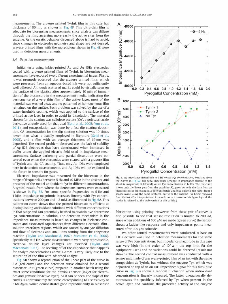
measurements. The gravure printed TyrInk film in this case hasthickness of 80 nm, as shown in Fig. 4F. This ultra-thin film isadequate for biosensing measurements since analyte can diffusethrough the film, assessing more easily the active sites from theenzyme. As the erratic behavior discussed above is hard to avoid,since changes in electrodes geometry and shape are not desired,gravure printed films with the morphology shown in Fig. 4E wereused in detection measurements.
3.4. Detection measurements
Initial tests using inkjet-printed Au and Ag IDEs electrodescoated with gravure printed films of TyrInk in biosensing mea-surements have exposed two different experimental issues. Firstly,it was promptly observed that the gravure printed films, whichwere processed from an aqueous-based ink were not sufficientlywell adhered. Although scattered marks could be visually seen onthe surface of the plastics after approximately 10 min of immer-sion of the biosensors in the measurement media, indicating thepermanence of a very thin film of the active layer, most of thematerial was washed away and no patterned or homogeneous filmremained on the surface. Such problem was solved by the use of awater-insoluble coating, which was applied to the surface of theprinted active layer in order to avoid its dissolution. The materialchosen for the coating was cellulose acetate (CA), a polysaccharidederivative already used for that goal (Setti et al., 2005; Yun et al.,2011), and encapsulation was done by a fast dip-coating deposi-tion. CA concentration for the dip-coating solution was 10 timeslower than what is usually employed in literature (Setti et al.,2005), and a film with an average thickness of 69 nm wasdeposited. The second problem observed was the lack of stabilityof Ag IDE electrodes that have deteriorated when immersed inbuffer under the applied electric field used in impedance mea-surements. Surface darkening and partial dissolution were ob-served even when the electrodes were coated with a gravure filmof TyrInk and the CA coating. Thus, only Au IDEs were employedhere in detection measurements, and Ag IDEs will be explored inthe future in sensors for gases.
Electrical impedance was measured for the biosensor in therange of frequencies between 5 Hz and 10 MHz in the absence andpresence of the model antioxidant Pyr in different concentrations.A typical result, from where the detections curves were extractedis shown in Fig. S2. For some specific frequencies as 5 Hz and1 kHz, impedance magnitude increases linearly with Pyr concen-trations between 200 mm and 1.2 mM, as illustrated in Fig. 5A. Thiscalibration curve shows that the printed biosensor is efficient atdistinguishing antioxidant solutions with different concentrationsin that range and can potentially be used to quantitative determinePyr concentrations in solution. The detection mechanism in theimpedance measurement is based on changes in dielectric con-stants and associated capacitances from different electrode–film–
solution interfaces regions, which are caused by analyte diffusionand flow of electrons and small ions coming from the enzymaticreaction (Taylor and Macdonald, 1987; Zucolotto et al., 2006).Specifically at 5 Hz, where measurements were very reproducible,electrical double layer changes are assessed (Taylor andMacdonald, 1987). The leveling off of the impedance that happensfor analyte concentrations above 1.2 mM is very likely due to thesaturation of the film with adsorbed analyte.
Fig. 5B shows a reproduction of the linear part of the curve in5 A (red curve) and the detection curve obtained for a secondbiosensor unit (green curve) prepared in another batch using theexact same conditions for the previous sensor (inkjet for electro-des and gravure for active layer). As it can be seen, the slope of thecurves is approximately the same, corresponding to a sensitivity of5.68 Ω/mm, which demonstrates good reproducibility in biosensor
fabrication using printing. However, from these pair of curves isalso possible to see that sensor resolution is limited to 200 mM,since when additions of 100 mM are made (green curve) the sensorshows a ladder-like response and only impedances points mea-sured after 200 mM coincide.
Two other control measurements were conducted. A bare AuIDE electrode was used in detection measurements for the samerange of Pyr concentrations, but impedance magnitude in this casewas very high (in the order of 107 Ω – the top limit for theequipment used) and no variations could be detected (result notshown). The second control measurement was conducted with asensor unit made of a gravure-printed film of an ink with the samecomposition as TyrInk, but without the enzyme Tyr, which wasdeposited on top of an Au IDE. Impedance signal for this film (bluecurve in Fig. 5B) shows a random fluctuation when antioxidantconcentration is linearly increased. The latter unequivocally de-monstrates the specificity inferred by Tyr when present in theactive layer, and confirms the preserved activity of the enzyme

F.J. Pavinatto et al. / Biosensors and Bioelectronics 67 (2015) 553–559 559
when deposited in the gravure film, already showed by opticalmeasurements (Fig. 3).
4. Conclusions
An all-printed and flexible biosensor for antioxidants was success-fully fabricated in this work. Interdigitated electrodes made of Au weredirectly deposited by inkjet in a one-step process using a commercialnanoparticle-based ink, which had its sintering conditions adapted toplastic-compatible temperatures. Traces with resolution of 45 mm andconductivity as high as 12% of bulk Au were printed and showed greatstability to flexions and to the use in biosensing via impedancemeasurements. A tailor-made ink was prepared having 1% w/v oftrehalose, 0.05% v/v of triton X-100, 68.5 mg/mL of Tyr and 6mg/mL ofCMC. This ink was efficiently gravure printed on plasma treatedplastics, giving rise to homogenous patterned films. Additives areneeded not only to provide a good printing, but also to improveenzyme stability and propitiate resistance to harsh gravure conditions,which resulted in 15.5% of preserved activity after printing. Theprinted biosensor has a sensitivity of 5.68 Ω/mm and limit of detectionof 200 mM, which is well below the concentration of antioxidants inwines and olive oil (Carlsen et al., 2010). Due to the fast and large-areaprinting characteristics of gravure, and its compatibility with roll-to-roll (R2R) processes, it is believed that the biosensor fabricated herehave great potential for the deployment of a technological product forthe analysis of dietary products.
Acknowledgements
The authors would like to acknowledge Fundação de Amparodo Estado de São Paulo (Fapesp), which funded this researchunder project # 2011/05742-0. Also, we are grateful to Prof. Dr.Vivek Subramanian by the use of his printing-related facilities, Dr.Marcelo Assunção Pereira e Silva for complementary AFM mea-surements, and Prof. Dr. Antonio Riul Jr. for helpful discussionsregarding the interpretation of impedance data. C.W.A. Paschoalacknowledges CNPq and CAPES Brazilian funding agencies, and R.Ramesh for all support at University of California.
Appendix A. Supplementary materials
Supplementary data associated with this article can be found inthe online version at http://dx.doi.org/10.1016/j.bios.2014.09.039.
References
Abe, K., Suzuki, K., Citterio, D., 2008. Anal. Chem. 80, 6928–6934. http://dx.doi.org/10.1021/ac800604v.
Allen, M., Leppäniemi, J., Vilkman, M., Alastalo, A., Mattila, T., 2010. Nanotechnology21, 475204. http://dx.doi.org/10.1088/0957-4484/21/47/475204.
Altenberend, U., Molina-Lopez, F., Oprea, A., Briand, D., Bârsan, N., De Rooij, N.F.,Weimar, U., 2013. Sens. Actuators B Chem. 187, 280–287. http://dx.doi.org/10.1016/j.snb.2012.11.025.
Apetrei, C., Alessio, P., Constantino, C.J.L., de Saja, J. a, Rodriguez-Mendez, M.L.,Pavinatto, F.J., Ramos Fernandes, E.G., Zucolotto, V., Oliveira, O.N., 2011. Biosens.Bioelectron. 26, 2513–2519. http://dx.doi.org/10.1016/j.bios.2010.10.047.
Arias, A.C., MacKenzie, J.D., McCulloch, I., Rivnay, J., Salleo, A., 2010. Chem. Rev. 110,3–24. http://dx.doi.org/10.1021/cr900150b.
Arrabito, G., Galati, C., Castellano, S., Pignataro, B., 2013. Lab Chip 13, 68–72. http://dx.doi.org/10.1039/c2lc40948h.
Barroso, M.F., de-los-Santos-Álvarez, N., Delerue-Matos, C., Oliveira, M.B.P.P., 2011.Biosens. Bioelectron. 30, 1–12. http://dx.doi.org/10.1016/j.bios.2011.08.036.
Berggren, C., Bjarnason, B., Johansson, G., 2001. Electroanalysis 13, 173–180. http://dx.doi.org/10.1002/1521-4109(200103)13:3o173::AID-ELAN17343.0.CO;2-B.
Carlsen, M.H., Halvorsen, B.L., Holte, K., Bøhn, S.K., Dragland, S., Sampson, L., Willey,C., Senoo, H., Umezono, Y., Sanada, C., Barikmo, I., Berhe, N., Willett, W.C.,
Phillips, R., Jacobs, D.R., Blomhoff, R., 2010. Nutr. J. 9, 3. http://dx.doi.org/10.1186/1475-2891-9-3.
Cho, H., Parameswaran, M., (Ash), Yu, H.-Z., 2007. Sens. Actuators B Chem. 123,749–756. http://dx.doi.org/10.1016/j.snb.2006.10.022.
Claramunt, S., Monereo, O., Boix, M., Leghrib, R., Prades, J.D., Cornet, a., Merino, P.,Merino, C., Cirera, a., 2013. Sens. Actuators B Chem. 187, 401–406. http://dx.doi.org/10.1016/j.snb.2012.12.093.
Correia, V., Caparros, C., Casellas, C., Francesch, L., Rocha, J.G., Lanceros-Mendez, S.,2013. Smart Mater. Struct. 22, 105028. http://dx.doi.org/10.1088/0964-1726/22/10/105028.
Delaney, J.T., Smith, P.J., Schubert, U.S., 2009. Soft Matter 5, 4866. http://dx.doi.org/10.1039/b909878j.
Derby, B., 2008. J. Mater. Chem. 18, 5717. http://dx.doi.org/10.1039/b807560c.Derby, B., 2012. Science 338 (80), 921–926. http://dx.doi.org/10.1126/
science.1226340.Di Risio, S., Yan, N., 2007. Macromol. Rapid Commun. 28, 1934–1940. http://dx.doi.
org/10.1002/marc.200700226.Fraga, C.G., Galleano, M., Verstraeten, S.V., Oteiza, P.I., 2010. Mol. Asp. Med. 31,
435–445. http://dx.doi.org/10.1016/j.mam.2010.09.006.Gonzalez-Macia, L., Morrin, A., Smyth, M.R., Killard, A.J., 2010. Analyst 135, 845–867.
http://dx.doi.org/10.1039/b916888e.Ilkhanizadeh, S., Teixeira, A.I., Hermanson, O., 2007. Biomaterials 28, 3936–3943.
http://dx.doi.org/10.1016/j.biomaterials.2007.05.018.Jabrane, T., Dubé, M., Griffiths, M., Mangin, P.J., 2011. J-FOR J. Sci. Technol. For. Prod.
Process. 1, 6–13.Jabrane, T., Jeaidi, J., Mangin, P.J., 2008. Adv. Print. Media Technol. 35, 279–288.Kang, B., Lee, W.H., Cho, K., 2013. ACS Appl. Mater. Interfaces 5, 2302–2315. http:
//dx.doi.org/10.1021/am302796z.Kaushik, J.K., Bhat, R., 2003. J. Biol. Chem. 278, 26458–26465. http://dx.doi.org/
10.1074/jbc.M300815200.Kipphan, H. (Ed.), 2001. Handbook of Print Media: Technologies and Production
Methods. Springer, Heildelberg.Mello, L.D., Kubota, L.T., 2007. Talanta 72, 335–348. http://dx.doi.org/10.1016/j.
talanta.2006.11.041.Monereo, O., Claramunt, S., Marigorta, M.M., De, Boix, M., Leghrib, R., Prades, J.D.,
Cornet, a, Merino, P., Merino, C., Cirera, a, 2013. Talanta 107, 239–247. http://dx.doi.org/10.1016/j.talanta.2013.01.022.
Pavinatto, F.J., Khan, Y., Arias, A.C., 2014. manuscript in preparation.Pavinatto, F.J., Fernandes, E.G.R., Alessio, P., Constantino, C.J.L., de Saja, J. a.,
Zucolotto, V., Apetrei, C., Oliveira Jr, O.N., Rodriguez-Mendez, M.L., 2011. J.Mater. Chem. 21, 4995. http://dx.doi.org/10.1039/c0jm03864d.
Prieto-Simón, B., Cortina, M., Campàs, M., Calas-Blanchard, C., 2008. Sens. ActuatorsB Chem. 129, 459–466. http://dx.doi.org/10.1016/j.snb.2007.08.004.
Reddy, A.S.G., Narakathu, B.B., Atashbar, M.Z., Rebros, M., Rebrosova, E., Joyce, M.K.,2011a. Procedia Eng. 25, 956–959. http://dx.doi.org/10.1016/j.proeng.2011.12.235.
Reddy, A.S.G., Narakathu, B.B., Atashbar, M.Z., Rebros, M., Rebrosova, E., Joyce, M.K.,2011b. Procedia Eng. 25, 120–123. http://dx.doi.org/10.1016/j.proeng.2011.12.030.
Sanjana, N.E., Fuller, S.B., 2004. J. Neurosci. Methods 136, 151–163. http://dx.doi.org/10.1016/j.jneumeth.2004.01.011.
Saum, A.G.E., Cumming, R.H., Rowell, F.J., 2000. Biosens. Bioelectron. 15, 305–313.Saunders, R., Gough, J., Derby, B., 2010. In: Ringeisen, B.R., Spargo, B.J., Wu, P.K.
(Eds.), Cell and Organ Printing. Springer, Netherlands, Dordrecht,pp. 35–50. http://dx.doi.org/10.1007/978-90-481-9145-1.
Sen, S., Chakraborty, R., 2011. In: Andreescu, S. (Ed.), Oxidative Stress: Diagnostics,Prevention, and Therapy. American Chemical Society, Washington, DC,pp. 1–37.
Setti, L., Fraleoni-Morgera, A., Ballarin, B., Filippini, A., Frascaro, D., Piana, C., 2005.Biosens. Bioelectron. 20, 2019–2026. http://dx.doi.org/10.1016/j.bios.2004.09.022.
Singh, K.V., Bhura, D.K., Nandamuri, G., Whited, A.M., Evans, D., King, J., Solanki, R.,2011. Langmuir 27, 13931–13939. http://dx.doi.org/10.1021/la202546a.
Søndergaard, R.R., Hösel, M., Krebs, F.C., 2013. J. Polym. Sci. Part B Polym. Phys. 51,16–34. http://dx.doi.org/10.1002/polb.23192.
Suman, O'Reilly, E., Kelly, M., Morrin, A., Smyth, M.R., Killard, A.J., 2011. Anal. Chim.Acta 697, 98–102. http://dx.doi.org/10.1016/j.aca.2011.04.036.
Sumerel, J., Lewis, J., Doraiswamy, A., Deravi, L.F., Sewell, S.L., Gerdon, A.E., Wright,D.W., Narayan, R.J., 2006. Biotechnol. J. 1, 976–987. http://dx.doi.org/10.1002/biot.200600123.
Taylor, D.M., Macdonald, a G., 1987. J. Phys. D. Appl. Phys., 20; , pp. 1277–1283. http://dx.doi.org/10.1088/0022-3727/20/10/010.
Tsouti, V., Boutopoulos, C., Zergioti, I., Chatzandroulis, S., 2011. Biosens. Bioelectron.27, 1–11. http://dx.doi.org/10.1016/j.bios.2011.05.047.
Van Gerwen, P., Laureyn, W., Laureys, W., Huyberechts, G., Op De Beeck, M., Baert,K., Suls, J., Sansen, W., Jacobs, P., Hermans, L., Mertens, R., 1998. Sens. ActuatorsB Chem. 49, 73–80. http://dx.doi.org/10.1016/S0925-4005(98)00128-2.
Varshney, M., Li, Y., 2009. Biosens. Bioelectron. 24, 2951–2960. http://dx.doi.org/10.1016/j.bios.2008.10.001.
Villar, G., Graham, A.D., Bayley, H., 2013. Science 340, 48–52. http://dx.doi.org/10.1126/science.1229495.
Yun, Y.H., Lee, B.K., Choi, J.S., Kim, S., Yoo, B., Kim, Y.S., Park, K., Cho, Y.W., 2011. Anal.Sci. 27, 375.
Zucolotto, V., Pinto, A.P., Tumolo, T., Moraes, M.L., Baptista, M.S., Riul, A., Araújo, A.P.U., Oliveira, O.N., 2006. Biosens. Bioelectron. 21, 1320–1326. http://dx.doi.org/10.1016/j.bios.2005.06.001.


