Biomolecular Structure and Modeling: Problem and ...
32
This is page 37 Printer: Opaque this Biomolecular Structure and Modeling: Problem and Application Perspective All things come out of the one, and the one out of all things. Change, that is the only thing in the world which is unchanging. Heraclitus of Ephesus (550–475 BC). 2.1 Computational Challenges in Structure and Function 2.1.1 Analysis of the Amassing Biological Databases The experimental progress described in the previous chapter has been accompa- nied by an increasing desire to relate the complex three-dimensional (3D) shapes of biomolecules to their biological functions and interactions with other molec- ular systems. Structural biology, computational biology, genomics, proteomics, bioinformatics, chemoinformatics, and others are natural partner disciplines in such endeavors. Structural biology focuses on obtaining a detailed structural resolution of macromolecules and relating structure to biological function. Computational biology was first associated with the discipline of finding sim- ilarities among nucleotide strings in known genetic sequences, and relating these relationships to evolutionary commonalities; the term has grown, however, to en-
Transcript of Biomolecular Structure and Modeling: Problem and ...

This is page 37Printer: Opaque this
�
Biomolecular Structure and Modeling:Problem and Application Perspective
All things come out of the one, and the one out of all things. Change,that is the only thing in the world which is unchanging.
Heraclitus of Ephesus (550–475 BC).
2.1 Computational Challenges in Structure andFunction
2.1.1 Analysis of the Amassing Biological Databases
The experimental progress described in the previous chapter has been accompa-nied by an increasing desire to relate the complex three-dimensional (3D) shapesof biomolecules to their biological functions and interactions with other molec-ular systems. Structural biology, computational biology, genomics, proteomics,bioinformatics, chemoinformatics, and others are natural partner disciplines insuch endeavors.
Structural biology focuses on obtaining a detailed structural resolution ofmacromolecules and relating structure to biological function.
Computational biology was first associated with the discipline of finding sim-ilarities among nucleotide strings in known genetic sequences, and relating theserelationships to evolutionary commonalities; the term has grown, however, to en-

38 2. Biomolecular Structure and Modeling: Problem and Application Perspective
compass virtually all computational enterprises to molecular biology problems[126].
Comparative genomics — the search and comparison of sequences amongspecies — is a natural outgrowth of the sequencing projects [116]. So arestructural and functional genomics, the characterization of the 3D structure andbiological function of these gene products [142, 20, 14, 111, 37].
In the fall of 2000, the U.S. National Institute of General Medical Sciences(NIGMS) launched a structural genomics initiative by funding seven researchgroups aiming to solve collectively the 3D structures of 10,000 proteins, eachrepresenting a protein family, over the next decade. This important step towardthe assembly of a full protein library requires improvements in both structuralbiology’s technology and methodology. (See the November 2000 supplement is-sue of Nature Structural Biology, volume 7, devoted to structural genomics andthe progress report [37]). Ultimately, the functional identification of unknownproteins is likely to dramatically increase our understanding of human diseaseprocesses and the development of structure-based drug therapies.
Proteomics is another current buzzword defining the related discipline of pro-tein structure and function (see [67] for an introduction), and even cellomics hasbeen introduced.1 Cellomics reflects the expanded interest of gene sequencers inintegrated cellular structure and function. The Human Proteomics Project — acollaborative venture to churn out atomic structures using high-throughput androbotics-aided methods based on NMR spectroscopy and X-ray crystallography,rather than sequences — may well be on its way.
New instruments that have revolutionized genomics known as DNA microar-rays, biochips, or gene expression chips (introduced in Chapter 1 and Box 1.5)allow researchers to determine which genes in the cell are active and to identifygene networks. A good introduction to these areas is volume 10 of the year 2000,pages 341–384, of Current Opinion in Structural Biology entitled “Sequencesand Topology. Genome and Proteome Informatics”, edited by P. Bork and D.Eisenberg.
The range of genomic sciences also continues [156] to the metabolome, theendeavor to define the complete set of metabolites (low-molecular cellular inter-mediates) in cells, tissues, and organs. Experimental techniques for performingthese integrated studies are continuously being developed. For example, yeastgeneticists have developed a clever technique for determining whether two pro-teins interact and, thereby by inference, participate in related cellular functions[210]. Such approaches to proteomics provide a powerful way to discover func-tions of newly identified proteins. DNA chip technology is also thought to holdthe future of individualized health care now coined personalized medicine orpharmacogenomics; see Chapter 14 and Box 1.5.
1A glossary of biology disciplines coined with “ome” or “omic” terms can be found athttp://www.genomicglossaries.com/content/omes.asp.

2.1. Computational Challenges 39
It has been said that current developments in these fields are revolutionaryrather than evolutionary. This view reflects the clever exploitation of biomolec-ular databases with computing technology and the many disciplines contributingto the biomolecular sciences (biology, chemistry, physics, mathematics, statis-tics, and computer science). Bioinformatics is an all-embracing term for manyof these exciting enterprises [99, 148] (structural bioinformatics is an impor-tant branch); chemoinformatics has also followed (see Chapter 14) [86]. Somegenome-technology company names are indicative of the flurry of activity andgrand expectations from our genomic era. Consider titles like Genetics ComputerGroup, Genetix Ltd., Genset, Protana, Protein Pathways, Inc., PyrosequencingAB, Sigma-Genosys, or Transgenomic Incorporated.
This excitement in the field’s developments and possibilities is echoed by thechief executive of the software giant Oracle Corp., Larry Ellison, who surroundshimself by molecular biologists — the scientists, board members, and fellowsof his Ellison Medical Foundation; explaining to a Wall Street Journal reporterhis preference of molecular biology over racing sailboats, Ellison said: “The raceis more interesting, the people in the race are more interesting and the prize isbigger.” (Wall Street Journal, January 9, 2003).
Although the number of sequence databases has grown very rapidly and ex-ceeds the amount of structural information,2 the 1990s saw an exponential riseof structural databases as well. From only 50 solved 3D structures in the Pro-tein Data Bank (PDB) in 1975, to 500 in 1988, another order of magnitude wasreached in 1996 (around 5000 entries). In fact, the rate of growth of structuralinformation is approaching the rate of increase of amino acid sequence infor-mation (see Figure 2.1 and Table 2.1). It is no longer a rare event to see a newcrystal structure on the cover of Nature or Science. Now, on a weekly basis,groups compete to have their newly-solved structure on the cover of prominentjournals, including the newer, colorful publications like Nature Structural Biol-ogy and Structure. The fraction of NMR-deduced structures deposited in the PDBis also rising steadily, reflecting roughly 15% by the end of 2001 (for updatedinformation, see www.rcsb.org/pdb/holdings.html).
This trend, coupled with tremendous advances in genome sequencing projects[120], argues strongly for increased usage of computational tools for analysis ofsequence/structure/function relationships and for structure prediction. Thus, be-sides genomics-based analyses and comparisons, important are accurate, reliable,and rapid theoretical tools for describing structural and functional aspects of geneproducts. (See [111], for example, for computational challenges in genomics).
2In 1991, it was pointed out [15] that the amount of 3D protein information is lagging by orders ofmagnitude behind the accessible sequence data [15].

40 2. Biomolecular Structure and Modeling: Problem and Application Perspective
1970 1975 1980 1985 1990 1995 200010
0
101
102
103
104
105
106
PDB PIR NRPR
Figure 2.1. The growth of protein sequence databases (named PIR and NRPR)versus structural database of macromolecules (PDB). See Table 2.1 andwww.dna.affrc.go.jp/htdocs/growth/P-history.html. The International Protein Infor-mation Resource (PIR) of protein sequences (pir.georgetown.edu) is a comprehensive,annotated public-domain database reflecting a collaboration among the United States(Georgetown University Medical Center in Washington, D.C.), Germany (MunichInformation Center for Protein Sequences in Martinsried), and Japan (InternationalProtein Sequence Database in Tsukuba). PIR is extensively cross referenced and linked tomany molecular databases of genes, genomes, protein structures, structural classification,literature, and more. The NRPR database represents merged, non redundant proteindatabase entries from several databases: PIR, SWISS-PROT, Genpept, and PDB.
2.1.2 Computing Structure From Sequence
One of the most successful approaches to date on structure prediction comes fromhomology modeling (also called comparative modeling) [3, 8].
In general, a large degree of sequence similarity often suggests similarity in 3Dstructure. It has been reported, for example, that a sequence identity of greaterthan 40% usually implies more than 90% 3D-structure overlap (defined as per-centage of C
�
atoms of the proteins that are within 3.5 A of each other in arigid-body alignment; see definitions in Chapter 3) [184]. Thus, sequence sim-ilarity of at least 50% suggests that the associated structures are similar overall.Conversely, small sequence similarity generally implies structural diversity.

2.1. Computational Challenges 41
Table 2.1. Growth of protein sequence databases.
Year PDBa PIRb NRPRc
1972 11973 61974 91975 141976 581977 851978 1041979 1281980 1501981 1681982 1971983 2341984 258 28981985 280 36151986 296 46121987 318 67961988 382 105271989 459 143721990 597 267981991 790 361501992 1006 472341993 1727 647601994 3091 755111995 4056 82066 1598081996 5240 91006 2041231997 6833 105741 2582721998 8942 117482 3242371999 11364 168808 3606742000 14063 198801 4977872001 16973 274514 744991
aFrom the Protein Data Bank (PDB), www.rcsb.org/pdb/holdings.html.bFrom the Protein International Resource (PIR), www-nbrf.georgetown.edu/cgi-bin/dbinfo.cFrom the ‘Non Redundant Proteins database merged Regular release’,
www.dna.affrc.go.jp/htdocs/growth/P-history.html.
There are many exceptions, however, as demonstrated humorously in a contestpresented to the protein folding community (see Box 2.1). The myoglobin andhemoglobin pair is a classic example where large structural, as well as evolution-ary, similarity occurs despite little sequence similarity (20%). Other exceptional

42 2. Biomolecular Structure and Modeling: Problem and Application Perspective
B1 MTYKLILNGKTLKGETTTEAVDAATAEKVFKQYANDNGVDGEWTYDDATKTFTVTE Janus MTKKAILALNTAKFLRTQAAVLAAKLEKLGAQEANDNAVDLEDTADDLYKTLLVLARop GTKQEKTALNMARFIRSQTLTLLEKLNELDADEQADICESLHDHADELYRSCLARF
B1 domain Rop
Figure 2.2. Ribbon representations of the B1 domain of IgG-binding protein G and the Ropmonomer (first 56 residues), which Janus resembles [46], with corresponding sequences.Half of the protein G
�domain (B1) residues were changed to produce Janus in response
to the Paracelsus challenge (see Box 2.1 and [177]). The origin of the residues is indicatedby the following color schemes: residues from B1: red; residues from Rop: blue; residuesin both: green; residues in neither: black. While experimental coordinates of protein G andRop are known, the structure of Janus was deduced by modeling. The single-letter aminoacid acronyms are detailed in Table ??, Chapter 3.
examples and various sequence/structure relationships are discussed separately inChapter 3, as well as Homework 6 and [79] for example.
More general than prediction by sequence similarity is structure prediction denovo [8], a Grand Challenge of the field, as described next.

2.2. Protein Folding 43
2.2 Protein Folding – An Enigma
2.2.1 ‘Old’ and ‘New’ Views
There has been much progress on the protein folding challenge since CyrusLevinthal first posed the well-known “paradox” named after him; see [97] fora historical perspective. Levinthal suggested that well defined folding pathwaysmight exist [123] since real proteins cannot fold by exhaustively traversingthrough their entire possible conformational space [124]. (See [213, 51, 214] fora recent related discussion of whether the number of protein conformers dependsexponentially or nonexponentially on chain length). Levinthal’s paradox led to thedevelopment of two views of folding — the ‘old’ and the ‘new’ — which havebeen merging of late [49, 50, 122].
The old view accents the existence of a specific folding pathway characterizedby well-defined intermediates. The new version emphasizes the rugged, heteroge-neous multidimensional energy landscape governing protein folding, with manycompeting folding pathways [228]. Yet, the boundary between the two views ispliant and the intersection substantial [97]. This integration has resulted froma variety of information sources: theories on funnel-shaped energy landscape(e.g., [158, 50, 25]); folding and unfolding simulations of simplified models (e.g.,[77, 87, 112, 141, 190, 71]), at high temperatures or low pH concentrations (e.g.,[122, 240]); NMR spectroscopic experiments that monitor protein folding inter-mediates (e.g., [60, 157]); predictions of secondary and/or tertiary structure onthe basis of evolutionary information [183]; and statistical mechanical theories.
Such studies suggest that while wide variations in folding pathways may occur,‘fast folders’ possess a unifying pattern for the evolution of native-structure con-tacts. In particular, the pathway and other kinetic aspects governing the folding ofa particular protein depend on the free energy landscape, which is temperature-dependent. Namely, the folding ensemble is sensitive to shallow energy traps atlower temperatures. Thus, changes in temperature can substantially change thefolding kinetics.
2.2.2 Folding Challenges
The great progress in the field can also be seen by evaluations of biannual predic-tion exercises held in Asilomar, California (termed CASP for Critical Assessmentof Techniques for Protein Structure Prediction) [58, 114, 209] (see PredictionCenter.llnl.gov, and special issues of the journal Proteins: vol. 23, 1995; Suppl. 1,1997; Suppl. 3, 1999, and Suppl. 5, 2001).
The CASP organizers assign specific proteins for theoretical prediction thatprotein crystallographers and NMR spectroscopists expect to complete by the nextCASP meeting. Prediction assessors then consider several categories of structuralprediction tools, for example: comparative (homology) modeling, fold recogni-tion (i.e., based on a library of known protein folds), and ab initio prediction(i.e., using first principles). The quality of the prediction has been characterized

44 2. Biomolecular Structure and Modeling: Problem and Application Perspective
in terms of C�
root-mean-square (RMS) deviations, the best of which is in therange of 2–6 A; the lowest values have been obtained from the best comparativemodeling approaches.
To the fourth CASP meeting in December 2000, two additional competitiveexperiments were added, dealing with protein structure prediction and ratio-nal drug design: CAFASP2 (assessing automatic methods for predicting proteinstructure), and CATFEE (evaluating methods for protein ligand docking); seepredictioncenter.llnl.gov/casp4/Casp4.html). These additions reflect the rapiddevelopments of many genome efforts and the rise of computational biology asan important discipline of biology, medicine, and biotechnology.
Results of the CASP4 meeting indicated considerable progress in fully-automated approaches for structure prediction and hence promise that computermodeling might become an alternative to experimental structure determination.See [146] for an overview with articles collected on CASP4 in that special issueof Proteins (Suppl. 5, 2001), as well as a related discussion of field progress andprospects [13] and of the limitations and challenges in the comparative modelingsection of CASP4 [134, 147, 182].
Results of the CASP5 meeting [209] revealed the evolving importance of theCASP initative for motivating progress in protein prediction and helping relatedglobal initatives, like the “Ten Most Wanted” proteins of unknown structure thatthe community aims to solve because of suggested biological importance (seetmw.llnl.gov/). In particular, the meeting demonstrated that comparative model-ing approaches can produce reasonably good structural models but that it is stilldifficult to predict the structure of regions that are substantially different fromthe target. Recognizing novel folds remains a challenge, as well as predictingsecondary structures and long-range contacts.
Modeling work in the field is invaluable because it teaches us to ask, and seekanswers to, systematic questions about sequence/structure/function relationshipsand about the underlying forces that stabilize biomolecular structures. Still, giventhe rapid improvements in the experimental arena, the pace at which modelingpredictions improve must be expeditious to make a significant contribution toprotein structure prediction from sequence. To this end, computational biologistsare soliciting candidates for the “Ten Most Wanted” proteins.
The annual Johns Hopkins Coolfront Protein Folding meetings also reflect thegreat progress made in this exciting field. As gleaned from reports of the fourth[160] and fifth [220] meetings, progress is rapid on many theoretical and ex-perimental fronts. Particularly exciting are emerging areas of study dealing withprotein folding in membranes and protein folding diseases (e.g., from misfoldingor chaperone interference; see below). Furthermore, progress on both experi-mental ultrafast methods for studying protein folding kinetics and theoreticalpredictions based on sequence and structural genomics is impressive.

2.2. Protein Folding 45
Box 2.1: Paracelsus Challenge
In 1994, George Rose and Trevor Creamer posed a challenge, named after a 16th-centuryalchemist: change the sequence of a protein by 50% or less to create an entirely different3D global folding pattern [177]. Though this challenge might sound not particularlydifficult, imagine altering at most 50% of the ingredients for a chocolate cake recipeso as to produce bouillabaisse instead! Rose and Creamer offered a reward of $1000 toentice entrants.
The transmutation was accomplished four years later by Lynne Regan and coworkers[46], who converted the four-stranded
�-sheet B1 domain of protein G — which has
the�
sheets packed against a single helix — into a four-helix bundle of two associatinghelices called Janus (see Figure 2.2). These contestants achieved this wizardry byreplacing residues in a
�-sheet-encoding domain (i.e., those with high
�-sheet-forming
propensities) with those corresponding to the four-helix-bundle protein Rop (repressorof primer). Other modifications were guided by features necessary for Rop stability (i.e.,internal salt bridge), and the combined design was guided by energy minimization andsecondary-structure prediction algorithms.
The challenge proposers, though delighted at the achievement they stimulated, concludedthat in the future only tee-shirt prizes should be offered rather than cash!
2.2.3 Folding by Dynamics Simulations?
While molecular dynamics simulations are beginning to approach the timescalesnecessary to fold small peptides [47] or small proteins [55], we are far from find-ing the Holy Grail, if there is one [12]. Indeed, IBM’s ambitious announcement inDecember 1999 of building a ‘Blue Gene’ petaflop computer (i.e., capable of
�������
floating-point operations per second) for folding proteins by 2005 depends on thecomputational models guiding this ubiquitous cellular process.3 For example,some proteins require active escorts to assist in their folding in vivo. These chap-erone molecules assist in the folding and rescue misbehaved polymers. Thoughmany details are not known about the mechanisms of chaperone assistance (seebelow), we recognize that chaperones help by guiding structure assembly andpreventing aggregation of misfolded proteins. For an overview of chaperones, see
3This $100 million program to build the world’s most powerful supercomputer depends on a mil-lion processors running in parallel with the PIM (processor in memory) design. This architecture putsthe memory and processor on the same chip: a total of 40,000 chips with 25 or more processors perchip. Besides the crucial role of software in the success leading to protein folding ‘in silico’, it is achallenge to eliminate bottlenecks in this enormous parallel system that might result between memoryand processors when sequential (rather than parallel) computations must be performed.
46 2. Biomolecular Structure and Modeling: Problem and Application Perspective
GroEL/GroES Complex
Top View
GroES Ring
GroEL cis Ring
GroEL trans Ring
10 Ao
80 Ao
140 Ao
Bottom View
80 Ao
71 Ao
33 Ao
o184 A
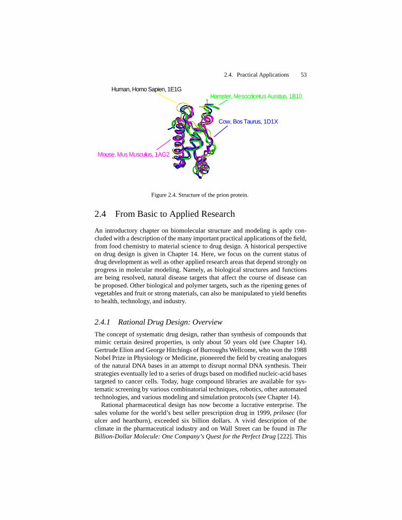
Figure 2.3. The bullet-shaped architecture of the GroEL/GroES chaperonin/co-chaperonincomplex sequence. Overall assembly and dimensions are shown from a side view (left).The top ring is the GroES ‘cap’, and the other layers are GroEL rings. Sidechains areshown in grey. As seen from the top and bottom views (right), a central channel forms inthe interior, conducive to protein folding. The protein is organized as three rings that sharea 7-fold rotational axis of symmetry (middle), where GroEL contains 14 identical pro-teins subunits assembled in two heptameric rings, and GroES contains 7 smaller identicalsubunits in its heptamer ring.
[90, 217], for example, and the 1 August 2001 issue of the Journal of StructuralBiology, volume 135, devoted to chaperones.
2.2.4 Folding Assistants
Current studies on chaperone-assisted folding, especially of the archetypal chap-erone duo, the E. Coli bacterial chaperonin GroEL and its cofactor GroES, areproviding insights into the process of protein folding [68, 205] (see Figure 2.3and Box 2.2). The rescue acts of chaperones depend on the subclass of these es-corts and the nature of the protein being aided. Some chaperones can assist alarge family of protein substrates, while others are more restrictive (see Box 2.2);detailed structural explanations remain unclear. Many families of chaperones arealso known, varying in size from small monomers (e.g., 40 or 70 kDa for DnaJand DnaK of Hsp70) to large protein assemblies (e.g., 810 kDa for GroEL or880 kDa for the GroEL/GroES complex).

2.2. Protein Folding 47
The small assistants bind to short runs of hydrophobic residues4 to delay pre-mature folding and prevent aggregation. Larger chaperones are likely needed toprevent aggregation of folded compact intermediates in the cell termed ‘moltenglobules’, requiring a complex trap-like mechanism involving co-chaperones (seealso Box 2.2).
Recent work also suggests that small chaperones in the PapD-like superfamily— which directs the folding of various surface organelles — facilitate folding byproviding direct steric information to their substrates, in addition to capping theirinteractive surfaces [154, 10]. In certain assemblies (pilus subunits called pilins),the chaperone donates a
�-strand that completes the fold of the pilin to promote
correct folding and binding to neighboring subunits.Since macromolecular crowding affects aggregation and diffusion properties of
proteins, uncertainties remain regarding the interpretation of in vitro experimentson chaperone-assisted folding. Different models for chaperone activity have alsobeen proposed: a ‘pathway’ route for direct folding, and a ‘network’ model, in-volving iterative folding. They differ by the extent to which unfolded proteinsare released to the cellular medium while they remain vulnerable to aggregation[64, 63].
2.2.5 Unstructured Proteins
Though our discussion has focused on the concept of native folds, not all proteinsare intrinsically structured [61]. The intrinsic lack of structure can be advanta-geous, for example in binding versatility to different targets or in ability to adaptdifferent conformations. Unfolded or non-globular structures are recognized inconnection with regulatory functions, such as binding of protein domains to spe-cific cellular targets [61, 230]. Examples include DNA and RNA-binding regionsof certain protein complexes (e.g., basic region of leucine zipper protein GCN4,DNA-binding domain of NFATC1, RNA recognition regions of the HIV-1 Revprotein). Here, the unstructured regions become organized only upon binding tothe DNA or RNA target. This folding flexibility offers an evolutionary advantage,which might be more fully appreciated in the future, as more gene sequences thatcode for unstructured proteins are discovered and analyzed.
4The terms hydrophobic (‘water-hating’) and hydrophilic (‘water-loving’) characterize water-soluble and water-insoluble molecular groups, respectively.

48 2. Biomolecular Structure and Modeling: Problem and Application Perspective
Box 2.2: Studies on Protein Escorts
The archetypal chaperone GroEL is a member of a chaperone class termed chaperonins;hsp60 of mitochondria and chloroplasts is another member of this class. These chaper-ones bind to partially-folded peptide chains and assist in the folding with the consumptionof ATP. The solved crystal structure of GroEL/GroES [233] suggests beautifully, in broadterms, how the large central channel inside a barrel-shaped chaperone might guide pro-tein compaction in its container and monitor incorrect folding by diminishing aggregation(see Figure 2.3). The two-ringed GroEL chaperonin (middle and bottom levels in Fig-ure 2.3) attaches to its partner chaperonin GroES (top ring) upon ATP binding, causinga major conformational change; the size of GroEL nearly doubles, and it assumes a cageshape, with GroES capping over it. This capping prevents the diffusion of partially foldedcompact intermediates termed ‘molten globules’ and offers them another chance at fold-ing correctly.
Experiments that track hydrogen exchange in unfolded rubisco protein by radioactivetritium (a hydrogen isotope) labeling suggest how misfolded proteins fall into this cavityand are released: a mechanical stretching force triggered by ATP binding partially ortotally unfolds the misfolded proteins, eventually releasing the captive protein [192].
These results also support an iterative annealing (or network model) for chaperone-guided folding, in which the process of forceful unfolding of misfolded molecules, theirtrapping in the cavity, and their subsequent release is iterated upon until proper folding.
The identification of preferential substrates for GroEL in vivo [98], namely multidomainE. Coli proteins with complex �
�folds, further explains the high-affinity interactions
formed between the misfolded or partially folded proteins and binding domains ofGroEL. These proteins require the assistance of a chaperone because assembly of�
-sheet domains requires long-range coordination of specific contacts (not the case forformation of � -helices). Natural substrates for other chaperones, like Eukaryotic type IIchaperonin CCT, also appear selective, favoring assistance to proteins like actin [129].
However, such insights into folding kinetics are only the tip of the iceberg. Chaperonetypes and mechanisms vary greatly, and the effects of macromolecular crowding (notmodeled by in vitro experiments) complicate interpretations of folding mechanisms invivo. Unlike chaperonins, members of another class of chaperones that includes the heat-shock protein Hsp70 bind to exposed hydrophobic regions of newly-synthesized proteinsand short linear peptides, reducing the likelihood of aggregation or denaturation. Theseare classified as ‘stress proteins’ since their amount increases as environmental stressesincrease (e.g., elevated temperatures). Other chaperones are known to assist in proteintranslocation across membranes.

2.3. Protein Misfolding 49
2.3 Protein Misfolding – A Conundrum
2.3.1 Prions and Mad Cows
Further clues into the protein folding enigma are also emerging from anotherpuzzling discovery involving certain proteins termed prions. These misfolded pro-teins — triggered by a conformational change rather than a sequence mutation —appear to be the source of infectious agent in fatal neurodegenerative diseases likebovine spongiform encephalopathy (BSE) or ‘mad cow disease’ (identified in themid 1980s in Britain), and the human equivalent Creutzfeld-Jacob disease (CJD).5
The precise mechanism of protein-misfolding induced diseases is not known,but connections to neurodegenerative diseases, which include Alzheimer’s, aregrowing and stimulating much interest in protein misfolding [42, 52].
Stanley Prusiner, a neurology professor at the University of California at SanFrancisco, coined the term prion to emphasize the infectious source as the protein(‘proteinaceous’), apparently in contradiction to the general notion that nucleicacids must be transferred to reproduce infectious agents. Prusiner won the 1998Nobel Prize in Physiology or Medicine for this “pioneering discovery of an en-tirely new genre of disease-causing agents and the elucidation of the underlyingprinciples of their mode of action”.
Prions add a new symmetry to the traditional roles long delegated to nucleicacids and proteins! Since the finding in the 1980s that nucleic acids (catalyticRNAs) can catalyze reactions — a function traditionally attributed to proteinsonly — the possibility that certain proteins, prions, carry genetic instructions —a role traditionally attributed to nucleic acids — completes the duality of functionsto both classes of macromolecules.
2.3.2 Infectious Protein?
Is it possible for an ailment to be transmitted by ‘infectious proteins’ rather thanviruses or other traditional infectious agents? The prion interpretation for the in-fection mechanism remains controversial for lack of clear molecular explanation.In fact, one editorial article stated that “whenever prions are involved, more openquestions than answers are available” [1]. Yet the theory is winning more con-verts with laboratory evidence that an infectious protein that causes mad cowdisease also causes a CJD variant in mice [187]. These results are somewhatfrightening because they suggest that the spread of this illness from one speciesto another is easier than has been observed for other diseases.
The proteinaceous theory suggests that the prion protein (see Figure 2.4) inthe most studied neurodegenerative prion affliction, scrapie (long known in sheepand goats), becomes a pathologic agent upon conversion of one or more of its
� -helical regions into�
-regions (e.g., parallel�
-helix [223]); once this confor-
5See information from the UK Department of Health on www.doh.gov.uk, the UK CJDSurveillance Unit at www.cjd.ed.ac.uk, and the CJD Disease Foundation at cjdfoundation.org.

50 2. Biomolecular Structure and Modeling: Problem and Application Perspective
mational change occurs, the conversion of other cellular neighbors proceeds by adomino-like mechanism, resulting in many abnormally-folded molecules whicheventually reap havoc in the mammal. This protein-only hypothesis was firstformulated by J.S. Griffith in 1967, but Prusiner first purified the hypotheticalabnormal protein thought to cause BSE. New clues are rapidly being added tothis intriguing phenomenon (see Box 2.3).
Both the BSE and CJD anomalies implicated with prions have been linkedto unusual deposits of protein aggregates in the brain. (Recent studies on micealso open the possibility that aberrant proteins might also accumulate in muscletissue). It is believed that a variant of CJD has caused the death of dozens ofpeople in Britain (and a handful in other parts of the world) since 1995 who atemeat infected with BSE, some only teenagers. Recent studies also suggest thatdeaths from the human form of mad cow disease could be rising significantly andspreading within Europe as well as to other continents.
Since the incubation period of the infection is not known — one victim becamea vegetarian 20 years before dying of the disease — scientists worry about theextent of the epidemic in the years to come. The consequences of these deathshave been disastrous to the British beef industry and have led indirectly to otherproblems (e.g., the 2001 outbreak of foot-and-mouth disease, a highly infectiousdisease of most farm animals except horses). The panic has not subsided, as un-certainties appear to remain regarding the safety of various beef parts, as well assheep meat, and the possible spread of the disease to other parts of the world.
2.3.3 Other Possibilities
Many details of this intriguing prion hypothesis and its associated diseases are yetto be discovered and related to normal protein folding. Some scientists believe thata lurking virus or virino (small nonprotein-encoding virus) may be involved in theprocess, perhaps stimulating the conformational change of the prion protein, butno such evidence has yet been found. Only creation of an infection de novo inthe test tube is likely to convince the skeptics, but the highly unusual moleculartransformation implicated with prion infection is very difficult to reproduce in thetest tube.
Box 2.3: Prion: Structural Evidence
The detailed structural picture associated with the prion conformational change is only be-ginning to emerge as new data appear [2]. In 1997, Kurt Wutrich and colleagues at theSwiss Federal Institute of Technology in Zurich reported the first NMR solution structureof the 208-amino acid glycoprotein “prion protein cellular” PrP
�
anchored to nerve cellmembranes. The structure reveals a flexibly disordered assembly of helices and sheets (seeFig. 2.4). This organization of the harmless protein might help explain the conversion pro-cess to its evil isoform PrP
���
. It has been suggested that chaperone molecules may bindto PrP
�
and drive its conversion to PrP���
and that certain membrane proteins may also be

2.3. Protein Misfolding 51
involved in the transformation.
In early 1998, a team from the University of California at San Francisco discovereda type of prion, different from that associated with mad cow disease, that attaches to amajor structure in neuron cells and causes cells to die by transmitting an abnormal signal.This behavior was observed in laboratory rats who quickly died when a mutated type ofprion was placed into the brains of newborn animals; their brains revealed the abnormalprions stuck within an internal membrane of neuron cells. The researchers believe thatthis mechanism is the heart of some prion diseases. They have also found such abnormalprions in the brain tissue of patients who died from a rare brain disorder called Gerstmann-Straussler-Scheinker disease (GSS) — similar to Creutzfeld-Jacob disease (CJD) — thatdestroys the brain.
Important clues to the structural conversion process associated with prion diseases werefurther offered in 1999, when a related team at UCSF, reported the NMR structure of thecore segment of a prion protein rPrP that is associated with the scrapie prion protein PrP
� �
[104, 128]. The researchers found that part of the prion protein exhibits multiple confor-mations. Specifically, an intramolecular hydrogen bond linking crucial parts of the proteincan be disrupted by a single amino acid mutation, leading to different conformations. Thiscompelling evidence on how the molecule is changed to become infectious might suggesthow to produce scrapie-resistant or BSE-resistant species by animal cloning.
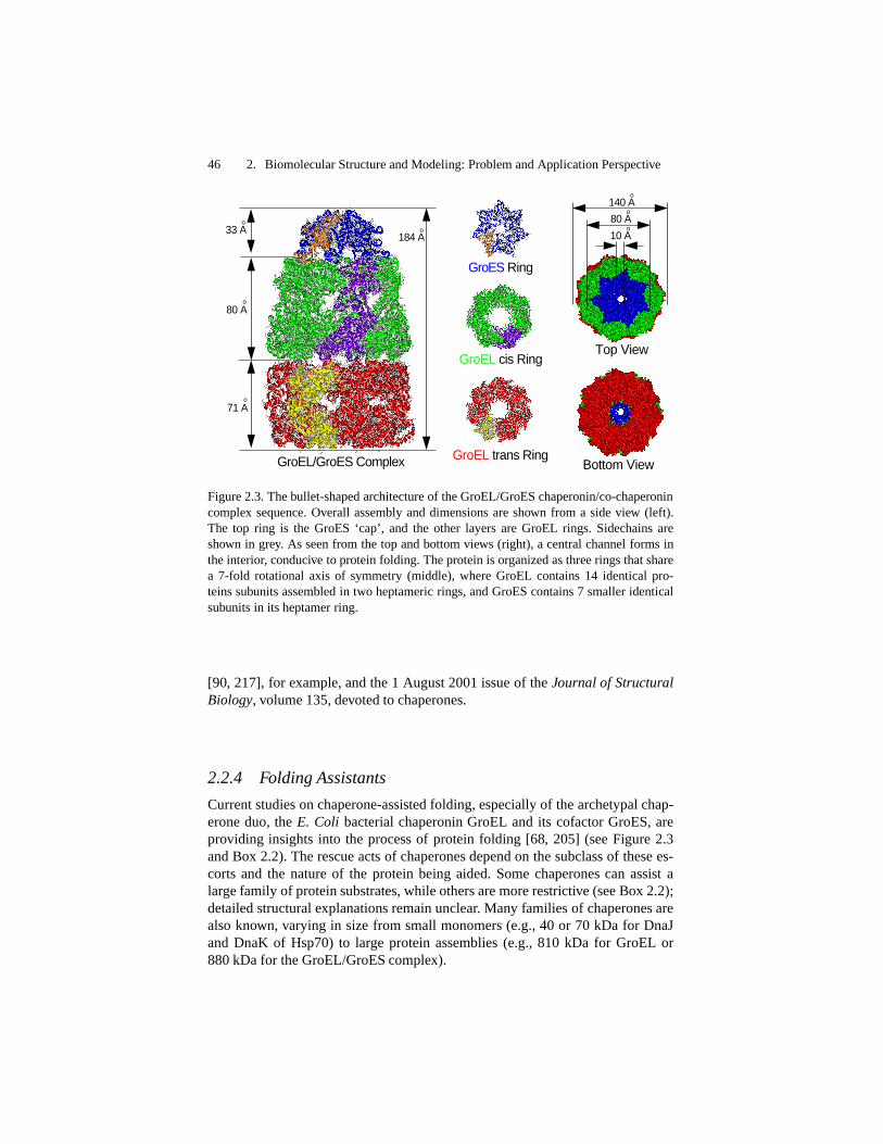
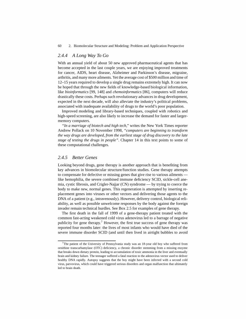
Prion views from several organisms (including human and cow) have been obtained[239], allowing analyses of species variations, folding, and misfolding relationships; see[223], for example. This high degree of similarity across species is shown in Figure 2.4.
Still, until prions are demonstrated to be infectious in vivo, the proteinaceous hypothesiswarrants reservation. Clues into how prions work may emerge from parallel work on yeastprions, which unlike their mammalian counterparts do not kill the organism but producetransmitted heritable changes in phenotype; many biochemical and engineering studies areunderway to explore the underlying mechanism of prion inheritance.
2.3.4 Other Misfolding Processes
There are other examples of protein misfolding diseases (e.g., references citedin [85, 52]). The family of amyloid diseases includes Alzheimer’s, Parkinson’s,and type II (late-onset) diabetes. For example, familial amyloid polyneuropathyis a heritable condition caused by the misfolding of the protein transthyretin. Theamyloid deposits that result interfere with normal nerve and muscle function.
Dobson [52] intriguingly suggests that understanding the evolution of proteinsholds the key to protein misfolding diseases. Namely, he argues that since evolu-tionary processes have selected sequences of amino acids that form close-packed,globular proteins, the effectively irreversible formation of amyloid fibrils reflectsa conversion of proteins to their ‘primordial’ rather than evolved states, possiblyfrom aging-induced mutations that destabilize native proteins.

52 2. Biomolecular Structure and Modeling: Problem and Application Perspective
As in mad cow disease, a molecular understanding of the misfolding processmay lead to treatments of the disorders. In the case of familial amyloid polyneu-ropathy, research has shown that incorporating certain mutant monomers in thetetramer protein transthyretin reduces considerably the formation of amyloid de-posits (amyloid fibrils); moreover, incorporating additional mutant monomerscan prevent misfolding entirely [85]. These findings suggest potential therapeuticstrategies for amyloid and related misfolding disorders. See also [164] for a newpharmacological approach for treating human amyloid diseases by using a small-molecule drug that targets a protein present in amyloid deposits; the drug linkstwo pentamers of that protein and leads to its rapid clearance by the liver.
Recent studies also suggest that misfolded proteins generated in the pathway ofprotein folding can be dangerous to the cell and cause harm (whether or not theyconvert normal chains into misfolded structures, as in prion diseases) [27, 216].The cellular mechanisms associated with such misfolded forms and aggregatesare actively being pursued.
2.3.5 Deducing Function From Structure
Having the sequence and also the 3D structure at atomic resolution, while ex-tremely valuable, is only the beginning of understanding biological function.How does a complex biomolecule accommodate its varied functions and inter-actions with other molecular systems? How sensitive is the 3D architecture of abiopolymer to its constituents?
Despite the fact that in many situations protein structures are remarkably stableto tinkering (mutations), their functional properties can be quite fragile. In otherwords, while a protein often finds ways to accommodate substitutions of a fewamino-acids so as not to form an entirely different overall folding motif [33],even the most minute sequence changes can alter biological activity significantly.Mutations can also influence the kinetics of the folding pathway.
An example of functional sensitivity to sequence is the altered transcriptionalactivity of various protein/DNA complexes that involve single base changesin the TATA-box recognition element and/or single protein mutations in TBP(TATA-Box binding protein) [161]. For example, changing just a single residuein the common nucleotide sequence of TATA-box element, TATAAAAG, toTAAAAAAG impairs binding to TBP and hence disables transcriptional activity.
In principle, theoretical approaches should be able to explain these relationsbetween sequence and structure from elementary physical laws and knowledge ofbasic chemical interactions. In practice, we are encountering immense difficultypinpointing what Nature does so well. After all, the notorious “protein folding”problem is a challenge to us, not to Nature.
Much work continues on this active front.
2.4. Practical Applications 53
Hamster, Mesocricetus Auratus, 1B10Human, Homo Sapien, 1E1G
Mouse, Mus Musculus, 1AG2
Cow, Bos Taurus, 1D1X
Figure 2.4. Structure of the prion protein.
2.4 From Basic to Applied Research
An introductory chapter on biomolecular structure and modeling is aptly con-cluded with a description of the many important practical applications of the field,from food chemistry to material science to drug design. A historical perspectiveon drug design is given in Chapter 14. Here, we focus on the current status ofdrug development as well as other applied research areas that depend strongly onprogress in molecular modeling. Namely, as biological structures and functionsare being resolved, natural disease targets that affect the course of disease canbe proposed. Other biological and polymer targets, such as the ripening genes ofvegetables and fruit or strong materials, can also be manipulated to yield benefitsto health, technology, and industry.
2.4.1 Rational Drug Design: Overview
The concept of systematic drug design, rather than synthesis of compounds thatmimic certain desired properties, is only about 50 years old (see Chapter 14).Gertrude Elion and George Hitchings of Burroughs Wellcome, who won the 1988Nobel Prize in Physiology or Medicine, pioneered the field by creating analoguesof the natural DNA bases in an attempt to disrupt normal DNA synthesis. Theirstrategies eventually led to a series of drugs based on modified nucleic-acid basestargeted to cancer cells. Today, huge compound libraries are available for sys-tematic screening by various combinatorial techniques, robotics, other automatedtechnologies, and various modeling and simulation protocols (see Chapter 14).
Rational pharmaceutical design has now become a lucrative enterprise. Thesales volume for the world’s best seller prescription drug in 1999, prilosec (forulcer and heartburn), exceeded six billion dollars. A vivid description of theclimate in the pharmaceutical industry and on Wall Street can be found in TheBillion-Dollar Molecule: One Company’s Quest for the Perfect Drug [222]. This

54 2. Biomolecular Structure and Modeling: Problem and Application Perspective
thriller describes the racy story of a new biotech firm for drugs to suppressthe immune system, specifically the discovery of an alternative treatment to cy-closporin, medication given to transplant patients. Since many patients cannottolerate cyclosporin, an alternative drug is often needed.
Tremendous successes in 1998, like Pfizer’s anti-impotence drug viagra andEntre-Med’s drugs that reportedly eradicated tumors in mice, have generatedmuch excitement and driven sales and earnings growth for drug producers. Aglance at the names of biotechnology firms is an amusing indicator of the hopeand prospects of drug research: Biogen, Cor Therapeutics, Genetech, Genzyme,Immunex, Interneuron Pharmaceuticals, Liposome Co., Millennium Pharmaceu-ticals, Myriad Genetics, NeXstar Pharmaceuticals, Regeneron Pharmaceuticals,to name a few. Yet, both the monetary cost and development time required foreach successful drug remains very high [16].
2.4.2 A Classic Success Story: AIDS Therapy
HIV Enzymes
A spectacular example of drugs made famous through molecular modelingsuccesses are inhibitors of the two viral enzymes HIV protease (HIV: humanimmunodeficiency virus) and reverse transcriptase for treating AIDS, acquiredimmune deficiency syndrome.
This world’s most deadly infectious disease is caused by an insidious retrovirus.Such a virus can convert its RNA genome into DNA, incorporate this DNA intothe host cell genome, and then spread from cell to cell. To invade the host, the viralmembrane of HIV must attach and fuse with the victim’s cell membrane; onceentered, the viral enzymes reverse transcriptase and integrase transform HIV’sRNA into DNA and integrate the DNA into that of the host [92].
Current drugs inhibit enzymes that are key to the life cycle of the AIDS virus(see Figure 2.5). Protease inhibitors like indinavir block the activity of proteases,protein-cutting enzymes that help a virus mature, reproduce, and become infec-tious [39]. Reverse transcriptase (RT) inhibitors block the action of an enzymerequired by HIV to make DNA from its RNA [173].
AIDS Drug Development
A typical drug cocktail is the triplet drug combination of the protease inhibitorindinavir with the two nucleoside analogue RT inhibitors AZT (zidovudine, or3
�
-azido-3�
-deoxythymidine) and 3TC. More than one drug is needed becausemutations in the HIV enzymes can confer drug resistance; thus, acting on differentsites as well as on different HIV proteins increases effectiveness of the therapy.
Two types of RT blockers are nucleoside analogues and non-nucleoside in-hibitors. Members of the former group like AZT interfere with the HIV activityby replacing a building block used to make DNA from the HIV RNA viruswith an inactive analog and thereby prevent accurate decoding of the viral RNA.Non-nucleoside RT inhibitors (e.g., nevirapine, calanolide molecules, and sustiva

2.4. Practical Applications 55
HIV−2 Protease
Asp25 Asp25’
Gly40Gly40’
Ile50Ile50’
Asp60 Asp60’
HIV−1 Reverse Trans criptase
A
B
Cend
Nend
N
N N
NH
C(CH3)3
OO
H
L−735,524 (Indinavir)
N
OH OH
Cend Nend
N
N
N
O
NO
O
(Nevirapine analogue)1051U91
Figure 2.5. Examples of AIDS drug targets — the HIV protease inhibitor and reversetranscriptase (RT) — with corresponding designed drugs. The protease inhibitor indinavir(crixivan) binds tightly to a critical area of the dimer protease enzyme (HIV-2, 198 residuestotal shown here [39]), near the flaps (residues 40 to 60 of each monomer), inducing aconformational change (flap closing) that hinders enzyme replication; intimate interac-tions between the ligand and enzyme are observed in residues 25 and 50 in each proteasemonomer. The non-nucleoside RT inhibitor 1051U91 (a nevirapine analogue), approvedfor use in combination with nucleoside analogue anti-HIV drugs like AZT, binds to a lo-cation near the active site of RT that does not directly compete with the oligonucleotidesubstrate. The large RT protein of 1000 residues contains two subdomains (A and B).

56 2. Biomolecular Structure and Modeling: Problem and Application Perspective
under development) are designed to bind with high affinity to the active site ofreverse transcriptase and therefore physically interfere with the enzyme’s action.
Design of such drugs was made possible in part by molecular modeling due tothe structure determination of the HIV protease by X-ray crystallography in 1989and RT a few years later [197]. Figure 2.5 shows molecular views of these HIVenzymes complexed with drugs.
Besides the HIV protease and reverse transcriptase, a third target is the HIVintegrase, which catalyzes the integration of a DNA copy of the viral genome intothe host cell chromosomes. Scientists at Merck have identified 1,3-diketo acidintegrase inhibitors that block strand transfer, one of the two specific catalyticfunctions of HIV-1 integrase [94]; this function has not been affected by previousinhibitors. This finding paves the way for developing effective integrase inhibitors.
AIDS Drug Limitations
Much progress has been made in this area since the first report of the rationaldesign of such inhibitors in 1990 [176] (see [16] for a review). In fact, the dra-matic decline of AIDS-related deaths by such drug cocktails can be attributed inlarge part to these new generation of designer drugs (see Box 2.4) since the firstintroduction of protease inhibitors in 1996. Indeed, the available drug cocktailsof protease inhibitors and nucleoside analogues RT inhibitors have been shown tovirtually suppress HIV, making AIDS a manageable disease.
However, the cocktails are not a cure. The virus returns once patients stopthe treatment. The mechanisms of drug resistant mutations and the interactionsamong them are not clearly understood [173]. In addition, few countires in thedeveloping world, like Africa, can afford the virus suppressing drugs; the drug-cocktail regimen is complex, requiring many daily pills taken at multiple timesand separated from eating, most likely for life; serious side effects also occur.In certain parts of the world, the situation is profoundly distressing: the lifeexpectancy of sexually-active Ugandans, for example, has fallen from 64 yearsbefore the epidemic to 42 today, and the number continues to drop.
Lurking Virus
As mentioned, even available treatments cannot restore the damage to the pa-tient’s immune system; the number of T-cell (white blood cells), which that HIVattaches itself to, is still lower than normal (which lowers the body’s defensesagainst infections), and there remain infected immune cells that the drugs cannotreach because of integration. Thus, new drugs are being sought to interrupt thefirst step in the viral life cycle — binding to a co-receptor on the cell surface torid the body of the cell’s latent reservoirs of the HIV virus, to chase the virus outof cells where it hides for subsequent treatment, or to drastically reduce the HIVreservoir so that the natural immune defenses can be effective. New structural andmechanistic targets are currently being explored (see Box 2.4).
A better understanding of the immune-system mechanism associated withAIDS, for example, may help explain how to prime the immune system to rec-

2.4. Practical Applications 57
ognize an invading AIDS virus. Unlike traditional AIDS drug cocktails whichinhibit division of already infected cells, fusion (or entry) inhibitors define anotherclass of drugs that seek to prevent HIV from entering the cell membrane. This en-try, called fusion, releases the virus’s genetic material and allows it to replicate.The promising drug T-20 or enfuvirtide (which must be injected into the skin) isa member of fusion-inhibitor drugs that, when added to a combination of stan-dard drugs, can significantly reduce HIV levels in the blood. As manifested byits complex components of invasion that include the fusion apparatus, the AIDSvirus has developed a complex, tricky, and multicomponent-protection infectionmachinery, as well as drug-resistant defense.
Besides integrase and fusion inhibitors, among the newer drugs to fight AIDSbeing developed are immune stimulators and antisense drugs. The former stim-ulate the body’s natural immune response, and the latter mimic the HIV geneticcode and prevent the virus from functioning.
Vaccine?
Still, many believe that only an AIDS vaccine offers true hope against thisdeadly disease. Yet the research on vaccines trails behind the development ofdrugs, which offer much greater financial incentives and lower risks The vaccineAIDSVAX by the California-base company VaxGen (ready by 2004 or 2005)could protect in part people from HIV, but the early 2003 results from the firstlarge-scale trial were not as encouraging as had been hoped. This vaccine isa genetically amplified version of a single protein from the outer shell of theAIDS virus; because the shell changes rapidly, the vaccine may offer only limitedprotection.
Another vaccine under development by an Oxford team (part of the Inter-national AIDS vaccine Initiative) is exploiting for vaccine development theimmunological data gleaned from Nairobi women who have remained unaffectedby AIDS despite many years of high-risk sexual behavior. These women’s T-cellswere found to fight off the disease by attacking two particular proteins producedby the AIDS virus. The DNA sequences making those proteins were subsequentlyidentified and used to create a vaccine specific to viral infections in East Africa;besides the DNA component associated with the relevant genes, the vaccine wasamplified with a benign virus copy with same DNA sequences inserted.
Other vaccines are being developed (e.g., Harvard, Merck) but, like those fur-ther along, response is far from ideal. Unfortuntately, experience suggests that aconstant level of exposure (e.g., booster shots) is needed to yield immunity, andthis defeats the main vaccine advantage of convenience and low cost. Observa-tions also suggest that more than one vaccine may be needed, since the HIV virusmutates and replicates quickly.6 Still, it is hoped that therapeutic vaccination in
6For example, there is an enormous variation in the HIV-1 envelope protein. It has also beenfound that nearly all of non-nucleoside reverse transcriptase inhibitors can be defeated by site-directedmutation of tyrosine 181 to cysteine in reverse transcriptase. For this reason, the derivatives of calano-

58 2. Biomolecular Structure and Modeling: Problem and Application Perspective
combination with anti-HIV-1 drug treatment, even if it fails to eradicate infection,will suppress AIDS infection and the rate of transmission, and ultimately decreasethe number of AIDS deaths substantially.
For a comprehensive overview of the biology of AIDS, the HIV life cycle,current status of the AIDS pandemic, and efforts for treating AIDS, see NatureInsight in the 19 April 2001 issue of Nature (Volume 410). This review was writ-ten at the 20 years after the first hints of the disease were reported in the summerof 1981, in clusters of gay men in large American cities; these groups exhibitedsevere symptoms of infection by certain pneumonias combined with those fromKaposi’s sarcoma (KS) cancer.
Box 2.4: Fighting AIDS
AIDS drugs attributed to the success of molecular modeling include AZT (zidovudine)sold by Bristol-Myers Squibb, and the newer drugs viracept (nelfinavir) made by AgouronPharmaceuticals, crixivan (indinavir) by Merck & Company, and amprenavir discovered atVertex Pharmaceuticals Inc. and manufactured by Glaxo Wellcome. Amprenavir, in partic-ular, approved by the U.S. Government in April 1999, is thought to cross the ‘blood-brainbarrier’ so that it can attack viruses that lurk in the brain, where the virus can hide. Thisgeneral class of inhibitors has advanced so rapidly that drug-resistant AIDS viruses havebeen observed.
Current structural investigations are probing the structural basis for the resistance mech-anisms, which remain mysterious, particularly in the case of nucleoside analogue RTinhibitors like AZT [121]. The solved complex of HIV-1 reverse transcriptase [100] of-fers intriguing insights into the conformational changes associated with the altered virusesthat influence the binding or reactivity of inhibitors like AZT and also suggests how toconstruct drug analogues that might impede viral resistance.
Basic research on the virus’s process of invading host cells — by latching onto receptors(e.g., the CD4 glycoprotein, which interacts with the viral envelope glycoprotein, gp120,and the transmembrane component glycoprotein, gp41), and co-receptors (e.g., CCR5 andCXCR4) — may also offer treatments, since developments of disease intervention and vac-cination are strongly aided by an understanding of the complex entry of HIV into cells; see[119] for example.
The HIV virus uses a spear-like agent on the virus’ protein coat to puncture the mem-brane of the cells which it invades; vaccines might be designed to shut the chemicalmechanism or stimuli that activate this invading harpoon of the surface protein. The solvedstructure of a subunit of gp41, for example, has been exploited to design peptide inhibitorsthat disrupt the ability of gp41 to contact the cell membrane [69]. A correlation has beennoted, for example, between co-receptor adaptation and disease progression.
lide A under current development are attractive drug targets because they appear more robust againstmutation [113].

2.4. Practical Applications 59
Novel techniques for gene therapy for HIV infections are also under development, suchas internal antibodies (intrabodies) against the Tat protein, a vehicle for HIV infection ofthe immune cells; it is hoped that altered T-cells that produce their own anti-Tat intrabodywould lengthen the survival time of infected cells or serve as an HIV ‘dead-end’.
Other clues to AIDS treatments may come from the finding that HIV-1 originally camefrom a subspecies of chimpanzees [80]. Since chimps have likely carried the virus forhundreds of thousands of years but not become ill from it, understanding this observationmight help fight HIV-pathogeny in humans. Help may also come from the interesting find-ing that a subset of humans have a genetic mutation (32 bases deleted from the 393 of geneCCR5) that creates deficient T-cell receptor; this mutation intriguingly slows the onset ofAIDS. Additionally, a small subset of people is endowed with a huge number of helper(CD4) T-cells which can coordinate an attack on HIV and thus keep the AIDS virus underexquisite control for many years; such people may not even be aware of the infection foryears.
2.4.3 Other Drugs
Another example of drug successes based on molecular modeling is the design ofpotent thrombin inhibitors. Thrombin is a key enzyme player in blood coagula-tion, and its repressors are being used to treat a variety of blood coagulation andclotting-related diseases. Merck scientists reported [17] how they built upon crys-tallographic views of a known thrombin inhibitor to develop a variety of inhibitoranalogues. In these analogues, a certain region of the known thrombin inhibitorwas substituted by hydrophobic ligands so as to bind better to a certain enzymepocket that emerged crucial for the fit. Further modeling helped select a subsetof these ligands that showed extremely compact thrombin/enzyme structures; thiscompactness helps oral absorption of the drug. The most potent inhibitor thatemerged from these modeling studies has demonstrated good efficacy on animalmodels [17].
Other examples of drugs developed in large part by computational techniquesinclude the antibacterial agent norfloxacin of Kyorin Pharmaceuticals (noroxinis one of its brand names), glaucoma treatment dorzolamide (“trusopt”/Merck),Alzheimer’s disease treatment donepezil (“aricept”/ Eisai), and migraine medicinezolmitriatan (“zomig”) discovered by Wellcome and marketed by Zeneca [16].The headline-generating drug that combats impotence (viagra) was also found bya rational drug approach. It was interestingly an accidental finding: the compoundhad been originally developed as a drug for hypertension and then angina.
There are also notable examples of herbicides and fungicides that were success-fully developed by statistical techniques based on linear and nonlinear regressionand classical multivariate analysis (or QSAR, see Chapter 14): the herbicidemetamitron — a bestseller in 1990 in Europe for protecting sugar beet crops —was discovered by Bayer AG in Germany.
60 2. Biomolecular Structure and Modeling: Problem and Application Perspective
2.4.4 A Long Way To Go
With an annual yield of about 50 new approved pharmaceutical agents that hasbecome accepted in the last couple years, we are enjoying improved treatmentsfor cancer, AIDS, heart disease, Alzheimer and Parkinson’s disease, migraine,arthritis, and many more ailments. Yet the average cost of $500 million and time of12–15 years required to develop a single drug remains extremely high. It can nowbe hoped that through the new fields of knowledge-based biological information,like bioinformatics [99, 148] and chemoinformatics [86], computers will reducedrastically these costs. Perhaps such revolutionary advances in drug development,expected in the next decade, will also alleviate the industry’s political problems,associated with inadequate availability of drugs to the world’s poor population.
Improved modeling and library-based techniques, coupled with robotics andhigh-speed screening, are also likely to increase the demand for faster and larger-memory computers.
“In a marriage of biotech and high tech,” writes the New York Times reporterAndrew Pollack on 10 November 1998, “computers are beginning to transformthe way drugs are developed, from the earliest stage of drug discovery to the latestage of testing the drugs in people”. Chapter 14 in this text points to some ofthese computational challenges.
2.4.5 Better Genes
Looking beyond drugs, gene therapy is another approach that is benefiting fromkey advances in biomolecular structure/function studies. Gene therapy attemptsto compensate for defective or missing genes that give rise to various ailments —like hemophilia, the severe combined immune deficiency SCID, sickle-cell ane-mia, cystic fibrosis, and Crigler-Najjar (CN) syndrome — by trying to coerce thebody to make new, normal genes. This regeneration is attempted by inserting re-placement genes into viruses or other vectors and delivering those agents to theDNA of a patient (e.g., intravenously). However, delivery control, biological reli-ability, as well as possible unwelcome responses by the body against the foreigninvader remain technical hurdles. See Box 2.5 for examples of gene therapy.
The first death in the fall of 1999 of a gene-therapy patient treated with thecommon fast-acting weakened cold virus adenovirus led to a barrage of negativepublicity for gene therapy.7 However, the first true success of gene therapy wasreported four months later: the lives of most infants who would have died of thesevere immune disorder SCID (and until then lived in airtight bubbles to avoid
7The patient of the University of Pennsylvania study was an 18-year old boy who suffered fromornithine transcarbamylase (OTC) deficiency, a chronic disorder stemming from a missing enzymethat breaks down dietary protein, leading to accumulation of toxic ammonia in the liver and eventuallybrain and kidney failure. The teenager suffered a fatal reaction to the adenovirus vector used to deliverhealthy DNA rapidly. Autopsy suggests that the boy might have been infected with a second coldvirus, parvovirus, which could have triggered serious disorders and organ malfunction that ultimatelyled to brain death.

2.4. Practical Applications 61
the risk of infection) were not only saved, but able to live normal lives followinggene therapy treatments that restore the ability of a gene essential to make T cells[35]. Unfortunately, complications arose is several of the treated infants by late2002 (see Box 2.5).
Though such medical advances appear just short of a miracle, it remains tobe seen how effective gene therapy will be on a wide variety of diseases andover a long period. Still, given that gene therapy is a young science in a state ofcontinuous flux, results to date indicate a promising future for the field [6].
A related technique for designing better genes is another relatively new tech-nique known as directed molecular evolution. Unlike protein engineering, inwhich natural proteins are improved by making specific changes to them, di-rected evolution involves mutating genes in a test tube and screening the resulting(‘fittest’) proteins for enhanced properties. Companies specializing in this newDarwinian mimicking (e.g., Maxigen, Diversa, and Applied Molecular Evolu-tion) are applying such strategies in an attempt to improve the potency or reducethe cost of existing drugs, or improve the stain-removing ability of bacterialenzymes in laundry detergents. Beyond proteins, such ideas might also be ex-tended to evolve better viruses to carry genes into the body for gene therapy orevolve metabolic pathways to use less energy and produce desired nutrients (e.g.,carotenoid-producing bacteria).
Box 2.5: Gene Therapy Examples
A prototype disease model for gene therapy is hemophilia, whose sufferers lack keyblood-clotting protein factors. Specifically, Factor VIII is missing in hemophilia A patients(the common form of the disease); the much-smaller Factor IX is missing in hemophilia Bpatients (roughly 20% of hemophiliacs in the United States).
Early signs of success in treatment of hemophilia B using adeno-associated virus (a vec-tor not related to adenovirus, which is slower acting and more suitable for maintenance andprevention) were reported in December 1999. However, introducing the much larger geneneeded for Factor VIII, as required by the majority of hemophiliacs, is more challenging.Here, the most successful treatments to date only increase marginally this protein’s level.Yet even those minute amounts are reducing the need for standard hemophilia treatment(injections of Factor IX) in these patients.
Larger vectors to stimulate the patient’s own cells to repair the defective gene are thussought, such as retroviruses (e.g., lentiviruses, the HIV-containing subclass), or non-virusparticles, like chimeraplasts (oligonucleotides containing a DNA/RNA blend), which canin theory correct point mutations by initiating the cell’s DNA mismatch repair machinery.
An interesting current project involving chimeraplasts is being tested in children ofAmish and Mennonite communities to treat the debilitating Crigler-Najjar (CN) syndrome.Sufferers of this disease lack a key enzyme which break down the toxic waste productbilirubin, which in the enzyme’s absence accumulates in the body and causes jaundice and

62 2. Biomolecular Structure and Modeling: Problem and Application Perspective
overall toxicity. Children with CN must spend up to 18 hours a day under a blue light toclear bilirubin and seldom reach adulthood, unless they are fortunate to receive and respondto a liver transplant. Chimeraplasty offers these children hope, and might reveal to be saferthan the adenovirus approach, but the research is preliminary and the immune response iscomplex and mysterious.
Recent success was reported for treating children suffering from the severe immunedisorder SCID type XI [35]. Gene therapy involves removing the bone marrow from in-fants, isolating their stem-cells, inserting the normal genes to replace the defective genesvia retroviruses, and then re-infusing the stem cells into the blood stream. As hoped, theinserted stem cells were able to generate the cells needed for proper immune functioningin the patients, allowing the babies to live normal lives. Though successful for 2–3 yearsfor most infants, complications arose when several infants developed leukemia-like condi-tions. Scientists believe that the retrovirus vectors lodged near a cancer-causing gene andactivated it. Of course, it remains to be seen whether the overall benefits outweigh the risks,and how in the long term children’s new immune systems will behave (i.e., deteriorate overtime or continue to function properly).
Though clearly many bumps in the road are expected when new therapies are developed,scientists remain hopeful. Indeed, success in any such gene therapy endeavors would leadto enormous progress in treating inherited diseases caused by point mutations.
2.4.6 Designed Compounds and Foods
From our farms to medicine cabinets to supermarket aisles, designer foods are bigbusiness.
As examples of these practical applications, consider the transgenic organismsdesigned to manufacture medically-important compounds: bacteria that producehuman insulin, goats whose milk contains proteins to make silk for use in surgicalthread or bulletproof clothing, silkworms that produce mammalian-type collagenand silk for use in tissue engineering and other medical applications, and the foodproduct chymosin to make cheese, a substitute for the natural rennet enzyme tra-ditionally extracted from cows’ stomachs. Genetically modified bacteria, moregenerally, hold promise for administering drugs and vaccines more directly tothe body (e.g., the gut) without the severe side effects of conventional therapies.For example, a strain of the harmless bacteria Lactococcus lactis modified to se-crete the powerful anti-inflammatory protein interleukin-10 (IL-10) has shownto reduce bowel inflammation in mice afflicted with inflammatory bowel dis-ease (IBD), a group of debilitating ailments that includes Crohn’s disease andulcerative colitis.
The production of drugs in genetically-altered plants — “biopharming” or“molecular pharming” — represents a growing trend in agricultural biotechnol-ogy. The goal is to alter gene structure of plants so that medicines can be grown onthe farm, such as to yield an edible vaccine from a potato plant against hepatitis B,or a useful antibody to be extracted from a tobacco plant. As in bioengineeredfoods, many obstacles must be overcome to make such technologies effective

2.4. Practical Applications 63
as medicines, environmentally safe, and economically profitable. Proponents ofmolecular pharming hope eventually for far cheaper and higher yielding drugs.
Genetically-engineered crops are also helping farmers and consumers by im-proving the taste and nutritional value of food, protecting crops from pests, andenhancing yields. Examples include the roughly one-half of the soybean and one-third of the corn grown in the United States, sturdier salad tomatoes,8 corn pollenthat might damage monarch butterflies, papaya plants designed to withstand thepapaya ringspot virus, and caffeine-free plants (missing the caffeine gene) thatproduce decaffeinated cups of java.
Closer to the supermarket, one of the fastest growing category of foods todayis nutraceuticals (a.k.a. functional foods or pharmaceuticals), no longer relegatedonly to health-food stores. These foods are designed to improve our overall nutri-tion as well as to help ward off disease, from cancer prevention to improved brainfunction. See Box 2.6 for examples. Related are diet ingredients and supplementscustomized to genetic variations based on gene/diet connections (nutritional ge-nomics or nutrigenomics), such as diets low in certain proteins (for patients withphenylketonuria) or high in liver, broccoli, and other folic-acid rich foods (forpeople with a genetic variation that produces a less efficient enzyme involved inprocessing folic acid).
The general public (first in Europe and now in the United States) has resistedgenetically-modified or biotech crops, and this was followed by several blockadesof such foods by leading companies, as well as global biosafety accords to protectthe environment. Protesters have painted these products as unnatural, hazardous,evil, and environmentally dangerous (‘Frankenfoods’).9
With the exception of transferred allergic sensitivities — as in Brazil nut al-lergies realized in soybeans that contained a gene from Brazil nuts — mostnegative reactions concerning food safety are not scientifically well-groundedin this writer’s opinion. In fact, not only do we abundantly use various spraysand chemicals to kill flies, bacteria, and other organisms in our surroundings andon the farm; each person consumes around 500,000 kilometers of DNA on an
8The Flavr Savr tomato that made headlines when introduced in 1993 contained a gene that reducesthe level of the ripening enzyme polygalacturonase. However, consumers were largely disappointed:though beautiful, the genetically engineered fruit lacked taste. This is because our understanding offruit ripening is still limited; a complex, coordinated series of biochemical steps is involved — modi-fying cell wall structure, improving texture, inducing softening, and producing compounds in the fruitthat transform flavor, aroma, and pigmentation. Strawberries and other fruit are known to suffer sim-ilarly from the limitations of our understanding of genetic regulation of ripening and, perhaps, alsofrom the complexity of human senses! See [215], for example, for a recent finding that a tomato plantwhose fruit cannot ripen carries a mutation in a gene encoding a transcription factor.
9Amusing Opinion/Art ads that appeared in The New York Times on 8 May 2000 include provoca-tive illustrations with text lines like “GRANDMA’S MINI-MUFFINS are made with 100%NATURAL irradiated grain and other ingredients”; “TOTALLY ORGANIC Biomatter madewith Nucleotide Resequencing”; “The Shady Glen Farms Promise: Our Food is fresh fromthe research labs buried deep under an abandoned farm”. [Note: the font size and formdifferences here are intentional, mimicking the actual ads].

64 2. Biomolecular Structure and Modeling: Problem and Application Perspective
average day! Furthermore, there are many potential benefits from genetically-engineered foods, like higher nutrients and less dependency on pesticides, andthese considerations might win in the long run. Still, environmental effects mustbe carefully monitored so that genetically-altered food will succeed in the longrun (see Box 2.6 for possible problems).
Perhaps to counter fear of introduced allergens, bioengineering is also beingused to reduce or remove compounds that cause allergic reactions in people.Though at a relatively early stage, various companies worldwide are using ge-netic engineering to try to reduce allergies from foods like wheat, rice, soybean,ryegrass, and peanuts. Genes responsible for producing allergenic proteins canbe removed (i.e., knocked out), as done for soybeans, or the associated proteinsredesigned, as in peanuts, so that allergenicity is lost but other nut characteristicsare retained. As above, care must be taken to retain flavor, freshness, and looks ofthe original product, and not to introduce other possible allergens.
In addition to tampering with plants to remove allergens, such biotech com-panies are also expanding effort on the removal of genes associated with naturaltoxins. For example, companies (with support of national security organizations)are attempting to remove the toxin ricin — one of the deadliest substances known— from castor plants. Castor beans have been cultivated for centuries, and theplant’s natural oils (which lack toxicity) are widely used as laxatives and as com-ponent in brake fluid, dyes, soaps, and cosmetics. However, the toxic protein ricincan also be extracted from the castor plant, and has been associated with terror-ist groups like Al Qaeda, with production of weapons “for mass destruction” inIraq, and with a famous killing of the spy Georgi Markov on a London sidewalkin 1978 by Bulgarian agents who injected ricin from an umbrella tip into the de-fector’s leg. Once removed, ricin-free castor plants can become more attractive togrowers.
Box 2.6: Nutraceuticals Examples
The concept of fortified food is not new. Vitamin-D supplemented milk has eradicatedrickets, and fortified breakfast cereals have saved many poor diets. In fact, classic bio-engineering has been used for a long time to manipulate genes through conventional plantand animal inter-breeding. But the new claims — relying on our increased understand-ing of our body’s enzymes and many associated vital processes — have been makingheadlines. (“Stressed Out? Bad Knee? Try a Sip of These Juices.”, J.E. Barnes and G.Winter, New York Times, Business, 27 May 2001). Tea brews containing sedative roots likekava promise to tame tension and ease stress. Fruit-flavored tonics with added glucosamine(building block of cartilage) and calcium are claimed to soothe stiff knees of aging bod-ies. (See Chapter 3 on the fibrous protein collagen). Fiber-rich grains are now touted asheart-disease reducers. Herb-coated snacks, like potato corn munchies coated with ginkgobiloba, are advertised as memory and alertness boosters.
With this growing trend of designer foods, the effect of these manipulations on our en-vironment demands vigilant watch. This is because it is possible to create ‘super-resistant

2.4. Practical Applications 65
weeds’ or genetically-improved fish that win others in food or mate competitions. Thispotential danger emerges since, unlike conventional cross-breeding (e.g., producing a tan-gelo from a tangerine and grapefruit), genetic engineering can overcome the species barrier— by inserting nut genes in soybeans or fish genes in tomatoes, for example. This newertype of tinkering can have unexpected results in terms of toxins or allergens which, oncereleased to the environment, cannot be stopped easily. For example, the first genetically-modified animal to reach American dinner plates is likely to be a genetically-altered salmonendowed with fortified genes that produce growth hormones, making the fish grow twice asfast as normal salmon. The effect of these endowed fish on the environment is yet unknown.
Popular examples of fortified food products with added vitamins and minerals (e.g., cal-cium and vitamin E) that also help protect against osteoporosis are orange juice, specialtyeggs, and some vegetarian burritos. Other designer disease-fighting foods include drinksenriched with echinacea to combat colds; juices filled with amino acids and herbs claimedto boost muscle and brain function; margarines containing plant stanol esters (from soy-bean or pine trees) to fight heart disease and cancer (by blocking cholesterol absorptionfrom the digestive tract), as well as green teas enriched with ginseng and other herbs; su-peryogurts to enhance the immune system; and tofu and yams to combat hot flashes. Suchfunctional foods are also touted to lower cholesterol, provide energy, fight off depression,or to protect against salmonella and E. coli poisoning (e.g., yogurt fortified with certainbacteria). Many other enriched food products are under design, for example fruit with in-creased vitamin C levels using a recently-isolated gene in strawberries (GalUR) that playsan important role in the production of vitamin C.
Will Ginkgo Biloba chips, Tension Tamer cocktails, or Quantum Punch juice becomepart of our daily diet (and medicine cabinet) in this millennium?
2.4.7 Designer Materials
New specialty materials are also being developed in industry with the neededthermochemistry, stereochemistry (e.g., compounds that bind to one chemical butnot its mirror image), and kinetic properties. Examples are enzymes for man-ufacturing detergents, adhesives and coatings, photography film, or biosensorsfor explosives [130]. Fullerene nanotubes (giant linear fullerene chains that cansustain enormous elastic deformations [234]), formed from condensed carbon va-por, have many potential applications. These range from architectural componentsof bridges and buildings, cars, and airplanes to heavy-duty shock absorbers, tocomponents of computer processors, scanning microscopes, and semiconductors.
Long buckyball nanotubes have even been proposed as elements of ‘elevators’to space in the new millennium [234]. These applications arise from their smallsize (their thickness is five orders of magnitude smaller than human hair), amaz-ing electronic properties, and enormous mechanical strength of these polymers.In particular, these miniscule carbon molecules conduct heat much faster thansilicon, and could therefore replace the silicon-based devices used in microelec-

66 2. Biomolecular Structure and Modeling: Problem and Application Perspective
tronics, possibly overcoming current limitations of computer memory and speed.
2.4.8 Cosmeceuticals
Cosmeceutical companies are also rising — companies that specialize in design ofcosmetics with bioactive ingredients (such as designer proteins and enzymes), in-cluding cosmetics that are individually customized (by pharmacogenomics) basedon genetic markers, such as single nucleotide polymorphisms (SNPs). Most pop-ular are products for sun or age-damaged skin containing alpha hydroxy acids(mainly glycolic and lactic acid), beta hydroxy acids (e.g., salicylic acid), andvarious derivatives of vitamin A or retinol (e.g., the tretinoin-containing Retin-A and Renova topical prescriptions). Besides reducing solar scars and wrinkling,products can also aid combat various skin diseases. Many of these compoundswork by changing the metabolism of the epidermis, for example by increasing therate of cell turnover, thereby enhancing exfoliation and the growth of new cells.New cosmeceuticals contain other antioxidants, analogues of various vitamins (A,D, and E), and antifungal agents.
The recent information gleaned from the Human Genome Project can help rec-ognize changes that age and wrinkle skin tissue, or make hair or teeth gray. Thisin turn can lead to the application of functional genomics technology to developagents that might help rejuvenate the skin, or color only target gray hair or toothenamel. Computational methods have an important role in such developments byscreening and optimizing designer peptides or proteins. Such biotechnology re-search to produce products for personal care will likely rise sharply in the comingyears.

2.4. Practical Applications 67

68 2. Biomolecular Structure and Modeling: Problem and Application Perspective


![Biomolecular modeling using NAMD on TeraGrid …charm.cs.illinois.edu/newPapers/10-31/talk.pdfBiomolecular modeling using NAMD on TeraGrid machines ... 14 [1] Castelmur et al., PNAS,](https://static.fdocuments.net/doc/165x107/5b3956fb7f8b9a40428e73af/biomolecular-modeling-using-namd-on-teragrid-charmcs-modeling-using-namd-on-teragrid.jpg)