Biomimetic protein nanoparticles facilitate enhanced dendritic cell ...
11
UC Irvine UC Irvine Previously Published Works Title Biomimetic protein nanoparticles facilitate enhanced dendritic cell activation and cross- presentation Permalink https://escholarship.org/uc/item/1nb80207 Journal ACS Nano, 7(11) ISSN 1936-0851 Authors Molino, NM Anderson, AKL Nelson, EL et al. Publication Date 2013-11-26 DOI 10.1021/nn403085w License CC BY-NC-ND 4.0 Peer reviewed eScholarship.org Powered by the California Digital Library University of California
Transcript of Biomimetic protein nanoparticles facilitate enhanced dendritic cell ...
UC IrvineUC Irvine Previously Published Works
TitleBiomimetic protein nanoparticles facilitate enhanced dendritic cell activation and cross-presentation
Permalinkhttps://escholarship.org/uc/item/1nb80207
JournalACS Nano, 7(11)
ISSN1936-0851
AuthorsMolino, NMAnderson, AKLNelson, ELet al.
Publication Date2013-11-26
DOI10.1021/nn403085w
LicenseCC BY-NC-ND 4.0 Peer reviewed
eScholarship.org Powered by the California Digital LibraryUniversity of California
MOLINO ET AL . VOL. 7 ’ NO. 11 ’ 9743–9752 ’ 2013
www.acsnano.org
9743
October 03, 2013
C 2013 American Chemical Society
Biomimetic Protein NanoparticlesFacilitate Enhanced Dendritic CellActivation and Cross-PresentationNicholas M. Molino,† Amanda K. L. Anderson,‡,§ Edward L. Nelson,‡ and Szu-Wen Wang*,†
†Department of Chemical Engineering and Materials Science, University of California, Irvine, California 92697-2575, United States, and ‡Institute for Immunology,University of California, Irvine, California 92697-4120, United States. §Present address: Epic Sciences, La Jolla, California 92037, United States.
Although recent years have seen ad-vances in cancer therapies, commontreatment strategies (e.g., chemother-
apy, radiation therapy, and surgical resection)still rely on techniques that lack specificity andrisk side effects, including toxicity.1 Recently, amore targeted approach to cancer therapyhas included harnessing the body's immunesystem for tumor destruction. While cancervaccination is a promising strategy, criticalbarriers to becoming a viable treatment in-clude immune tolerance and the inability toprovoke a robust enough immune responseto overcome the weak immunogenicity ofmany cancer antigens.1,2
In contrast, the natural immune systemhas evolved to become particularly adept atrecognizing key geometries and pathogenicfeatures,most notably thoseof viruses. Viruses,virus-like particles (VLPs), and other proteinnanoparticles have proven to be well-suitedas vaccine platforms,3 and examples of theirdevelopment exist in the clinic (e.g., Gardasil)and in clinical trials.3,4 VLPs typically contain
a hollow core and multiple interfaces(i.e., internal, external, and intersubunit) forengineering functional elements,3,5�7 al-lowing fine control over physical propertiessuch as particle stability, surface chemistry,and biological interaction.6,8�11 However,many viral-based vaccine platforms exhibitstrong self-adjuvanting properties thatmay not always be desired, dependingon the preferred immunotherapeutic out-come.3,12 Additionally, VLPs and attenu-ated viruses may be difficult to produceand purify in large quantities using com-mon protein expression systems, andtherefore alternative platforms should beexplored.12
Our group has been developing the struc-tural core of the nonviral E2 subunit ofpyruvate dehydrogenase as a protein nano-particle platform for therapeutic applicat-ion.8�11,13,14 E2 is a caged protein structureexhibiting unusually high thermostability andcomprises 60 identical self-assemblingmono-mers that produce a hollow dodecahedral
* Address correspondence [email protected].
Received for review June 18, 2013and accepted October 3, 2013.
Published online10.1021/nn403085w
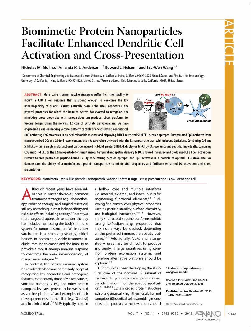
ABSTRACT Many current cancer vaccine strategies suffer from the inability to
mount a CD8 T cell response that is strong enough to overcome the low
immunogenicity of tumors. Viruses naturally possess the sizes, geometries, and
physical properties for which the immune system has evolved to recognize, and
mimicking those properties with nanoparticles can produce robust platforms for
vaccine design. Using the nonviral E2 core of pyruvate dehydrogenase, we have
engineered a viral-mimicking vaccine platform capable of encapsulating dendritic cell
(DC)-activating CpG molecules in an acid-releasable manner and displaying MHC I-restricted SIINFEKL peptide epitopes. Encapsulated CpG activated bone
marrow-derived DCs at a 25-fold lower concentration in vitro when delivered with the E2 nanoparticle than with unbound CpG alone. Combining CpG and
SIINFEKL within a single multifunctional particle induced∼3-fold greater SIINFEKL display on MHC I by DCs over unbound peptide. Importantly, combining
CpG and SIINFEKL to the E2 nanoparticle for simultaneous temporal and spatial delivery to DCs showed increased and prolonged CD8 T cell activation,
relative to free peptide or peptide-bound E2. By codelivering peptide epitopes and CpG activator in a particle of optimal DC-uptake size, we
demonstrate the ability of a noninfectious protein nanoparticle to mimic viral properties and facilitate enhanced DC activation and cross-
presentation.
KEYWORDS: biomimetic . virus-like particle . nanoparticle vaccine . protein cage . cross-presentation . CpG . dendritic cell
ARTIC
LE
MOLINO ET AL . VOL. 7 ’ NO. 11 ’ 9743–9752 ’ 2013
www.acsnano.org
9744
capsule ∼25 nm in diameter.13,15�17 This size fallswithin the narrow size range of 20�45 nm, which isreported to be optimal for passive diffusion to regionsof high immune activity (i.e., the lymph nodes) foruptake by the body's most potent antigen-presentingcell, the dendritic cell (DC).7,18,19 Because E2 is a non-viral particle, it does not possess any infectious abilityor native biological function for entrance into mam-malian cells. We have engineered an E2 particle thatcontains recombinantly introduced internal cysteineresidues for packaging of bioactive molecules andcellular delivery.11 Other groups have explored E2as a platform for inducing helper T cell and humoralresponses to HIV in vivo.20,21 These recent studies,along with our demonstrated ability to modulate im-mune interaction with E2 in vitro and to deliver ther-apeutic molecules to cells, have prompted us toexplore the redesign of our protein nanoparticle as aviral-mimicking DC-based vaccine platform.9�11
DCs have been identified as the key target for cell-mediated immunotherapies because of their antigenprocessing capabilities and orchestration of down-stream adaptive immune responses.22�24 Importantfor cancer, DCs are particularly efficient at capturingand presenting endogenous antigen via MHC I (i.e.,cross-presentation), leading to a strong CD8 T celleffector response.22,23,25 Viruses are strong inducersof CD8 T cell immunity, and therefore, nanoparticles byvirtue of their similar size have been explored for thedelivery of antigens to DCs.24,26,27 In addition to thepackaging of antigens, nanoparticles may also encap-sulate DC-activating molecules (e.g., CpG DNA motifs)
to mediate the magnitude and type of immuneresponse.28
Reports have shown that a requisite for a strongantitumor response includes simultaneous codeliveryof antigen and activator to the same DC subcellularcompartment, as would happen with a natural viralinfection.29,30 Many current strategies employ systemicdelivery of antigen togetherwith adjuvant, thereby notlikely meeting this criteria, as it places a high constrainton both free antigen and activator arriving in the sameDC subcellular compartment simultaneously in vivo.Recent attempts to overcome this barrier have in-cluded the use of nanoparticles for packaged deliveryof vaccine components.3,31,32 Furthermore, nanoparti-cles protect the molecular cargo while also shieldingthe host from toxic or immune-impairing side effects,which have been linked to systemic delivery of Toll-likereceptor (TLR) agonists in humans.33�35 A nanoparticlevaccine to mimic the natural properties of viruses for acell-mediated immune response should deliver anti-gen to DCs, facilitate increased levels of antigen cross-presentation, and provide the necessary signals toinduce immune activation.Natural viruses display repeating patterns of anti-
gen, and they also package geneticmaterial. Therefore,we hypothesized that by mimicking the simultaneouspackaging and transport of a repeatedMHC I-restrictedpeptide epitope and a DNA-based DC activator (CpG)within the nonviral E2 particle, we can induce DCmaturation and antigen cross-presentation to a greaterextent than with free CpG or free peptide, respectively.No studies to date have demonstrated the modular
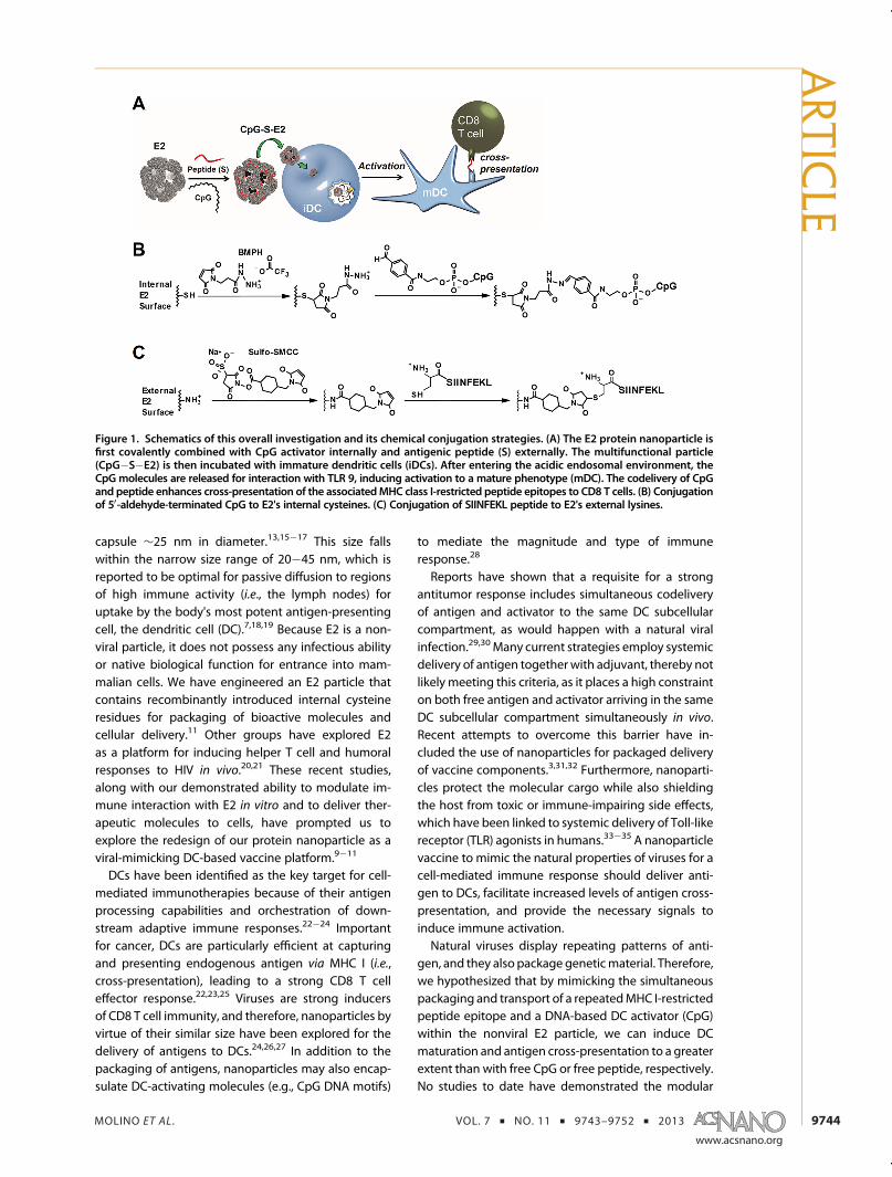
Figure 1. Schematics of this overall investigation and its chemical conjugation strategies. (A) The E2 protein nanoparticle isfirst covalently combined with CpG activator internally and antigenic peptide (S) externally. The multifunctional particle(CpG�S�E2) is then incubated with immature dendritic cells (iDCs). After entering the acidic endosomal environment, theCpG molecules are released for interaction with TLR 9, inducing activation to a mature phenotype (mDC). The codelivery of CpGand peptide enhances cross-presentation of the associatedMHC class I-restricted peptide epitopes to CD8 T cells. (B) Conjugationof 50-aldehyde-terminated CpG to E2's internal cysteines. (C) Conjugation of SIINFEKL peptide to E2's external lysines.
ARTIC
LE
MOLINO ET AL . VOL. 7 ’ NO. 11 ’ 9743–9752 ’ 2013
www.acsnano.org
9745
00reprogramming00 of an empty protein nanoscaffoldshell of noninfectious origin for eliciting immune re-sponse to the most potent antigen-presenting cell. Wereport, for the first time, the biomimetic design andcharacterization of a protein nanoparticle that is func-tionalized with CpG and peptide epitopes, for whichrelease can be triggered by DC uptake (Figure 1A). TheDC-activating properties of acid-releasable, encapsu-lated CpG are measured, and we also evaluate theCD8 T cell-activating properties of DCs that have beenloaded with E2 nanoparticles harboring both CpG andantigen.
RESULTS AND DISCUSSION
E2 Nanocapsules Can Be Simultaneously Functionalized withCpG Activator and Antigenic Peptide Epitopes. To induce asufficiently strong CD8 T cell immune response, bothantigen and activator molecules should colocalizewithin the sameDCendosomal compartment.30 Therefore,in the design of E2 as a vaccine platform, the combina-tion of antigenic peptides and CpG within the sameparticle is critical. Our previously characterized E2 con-taining the functional amino acid mutation D381C(referred to as E2 in this study) was used as the startingscaffold.13 The design of this E2 nanoparticle enabledencapsulation of an endosomal TLR ligand (i.e., CpG forTLR 9) for release in the acidic environment that occursduring DC endocytosis and processing of antigen.36
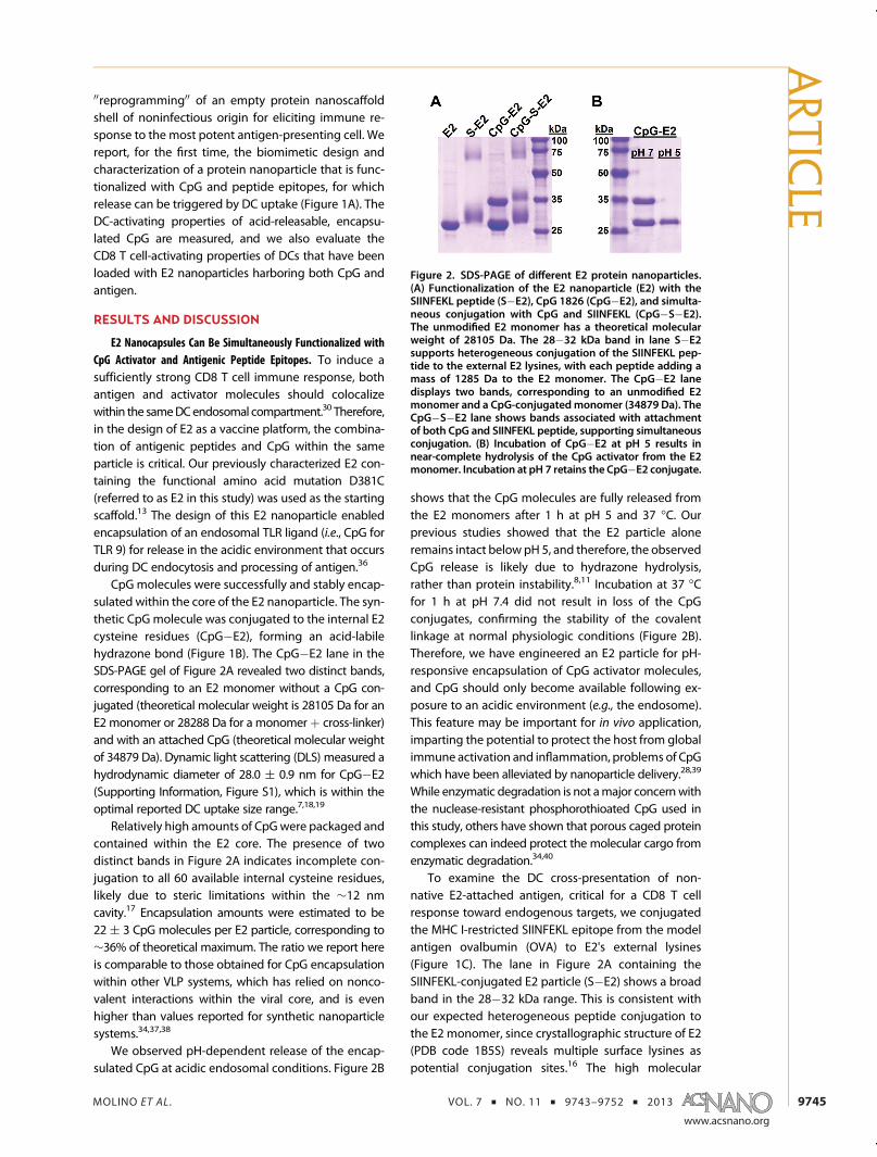
CpG molecules were successfully and stably encap-sulated within the core of the E2 nanoparticle. The syn-thetic CpG molecule was conjugated to the internal E2cysteine residues (CpG�E2), forming an acid-labilehydrazone bond (Figure 1B). The CpG�E2 lane in theSDS-PAGE gel of Figure 2A revealed two distinct bands,corresponding to an E2 monomer without a CpG con-jugated (theoretical molecular weight is 28105 Da for anE2 monomer or 28288 Da for a monomerþ cross-linker)and with an attached CpG (theoretical molecular weightof 34879 Da). Dynamic light scattering (DLS) measured ahydrodynamic diameter of 28.0 ( 0.9 nm for CpG�E2(Supporting Information, Figure S1), which is within theoptimal reported DC uptake size range.7,18,19
Relatively high amounts of CpGwere packaged andcontained within the E2 core. The presence of twodistinct bands in Figure 2A indicates incomplete con-jugation to all 60 available internal cysteine residues,likely due to steric limitations within the ∼12 nmcavity.17 Encapsulation amounts were estimated to be22 ( 3 CpG molecules per E2 particle, corresponding to∼36% of theoretical maximum. The ratio we report hereis comparable to those obtained for CpG encapsulationwithin other VLP systems, which has relied on nonco-valent interactions within the viral core, and is evenhigher than values reported for synthetic nanoparticlesystems.34,37,38
We observed pH-dependent release of the encap-sulated CpG at acidic endosomal conditions. Figure 2B
shows that the CpG molecules are fully released fromthe E2 monomers after 1 h at pH 5 and 37 �C. Ourprevious studies showed that the E2 particle aloneremains intact below pH 5, and therefore, the observedCpG release is likely due to hydrazone hydrolysis,rather than protein instability.8,11 Incubation at 37 �Cfor 1 h at pH 7.4 did not result in loss of the CpGconjugates, confirming the stability of the covalentlinkage at normal physiologic conditions (Figure 2B).Therefore, we have engineered an E2 particle for pH-responsive encapsulation of CpG activator molecules,and CpG should only become available following ex-posure to an acidic environment (e.g., the endosome).This feature may be important for in vivo application,imparting the potential to protect the host from globalimmune activation and inflammation, problems of CpGwhich have been alleviated by nanoparticle delivery.28,39
While enzymatic degradation is not amajor concernwiththe nuclease-resistant phosphorothioated CpG used inthis study, others have shown that porous caged proteincomplexes can indeed protect the molecular cargo fromenzymatic degradation.34,40
To examine the DC cross-presentation of non-native E2-attached antigen, critical for a CD8 T cellresponse toward endogenous targets, we conjugatedthe MHC I-restricted SIINFEKL epitope from the modelantigen ovalbumin (OVA) to E2's external lysines(Figure 1C). The lane in Figure 2A containing theSIINFEKL-conjugated E2 particle (S�E2) shows a broadband in the 28�32 kDa range. This is consistent withour expected heterogeneous peptide conjugation tothe E2 monomer, since crystallographic structure of E2(PDB code 1B5S) reveals multiple surface lysines aspotential conjugation sites.16 The high molecular
Figure 2. SDS-PAGE of different E2 protein nanoparticles.(A) Functionalization of the E2 nanoparticle (E2) with theSIINFEKL peptide (S�E2), CpG 1826 (CpG�E2), and simulta-neous conjugation with CpG and SIINFEKL (CpG�S�E2).The unmodified E2 monomer has a theoretical molecularweight of 28105 Da. The 28�32 kDa band in lane S�E2supports heterogeneous conjugation of the SIINFEKL pep-tide to the external E2 lysines, with each peptide adding amass of 1285 Da to the E2 monomer. The CpG�E2 lanedisplays two bands, corresponding to an unmodified E2monomer and a CpG-conjugated monomer (34879 Da). TheCpG�S�E2 lane shows bands associated with attachmentof both CpG and SIINFEKL peptide, supporting simultaneousconjugation. (B) Incubation of CpG�E2 at pH 5 results innear-complete hydrolysis of the CpG activator from the E2monomer. Incubation at pH 7 retains the CpG�E2 conjugate.
ARTIC
LE
MOLINO ET AL . VOL. 7 ’ NO. 11 ’ 9743–9752 ’ 2013
www.acsnano.org
9746
weight bands of lighter intensities observed for theSIINFEKL-containing constructs (S�E2 and CpG�S�E2)are due to reaction with sulfo-SMCC; these bands arealso present in E2 þ sulfo-SMCC alone, and suggest asmall population of cross-linked E2 subunits. Measure-ment of peptide conjugation yielded a ratio of 2.9( 0.3peptides per proteinmonomer, comparable to reportedSIINFEKL conjugation with other VLP systems.41 DLSsize measurements showed a size of 34.8 ( 4.2 nm(Supporting Information, Figure S1), within the reportedoptimal size range for vaccine delivery.7,18,19
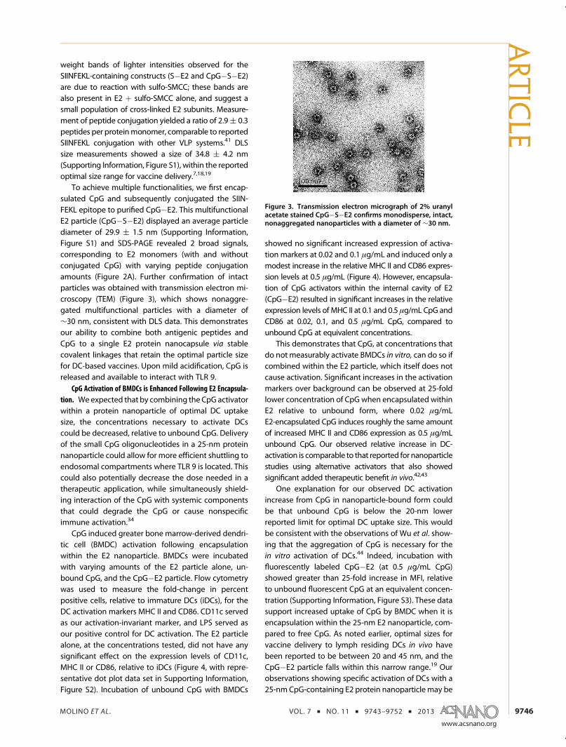
To achieve multiple functionalities, we first encap-sulated CpG and subsequently conjugated the SIIN-FEKL epitope to purified CpG�E2. This multifunctionalE2 particle (CpG�S�E2) displayed an average particlediameter of 29.9 ( 1.5 nm (Supporting Information,Figure S1) and SDS-PAGE revealed 2 broad signals,corresponding to E2 monomers (with and withoutconjugated CpG) with varying peptide conjugationamounts (Figure 2A). Further confirmation of intactparticles was obtained with transmission electron mi-croscopy (TEM) (Figure 3), which shows nonaggre-gated multifunctional particles with a diameter of∼30 nm, consistent with DLS data. This demonstratesour ability to combine both antigenic peptides andCpG to a single E2 protein nanocapsule via stablecovalent linkages that retain the optimal particle sizefor DC-based vaccines. Upon mild acidification, CpG isreleased and available to interact with TLR 9.
CpG Activation of BMDCs is Enhanced Following E2 Encapsula-tion. Weexpected that by combining the CpG activatorwithin a protein nanoparticle of optimal DC uptakesize, the concentrations necessary to activate DCscould be decreased, relative to unbound CpG. Deliveryof the small CpG oligonucleotides in a 25-nm proteinnanoparticle could allow for more efficient shuttling toendosomal compartments where TLR 9 is located. Thiscould also potentially decrease the dose needed in atherapeutic application, while simultaneously shield-ing interaction of the CpG with systemic componentsthat could degrade the CpG or cause nonspecificimmune activation.34
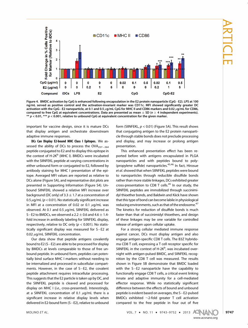
CpG induced greater bone marrow-derived dendri-tic cell (BMDC) activation following encapsulationwithin the E2 nanoparticle. BMDCs were incubatedwith varying amounts of the E2 particle alone, un-bound CpG, and the CpG�E2 particle. Flow cytometrywas used to measure the fold-change in percentpositive cells, relative to immature DCs (iDCs), for theDC activation markers MHC II and CD86. CD11c servedas our activation-invariant marker, and LPS served asour positive control for DC activation. The E2 particlealone, at the concentrations tested, did not have anysignificant effect on the expression levels of CD11c,MHC II or CD86, relative to iDCs (Figure 4, with repre-sentative dot plot data set in Supporting Information,Figure S2). Incubation of unbound CpG with BMDCs
showed no significant increased expression of activa-tion markers at 0.02 and 0.1 μg/mL and induced only amodest increase in the relative MHC II and CD86 expres-sion levels at 0.5 μg/mL (Figure 4). However, encapsula-tion of CpG activators within the internal cavity of E2(CpG�E2) resulted in significant increases in the relativeexpression levels of MHC II at 0.1 and 0.5 μg/mL CpG andCD86 at 0.02, 0.1, and 0.5 μg/mL CpG, compared tounbound CpG at equivalent concentrations.
This demonstrates that CpG, at concentrations thatdo not measurably activate BMDCs in vitro, can do so ifcombined within the E2 particle, which itself does notcause activation. Significant increases in the activationmarkers over background can be observed at 25-foldlower concentration of CpG when encapsulated withinE2 relative to unbound form, where 0.02 μg/mLE2-encapsulated CpG induces roughly the same amountof increased MHC II and CD86 expression as 0.5 μg/mLunbound CpG. Our observed relative increase in DC-activation is comparable to that reported for nanoparticlestudies using alternative activators that also showedsignificant added therapeutic benefit in vivo.42,43
One explanation for our observed DC activationincrease from CpG in nanoparticle-bound form couldbe that unbound CpG is below the 20-nm lowerreported limit for optimal DC uptake size. This wouldbe consistent with the observations of Wu et al. show-ing that the aggregation of CpG is necessary for thein vitro activation of DCs.44 Indeed, incubation withfluorescently labeled CpG�E2 (at 0.5 μg/mL CpG)showed greater than 25-fold increase in MFI, relativeto unbound fluorescent CpG at an equivalent concen-tration (Supporting Information, Figure S3). These datasupport increased uptake of CpG by BMDC when it isencapsulation within the 25-nm E2 nanoparticle, com-pared to free CpG. As noted earlier, optimal sizes forvaccine delivery to lymph residing DCs in vivo havebeen reported to be between 20 and 45 nm, and theCpG�E2 particle falls within this narrow range.19 Ourobservations showing specific activation of DCs with a25-nm CpG-containing E2 protein nanoparticle may be
Figure 3. Transmission electron micrograph of 2% uranylacetate stained CpG�S�E2 confirms monodisperse, intact,nonaggregated nanoparticles with a diameter of ∼30 nm.
ARTIC
LE
MOLINO ET AL . VOL. 7 ’ NO. 11 ’ 9743–9752 ’ 2013
www.acsnano.org
9747
important for vaccine design, since it is mature DCsthat display antigen and orchestrate downstreamadaptive immune responses.
DCs Can Display E2-bound MHC Class I Epitopes. We as-sessed the ability of DCs to process the OVA257�264
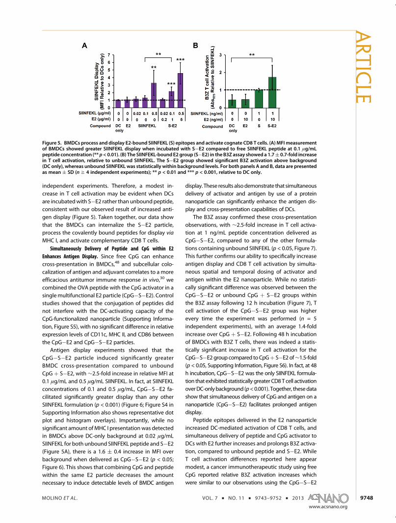
peptide conjugated to E2 and to display this epitope inthe context of H-2Kb (MHC I). BMDCs were incubatedwith the SIINFEKL peptide at varying concentrations ineither unbound form or conjugated to E2, followed byantibody staining for MHC I presentation of the epi-tope. Averaged MFI values are reported as relative toDCs alone (Figure 5A), and representative dot plots arepresented in Supporting Information (Figure S4). Un-bound SIINFEKL showed a relative MFI increase overbackground (DC only) of 3.3( 1.7 at a concentration of0.5 μg/mL (p< 0.01). No statistically significant increasein MFI at a concentration of 0.02 or 0.1 μg/mL wasobserved. At 0.1 and 0.5 μg/mL SIINFEKL delivered asS�E2 to BMDCs, we observed a 2.2( 0.6 and 4.6( 1.4-fold increase in antibody labeling for SIINFEKL display,respectively, relative to DC only (p < 0.001). No statis-tically significant display was measured for S�E2 at0.02 μg/mL SIINFEKL concentration.
Our data show that peptide antigens covalentlybound to E2 (S�E2) are able to be processed for displayby BMDCs at levels comparable to those of free un-bound peptide. In unbound form, peptides can poten-tially bind surface MHC I markers without needing tobe internalized and processed in subcellular compart-ments. However, in the case of S�E2, the covalentpeptide attachment requires intracellular processing.This suggests that the E2 particle is taken up byDC, andthe SIINFEKL peptide is cleaved and processed fordisplay on MHC I (i.e., cross-presented). Interestingly,at a SIINFEKL concentration of 0.1 μg/mL there is asignificant increase in relative display levels whendelivered in E2-bound form (S�E2), relative to unbound
form (SIINFEKL, p < 0.01) (Figure 5A). This result showsthat conjugating antigen to the E2 protein nanoparti-cle through stable bonds does not preclude processingand display, and may increase or prolong antigenpresentation.
This enhanced presentation effect has been re-ported before with antigens encapsulated in PLGAnanoparticles and with peptides bound to poly-(propylene sulfide) nanoparticles.45,46 In fact, Hirosueet al. showed that when SIINFEKL peptides were boundto nanoparticles through reducible disulfide bondsrather thanmore stable linkages, DCs exhibited greatercross-presentation to CD8 T cells.45 In our study, theSIINFEKL peptides are immobilized through succinimi-dyl thioether bonds, and Baldwin and Kiick have shownthat this type of bond canbecome labile inphysiologicalreducingenvironments, suchas that of theendosome.47
The kinetics for reduction of disulfide bonds is muchfaster than that of succinimidyl thioethers, and designof these linkages may be one variable for controlledrelease of antigen upon cellular uptake.
For a strong cellular mediated immune responseagainst cancer, DCs must display antigen and alsoengage antigen-specific CD8 T cells. The B3Z hybrido-ma CD8 T cell, expressing a T cell receptor specific forSIINFEKL in the context of H-2Kb, was incubated over-night with antigen-pulsed BMDC, and SIINFEKL recog-nition by the CD8 T cell was measured. The resultsshown in Figure 5B demonstrate that BMDC loadedwith the S�E2 nanoparticle have the capability tofunctionally engage CD8 T cells, a critical event linkinginnate and adaptive immunity for a cell-mediatedeffector response. While no statistically significantdifference between the effects of bound and unboundpeptide is evident based on averages, the S�E2-pulsedBMDCs exhibited ∼2-fold greater T cell activationcompared to the free peptide in four out of five
Figure 4. BMDC activation by CpG is enhanced following encapsulation in the E2 protein nanoparticle (CpG�E2). LPS at 100ng/mL served as positive control and the activation-invariant marker was CD11c. MFI showed significantly greater DCactivation with the CpG�E2 nanoparticle, at 0.1 and 0.5 μg/mL CpG for MHC II and CD86 markers and 0.02 μg/mL for CD86,compared to free CpG at equivalent concentrations. Data are presented as mean ( SD (n g 4 independent experiments).** p < 0.01, *** p < 0.001, relative to unbound CpG at equivalent concentration for the given marker.
ARTIC
LE
MOLINO ET AL . VOL. 7 ’ NO. 11 ’ 9743–9752 ’ 2013
www.acsnano.org
9748
independent experiments. Therefore, a modest in-crease in T cell activation may be evident when DCsare incubatedwith S�E2 rather than unboundpeptide,consistent with our observed result of increased anti-gen display (Figure 5). Taken together, our data showthat the BMDCs can internalize the S�E2 particle,process the covalently bound peptides for display via
MHC I, and activate complementary CD8 T cells.Simultaneously Delivery of Peptide and CpG within E2
Enhances Antigen Display. Since free CpG can enhancecross-presentation in BMDCs,48 and subcellular colo-calization of antigen and adjuvant correlates to a moreefficacious antitumor immune response in vivo,30 wecombined the OVA peptide with the CpG activator in asinglemultifunctional E2 particle (CpG�S�E2). Controlstudies showed that the conjugation of peptides didnot interfere with the DC-activating capacity of theCpG-functionalized nanoparticle (Supporting Informa-tion, Figure S5), with no significant difference in relativeexpression levels of CD11c, MHC II, and CD86 betweenthe CpG�E2 and CpG�S�E2 particles.
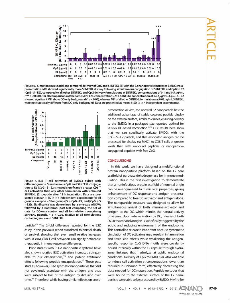
Antigen display experiments showed that theCpG�S�E2 particle induced significantly greaterBMDC cross-presentation compared to unboundCpG þ S�E2, with ∼2.5-fold increase in relative MFI at0.1 μg/mL and 0.5 μg/mL SIINFEKL. In fact, at SIINFEKLconcentrations of 0.1 and 0.5 μg/mL, CpG�S�E2 fa-cilitated significantly greater display than any otherSIINFEKL formulation (p < 0.001) (Figure 6; Figure S4 inSupporting Information also shows representative dotplot and histogram overlays). Importantly, while nosignificant amount ofMHC I presentationwas detectedin BMDCs above DC-only background at 0.02 μg/mLSIINFEKL for both unbound SIINFEKL peptide and S�E2(Figure 5A), there is a 1.6 ( 0.4 increase in MFI overbackground when delivered as CpG�S�E2 (p < 0.05;Figure 6). This shows that combining CpG and peptidewithin the same E2 particle decreases the amountnecessary to induce detectable levels of BMDC antigen
display. These results alsodemonstrate that simultaneousdelivery of activator and antigen by use of a proteinnanoparticle can significantly enhance the antigen dis-play and cross-presentation capabilities of DCs.
The B3Z assay confirmed these cross-presentationobservations, with ∼2.5-fold increase in T cell activa-tion at 1 ng/mL peptide concentration delivered asCpG�S�E2, compared to any of the other formula-tions containing unbound SIINFEKL (p < 0.05, Figure 7).This further confirms our ability to specifically increaseantigen display and CD8 T cell activation by simulta-neous spatial and temporal dosing of activator andantigen within the E2 nanoparticle. While no statisti-cally significant difference was observed between theCpG�S�E2 or unbound CpG þ S�E2 groups withinthe B3Z assay following 12 h incubation (Figure 7), Tcell activation of the CpG�S�E2 group was higherevery time the experiment was performed (n = 5independent experiments), with an average 1.4-foldincrease over CpG þ S�E2. Following 48 h incubationof BMDCs with B3Z T cells, there was indeed a statis-tically significant increase in T cell activation for theCpG�S�E2 group compared toCpGþ S�E2 of∼1.5-fold(p < 0.05, Supporting Information, Figure S6). In fact, at 48h incubation, CpG�S�E2 was the only SIINFEKL formula-tion that exhibited statistically greater CD8T cell activationoverDC-onlybackground (p<0.001). Together, thesedatashow that simultaneous delivery of CpG and antigen on ananoparticle (CpG�S�E2) facilitates prolonged antigendisplay.
Peptide epitopes delivered in the E2 nanoparticleincreased DC-mediated activation of CD8 T cells, andsimultaneous delivery of peptide and CpG activator toDCs with E2 further increases and prolongs B3Z activa-tion, compared to unbound peptide and S�E2. WhileT cell activation differences reported here appearmodest, a cancer immunotherapeutic study using freeCpG reported relative B3Z activation increases whichwere similar to our observations using the CpG�S�E2
Figure 5. BMDCs process and display E2-bound SIINFEKL (S) epitopes and activate cognate CD8 T cells. (A) MFI measurementof BMDCs showed greater SIINFEKL display when incubated with S�E2 compared to free SIINFEKL peptide at 0.1 μg/mLpeptide concentration (** p< 0.01). (B) The SIINFEKL-bound E2 group (S�E2) in the B3Z assay showeda 1.7( 0.7-fold increasein T cell activation, relative to unbound SIINFEKL. The S�E2 group showed significant B3Z activation above background(DC only), whereas unbound SIINFEKL was statistically within background levels. For both panels A and B, data are presentedas mean ( SD (n g 4 independent experiments); ** p < 0.01 and *** p < 0.001, relative to DC only.
ARTIC
LE
MOLINO ET AL . VOL. 7 ’ NO. 11 ’ 9743–9752 ’ 2013
www.acsnano.org
9749
particle.49 The 2-fold difference reported for the B3Zassay in this previous report translated to animal deathor survival, showing that even small relative increaseswith in vitro CD8 T cell activation can signify noticeabletherapeutic immune response differences.
Prior studies with PLGA nanoparticle systems havealso shown relative B3Z activation increases compar-able to our observations,46 and potent antitumoreffects following peptide encapsulation.43 These paststudies, however, used synthetic nanoparticles that didnot covalently associate with the antigen, and thuswere subject to loss of the antigen by diffusion overtime.43 Therefore, while having similar effects on cross-
presentation in vitro, the nonviral E2 nanoparticle has theadditional advantage of stable covalent peptide displayon theexternal surface, similar to viruses, ensuringdeliveryto the BMDCs in a packaged size reported optimal forin vivo DC-based vaccination.7,19 Our results here showthat we can specifically activate BMDCs with theCpG�S�E2 particle, and that associated antigen can beprocessed for display via MHC I to CD8 T cells at greaterlevels than with unbound peptides or nanoparticle-conjugated peptides with free CpG.
CONCLUSIONS
In this work, we have designed a multifunctionalprotein nanoparticle platform based on the E2 corescaffold of pyruvate dehydrogenase for immune mod-ulation. This is the first investigation to demonstratethat a noninfectious protein scaffold of nonviral origincan be re-engineered to mimic viral properties, givingenhancement of DC response and antigen presenta-tion compared to free DC activator and antigen alone.The nanoparticle structure was designed to allow forsimultaneous arrival of both immune-activator andantigen to the DC, which mimics the natural activityof viruses. Upon internalization by DC, release of bothDC activator and antigen is specifically triggered by theacidic and reducing environment of the endosome.This controlled release is important because systematiccirculation of DC activators may result in inflammationand toxic side effects while weakening the antigen-specific response. CpG DNA motifs were covalentlybound internally within the E2 capsule through hydra-zone linkages that hydrolyze at acidic endosomalconditions. Delivery of CpG to BMDCs in vitro was ableto induce cell activation at concentrations lower thanrequired in unbound form, effectively decreasing thedose needed for DC maturation. Peptide epitopes thatwere bound to the external surface of the E2 nano-particlewere able to beprocessed by BMDCs in vitro for
Figure 6. Simultaneous spatial and temporal delivery of CpG and SIINFEKL (S) with the E2 nanoparticle increases BMDC cross-presentation. MFI showed significantlymore SIINFEKL display following simultaneous conjugation of SIINFEKL and CpG to E2(CpG�S�E2), compared to all other SIINFEKL and CpG delivery formulations at SIINFEKL concentrations of 0.1 and 0.5 μg/mL(*** p<0.001, for all comparisons at the sameSIINFEKL concentration). At a SIINFEKL concentration of 0.02μg/mL, CpG�S�E2showedsignificantMFI aboveDC-onlybackground (*p<0.05),whereasMFIof all otherSIINFEKL formulationsat0.02μg/mLSIINFEKLwere not statistically different from DC-only background. Data are presented as mean ( SD (ng 4 independent experiments).
Figure 7. B3Z T cell activation of BMDCs pulsed withdifferent groups. Simultaneous CpG and SIINFEKL conjuga-tion to E2 (CpG�S�E2) showed significantly greater CD8 Tcell activation than any other formulation with unboundSIINFEKL (S) peptide after 12 h incubation. Data are pre-sented asmean( SD (ng 4 independent experiments for allgroups, except n = 3 for groups [Sþ CpG�E2] and [CpGþ SþE2]). Significance was determined by a one-way ANOVAfollowed by a Bonferroni post-test comparing the set ofdata for DC-only control and all formulations containingSIINFEKL peptide. * p < 0.05, relative to all formulationscontaining unbound SIINFEKL.
ARTIC
LE
MOLINO ET AL . VOL. 7 ’ NO. 11 ’ 9743–9752 ’ 2013
www.acsnano.org
9750
MHC I display, likely through cross-presentation, andthe DCs were able to further functionally engageantigen specific CD8 T cells.The combination of both CpG and peptide epitopes
on a single multifunctional E2 particle increased MHC Idisplay and CD8 T cell activation, relative to unboundforms of the individual components. This shows theability to enhance cross-presentation of nanoparticle-associated antigens by codelivering an endosomallyrestricted TLR 9 ligand. With the use of a modelantigen, we demonstrate the potential of engineeringa nonviral protein nanoparticle system to induce a CD8T cell mediated immune response, which is necessaryfor anticancer responses and mimics the activity of
viruses, without being infectious. Decreasing theamount of adjuvant molecules needed for DC-activa-tion while increasing antigen cross-presentation mayhelp reduce unwanted side effects or altered immuneresponses (e.g., tolerance) evident with the systemicadministration of these individual components. Thisstudy provides the groundwork for optimizing the de-sign of an E2 nanoparticle for targeting therapeuticallyrelevant antigenic peptides to the immune system foranticancer responses. More broadly, it demonstratesthat using biomimetic strategies which emulate viruses,such as size, symmetry, and simultaneous intracellulardelivery of antigen and activator to DCs, can be aneffective strategy for eliciting immune response.
METHODSMaterials. All buffer reagents were purchased from Fisher
Scientific, unless otherwise noted. The oligodeoxynucleotideTLR 9 ligand CpG 1826 (50-tccatgacgttcctgacgtt-30) (CpG) wassynthesized with a phosphorothioated backbone and 50 ben-zaldehyde modification by TriLink Biotechnologies. The CpG1826 oligonucleotidewith a 50 Alexa Fluor 488modification wassynthesized by Integrated DNA Technologies. The MHC I im-munodominant peptide SIINFEKL (OVA257�264) was synthesizedwith an N-terminal cysteine by Genscript. All cell culture mediawas comprised of RPMI 1640 (Mediatech) supplemented with10% heat-inactivated fetal bovine serum (Gibco), 1 mM sodiumpyruvate (Hyclone), 2 mM L-glutamine (Lonza), 100 units/mlpenicillin (Hyclone), 100 μg/mL streptomycin (Hyclone), 50 μM2-mercaptoethanol (Fisher), 0.1 mM nonessential amino acids(Lonza) (complete RPMI media). NP-40 and chlorophenol redβ-galactoside were from Sigma.
Cell Lines. B3Z, a CD8 T cell hybridoma containing a T cellreceptor specific for the MHC I ovalbumin epitope SIINFEKL inthe context of H-2Kb, was kindly provided by Prof. Nilabh Shastri(University of California, Berkeley). Cells were maintained incomplete RPMI media at less than 7 � 105 cells/ml.50
E2 Nanocapsule Preparation. The D381C E2 protein (E2) wasprepared as previously described.13 D381C is an E2mutant witha non-native cysteine introduced to the internal cavity of thenanoparticle for site-directed functionalization. Briefly, proteinswere expressed in E. coli, cells were lysed, and soluble cell lysateswere applied to a HiPrep Q Sepharose anion exchange column(GE Healthcare) followed by a Superose 6 (GE Healthcare) sizeexclusion column for purification. The purified proteins wereanalyzed by dynamic light scattering (Zetasizer Nano ZS,Malvern) for size measurements. Electrospray ionization massspectrometry and SDS-PAGE were performed for molecularweight and purity confirmation. Final protein preparations werestored in 50 mM potassium phosphate at pH 7.4 with 100 mMNaCl at 4 �C for short-term and �80 �C for long-term storage.
Lipopolysaccharide (LPS), a component of gram-negativebacterial cell walls, is recognized by TLR 4 expressing immune cells(e.g., DCs), causing potentially unwanted immune activation.Residual LPS was removed following the method described byAida and Pabst.51 Briefly, Triton X-114 (Sigma) was added to thepurified protein at 1% (v/v), chilled to 4 �C, vortexed vigorously,andheated to37 �C. Themixturewas then centrifuged at 18,000�g and 37 �C for 30 s, and the protein-containing aqueous portionwas separated from the detergent. This total process repeatedg 8times. Residual Triton was removed with detergent removal spincolumns (Pierce). LPS levelswere below8 EU (0.8 ng) permilligramof E2 protein (LAL ToxinSensor gel clot assay, Genscript), signifi-cantly lower than levels that activate DCs in our assays.
CpG and Peptide Conjugation. For CpG conjugation (Figure 1B),the cysteines of the E2 internal cavity were first reducedwith 10-fold excess of TCEP (Pierce) for 30 min followed by
incubation with the N-(β-maleimidopropionic acid) hydrazide(BMPH) linker (Pierce) at a 10-fold excess for 2 h at roomtemperature (RT). Unreacted linker was removed using ZebaSpin Desalting columns with a 40 kDa cutoff (Pierce). Thealdehyde-modified CpG 1826 was added at 5-fold excess overprotein monomer, incubated overnight at RT, and excess CpGremoved by desalting spin columns. Conjugation was esti-mated by SDS-PAGE and measured by band intensity analysiswith the NIH ImageJ software normalized to protein concentra-tion measured with the BCA assay (Pierce). Conjugation mea-surements are given as an average number of CpG moleculesper E2 particle (n = 3). For CpG acid-hydrolysis assays, theconjugated E2 nanocapsules were dialyzed against 50 mMpotassium phosphate with 100 mM NaCl at either pH 7.4(negative control) or pH 5 using drop dialysis membranes(Millipore) for 60�90 min. The nanoparticles were then re-moved from dialysis, incubated at 37 �C for an additional 1 h,and examined by SDS-PAGE.
For peptide conjugation (Figure 1C), the E2 protein was firstincubated with sulfosuccinimidyl 4-(N-maleimidomethyl)-cyclohexane-1-carboxylate (SMCC, Pierce) at a 20-fold excessto protein monomer for 30 min at room temperature followedby removal of unreacted linker by desalting spin columns.SMCC-functionalized E2 was combined with a 10-fold excessto protein monomer of the CSIINFEKL (TCEP reduced) peptidefor 2 h at RT. Excess peptide was removed by desalting spincolumns. Conjugation of the peptide to the E2 protein wasassessed by SDS-PAGE, and the number of peptides attached perparticlewasmeasured by the difference in free thiol concentration(i.e., unreacted peptides) between a conjugation reactionwith andwithout the SMCC cross-linker (nonspecific loss of free thiols overthe incubation time). Free thiol concentration was determinedusing Ellman's assay (Pierce), following manufacturer's instruc-tions. Conjugation measurements are given as an average (standard deviation of CSIINFEKL peptides per E2 particle (n = 3).
For particles to which both peptide and CpGs were attached,the reaction schemes for the individual components were carriedout as described above. The CpG oligonucleotide was conjugatedfirst, followed by peptide conjugation with extent of conjugationof bothcomponents assessedby SDS-PAGE. Transmission electronmicrographs of 2% uranyl acetate stained nanoparticles on Cu150 mesh Formvar-carbon coated grids were obtained on aJEM1200EX (JEOL) with a BioScan600W digital camera (Gatan).
Bone Marrow-Derived Dendritic Cells (BMDCs). Discarded excesstissue from C57Bl/6 wild-type mice was kindly provided by thelaboratories of Prof. Paolo Casali and Prof. Wendy Liu at theUniversity of California, Irvine. Bone marrow-derived dendriticcells (BMDCs) were prepared following the method describedby Lutz et al.52 Briefly, the femurs and tibias were rinsed in 70%ethanol, epiphyses removed, and the marrow flushed. Cellswerebrokenup toa single cell suspensionandapplied to a 70μmcell strainer (Fisher). Red blood cells were depleted with ACK
ARTIC
LE
MOLINO ET AL . VOL. 7 ’ NO. 11 ’ 9743–9752 ’ 2013
www.acsnano.org
9751
lysing buffer (Lonza), followed by washing with PBS. The marrowcells were plated at 2 � 105 cells/ml (10 mL total) on sterilebacteriological Petri dishes (Fisher) in complete RPMI mediasupplemented with 20 ng/mL murine recombinant GM-CSF(eBioscience) (DC media). Cells were maintained at 37 �C and5%CO2, and 10mL freshDCmediawas addedonday 3.Onday 6,50% of the media was replaced, and the nonadherent cells werepelleted and added back to the plates. Loosely and nonadherentcells were collected and used as immature BMDCs on day 8.
BMDC Activation. Immature BMDCs (iDCs) harvested on day 8were plated at 5� 105 cells/well in 24-well plates and allowed tosettle overnight. The E2 nanocapsule, unbound CpG, CpG-conjugated nanocapsules, or 100 ng/mL LPS (positive control)were added and incubated with cells for 24 h at 37 �C. Aftercollecting DCs by gentle pipetting, surface expression of CD11c,MHC II, and CD86 was assessed by labeling with fluorescentlytagged monoclonal antibodies (eBioscience for FITC-anti-CD11c and FITC-anti-CD86 and Biolegend for PE-anti-MHC II).Cells were analyzed by flow cytometry, collecting 5� 104 eventsper sample, using the Accuri C6 (BD Biosciences). The data arereported as fold-increase in percent positive cells (n g 4independent experiments), relative to iDCs. Representativegating can be found in Supporting Information Figure S2.
Antigen Display and B3Z Assays. BMDCs harvested on day 8wereplated at 2.5 � 105 per well in 48 well plates and allowed tosettle overnight. The E2 nanocapsule, SIINFEKL peptide (with orwithout unbound CpG), the SIINFEKL-conjugated E2 (S�E2,with or without unbound CpG), or the CpG and SIINFEKL doubleconjugated E2 (CpG�S�E2) nanocapsule were added for18 h. Cell surface display of the SIINFEKL epitope in the contextof H-2kb was labeled with PE-tagged monoclonal antibody25-D1.16 (Biolegend) and measured with flow cytometry(collecting 5 � 104 events per sample). The data are presentedas MFI (n g 4 independent experiments) relative to DCs only(nonspecific antibody labeling of BMDCs).
For the T cell activation assays, BMDCs harvested on day 8were plated at 1 � 105 cells/well in a 96-well plate and allowedto settle overnight. The E2 nanocapsule, SIINFEKL peptide (withor without unbound CpG), S�E2 (with or without unboundCpG), or CpG�S�E2 were added to the BMDCs, washed awayafter 1 h, and the B3Z CD8 T cells added at 1� 105 cells/well foran additional 12 or 48 h. The B3Z cells are activated by theSIINFEKL epitope in the context of H-2kb, which can be mea-sured by lacZ activity.50 Cells were washed with PBS andincubated with 100 μL of Z buffer (100 mM 2-mercaptoethanol,9 mM MgCl2, 0.125% NP-40, and 0.15 mM chlorophenol redβ-galactoside) for 4 h at 37 �C. Following incubation, 50 μL of stopbuffer (300mMglycine and15mMEDTA inwater) was added, andabsorbance at 570 nm was measured. The data are presented asan average absorbance relative to unbound SIINFEKL peptide(n g 4 independent experiments, unless otherwise noted).
Statistical Analysis. Statistical analyses were carried out usingInStat version 3.10. Data are reported asmean( standard deviation(SD) of at least four independent experiments (unless otherwisenoted), with the value of a single independent experiment beingthe average of at least two replicates for that set. Statisticalsignificance was determined by performing a one-way analysis ofvariance (ANOVA) followed by a Bonferroni post-test over pairswithin thegroup.Pvalues less than0.05wereconsidered significant.
Conflict of Interest: The authors declare no competingfinancial interest.
Acknowledgment. We thank the laboratories of Prof. PaoloCasali and Prof. Wendy Liu at UCI for providing excess animaltissue, and the laboratory of Prof. Aaron Esser-Kahn for use of theirflow cytometer. We thank Prof. Nilabh Shastri at UC Berkeley forproviding the B3Z CD8 T cell hybridoma cell line. DLS and massspectrometry were carried out at the UCI Laser SpectroscopyFacility and the UCI Mass Spectrometry Facility, respectively. Weare also grateful to Dr. Sergey Ryazantsev for assistance withobtaining TEM images in the EMi Laboratory at UCLA. This workwas supported by NIH (R21 EB010161), in part by the NationalCancer Institute of NIH (P30CA062203), and the University ofCalifornia Cancer Research Coordinating Committee. The contentis solely the responsibility of the authors and does not necessarilyrepresent the official views of the National Institutes of Health.
Supporting Information Available: Additional data for DLS ofthe E2, CpG�E2, S�E2, and CpG�S�E2 particles, representativedot plot flow cytometry data for DC activation and SIINFEKLdisplay, DC uptake of fluorescent CpG and CpG�E2, and relativeMFI DC-activation data for the CpG�S�E2 particle. Thismaterialis available free of charge via the Internet at http://pubs.acs.org.
REFERENCES AND NOTES1. Aly, H. A. Cancer Therapy and Vaccination. J. Immunol.
Methods 2012, 382, 1–23.2. Klebanoff, C. A.; Acquavella, N.; Yu, Z. Y.; Restifo, N. P.
Therapeutic Cancer Vaccines: Are We There Yet? Immunol.Rev. 2011, 239, 27–44.
3. Plummer, E. M.; Manchester, M. Viral Nanoparticles andVirus-Like Particles: Platforms for Contemporary VaccineDesign. Wiley Interdiscip. Rev.: Nanomed. Nanobiotechnol.2011, 3, 174–196.
4. Kushnir, N.; Streatfield, S. J.; Yusibov, V. Virus-Like Particlesas a Highly Efficient Vaccine Platform: Diversity of Targetsand Production Systems and Advances in Clinical Devel-opment. Vaccine 2012, 31, 58–83.
5. Pokorski, J. K.; Steinmetz, N. F. The Art of Engineering ViralNanoparticles. Mol. Pharm. 2011, 8, 29–43.
6. Uchida, M.; Klem, M. T.; Allen, M.; Suci, P.; Flenniken, M.;Gillitzer, E.; Varpness, Z.; Liepold, L. O.; Young, M.; Douglas,T. Biological Containers: Protein Cages as MultifunctionalNanoplatforms. Adv. Mater. 2007, 19, 1025–1042.
7. Bachmann, M. F.; Jennings, G. T. Vaccine Delivery: A Matterof Size, Geometry, Kinetics and Molecular Patterns. Nat.Rev. Immunol. 2010, 10, 787–796.
8. Dalmau, M.; Lim, S.; Wang, S. W. Design of a pH-DependentMolecular Switch in a Caged Protein Platform. Nano Lett.2009, 9, 160–166.
9. Molino, N.M.; Bilotkach, K.; Fraser, D. A.; Ren, D.; Wang, S.W.Cell Uptake and Complement Responses Toward Polymer-Functionalized Protein Nanocapsules. Biomacromolecules2012, 13, 974–981.
10. Ren, D. M.; Dalmau, M.; Randall, A.; Shindel, M. M.; Baldi, P.;Wang, S. W. Biomimetic Design of Protein Nanomaterialsfor Hydrophobic Molecular Transport. Adv. Funct. Mater.2012, 22, 3170–3180.
11. Ren, D. M.; Kratz, F.; Wang, S. W. Protein NanocapsulesContaining Doxorubicin as a pH-Responsive DeliverySystem. Small 2011, 7, 1051–1060.
12. Grgacic, E. V.; Anderson, D. A. Virus-Like Particles: Passportto Immune Recognition. Methods 2006, 40, 60–65.
13. Dalmau, M.; Lim, S.; Chen, H. C.; Ruiz, C.; Wang, S. W.Thermostability and Molecular Encapsulation Within anEngineered Caged Protein Scaffold. Biotechnol. Bioeng.2008, 101, 654–664.
14. Dalmau, M.; Lim, S.; Wang, S. W. pH-Triggered Disassemblyin a Caged Protein Complex. Biomacromolecules 2009, 10,3199–3206.
15. Domingo, G. J.; Chauhan, H. J.; Lessard, I. A. D.; Fuller, C.;Perham, R. N. Self-Assembly and Catalytic Activity of thePyruvate Dehydrogenase Multienzyme Complex fromBacillus stearothermophilus. Eur. J. Biochem. 1999, 266,1136–1146.
16. Izard, T.; Aevarsson, A.; Allen, M. D.; Westphal, A. H.;Perham, R. N.; de Kok, A.; Hol, W. G. Principles of Quasi-Equivalence and EuclideanGeometryGovern the Assemblyof Cubic and Dodecahedral Cores of Pyruvate Dehydro-genase Complexes. Proc. Natl. Acad. Sci. U.S.A. 1999, 96,1240–1245.
17. Milne, J. L. S.; Shi, D.; Rosenthal, P. B.; Sunshine, J. S.;Domingo, G. J.; Wu, X. W.; Brooks, B. R.; Perham, R. N.;Henderson, R.; Subramaniam, S. Molecular Architectureand Mechanism of an Icosahedral Pyruvate Dehydrogen-ase Complex: AMultifunctional Catalytic Machine. EMBO J.2002, 21, 5587–5598.
18. Reddy, S. T.; Rehor, A.; Schmoekel, H. G.; Hubbell, J. A.;Swartz, M. A. In Vivo Targeting of Dendritic Cells in LymphNodes with Poly(Propylene Sulfide) Nanoparticles. J. Con-trolled Release 2006, 112, 26–34.
ARTIC
LE
MOLINO ET AL . VOL. 7 ’ NO. 11 ’ 9743–9752 ’ 2013
www.acsnano.org
9752
19. Reddy, S. T.; van der Vlies, A. J.; Simeoni, E.; O'Neil, C. P.;Swartz, M. A.; Hubbell, J. A. Exploiting Lymphatic Transportand Complement Activation in Nanoparticle Vaccines.Tissue Eng., Part A 2008, 14, 734–735.
20. Caivano, A.; Doria-Rose, N. A.; Buelow, B.; Sartorius, R.;Trovato, M.; D'Apice, L.; Domingo, G. J.; Sutton, W. F.;Haigwood, N. L.; De Berardinis, P. HIV-1 Gag p17 Presentedas Virus-Like Particles on the E2 Scaffold from Geobacillusstearothermophilus Induces Sustained Humoral and Cel-lular Immune Responses in the Absence of IFNγ Produc-tion by CD4þ T Cells. Virology 2010, 407, 296–305.
21. Jaworski, J. P.; et al. Co-Immunization with MultimericScaffolds and DNA Rapidly Induces Potent AutologousHIV-1 Neutralizing Antibodies and CD8(þ) T Cells. PLOSOne 2012, 7, e31464.
22. Apetoh, L.; Locher, C.; Ghiringhelli, F.; Kroemer, G.; Zitvogel,L. Harnessing Dendritic Cells in Cancer. Semin. Immunol.2011, 23, 42–49.
23. Tacken, P. J.; de Vries, I. J.; Torensma, R.; Figdor, C. G.Dendritic-Cell Immunotherapy: From Ex Vivo Loading toIn Vivo Targeting. Nat. Rev. Immunol. 2007, 7, 790–802.
24. Ueno, H.; Klechevsky, E.; Schmitt, N.; Ni, L.; Flamar, A. L.;Zurawski, S.; Zurawski, G.; Palucka, K.; Banchereau, J.; Oh, S.Targeting Human Dendritic Cell Subsets for ImprovedVaccines. Semin. Immunol. 2011, 23, 21–27.
25. Joffre, O. P.; Segura, E.; Savina, A.; Amigorena, S. Cross-Presentation by Dendritic Cells. Nat. Rev. Immunol. 2012,12, 557–569.
26. Reddy, S. T.; Swartz, M. A.; Hubbell, J. A. Targeting DendriticCells with Biomaterials: Developing the Next Generationof Vaccines. Trends Immunol. 2006, 27, 573–579.
27. Joshi, M. D.; Unger, W. J.; Storm, G.; van Kooyk, Y.;Mastrobattista, E. Targeting Tumor Antigens to DendriticCells Using Particulate Carriers. J. Controlled Release 2012,161, 25–37.
28. Bourquin, C.; et al. Targeting CpG Oligonucleotides to theLymph Node by Nanoparticles Elicits Efficient AntitumoralImmunity. J. Immunol. 2008, 181, 2990–2998.
29. Burgdorf, S.; Scholz, C.; Kautz, A.; Tampe, R.; Kurts, C. Spatialand Mechanistic Separation of Cross-Presentation andEndogenous Antigen Presentation. Nat. Immunol. 2008,9, 558–566.
30. Nierkens, S.; den Brok, M. H.; Sutmuller, R. P. M.; Grauer, O. M.;Bennink, E.; Morgan,M. E.; Figdor, C. G.; Ruers, T. J. M.; Adema,G. J. In Vivo Colocalization of Antigen and Cytidyl GuanosylWithin Dendritic Cells Is Associated with the Efficacy ofCancer Immunotherapy. Cancer Res. 2008, 68, 5390–5396.
31. Krishnamachari, Y.; Geary, S. M.; Lemke, C. D.; Salem, A. K.Nanoparticle Delivery Systems in Cancer Vaccines. Pharm.Res. 2011, 28, 215–236.
32. Peek, L. J.; Middaugh, C. R.; Berkland, C. Nanotechnology inVaccineDelivery. Adv. Drug Delivery Rev. 2008, 60, 915–928.
33. Wilson, N. S.; et al. Systemic Activation of Dendritic Cellsby Toll-Like Receptor Ligands or Malaria Infection ImpairsCross-Presentation and Antiviral Immunity. Nat. Immunol.2006, 7, 165–172.
34. Storni, T.; Ruedl, C.; Schwarz, K.; Schwendener, R. A.;Renner, W. A.; Bachmann, M. F. Nonmethylated CG MotifsPackaged into Virus-Like Particles Induce Protective Cyto-toxic T Cell Responses in the Absence of Systemic SideEffects. J. Immunol. 2004, 172, 1777–1785.
35. Steinhagen, F.; Kinjo, T.; Bode, C.; Klinman, D. M. TLR-BasedImmune Adjuvants. Vaccine 2011, 29, 3341–3355.
36. Rodriguez, A.; Regnault, A.; Kleijmeer, M.; Ricciardi-Castag-noli, P.; Amigorena, S. Selective Transport of InternalizedAntigens to the Cytosol for MHC Class I Presentation inDendritic Cells. Nat. Cell Biol. 1999, 1, 362–8.
37. Zwiorek, K.; Bourquin, C.; Battiany, J.; Winter, G.; Endres, S.;Hartmann, G.; Coester, C. Delivery by Cationic Gelatin Nano-particles Strongly Increases the Immunostimulatory Effectsof CpG Oligonucleotides. Pharm. Res. 2008, 25, 551–562.
38. Roman, B. S.; Irache, J. M.; Gomez, S.; Tsapis, N.; Gamazo, C.;Espuelas, M. S. Co-Encapsulation of an Antigen and CpGOligonucleotides into PLGA Microparticles by TROMSTechnology. Eur. J. Pharm. Biopharm. 2008, 70, 98–108.
39. Tacken, P. J.; Zeelenberg, I. S.; Cruz, L. J.; van Hout-Kuijer,M. A.; van de Glind, G.; Fokkink, R. G.; Lambeck, A. J. A.;Figdor, C. G. Targeted Delivery of TLR Ligands to Humanand Mouse Dendritic Cells Strongly Enhances Adjuvanti-city. Blood 2011, 118, 6836–6844.
40. Sioud, M.; Leirdal, M. Design of Nuclease Resistant ProteinKinase CR DNA Enzymes with Potential Therapeutic Ap-plication. J. Mol. Biol. 2000, 296, 937–47.
41. Peacey, M.; Wilson, S.; Baird, M. A.; Ward, V. K. VersatileRHDV Virus-Like Particles: Incorporation of Antigens byGenetic Modification and Chemical Conjugation. Biotech-nol. Bioeng. 2007, 98, 968–977.
42. Molinari, P.; Crespo, M. I.; Gravisaco, M. J.; Taboga, O.;Moron, G. Baculovirus Capsid Display Potentiates OVACytotoxic and Innate Immune Responses. PLOS One2011, 6, e24108.
43. Zhang, Z.; Tongchusak, S.; Mizukami, Y.; Kang, Y. J.; Ioji, T.;Touma, M.; Reinhold, B.; Keskin, D. B.; Reinherz, E. L.;Sasada, T. Induction of Anti-Tumor Cytotoxic T Cell Re-sponses Through PLGA-Nanoparticle Mediated AntigenDelivery. Biomaterials 2011, 32, 3666–3678.
44. Wu, C. C. N.; Lee, J. D.; Raz, E.; Corr, M.; Carson, D. A.Necessity of Oligonucleotide Aggregation for Toll-LikeReceptor 9 Activation. J. Biol. Chem. 2004, 279, 33071–33078.
45. Hirosue, S.; Kourtis, I. C.; van der Vlies, A. J.; Hubbell, J. A.;Swartz, M. A. Antigen Delivery to Dendritic Cells by Poly-(Propylene Sulfide) Nanoparticles with Disulfide Conju-gated Peptides: Cross-Presentation and T Cell Activation.Vaccine 2010, 28, 7897–906.
46. Shen, H.; Ackerman, A. L.; Cody, V.; Giodini, A.; Hinson, E. R.;Cresswell, P.; Edelson, R. L.; Saltzman, W. M.; Hanlon, D. J.Enhanced and Prolonged Cross-Presentation FollowingEndosomal Escape of Exogenous Antigens Encapsulatedin Biodegradable Nanoparticles. Immunology 2006, 117,78–88.
47. Baldwin, A. D.; Kiick, K. L. Tunable Degradation ofMaleimide-Thiol Adducts in Reducing Environments. Bio-conjugate Chem. 2011, 22, 1946–53.
48. Datta, S. K.; et al. A Subset of Toll-Like Receptor LigandsInduces Cross-Presentation by BoneMarrow-DerivedDen-dritic Cells. J. Immunol. 2003, 170, 4102–4110.
49. den Brok, M. H.; Sutmuller, R. P.; Nierkens, S.; Bennink, E. J.;Toonen, L. W.; Figdor, C. G.; Ruers, T. J.; Adema, G. J.Synergy Between In Situ Cryoablation and TLR9 Stimula-tion Results in a Highly Effective In Vivo Dendritic CellVaccine. Cancer Res. 2006, 66, 7285–7292.
50. Sanderson, S.; Shastri, N. LacZ Inducible, Antigen/MHC-Specific T Cell Hybrids. Int. Immunol. 1994, 6, 369–376.
51. Aida, Y.; Pabst, M. J. Removal of Endotoxin from ProteinSolutions by Phase Separation Using Triton X-114.J. Immunol. Methods 1990, 132, 191–195.
52. Lutz, M. B.; Kukutsch, N.; Ogilvie, A. L.; Rossner, S.; Koch, F.;Romani, N.; Schuler, G. An Advanced Culture Method forGenerating Large Quantities of Highly Pure Dendritic CellsfromMouse BoneMarrow. J. Immunol. Methods 1999, 223,77–92.
ARTIC
LE