BIOMECHANICS: APPLICATIONS IN ORTHOPEDICS · BIOMECHANICS - Biomechanics: Applications In...
14
UNESCO-EOLSS SAMPLE CHAPTERS BIOMECHANICS - Biomechanics: Applications In Orthopedics - Luca Cristofolini ©Encyclopedia of Life Support Systems (EOLSS) BIOMECHANICS: APPLICATIONS IN ORTHOPEDICS Luca Cristofolini Dipartimento di Ingegneria Industriale, Università di Bologna, Italy Keywords: Orthopedics, musculoskeletal system, movement analysis, joint forces, joint kinematics, implantable devices, prostheses, orthoses, in vitro testing, Finite Element methods, total joint replacement, fixation devices, pre-clinical validation. Contents 1. Introduction 2. Biomechanics of the musculoskeletal system 3. Engineering tools for organ-level and tissue-level biomechanical investigations in orthopedics 4. Biomechanics of orthopedic devices 5. Future directions in orthopedic biomechanics Acknowledgments Glossary Bibliography Biographical sketch Summary This chapter summarizes the application of biomechanics in orthopedics. An overview is provided of the applications of biomechanics to basic science. This includes understanding how the musculoskeletal system works and moves; measuring indicators of movement that can describe the state of health/disease of a subject; building models of the entire musculoskeletal system (or of a portion of it) to describe, interpret and predict its function under different conditions; measuring the mechanical and structural properties of organs of our musculoskeletal system alone, and in presence of an orthopedic device. Descriptions of the tools that can be used in vitro and in silico to measure/predict the most relevant mechanical quantities (forces, moments, strain, displacement, strength, mode of failure) in bony structures are provided. In the last part, the most applicative role of biomechanics is described: design and validation of orthopedic devices is an extremely relevant issue (both to manufacturers, practitioners and patients), which involves a great deal of biomechanical experiments and simulations. 1. Introduction In this chapter the role of biomechanics in orthopedics is discussed. There are several possible ways of classifying applications of biomechanics in the field of orthopedics. One possible classification relates to the scopes: some activity is directed to the understanding of the basic principles of our musculoskeletal system, i.e. it relates to basic research, and is typically carried out at non-profit and/or public-funded research institutes. Other applications relate to the design and/or validation of implantable devices, i.e. it consists of applied research and is typically performed by Research and 301
Transcript of BIOMECHANICS: APPLICATIONS IN ORTHOPEDICS · BIOMECHANICS - Biomechanics: Applications In...
UNESCO-EOLS
S
SAMPLE C
HAPTERS
BIOMECHANICS - Biomechanics: Applications In Orthopedics - Luca Cristofolini
©Encyclopedia of Life Support Systems (EOLSS)
BIOMECHANICS: APPLICATIONS IN ORTHOPEDICS Luca Cristofolini
Dipartimento di Ingegneria Industriale, Università di Bologna, Italy Keywords: Orthopedics, musculoskeletal system, movement analysis, joint forces, joint kinematics, implantable devices, prostheses, orthoses, in vitro testing, Finite Element methods, total joint replacement, fixation devices, pre-clinical validation. Contents 1. Introduction 2. Biomechanics of the musculoskeletal system 3. Engineering tools for organ-level and tissue-level biomechanical investigations in orthopedics 4. Biomechanics of orthopedic devices 5. Future directions in orthopedic biomechanics Acknowledgments Glossary Bibliography Biographical sketch Summary This chapter summarizes the application of biomechanics in orthopedics. An overview is provided of the applications of biomechanics to basic science. This includes understanding how the musculoskeletal system works and moves; measuring indicators of movement that can describe the state of health/disease of a subject; building models of the entire musculoskeletal system (or of a portion of it) to describe, interpret and predict its function under different conditions; measuring the mechanical and structural properties of organs of our musculoskeletal system alone, and in presence of an orthopedic device. Descriptions of the tools that can be used in vitro and in silico to measure/predict the most relevant mechanical quantities (forces, moments, strain, displacement, strength, mode of failure) in bony structures are provided. In the last part, the most applicative role of biomechanics is described: design and validation of orthopedic devices is an extremely relevant issue (both to manufacturers, practitioners and patients), which involves a great deal of biomechanical experiments and simulations. 1. Introduction In this chapter the role of biomechanics in orthopedics is discussed. There are several possible ways of classifying applications of biomechanics in the field of orthopedics. One possible classification relates to the scopes: some activity is directed to the understanding of the basic principles of our musculoskeletal system, i.e. it relates to basic research, and is typically carried out at non-profit and/or public-funded research institutes. Other applications relate to the design and/or validation of implantable devices, i.e. it consists of applied research and is typically performed by Research and
301
UNESCO-EOLS
S
SAMPLE C
HAPTERS
BIOMECHANICS - Biomechanics: Applications In Orthopedics - Luca Cristofolini
©Encyclopedia of Life Support Systems (EOLSS)
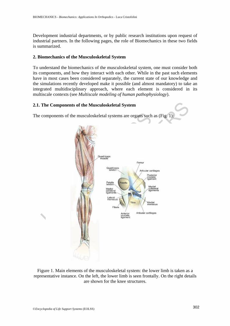
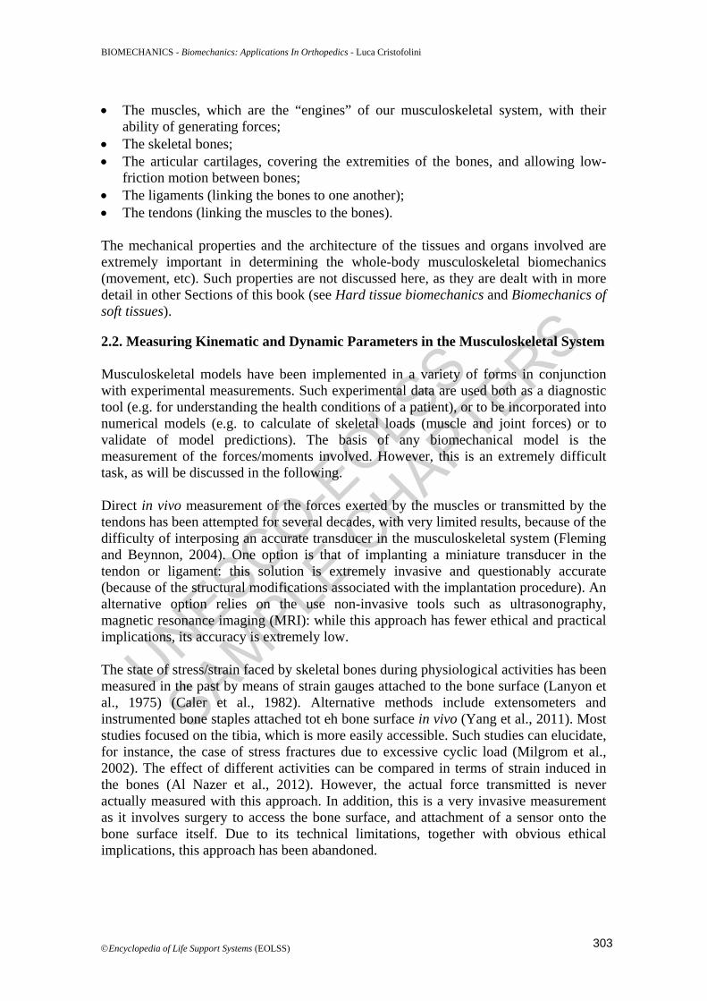
Development industrial departments, or by public research institutions upon request of industrial partners. In the following pages, the role of Biomechanics in these two fields is summarized. 2. Biomechanics of the Musculoskeletal System To understand the biomechanics of the musculoskeletal system, one must consider both its components, and how they interact with each other. While in the past such elements have in most cases been considered separately, the current state of our knowledge and the simulations recently developed make it possible (and almost mandatory) to take an integrated multidisciplinary approach, where each element is considered in its multiscale contexts (see Multiscale modeling of human pathophysiology). 2.1. The Components of the Musculoskeletal System The components of the musculoskeletal systems are organs such as (Fig. 1):
Figure 1. Main elements of the musculoskeletal system: the lower limb is taken as a representative instance. On the left, the lower limb is seen frontally. On the right details
are shown for the knee structures.
302
UNESCO-EOLS
S
SAMPLE C
HAPTERS
BIOMECHANICS - Biomechanics: Applications In Orthopedics - Luca Cristofolini
©Encyclopedia of Life Support Systems (EOLSS)
• The muscles, which are the “engines” of our musculoskeletal system, with their ability of generating forces;
• The skeletal bones; • The articular cartilages, covering the extremities of the bones, and allowing low-
friction motion between bones; • The ligaments (linking the bones to one another); • The tendons (linking the muscles to the bones). The mechanical properties and the architecture of the tissues and organs involved are extremely important in determining the whole-body musculoskeletal biomechanics (movement, etc). Such properties are not discussed here, as they are dealt with in more detail in other Sections of this book (see Hard tissue biomechanics and Biomechanics of soft tissues). 2.2. Measuring Kinematic and Dynamic Parameters in the Musculoskeletal System Musculoskeletal models have been implemented in a variety of forms in conjunction with experimental measurements. Such experimental data are used both as a diagnostic tool (e.g. for understanding the health conditions of a patient), or to be incorporated into numerical models (e.g. to calculate of skeletal loads (muscle and joint forces) or to validate of model predictions). The basis of any biomechanical model is the measurement of the forces/moments involved. However, this is an extremely difficult task, as will be discussed in the following. Direct in vivo measurement of the forces exerted by the muscles or transmitted by the tendons has been attempted for several decades, with very limited results, because of the difficulty of interposing an accurate transducer in the musculoskeletal system (Fleming and Beynnon, 2004). One option is that of implanting a miniature transducer in the tendon or ligament: this solution is extremely invasive and questionably accurate (because of the structural modifications associated with the implantation procedure). An alternative option relies on the use non-invasive tools such as ultrasonography, magnetic resonance imaging (MRI): while this approach has fewer ethical and practical implications, its accuracy is extremely low. The state of stress/strain faced by skeletal bones during physiological activities has been measured in the past by means of strain gauges attached to the bone surface (Lanyon et al., 1975) (Caler et al., 1982). Alternative methods include extensometers and instrumented bone staples attached tot eh bone surface in vivo (Yang et al., 2011). Most studies focused on the tibia, which is more easily accessible. Such studies can elucidate, for instance, the case of stress fractures due to excessive cyclic load (Milgrom et al., 2002). The effect of different activities can be compared in terms of strain induced in the bones (Al Nazer et al., 2012). However, the actual force transmitted is never actually measured with this approach. In addition, this is a very invasive measurement as it involves surgery to access the bone surface, and attachment of a sensor onto the bone surface itself. Due to its technical limitations, together with obvious ethical implications, this approach has been abandoned.
303
UNESCO-EOLS
S
SAMPLE C
HAPTERS
BIOMECHANICS - Biomechanics: Applications In Orthopedics - Luca Cristofolini
©Encyclopedia of Life Support Systems (EOLSS)
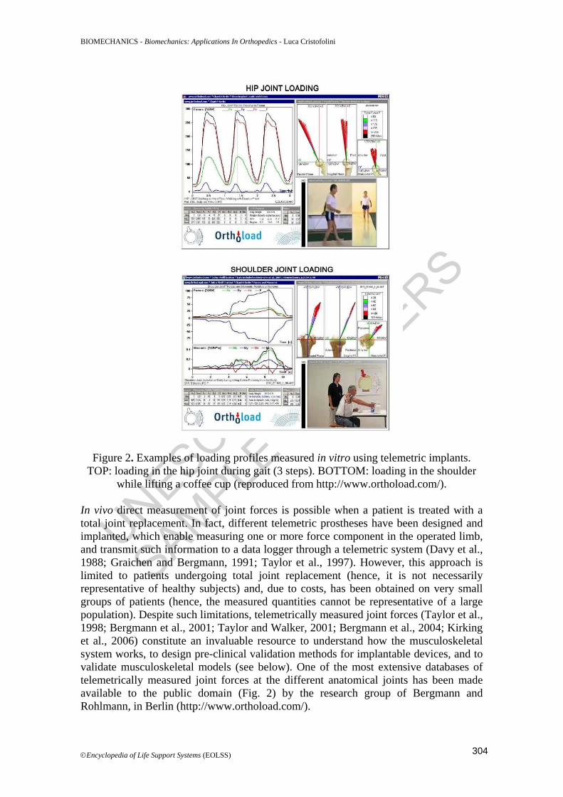
Figure 2. Examples of loading profiles measured in vitro using telemetric implants. TOP: loading in the hip joint during gait (3 steps). BOTTOM: loading in the shoulder
while lifting a coffee cup (reproduced from http://www.orthoload.com/). In vivo direct measurement of joint forces is possible when a patient is treated with a total joint replacement. In fact, different telemetric prostheses have been designed and implanted, which enable measuring one or more force component in the operated limb, and transmit such information to a data logger through a telemetric system (Davy et al., 1988; Graichen and Bergmann, 1991; Taylor et al., 1997). However, this approach is limited to patients undergoing total joint replacement (hence, it is not necessarily representative of healthy subjects) and, due to costs, has been obtained on very small groups of patients (hence, the measured quantities cannot be representative of a large population). Despite such limitations, telemetrically measured joint forces (Taylor et al., 1998; Bergmann et al., 2001; Taylor and Walker, 2001; Bergmann et al., 2004; Kirking et al., 2006) constitute an invaluable resource to understand how the musculoskeletal system works, to design pre-clinical validation methods for implantable devices, and to validate musculoskeletal models (see below). One of the most extensive databases of telemetrically measured joint forces at the different anatomical joints has been made available to the public domain (Fig. 2) by the research group of Bergmann and Rohlmann, in Berlin (http://www.orthoload.com/).
304
UNESCO-EOLS
S
SAMPLE C
HAPTERS
BIOMECHANICS - Biomechanics: Applications In Orthopedics - Luca Cristofolini
©Encyclopedia of Life Support Systems (EOLSS)
Alternatively, muscle activity can be measured in vitro using electromyography (EMG, (Bronzino, 2006)). EMG relies upon measurement of the electrical signal associated with muscle activity, which can be either collected using needles (local measurement, more invasive) or surface electrodes (less invasive, but providing a worse signal. EMG measurements are affected by large errors (e.g. noise, crass-talk between neighboring muscles). For these reasons, EMG is suitable to detect the timing of activation in a binary way (on-off) rather than actually measuring the force exerted by the single muscle. Human movement analysis allows gathering quantitative information about the mechanics of the musculoskeletal system during motor tasks. Quantification of human motion incorporates an amount of directly measured quantities (e.g. motions and accelerations of the different segments, ground reaction forces) and numerical models describing the relationship between the segments. More details about the methods of movement analysis can be found elsewhere (see Biodynamics and Human motion). The following quantities can be obtained, which can be used as an input for biomechanical musculoskeletal models: instantaneous positions of markers located on the skin surface using stereophotogrammetry (motion capture), external forces using dynamometers such as force plates, electrical activity of muscles using electromyography (EMG). Marker locations are necessary for the calculation of joint kinematics with different approaches (e.g., inverse kinematics), which, in conjunction with the measured external forces are used for the calculation of joint torques, representing the resultant action of all muscles crossing a joint, through inverse dynamics methods. EMG recordings are typically used for EMG-driven models or for validation purposes by comparison with predicted muscle activations. All such kinematic and dynamic variables are strongly subject dependent. In fact, the motion patterns, and the associated loading profile, exhibit a highly personalized trend, in relation to individual anatomy, motor strategy, habits, neuro-motor control, diseases, etc. 2.3. Modeling the Musculoskeletal System A deep knowledge of the loads acting on the skeletal system during human movements may have significant clinical implications and could help improving the diagnosis, planning and treatment in several orthopedics and neurological contexts. Muscle and joint function analyses can be performed when the following information is simultaneously available: body-segmental motion, external forces applied to the body, accurate knowledge of muscle and joint loads. Therefore, a valuable solution for analyzing internal loads in living subjects is represented by the prediction through computational models of the musculoskeletal system driven by the biomechanical quantities that can be measured in vivo (see above). Musculoskeletal models can be: • Generic, i.e. based on an average anatomy, identified to represent a chosen
population: such models are suitable for predicting general trends, effects of different motor tasks etc. Such models can be extremely useful in basic research about musculoskeletal function.
305
UNESCO-EOLS
S
SAMPLE C
HAPTERS
BIOMECHANICS - Biomechanics: Applications In Orthopedics - Luca Cristofolini
©Encyclopedia of Life Support Systems (EOLSS)
• Subject-specific, i.e. based on the anatomy of an individual subject: such models can predict what happens in a given subject, with his/her anatomy, disease, etc. These models are the best candidates for use in clinical applications.
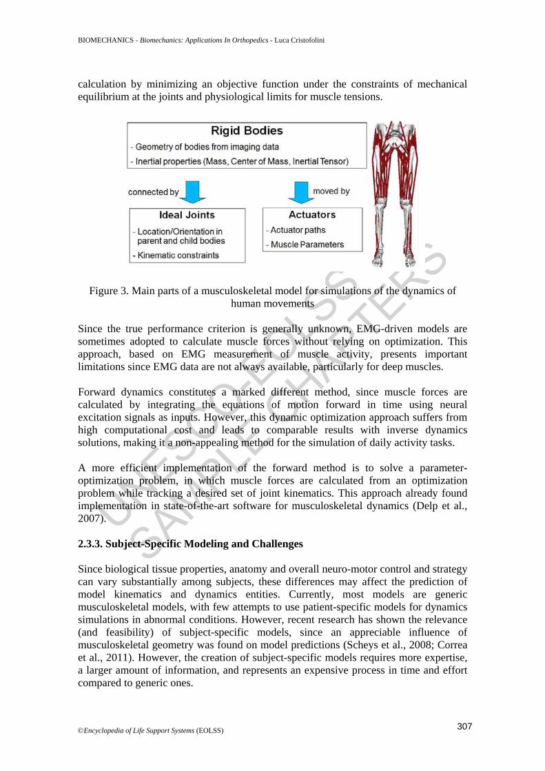
2.3.1. Modeling Applications Thanks to the steady increase in computing power, in combination with more efficient and reliable modeling strategies, the development of musculoskeletal models is continuously improving, with a wide range of applications in biomechanical problems. For instance, modeling approaches were adopted to study how muscle geometry affects the joint moment-generating capacity (Lenaerts et al., 2008; Valente et al., 2012), how muscle moment arms and lengths are altered by surgical procedures (Delp et al., 1994), or to evaluate potential of muscles to accelerate the body segments during movement (Liu et al., 2006). However, the most common and challenging application is represented by the estimation of actual forces transmitted by the muscles and joints during movement (Erdemir et al., 2007; Pandy and Andriacchi, 2010), since such loads are related to several clinical scenarios. Some important aspects are represented by the prediction of clinical outcomes after total joint replacements (joint function, bone remodeling, primary stability, muscular dysfunction) (Claes et al., 2000; Bitsakos et al., 2005), planning of surgical and rehabilitation treatments for gait-related disorders (cerebral palsy, stroke, osteoarthritis) (Arnold and Delp, 2005; Steele et al., 2012; Taddei et al., 2012), understanding the osteoporotic fractures (Viceconti et al., 2012). 2.3.2. Modeling Development and Simulation Methods Models (including biomechanical ones) are simplified representations of a more complex reality. Because of their nature, it cannot be for granted that models predictions are representative of reality. For this reason, all models need to undergo a systematic verification and validation procedure (Babuska and Oden, 2004). When dealing with multibody-dynamics models of the musculoskeletal system, what should be included in the model depends on the purpose of the model itself. When the overall goal is to analyze the biomechanics of movement and the skeletal loads, bone segments can be described as rigid bodies, connected by ideal joints and moved by muscle-tendon actuators (Fig. 3). The model identification process involves several and fairly complex operations, unless generic models based on average geometry derived from suitable population studies are adopted and scaled onto specific anthropometries. A framework commonly adopted for the prediction of muscle and joint loading in human movement includes musculoskeletal models in conjunction with body-segmental and ground reaction force measurements and inverse or forward dynamics methods. Inverse dynamics is based on gait analysis measurements applied to a recursive Newton-Euler algorithm to calculate the net joint moments exerted. Then, muscle forces are calculated by applying optimization methods to solve an indeterminate problem, under the hypothesis that the human body adopts some optimal performance criterion. For example, common objective functions are the sum of the squares of muscle stresses, or the sum of the squares of muscle activations. Static optimization allows muscle force
306
UNESCO-EOLS
S
SAMPLE C
HAPTERS
BIOMECHANICS - Biomechanics: Applications In Orthopedics - Luca Cristofolini
©Encyclopedia of Life Support Systems (EOLSS)
calculation by minimizing an objective function under the constraints of mechanical equilibrium at the joints and physiological limits for muscle tensions.
Figure 3. Main parts of a musculoskeletal model for simulations of the dynamics of human movements
Since the true performance criterion is generally unknown, EMG-driven models are sometimes adopted to calculate muscle forces without relying on optimization. This approach, based on EMG measurement of muscle activity, presents important limitations since EMG data are not always available, particularly for deep muscles. Forward dynamics constitutes a marked different method, since muscle forces are calculated by integrating the equations of motion forward in time using neural excitation signals as inputs. However, this dynamic optimization approach suffers from high computational cost and leads to comparable results with inverse dynamics solutions, making it a non-appealing method for the simulation of daily activity tasks. A more efficient implementation of the forward method is to solve a parameter-optimization problem, in which muscle forces are calculated from an optimization problem while tracking a desired set of joint kinematics. This approach already found implementation in state-of-the-art software for musculoskeletal dynamics (Delp et al., 2007). 2.3.3. Subject-Specific Modeling and Challenges Since biological tissue properties, anatomy and overall neuro-motor control and strategy can vary substantially among subjects, these differences may affect the prediction of model kinematics and dynamics entities. Currently, most models are generic musculoskeletal models, with few attempts to use patient-specific models for dynamics simulations in abnormal conditions. However, recent research has shown the relevance (and feasibility) of subject-specific models, since an appreciable influence of musculoskeletal geometry was found on model predictions (Scheys et al., 2008; Correa et al., 2011). However, the creation of subject-specific models requires more expertise, a larger amount of information, and represents an expensive process in time and effort compared to generic ones.
307
UNESCO-EOLS
S
SAMPLE C
HAPTERS
BIOMECHANICS - Biomechanics: Applications In Orthopedics - Luca Cristofolini
©Encyclopedia of Life Support Systems (EOLSS)
Indeed, to be effective in a clinical context, the methods that need to be developed to gather the necessary information should satisfy requirements of applicability. The adopted methods should satisfy ethical and technological constraints, while requiring limited time and operator interaction. Typically, models are created from clinical images (CT and MRI) and gait analysis measurements: data for musculoskeletal geometry needs to be extracted and some information cannot be always collected in the clinical practice. Because of the limited amount of quantitative in vivo data available about internal forces, a thorough validation of model predictions is still unfeasible. Validation remains a major challenge faced by musculoskeletal modeling, and highlighting the need of a deep understanding on the sensitivity of model predictions to the modeling hypotheses and the uncertainties associated to each of the large amount of parameters involved. This, in conjunction with the mentioned limitation on subject-specific modeling, represents a constraint for current applicability of personalized musculoskeletal model into clinical contexts. However, the field is increasingly growing and represents an important resource for computer-aided medicine challenges. Recently, significant steps towards the improvement and speeding up of personalized modeling have been made. 3. Engineering Tools for Organ-Level and Tissue-Level Biomechanical Investigations in Orthopedics 3.1. The Role of In Vitro Experiments In a historical perspective, implantable devices have originally been tested pre-clinically in vitro, long before computer models could provide truly relevant information. The advantage of in vitro testing is that a real specimen (including a physical prototype and possibly a bone segment) is subjected to real loads. This brings results from in vitro experiments closer to “reality” compared to other approaches such as numerical modeling. If we exclude the tests on the implantable device alone, which are often regulated by internationals standards, in vitro experiments focus on the biomechanical function of the device when implanted in the host bone(s). Therefore, in vitro tests often include bone segments. These can either be synthetic bone replicas, or animal bones, or human cadaveric specimens. Synthetic models (usually made of composite material) offer the advantage of being highly reproducible, available in large numbers, easy to handle, and relatively inexpensive. Synthetic models suffer from some limitations: (i) their high reproducibility makes them unsuitable to represent the variability among subjects; (ii) while they are designed to provide similar mechanical properties in the elastic range, they have quite a different behavior when loaded to failure. Animal tissue specimens are relatively easy to obtain from food supply (conversely, breeding animals for testing purposes is subjected to strict ethical rules), and they have mechanical properties that are similar to human bone. However, in most cases the anatomy and the structure are different from human bone because of the biomechanical function of such animals (mostly quadrupeds) compared to humans. Such limitations make synthetic bones and animal bones suitable for an early stage of pre-clinical validation, but not for the final phases where similarity to the patient bone is essential. For this reason, at some stage
308
UNESCO-EOLS
S
SAMPLE C
HAPTERS
BIOMECHANICS - Biomechanics: Applications In Orthopedics - Luca Cristofolini
©Encyclopedia of Life Support Systems (EOLSS)
D Bibliography Al Nazer, R., Lanovaz, J., Kawalilak, C., Johnston, J.D., Kontulainen, S., 2012. Direct in vivo strain measurements in human bone-a systematic literature review. J Biomech 45, 27-40. [Review paper summarizing the main methods for in vivo measurement of strain]
Arnold, A.S., Delp, S.L., 2005. Computer modeling of gait abnormalities in cerebral palsy: application to treatment planning. Theoretical Issues in Ergonomics Science 6, 305-312. [Scientific paper describing computer-aided treatment of cerebral palsy]
Babuska, I., Oden, J.T., 2004. Verification and validation in computational engineering and science: basic concepts. Computer Methods in Applied Mechanics and Engineering 193, 4057-4066. [Paper describing the main concepts and methods to verify and validate numerical models]
Bayraktar, H.H., Gupta, A., Kwon, R.Y., Papadopoulos, P., Keaveny, T.M., 2004. The modified super-ellipsoid yield criterion for human trabecular bone. J Biomech Eng 126, 677-684. [Scientific paper describing a failure criterion for bone tissue]
Bergmann, G., Deuretzbacher, G., Heller, M., Graichen, F., Rohlmann, A., Strauss, J., Duda, G.N., 2001. Hip contact forces and gait patterns from routine activities. J Biomech 34, 859-871. [Scientific paper reporting a set of data concerning physiological loading during daily tasks]
Bergmann, G., Graichen, F., Rohlmann, A., 2004. Hip joint contact forces during stumbling. Langenbecks Arch Surg 389, 53-59. [Scientific paper reporting a set of data concerning physiological loading during daily tasks]
Birolini, A., 2010. Reliability Engineering, 6th Edition. Springer-Verlag Berlin Heidelberg, [Handbook of reliability engineering and statistics]
Bitsakos, C., Kerner, J., Fisher, I., Amis, A.A., 2005. The effect of muscle loading on the simulation of bone remodelling in the proximal femur. J Biomech 38, 133-139. [Scientific paper addressing musculoskeletal loading, its determinants and consequences]
Bordini, B., Stea, S., De Clerico, M., Strazzari, S., Sasdelli, A., Toni, A., 2007. Factors affecting aseptic loosening of 4750 total hip arthroplasties: multivariate survival analysis. BMC Musculoskelet Disord 8, 69. [Scientific paper describing a study on micromotions and stability of an implanted joint prosthesis]
Bourne, R.B., Finlay, J.B., 1986. The influence of tibial component intramedullary stems and implant-cortex contact on the strain distribution of the proximal tibia following total knee arthroplasty. An in vitro study. Clin Orthop Relat Res 95-99. [Scientific paper describing a study on load transfer for an implanted joint prosthesis]
Bowman, N.K., Bucher, T.A., Bassily, A.A., 2006. Fracture of the stem of the femoral component after resurfacing arthroplasty of the hip. J Bone Joint Surg Br 88, 1652-1653. [Scientific paper addressing describing mechanical failure of a recent hip prosthesis]
325
Guest6
Rectangle
Guest6
Text Box
TO ACCESS ALL THE 30 PAGES OF THIS CHAPTER, Visit: http://www.eolss.net/Eolss-sampleAllChapter.aspx
Guest6
Text Box
- - -
UNESCO-EOLS
S
SAMPLE C
HAPTERS
BIOMECHANICS - Biomechanics: Applications In Orthopedics - Luca Cristofolini
©Encyclopedia of Life Support Systems (EOLSS)
Bronzino, J.D., 2006. The Biomedical Engineering Handbook, Third Edition. CRC: Taylor & Francis, Boca Raton, (ISBN-10: 0849321247) [General, extensive reference covering most fields of bioengineering]
Bryan, R., Nair, P.B., Taylor, M., 2009. Use of a statistical model of the whole femur in a large scale, multi-model study of femoral neck fracture risk. J Biomech 42, 2171-2176. [Scientific paper describing an investigation of femur fractures based on a statistical numerical approach]
Caler, W.E., Carter, D.R., Vasu, R., McCarthy, J.C., Harris, W.H., 1982. In vivo intracortical loading histories calculated from bone strain telemetry. In: Biomechanics - Principles and applications. Huiskes, R., van Campen, D.H., de Wijn, J.R. (Eds). Martinus Nijhoff Publishers, pp. 241-245. [Scientific paper reporting a set of data concerning physiological loading during daily tasks]
Carbone, A., Howie, D.W., McGee, M., Field, J., Pearcy, M., Smith, N., Jones, E., 2006. Aging performance of a compliant layer bearing acetabular prosthesis in an ovine hip arthroplasty model. J Arthroplasty 21, 899-906. [Scientific paper addressing the in vivo effect and consequences of the presence of a prosthesis on the strain distribution in the host bone]
Claes, L., Fiedler, S., Ohnmacht, M., Duda, G.N., 2000. Initial stability of fully and partially cemented femoral stems. Clin. Biomech. 15, 750-755. [Scientific paper describing a study on micromotions and stability of an implanted joint prosthesis]
Clarke, S.G., Phillips, A.T., Bull, A.M., 2012. Validation of FE micromotions and strains around a press-fit cup: introducing a new micromotion measuring technique. Ann Biomed Eng 40, 1586-1596. [Scientific paper describing a study on micromotions and stability of an implanted joint prosthesis]
Clech, J.P., Keer, L.M., Lewis, J.L., 1984. A crack model of a bone cement interface. J Biomech Eng 106, 235-243. [Scientific paper reporting an experimental-numerical approach to the bone-cement interface and its failure]
Correa, T.A., Baker, R., Graham, H.K., Pandy, M.G., 2011. Accuracy of generic musculoskeletal models in predicting the functional roles of muscles in human gait. J Biomech 44, 2096-2105. [Scientific paper addressing musculoskeletal loading, its determinants and consequences]
Cristofolini, L., Schileo, E., Juszczyk, M., Taddei, F., Martelli, S., Viceconti, M., 2010. Mechanical testing of bones: the positive synergy of FE models and in vitro experiments. Philos Transact A Math Phys Eng Sci 368, 2725-2763. [Perspective paper providing an overview and some recommendations for an optimal use of in vitro experiments and numerical models]
Cristofolini, L., Viceconti, M., 2000. Development and validation of a technique for strain measurement inside polymethyl methacrylate. The Journal of Strain Analysis for Engineering Design 35, 21-33. [Scientific paper describing a method (and its validation) to include a strain gauge inside acrylic bone cement to measure its state of strain]
Cristofolini, L., Viceconti, M., 2000. Mechanical validation of whole bone composite tibia models. J Biomech 33, 279-288. [Scientific paper describing a set of composite bone surrogates (tibias), and their validation in comparison with human specimens]
Cristofolini, L., Viceconti, M., Cappello, A., Toni, A., 1996. Mechanical validation of whole bone composite femur models. J Biomech 29, 525-535. [Scientific paper describing a set of composite bone surrogates (femurs), and their validation in comparison with human specimens]
Currey, J.D., 2001. Bone strength: what are we trying to measure? Calcif Tissue Int 68, 205-210. [Critical overview of the failure criteria (and methods to describe and predict failure) in bone tissue and bones]
Davy, D.T., Kotzar, G.M., Brown, R.H., Heiple, K.G., Goldberg, V.M., Heiple, K.G.J., Berilla, J., Burstein, A., 1988. Telemetric force measurements across the hip after total arthroplasty. J. Bone Jt. Surg. Am. 70, 45-50. [Scientific paper reporting a set of data concerning physiological loading during daily tasks]
Delp, S.L., Anderson, F.C., Arnold, A.S., Loan, P., Habib, A., John, C.T., Guendelman, E., Thelen, D.G., 2007. OpenSim: open-source software to create and analyze dynamic simulations of movement. IEEE Trans Biomed Eng 54, 1940-1950. [Scientific paper describing an open source software for the modelization of human movement]
326
UNESCO-EOLS
S
SAMPLE C
HAPTERS
BIOMECHANICS - Biomechanics: Applications In Orthopedics - Luca Cristofolini
©Encyclopedia of Life Support Systems (EOLSS)
Delp, S.L., Ringwelski, D.A., Carroll, N.C., 1994. Transfer of the rectus femoris: effects of transfer site on moment arms about the knee and hip. J Biomech 27, 1201-1211. [Scientific paper investigating musculoskeletal loading after tendon transfer surgery]
Erdemir, A., McLean, S., Herzog, W., van den Bogert, A.J., 2007. Model-based estimation of muscle forces exerted during movements. Clin Biomech (Bristol, Avon) 22, 131-154. [Scientific paper addressing musculoskeletal loading, its determinants and consequences]
Eskelinen, A., Remes, V., Helenius, I., Pulkkinen, P., Nevalainen, J., Paavolainen, P., 2005. Total hip arthroplasty for primary osteoarthrosis in younger patients in the Finnish arthroplasty register. 4,661 primary replacements followed for 0-22 years. Acta Orthop 76, 28-41. [Extensive report of the clinical outcome of joint replacement, based on follow-up recorded in a National register]
Fleming, B.C., Beynnon, B.D., 2004. In vivo measurement of ligament/tendon strains and forces: a review. Ann Biomed Eng 32, 318-328. [Overview of musculoskeletal loading, its determinants and consequences]
Graichen, F., Bergmann, G., 1991. Four-channel telemetry system for in vivo measurement of hip joint forces. J. Biomed. Eng. 13, 370-374. [Scientific paper reporting a set of data concerning physiological loading during daily tasks]
Havelin, L.I., Engesaeter, L.B., Espehaug, B., Furnes, O., Lie, S.A., Vollset, S.E., 2000. The Norwegian Arthroplasty Register: 11 years and 73,000 arthroplasties. Acta Orthop Scand 71, 337-353. [Extensive report of the clinical outcome of joint replacement, based on follow-up recorded in a National register]
Henninger, H.B., Reese, S.P., Anderson, A.E., Weiss, J.A., 2010. Validation of computational models in biomechanics. Proc Inst Mech Eng H 224, 801-812. [Overview of the methods to build reliable numerical models and validate them]
Herberts, P., Malchau, H., 2000. Long-term registration has improved the quality of hip replacement: a review of the Swedish THR Register comparing 160,000 cases. Acta Orthop Scand 71, 111-121. [Extensive report of the clinical outcome of joint replacement, based on follow-up recorded in a National register]
Juszczyk, M., Pallini, F., Schileo, E., Taddei, F., Cristofolini, L., 2006. Improvement of experimental-numerical cross-validation in studies of the proximal femur. J Biomech 39, S473. [Scientific paper describing a transducer to accurately measure the point of load application, and improve validation of numerical models]
Kirking, B., Krevolin, J., Townsend, C., Colwell, C.W., Jr., D'Lima, D.D., 2006. A multiaxial force-sensing implantable tibial prosthesis. J Biomech 39, 1744-1751. [Scientific paper describing an instrumented knee prosthesis to measure in vivo loads]
Lanyon, L.E., Hampson, W.G.J., Goodship, A.E., Shah, J.S., 1975. Bone deformation recorded in vivo from strain gauges attached to the human tibial shaft. Acta Orthop. Scand. 46, 256-268. [Scientific paper reporting strain in the bone measured in vivo]
Lenaerts, G., De Groote, F., Demeulenaere, B., Mulier, M., Van der Perre, G., Spaepen, A., Jonkers, I., 2008. Subject-specific hip geometry affects predicted hip joint contact forces during gait. J Biomech 41, 1243-1252. [Scientific paper addressing the effect and consequences of the presence of a prosthesis on the strain distribution in the host bone]
Lengsfeld, M., Burchard, R., Gunther, D., Pressel, T., Schmitt, J., Leppek, R., Griss, P., 2005. Femoral strain changes after total hip arthroplasty--patient-specific finite element analyses 12 years after operation. Med Eng Phys 27, 649-654. [Scientific paper addressing the effect and consequences of the presence of a prosthesis on the strain distribution in the host bone]
Lewis, G., 1997. Properties of acrylic bone cement: state of the art review. J. Appl. Biomaterials 38, 155-182. [Overview of mechanical, chemical and processing properties of acrylic cements for orthopedic applications]
Little, J.P., Taddei, F., Viceconti, M., Murray, D.W., Gill, H.S., 2007. Changes in femur stress after hip resurfacing arthroplasty: response to physiological loads. Clin Biomech (Bristol, Avon) 22, 440-448. [Scientific paper addressing the effect and consequences of the presence of a prosthesis on the strain distribution in the host bone]
327
UNESCO-EOLS
S
SAMPLE C
HAPTERS
BIOMECHANICS - Biomechanics: Applications In Orthopedics - Luca Cristofolini
©Encyclopedia of Life Support Systems (EOLSS)
Liu, M.Q., Anderson, F.C., Pandy, M.G., Delp, S.L., 2006. Muscles that support the body also modulate forward progression during walking. J Biomech 39, 2623-2630. [Scientific paper addressing musculoskeletal loading, its determinants and consequences]
Long, J.P., Santner, T.J., Bartel, D.L., 2009. Hip resurfacing increases bone strains associated with short-term femoral neck fracture. Journal Orthopaedic Research 27, 1319-1325. [Scientific paper addressing the effect and consequences of the presence of a prosthesis on the strain distribution in the host bone]
Merx, H., Dreinhofer, K., Schrader, P., Sturmer, T., Puhl, W., Gunther, K.P., Brenner, H., 2003. International variation in hip replacement rates. Ann Rheum Dis 62, 222-226. [Scientific paper comparing the musculoskeletal loading regimens in for different populations]
Milgrom, C., Finestone, A., Sharkey, N., Hamel, A., Mandes, V., Burr, D., Arndt, A., Ekenman, I., 2002. Metatarsal strains are sufficient to cause fatigue fracture during cyclic overloading. Foot Ankle Int 23, 230-235. [Scientific paper investigate the magnitude, and possible consequences, of in vivo loads in the metatarsal bones]
Ohman, C., Dall'Ara, E., Baleani, M., Van Sint Jan, S., Viceconti, M., 2008. The effects of embalming using a 4% formalin solution on the compressive mechanical properties of human cortical bone. Clin Biomech (Bristol, Avon) 23, 1294-1298. [Investigation on the most suitable method for preserving bone tissue without altering its mechanical properties]
Pandy, M.G., Andriacchi, T.P., 2010. Muscle and joint function in human locomotion. Annu Rev Biomed Eng 12, 401-433. [Scientific paper addressing musculoskeletal loading, its determinants and consequences]
Perry, C.C., 1984. The resistance strain gauge revisited. Experimental Mechanics 24, 286-299. [Overview of strain gauge instrumentation and methods]
Popper, K., 2003. The logic of scientific discovery. Taylor and Francis, London/New York [One of the most important introductions to epistemology]
Puolakka, T.J., Pajamaki, K.J., Halonen, P.J., Pulkkinen, P.O., Paavolainen, P., Nevalainen, J.K., 2001. The Finnish Arthroplasty Register: report of the hip register. Acta Orthop Scand 72, 433-441. [Extensive report of the clinical outcome of joint replacement, based on follow-up recorded in a National register]
Reggiani, B., Cristofolini, L., Varini, E., Viceconti, M., 2007. Predicting the subject-specific primary stability of cementless implants during pre-operative planning: preliminary validation of subject-specific finite-element models. J Biomech 40, 2552-2558. [Scientific paper describing a study on micromotions and stability of an implanted joint prosthesis]
Rockwood, C.A.J., Green, D.P., Bucholz, R.W., 1991. Rockwood and Green's fractures in adults. J.B. Lippincott, Philadelphia [Handbook describing one of the most extensive and acknowledged classification of bone fractures, including diagnosis, treatment and prognosis]
Scheys, L., Spaepen, A., Suetens, P., Jonkers, I., 2008. Calculated moment-arm and muscle-tendon lengths during gait differ substantially using MR based versus rescaled generic lower-limb musculoskeletal models. Gait Posture 28, 640-648. [Scientific paper addressing musculoskeletal loading, its determinants and consequences]
Schileo, E., Taddei, F., Cristofolini, L., Viceconti, M., 2008. Subject-specific finite element models implementing a maximum principal strain criterion are able to estimate failure risk and fracture location on human femurs tested in vitro. J Biomech 41, 356-367. [Investigation of the failure criteria (and methods to describe and predict failure) in bone tissue and bones]
Schileo, E., Taddei, F., Malandrino, A., Cristofolini, L., Viceconti, M., 2007. Subject-specific finite element models can accurately predict strain levels in long bones. J Biomech 40, 2982-2989. [Scientific paper describing a method (and its validation) to build numerical models of the bones that incorporate tissue inhomogeneity to predict bone strain]
Skurla, C.P., Pluhar, G.E., Frankel, D.J., Egger, E.L., James, S.P., 2005. Assessing the dog as a model for human total hip replacement. Analysis of 38 canine cemented femoral components retrieved at post-mortem. J Bone Joint Surg Br 87, 120-127. [Scientific paper addressing the effect and consequences of the presence of a prosthesis in terms of bone adaptation]
328
UNESCO-EOLS
S
SAMPLE C
HAPTERS
BIOMECHANICS - Biomechanics: Applications In Orthopedics - Luca Cristofolini
©Encyclopedia of Life Support Systems (EOLSS)
Sorbie, C., 2003. Arthroplasty in the treatment of subcapital hip fracture. Orthopedics 26, 337-341; quiz 342-333. [Scientific paper describing the use of total hip replacement to treat the fractured hip]
Speirs, A.D., Heller, M.O., Taylor, W.R., Duda, G.N., Perka, C., 2007. Influence of changes in stem positioning on femoral loading after THR using a short-stemmed hip implant. Clin Biomech (Bristol, Avon) 22, 431-439. [Scientific paper addressing the effect and consequences of the presence of a prosthesis on the strain distribution in the host bone]
Steele, K.M., Demers, M.S., Schwartz, M.H., Delp, S.L., 2012. Compressive tibiofemoral force during crouch gait. Gait Posture 35, 556-560. [Scientific paper reporting a set of data concerning physiological loading during daily tasks]
Sutton, M.A., 2008. Editorial: Special issue on Image correlation methods. The Journal of Strain Analysis for Engineering Design 43, 1-2. [Overview of the methods, advantages and limitations of strain measurement by means of digital image correlation]
Taddei, F., Martelli, S., Valente, G., Leardini, A., Benedetti, M.G., Manfrini, M., Viceconti, M., 2012. Femoral loads during gait in a patient with massive skeletal reconstruction. Clin Biomech (Bristol, Avon) 27, 273-280. [Scientific paper reporting internal loads in a tumor patient after reconstructive surgery]
Taddei, F., Schileo, E., Helgason, B., Cristofolini, L., Viceconti, M., 2007. The material mapping strategy influences the accuracy of CT-based finite element models of bones: An evaluation against experimental measurements. Med Eng Phys 29, 973-979. [Scientific paper describing a method (and its validation) to build numerical models of the bones that incorporate tissue inhomogeneity]
Taylor, S.J., Walker, P.S., 2001. Forces and moments telemetered from two distal femoral replacements during various activities. J Biomech 34, 839-848. [Scientific paper reporting a set of data concerning physiological loading during daily tasks]
Taylor, S.J., Walker, P.S., Perry, J.S., Cannon, S.R., Woledge, R., 1998. The forces in the distal femur and the knee during walking and other activities measured by telemetry. J Arthroplasty 13, 428-437. [Scientific paper reporting a set of data concerning physiological loading during daily tasks]
Taylor, S.J.G., Perry, J.S., Meswania, J.M., Donaldson, N., Walker, P.S., Cannon, S.R., 1997. Telemetry of forces from proximal femoral replacements and relevance to fixation. J Biomech. 30, 225-234. [Scientific paper reporting a set of data concerning physiological loading during daily tasks]
Trabelsi, N., Yosibash, Z., Wutte, C., Augat, P., Eberle, S., 2011. Patient-specific finite element analysis of the human femur--a double-blinded biomechanical validation. J Biomech 44, 1666-1672. [Scientific paper describing a method (and its validation) to build numerical models of the bones]Valente, G., Martelli, S., Taddei, F., Farinella, G., Viceconti, M., 2012. Muscle discretization affects the loading transferred to bones in lower-limb musculoskeletal models. Proc Inst Mech Eng H 226, 161-169. [Scientific paper addressing musculoskeletal loading, its determinants and consequences]
Viceconti, M., Affatato, S., Baleani, M., Bordini, B., Cristofolini, L., Taddei, F., 2009. Pre-clinical validation of joint prostheses: a systematic approach. J. Mechanical Behavior of Biomedical Materials 2, 120-127. [Overview of the approach for an extensive pre-clinical validation of implantable devices]
Viceconti, M., Baleani, M., Squarzoni, S., Toni, A., 1997. Fretting wear in a modular neck hip prosthesis. J Biomed Mater Res 35, 207-216. [Scientific paper describing the wear associated with microscopic relative movements in modular prostheses]
Viceconti, M., Brusi, G., Pancanti, A., Cristofolini, L., 2006. Primary stability of an anatomical cementless hip stem: a statistical analysis. J Biomech 39, 1169-1179. [Scientific paper describing a study on micromotions and stability of an implanted joint prosthesis]
Viceconti, M., Casali, M., Massari, B., Cristofolini, L., Bassini, S., Toni, A., 1996. The 'standardized femur program' proposal for a reference geometry to be used for the creation of finite element models of the femur. J Biomech 29, 1241. [Scientific paper recommending standardization of the femur geometry for pre-clinical validation purposes]
Viceconti, M., Cristofolini, L., Baleani, M., Toni, A., 2001. Pre-clinical validation of a new partially cemented femoral prosthesis by synergetic use of numerical and experimental methods. J Biomech 34, 723-731. [Scientific paper describing a study on load transfer, micromotions and stability of an implanted joint prosthesis]
329
UNESCO-EOLS
S
SAMPLE C
HAPTERS
BIOMECHANICS - Biomechanics: Applications In Orthopedics - Luca Cristofolini
©Encyclopedia of Life Support Systems (EOLSS)
Viceconti, M., Ruggeri, O., Toni, A., Giunti, A., 1996. Design-related fretting wear in modular neck hip prosthesis. J Biomed Mater Res 30, 181-186. [Scientific paper describing the wear associated with microscopic relative movements in modular prostheses]
Viceconti, M., Taddei, F., Cristofolini, L., Martelli, S., Falcinelli, C., Schileo, E., 2012. Are spontaneous fractures possible? An example of clinical application for personalised, multiscale neuro-musculo-skeletal modelling. J Biomech 45, 421-426. [Scientific paper describing a multiscale investigation to estimate the possibility of fracture of the human femur during para-physiological loading]
Yang, P.F., Bruggemann, G.P., Rittweger, J., 2011. What do we currently know from in vivo bone strain measurements in humans? J Musculoskelet Neuronal Interact 11, 8-20. [Critical review of the information available from strain measurement in living subjects]
Yosibash, Z., Tal, D., Trabelsi, N., 2010. Predicting the yield of the proximal femur using high-order finite-element analysis with inhomogeneous orthotropic material properties. Philos Transact A Math Phys Eng Sci 368, 2707-2723. [Investigation of the failure criteria (and methods to describe and predict failure) in bone tissue and bones]
Zhou, Z.R., Pellerin, V., Vincent, L., Fretting-Wear of Aluminium and Titanium Alloys. 1990, Titanium and Aluminium, Paris, I.I.T.T. International 145-153 [Paper describing the effect of fretting on aluminum and titanium surfaces] Biographical Sketch Luca Cristofolini is a Full Professor of Biomechanics at the University of Bologna since 2012. Between 2004 and 2012 he was an Associate Professor of Biomechanics, and between 1995 and 2004 a Research Assistant in Experimental Mechanics. Luca earned his PhD in Bioengineering in 1996, and his Master in Mechanical Engineering cum laude in 1992.
His research field covers experimental stress analysis and in vitro biomechanical simulations. His research is focused on orthopedic biomechanics: (1) design and validation of implantable devices (improvement of total joint replacement, including bone-implant interaction); and (2) multiscale mechanical testing of bone structures (strain distribution, stiffness, strength, fracture). While his expertise is on experimental mechanics, his work is strongly integrated with finite element (FE) modeling (identification of model parameters, experimental validation of models, exploitation of models to optimize in vitro experiments). He has continuing research collaboration with Istituto Ortopedico Rizzoli, and with several foreign Research Institutions. He played a role in 26 national and 10 international research projects.
He won the Clinical Biomechanics award in 2002. In 1997 he won the Capocaccia Award (Italian Association for Stress Analysis, AIAS). His PhD thesis was awarded by the Italian Lions Club as the best PhD thesis in Bioengineering. He published over 130 papers in ISI indexed international journals and is co-inventor in a European Patent.
He is in the Editorial Board of J. Biomechanics, and is a Referee for several Journals in the field of Biomechanics. He is a member of the European Society of Biomechanics (ESB) since 2003. He serves in the Council of the ESB since 2012. He serves in the Executive Board of the Italian Chapter of the European Society of Biomechanics since 2011. He is a member of the Italian Association for stress Analysis (AIAS), of the Italian Society for Bioengineering (GNB).
330