BIOLOGY - Saddleback College · If actively growing cells are fed 14C ... Unsaturated fatty acids...
73
BIOLOGY Chapter 3 BIOLOGICAL MACROMOLECULES
Transcript of BIOLOGY - Saddleback College · If actively growing cells are fed 14C ... Unsaturated fatty acids...

BIOLOGY
Chapter 3 BIOLOGICAL MACROMOLECULES

Figure 5.1

Large Biological Molecule Terms
• 4 classes of bio. molecules
Carbohydrates (sugars)
Proteins
Nucleic acids
Lipids
• Monomers (subunits): single-part
– Building blocks for macromolecules
• Polymers: many-parts
– Composed of many monomers
– Covalently bonded
Figure 3.1

Figure 2.13
Sugars
Hyd
roly
sis
De
hyd
rati
on
syn
the
sis
Carbohydrate
Energy Energy
Dehydration
Synthesis
• Remove H2O
• Form bonds
• Polymer
formation
• Anabolic
Hydrolysis
• Add H2O
• Break bonds
• Catabolic
• “Digestion”
animation

Figure 5.2(a) Dehydration reaction: synthesizing a polymer
Short polymer Unlinked monomer
Dehydration removesa water molecule,forming a new bond.
Longer polymer
(b) Hydrolysis: breaking down a polymer
Hydrolysis addsa water molecule,breaking a bond.
1
1
1
2 3
2 3 4
2 3 4
1 2 3

Figure 3.2, 3.3
(a) Dehydration reaction: synthesizing a polymer
Short polymer Unlinked monomer
Dehydration removesa water molecule,forming a new bond.
Longer polymer
1 2 3 4
1 2 3

Figure 5.2b
(b) Hydrolysis: breaking down a polymer
Hydrolysis addsa water molecule,breaking a bond.
1 2 3 4
1 2 3
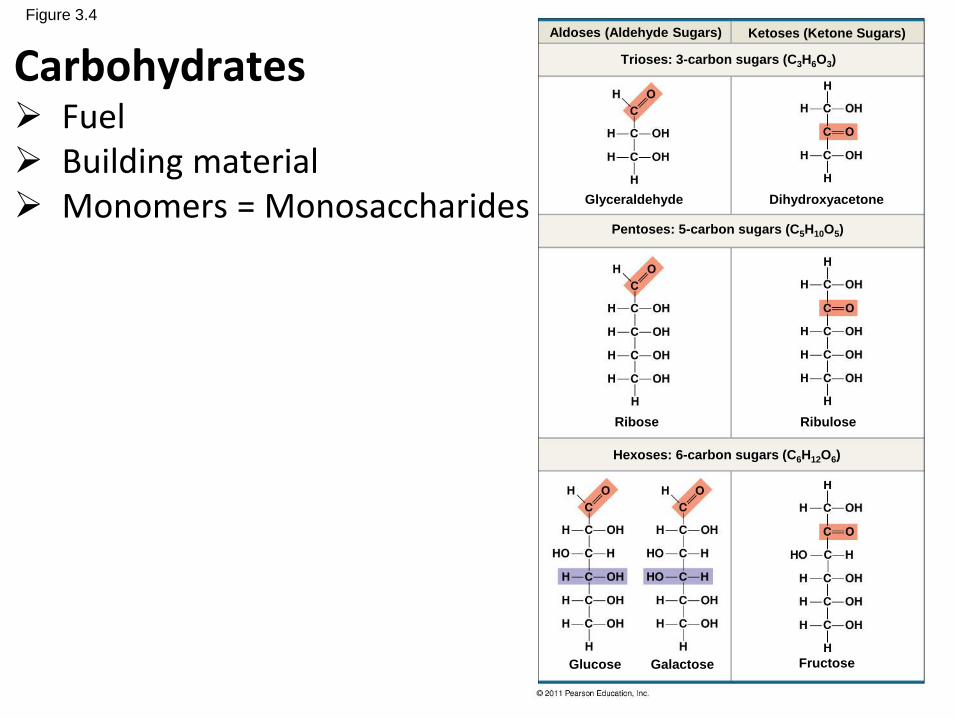
Figure 3.4Aldoses (Aldehyde Sugars) Ketoses (Ketone Sugars)
Glyceraldehyde
Trioses: 3-carbon sugars (C3H6O3)
Dihydroxyacetone
Pentoses: 5-carbon sugars (C5H10O5)
Hexoses: 6-carbon sugars (C6H12O6)
Ribose Ribulose
Glucose Galactose Fructose
Carbohydrates Fuel Building material Monomers = Monosaccharides

Figure 3.4
Aldose (Aldehyde Sugar) Ketose (Ketone Sugar)
Glyceraldehyde
Trioses: 3-carbon sugars (C3H6O3)
Dihydroxyacetone
Functional Groups

Figure 3.6
(a) Linear and ring forms
(b) Abbreviated ring structure
1
2
3
4
5
6
6
5
4
32
1 1
23
4
5
6
1
23
4
5
6

Figure 3.7, 3.8
(a) Dehydration reaction in the synthesis of maltose
(b) Dehydration reaction in the synthesis of sucrose
Glucose Glucose
Glucose
Maltose
Fructose Sucrose
1–4glycosidic
linkage
1–2glycosidic
linkage
1 4
1 2
DisaccharidesCarbohydrates

Figure 3.9
(a) Starch (amylose/amylopectin):a plant polysaccharide
(b) Glycogen:an animal polysaccharide
Chloroplast Starch granules
Mitochondria Glycogen granules
Amylopectin
Amylose
GlycogenWhere stored?Relate this to negative feedback & low blood sugar.
1 m
0.5 m
Polysaccharides 1) Storage Forms

Figure 5.7a
(a) and glucose ring structures
Glucose Glucose
4 1 4 1
Polysaccharidesalpha vs beta glucose

Figure 5.7b
(b) Starch: 1–4 linkage of glucose monomers
(c) Cellulose: 1–4 linkage of glucose monomers
41
41
Polysaccharidesalpha vs beta glucose arrangement
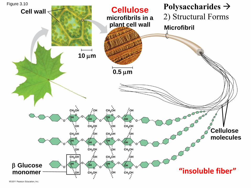
Figure 3.10
Cell wall
Microfibril
Cellulosemicrofibrils in aplant cell wall
Cellulosemolecules
Glucosemonomer
10 m
0.5 m
Polysaccharides
2) Structural Forms
“insoluble fiber”

Which polysaccharide has the
greatest number of branches?
a) cellulose
b) chitin
c) amylose
d) amylopectin
e) glycogen

Fig. 5-9

Why are human enzymes that digest
starch unable to digest cellulose?
a) Cellulose is made of amino-containing sugars that cannot
be metabolized.
b) Cellulose contains L-glucose instead of D-glucose; starch-
digesting enzymes are specific for polymers of D-glucose.
c) Cellulose has beta-glycosidic linkages; starch-digesting
enzymes cleave only alpha-glycosidic linkages.
d) Cellulose has beta-galactoside linkages that only bacterial
beta-galactosidases can cleave.
e) Cellulose fibers are covalently cross-linked; starch-digesting
enzymes cannot cleave these cross-links.
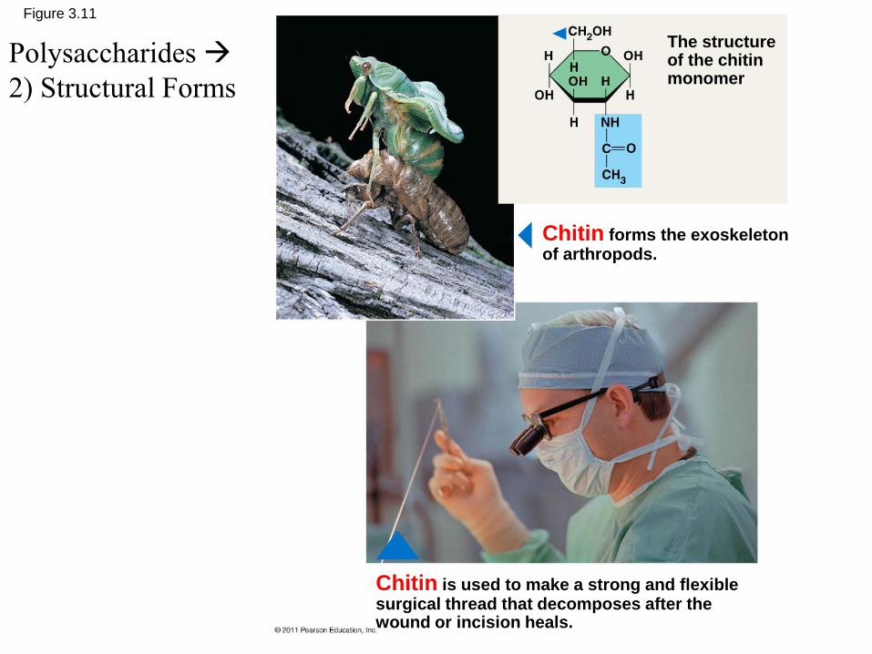
Figure 3.11
Chitin forms the exoskeletonof arthropods.
The structureof the chitinmonomer
Chitin is used to make a strong and flexiblesurgical thread that decomposes after thewound or incision heals.
Polysaccharides
2) Structural Forms

Subunits
a) proteins
b) starch
c) nucleic acids
d) fatty acids
If actively growing cells are fed 14C-labeled glucose,
what macromolecules will become radioactive first?

Figure 3.13
(a) One of three dehydration reactions in the synthesis of a fat
(b) Fat molecule (triacylglycerol)
Fatty acid(in this case, palmitic acid)
Glycerol
Ester linkage
Lipids

Figure 5.10a
(a) One of three dehydration reactions in the synthesis of a fat
Fatty acid(in this case, palmitic acid)
Glycerol

Figure 5.10b
(b) Fat molecule (triacylglycerol)
Ester linkage

Figure 3.14
• Stearic acid is a common saturated fatty acid.

Figure 3.15
• Oleic acid is a common unsaturated fatty acid.

Figure 3.16
• Saturated fatty acids have hydrocarbon
chains connected by single bonds only.
Unsaturated fatty acids have one or more
double bonds. Each double bond may be
in a cis or trans configuration. In the cis
configuration, both hydrogens are on the
same side of the hydrocarbon chain. In
the trans configuration, the hydrogens
are on opposite sides. A cis double bond
causes a kink in the chain.

Figure 3.17
• Alpha-linolenic acid is an example of an omega-3 fatty acid. It has three cis double bonds
and, as a result, a curved shape. For clarity, the carbons are not shown. Each singly bonded
carbon has two hydrogens associated with it, also not shown.

Figure 5.11
(a) Saturated fat(b) Unsaturated fat
Structuralformula of asaturated fatmolecule
Space-fillingmodel of stearicacid, a saturatedfatty acid
Structuralformula of anunsaturated fatmolecule
Space-filling modelof oleic acid, anunsaturated fattyacid
Cis double bondcauses bending.
Saturated vs unsaturated fat comparison

Figure 3.19
Choline
Phosphate
Glycerol
Fatty acids
Hydrophilichead
Hydrophobictails
(c) Phospholipid symbol(b) Space-filling model(a) Structural formula
Hyd
rop
hil
ic h
ea
dH
yd
rop
ho
bic
ta
ils
Lipids
•phospholipid

Figure 3.20
• The phospholipid bilayer is the major component of all cellular membranes.

Compared to tropical fish, arctic
fish oils have
a) more unsaturated fatty acids.
b) more cholesterol.
c) fewer unsaturated fatty acids.
d) more trans-unsaturated fatty acids.
e) more hydrogenated fatty acids.
Lipids

Figure 3.21
Lipids
•Steroids cholesterol

Lipid Functions
• Energy storage
• Insulation
• Cushioning organs
• Prevents water loss
• Chemical messengers
• Membranes

Lipids
a) are made from glycerol and fatty acids.
b) contain nitrogen.
c) have low energy content.
d) are acidic when mixed with water.
e) do not dissolve well in water.
All lipids

Figure 3.22Proteins
•Monomers amino acids

Figure 3.23

Figure 3.24Dehydration Synthesis
Condensation RXN

Figure 5.17
Peptide bond
New peptidebond forming
Sidechains
Back-bone
Amino end(N-terminus)
Peptidebond
Carboxyl end(C-terminus)
Dehydration Synthesis
Condensation RXN

Figure 3.25
Primary structure
Aminoacids
Amino end
Carboxyl end
Primary structure of transthyretin
Protein structure
DNA RNA Protein

Figure 3.28
Secondarystructure
Tertiarystructure
Quaternarystructure
Hydrogen bond
helix
pleated sheet
strand
Hydrogenbond
Transthyretinpolypeptide
Transthyretinprotein
Protein structure

Figure 5.20c
Secondary structure
Hydrogen bond
helix
pleated sheet
strand, shown as a flatarrow pointing towardthe carboxyl end
Hydrogen bond

Figure 5.20d
Secondary Structure pleated sheet

Figure 5.20e
Tertiary structure
Transthyretinpolypeptide

Figure 3.29
1) Hydrogenbond
4) Disulfidebridge
Polypeptidebackbone
5) Ionic bond
2) Hydrophobic
interactions and
3) van der Waals
interactions
Tertiary structure• Possible bonds

Figure 5.20b
Secondarystructure
Tertiarystructure
Quaternarystructure
Hydrogen bond
helix
pleated sheet
strand
Hydrogenbond
Transthyretinpolypeptide
Transthyretinprotein
Protein structure

Figure 5.20g
Quaternary structure
Transthyretinprotein
(four identicalpolypeptides)
Collagen
Figure 5.20h

Figure 5.20i
Hemoglobin
Heme
Iron
subunit
subunit
subunit
subunit
Quaternary structure

Figure 5.19
Primary
Structure
Secondary
and Tertiary
Structures
Quaternary
StructureFunction
Red Blood Cell
Shape
5 µm
Proteins do not associatewith one another; eachcarries oxygen.
Proteins aggregate into afiber; capacity tocarry oxygenis reduced.
5 µm
Normalhemoglobin
Normal 𝛃subunit
𝛃
𝛃 𝛂
𝛂
𝛂
𝛂
𝛃
𝛃
Sickle-cellhemoglobin
Sickle-cell 𝛃subunit
Sic
kle
-cell
No
rma
l
1
2
3
4
5
6
7
1
2
3
4
5
6
7

Protein Structure
a) primary
b) tertiary
c) quarternary
d) all of the above
e) primary and tertiary
structures only
The sickle-cell hemoglobin
mutation alters what level(s)
of protein structure?

Figure 5.22
Normal protein Denatured protein
tu
Denature
• Temperature heat
• pH
• [Ionic]
• Solvents

Macromolecular Structures and Bonds
a) Acidic pH denatures (unfolds and inactivates) proteins by disrupting their hydrogen bonds.
b) Citrus juice denatures proteins by disrupting their ionic bonds.
c) Citrus juice contains enzymes that hydrolyze peptide bonds to break apart proteins.
d) Citrus juice dissolves cell membranes by disrupting hydrophobic interactions.
Ceviche is prepared by marinating fresh raw fish in citrus juice for several hours, until the flesh becomes opaque and firm, as if cooked. How does citrus juice render the seafood safe to eat?

Figure 5.23
The cap attaches, causingthe cylinder to changeshape in such a way thatit creates a hydrophilicenvironment for thefolding of the polypeptide.
Cap
Polypeptide
Correctlyfoldedprotein
Chaperonin(fully assembled)
Steps of ChaperoninAction:
An unfolded poly-peptide enters thecylinder fromone end.
Hollowcylinder
The cap comesoff, and theproperly foldedprotein isreleased.
1
2 3
Chaperonin or chaperone proteins
Misfolded proteins Alzheimer’s, Parkinson’s, and mad cow disease

Figure 5.15a
Enzymatic proteins
Enzyme
Example: Digestive enzymes catalyze the hydrolysis
of bonds in food molecules.
Function: Selective acceleration of chemical reactions
Protein function #1
8 generalized functions of proteins

Figure 5.15b
Storage proteins
Ovalbumin Amino acidsfor embryo
Function: Storage of amino acids
Examples: Casein, the protein of milk, is the major
source of amino acids for baby mammals. Plants have
storage proteins in their seeds. Ovalbumin is the
protein of egg white, used as an amino acid source
for the developing embryo.
Protein function #2

Figure 5.15c
Hormonal proteins
Function: Coordination of an organism’s activities
Example: Insulin, a hormone secreted by the
pancreas, causes other tissues to take up glucose,
thus regulating blood sugar concentration
Highblood sugar
Normalblood sugar
Insulinsecreted
Protein function #3

Figure 5.15d
Muscle tissue
Actin Myosin
100 m
Contractile and motor proteins
Function: Movement
Examples: Motor proteins are responsible for the
undulations of cilia and flagella. Actin and myosin
proteins are responsible for the contraction of
muscles.
Protein function #4

Figure 5.15e
Defensive proteins
Virus
Antibodies
Bacterium
Function: Protection against disease
Example: Antibodies inactivate and help destroy
viruses and bacteria.
Protein function #5

Figure 5.15f
Transport proteins
Transportprotein
Cell membrane
Function: Transport of substances
Examples: Hemoglobin, the iron-containing protein of
vertebrate blood, transports oxygen from the lungs to
other parts of the body. Other proteins transport
molecules across cell membranes.
Protein function #6

Figure 5.15g
Signalingmolecules
Receptorprotein
Receptor proteins
Function: Response of cell to chemical stimuli
Example: Receptors built into the membrane of a
nerve cell detect signaling molecules released by
other nerve cells.
Protein function #7

Figure 5.15h
60 m
Collagen
Connectivetissue
Structural proteins
Function: Support
Examples: Keratin is the protein of hair, horns,
feathers, and other skin appendages. Insects and
spiders use silk fibers to make their cocoons and webs,
respectively. Collagen and elastin proteins provide a
fibrous framework in animal connective tissues.
Protein function #8

8 Protein Functions
1. Structural
2. Storage
3. Transport
4. Hormonal
5. Receptor
6. Contractile
7. Defense
8. Enzymatic
1. Connective tissue (tendons,
ligaments)
2. Albumin, casein
3. Hemoglobin, ion channels, etc
4. Insulin
5. Detects other signals (stimuli)
6. Movement
7. Immune system (antibodies)
8. Increase chemical reactions
(Digestion, cellular respiration)

Figure 3.31

Figure 5.26aSugar-phosphate backbone
5 end
5C
3C
5C
3C
3 end
(a) Polynucleotide, or nucleic acid
(b) Nucleotide
Phosphategroup Sugar
(pentose)
Nucleoside
Nitrogenousbase
5C
3C
1C
Nucleic Acids
• Nucleotide

Figure 5.26b
Nitrogenous bases
Cytosine (C)
Thymine (T, in DNA)
Uracil (U, in RNA)
Adenine (A) Guanine (G)
Sugars
Deoxyribose (in DNA)
Ribose (in RNA)
(c) Nucleoside components
Pyrimidines
Purines
Nitrogenous Bases

Base pair
Phosphate
Sugar
Nucleotide
Nucleic Acids Store Genetic Information
• Structure of DNA (deoxyribonucleic acid)• Double–stranded (dbl helix)
• Nucleotides contain
– Deoxyribose (sugar)
– Nitrogenous bases» Adenine
» Guanine
» Cytosine
» Thymine
• Pairing
» Adenine - Thymine
» Guanine - Cytosine

Figure 5.27
Sugar-phosphatebackbones
Hydrogen bonds
Base pair joinedby hydrogen bonding
Base pair joinedby hydrogen
bonding
(b) Transfer RNA(a) DNA
5 3
53
Base pair rules

Nucleic Acids Store Genetic Information
• Structure of RNA (ribonucleic acid)• Single–stranded
• Nucleotides contain
– Ribose
– Nitrogenous bases
» Adenine
» Guanine
» Cytosine
» Uracil

RNA and DNA
a) DNA encodes hereditary information; RNA does
not.
b) DNA forms duplexes; RNA does not.
c) DNA contains thymine; RNA contains uracil.
d) all of the above
How does RNA differ from DNA?

Figure 5.25-3
Synthesis ofmRNA
mRNA
DNA
NUCLEUS
CYTOPLASM
mRNA
Ribosome
AminoacidsPolypeptide
Movement ofmRNA intocytoplasm
Synthesisof protein
1
2
3
Nucleic Acids
Transcription
• DNA RNA
Translation
• RNA Protein

Nucleic acid functions?
• DNA: instructions for making proteins via
RNA• Information storage
• Information transfer
• RNA: instructions for making proteins • Protein synthesis DNA → RNA → Proteins
• Proteins: direct most of life’s processes
• Transfer of chemical energy

Figure 5.UN02

Figure 5.UN02a
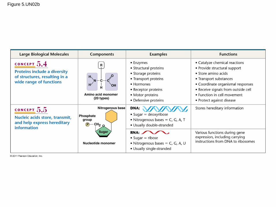
Figure 5.UN02b