BIOLOGY PAPER 3 REVISION KITS 2018
4
SEPT 2018 - PAPER 3 BIOLOGY PAPER 3 REVISION KITS 2018
Transcript of BIOLOGY PAPER 3 REVISION KITS 2018
SEPT 2018 - PAPER 3
BIOLOGY PAPER 3REVISION KITS 2018
1. Take 2 clean test tubes and into each add 5cm3 of dilute hydrogen peroxide. Label the test tubes as A
and B.
Cut 2 cubes of irish potato measuring 1cm3 each. Boil one cube in a boiling tube with some water for
about 5 minutes.
Drop the boiled cube into test tube A and unbolied cube in test tube B. State your observations
(a) Test tube A (1mk)
………………………………………………………………………………………………….
………………………………………………………………………………………………….
Test tube B (1mk)
………………………………………………………………………………………………….
………………………………………………………………………………………………….
(b) Account for your observations in
Test tube A (1mk)
………………………………………………………………………………………………….
………………………………………………………………………………………………….
Test tube B (2mks)
………………………………………………………………………………………………….
………………………………………………………………………………………………….
(c) Take a small piece of substance Z provided and add to it 2cm3 of sodium hydrogen carbonate.
(i) State your observations (1mk)
………………………………………………………………………………………………….
………………………………………………………………………………………………….
(ii) Which physiological process in the body is illustrated above (1mk)
………………………………………………………………………………………………….
………………………………………………………………………………………………….
(iii) State the part of the body where the process takes place. (1mk)
………………………………………………………………………………………………….
………………………………………………………………………………………………….
(iv)What is the significance of the process (1mk)
………………………………………………………………………………………………….
………………………………………………………………………………………………….
(d) Put 2cm3 of liquid labelled as C into a test tube. Squeeze some juice from specimen X into a beaker.
Draw some of the juice into a dropper. Add 3 drops of the juice into the test tube with solution C.
(i) State your observation. (1mk)
………………………………………………………………………………………………….
………………………………………………………………………………………………….
(ii) State the part of the human body where the physiological process demonstrated above occurs
and the enzyme that carriers out the process. (2mks)
Part of body
………………………………………………………………………………………………….
Enzyme
………………………………………………………………………………………………….
(iii) Which gland produces the enzyme stated in (ii) above. (1mk)
………………………………………………………………………………………………….
(iv) Which hormone stimulates the production of the enzyme stated in (II) above. (1mk)
………………………………………………………………………………………………….
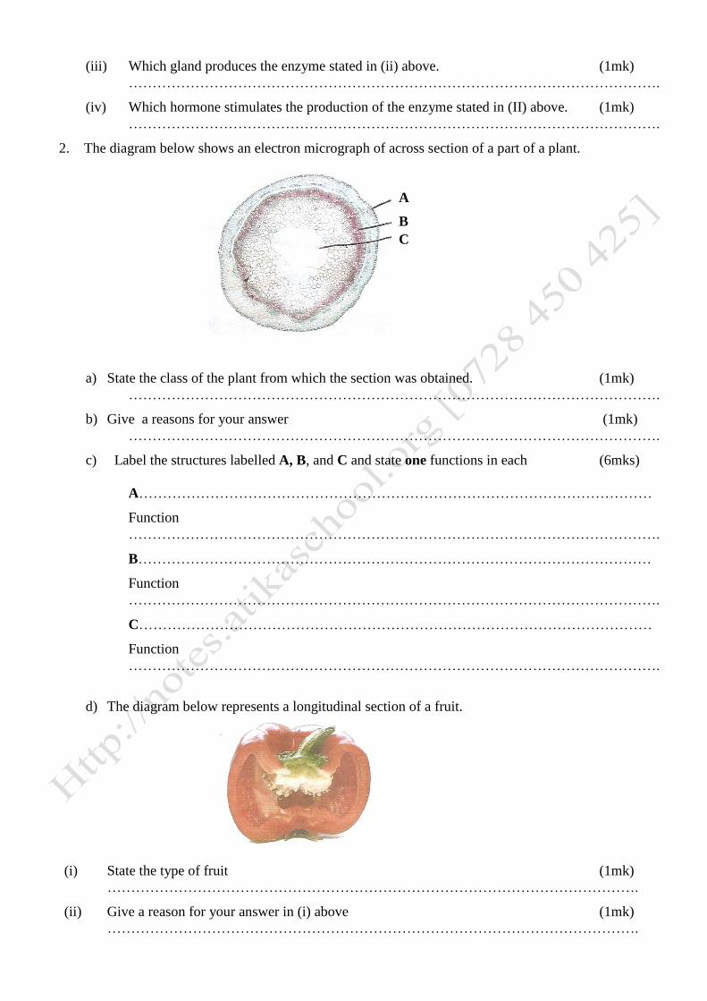
2. The diagram below shows an electron micrograph of across section of a part of a plant.
a) State the class of the plant from which the section was obtained. (1mk)
………………………………………………………………………………………………….
b) Give a reasons for your answer (1mk)
………………………………………………………………………………………………….
c) Label the structures labelled A, B, and C and state one functions in each (6mks)
A………………………………………………………………………………………………
Function
………………………………………………………………………………………………….
B………………………………………………………………………………………………
Function
………………………………………………………………………………………………….
C………………………………………………………………………………………………
Function
………………………………………………………………………………………………….
d) The diagram below represents a longitudinal section of a fruit.
(i) State the type of fruit (1mk)
………………………………………………………………………………………………….
(ii) Give a reason for your answer in (i) above (1mk)
………………………………………………………………………………………………….
A
B
C
(iii) State the type of placentation in the fruit and give a reason for your answer (2mks)
Placentation
………………………………………………………………………………………………….
Reason
………………………………………………………………………………………………….
………………………………………………………………………………………………….
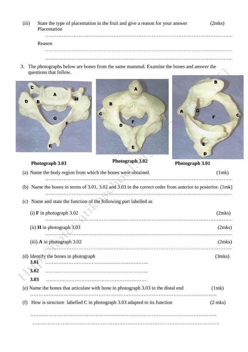
3. The photographs below are bones from the same mammal. Examine the bones and answer the
questions that follow.
(a) Name the body region from which the bones were obtained. (1mk)
………………………………………………………………………………………………….
(b) Name the bones in terms of 3.01, 3.02 and 3.03 in the correct order from anterior to posterior. (1mk)
………………………………………………………………………………………………….
(c) Name and state the function of the following part labelled as
(i) F in photograph 3.02 (2mks)
………………………………………………………………………………………………….
(ii) H in photograph 3.03 (2mks)
………………………………………………………………………………………………….
(iii) A in photograph 3.02 (2mks)
………………………………………………………………………………………………….
(d) Identify the bones in photograph (3mks)
3.01 ……………………………………………………..
3.02 ……………………………………………………..
3.03 ……………………………………………………
(e) Name the bones that articulate with bone in photograph 3.03 in the distal end (1mk)
………………………………………………………………………………………………….
(f) How is structure labelled C in photograph 3.03 adapted to its function (2 mks)
………………………………………………………………………………………………….
………………………………………………………………………………………………….
Photograph 3.03 Photograph 3.01 Photograph 3.02
A
C
D B
G
E
F
H
A
B
C
D
E
F
A
B
C
D
F
G
E
F
G