BIOLOGICAL THREAT AGENTS - HPSC603,en.pdf · National Disease Surveillance Centre Biological Threat...
21
National Disease Surveillance Centre Biological Threat Agents BIOLOGICAL THREAT AGENTS This document is intended to give an overview of the clinical management and public health implications of selected biological threat agents thought most likely to be used in terrorist attacks. The likelihood of such attacks were considered to be remote until the airliner attacks in the US on September 11 th , 2001 and the mailing of finely milled anthrax spores to agencies and individuals in the US during October 2001. Systems for dealing with such incidents have been put in place by governments across the world in order to address the unlikely widespread use of such tactics by terrorist organisations. Early detection is essential for ensuring a prompt response to biological attacks. Features that should alert healthcare providers to the possibility of a bioterrorism related outbreak include: • A rapidly increasing disease incidence in a normally healthy population • An unusual increase in the number of people seeking care, particularly with fever, respiratory or gastrointestinal complaints • An endemic disease rapidly emerging at an uncharacteristic time or in an unusual pattern (e.g. an increase in what appears to be chickenpox-like illness among adult patients, but which might be smallpox) • Clusters of patients arriving from a single locale • Large numbers of potentially fatal cases (e.g. a large number of cases of acute flaccid paralysis with prominent bulbar palsies, suggestive of a release of botulinum toxin) • Any one patient presenting with a disease that is relatively uncommon and has bioterrorism potential The agents thought most likely to be used in such attacks include: 1. Anthrax, 2. Smallpox, 3. Botulinum toxin, 4. Plague, and 5. Tularaemia Version 1 18/6/02 1
Transcript of BIOLOGICAL THREAT AGENTS - HPSC603,en.pdf · National Disease Surveillance Centre Biological Threat...
National Disease Surveillance Centre Biological Threat Agents
BIOLOGICAL THREAT AGENTS
This document is intended to give an overview of the clinical management and public health
implications of selected biological threat agents thought most likely to be used in terrorist
attacks. The likelihood of such attacks were considered to be remote until the airliner attacks
in the US on September 11th, 2001 and the mailing of finely milled anthrax spores to agencies
and individuals in the US during October 2001. Systems for dealing with such incidents have
been put in place by governments across the world in order to address the unlikely widespread
use of such tactics by terrorist organisations. Early detection is essential for ensuring a prompt
response to biological attacks.
Features that should alert healthcare providers to the possibility of a bioterrorism related
outbreak include:
• A rapidly increasing disease incidence in a normally healthy population
• An unusual increase in the number of people seeking care, particularly with fever,
respiratory or gastrointestinal complaints
• An endemic disease rapidly emerging at an uncharacteristic time or in an unusual
pattern (e.g. an increase in what appears to be chickenpox-like illness among adult
patients, but which might be smallpox)
• Clusters of patients arriving from a single locale
• Large numbers of potentially fatal cases (e.g. a large number of cases of acute flaccid
paralysis with prominent bulbar palsies, suggestive of a release of botulinum toxin)
• Any one patient presenting with a disease that is relatively uncommon and has
bioterrorism potential
The agents thought most likely to be used in such attacks include:
1. Anthrax,
2. Smallpox,
3. Botulinum toxin,
4. Plague, and
5. Tularaemia
Version 1 18/6/02 1
National Disease Surveillance Centre Biological Threat Agents
Anthrax Epidemiology
Anthrax is a zoonotic disease caused by Bacillus anthracis, a spore-forming rod, which
occurs primarily in large domestic and wild herbivores. Cattle, horses, goats, sheep and pigs
are most commonly infected and distribution is worldwide. 1 Many species of animals and
birds can acquire the disease naturally. The reservoir of B anthracis is the soil and spores can
remain viable for decades.2 Human infection usually follows contact from infected animals
or contaminated animal products. Infection occurs usually through the skin and less
commonly through the respiratory or gastrointestinal systems.1
A previously undescribed form of anthrax - inhalational anthrax - appeared during the latter
half of the 19th century, in woolsorters in England, caused by aerosolisation of anthrax spores
generated during the industrial processing of animal hides. Inhalational anthrax carried a
much higher mortality rate than the cutaneous form of the disease.
About 2000 cases of cutaneous anthrax are reported globally each year. There are about 5
cases each year in the US and fewer than 1 case a year in the UK.3,4 There have been no
human or animal anthrax notifications in Ireland during the last 25 years.
Anthrax as a Bioterrorist Agent
Anthrax spores are highly infective and small doses have the potential of producing
considerable harm. If intentionally released, spores would most likely produce effects
following inhalation. Anthrax is not contagious; there is no person-to-person spread – if cases
of inhalational disease were to appear in Ireland it is most likely to be due to intentional
release of B anthracis spores. There is a small risk associated with cutaneous lesions where
infested skin lesions may rarely be associated with transmission of cutaneous anthrax. Such
transmission requires an existing break in the skin.
Clinical Features
Human disease is potentially fatal. If systemic, toxaemia is common.
Anthrax can present in 3 ways:
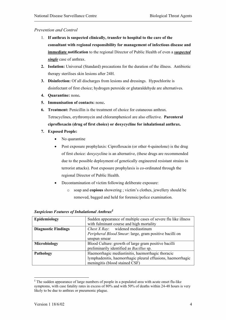
1. Cutaneous: by skin inoculation – 95% of cases. Commonly affects hands, forearms
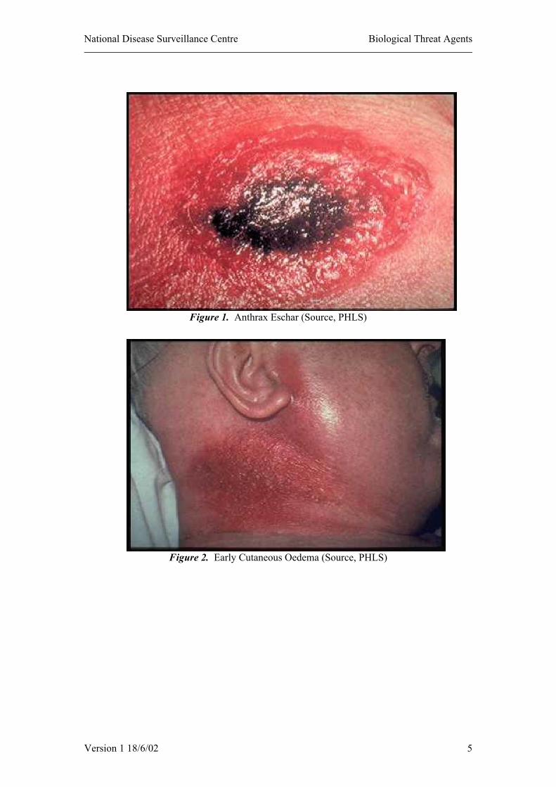
and head. Cardinal feature is painless swelling of skin. This begins with itching at the
point of contact, followed by a papular lesion, which becomes vesicular, ulcerates and
becomes weepy. Over the next 2-6 days, develops into a depressed, blackened eschar.
Can progress to toxaemia. Mortality with treatment is <1%.
Version 1 18/6/02 2
National Disease Surveillance Centre Biological Threat Agents
2. Gastrointestinal: by eating infected meat – uncommon. Characterised by abdominal
pain, ascites, diarrhoea and haematemesis, followed by fever and septicaemia.
Mortality rate is generally over 50%.
3. Respiratory: the least common (only 18 naturally occurring cases in the US during
the 20th Century) but a much commoner presentation following a bioterrorist attack.
Mortality is about 90%. The incubation period can be 1-60 days (generally 1-5 days).
Early diagnosis is difficult and requires a high index of suspicion. There are 2 stages
to the illness:
a. Initial Phase: Hours to a few days.
Non-specific symptoms (fever, cough, headache vomiting, rigors,
chest and abdominal pain).
•
•
•
•
•
•
•
•
Signs and laboratory findings non-specific.
Some patients may appear apparently to recover.
The remainder progress to the second fulminant stage of the
disease.
b. Fulminant Phase: Sudden onset, bacteraemia and toxaemia, fever, dyspnoea,
sweating, bacteraemia and septic shock.
Stridor can develop as a result of adenopathic mediastinal
compression.
Chest X ray shows mediastinal widening due to hilar adenopathy
but infiltrates are generally absent.
Up to 50% can develop haemorrhagic meningitis with signs of
meningism and raised intracranial pressure. In these cases, death
generally follows in 24-36 hours.
Mortality rate is proportional to the incubation period – most
fatalities occur in victims who develop symptoms within 3 days of
exposure.
Diagnosis
A widened mediastinum on Chest X Ray in a previously healthy individual who has
symptoms of overwhelming flu-like illness is virtually pathognomic of inhalational anthrax.
Detection at this stage is unlikely to be of benefit to such a patient, but might lead to earlier
diagnosis in others.
B anthracis can be seen following Gram staining of peripheral blood smears. Culture of the
organism from blood is often only possible late in the disease.
Version 1 18/6/02 3
National Disease Surveillance Centre Biological Threat Agents
Prevention and Control
1. If anthrax is suspected clinically, transfer to hospital to the care of the
consultant with regional responsibility for management of infectious disease and
immediate notification to the regional Director of Public Health of even a suspected
single case of anthrax.
2. Isolation: Universal (Standard) precautions for the duration of the illness. Antibiotic
therapy sterilises skin lesions after 24H.
3. Disinfection: Of all discharges from lesions and dressings. Hypochlorite is
disinfectant of first choice; hydrogen peroxide or glutaraldehyde are alternatives.
4. Quarantine: none.
5. Immunisation of contacts: none.
6. Treatment: Penicillin is the treatment of choice for cutaneous anthrax.
Tetracyclines, erythromycin and chloramphenicol are also effective. Parenteral
ciprofloxacin (drug of first choice) or doxycycline for inhalational anthrax.
7. Exposed People:
No quarantine •
•
•
Post exposure prophylaxis: Ciprofloxacin (or other 4-quinolone) is the drug
of first choice: doxycycline is an alternative, (these drugs are recommended
due to the possible deployment of genetically engineered resistant strains in
terrorist attacks). Post exposure prophylaxis is co-ordinated through the
regional Director of Public Health.
Decontamination of victim following deliberate exposure:
o soap and copious showering ; victim’s clothes, jewellery should be
removed, bagged and held for forensic/police examination.
Suspicious Features of Inhalational Anthrax§
Epidemiology Sudden appearance of multiple cases of severe flu like illness with fulminant course and high mortality
Diagnostic Findings Chest X Ray: widened mediastinum Peripheral Blood Smear: large, gram positive bacilli on unspun smear
Microbiology Blood Culture: growth of large gram positive bacilli preliminarily identified as Bacillus sp.
Pathology Haemorrhagic mediastinitis, haemorrhagic thoracic lymphadenitis, haemorrhagic pleural effusions, haemorrhagic meningitis (blood stained CSF)
§ The sudden appearance of large numbers of people in a populated area with acute onset flu-like symptoms, with case fatality rates in excess of 80% and with 50% of deaths within 24-48 hours is very likely to be due to anthrax or pneumonic plague.
Version 1 18/6/02 4
National Disease Surveillance Centre Biological Threat Agents
Figure 1. Anthrax Eschar (Source, PHLS)
Figure 2. Early Cutaneous Oedema (Source, PHLS)
Version 1 18/6/02 5
National Disease Surveillance Centre Biological Threat Agents
Smallpox Epidemiology
Smallpox is a viral disease, unique to humans and caused by a DNA orthopoxvirus, variola.
There are 2 forms of the disease, variola major (ordinary form) and variola minor (alastrim).
In unvaccinated subjects, variola major has a mortality rate of over 30% (3% in vaccinated)
while variola minor has a mortality rate of 1% or less. It is spread by airborne transmission.
The rate of spread of smallpox through a population is slower than measles and chickenpox.
Patients spread smallpox mainly to household and family friends. The secondary attack rate
among unvaccinated contacts is about 50%.5
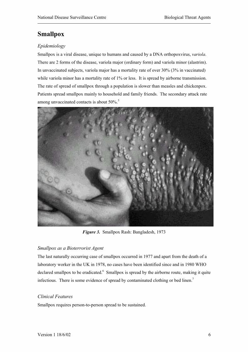
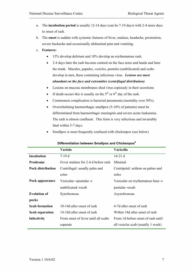
Figure 3. Smallpox Rash: Bangladesh, 1973
Smallpox as a Bioterrorist Agent
The last naturally occurring case of smallpox occurred in 1977 and apart from the death of a
laboratory worker in the UK in 1978, no cases have been identified since and in 1980 WHO
declared smallpox to be eradicated.6 Smallpox is spread by the airborne route, making it quite
infectious. There is some evidence of spread by contaminated clothing or bed linen.7
Clinical Features
Smallpox requires person-to-person spread to be sustained.
Version 1 18/6/02 6
National Disease Surveillance Centre Biological Threat Agents
a. The incubation period is usually 12-14 days (can be 7-19 days) with 2-4 more days
to onset of rash.
b. The onset is sudden with systemic features of fever, malaise, headache, prostration,
severe backache and occasionally abdominal pain and vomiting.
c. Features:
15% develop delirium and 10% develop an erythematous rash. •
•
•
•
•
•
•
2-4 days later the rash become centred on the face arms and hands and later
the trunk. Macules, papules, vesicles, pustules (umbilicated) and scabs
develop in turn, these containing infectious virus. Lesions are most
abundant on the face and extremities (centrifugal distribution)
Lesions on mucous membranes shed virus copiously in their secretions
If death occurs this is usually on the 5th or 6th day of the rash.
Commonest complication is bacterial pneumonia (mortality over 50%).
Overwhelming haemorrhagic smallpox (5-10% of patients) must be
differentiated from haemorrhagic meningitis and severe acute leukaemia.
The rash is almost confluent. This form is very infectious and invariably
fatal within 5-7 days.
Smallpox is most frequently confused with chickenpox (see below)
Differentiation between Smallpox and Chickenpox6
Variola Varicella
Incubation 7-19 d 14-21 d
Prodrome Fever malaise for 2-4 d before rash Minimal
Pock distribution Centrifugal: usually palm and
soles
Centripetal: seldom on palms and
soles
Pock appearance Vesicular→pustular→
umbilicated→scab
Vesicular on erythematous base→
pustular→scab
Evolution of
pocks
Synchronous Asynchronous
Scab formation 10-14d after onset of rash 4-7d after onset of rash
Scab separation 14-18d after onset of rash Within 14d after onset of rash
Infectivity From onset of fever until all scabs
separate
From 1d before onset of rash until
all vesicles scab (usually 1 week)
Version 1 18/6/02 7
National Disease Surveillance Centre Biological Threat Agents
Diagnosis
The discovery of a single, suspected case of smallpox, must be treated as an international
health emergency and must be notified to the consultant with regional responsibility for
management of infectious disease and to the regional Director of Public Health
immediately. All suspected cases of smallpox in the community should be isolated at the
point of diagnosis for further expert evaluation. Diagnosis is through a combination of
clinical features and laboratory findings. Characteristic virion appearance on EM. Specific
ELISA. PCR techniques are becoming more accurate. Virus is more likely to be isolated
from blood during early phases and from vesicles and pustules in the latter phases.
Prevention and Control
1. Seek immediate advice from consultant with regional responsibility for
management of infectious disease and immediate notification to the regional
Director of Public Health of even a suspected single case of smallpox.
2. Isolation: Airborne precautions for the duration of the illness. Isolation should be in
specific isolation units, a non-hospital facility or at home wherever possible.
3. Quarantine: Strict quarantine with respiratory isolation should be applied for 18
days to all persons in direct contact with the index case, especially the unvaccinated.
Infectiousness is from the onset of fever and peaks on the 2nd and 3rd day of fever.
4. Immunisation of contacts: All face-to-face contacts and all healthcare staff.
Vaccination of selected individuals and groups will take place on the advice of the
Expert Committee.
5. Treatment: Supportive therapy only. Antivirals not particularly effective.
Antibiotics for secondary pneumonia.
6. Exposed People:
Quarantine as above. If many contacts may be logistically difficult to isolate
all therefore measure temperature daily. Any rise during the 17 days
following exposure should prompt immediate quarantine.
•
• Vaccine: vaccinia vaccine. Smallpox vaccine contains live vaccinia virus, a
virus in the orthopoxvirus family, which is closely related to variola virus. It
does not contain smallpox virus and cannot transmit smallpox. Vaccination
is likely to offer significant protection against disease and death if
administered up to 4 days after exposure. If symptoms appear, they are
milder and mortality is less in vaccinated than in non-vaccinated persons.
Even when immunity has waned, vaccinated persons shed less virus and are
Version 1 18/6/02 8
National Disease Surveillance Centre Biological Threat Agents
less likely to transmit the disease. A single dose vaccine usually prevents
smallpox infection for at least 10 years.
The vaccine is freeze-dried and sealed in ampoules for later re-suspension in
sterile buffer and subsequent intradermal inoculation by multiple puncture
with a bifurcated needle. A certified clinical ‘take’ of vaccinia vaccine is the
development of one or more pocks over 6-8 days, then progressive crusting in
the following 8-10 days. Smallpox vaccination should be undertaken by
those who have completed training in the procedure.
Existing vaccines have proven efficacy but also have a high incidence of
adverse side-effects. The risk of adverse events is sufficiently high that
vaccination is not warranted if there is no or little real risk of exposure.
Vaccine is contraindicated for certain groups. These include
• Pregnant women,
• Persons with immune disorders or experiencing therapeutically-
induced immunosuppression,
• Persons with HIV infection, and
• Persons with a history of eczema.
7. Healthcare Staff: managing suspected cases:
• Full airborne precautions (as above, victims to be managed by staff known
to be vaccinated where possible, high efficacy masks, gloves, gowns).
• Manage in negative pressure room where possible (in isolation facility or
at home if large outbreak).
• Terminal decontamination (upon death or transfer)
• Immediate emergency vaccination (as above) including paramedical,
mortuary and other rescue, fire and police staff.
8. Laboratory: Specimens should be transferred to National Virus Reference
Laboratory Belfield.
Version 1 18/6/02 9
National Disease Surveillance Centre Biological Threat Agents
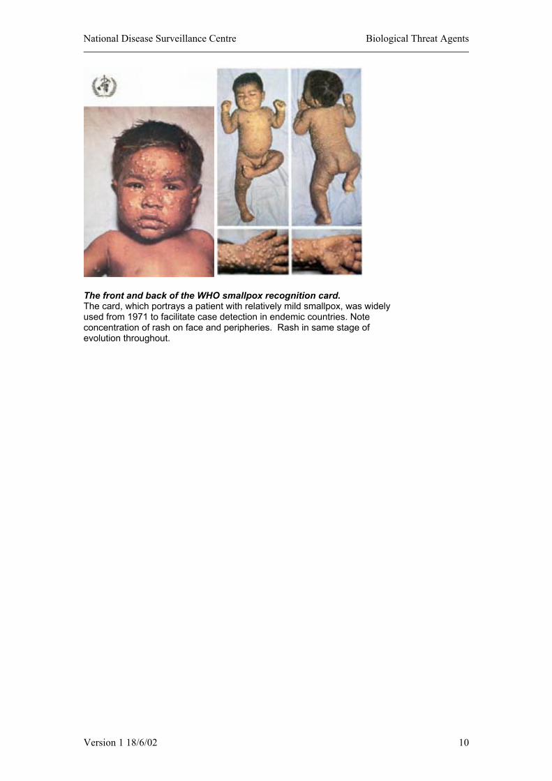
The front and back of the WHO smallpox recognition card. The card, which portrays a patient with relatively mild smallpox, was widely used from 1971 to facilitate case detection in endemic countries. Note concentration of rash on face and peripheries. Rash in same stage of evolution throughout.
Version 1 18/6/02 10
National Disease Surveillance Centre Biological Threat Agents
Botulinum Toxin Introduction
Botulinum toxin has been identified as a possible agent for use during a bioterrorist attack.
Epidemiology
Botulism is a produced as a result of ingesting Botulinum neurotoxin, produced by
Clostridium botulinum. Improperly prepared food or canned products usually cause botulism.8
Wound botulism occurs following penetrating injuries complicated by soil contamination and
may also follow the injection of illegal drugs.
Botulinum Toxin as a Bioterrorist Agent
If used in a bioterrorist attack, botulinum toxin could be released by aerosol and inhaled or
used to contaminate food. Botulinum toxin is a neurotoxin and acts by blocking
neurotransmission.9
Clinical Features
The typical clinical picture involves bulbar palsy and skeletal muscle weakness. The onset of
signs is dose dependent and may vary from 2 hours to 8 days in the case of foodborne
poisoning and from 1 hour to several days following aerosol exposure. Both forms of
exposure have the same set of symptoms; in addition, abdominal cramps, nausea, vomiting,
and diarrhoea generally precede foodborne poisoning. Progressive muscular paralysis leads
to pharyngeal and diaphragmatic paralysis and if untreated, generally death. Aggressive
treatment has reduced the mortality rate to about 5%.10
Patients generally present with difficulty seeing, speaking +/- swallowing. Motor signs
predominate.
1. Foodborne: condition on presentation
Symptoms: diplopia, dysphagia, dysarthria, dysphonia dry mouth fatigue,
dizziness, nausea vomiting, leg and arm weakness.
•
•
•
•
Signs: Alertness, afebrile, ptosis, paralysis of gaze, nystagmus, facial palsy,
fixed/dilated pupils, diminished gag, arm and leg weakness.
2. Inhalational:
3rd day after exposure: mucus in throat, dysphagia, dizziness
4th day after exposure: difficulty moving eyes, mild papillary dilation and
nystagmus, indistinct speech, unsteady gait, extreme weakness.
Version 1 18/6/02 11
National Disease Surveillance Centre Biological Threat Agents
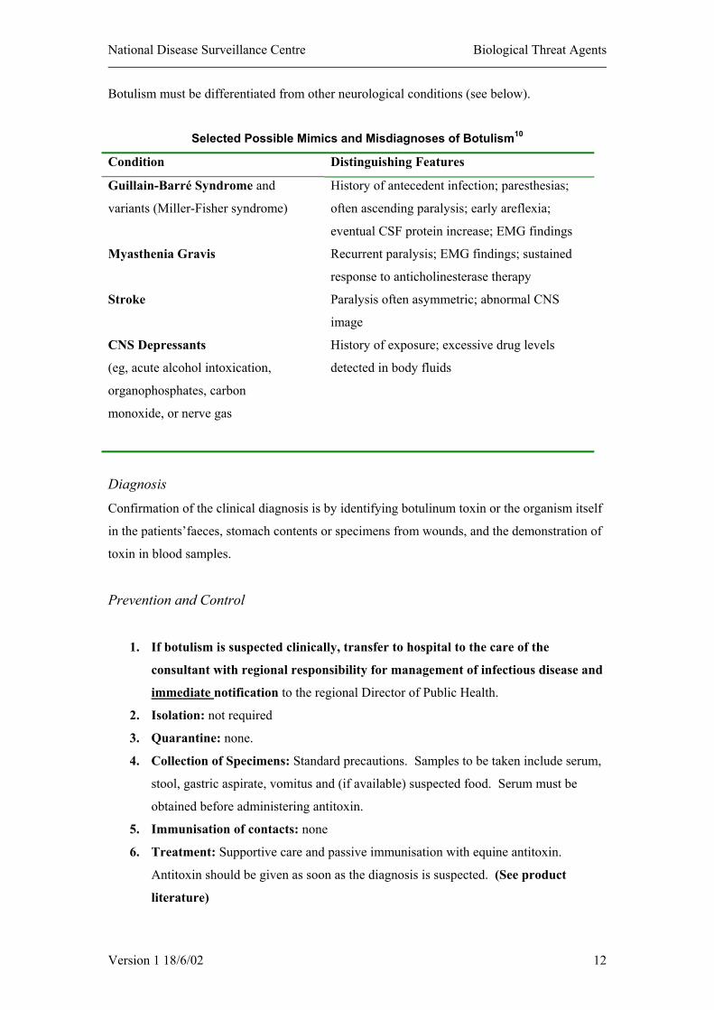
Botulism must be differentiated from other neurological conditions (see below).
Selected Possible Mimics and Misdiagnoses of Botulism10
Condition Distinguishing Features
Guillain-Barré Syndrome and
variants (Miller-Fisher syndrome)
History of antecedent infection; paresthesias;
often ascending paralysis; early areflexia;
eventual CSF protein increase; EMG findings
Myasthenia Gravis Recurrent paralysis; EMG findings; sustained
response to anticholinesterase therapy
Stroke Paralysis often asymmetric; abnormal CNS
image
CNS Depressants
(eg, acute alcohol intoxication,
organophosphates, carbon
monoxide, or nerve gas
History of exposure; excessive drug levels
detected in body fluids
Diagnosis
Confirmation of the clinical diagnosis is by identifying botulinum toxin or the organism itself
in the patients’faeces, stomach contents or specimens from wounds, and the demonstration of
toxin in blood samples.
Prevention and Control
1. If botulism is suspected clinically, transfer to hospital to the care of the
consultant with regional responsibility for management of infectious disease and
immediate notification to the regional Director of Public Health.
2. Isolation: not required
3. Quarantine: none.
4. Collection of Specimens: Standard precautions. Samples to be taken include serum,
stool, gastric aspirate, vomitus and (if available) suspected food. Serum must be
obtained before administering antitoxin.
5. Immunisation of contacts: none
6. Treatment: Supportive care and passive immunisation with equine antitoxin.
Antitoxin should be given as soon as the diagnosis is suspected. (See product
literature)
Version 1 18/6/02 12
National Disease Surveillance Centre Biological Threat Agents
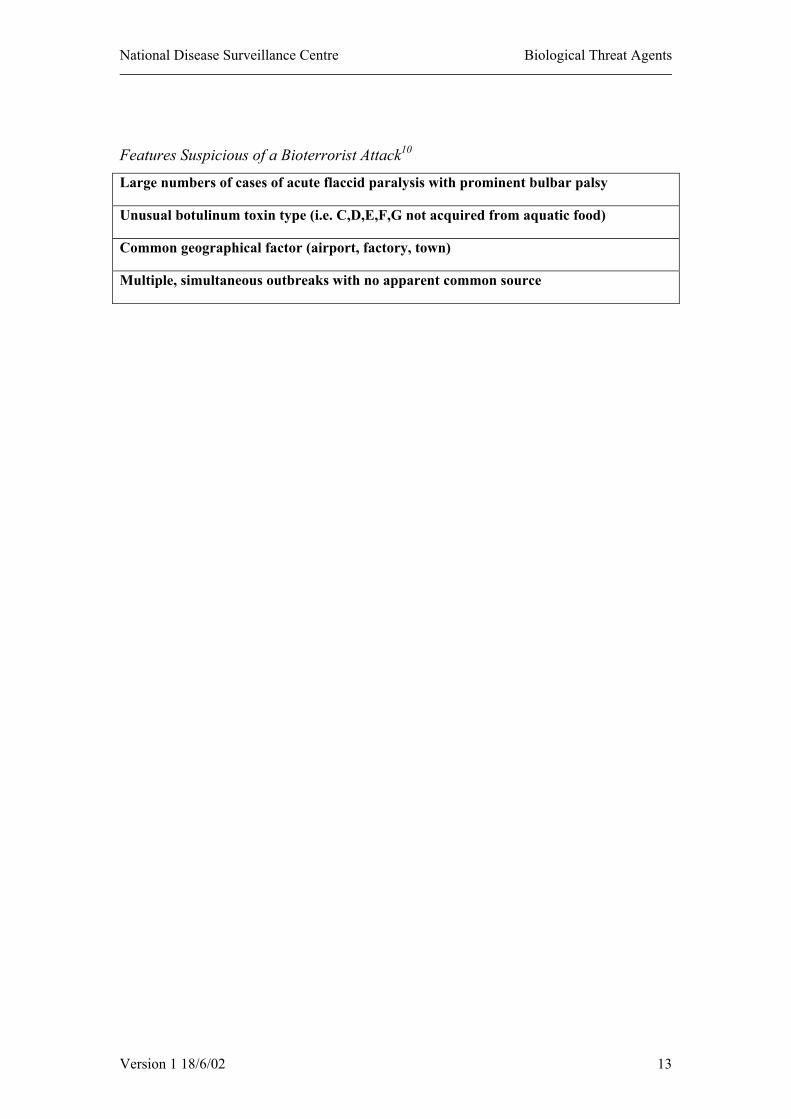
Features Suspicious of a Bioterrorist Attack10
Large numbers of cases of acute flaccid paralysis with prominent bulbar palsy
Unusual botulinum toxin type (i.e. C,D,E,F,G not acquired from aquatic food)
Common geographical factor (airport, factory, town)
Multiple, simultaneous outbreaks with no apparent common source
Version 1 18/6/02 13
National Disease Surveillance Centre Biological Threat Agents
Plague Introduction
The causative organism is Yersinia pestis, a gram-negative rod. If used in a bioterrorist
attack, Y. pestis could be released by aerosol and inhaled or spread by infected vectors (fleas).
Person-to-person spread occurs readily. Plague is highly infectious.11
Clinical Features
There are 3 principal clinical forms of plague, bubonic (90% of cases), primary septicaemic
(10%) and pneumonic (infectious) (1%). If used as an aerosolised biological warfare agent,
the manifestations of plague would be:
•
•
•
•
•
•
•
•
•
•
•
•
•
epidemic pneumonia with blood tinged sputum or
bubonic and septicaemic plague, or both if fleas were used as carriers.12
1. Bubonic Plague:
Incubation 1-8 days.
Onset sudden with fever, rigors, headache, confusion with nausea and
vomiting.
Buboes (extremely painful, enlarged, inflamed lymph nodes) appear 6-8
hours after onset of symptoms. Even almost comatose patients will
protect buboes from movement or contact.
Up to 15% will develop pneumonic plague and become potentially
infectious.
Mortality rate is about 12%.
2. Septicaemic Plague:
May be primary or secondary to the bubonic form.
Presentation as for any gram-negative septicaemia: fever, rigors, nausea,
vomiting, diarrhoea and abdominal pain (due to hepatosplenomegaly).
Purpura, disseminated intravascular coagulopathy (DIC), peripheral
cyanosis and necrosis develop.
Mortality rate is about 30%.
3. Pneumonic Plague: the most likely form after a biological warfare attack.
May occur primarily, from aerosol inhalation or secondarily from
haematogenous dissemination.
Incubation: 1-3 days.
Version 1 18/6/02 14
National Disease Surveillance Centre Biological Threat Agents
Sudden onset malaise, high fever, rigors, headache, myalgia, cough and
blood-tinged sputum
•
•
•
•
•
•
•
•
Pneumonic symptoms develop rapidly with dyspnoea, stridor and
cyanosis.
Terminally respiratory failure and shock
Chest X Ray: patchy or consolidated bronchopneumonia.
The high mortality from plague has fallen dramatically with the advent of
effective antibiotic therapy. In cases reported to WHO during 1983-1997
the overall fatality rate was 8%.
Diagnosis
Characteristic physical findings (see above and table)
Presumptive diagnosis on identifying gram-negative coccobacilli
o peripheral blood,
o lymph node needle aspirate,
o sputum or other specimens
Formal diagnosis on culturing organism from
o blood,
o sputum
o bubo aspirates also
o Ag-ELISA
Prevention and Control
1. If plague is suspected clinically, transfer to hospital to the care of the
consultant with regional responsibility for management of infectious disease
and immediate notification to the regional Director of Public Health.
2. Isolation: single room, droplet precautions (– single room, staff masks only).
3. Quarantine: none.
4. Collection of Specimens: Standard precautions.
5. Treatment: Gentamicin or Streptomycin parenterally for severe cases;
Ciprofloxacin or Doxycycline for milder cases when an oral drug is preferred
6. Contacts of cases: All contacts of cases of symptomatic pneumonic plague should
receive antibiotic prophylaxis with ciprofloxacin or doxycycline. Contacts will
include health care workers attending patients with suspected or confirmed
pneumonic plague (who have not received 72 hours of antibiotic treatment) and
laboratory staff who handle specimens from patients subsequently shown to have
Version 1 18/6/02 15
National Disease Surveillance Centre Biological Threat Agents
plague. In addition exposed persons should monitor themselves for development of
fever and/or respiratory symptoms. Any exposed person developing symptoms
should be hospitalised, isolated and started on treatment for plague, pending
microbiological diagnosis of the cause of the illness. Post exposure
chemoprophylaxis will be co-ordinated by the Director of Public Health.
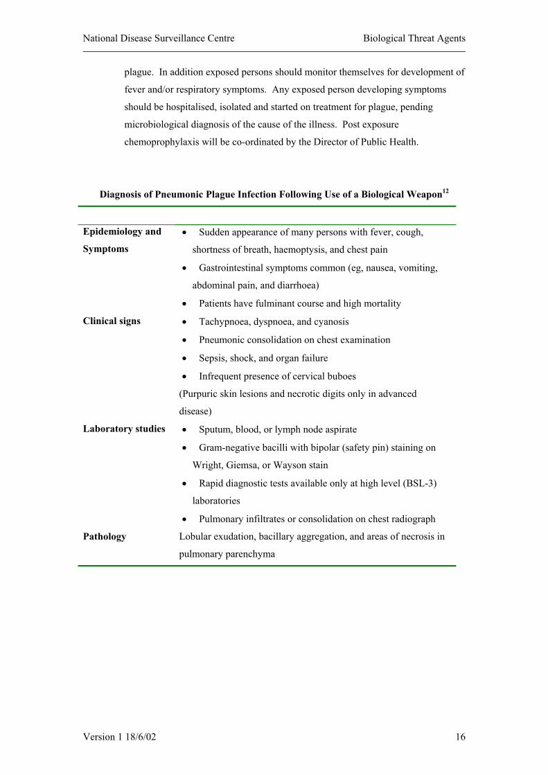
Diagnosis of Pneumonic Plague Infection Following Use of a Biological Weapon12
Epidemiology and
Symptoms
•
•
•
Sudden appearance of many persons with fever, cough,
shortness of breath, haemoptysis, and chest pain
Gastrointestinal symptoms common (eg, nausea, vomiting,
abdominal pain, and diarrhoea)
Patients have fulminant course and high mortality
Clinical signs •
•
•
•
•
•
•
•
Tachypnoea, dyspnoea, and cyanosis
Pneumonic consolidation on chest examination
Sepsis, shock, and organ failure
Infrequent presence of cervical buboes
(Purpuric skin lesions and necrotic digits only in advanced
disease)
Laboratory studies Sputum, blood, or lymph node aspirate
Gram-negative bacilli with bipolar (safety pin) staining on
Wright, Giemsa, or Wayson stain
Rapid diagnostic tests available only at high level (BSL-3)
laboratories
Pulmonary infiltrates or consolidation on chest radiograph
Pathology Lobular exudation, bacillary aggregation, and areas of necrosis in
pulmonary parenchyma
Version 1 18/6/02 16
National Disease Surveillance Centre Biological Threat Agents
Tularaemia Epidemiology
Tularaemia is a zoonosis caused by Francisella tularensis, a non-motile, aerobic,
Gram-negative cocco-bacillus.13 It is distributed world-wide, particularly in North America,
many parts of continental Europe, the former Soviet Union, China and Japan.14 Human-to
human transmission does not occur.15
F. tularensis is one of the most infectious pathogenic bacteria known, requiring inoculation or
inhalation of as few as 10 organisms to produce recognisable clinical disease. Tularaemia
(also known as rabbit, or deer fly fever) is acquired under natural conditions by inoculation of
the organism (taken from animal blood or tissue fluids) in the bites of deerfly, mosquitoes or
ticks. Less commonly, inhaling contaminated dusts or ingesting contaminated food or water
can also produce clinical disease. Fewer than 200 cases of tularaemia are reported annually in
the US, case fatality rates being in the order of 1-2%.
Clinical Features
Under normal conditions, tularaemia can present in a variety of ways – ulceroglandular,
glandular, oculoglandular, oropharyngeal, pneumonic, typhoidal and septic forms.
Ulceroglandular and typhoidal are commonest. Onset is usually sudden with fever and flu-
like symptoms (headache, chills and rigors, generalised myalgia, coryza and sore throat).
1. Ulceroglandular: inoculation of the organism through skin or mucous membranes.
Presents with lesions on the skin or mucous membranes (including the conjunctivae).
Lymphadenopathy is generally a prominent feature. Accounts for 75% of naturally
acquired cases.
2. Typhoidal: this form occurs mainly after inhalation of infectious aerosols accounts
for about 25% of naturally occurring cases. This form tends to present with features
of systemic disease; fever and collapse. Gastrointestinal symptoms can be prominent,
hence the confusing name.15 Untreated, mortality from this form of the disease can
range from 35-60%.15
Diagnosis
Diagnosis of tularaemia can be very difficult given the non-specificity of symptoms (See
Table below).
Version 1 18/6/02 17
National Disease Surveillance Centre Biological Threat Agents
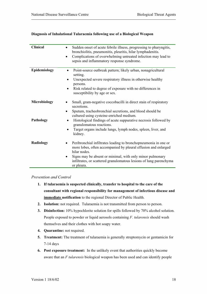
Diagnosis of Inhalational Tularaemia following use of a Biological Weapon
Clinical • Sudden onset of acute febrile illness, progressing to pharyngitis, bronchiolitis, pneumonitis, pleuritis, hilar lymphadenitis.
• Complications of overwhelming untreated infection may lead to sepsis and inflammatory response syndrome.
Epidemiology • Point-source outbreak pattern; likely urban, nonagricultural
setting. • Unexpected severe respiratory illness in otherwise healthy
persons. • Risk related to degree of exposure with no differences in
susceptibility by age or sex.
Microbiology • Small, gram-negative coccobacilli in direct stain of respiratory secretions.
• Sputum, tracheobronchial secretions, and blood should be cultured using cysteine-enriched medium.
Pathology • Histological findings of acute suppurative necrosis followed by granulomatous reactions.
• Target organs include lungs, lymph nodes, spleen, liver, and kidney.
Radiology • Peribronchial infiltrates leading to bronchopneumonia in one or
more lobes, often accompanied by pleural effusion and enlarged hilar nodes.
• Signs may be absent or minimal, with only minor pulmonary infiltrates, or scattered granulomatous lesions of lung parenchyma or pleura.
Prevention and Control
1. If tularaemia is suspected clinically, transfer to hospital to the care of the
consultant with regional responsibility for management of infectious disease and
immediate notification to the regional Director of Public Health.
2. Isolation: not required. Tularaemia is not transmitted from person to person.
3. Disinfection: 10% hypochlorite solution for spills followed by 70% alcohol solution.
People exposed to powder or liquid aerosols containing F. tularensis should wash
themselves and their clothes with hot soapy water.
4. Quarantine: not required.
5. Treatment: The treatment of tularaemia is generally streptomycin or gentamicin for
7-14 days
6. Post exposure treatment: In the unlikely event that authorities quickly become
aware that an F tularensis biological weapon has been used and can identify people
Version 1 18/6/02 18
National Disease Surveillance Centre Biological Threat Agents
during the early incubation period exposed persons should be treated with 14 days of
oral doxycycline or ciprofloxacin.
7. Healthcare Staff: managing suspected cases: universal precautions, prophylaxis to
laboratory staff who have been exposed to a potentially infective exposure (spill,
centrifuge accident, needlestick injury).
Useful Internet Links
National Disease Surveillance Centre www.ndsc.ie
Department of Health and Children www.doh.ie
Public Health Laboratory Service (UK) www.phls.co.uk
Centres for Disease Control, (Bioterrorism Section)
Atlanta, US
www.bt.cdc.gov
Version 1 18/6/02 19
National Disease Surveillance Centre Biological Threat Agents
Version 1 18/6/02 20
References
1. Freidlander AM. Anthrax. In: Medical Aspects of Chemical and Biological Warfare. Eds Frederick R. Sidell, Ernest T. Takafuji, David R. Franz. Uniformed Services University of the Health Sciences, Bethesda: 1997. 2. Slack RCB. Bacillus. In: Medical Microbiology. Eds Greenwood D, Slack RCB Peutherer J. 14th Edn. Churchill Livingstone, Edinburgh:1992. 3. Inglesby TV, Henderson DA, Bartlett JG, Ascher MS, Eitzen E, Friedlander AM, Hauer J, McDade J, Osterholm MT, O'Toole T, Parker G, Perl TM, Russell PK, Tonat K. Anthrax as a biological weapon: medical and public health management. Working Group on Civilian Biodefense. JAMA, 1999;281:1735-45. 4. Public Health Laboratory Service, London: 2001. 5. Pennington TH. Poxviruses. In: Medical Microbiology. Eds Greenwood D, Slack RCB Peutherer J. 14th Edn. Churchill Livingstone, Edinburgh:1992. 6. McClain DJ. Smallpox. In: Medical Aspects of Chemical and Biological Warfare. Eds Frederick R. Sidell, Ernest T. Takafuji, David R. Franz. Uniformed Services University of the Health Sciences, Bethesda: 1997. 7. Henderson DA, Inglesby TV, Bartlett JG, Ascher MS, Eitzen E, Jahrling PB, Hauer J, Layton M, McDade J, Osterholm MT, O'Toole T, Parker G, Perl T, Russell PK, Tonat K. Smallpox as a biological weapon: medical and public health management. Working Group on Civilian Biodefense. JAMA, 1999;281:2127-37. 8. Colee JG. Clostriodium. In: Medical Microbiology. Eds Greenwood D, Slack RCB Peutherer J. 14th Edn. Churchill Livingstone, Edinburgh:1992. 9. Middlebrook JL and Franz DR. Botulinium Toxins. In: Medical Aspects of Chemical and Biological Warfare. Eds Frederick R. Sidell, Ernest T. Takafuji, David R. Franz. Uniformed Services University of the Health Sciences, Bethesda: 1997. 10. Arnon SS, Schechter R, Inglesby TV, Henderson DA, Bartlett JG, Ascher MS, Eitzen E, Fine AD, Hauer J, Layton M, Lillibridge S, Osterholm MT, O'Toole T, Parker G, Perl TM, Russell PK, Swerdlow DL, Tonat K. Botulinum toxin as a biological weapon: medical and public health management. Working Group on Civilian Biodefense. JAMA, 2001; 285:1059-70. 11. McGovern TW, Freidlander AM. Plague. In: Medical Aspects of Chemical and Biological Warfare. Eds Frederick R. Sidell, Ernest T. Takafuji, David R. Franz. Uniformed Services University of the Health Sciences, Bethesda: 1997. 12. Inglesby TV, Dennis DT, Henderson DA, Bartlett JG, Ascher MS, Eitzen E, Fine AD, Friedlander AM, Hauer J, Koerner JF, Layton M, McDade J, Osterholm MT, O'Toole T, Parker G, Perl TM, Russell PK, Schoch-Spana M, Tonat K. Plague as a biological weapon: medical and public health management. Working Group on Civilian Biodefense. JAMA, 2000, 283:2281-90. 13. Evans ME and Freidlander AM. Tularemia. In: Medical Aspects of Chemical and Biological Warfare. Eds Frederick R. Sidell, Ernest T. Takafuji, David R. Franz. Uniformed Services University of the Health Sciences, Bethesda: 1997. ss 14. Chin J. Control of Communicable Diseases Manual. American Public Health Association, Washington: 2000. 15. Dennis DT, Inglesby TV, Henderson DA, Bartlett JG, Ascher MS, Eitzen E, Fine AD, Friedlander AM, Hauer J, Layton M, Lillibridge SR, McDade JE, Osterholm MT, O'Toole T, Parker G, Perl TM,
National Disease Surveillance Centre Biological Threat Agents
Version 1 18/6/02 21
Russell PK, Tonat K. Tularemia as a biological weapon: medical and public health management. JAMA. 2001 ;285:2763-73.