Biological Synthesis of Zinc Oxide Nanoparticles from Leaf … · Biological Synthesis of Zinc...
14
International Journal of Materials Science ISSN 0973-4589 Volume 12, Number 1 (2017), pp. 73-86 © Research India Publications http://www.ripublication.com Biological Synthesis of Zinc Oxide Nanoparticles from Leaf Extract of Curcuma neilgherrensis Wight G. Parthasarathy* P.hD, Research Scholar, Department of Electronics, Erode Arts and Science College (Autonomous), Erode-638 009, TamilNadu, India. Dr. M. Saroja Associate Professor of Electronics, Department of Electronics, Erode Arts and Science College (Autonomous), Erode-638 009, TamilNadu, India. Dr. M.Venkatachalam Head & Associate Professor of Electronics, Department of Electronics, Erode Arts and Science College (Autonomous), Erode-638 009, Tamil Nadu, India. Dr. V.K. Evanjelene R and D Manager, Alpha Omega Hi Tech Bio Research centre, Salem-636 008, TamilNadu, India. Abstract Biologically synthesized nanoparticles have been broadly using in the field of medicine. Research in nanotechnology highlights the possibility of green chemistry pathways to produce technologically important nanomaterials. In the present study, the objective was to study the synthesis and analyse the Zinc Oxide Nanoparticles (ZnO NPs) from Curcuma neilgherrensis leaf extract. The samples were characterized by Ultra-Violet Spectroscopy (UV), Fourier transform infrared spectroscopy (FTIR), Scanning Electron Microscopy (SEM), X-ray diffraction (XRD). These ZnO NPs were
Transcript of Biological Synthesis of Zinc Oxide Nanoparticles from Leaf … · Biological Synthesis of Zinc...
International Journal of Materials Science
ISSN 0973-4589 Volume 12, Number 1 (2017), pp. 73-86
© Research India Publications
http://www.ripublication.com
Biological Synthesis of Zinc Oxide Nanoparticles
from Leaf Extract of Curcuma neilgherrensis Wight
G. Parthasarathy*
P.hD, Research Scholar, Department of Electronics, Erode Arts and Science College (Autonomous),
Erode-638 009, TamilNadu, India.
Dr. M. Saroja
Associate Professor of Electronics, Department of Electronics, Erode Arts and Science College (Autonomous),
Erode-638 009, TamilNadu, India.
Dr. M.Venkatachalam
Head & Associate Professor of Electronics, Department of Electronics, Erode Arts and Science College (Autonomous),
Erode-638 009, Tamil Nadu, India.
Dr. V.K. Evanjelene
R and D Manager, Alpha Omega Hi Tech Bio Research centre,
Salem-636 008, TamilNadu, India.
Abstract
Biologically synthesized nanoparticles have been broadly using in the field
of medicine. Research in nanotechnology highlights the possibility of green
chemistry pathways to produce technologically important nanomaterials. In
the present study, the objective was to study the synthesis and analyse the
Zinc Oxide Nanoparticles (ZnO NPs) from Curcuma neilgherrensis leaf
extract. The samples were characterized by Ultra-Violet Spectroscopy
(UV), Fourier transform infrared spectroscopy (FTIR), Scanning Electron
Microscopy (SEM), X-ray diffraction (XRD). These ZnO NPs were
74 G. Parthasarathy, Dr. M. Saroja, Dr. M.Venkatachalam & Dr. V.K. Evanjelene
evaluated for antibacterial activity. The maximum diameter of inhibition
zones around the disk used for Pseudomonas aeruginosa indicates the
resistance to ZnO NPs followed by Escherichia coli. Among the five
bacterial species tested, the Pseudomonas aeuroginosa is more susceptible
when compared with other three species. It is concluded that the biological
synthesis of ZnO NPs is very rapid, effortless, cost effective and eco-
friendly and without any side effects and ZnO NPs may be used for the
preparation of antibacterial formulations against Pseudomonas aeuroginosa. Different functional groups were found to be present
signifying the presence of different compounds in the extract.
Keywords: Curcuma neilgherrensis, Zinc Oxide Nanoparticles,
Antibacterial activity, FTIR, SEM, UV, XRD.
INTRODUCTION
Nanotechnology is an imperative division in the most important fields of biology,
chemistry, physics and material sciences. Nanoparticles possess a wide array of
application in the different fields’ viz., drug, electronics, and therapeutics and as
analytical agents. The nanomaterials can be synthesized by different methods
including chemical, physical, irradiation and biological methods. The improvement of
new chemical or physical methods has resulted in environmental contaminations,
since the chemical procedures involved in the synthesis of nanomaterials generate a
large amount of hazardous byproducts. Thus, there is a need of “green synthesis” that
includes a clean, safe, eco-friendly and environmentally nontoxic method of
nanoparticle synthesis. Moreover in this process there is no need to use high pressure,
energy, temperature and toxic chemicals.
Among the metal oxide nanoparticles, zinc oxide is attention-grabbing because it has
enormous applications in various areas such as optical, piezoelectric, magnetic, and
gas sensing. Besides these properties, ZnO nanostructure exhibits high catalytic
efficiency, strong adsorption capability and are used more and more frequently in the
manufacture of sunscreens, ceramics and rubber processing, wastewater treatment,
and as a fungicide.
In modern years ZnO NPs have drawn attention of many researchers for their
distinctive optical and chemical behaviours which can be easily tuned by changing the
morphology. Within the large family of metal oxide NPs, ZnO NPs have been used in
diverse cutting edge applications like electronics, communication, sensors, cosmetics,
environmental protection, biology and the medicinal industry. Moreover, ZnO NPs
has a tremendous potential in biological applications like biological sensing,
biological labelling, gene delivery, drug delivery and nanomedicine5–8 along with its
Biological Synthesis of Zinc Oxide Nanoparticles from Leaf Extract of Curcuma neilgherrensis Wight 75
antibacterial, antifungal, acaricidal, pediculocidal, larvicidal and anti-diabetic
activities.
The major response concerned in the biosynthesis of ZnO nanoparticles mediated by
the leaf extract of Curcuma neilgherrensis is reduction/oxidation reaction where the
phytochemicals and enzymes present in biological resources take part for the
translation of metal compounds in to specific nanoparticles.
Curcuma neilgherrensis Wight (Family: Zingiberaceae); well-known as
“Kattukalvazhai” in Tamil is a folklore medicine widely used by the tribes of Western
Ghats for the management of diabetes mellitus. The traditional medicinal practitioners
of Kodagu District of Karnataka have recognized its usage in diabetes mellitus, even
so the Palliayar tribes of Tamilnadu are using its tuber for edible purposes. The leaf is
considered as the constructive part for counteracting the ailing effects of diabetes
mellitus. In spite of its reputation, it has not yet been investigated scientifically and
hence its leaves were contemplation worth to study in detail. The existing textual
information about the herb is very minimum and inadequate. C. neilgherrensis is a
herb with small conical rhizomes inside whitish in color, ending in root tubers,
fusiform. Leaves green, lanceolate/oblong - lanceolate in shape, 25cm in length and 8
cm wide. Inflorescence there in both lateral and central, long with a distinct coma.
Coma bracts are oblong- lanceolate, fused only at base, light to dark pink or violet in
colour. Fertile bracts are combined about lower 1/3, slightly curved, margin wavy,
green, green with a pink or violet spot at the tip, and densely pubescent. The
bracteoles are triangular in shape. Flowers are longer than the bracts, 3-4 in each
bract, and light yellow in colour. Calyx three lobed at apex, violet dotted, and densely
pubescent. Corolla tube light yellow in colour, lobes unequal, pubescent, hooded at
tip. Labellum shows a median cleft, yellow with a deep yellow median band.
MATERIALS AND METHODS
Extraction of the plant material
The fresh plant materials were washed with running tap water and shade dried. The
leaves of Curcuma neilgherrensis were crushed to coarsely powdered by grinder.
These coarse powders (25g) were then subjected to successive extraction in 250ml of
each solvent (methanol) by using Soxhlet apparatus. The collected extracts were
stored and then taken up for further investigations.
Phytochemical analysis
Preliminary phytochemicals analysis was carried out for all the Curcuma neilgherrensis extracts as per standard methods described by Brain and Turner 1975
and Evans 1996.
76 G. Parthasarathy, Dr. M. Saroja, Dr. M.Venkatachalam & Dr. V.K. Evanjelene
Detection of alkaloids
Curcuma neilgherrensis extracts were dissolved individually in dilute hydrochloric
acid and filtered. The filtrates were used to test the presence of alkaloids.
Detection of Flavonoids
a) Lead acetate test: Extracts were treated among few drops of lead acetate
solution. Formation of yellow colour precipitate indicates the presence of
flavonoids.
b) H2SO4 test: Extracts were treated with few drops of H2SO4. Formation of
orange colour indicate the presence of flavonoids.
Detection of Steroids
2ml of acetic anhydride was added to 0.5g of the extracts, each with 2ml of H2 SO4.
The colour changed from violet to blue or green in some samples indicate the
presence of steroids.
Detection of Terpenoids
Salkowski’s test
0.2g of the extract of the whole plant sample be assorted with 2ml of chloroform and
concentrated H2SO4 (3ml) was carefully added to form a layer. A reddish brown
coloration of the inner face was indicates the presence ofterpenoids.
Detection of Anthraquinones
About 0.2g of the leaf extract was boiled with 10% HCl for few minutes in a water
bath. It was filtered and allowed to cool. Equal volume of CHCl3 was added to the
filtrate. Few drops of 10% NH3 were added to the mixture and heated. Formation of
pink colour indicates the presence anthraquinones.
Detection of Phenols
a) Ferric chloride test: Leaf Extracts were treated with few drops of ferric
chloride solution. Formation of bluish black colour indicates the presence of
phenol.
Biological Synthesis of Zinc Oxide Nanoparticles from Leaf Extract of Curcuma neilgherrensis Wight 77
b) Lead acetate test: Leaf Extracts was treated with few drops of lead acetate
solution. Formation of yellow colour precipitate indicates the presence of
phenol.
Detection of Saponins
About 0.2g of the leaf extract was shaken with 5ml of distilled water. Formation of
frothing (appearance of creamy miss of small bubbles) shows the presence of
saponins.
Detection of Tannins
A small quantity of extract was assorted with water and heated on water bath. The
mixture was filtered and ferric chloride was added to the filtrate. A dark green colour
formation indicates the presence of tannins.
Detection of Carbohydrates
Extracts were dissolved separately in 5ml distilled water and filtered. The filtrate was
used to test the presence of carbohydrates.
Detection of Oils and Resins
Test solution was applied on filter paper. It develops a clear appearance on the filter
paper. It indicates the presence of oils and resins.
Antimicrobial activity
The disc diffusion method (Bauer et al., 1966) was used to display the antimicrobial
activity. In vitro antimicrobial activity was screened by via Muller Hinton Agar
(MHA) obtained from Hi-media (Mumbai). The MHA plates were prepared by
pouring 15 ml of molten media into sterilized petriplates. The plates were allowed to
coagulate for 5 minutes and 0.1% inoculums suspension was swabbed uniformly and
the inoculums be allowed to dry for 5 minutes. The concentration of extracts is 40
mg/disc was loaded on 6 mm sterile disc. The loaded disc was positioned on the
surface of medium and the extract was allowed to spread for 5 minutes and the plates
were kept for incubation at 370C for 24 hrs. At the end of incubation, inhibition zones
formed around the disc were measured with transparent ruler in millimeter.
78 G. Parthasarathy, Dr. M. Saroja, Dr. M.Venkatachalam & Dr. V.K. Evanjelene
Synthesis of ZnO Nano Particles
Preparation of zinc oxide NPs, for the synthesis of NPs, 50 ml of plant leaves extract
was taken and boiled at 600–800C by using a stirrer-heater. Then, 5 g of zinc nitrate
was added to the solution as the temperature reached at 600C. This mixture was then
boiled until it transformed to a deep yellow coloured suspension. This paste was then
collected in a ceramic crucible and heated in an air heated furnace at 600C for 2 h. A
light white coloured powder was obtained and this powder was carefully collected and
sent for different characterizations. The material was powered by a mortar and pestle
so, that got a fine powder, which is easy for further characterizations.
FT-IR Spectroscopy
Infrared light from suitable source passes through a scanning Michelson inferometer
and Fourier Transformation gives a plot of intensity versus frequency. When a
powdered plant sample is placed in the beam, it absorbs particular frequencies, so that
their intensities are reduced in the inferogram and the ensuing Fourier transform is the
infrared absorption spectrum of the sample.
Ultra- Violet Spectroscopy
The UV spectrum provides a useful means of detecting conjugated unsaturated
chromophores within a molecule such as polyenes, α, β-unsaturated ketones and
aromatic compounds. This can be particularly helpful in the identification of
chromophores and flavones. The UV spectrum may be caused by the summation of
chromophores from different parts of a polyfunctional molecule, and this should be
considered in the light of deduction drawn from other spectroscopic methods and
chemical degradation.
SEM analysis
Scanning electron microscopic (SEM) analysis was performed with the Hitachi S-
4500 SEM machine. Thin films of the sample were prepared on a carbon coated
copper grid by simply dropping a very minute amount of the sample on the grid, with
excess solution being removed using blotting paper. The film on the SEM grid was
then allowed to dry by putting the grids under a mercury lamp for 5 min.
XRD Analysis
ZnO nanoparticles were examined by X-ray diffractometer. The powdered metal was
sticked in the cubes of XRD and then the result was taken in the XRD equipment.
Biological Synthesis of Zinc Oxide Nanoparticles from Leaf Extract of Curcuma neilgherrensis Wight 79
RESULTS AND DISCUSSION
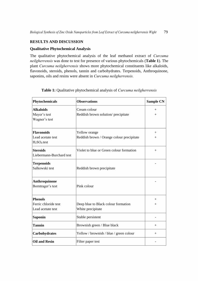
Qualitative Phytochemical Analysis
The qualitative phytochemical analysis of the leaf methanol extract of Curcuma neilgherrensis was done to test for presence of various phytochemicals (Table 1). The
plant Curcuma neilgherrensis shows more phytochemical constituents like alkaloids,
flavonoids, steroids, phenols, tannin and carbohydrates. Terpenoids, Anthroquinone,
saponins, oils and resins were absent in Curcuma neilgherrensis.
Table 1: Qualitative phytochemical analysis of Curcuma neilgherrensis
Phytochemicals Observations Sample CN
Alkaloids
Mayer’s test
Wagner’s test
Cream colour
Reddish brown solution/ precipitate
+
+
Flavonoids
Lead acetate test
H2SO4 test
Yellow orange
Reddish brown / Orange colour precipitate
+
+
Steroids
Liebermann-Burchard test
Violet to blue or Green colour formation +
Terpenoids
Salkowski test
Reddish brown precipitate
-
Anthroquinone
Borntrager’s test
Pink colour
-
Phenols
Ferric chloride test
Lead acetate test
Deep blue to Black colour formation
White precipitate
+
+
Saponin Stable persistent -
Tannin Brownish green / Blue black +
Carbohydrates Yellow / brownish / blue / green colour +
Oil and Resin Filter paper test -
80 G. Parthasarathy, Dr. M. Saroja, Dr. M.Venkatachalam & Dr. V.K. Evanjelene
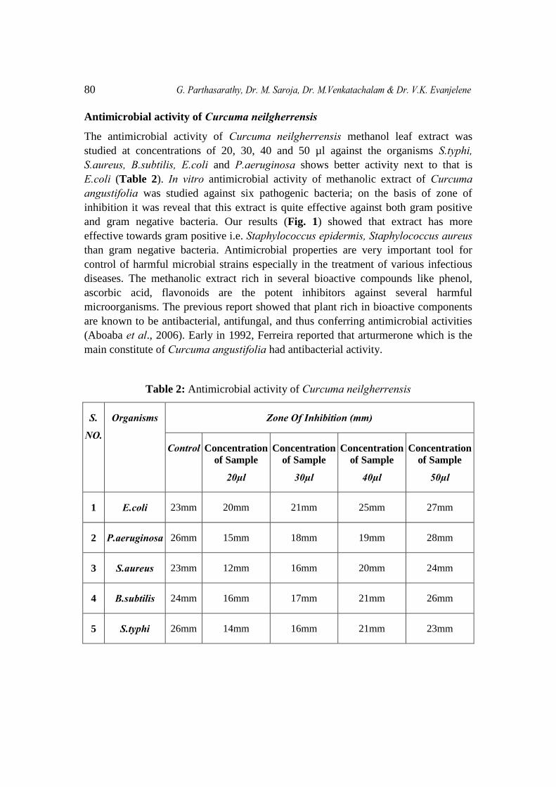
Antimicrobial activity of Curcuma neilgherrensis
The antimicrobial activity of Curcuma neilgherrensis methanol leaf extract was
studied at concentrations of 20, 30, 40 and 50 µl against the organisms S.typhi, S.aureus, B.subtilis, E.coli and P.aeruginosa shows better activity next to that is
E.coli (Table 2). In vitro antimicrobial activity of methanolic extract of Curcuma angustifolia was studied against six pathogenic bacteria; on the basis of zone of
inhibition it was reveal that this extract is quite effective against both gram positive
and gram negative bacteria. Our results (Fig. 1) showed that extract has more
effective towards gram positive i.e. Staphylococcus epidermis, Staphylococcus aureus
than gram negative bacteria. Antimicrobial properties are very important tool for
control of harmful microbial strains especially in the treatment of various infectious
diseases. The methanolic extract rich in several bioactive compounds like phenol,
ascorbic acid, flavonoids are the potent inhibitors against several harmful
microorganisms. The previous report showed that plant rich in bioactive components
are known to be antibacterial, antifungal, and thus conferring antimicrobial activities
(Aboaba et al., 2006). Early in 1992, Ferreira reported that arturmerone which is the
main constitute of Curcuma angustifolia had antibacterial activity.
Table 2: Antimicrobial activity of Curcuma neilgherrensis
S.
NO.
Organisms Zone Of Inhibition (mm)
Control Concentration
of Sample
20µl
Concentration
of Sample
30µl
Concentration
of Sample
40µl
Concentration
of Sample
50µl
1 E.coli 23mm 20mm 21mm 25mm 27mm
2 P.aeruginosa 26mm 15mm 18mm 19mm 28mm
3 S.aureus 23mm 12mm 16mm 20mm 24mm
4 B.subtilis 24mm 16mm 17mm 21mm 26mm
5 S.typhi 26mm 14mm 16mm 21mm 23mm
Biological Synthesis of Zinc Oxide Nanoparticles from Leaf Extract of Curcuma neilgherrensis Wight 81
Fig.1 (a) Fig.1 (b)
Fig.1 (c) Fig.1 (d)
Fig.1 (e)
Fig. 1: Antibacterial activity of ZnO Nanoparticles synthesised using 50ml of
Curcuma neilgherrensis extract against multiple pathogenic bacteria
82 G. Parthasarathy, Dr. M. Saroja, Dr. M.Venkatachalam & Dr. V.K. Evanjelene
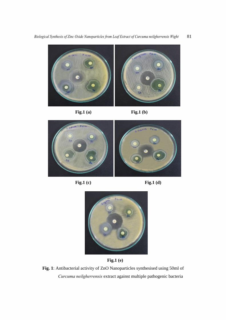
UV Spectrum of Curcuma neilgherrensis extract
Fig. 2: UV spectra of ZnO Nanoparticles synthesised using 50ml of
Curcuma neilgherrensis leaf extract
The UV analysis of the methanol extract synthesized zinc oxide nanoparticles of
Curcuma neilgherrensis leaf extract (Fig. 2). The maximum absorption peak was
obtained at 208 nm wavelength. And there are other peaks also which are 217 nm and
221 nm. The peak 208 nm is polyacetylenes which is Falcarinone, 217 nm is hydroxyl
coumarins and 221 nm is Phenolic acids that are salicylic acids.
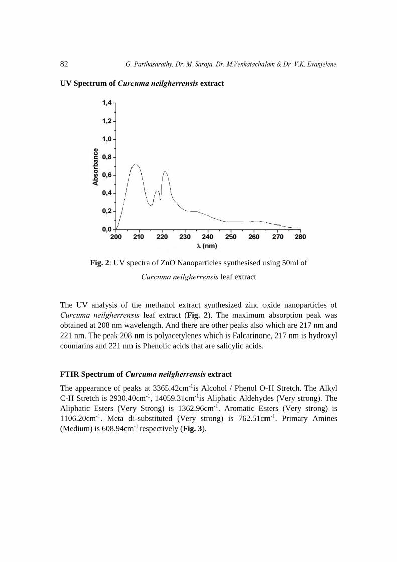
FTIR Spectrum of Curcuma neilgherrensis extract
The appearance of peaks at 3365.42cm-1is Alcohol / Phenol O-H Stretch. The Alkyl
C-H Stretch is 2930.40cm-1, 14059.31cm-1is Aliphatic Aldehydes (Very strong). The
Aliphatic Esters (Very Strong) is 1362.96cm-1. Aromatic Esters (Very strong) is
1106.20cm-1. Meta di-substituted (Very strong) is 762.51cm-1. Primary Amines
(Medium) is 608.94cm-1 respectively (Fig. 3).
Biological Synthesis of Zinc Oxide Nanoparticles from Leaf Extract of Curcuma neilgherrensis Wight 83
Fig. 3: FI-IR Spectra of ZnO Nanoparticles synthesised using 50ml of
Curcuma neilgherrensis leaf extract
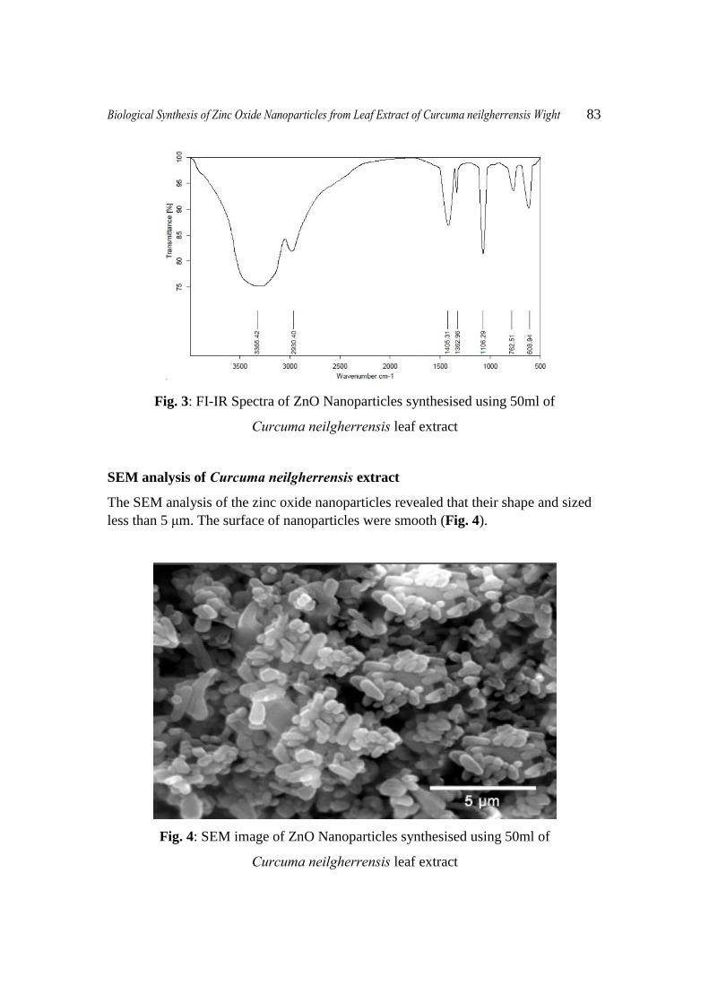
SEM analysis of Curcuma neilgherrensis extract
The SEM analysis of the zinc oxide nanoparticles revealed that their shape and sized
less than 5 μm. The surface of nanoparticles were smooth (Fig. 4).
Fig. 4: SEM image of ZnO Nanoparticles synthesised using 50ml of
Curcuma neilgherrensis leaf extract
84 G. Parthasarathy, Dr. M. Saroja, Dr. M.Venkatachalam & Dr. V.K. Evanjelene
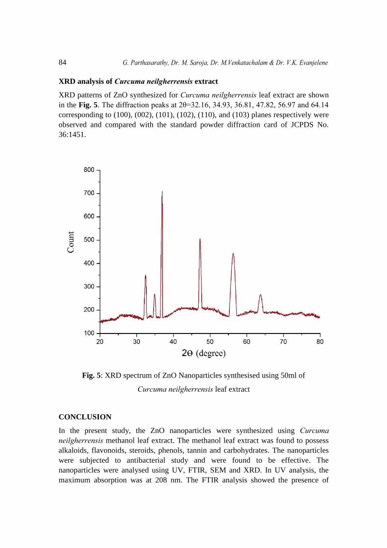
XRD analysis of Curcuma neilgherrensis extract
XRD patterns of ZnO synthesized for Curcuma neilgherrensis leaf extract are shown
in the Fig. 5. The diffraction peaks at 2θ=32.16, 34.93, 36.81, 47.82, 56.97 and 64.14
corresponding to (100), (002), (101), (102), (110), and (103) planes respectively were
observed and compared with the standard powder diffraction card of JCPDS No.
36:1451.
Fig. 5: XRD spectrum of ZnO Nanoparticles synthesised using 50ml of
Curcuma neilgherrensis leaf extract
CONCLUSION
In the present study, the ZnO nanoparticles were synthesized using Curcuma neilgherrensis methanol leaf extract. The methanol leaf extract was found to possess
alkaloids, flavonoids, steroids, phenols, tannin and carbohydrates. The nanoparticles
were subjected to antibacterial study and were found to be effective. The
nanoparticles were analysed using UV, FTIR, SEM and XRD. In UV analysis, the
maximum absorption was at 208 nm. The FTIR analysis showed the presence of
Biological Synthesis of Zinc Oxide Nanoparticles from Leaf Extract of Curcuma neilgherrensis Wight 85
Alcohol / Phenol O-H Stretch, Alkyl C-H Stretch, Aliphatic Aldehydes, Aliphatic
Esters, Aromatic Esters, Meta di-substituted and Primary Amines (Medium). The
SEM analysis revealed that the nanoparticles were of less than 5 μm shape. The size
of the nanoparticles are 3.34, 3.63, 5.08, 2.27, 1.47 and 1.56. From this study, it was
evident that the plant Curcuma neilgherrensis can be used to synthesize nanoparticles
using green chemistry methods for various applications.
FINANCIAL SUPPORT AND SPONSORSHIP:
Nil.
CONFLICT OF INTEREST:
There are no conflicts of interest.
ACKNOWLEDGEMENTS
The author is sincere thanks to the Managing Director of Alpha Omega Hi-tech Bio
Research Centre, Salem for supporting and providing the facility to work in the lab.
REFERENCES
[1] C. Dagdeviren, S. W. Hwang, Y. Su, S. Kim: Transient, Biocompatible
Electronics and Energy Harvesters Based on ZnO. Small 2013; 9(20): 3398–
3404.
[2] L. Wang, Y. Kang, X. Liu, S. Zhang: ZnO nanorod gas sensor for ethanol
detection. Sens. Actuators B 2012; 162(1): 237–243.
[3] S. E. Cross, B. Innes, M. S. Roberts, T. Tsuzuki: Human skin penetration of
sunscreen nanoparticles: in vitro assessment of a novel micronised zinc oxide
formulation. Skin Pharmacol. Physiol 2007; 20(3): 148–154.
[4] J. Zhou, N. Xu and Z. L. Wang: Dissolving behavior and stability of ZnO
wires in bioStuids: a study on biodegradability and biocompatibility of ZnO
nanostructures. Adv. Mater 2006; 18(18): 2432–2435.
[5] J. W. Rasmussen, E. Martinez, P. Louka and D. G. Wingett: Zinc oxide
nanoparticles for selective destruction of tumor cells and potential for drug
delivery applications. Expert Opin. Drug Delivery 2010; 7(9): 1063–1077.
[6] G. Applerot, A. Lipovsky, R. Dror and N. Perkas: Enhanced antibacterial
activity of nanocrystalline ZnO due to increased ROS-mediated cell injury,
Adv. Funct. Mater 2009; 19(6): 842–852.
86 G. Parthasarathy, Dr. M. Saroja, Dr. M.Venkatachalam & Dr. V.K. Evanjelene
[7] D. Sharmaa, J. Rajputa, B. S. Kaitha and M. Kaurb: Synthesis of ZnO
nanoparticles and study of their antibacterial and antifungal properties. Thin
Solid Films 2010; 519(3): 1224– 1229.
[8] S. Nair: Role of size scale of ZnO nanoparticles and microparticles on toxicity
toward bacteria and osteoblast cancer cells. J. Mater. Sci.: Mater. Med 2009;
20 Suppl. 1: S235–S241.
[9] V. Kirthi, A. A. Rahuman, G. Rajakumar, S. Marimuthu: Acaricidal,
pediculocidal and larvicidal activity of synthesized ZnO nanoparticles using
wet chemical route against blood feeding parasites. Parasitol. Res 2011; 109:
461–472.
[10] Alkaladi, A. M. Abdelazim and M.A: Antidiabetic Activity of Zinc Oxide and
Silver Nanoparticles on Streptozotocin-Induced Diabetic Rats. Int. J. Mol. Sci
2014; 15: 2015–2023.
[11] R. Seshadri, in: C.N.R. Rao, A. Muller, A.K. Cheetham (Eds.): The Chemistry
of Nanomaterials. Wiley-VCH Verlag GmbH, Weinheim 2004; 1: 94–112.
[12] L. Theodore, Nanotechnology: Basic Calculations for Engineers and
Scientists. Wiley, Hoboken; 2006.
[13] X. Wang, J. Lu, M. Xu, B. Xing: Sorption of pyrene by regular and
nanoscaled metal oxide particles influence of adsorbed organic matter.
Environmental Science and Technology 2008; 42: 7267–7272.
[14] J. Nicole, R. Binata, R.T. Koodali, M.C. Adhar: FEMS Microbiol. Lett 2008;
279: 71–76.
[15] Zhang M, Liu M, Prest H and Fischer’s: Nanoparticles secreted from iry
rootlets for surface climbing. Nanoletters 2008; 8: 1277 – 1280.
[16] Jeong S, Yeo S and Yis: Antibacterial characterization of silver nanoparticles
against E. coli ATCC-15224. Journal of Material Science 2005; 40: 5407.
[17] Savithramma N, Lingarao M, Rukmini K and Suvarnalatha DP: Antimicrobial
activity of silver nanoparticles synthesized by using medicinal plants.
International Journal of Chem Tech Research 2011; 3: 1394-1402.
[18] Aboaba OO, Smith SI, Olude FO: Antimicrobial effect of edible Plant extract
on Escherichia coli 0157: H7. Pak J Nutri 2006; 5(4): 325-327.
[19] Negi, P.S, Jayaprakasha, G.K. Rao, L.J.M. Sakariah, K.K: Antibacterial
activity of turmeric oil: A byproduct from curcumin manufacture. J of Agri
and Food Chem 1999; 47, 4297-4300.