Biological Approaches for Cartilage Repairorthodoc.aaos.org/WilliamFBennettMD/Biological Approaches...
9
Biological Approaches for Cartilage Repair Alberto Gobbi, MD Lyndon Bathan, MD ABSTRACT: The social impact of bone and cartilage pathologies entails high costs in terms of therapeutic treatments and loss of income. As a result, the current research trend includes preventive interventions and therapeutic solutions that can lead to an enhancement of tissue regeneration and the reduction of degenera- tive mechanisms. Many options have heen made available to address problems regarding cartilage damage, each with its own advantages and disadvantages. Several stud- ies are currently in progress to clarify some of the questions that remain unanswered about the long- term durability of these procedures and the possible modifications that can be made to achieve better re- sults. Biotechnology is progressing at a rapid pace that al- lows the introductions of several products for clinical application; however, randomized, prospective studies for these innovations should be conducted to validate the safety and efficacy of cartilage regeneration. [JKnee Surg. 2009;22:36-44J INTRODUCTION According to a study conducted by the World Health Organization, musculoskeletal injuries are the most common cause of severe long-term pain and disability, affecting millions of people worldwide.59 Accordingly, The authors arefrom the Orthopaedic Artliroscopic Surgery Interna- tional Bioresearch Foundation, Milan-, Italy. Correspondence: Alberto Gobbi, MD, Orthopaedic Arihroscopic Surgery International Bioresearch Foundation, Via Amadeo 24, 20133 Milano, Italy. 2000 to 2010 has been called the "decade of bone and joints" to launch global awareness and promote further research in the prevention, diagnosis, and treatment of joint injuries. The social impact of bone and cartilage pathologies entails high costs through therapeutic treatments and loss of income. In the United States, osteoarthritis medi- cines cost $5.31 billion in 2007, 4 and musculoskeletal conditions including osteoarthritis cost nearly $128 bil- lion per year in direct medical expenses (ie, total joint replacement procedures and loss of income and produc- tion in 2003). 5 For these reasons, the trend of the research is now going toward preventive interventions and therapeu- tic solutions that can lead to an enhancement of tissue regeneration and the reduction of degenerative mecha- nisms. CARTILAGE TREATMENT Hyaline cartilage combines a smooth surface and the ability to withstand an extreme amount of pressure. It is extremely important to reconstruct a perfect surface that will withstand heavy loads. Unfortunately, articular carti- lage lesions, with their inherent limited healing potential, remain a challenging problem for orthopedic surgeons. In the past few decades, surgeons often replaced the ar- ticular surface with expensive and sophisticated implants when articular lesions become full-blown osteoarthritis. However, recent studies have used ne\ orthobiological techniques in cartilage lesions with increasing frequency and effectiveness as a way to regenerate tissue homeo- stasis and delay the progression of osteoarthritis. Growth factors and mesenchymal stem cells have been used suc- cessfully in many medical fields, such as maxillofacial, cosmetic, spine, orthopedic, and general wound healing applications. 36 January 2009 / Vol 22 No 1
Transcript of Biological Approaches for Cartilage Repairorthodoc.aaos.org/WilliamFBennettMD/Biological Approaches...

Biological Approaches for Cartilage RepairAlberto Gobbi, MDLyndon Bathan, MD
ABSTRACT: The social impact of bone and cartilagepathologies entails high costs in terms of therapeutictreatments and loss of income. As a result, the currentresearch trend includes preventive interventions andtherapeutic solutions that can lead to an enhancementof tissue regeneration and the reduction of degenera-tive mechanisms.
Many options have heen made available to addressproblems regarding cartilage damage, each with itsown advantages and disadvantages. Several stud-ies are currently in progress to clarify some of thequestions that remain unanswered about the long-term durability of these procedures and the possiblemodifications that can be made to achieve better re-sults.
Biotechnology is progressing at a rapid pace that al-lows the introductions of several products for clinicalapplication; however, randomized, prospective studiesfor these innovations should be conducted to validatethe safety and efficacy of cartilage regeneration.
[JKnee Surg. 2009;22:36-44J
INTRODUCTION
According to a study conducted by the World HealthOrganization, musculoskeletal injuries are the mostcommon cause of severe long-term pain and disability,affecting millions of people worldwide.59 Accordingly,
The authors are from the Orthopaedic Artliroscopic Surgery Interna-tional Bioresearch Foundation, Milan-, Italy.
Correspondence: Alberto Gobbi, MD, Orthopaedic ArihroscopicSurgery International Bioresearch Foundation, Via Amadeo 24, 20133Milano, Italy.
2000 to 2010 has been called the "decade of bone andjoints" to launch global awareness and promote furtherresearch in the prevention, diagnosis, and treatment ofjoint injuries.
The social impact of bone and cartilage pathologiesentails high costs through therapeutic treatments andloss of income. In the United States, osteoarthritis medi-cines cost $5.31 billion in 2007,4 and musculoskeletalconditions including osteoarthritis cost nearly $128 bil-lion per year in direct medical expenses (ie, total jointreplacement procedures and loss of income and produc-tion in 2003).5
For these reasons, the trend of the research is nowgoing toward preventive interventions and therapeu-tic solutions that can lead to an enhancement of tissueregeneration and the reduction of degenerative mecha-nisms.
CARTILAGE TREATMENT
Hyaline cartilage combines a smooth surface and theability to withstand an extreme amount of pressure. It isextremely important to reconstruct a perfect surface thatwill withstand heavy loads. Unfortunately, articular carti-lage lesions, with their inherent limited healing potential,remain a challenging problem for orthopedic surgeons.In the past few decades, surgeons often replaced the ar-ticular surface with expensive and sophisticated implantswhen articular lesions become full-blown osteoarthritis.However, recent studies have used ne\ orthobiologicaltechniques in cartilage lesions with increasing frequencyand effectiveness as a way to regenerate tissue homeo-stasis and delay the progression of osteoarthritis. Growthfactors and mesenchymal stem cells have been used suc-cessfully in many medical fields, such as maxillofacial,cosmetic, spine, orthopedic, and general wound healingapplications.
36 January 2009 / Vol 22 No 1
Biological Approaches for Cartilage Repair
CONSERVATIVE TREATMENT ANDPREVENTIVE BIOLOGIC SOLUTION
Nonsurgical treatment of cartilage lesions, includingdiet, intra-articular injections, and rehabilitation were rele-gated to pain control and activity modifications. Recent stud-ies on pulsed electromagnetic fields and platelet-rich plasmainjections have shown that these methods have the capacityto help heal cartilage tissue and delay osteoarthritis.
A recent study by Focht et al19 analyzed 316 adultswith obesity who underwent an arthritis, diet, and activitypromotion trial. This 18-month single-blind, randomizedcontrolled trial, compared the effects of exercise alone,dietary weight loss alone, a combination of exercise plusdietary weight loss, and a healthy lifestyle control inter-vention. The study concluded that exercise and dietaryweight loss, compared with the healthy lifestyle controlintervention, resulted in improved mobility-related self-efficacy and pain reduction.19
In an animal study, Ciombor et al13 showed that pulsedelectromagnetic fields preserved the morphology of ar-ticular cartilage and retarded the development of osteo-arthritic lesions in Hartley guinea pigs compared with acontrol group. The study concluded that pulsed electro-magnetic fields were disease modifying in this population.This supported a previous in vitro study of human chon-drocytes that showed increased cell proliferation with ex-posure to pulsed electromagnetic fields.16 The study notedthat electric and electromagnetic fields increased gene ex-pression and synthesis of growth factors, and that this mayamplify field effects through autocrine and paracrine sig-naling. Electric and electromagnetic fields may producea sustained regulation of growth factors that enhance, butdo not disorganize, endochondral bone formation.1
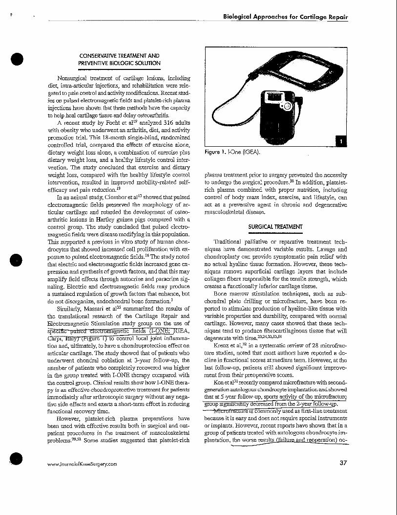
Similarly, Massari et al35 summarized the results ofthe translational research of the Cartilage Repair andElectromagnetic Stimulation study group on the use ofspecific puked electromagnetic fields (I-ONlJ; ICibA,Carpi, ltaiy,r(jtngure I) to control local joint inflamma-tion and, ultimately, to have a chondroprotection effect onarticular cartilage. The study showed that of patients whounderwent chondral coblation at 3-year follow-up, thenumber of patients who completely recovered was higherin the group treated with I-ONE therapy compared withthe control group. Clinical results show how I-ONE thera-py is an effective chondroprotective treatment for patientsimmediately after arthroscopic surgery without any nega-tive side effects and exerts a short-term effect in reducingfunctional recovery time.
However, platelet-rich plasma preparations havebeen used with effective results both in surgical and out-patient procedures in the treatment of muscoloskeletalproblems.29-32 Some studies suggested that platelet-rich
Figure 1.1-One (IGEA).
plasma treatment prior to surgery prevented the necessityto undergo the surgical procedure.38 In addition, platelet-rich plasma combined with proper nutrition, includingcontrol of body mass index, exercise, and lifestyle, canact as a preventive agent in chronic and degenerativemusculoskeletal disease.
SURGICAL TREATMENT
Traditional palliative or reparative treatment tech-niques have demonstrated variable results. Lavage andchondroplasty can provide symptomatic pain relief withno actual hyaline tissue formation. However, these tech-niques remove superficial cartilage layers that includecollagen fibers responsible for the tensile strength, whichcreates a functionally inferior cartilage tissue.
Bone marrow stimulation techniques, such as sub-chondral plate drilling or microfracture, have been re-ported to stimulate production of hyaline-like tissue withvariable properties and durability, compared with normalcartilage. However, many cases showed that these tech-niques tend to produce fibrocartilaginous tissue that willdegenerate with time.23124'32^3154
Kreuz et al,32 in a systematic review of 28 microfrac-ture studies, noted that most authors have reported a de-cline in functional scores at medium term. However, at thelast follow-up, patients still showed significant improve-ment from their preoperative scores.
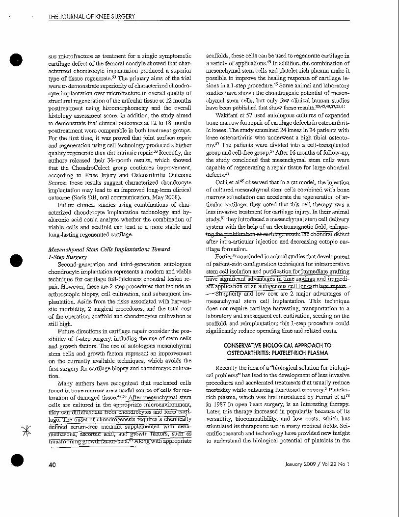
Kon et al31 recently compared microfracture with second-generation autologous chondrocyte implantation, and showedthat at 5-year follow-up, sports activity of the microfracturegroup sigmncantly decreased trom the 2-3'ear follow-up.
Miciulraecure is commonly used as first-line treatmentbecause it is easy and does not require special instrumentsor implants. However, recent reports have shown that in agroup of patients treated with autologous chondrocyte im-plantation, the worse results ffim'lnre and reoperation) oc-
wwwJournalofKneeSurgery.com 37

THE JOURNAL OF KNEE SURGERY
Figure 2. Open second-generation autologous chondrocyteimplantation.
curred in patients previously treated with microfracture,compared With patients treated, with autologous"T5tondro-cyte implantation as first-line surgery.32
" Usteoctiondral autologous transplantation and mosa-icplasty can restore normal cartilage tissue, but the ap-plication is restricted to small defects and there are someconcerns about donor-site morbidity.28
First introduced by Peterson,10 autologous chondro-cyte implantation has been proven capable of restoringhyaline cartilage tissue. Recent studies41-46-47 suggestedthe durability of this treatment, especially at long-termfollow-up, primarily due to its ability to produce hyaline-like cartilage that is mechanically and functionally stable.Autologous chondrocyte implantation also allows inte-gration with the adjacent articular surface. However, thismethod requires 2 surgical procedures and showed localmorbidity for periostea! harvest11
The complexity of the Peterson periosteal techniqueand the possible complication of periosteal patch hyper-trophy prompted surgeons to seek alternative techniquesto enhance cell delivery and outcome.
At present, the most promising technique is tissue en-gineering, in which cells are combined with scaffolds topreform a tissue; in general, the concept involves culturedautogenous chondrocytes integrated in biodegradable andbiocompatible scaffolds. After the chondrocytes are culti-vated and seeded on the scaffold, they must reacquire andmaintain their chondrogenic phenotype to synthesize anextracellular matrix containing type II collagen, glycos-arnlnoglycans, and proteoglycans, all of which are neces-sary to produce hyaline cartilage.
SECOND-GENERATION AUTOLOGOUSCHONDROCYTE IMPLANTATION
The ideal scaffold should be biocompatible, biode-gradable, not trigger any inflammatory response, and not
Figure 3. Arthroscopic second-generation autologous chon-drocyte implantation.
cytotoxic. It should offer a temporary support to cells topromote replacement of a newly synthesized matrix andpossibly induce proliferation of the transplanted cells. Thematrix should also be permeable to nutrients and providefirm adhesion to the surrounding cartilage wound edgesto promote integration. In addition, the scaffold must bereproducible and readily available, as well as versatile forrepair and resurfacing.12^5'42^5
After a systematic review of the available literatureabout different scaffolds in the market, which includesporcine collagen I and HI (ie, membrane-seeded autolo-^goTis^chondrocyte implantation [Genzyme Europe BY.Namderi, me .Netherlands]), three-dimensional bovine^•lullagan tiNleoCart; Histogenics. Waltharn Mas<;)j ^nripoTyactic acid and polyglycolic acid fletrp (Rioserrl,—BangkoK, Thailand), we concluded that a hyaluronan-based scaffold may be optimal for chondrocyte proliferq.-flonT" "
Among several other proteins, hyaluronan is a natu-rally occurring and highly conserved glycosaminoglycanwidely distributed in the body. It has proven to be an idealmolecule for tissue engineering strategies in cartilagerepair because of its impressive multifunctional activ-ity through its structural and biological role. The three-dimensional nonwoven scaffold, HYAFF (Fidia Farma-ceutici, Padova, Italy), supports the in vitro growtlTofhighly viable chonorocytes and promotes the expressionof the original chondrogenic phenotype,12 Chondrocytesthat were previously expanded on plastic and seeded intothe scaffold produce a characteristic extracellular matrixrich in proteoglycans and express typical markers of hya-line cartilage, such as collagen II and aggrecan.25 Whenimplanted in full-thickness defects of the femoral condylein rabbits, chondrocytes regenerated a cartilage-like tis-sue.26,55
38 January 2009 / Vol 22 No 1

Biological Approaches for Cartilage Repair
The main indications for second-generation cartilagetransplantation are symptomatic focal, fall-thickness car-tilage lesions (ie, International Cartilage Repair Societygrades HI to IV) in the absence of significant arthritis inphysiologically young patients (between ages 15 and 50).Additional factors to consider include the patient's moti-vation and willingness to comply with the postimplanta-tion rehabilitation regimen. Defect sizes (range, 2-12 cm2)have been shown to be favorable to regeneration. Osteo-chondritis dissecans is not a contraindication for cartilagetransplantation as long as the bone loss is not >8 mm.47
Adverse events of second-generation autologouschondrocyte implantation are apparently lower than firstgeneration, as reported by Mandelbaum.33
Several reports from controlled trials in patients un-dergoing surgery with the use of these hyaluronic acidscaffolds have been presented.21-2234'42 However, the larg-est collection of data using the hyaluronic acid scaffold inclinical practice is represented by a multicenter observa-tional study conducted in Italian Orthopedic Centers since2001.21'22-34
Autologous chondrocyte implantation can be per-formed through the conventional arthrotomy approach.However, recent advances in scaffold technology hasenabled surgeons to perform this technique arthroscopi-cally (Figures 2 and S).34 Some technical limitations pre-vail, including treatment of patellar lesions and posteriorportions of femoral condyles or the tibial plateau. Theselimits are common to all arthroscopic techniques andcould partly be resolved with the development of new ar-throscopic tools.
In a prospective, nonrandomized study22 on patello-femoral lesions treated with second-generation anfnln-gous ciiondrocyte implantation, we analyzed a group ofpatients at the 5-}'ear follow-up, using International KneeDocumentation Committee (IKDC) subjective and objec-tive scores, the EuroQoL pain scale, and Tegner scores.The authors noted significant improvement from preop-erative scores to the final follow-up. Objective preopera-tive data improved from 8 of 34 (23.5%) patients classi-fied as IKDC A or B scores to 31 of 34 (91.2%) classifiedIKDC A or B scores at the 5-year follow-up. Mean subjec-tive scores improved from 46.09 points preoperatively to70.39 points 5 years postoperatively, and Tegner scoresimproved from 2.56 to 4.68. EuroQol visual analog scalescores improved from 54.81 to 78.24.
In another study, we compared second-generationautologous chondrocyte implantation with microfractureand found a higher improvement in the IKDC objectiveand subjective scores in the group treated with second-generation autologous chondrocyte implantation at the5-year follow-up.31 Analyzing the resumption of sportsactivity obtained with the Tegner score, we observed sim-
(marker analysis! f cell expansion J
Figure4. ChondroCelect (TiGenix NV) scientific monogram.Reprinted with permission from TiGenix NV.
ilar level in both groups at the 2-year follow-up, whichremained stable at the 5-year follow-up in the second-generation autologous chondrocyte implantation group,whereas return to sport activities worsened in the micro-fracture group.31
FUTURE IMPLICATIONS
Third-Generation Autologous ChondrocyteImplantation
Promising results have been shown with autologouschondrocyte implantation. Autologous chondrocyte im-plantation is a technology that involves the implantationof an expanded chondrocyte population derived from acartilage biopsy. These expansions result in the loss ofphenotypic traits,'also called dedijferennanon.7 This pro-duces cnondrocytes with a decreased capacity to regener-ate hyaline cartilage cells.
" uharactenzed chondrocyte implantation is a newgeneration of autologous chondrocyte implantationprocedures that uses ChondroCelect (TiGenix NV,Haasrode, Belgium). CfTondroCelect was developed tolimit the loss of phenotype and is an. expanded popula-tion ot cnondrocyte with a proven ability to producestable cartilage in vivo (Figure 4). The highly con-trolled and. consistent manutactunng process is basedon the expression of a marker profile to characterizethis cell population.
A recent prospective, randomized controlled trial thatcompared characterized chondrocyte implantation ver-
wwwJournalofKneeSurgery.com 39
THE JOURNAL OF KNEE SURGERY
sus microfracture as treatment for a single symptomaticcartilage defect of the femoral condyle showed that char-acterized chondrocyte implantation produced a superiortype of tissue regenerate.33 The primary aims of the trialwere to demonstrate superiority of characterized chondro-cyte implantation over microfracture in overall quality ofstructural regeneration of the articular tissue at 12 monthsposttreatment using histomorphometry and the overallhistology assessment score. In addition, the study aimedto demonstrate that clinical outcomes at 12 to 18 monthsposttreatment were comparable in both treatment groups.For the first time, it was proved that joint surface repairand regeneration using cell technology produced a higherquality regenerate than did intrinsic repair.53 Recently, theauthors released their 36-month results, which showedthat the ChondroCelect group continues improvement,according to Knee Injury and Osteoarthritis OutcomeScores; these results suggest characterized chondrocyteimplantation may lead to an improved long-term clinicaloutcome (Saris DB, oral communication, May 2008).
Future clinical studies using combinations of char-acterized chondrocyte implantation technology and hy-aluronic acid could analyze whether the combination ofviable cells and scaffold can lead to a more stable andlong-lasting regenerated cartilage.
Mesenchymal Stem Cells Implantation: Toward1-Step Surgery
Second-generation and third-generation autologouschondrocyte implantation represents a modem and viabletechnique for cartilage full-thickness chondral lesion re-pair. However, these are 2-step procedures that include anarthroscopic biopsy, cell cultivation, and subsequent im-plantation. Aside from the risks associated with harvest-site morbidity, 2 surgical procedures, and the total costof the operation, scaffold and chondrocytes cultivation isstill high.
Future directions in cartilage repair consider the pos-sibility of 1-step surgery, including the use of stem cellsand growth factors. The use of autologous mesenchymalstem cells and growth factors represent an improvementon the currently available techniques, which avoids thefirst surgery for cartilage biopsy and chondrocyte cultiva-tion.
Many authors have recognized that nucleated cellsfound in bone marrow are a useful source of cells for res-toration of damaged tissue.49-S8_After meseuchymal stemcells are cultured in the appropriate microenvironment,they can diiierentiate trom cnondrocytes and form carti-lage, me onset of chondrogenesis requires a chemicallydenned serum-free medium supplemented with dexa-tnetnasone, ascoroic acid, and growth ractors, such astransromuug giowdl faciui-beia/' Along with appropriate
scaffolds, these cells can be used to regenerate cartilage ina variety of applications.49 In addition, the combination ofmesenchymal stem cells and platelet-rich plasma make itpossible to improve the healing response of cartilage le-sions in a 1-step procedure.45 Some animal and laboratorystudies have shown the chondrogenic potential of mesen-chymal stem cells, but only few clinical human studieshave been published that show these results.20'45'49-57-58-61
Wakitani et 57 used autologous cultures of expandedbone marrow for repair of cartilage defects in osteoarthrit-ic knees. The study examined 24 knees in 24 patients withknee osteoarthritis who underwent a high tibial osteoto-my.57 The patients were divided into a cell-transplantedgroup and cell-free group.57 After 16 months of follow-up,the study concluded that mesenchymal stem cells werecapable of regenerating a repair tissue for large chondraldefects.57
Ochi et al45 observed that in a rat model, the injectionof cultured mesenchymal stem cells combined with bonemarrow stimulation can accelerate the regeneration of ar-ticular cartilage; they noted that this cell therapy was aless invasive treatment for cartilage injury. In their animalstudy,61 they introduced a mesenchymal stem cell deliverysystem with the help of an electromagnetic field, enhanc-
•fav, trip prnlifpntion of cartilcige uuJ Jti Ihe chundral defectafter intra-articular injection and decreasing ectopic car-tilage formation.
Fortier20 concluded in animal studies that developmentof patient-side configuration techniques for intraoperativestem cell isolation and purification for immediate grafting
Have significant advantages in time savings and immedi-ate application of an autogenous cell fc
dfnplicity and low cost are 2 major advantages ofmesenchymal stem cell implantation. This techniquedoes not require cartilage harvesting, transportation to alaboratory and subsequent cell cultivation, seeding on thescaffold, and reimplantation; this 1-step procedure couldsignificantly reduce operating time and related costs.
CONSERVATIVE BIOLOGICAL APPROACH TO
OSTEOARTHRITIS: PLATELET-RICH PLASMA
Recently the idea of a "biological solution for biologi-cal problems" has lead to the development of less invasiveprocedures and accelerated treatments that usually reducemorbidity while enhancing functional recovery.3 Platelet-rich plasma, which was first introduced by Ferrari et al18
in 1987 in open heart surgery, is an interesting therapy.Later, this therapy increased in popularity because of itsversatility, biocompatibility, and low costs, which hasstimulated its therapeutic use in many medical fields. Sci-entific research and technology have provided new insightto understand the biological potential of platelets in the
40 January 2009 / Vol 22 No 1

Biological Approaches for Cartilage Repair
Figure 5. Centrigel (ReGenTHT Regen PRP Kit; ReGenLabSA, Mollens, Switzerland).
wound-healing process.3-52 It is well known that plateletshave many functions beyond simple hemostasis plateletscontain many important bioactive proteins and growthfactors, such as platelet-derived growth factor, insulin-like growth factor, transforming growth factor, epidermalgrowth factor, fibroblast growth factor, and vascular endo-thelial growth factor. These factors, if secreted, regulatekey processes involved in tissue repair, including cell pro-liferation, chemotaxis, migration, cellular differentiation,and extracellular matrix synthesis.6-39
The rationale for topical use of platelet-enriched prepa-rations is to stimulate the natural healing cascade and tissueregeneration by a supraphysiological release of platelet-derived factors directly in the site of treatment. Several sys-tems are available to prepare the platelet-rich plasma and theplatelet gel. Through these systems, a 2- to 6-fold enrichmentof platelet concentration is obtained to achieve comparablegrowth factor enrichment (Figures 5 and 6). The amount ofgrowth factor available to the tissue healing depends on thegrowth factors actually stored in platelets and the kinetics ofthe adsorption of the platelet gel.17 In addition, evidence ismounting to show that the crucial factors in the effectivenessof this treatment are the number of platelets used (ie, 5=1.2to 2.0X ICP/mL in platelet-rich plasma) and the way in whichthey are processed, as the growth factor content of the gel ishighly sensitive to these 2 variables.8-9-36-37
Recent studies2-15-29-40 have documented the effec-tiveness of growth factors in chondrogenesis and in pre-venting degeneration of the joints. Injarricular. Kon29
sfutfed~3tr patients with symptomatic degenerative dis-ease of the knee joints treated with 3 platelet-rich plasmamtra-articular injections weekly. The 6-month follow-upsEowBd~posirive effects on function and symptoms wiQ\n improvement of 85% in scores evaluated for pa-
tients with median age <60 years, whereas in patients
Figure 6. Harvest SmartPReP 2 APC60 Process Kit (HarvestTechnologies, Plymouth, Mass).
>60 years, the improvement shown was only 30%. Na-kagawa et al40 demonstrated the efficacy of autologpusplatelet-rich plasma in stimulating the proliferation andcollagen syntnesis of human chondrocytes, suggestingthe use of this method in the treatment of cartilage de-fects. Amnia ei aF showed that an intra-articular injec-titm"oFplateiet-rich plasma could induce an increase inproduction of hyaluronic acid structure and promote•angiogenesis and cell proliferation. Cugat et al" usedplatelet-rich growth factors to treat cfaonorat aetect inathletes and obtained good results. According to theirexperiences witn ottier connective tissue repair, theyshowed that platelet-rich growth factors in physiologicalconcentration are effective for the recovery of connectivetissue. In addition, local treatment is safe and does notalter the systemic concentrations of these proteins. Theyalso had good results using platelet-rich growth, factors inarticular cartilage lesion treatment and presented the fol-lowing positive results: platelet-rich growth factors canincrease total glycosaminoglycansjsollagen II synthesis'and can deiima&d Uegtadation. In addition, platelet-richgrowth factors induce chondrogenesis of mesenchymalstem uclls aud promote chondrocyte proliferation, dif-ferentiation, and adhesion.' ' "
\vwwJournalofKneeSurgery.com 41

THE JOURNAL OF KNEE SURGERY
TABLEREHABILITATION PROTOCOLPhase Objective Criteria to Progress
Phase 1: cartilage protection andrecovery of walk
Phase 2: cartilage transition andrecovery of running
Phase 3: cartilage maturation andathletic recovery
Phase 4: cartilage turnover andmaintenance
Protect the transplant; decrease painand effusion; increase range of move-ment; delay muscle atrophy
Return to a correct running pathway
Sustain high loads and impact activi-ties; prepare athlete for a return tocompetition with good recoveryof aerobic endurance; recovery ofsports-specific skills; stimulate carti-lage tissue remodeling
Maintain a good quality of life andgood physical condition; avoid excessbody tat; prevent risk of reinjury;return to team and competitions
Full active knee extension; knee flexion>120°; no swelling; no pain during weightbearing; recovery of correct walk pattern;adequate muscle recruitment [ie, quadriceps)
Running without pain or swelling at 8 km/hfor 15 feet; adequate recovery of coordinationand neuromuscular control; >80% recovery ofstrength in the contralateral limb; >80% single-leg hop test in the contralateral limb
Can ascend and descend stairs and, for ath-letes, running without pain or effusion at 10km/h for 15 feet and without a significantincrease of blood lactic acid concentrationabove resting values; recovery of sports-specific skills
In an experimental study done on animals, Wu et al60
showed the effectiveness of intra-articular injections ofplatelet-rich plasma with chondrocytes grown in vivo thatresulted in the formation of new cartilage tissue.
Finally, as observed by Nishimoto et al,43 we believethat simultaneous concentration of platelets and bonemarrow cells, acting as a sources of growth factors and"working cells," could play important roles in future re-generative medicine.
REHABILITATION PROGRAM AFTERCARTILAGE TRANSPLANTATION
The importance of rehabilitation after cartilage trans-plantation cannot be overemphasized. These protocols arean important part of the success of cartilage regenerationstudies in Italy. A standardized postoperative functionalrehabilitation protocol is adopted based on current knowl-edge of the biology of graft healing and on functional cri-teria and therapy goal progression.48-56 Patients will prog-ress through 4 rehabilitation phases51:• Phase 1: Protection of the transplant and. the recovery
of normal gait• Phase 2: Recovery of a correct run.• Phase 3: Recovery of sports-specific skills.• Phase 4: Maintenance of the physical fitness attained
during rehabilitation and prevention of the risk of re-injury.Progression between phases is decided accord-
ing to the achievement of specific criteria (Table),
with particular care to avoid swelling and pain in thejoint.14-27-44-50
CONCLUSION
A biological approach to cartilage lesions is a new chal-lenge. A number of viable options have been made availableover the years to address problems concerning cartilage dam-age, and each technique has its advantages and disadvan-tages. Numerous studies are currently in progress to clarifysome of the questions that still remain unanswered regardingthe long-term durability of these procedures and the possiblemodifications that can still be done to achieve better results.
Biotechnology is progressing at a rapid pace, explor-ing new horizons and allowing the introduction of numer-ous products for clinical application. However, carefullyconducted randomized, prospective studies for each ofthese innovations should be conducted to validate thesafety and efficacy of cartilage regeneration.
ACKNOWLEDGMENT
The authors thank Dr Lorenzo Boldriiu from the Iso-kinetic Rehabilitation Network, Milan, Italy, for his con-tributions to the rehabilitation program and the platelet-rich plasma sections of this article.
REFERENCES
1. Aaron RK, Boyan BD, Ciombor DM, Schwartz Z, SimonBJ. Stimulation of growth factor synthesis by electric and
42 January 2009 / Vol 22 No 1

Biological Approaches for Cartilage Repair
electromagnetic fields. Clin Orthop. 2004;(419):30-37.2. Anitua E, Sanchez M, Nurden AT, et al. Platelet-released
growth factors enhance the secretion of hyaluronic acidand induce hepatocyte growth factor production by syno-•vial fibroblasts from arthritic patients. Rheumatology (Ox-ford). 2007;46:1769-1772.
3. Anitua E, Sanchez M, (Drive G, Andfa I. The potential im-pact of the preparation rich in growth factors (PRGF) indifferent medical fields. Biomaterials. 2007^8:4551-4560.
4. Arnold M, Comer B, McGuire S. The pharma report: Top20 pharma companies. Medical Marketing & Media. May1, 2008:45-55.
5. Arthritis Foundation. Arthritis Foundation Web site.http:/Avww.arthritis.org/cost-arthritis.php. Accessed May27, 2008.
6. Bennett NT, Schultz GS. Growth factors and wound heal-ing: Biochemical properties of growth factors and theirreceptors.Am JSurg. 1993;165:728-737.
7. Benya, PD, Shaffer JD. Dedifferentiated chondrocytesreexpress the differentiated collagen phenotype when cul-tured .in agarose gels. Cell. 1982;30:215-224.
8. Borzini P, Mazzucco L. Platelet gels and releasates. CurrOpin Haematol. 2005;12:473-479.
9. Borzini P, Mazzucco L. Tissue regeneration and in-locoadministration of platelet derivatives. Clinical outcome,heterogeneous products, heterogeneity of the effect ormechanisms. Transfusion. 2005;45:1759-1767.
10. Britfberg M, Lindahl A, Nilsson A, Ohlsson C, IsakssonO, Peterson L. Treatment of deep cartilage defects in theknee with autologous chondrocyte transplantation. NEnglJMed. 1994;33 1:889-895.
11. Brittberg M, Peterson L, Sjogren-Jansson E, Tallheden T,Lindahl A. Articular cartilage engineering with autologouschondrocyte transplantation. A review of recent develop-ments. J Bone Joint Surg Am. 2003;85(suppl3):109-115.
12. Brun P, Abatangelo G, Radice M, et al. Chondrocyte ag-gregation and reorganization into three-dimensional scaf-folds. J Biomed Mater Res. 1999;46:337-346.
13. Ciombor DM, Aaron RK, Wang S, Simon B. Modificationof osteoarthritisby pulsed electromagnetic field: A morpho-logical study. Osteoarthruis Cartilage. 2003;! 1:455-462.
14. Greta D, Delia Villa S, Roi GS. Rehabilitation after ar-throscopic autologous chondrocyte transplantation withthree dimensional hyaluronan-based scaffolds of the knee.In: Zanasi S, Brittberg M, Maracci M, eds. Basic Science,Clinical Repair and Reconstruction of Articular CartilageDefects: Current Status and Prospects. Bologna, Italy:Timeo Editore; 2006:677-684.
15. Cugat R, Carrillo JM, Serra I, et al. Articular cartilagedefects reconstruction by plasma rich growth factors. In:Zanasi S, Brittberg M, Maracci M, eds. Basic Science,Clinical Repair and Reconstruction of Articular CartilageDefects: Current Status and Prospects. Bologna, Italy:Timeo Editore; 2006:801-807.
16. De Mattei M, Caruso A, Pezzetti F, et al. Effects of poisedelectromagnetic fields on human articular chondrocyteproliferation. Connect Tissue Res. 2001;42:269-279.
17. Everts PA, Knape JT, Weibrich G, et al. Platelet-rich plas-ma and platelet gel: A review. J Extra Corpor Teclmol.2006;38:174-187.
18. Ferrari M, Zia S, Valbonesi M, et al. A new techniquefor hemodflution, preparation of autologous platelet-richplasma and intraoperative blood salvage in cardiac sur-gery. IntJArtif Organs. 1987;10:47-50.
19. Focht BC, Rejeski WJ, Ambrosius WT, Katula JA, Messi-
20.
21.
22.
23.
24.
25.
26.
27.
28.
29.
30.
3 1 .
er SP. Exercise, self-efficacy, and mobility performance inoverweight and obese older adults with knee osteoarthri-tis. Arthritis Rheum. 2005;53:659-665.Fortier LA. Stem cells: Classifications, controversies, andclinical applications. Vet Surg. 2005;34:415-423.Gobbi A, Kon E, Berruto M, Francisco R, Filardo G, Mar-cacci M. Patellofemoral full-thickness chondral defectstreated with Hyalograft-C: A clinical, arthroscopic, andhistologic review. Am J Sports Med. 2006;34:1763-1773.Gobbi A, Kon E, Filardo G, et al. Patellofemoral full-thickness chondral defects treated with second generationACI: A clinical review at 5 years follow-up. Am J SportsMed. In press.Gobbi A, Nunag P, Malinowski K.Treatment of full thick-ness chondral lesions of the knee with microfracture in agroup of athletes. Knee Surg Sports Traumatol Arthrosc.2005:13:213-221.Gomoll AH, Rosenberger R, Bryant T, Minas T. Marrowstimulation techniques increase the failure rate of subse-quent autologous chondrocyte implantation. In: Cole BJ,Hamer CD, eds. AOSSMSpecialty Day San Francisco Ab-stract Book. 2008:24.Grigolo B, Lisignoli G, Piacentini A, et al. Evidencefor redifferentiation of human chondrocytes grown on ahyaluronan-based biomaterial (HYAFF 11): Molecular,immunoMstochemical and ultrastructaral analysis. Bio-materials. 2002;23:1187-1195.Grigolo B, Roseti L, Fiorini M, et al. Transplanta-tion of chondrocytes seeded on a hyaluronan derivative(HYAFF-®11) into cartilage defects in rabbits. Biomate-rials. 2001;22:2417-2424.Hambly K, Bobic V, Wondrasch B, Van Assche D, Marlo-vits S. Autologous chondrocyte implantation postopera-tive care and rehabilitation: Science and practice. Am JSports Med. 2006;34:1020-1038.Hangody L, Fiiles P. Autologous osteochondral mosaic-plasty for the treatment of full-thickness defects of weight-bearing joints: Ten years of experimental and clinical ex-pertise. J Bone Joint Surg Am. 2003;85(suppl 2):25-32.Kon E. Utilisation of platelet-derived growth factors forthe treatment of degenerative cartilage pathology. Paperpresented at: 7th World Congress of International Carti-lage Research Society; October 2007; Warsaw, Poland.Kon E, Filardo G, Delcogliano M, Montaperto C, Mar-cacci M. Second generation autologous chondrocyteimplantation: Systematic review. Sports Med Arthrosc.2008:16:221-229.Kon E, Gobbi A, Filardo G, et al. Arthroscopic secondgeneration autologous chondrocyte implantation com-pared with microfracture in the knee. A prospective com-parative ijtudy.., [iH^LSrjorts Med. In press.Kreuz PC, Niemeyer R Pestka 1M, et atof microfracture for articular cartilage repair in the knee.An evidence-based systematic review. Am J Sports Med. In
33. MmrHHnrnimj H N f ^ l ^' i^mfinn fFllJ^VT^H therapy. Pa-per presented at: Arthroscopy AssociationoFWoohAmer^ica Specialty Day; February 2007; San Diego, CA.
34. Marcacci M, Berruto M, Brocchetta D, et al. Articularcartilage engineering with Hyalograft C: 3-year clinicalresults. Clin Orthop. 2005;(435):96-105.
35. Massari L, Benazzo F, De Mattei M, Setti S, Fini M,CRES Study Group. Effects of electrical physical stimulion articular cartilage. JBone Joint Surg Am. 2007;89(sup-pl 3):152-161.
wwwJournalofKneeSurgery.com 43

THE JOURNAL OF KNEE SURGERY
36. Mazzucco L, Balbo V, Cattana E, Borzini P. Platelet richplasm and platelet gel preparation using Plateltex. VoxSanguinis. 2008;94:202-208.
37. Mazzucco L, Schiraldi M, Cattana E, et aL Improvementof mesenchymal cell expansion and differentiation inbone forming cell by means of platelet derived factors.Vox Sanguinis. 2007;93(snppl l):47-53.
38. Mishra A, Pavelko T. Treatment of chrome elbow tendino-sis with buffered platelet-rich plasma. Am J Sports Med.2006;34:1774-1778.
39. Molloy T, Wang Y, Murrell G. The roles of growth factors intendon and ligament healing. Spans Med. 2003^3:381-394.
40. Nakagawa K, Sasho T, Arai M, et al. Effects of autologousplatelet-rich plasma on the metabolism of human articularchondrocytes. Paper presented at: 7th World Congress ofInternational Cartilage Research Society; October 2007;Warsaw, Poland.
41. Nakamae A, Engebretsen L, Peterson L. Autologouschondrocyte transplantation for the treatment of massivecartilage lesion of hte distal tibia: A case report with 8-year follow-up. Knee Surg Sports Traianatol Artlnvsc.2007;15:1469-1472.
42. Nehrer S, Schatz K, Marlovits S, et al. Preliminary resultsof matrix-assisted chondrocyte transplantation using a hy-aluronan matrix. Paper presented at International Carti-lage Research Society Symposium; June 2002; Toronto,Ontario, Canada.
43. Nishimoto S, Oyama T, Matsuda K. Simultaneous concen-tration of platelets and marrow cells: A simple and usefultechnique to obtain source cells and growth factors for regen-erative medicine. Wound Repair Regen. 2007; 15:156-162.
44. Nugent-Derfus GE, TakaraT, O'Neill IK, et al. Continu-ous passive motion applied to whole joints stimulateschondrocyte biosynthesis of PRG4. OsteoArthritis Carti-lage. 2007;15:566-574.
45. OcM M, Kanaya A, Nishimori M. Cell therapy for promo-tion of cartilage regeneration after drilling and ACL healing.Paper presented at 6th Biennial International Society of Ar-throscopy, Knee Surgery and Orthopaedic Sports MedicineCongress; May 2007; Florence, Italy,
46. Peterson L, Brittberg M, Kiviranta I, Akerlund EL, Lin-dahl A. Autologous chondrocyte transplantation. Bio-mechanics and long-term durability. Am J Sports Med.2002;30:2-12.
47. Peterson L, Minas T, Brittberg M, Lindahl A. Treatmentof osteochondritis dissecans of the knee with autologouschondrocyte transplantation: Results at two to ten years. /Bone Joint Surg Am. 2003;85(suppl 2): 17-24.
48. Reinold MM, Wfflc KB, Macrina LC, Dugas JR, Cain EL.Current concepts in the rehabilitation following articular
cartilage repair procedures in the knee. J Ortliop SpansPhys Tlier. 2006;36:774-794.
49. Robey PG, Bianco P. The use of adult stem cells in rebuild-ing the human face. JAm DentAssoc. 2006; 137:961-972.
50. Salter RB. History of rest and motion and the scientificbasis for early continous passive motion. Hand Clin.
5 1 . Salter RB . The physiologic basis of continous passive mo-tion for articular cartilage healing and regeneration. HandClin. 1994;1Q:211-219.
52. Sampson S, Gerhardt M, Mandelbaum B. Platelet richplasma injection grafts formuscoloskeletal injuries: A re-view [published online ahead of print July 16,2008]. CurrRev Muscoloskelet Med. doi: 10.10Q7M2178-003-9032-5.
53. Saris DB, Vanlauwe J, Victor J, et al. Characterized chon-drocyte implantation results in better structural repairwhen treating symptomatic cartilage defects of the kneein a randomized clinical trial versus microfracture. Am JSports Med. 2008;36:235-246.
54. Sgagh'one NA, Miniaci A, Gillogly SD, Carter TR. Up-date on advanced surgical techniques in the treatment oftraumatic focal articular cartilage lesions in the knee. Ar-tlwoscopy. 2002;18(suppl l):9-32.
55. Solchaga LA, Yoo JU, Lundberg M, et al. Hyaluronan-based polymers in the treatment of osteochondral defects.J Orthop Res. 2000;18:773-780.
56. Steadman JR, Rodkey WG, Briggs KK. Microfracture totreat full-thickness chondral defects: Surgical technique, re-habilitation, and outcomes. J Knee Swr£.2002;15: 170-176.
57. Wakitani S, Imoto K, Yamamoto T, Saito M, Murata N,Yoneda M. Human autologous culture expanded bonemarrow mesenchymal cell transplantation for repair ofcartilage defects in osteoarthritic knees. OsteoarthritisCartilage. 2002;10:199-206.Wilke MM, Nydam DV, Nixon AJ. Enhanced early chon-drogenesis in articular defects following arthroscopicmesenchymal stem cell implantation in an equine model.J Orthop Res. 2007;25:913-925.Woolf AD, Pfleger B. Burden of major musculoskeletalconditions. Bull World Health Organ. 2003;81:646-656.
60. Wu W, Chen F, Liu Y, Ma Q, Mao T. Autologous injectabletissue-engineered cartilage by using platelet-rich plasma:Experimental study in a rabbit model. J Oral MaxillofacSurg, 2007;65;1951-1957.
61. Yanada S, Ochi M, Adachi N, Nobuto H, Agung M,Kawamata S. Effects of CD44 antibody— or RODS pep-tide — immobilized magnetic beads on cell proliferationand chondrogenesis of mesenchymal stem cells. JBiomedMater Res A. 2006;77:773-784.
58
59
44 January 2009 / Vol 22 No 1