Biological and Medical Significance of Calcium Phosphates · 2009-08-18 · REVIEWS...
17
Transcript of Biological and Medical Significance of Calcium Phosphates · 2009-08-18 · REVIEWS...


1. Introduction
Calcium phosphates are the most important inorganicconstituents of biological hard tissues. In the form ofcarbonated hydroxyapatite (HA), they are present in bone,teeth, and tendons to give these organs stability, hardness, andfunction. Calcium phosphate crystals are also found in ™dead∫nature as mineral deposits of considerable size, having grownover many years under sometimes extreme conditions ofpressure and temperature. In contrast, biologically formedcalcium phosphates are often nanocrystals that are precipi-tated under mild conditions (ambient pressure, near roomtemperature).
The biological formation of minerals by living organisms iscommonly called ™biomineralization∫.[1±9] Today more than60 minerals are known that are used by organisms, for
example, for protection (shell), as tools (teeth), as gravitysensors (octoconia or statoliths), or as a skeleton. In terms ofabsolute quantity, calcium phosphates are minor compared tocalcium carbonate (CaCO3) and silicon dioxide (as silicic acidSiO2 ¥nH2O), which both occur in huge amounts in marinesingle-cell organisms. Another very important class of bio-minerals are iron oxides that occur, for example, in snail teethor magnetotactic bacteria.[1] The presence of calcium phos-phates in vertebrates (such as humans) makes them partic-ularly important in biomedicine, as many diseases result fromirregularities in the skeletal system (i.e. in bone) or the dentalsystem (in teeth). It must also be stressed that, although thepresence of calcium phosphate in these hard tissues is crucialfor survival, there are occasions on which calcium phosphateminerals crystallize in an irregular way in undesired regions.These phenomena are called pathological crystallization orectopic mineralization, of which atherosclerosis, stone for-mation, or dental calculus are prominent examples.
Herein, we give an overview of the occurrence, formation,and significance of calcium phosphate minerals in livingorganisms, with a special emphasis on current biomedicalquestions.
Biological and Medical Significance of Calcium Phosphates
Sergey V. Dorozhkin and Matthias Epple*
Dedicated to Professor Sir John Meurig Thomas on the occasion of his 70th birthday
The inorganic part of hard tissues(bones and teeth) of mammals consistsof calcium phosphate, mainly of apa-titic structure. Similarly, most unde-sired calcifications (i.e. those appear-ing as a result of various diseases) ofmammals also contain calcium phos-phate. For example, atherosclerosisresults in blood-vessel blockage causedby a solid composite of cholesterolwith calcium phosphate. Dental cariesresult in a replacement of less solubleand hard apatite by more soluble andsofter calcium hydrogenphosphates.Osteoporosis is a demineralization ofbone. Therefore, from a chemical pointof view, processes of normal (bone and
teeth formation and growth) andpathological (atherosclerosis and den-tal calculus) calcifications are just anin vivo crystallization of calcium phos-phate. Similarly, dental caries andosteoporosis can be considered to bein vivo dissolution of calcium phos-phates. On the other hand, because ofthe chemical similarity with biologicalcalcified tissues, all calcium phosphatesare remarkably biocompatible. Thisproperty is widely used in medicinefor biomaterials that are either entirelymade of or coated with calcium phos-phate. For example, self-setting bonecements made of calcium phosphatesare helpful in bone repair and titanium
substitutes covered with a surface layerof calcium phosphates are used for hip-joint endoprostheses and tooth substi-tutes, to facilitate the growth of boneand thereby raise the mechanical sta-bility. Calcium phosphates have a greatbiological and medical significance andin this review we give an overview ofthe current knowledge in this subject.
Keywords: bioinorganic chemistry ¥biomaterials ¥ biomimetic synthesis ¥biomineralization ¥ materials science
[*] Prof. Dr. M. Epple, Dr. S. V. DorozhkinSolid-State Chemistry, Faculty of ChemistryUniversity of BochumUniversit‰tsstrasse 150, 44780 Bochum (Germany)Fax: (�49)234-321-4558E-mail : [email protected]
REVIEWS
Angew. Chem. Int. Ed. 2002, 41, 3130 ± 3146 ¹ 2002 WILEY-VCH Verlag GmbH&Co. KGaA, Weinheim 1433-7851/02/4117-3131 $ 20.00+.50/0 3131

REVIEWS M. Epple and S. V. Dorozhkin
2. Geological and Biological Occurrence
Calcium and phosphorus are widely distributed elementson our planet. The surface layer of the Earth contains about3.4 wt% of calcium and 0.10 wt% of phosphorus.[10] Combi-nations of oxides of these two elements with or withoutincorporation of water give different calcium phosphates.Unless doped with a colored transition-metal ion (often thecase in nature), all calcium phosphates are white solids. Mostcalcium phosphates are only sparingly soluble in water, andsome can be considered to be insoluble, but all dissolve inacids. Ortho- (PO4
3�), pyro- (P2O74�), and poly- ((PO3)nn�)
phosphates can be structurally distinguished. Although cal-cium pyrophosphates occur in some pathological calcifica-tions, only calcium orthophosphates will be considered here.They are the major component of all human calcified tissues,and natural calcium orthophosphates are the source forphosphorus-containing fertilizers.[11±14]
Geologically, natural calcium orthophosphates are found indifferent regions to fluoroapatite deposits, Ca10(PO4)6F2, orphosphorites. Most geological environments contain calciumphosphates, usually as accessory minerals (�5%). In somesedimentary rocks (phosphorites) and rarely in igneoussegregations (fluoroapatite), the concentration is high enoughto permit an economic use. The largest world deposits ofnatural phosphate rock are located in Morocco, Russia,Kazakhstan, and the USA (Florida, Tennessee).[11±14] Mostnatural calcium phosphates occur as small polycrystals. Largercrystals usually have the crystal structure of apatites (hex-agonal system, space group P63/m, or monoclinic system,space group P21/b). None of these crystals are pure com-pounds; they are always admixtures of other elements. For
example, calcium ions may be partially replaced by Sr, Ba,Mg, K, Na, Fe; phosphate ions may be replaced by AsO4
3�,CO3
2�, and VO43� ; hydroxide, chloride, bromide, carbonate,
and oxide ions may substitute fluoride ions in the crystallattice. Moreover, some ions in the crystal structure may bemissing, which leaves crystallographic defects. This leads tothe formation of nonstoichiometric compounds. Figure 1shows polycrystalline and single-crystalline calcium phos-phate minerals.
The major industrial application of calcium phosphateminerals is in the production of agricultural fertilizers. Naturalcalcium phosphates that are used for fertilizer production canbe of geological or of biological origin for example, guano
Figure 1. Polycrystalline (a) and single-crystalline (b) fluoroapatite (chem-ical formula: Ca10(PO4)6F2) of geological origin. The single crystal has agrey ± green color caused by incorporated transition metals.
3132 Angew. Chem. Int. Ed. 2002, 41, 3130 ± 3146
Matthias Epple studied chemistry at the Technical University ofBraunschweig and obtained his Ph.D. in 1992 (Prof. H. K.Cammenga). In 1993, he held a postdoctoral position at theUniversity of Washington (Seattle, Prof. J. C. Berg). From 1994to 1997 he worked on his Habilitation in the group of Prof.Reller at the University of Hamburg, interrupted by researchstays at the Royal Institution in London with Prof. J. M.Thomas. In 1998, he received the Heinz-Maier-Leibnitz prizeand a Heisenberg stipend from the DFG. Since 2000, he hasbeen Professor of Inorganic Chemistry at the University ofBochum. His research interests include the reactivity of solids,molecular crystals, synchrotron radiation, biomaterials, andbiomineralization. He has authored more than 90 publicationsin these fields. He is also a member of the advisory board for synchrotron radiation at the German Electron SynchrotronFacility (DESY, Hamburg) and a member of the board of the Ruhr Competence Center of Medical Technology.
Dr. Sergey V. Dorozhkin studied chemistry and chemical engineering at the Moscow Institute of Chemical Technology,Moscow, USSR. Later he joined the Research Institute of Fertilizers (Moscow, USSR) where he worked on the dissolutionmechanism of natural fluoroapatite. He received his Ph.D. degree under the supervision of Prof. Igor V. Melikhov(Chemistry Department of the M. V. Lomosonov Moscow State University, Russia) in 1992. Since that he has worked as apost-doctoral researcher on calcium phosphates and biomaterials at the Universities of Strasburg and Nantes (both France),as well as at the University of Aveiro (Portugal). From 2000 to 2002 he was a post-doctoral researcher on biomineralizationat the Department of Solid-State Chemistry of the University of Bochum (Germany).
S. V. DorozhkinM. Epple

REVIEWSBiomineralization of Calcium Phosphates
(mineralized excrements of birds, accumulated over thou-sands of years, e.g. in the South Sea at Nauru, Banaba, andMakatea). On the 21 km2 island of Nauru, about 2 million tonsof fertilizers are mined every year, which is leading to severeecological problems. The total capacity of industrial plants inthe world exceeds 25 million tons of phosphate fertilizers peryear (as P2O5).[12]
In biological systems, calcium orthophosphates occur as theprincipal inorganic constituent of normal (bones, teeth, fishenameloid, and some species of shells) and pathological(dental and urinary calculus and stones, atheroscleroticlesions) calcifications.[15±18] Structurally, they occur mainly inthe form of poorly crystallized nonstoichiometric sodium-,magnesium-, and carbonate-containing HA (often called™biological apatite∫ or dahllite). The main constituents ofhuman bones are calcium orthophosphates (�50 ± 60 wt%),collagen (�30 ± 40 wt%), and water (�10 wt%). In micro-scopic studies of the interface between implanted calciumphosphate biomaterials and the host bone, poorly crystallizednonstoichiometric carbonated apatite similar to that of boneapatite was found.[19±21] Detailed information on the chemicalcomposition of the most important human normal calcifiedtissues is given in Table 1. Figure 2 shows a picture of acalcined bone, that is, only the calcium phosphate skeleton,after burning off all organic components.
As a variety of stoichiometric calcium phosphates is known,abbreviations have traditionally been introduced to distin-guish between the different compounds. Important parame-ters are the molar Ca/P ratio and the solubility. Table 2presents the known calcium phosphate phases. For thechemically pure compounds, the Ca/P ratio can be between0.5 ± 2.0. In general, the lower this ratio, the more acidic andsoluble in water the calcium phosphate is (see ref. [22] for theapparent solubility of these phases as a function of pH valueand calcium concentration). A brief description of all calciumorthophosphates is given below. Table 3 contains their crys-tallographic data.
Figure 2. Calcined porous bone (spongiosa) showing the high porosity andthe interconnecting network of pores (magnification: 20.4� ).
MCPM (monocalcium phosphate monohydrate, Ca(H2-
PO4)2 ¥H2O) is the most acidic and water-soluble calciumphosphate compound. It precipitates from highly acidicsolutions that are normally used in the industrial productionof phosphorus-containing fertilizer (™triple superphos-phate∫).[12] At temperatures above 100�C, it transforms intoMCPA (monocalcium phosphate anhydrate, Ca(H2PO4)2).Because of its comparatively high acidity and solubility, MCPMis never found in biological calcifications. However, MCPM isused in some calcium phosphate cements in medicine.[23±27]
Other applications are as antacids, acidulents, and mineralsupplements for baking powders, foods, and beverages.[28]
MCPA is the anhydrous form of MCPM. It crystallizesunder similar conditions as MCPM but at temperatures above100 �C (e.g. from highly concentrated mother liquors infertilizer production). Like MCPM, MCPA never appears incalcified tissues, and there is no current application inmedicine; it is mainly used as a fertilizer.[12, 28]
DCPD (dicalcium phosphate dihydrate, CaHPO4 ¥ 2H2O;the mineral brushite) can be easily crystallized from aqueous
Angew. Chem. Int. Ed. 2002, 41, 3130 ± 3146 3133
Table 1. Comparative composition and structural parameters of inorganic phases of adult-human calcified tissues.[a][15, 21]
Composition Enamel Dentin Bone Hydroxyapatite (HA)
calcium [wt%][b] 36.5 35.1 34.8 39.6phosphorus (as P) [wt%][b] 17.7 16.9 15.2 18.5Ca/P (molar ratio)[b] 1.63 1.61 1.71 1.67sodium [wt%][b] 0.5 0.6 0.9 ±magnesium [wt%][b] 0.44 1.23 0.72 ±potassium [wt%][b] 0.08 0.05 0.03 ±carbonate (as CO3
2�) [wt%][c] 3.5 5.6 7.4 ±fluoride [wt%][b] 0.01 0.06 0.03 ±chloride [wt%][b] 0.30 0.01 0.13 ±pyrophosphate,(as P2O7
4�) [wt%][c] 0.022 0.10 0.07 ±total inorganic [wt%][c] 97 70 65 100total organic [wt%][c] 1.5 20 25 ±water [wt%][c] 1.5 10 10 ±a axis [ä][d] 9.441 9.421 9.41 9.430c axis [ä][d] 6.880 6.887 6.89 6.891crystallinity index, (HA� 100) 70 ± 75 33 ± 37 33 ± 37 100typical crystal sizes [nm][1, 105, 107] 100 � 50� 50�m 35� 25� 4 50� 25� 4 200 ± 600ignition products (800 �C) �-TCP � HA �-TCP� HA HA � CaO HAelasticity modulus (GPa)[261] 80 15 0.34 ± 13.8 10compressive strength (MPa) 10 100 150 100
[a] Because of the considerable variation found in biological samples, typical values are given in these cases. [b] Ashed samples. [c] Unashed samples.[d] Lattice parameters: � 0.003 ä.

REVIEWS M. Epple and S. V. Dorozhkin
solutions. DCPD transforms into dicalcium phosphate anhy-drate at temperatures above 80 �C. DCPD is of biologicalimportance because it is often found in pathological calcifi-cations (dental calculi, crystalluria, chondrocalcinosis,[15±17]
and urinary stones[18]). DCPD has been proposed as anintermediate in both bone mineralization and dissolution ofenamel in acids (dental caries).[15±18] In surgery, DCPD is usedin calcium phosphate cements[27, 29±34] and, in dentistry, intoothpaste together with fluoride-containing compounds (e.g.NaF) for protection against caries.[35±38] Other applications arein fertilizers,[12] glass production, calcium supplements infoods, and mineral supplements in cereals.[28]
DCPA (dicalcium phosphate anhydrate, CaHPO4; themineral monetite) is the anhydrous form of DCPD. DCPA,like DCPD, can be crystallized from aqueous solutions but at100 �C. Unlike DCPD, DCPA occurs in neither normal norpathological calcifications. It is used in calcium phosphatecements,[33, 39±44] and other applications are as polishing agents,
sources of calcium and phosphate in nutritional supplements,tabletting aids, and toothpaste components.[28]
OCP (octacalcium phosphate, Ca8(HPO4)2(PO4)4 ¥ 5H2O) isoften found as an intermediate phase during the precipitationof the thermodynamically more stable calcium phosphates(e.g. HA, calcium-deficient HA (CDHA)) from aqueoussolutions. OCP consists of apatitic layers (with atomicarrangements of calcium and phosphate ions similar to thoseof HA) separated by hydrated layers (water molecules). OCPis of great biological importance because it is one of the stablecomponents of human dental and urinary calculi.[45±47] It playsan important role in the in vivo formation of apatiticbiominerals. A ™central OCP inclusion∫ (also known as™central dark line∫) is seen by transmission electron micro-scopy in many biological apatites and in some syntheticallyprecipitated HA (see below for a detailed discussion).[48±51]
Although OCP has not been observed in vascular calcifica-tions, it has been strongly suggested as the precursor phase to
3134 Angew. Chem. Int. Ed. 2002, 41, 3130 ± 3146
Table 2. Properties of the biologically relevant calcium orthophosphates.[a][103, 104]
Ca/Pratio
Compound Formula Solubility at25 �C, � log(Ksp)
Solubility at37 �C, � log(Ksp)
pH stabilityrange in aqueoussolution at 25 �C
0.5 monocalcium phosphate monohydrate (MCPM) Ca(H2PO4)2 ¥H2O 1.14 no data 0.0 ± 2.00.5 monocalcium phosphate anhydrate (MCPA) Ca(H2PO4)2 1.14 no data [d]
1.0 dicalcium phosphate dihydrate (DCPD, ™brushite∫) CaHPO4 ¥ 2H2O 6.59 6.63 2.0 ± 6.01.0 dicalcium phosphate anhydrate (DCPA, ™monetite∫) CaHPO4 6.90 7.02 [d]
1.33 octacalcium phosphate (OCP) Ca8(HPO4)2(PO4)4 ¥ 5H2O 96.6 95.9 5.5 ± 7.01.5 �-tricalcium phosphate (�-TCP) �-Ca3(PO4)2 25.5 25.5 [b]
1.5 �-tricalcium phosphate (�-TCP) �-Ca3(PO4)2 28.9 29.5 [b]
1.2 ± 2.2 amorphous calcium phosphate (ACP) Cax(PO4)y ¥ nH2O [c] [c] [e]
1.5 ± 1.67 calcium-deficient hydroxyapatite (CDHA) Ca10�x(HPO4)x(PO4)6�x(OH)2-x (0� x� 1) � 85.1 � 85.1 6.5 ± 9.51.67 hydroxyapatite (HA) Ca10(PO4)6(OH)2 116.8 117.2 9.5 ± 122.0 tetracalcium phosphate (TTCP) Ca4(PO4)2O 38 ± 44 37 ± 42 [b]
[a] The solubility is given as the logarithm of the ion product of the given formulae (excluding hydrate water) with concentrations in mol L�1. [b] These compoundscannot be precipitated from aqueous solutions. [c] Cannot be measured precisely. However, the following values were reported: 25.7� 0.1 (pH 7.40), 29.9� 0.1(pH 6.00), 32.7� 0.1 (pH 5.28).[78] [d] Stable at temperatures above 100 �C. [e] Always metastable. The composition of a precipitate depends on the solutionpH value and composition.
Table 3. Crystallographic data of calcium phosphates.[72, 73]
Compound Space group Unit cell parameters[a] Z[b] Density [gcm�3]
MCPM triclinic P1≈ a� 5.6261(5), b� 11.889(2), c� 6.4731(8) 2 2.23�� 98.633(6), �� 118.262(6), �� 83.344(6)
MCPA triclinic P1≈ a� 7.5577(5), b� 8.2531(6), c� 5.5504(3) 2 2.58�� 109.87(1), �� 93.68(1), �� 109.15(1)
DCPD monoclinic Ia a� 5.812(2), b� 15.180(3), c� 6.239(2) 4 2.32�� 116.42(3)
DCPA triclinic P1≈ a� 6.910(1), b� 6.627(2), c� 6.998(2) 4 2.89�� 96.34(2), �� 103.82(2), �� 88.33(2)
OCP triclinic P1≈ a� 19.692(4), b� 9.523(2), c� 6.835(2) 1 2.61�� 90.15(2), �� 92.54(2), �� 108.65(1)
�-TCP monoclinic P21/a a� 12.887(2), b� 27.280(4), c� 15.219(2) 24 2.86�� 126.20(1)
�-TCP rhombohedral R3cH a�b� 10.439(1), c� 37.375(6) 21[c] 3.07�� 120
HA monoclinic P21/b a� 9.84214(8), b� 2a, c� 6.8814(7) 4 3.16�� 120 (monoclinic)
or hexagonal P63/m a�b� 9.4302(5), c� 6.8911(2) 2�� 120 (hexagonal)
TTCP monoclinic P21 a� 7.023(1), b� 11.986(4), c� 9.473(2) 4 3.05�� 90.90(1)
[a] a, b, c are given in ä and �, �, � in �. [b] Number of formula units per unit cell. [c] Per hexagonal unit cell.

REVIEWSBiomineralization of Calcium Phosphates
biological apatites found in natural and prosthetic heartvalves.[52, 53]
�-TCP (�-tricalcium phosphate) is the ™true calciumorthophosphate∫ of the stoichiometric compositionCa3(PO4)2. It cannot be precipitated from solution, butmay only be prepared by calcination, e.g. of CDHA (seebelow), at temperatures above 800 �C [Eq. (1)]:
Ca9(HPO4)(PO4)5OH � 3Ca3(PO4)2�H2O (1)
At temperatures above 1125 �C, it transforms into the high-temperature phase �-TCP. Being the stable phase at roomtemperature, �-TCP is less soluble in water than �-TCP(Table 2). Pure �-TCP never occurs in biological calcifica-tions. Only the magnesium-containing form called ™whitlock-ite∫ (chemical formula: �-(Ca,Mg)3(PO4)2) is found in dentalcalculi and urinary stones,[15±18, 54] dental caries, salivary stones,arthritic cartilage, as well as in some soft-tissue deposits.[15±18]
In biomedicine, �-TCP is used in calcium phosphate bonecements.[23, 24, 55±58] In combination with HA, �-TCP is used as a™biphasic calcium phosphate∫ (™BCP∫)[59±65] as a bone-sub-stitution ceramic. Other applications include fertilizers,[12]
polishing and dental powders, porcelains, pottery, enamel,and animal food supplements.[28]
�-TCP (�-tricalcium phosphate, �-Ca3(PO4)2) is a metasta-ble phase at room temperature, prepared from �-TCP atabove 1125 �C. �-TCP is more reactive in aqueous systemsthan �-TCP and can be hydrolyzed to a mixture of othercalcium phosphates. It never occurs in biological calcificationsand has a limited application in medicine in calciumphosphate cements.[26, 31, 33, 34, 41±44, 66] �-TCP is also used as afertilizer.[28]
ACP (amorphous calcium phosphate) is often encounteredas a transient phase during the formation of calciumphosphates in aqueous systems. Usually, ACP is the firstphase that is precipitated from a supersaturated solutionprepared by rapid mixing of solutions containing of calciumcations and phosphate anions.[67±71] The chemical compositionof ACP strongly depends on the solution pH value and theconcentrations of calcium and phosphate ions in the motherliquor. For example, ACP phases with Ca/P ratios in the rangeof 1.18:1 (precipitated at solution pH 6.6) to 1.53:1 (precipi-tated at solution pH 11.7)[72, 73] and even up to 2.5:1[15±17] havebeen described.
The structure of ACP is still uncertain. IR spectra of ACPshow broad, featureless phosphate absorption bands. Thecompounds are amorphous, according to X-ray diffractionexperiments. Electron microscopy of ACP usually revealsspherical particles with typical diameters of 20 ± 200 nm.However, it is likely that ACP has an apatitic short-rangestructure, but with a crystal size so small that it appearsamorphous in X-ray diffraction experiments (no coherentX-ray scattering). This is supported by X-ray absorptionspectroscopic data (EXAFS; extended X-ray absorption finestructure) on biogenic and synthetic samples.[74±77] On theother hand, it was proposed that the basic structural unit ofACP is a 9.5 ä diameter, roughly spherical cluster of ions withthe composition Ca9(PO4)6.[72, 73] These clusters were foundexperimentally as seed nuclei during the crystallization of
HA, and a model was developed to describe the crystalliza-tion of HA as a stepwise assembly of these units.[78] Bio-logically, ACP (often containing magnesium, carbonate, andpyrophosphate) is found in soft-tissue pathological calcifica-tions (e.g. heart-valve calcifications of uremic patients).[15±18]
In medicine, ACP is sometimes used in calcium phosphatecements.[31±33] Bioactive composites of ACP with polymershave properties suitable for use in dentistry[79±82] and sur-gery.[83±86]
CDHA (calcium-deficient hydroxyapatite) can be easilyprepared by the simultaneous addition of calcium- andphosphate-containing solutions into boiling water, followedby boiling the suspension for several hours. During this time,initially precipitated OCP or ACP (this depends on thesolution pH value) are transformed into CDHA. On heatingabove 700 �C, dry CDHAwith Ca/P� 1.5:1 will convert into �-TCP and that with 1.5:1�Ca/P� 1.67:1 will convert into amixture of HA and �-TCP (the above-mentioned biphasiccalcium phosphate, BCP).[59±65]
Because of its nonstoichiometric character, CDHA alwayscontains other ions. The extent depends on the counterions ofthe chemicals used for preparation (e.g. Na�, Cl�). There havebeen no direct determinations of the structures of CDHA andthe unit cell parameters are uncertain. As a first approxima-tion, CDHA may be considered as HA with some ionsmissing.[87] According to the chemical formula of CDHA(Table 2), there are vacant calcium ion sites (mainly Ca2sites,[88, 89] see HA below) and hydroxide ion sites in the crystalstructure of this compound. However, little is known aboutthe vacancies of phosphate ions: in CDHA, part of thephosphate ions is either protonated or substituted by otherions (e.g. carbonate).
Unsubstituted CDHA (i.e. containing calcium, phosphate,hydrogenphosphate, and hydroxide ions only) does not existin biological systems; it occurs only with ionic substitutions:Na�, K�, Mg2�, Sr2� for Ca2� ; carbonate for phosphate;fluoride, chloride, and carbonate for hydroxide, and somewater, form the so-called ™biological apatite∫ or dahllite–themain inorganic component of animal and human normal andpathological calcifications.[15, 16] Therefore, CDHA is a verypromising compound for the manufacture of artificial bonesubstitutes.
HA (hydroxyapatite, Ca10(PO4)6(OH)2) is the most stableand least soluble of all calcium orthophosphates (Table 2).Pure HA crystallizes in the monoclinic space group P21/b.However, at temperatures above 250 �C, there is a monoclinicto hexagonal phase transition in HA[72, 73] (space groupP63/m).[90, 91] Some impurities, like partial substitution ofhydroxide by fluoride or chloride ions, stabilize the hexagonalstructure of HA at ambient temperature. For this reason, thevery rare single crystals of natural HA always exhibit ahexagonal space group.
HA can be prepared in aqueous solutions by mixing exactlystoichiometric quantities of calcium- and phosphate-contain-ing solutions at pH� 9, followed by boiling for several daysunder a CO2-free atmosphere, filtration, and drying. Micro-crystalline samples of HA can also be prepared by solid-statereactions of other calcium phosphates (e.g. MCPM, DCPA,DCPD, OCP) with CaO, Ca(OH)2, or CaCO3 at temperatures
Angew. Chem. Int. Ed. 2002, 41, 3130 ± 3146 3135

REVIEWS M. Epple and S. V. Dorozhkin
above 1200 �C, in an atmosphere of equal volumes of waterand nitrogen. Single crystals of HA can be prepared byhydrothermal synthesis.[72, 73] A water-free synthesis can beperformed in ethanol from Ca(OEt)2 and H3PO4.[92, 93]
Pure HA never occurs in biological systems. However,becuase of the chemical similarities to bone and teeth mineral(Table 1), HA is widely used as a coating for orthopedic (e.g.hip-joint prosthesis) and dental implants (reviewed inrefs. [94, 95]), and a calcium phosphate cement with HA hasalso been developed.[29] Because of the great similarity tobone mineral, HA is also used in liquid chromatography ofproteins and other biological compounds.[96±101]
TTCP (tetracalcium phosphate Ca4(PO4)2O) is the mostbasic calcium orthophosphate. However, its solubility inwater is higher than that of HA (Table 2). TTCP cannot beprecipitated from aqueous solutions, and thus can only beprepared by a solid-state reaction above 1300 �C, forexample, by heating homogenized, equimolar quantities ofDCPA and CaCO3 in dry air, or in a stream of dry nitrogen[Eq. (2)]:[72, 73]
2CaHPO4� 2CaCO3 � Ca4(PO4)O� 2CO2�H2O (2)
TTCP is not very stable in aqueous solutions; it slowlyhydrolyses to HA and calcium hydroxide.[72, 73] Consequently,TTCP is never found in biological calcifications. In medicine,TTCP is widely used for the preparation of various self-settingcalcium phosphate cements.[27, 29±31, 39, 41, 102±104]
3. Biomineralization and Biological Hard Tissues
Biological mineralization (biomineralization) is the processof in vivo formation of inorganic minerals. As shown inTable 1 and discussed above, in the human body all normaland most pathological calcifications consist of calcium phos-phates. Other minerals such as calcium carbonate (found inmollusk shells, algae, fish, ascidians, and plants), calciumoxalate (present in plants), CaSO4 (jellyfish), SrSO4 (single-celled sea organisms of the genus acantharia), and BaSO4
(algae), silicon dioxide (marine algae and plants), and ironoxide (in bacteria, limpets, chitons, or mollusk teeth) are alsofound in biological systems,[1, 4, 5] but that is another story.Only the chemical and structural peculiarities of calcifiedtissues consisting of calcium phosphates will be discussedhere.
According to Weiner and Wagner, ™the term bone refers toa family of materials, all of which are built up of mineralizedcollagen fibrils∫.[105, 106] This family of materials also includesdentin (the material that constitutes the interior of a tooth),cementum (the thin layer between the root of a tooth and thejaw), and mineralized tendons.[105, 107] Let us start with the™real∫ bones.
3.1. Bone
Bone is the major calcification present in a human body.[1]
It serves as structural (mechanical) support for the body
and as the major reservoir of calcium and phosphate ionsnecessary for a wide variety of metabolic functions. Fromthe chemical point of view, bone is a composite material(Table 1) of calcium phosphate and collagen. The physio-logical fluids present in bone act as plasticizers. Porosity isan important property of bone, as it allows the body fluidsand cells to access the various regions of the osseoustissue while also influencing the mechanical anisotro-py.[1, 5, 15±17, 19±21, 105, 108±112]
Usually bone is composed of a relatively dense outerlayer (Corticalis ; the cortical or compact bone) surround-ing a less dense, porous tissue (Spongiosa ; cancellousbone), which is filled with a gel-like tissue known as bonemarrow (Figure 3). Bone is a highly complex materialthat exhibits a strongly hierarchical structure on differentlength scales (see refs. [1, 5, 105, 108 ± 112] for detailed dis-cussions).
Figure 3. A noncalcined cancellous bone (femoral head) showing thetransition from a more compact outer layer (corticalis) to a more porousinterior (spongiosa).
Microscopically, the constituent building blocks of bone aremineralized collagen fibrils of 80 ± 100 nm thickness and alength of a few to tens of microns (Figure 4). These fibrils arecomposites of biological apatite (i.e. CDHA with ionicsubstitutions) and molecules of type I collagen. The crystalsof biological apatite in bone are always plateletlike (elongatedalong the crystallographic c axis) and very thin; 2 ± 4 nm (inother words, just a few unit cells thick!–see Table 1). Thecrystals insert themselves in a parallel fashion into thecollagen fibrils, while the latter are formed by self-assemblyof collagen triple helices.[105] Recently, this lowest level ofhierarchical organization of bone has been successfullysimulated by HA precipitation on amphiphilic peptide nano-fibers.[113] However, the interface between collagen andcrystals of biological apatite is still poorly understood. It isnot known why the crystals of biological apatite are platelet-shaped.[1, 5, 105, 108±112]
In general, a sequence of temporal events can be recog-nized during bone formation. The first stage involves thesynthesis and extracellular assembly of the collagen I matrixframework of fibrils, followed by its mineralization. The
3136 Angew. Chem. Int. Ed. 2002, 41, 3130 ± 3146

REVIEWSBiomineralization of Calcium Phosphates
Figure 4. Schematic drawing of the mineralized collagen fibrils that are thebasic constituents of bone. Platelet-shaped nanocrystals of CDHA areincorporated in a parallel way between collagen molecules, with thecrystallographic c axis parallel to the fiber axis.
crystals of biological apatite grow with a specific crystallineorientation–the c axes of the crystals are roughly parallel tothe long axes of the collagen fibrils within which they aredeposited.[5, 105, 107] The same is true for dentin and enam-el,[114, 115] as well as for more primitive living organisms. Forexample, in the shell of the mollusk Lingula unguis whichconsists of CDHA, the crystal c axes are oriented parallel tothe �-chitin fibrils.[116] Therefore, the orientation of CDHAcrystals parallel to the long axes of an organic frameworkcould be a general feature of the calcium phosphate bio-mineralization process.
Unlike other mineralized tissues, bone continuously under-goes a so-called ™remodeling∫ process as it is resorbed byspecialized cells called osteoclasts and formed by another typeof cells called osteoblasts in a delicate equilibrium. Osteopo-rosis is the condition in which bone resorption dominates, andin osteopetrosis, the reverse process is dominant. That is whymature bone consists of a very complex assembly of bone™patches∫, each of which has a slightly different structure anda different age.[1, 5, 105, 107±112]
There is no general agreement on the chemical mechanismof bone formation. It is clear that the inorganic part of boneconsists of biological apatite, that is, CDHA in which someions have been replaced but (surprisingly!) without detect-able amounts of hydroxide ions.[117±119] However, variousin vitro experiments on the precipitation of CDHA and HArevealed that none of these compounds directly precipitatesfrom supersaturated aqueous solutions containing calciumand phosphate ions: some intermediate phases (so-called™precursors∫) are always involved.[15±17, 48±53, 67±71] Three com-pounds (DCPD, ACP, and OCP) are possible precursors toCDHA and HA precipitation in vitro. Therefore, the samecompounds are suggested as the precursors to in vivo boneformation. Evidently, the precursor phase of bone is of atransient nature, which complicates its detection, especiallyin vivo. In 1966, Brown et al. suggested that OCP is theoriginal precipitate on which biological apatite nucleatesin the following step.[120] This idea was extended in
their further investigations.[121±124] By use of high-resolutiontransmission electron microscopy, this hypothesis was sup-ported: computer-simulated lattice images of the ™centraldark line∫ in mineralized tissues revealed that it consisted ofOCP.[48±50]
Simultaneously with Brown, the research group led byPosner proposed that ACP is the initially precipitated phase ofbone formation in vivo.[125±127] This conclusion was drawn fromthe following facts:� When calcium orthophosphates are prepared by rapid
precipitation from aqueous solutions containing calciumcations and phosphate anions at pH� 8.5 in vitro, theinitial solid phase that appears is amorphous.
� Mature bone mineral is a mixture of ACP and poorlycrystallized CDHA.
� Early bone mineral has a lower crystallinity than maturebone,[125±133] which suggests that after being formed thecrystals of bone mineral undergo some transformationsduring maturation.For obvious reasons, there is only indirect evidence for the
in vivo crystal growth of bone mineral. Studies of animalbones of different ages showed that the X-ray diffractionpeaks become sharper with increasing age, that is, thecrystallinity and/or the domain size increase. This changeoccurs anisotropically, that is, it is more pronounced in thecrystallographic a axis [(310) reflections] than the c axis[(002) reflections].[134, 135] In addition to this, other changes,such as an increase of calcium content and a decrease ofHPO4
2� occur in bone mineral with age.[136, 137] Both crystalsize and carbonate content increase during aging in rats andcows.[137] From a chemical point of view, these changesindicate a slow transformation of a poorly crystallized CDHAinto a better crystallized HA.
There is a current debate on the question of whether boneformation is an active or a passive process. As an ™activeprocess∫, one describes the assembly of calcium phosphatenanocrystals within a spatially confined compartment of anosteoblast, that is, within a matrix vesicle. These structureshave been found by transmission electron microscopy forbone and tooth formation.[138±140] The term ™passive process∫comes from the observation that blood serum is supersatu-rated with respect to calcium phosphate precipitation,[141]
therefore mineralization should occur spontaneously at asuitable nucleus (i.e. on a collagen fibril). The collagen fibrilshave a specific structure with a periodicity of 67 nm and 35 ±40 nm gaps or holes between the ends of the collagenmolecules, where bone mineral is incorporated in themineralized fibril. A nucleation within these holes wouldlead to discrete crystals with a size related to the nucleatingcavity in the collagen fibril. It was proposed that thetemporary absence of specific inhibitors leads to precipitationand thereby regulates this physicochemical bone forma-tion.[142±144] The question of whether cells do actively formand deposit bone mineral or whether a systemic regulation ofinhibitors controls bone formation is still open.[145] The truthprobably lies somewhere in between, that is, calcium phos-phate nanocrystals are formed within cells from a super-saturatedmedium and excreted near the collagen fibers wherethey are finally deposited.
Angew. Chem. Int. Ed. 2002, 41, 3130 ± 3146 3137

REVIEWS M. Epple and S. V. Dorozhkin
3.2. Teeth
Teeth are the second major normal calcification presentin mammals.[1] The structure of teeth is even more com-plicated than that of bone (Figure 5). For example, unlikebone, teeth consist of at least two different biominerals:enamel (outside) and dentin (interior). As shown inTable 1, dentin and bone have many similarities, and inmost aspects they can be regarded as being essentially thesame material.[1, 72, 73, 105, 107±112, 136] Therefore, most statementsmade above for bone are also valid for dentin.
Figure 5. Schematic picture of a tooth and its local chemical composition.
Tooth enamel contains crystals of biological apatite that aremuch larger than those of bone and dentin (Table 1). Inaddition, its organic phase does not contain collagen. At theinterface between enamel and dentin, there is an ™enameloid∫phase; a hard tissue that contains enamel-like crystals ofbiological apatite and collagen fibrils.[1]
Enamel and enameloid consist of biological apatite crystalsthat are remarkably different from the other mineralizedtissues in humans and vertebrates. In enamel, needlelikecrystal rods are tens of microns long (up to 100 �m) butsometimes only 50 nm wide,[146±150] which is much larger thanthe mineral crystals of dentin and bone (Table 1), butnevertheless consist of carbonated CDHA.[151±153] On thesurface, there is also some fluoride content in place ofhydroxide ions[154] although the overall content of fluorideions in enamel is small (about 0.01 wt%;[16] see also Table 1).Note that fluoroapatite is not found in enamel.[1]
The enamel crystals are generally organized into parallelarrays under strict biological control. This structure can bededuced from the observation that, at every stage, the parallelarrays are well-ordered and that the crystal rods all have aremarkably uniform cross section (Figure 6).[146±148] The firstdetectable crystals in enamel formation are flat, thin rib-bons,[146±148] that were reported to be OCP,[109, 155±157] �-(Ca,Mg)3(PO4)2,[156] or DCPD.[117, 119] During maturation ofthe enamel, the mineral content increases from initially45 wt% to 98 ± 99 wt%,[117] accompanied by widening andthickening of the crystal rods.[117, 119, 158, 159] Simultaneously, the
Figure 6. Scanning electron micrograph of the forming enamel on acontinuously growing rat incisor, which shows ordered rods of calciumphosphates. Scale bar: 10 �m (taken from ref. [1] with permission).
Ca/P ratio increases[158, 159] and the carbonate content de-creases,[160±162] which finally results in the most highly miner-alized and hardest skeletal tissue.
Enamel crystals show the (100) face at the sides andpresumably the (001) face at the ends,[163, 164] as usual forHA. A ™central dark line∫ is observed by TEM in the centersof enamel crystals (also observed in bone and dentin), whichconsists of OCP.[48±51] As described above for bone, X-raydiffraction shows that the crystals of ™younger∫ dentin are lessordered than those of more mature dentin.[136] Therefore,maturation of dentin is a slow transformation of a poorlycrystallized CDHA into a better crystallized HA.
The development of individual enamel and dentin crystalswas studied by high-resolution transmission electron micros-copy.[165±167] Both processes appear to be roughly comparableand were described in a four-step process. The first two stepsinclude the initial nucleation and formation of nanometer-sized particles of CDHA. They are followed by formation ofribbonlike crystals, which until recently was considered to bethe first step of biological crystal formation in the tooth.[165±167]
These complicated processes, starting with the heterogeneousnucleation of inorganic calcium phosphate on an organicextracellular matrix, are controlled in both tissues by theorganic matrix and are under cellular control (odontoblastsand ameloblasts).[168] To complicate the process even further,regular and discrete domains of various charges or chargedensities on the surface of CDHA crystals derived from thematuration stage of enamel development were recentlydiscovered by a combination of atomic and chemical forcemicroscopy.[169] Organic molecules (e.g. amelogenin)[169] atphysiological solution pH values appear to bind on thecharged surface domains of CDHA.
On the other hand, dentin and enamel share a commonstarting location: the dentin ± enamel junction.[170±172] Thesteps of enamel crystal growth at the junction are a matterof current debate. Some authors claim that the enamel crystalsgrow epitaxially on the pre-existing dentin crystals, because ofa high continuity between enamel and dentin crystals.[173±175]
Others have shown that enamel crystals are formed at a givendistance from the dentin surface[155±157, 176] and could either
3138 Angew. Chem. Int. Ed. 2002, 41, 3130 ± 3146

REVIEWSBiomineralization of Calcium Phosphates
reach dentin crystals by a subsequent growth[177] or remaindistant.[176, 178] Thus, both structure and formation of the teethappear to be more complicated than those of the bone.
A physicochemical mineralization occurs every day on ourteeth. Enamel is only formed during dentinogenesis in the jaw,that is, it will never be repaired by cellular action. If it isetched, for example, by acidic food or beverages, CDHA isdissolved. Fortunately, the saliva in the mouth is supersatu-rated with respect to CDHA deposition (as is the bloodserum), and after a while, the surface layer is restored again.This process does not involve any biological action andtherefore can be classified as ™passive mineralization∫ (seealso the discussion above on bone formation). Replacementof some hydroxide ions with fluoride ions (which leads tofluorohydroxyapatite) lowers the solubility and thereforeimproves the acid resistance.[154]
3.3. Cartilage
Cartilage is usually (but not exclusively) part of theendoskeleton of animals[1, 179] and exists both in mineralizedand unmineralized forms. Only vertebrates develop mineral-ized cartilage, in some cases in the central portions of thevertebra and close to the surface of jaws. Except forpathological cases, the mineralization of cartilage occurs intwo situations in the body: First, during bone formation in theendochondral plate (in almost all vertebrates) and second, asfinal mineralized product (only in sharks and certain otherfishes[180]).[1]
Mineralized cartilage consists of the unmineralized carti-lage plus crystals of CDHA, as well as considerable amountsof amino acids, phosphoserine, and other biological com-pounds. The molecular organization of macromolecules ofcartilage and CDHA crystals is still not fully understood.Mineralized cartilage and bone coexist in close proximity inthe endochondral plate during bone formation. They havesimilar macromolecular constituents, and both containCDHA.[1] However, the shape of the CDHA crystals inmineralized cartilage, in general, resembles that in enamel:the crystals were found to be needlelike (CDHA crystals ofbone are platelike),[1, 105, 108±112] but much shorter (25 ±75 nm[181] or 50 ± 160 nm[182]) than those of enamel (up to100 �m[146±150]). The average thickness of the CDHA crystalsin mineralized cartilage was reported as 5 ± 7.5 nm[181] and1.8 nm.[182]
The process of cartilage mineralization has been well-described elsewhere.[183±185] Before the crystal formation, theorganic matrix (consisting of proteoglycans, type II collagenand water)[1] first takes up calcium and then phosphate.[185]
The first crystals of CDHA, those formed in cartilage, wereneedlelike and located inside cellular matrix vesicles.[183, 184]
After growth within the vesicles, the crystals extend out ofthese containers into the surrounding organic matrix. Theyaggregate into clusters of randomly oriented crystals. In asecond step, these clusters further aggregate to form themature mineralized-cartilage structure with a random ar-rangement of crystals.[181] Physicochemical investigations ofthe crystals revealed their very poor crystallinity and the
presence of significant amounts of nonapatitic calciumphosphates. The concentration of such nonapatitic phosphateswas found to increase during the early stages of cartilagemineralization but then decreased as the mineral contentsteadily rose, until full mineralization was achieved.[186]
Therefore, the CDHA crystals in the vesicles act as centersof cartilage mineralization. However, a detailed understand-ing of the mechanisms of crystal nucleation and growth inthese vesicles is not yet available.[1]
3.4. Shells
Rarely, calcium phosphates are encountered in molluskshells (that in most cases consist of calcium carbonate).[187, 188]
When biomineralization was ™invented∫ by nature about570 million years ago, there were both mollusks with calciumcarbonate and calcium phosphate shells. Over time, the oneswith calcium phosphate shells mostly disappeared (so-called™problematica∫), and today the overwhelming majority ofmollusk has shells of calcium carbonate.[1] Figure 7 showsfossilized shells of the species Lingula that consist of calciumphosphate (apatite).[116]
Figure 7. Fossilized shells of the brachiopod Lingula from theLower Triassic, consisting of calcium phosphate (taken fromhttp://inyo.topcities.com/ef/lingula.html with permission).
4. Pathological Crystallization of CalciumPhosphates
Unwanted deposition of calcium phosphates in the bodycan lead to severe diseases. Calcium phosphate depositionsare responsible, among other things, for urinarystones,[15, 189, 190] atherosclerosis,[141, 191±193] dental calculus,[45, 46]
calcification of artificial heart valves,[194±198] and calcifiedmenisci (™chondrocalcinosis∫).[199, 200] Figure 8 shows an ex-ample of atherosclerotic depositions of calcium phosphate(together with cholesterol) that was isolated from arter-ies.[193, 201] Blockage of arteries by such deposits is the majorcause of death in developed countries.
Angew. Chem. Int. Ed. 2002, 41, 3130 ± 3146 3139

REVIEWS M. Epple and S. V. Dorozhkin
Figure 8. Spherical calcium phosphate particles isolated from an athero-sclerotic lesion. Scale bar: 4 �m (taken from ref. [193] with permission).
As many body fluids (blood, saliva) are supersaturated withrespect to HA precipitation,[141] we may conclude thatcalcification is thermodynamically feasible but kineticallyhindered in most parts of the body. Therefore, suitableinhibitory mechanisms must be at work to prevent anunwanted mineralization in the body. The mechanisms of thisinhibition are a topic of current research in molecularmedicine, as it can be concluded that disruptions of thisinhibition are probably the cause of pathological calcifica-tions. In addition, the fine-tuned equilibrium of bone resorp-tion and formation may be based on such processes. Forinstance, in mice in which the genes that are responsible forthe production of the specific blood proteins (fetuine,[142, 144]
matrix Gla protein[143, 202]) were knocked out, uncontrolledcalcification in the arteries occurs. Obviously, these proteinsserve as inhibitors of calcium phosphate precipitation bysuitable complexation of the dissolved ions or by effectivelypreventing formed nuclei from further growth by preferentialadsorption.[203±205]
On the other hand, some mechanisms have been identifiedthat enhance crystallization.[141] Currently discussed, especial-ly for the case of atherosclerosis, are:� the heterogeneous nucleation of calcium phosphates on the
membranes of dead cells that contain phospholipids(phosphate groups act as nucleators),[192, 195]
� nucleation by antibodies that are specific for cholester-ol,[205, 206] and
� cellular action of osteoblast-like cells (so-called pericytes)within arteries that form bonelike tissue.[207]
For the case of atherosclerosis, obviously a number ofeffects are responsible for the pathological calcification; theserange from purely physicochemical effects (supersatura-tion)[141] over biologically induced nucleation to the bio-logically controlled deposition of calcium phosphates byspecialized cells.[141]
Similar effects exist during the calcification of artificialheart valves. The replacement of heart valves by implants ofeither biological (porcine heart valves) or synthetic origin isnow a common procedure in cardiosurgery (about 150000 areimplanted every year worldwide).[196] However, the implanteddevices tend to calcify after implantation (in some cases evenafter a few months), that is, they become stiff because of
deposition of calcium phosphate. The origin of this behavior isnot yet clear but, at least with heart valves of biological origin,a nucleation by membranes of dead cells (phospholipids)appears likely.[194±198]
5. Calcium Phosphates as Biomaterials
The treatment of injuries or diseases often requires surgicalaction. For the past 50 years, biomaterials have increasinglybeen applied to improve surgical procedures or to restore lostbody functions. Bone fractures are usually treated withmetallic wires, nails, screws, and plates, joints are replacedby artificial endoprostheses (hip or knee), and lost teeth arereplaced by metallic implants in the jaw, to name a fewexamples. As soon as foreign materials come into internalcontact with the body, the question of biocompatibilitybecomes paramount, as any adverse effect (namely toxicity,allergy, inflammation, corrosion, and mechanical failure)must be strictly avoided. The search for optimally designedbiomaterials is still ongoing as a joint effort of physicians,engineers, chemists, and physicists.[15, 194, 208±213]
Calcium phosphates generally have an excellent biocom-patibility, that is, they are well-accepted by the body andintegrate well, for example, into bone upon implantation. Thisis because of their almost ubiquitous presence in the body ineither the dissolved or solid form. Consequently, they havefound important applications as biomaterials, particularly forhard-tissue regeneration.[21, 47, 66, 214±221]
In the bulk form, calcium phosphates are used as artificialbone-substitution material for surgical treatment of bonedefects by orthopedic surgeons and maxillofacial sur-geons.[15, 16, 219, 222] A bone defect that is caused, for example,by tumor extraction, complicated fracture, or inflammationmust be filled with a suitable material to permit growth of newbone into this defect. Otherwise, ingrowth of fibrous tissuewould prevent bone formation within the defect. Because theideal substitute (the ™golden standard∫), a patient×s ownspongious bone from the Iliac crest (hip) is usually notavailable in sufficient quantities, and as materials of biologicalorigin are critically discussed because of possible infections orimmune reactions, the need for a fully synthetic material isevident. Today, many different calcium phosphate ceramicsare on the market for the treatment of bone defects (see, forexample, refs. [15, 16, 219, 222] for overviews).
Chemically, synthetic bone-substitution materials are usu-ally based on HA, �-TCP, or BCP (i.e. a composite of HA and�-TCP).[15, 16, 219, 222, 223] The requirements for an ideal substi-tute are usually:� a porosity with a pore diameter of some 100 �m size (to
permit ingrowth of bone cells; see Figures 2 and 3),� a biodegradation rate comparable to the formation of bone
tissue (i.e. between a few months and about two years), and� a sufficient mechanical stability.[15, 16, 219, 222]
HA is more stable than �- and �-TCP under physio-logical conditions, as it has a lower solubility and slowerresorption kinetics.[15, 16, 219, 222] Implants of calcined HA ofhigh crystallinity are present in a defect even years afterimplantation in a virtually unchanged form, therefore �-
3140 Angew. Chem. Int. Ed. 2002, 41, 3130 ± 3146
REVIEWSBiomineralization of Calcium Phosphates
TCP[218] or BCP[21, 61, 63, 64] ceramics are favored today. An idealmaterial should be degraded inside the defect simultaneouslywith the formation of a new bone, that is, the full restorationof the defect with biological material is desired. Figure 9shows three examples of calcium phosphate-based bone-substitution materials of different origins. Implant porosity isa very important property to allow cell invasion and boneingrowth.
Figure 9. Examples of porous calcium phosphate-based bone-substitutionmaterials: a) Cerabone (hydroxyapatite) from spongious calcined bovinebone (about 3� 1� 1 cm3); b) Algipore (hydroxyapatite) from hydro-thermal processing of calcium carbonate-containing algae with ammoniumphosphate. Scale bar: 100 �m; c) Cerasorb (synthetic phase-pure �-TCP)with CNC (computer numerical control)-drilled holes (about 1� 1�2 cm3).
A new concept in the treatment of bone defects wasintroduced with bone cements based on calcium phosphates,which harden inside the defect. Although different formula-tions are on the market (see the discussion of the differentcalcium phosphates above), they usually consist of solidcalcium phosphates that are mixed with a solution to inducethe precipitation of a CDHA-like phase [Eq. (3), not stoichio-metrically balanced]:[66, 102±104, 211, 212]
Ca(H2PO4) ¥ 2H2O (s)��-Ca3(PO4)2 (s)�CaCO3 (s)�Na2HPO4 (aq) �Ca8.8(HPO4)0.7(PO4)4.5(CO3)0.7(OH)1.3 (s)
(3)
The advantage of this procedure is that the cement adaptsbetter to the defect geometry than ceramic materials that areimplanted as solids. The structure and composition of thehardened calcium phosphate is close to that of bone mineral;therefore, a facilitated resorption is observed.[66]
Calcium phosphate coatings on metals are often applied inmedicine. Metallic implants are encountered in endoprosthe-ses (total hip-joint replacements) and artificial tooth sockets.The requirement for mechanical stability necessitates the use
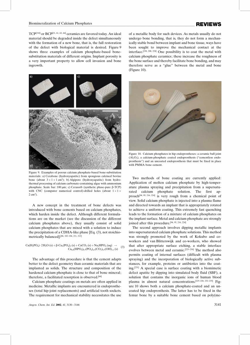
of a metallic body for such devices. As metals usually do notundergo bone bonding, that is, they do not form a mechan-ically stable bond between implant and bone tissue, ways havebeen sought to improve the mechanical contact at theinterface.[194, 208, 224] One possibility is to coat the metal withcalcium phosphate ceramics; these increase the roughness ofthe bone surface and thereby facilitate bone bonding, and maytherefore serve as a ™glue∫ between the metal and bone(Figure 10).
Figure 10. Calcium phosphates in hip endoprostheses: a ceramic ball joint(Al2O3), a calcium-phosphate coated endoprosthesis (™cementless endo-prosthesis∫) and an uncoated endoprosthesis that must be fixed in placewith PMMA bone cement.
Two methods of bone coating are currently applied:Application of molten calcium phosphate by high-temper-ature plasma spraying and precipitation from a supersatu-rated calcium phosphate solution. The first ap-proach[94, 95, 216, 224] is very rough from a chemical point ofview. Solid calcium phosphate is injected into a plasma flameand directed towards an implant that is appropriately rotatedto achieve a uniform coating. This extremely fast quenchingleads to the formation of a mixture of calcium phosphates onthe implant surface. Metal and calcium phosphate are stronglyjoined after this procedure.[94, 95, 216, 224]
The second approach involves dipping metallic implantsinto supersaturated calcium phosphate solutions. This methodwas strongly promoted by the work of Kokubo and co-workers and van Blitterswijk and co-workers, who showedthat after appropriate surface etching, a stable interfaceevolves between metal and ceramic.[225±230] The method alsopermits coating of internal surfaces (difficult with plasmaspraying) and the incorporation of biologically active sub-stances, for example, proteins or antibiotics into the coat-ing.[231] A special case is surface coating with a biomimeticdefect apatite by dipping into simulated body fluid (SBF), asolution that contains the inorganic ions of human bloodplasma in almost natural concentrations.[225±228, 232±235] Fig-ure 10 shows both a calcium phosphate-coated and an un-coated hip endoprosthesis. The latter has to be fixed in thefemur bone by a suitable bone cement based on poly(me-
Angew. Chem. Int. Ed. 2002, 41, 3130 ± 3146 3141

REVIEWS M. Epple and S. V. Dorozhkin
thylmethacrylate) (PMMA). Note that this polymer is notbiodegradable and remains in the operation site.[224, 236]
The same principles are valid for tooth implant systems thatare fixed into the jawbone, onto which artificial teeth areattached. In general, the mechanical contact between implantand bone is crucial, as considerable forces have to bewithstood. Coating of such dental implants with calciumphosphates (usually by plasma spraying) leads to better andfaster bone attachment. Figure 11 shows such a plasma-spray-coated tooth implant in low and high magnification. Finally,Figure 12 shows the surface of a nickel-titanium shape-memory alloy (NiTi, ™Nitinol∫) that was coated with calciumphosphate from solution to improve its biocompatibility.[237]
Figure 11. Dental implants (by Friadent) coated with calcium phosphateby a plasma-spray process. a: � 10, b: � 1000. Note the irregular, roughstructure of the deposited calcium phosphate at the higher magnification.Scale bars 1 mm and 10 �m, respectively.
Figure 12. The surface of a nickel ± titanium shape-memory alloy (™Niti-nol∫) that was coated with a calcium phosphate layer by dipping into ansupersaturated calcium phosphate solution. The front part shows theetched metal surface from which the calcium phosphate layer has beenmechanically removed. Scale bar: 2 �m.
6. Biomimetic Crystallization of CalciumPhosphates
Nature×s ability to assemble inorganic compounds into thebiological structures (shells, spicules, teeth, bone, skeletons) isstill not reproducible by synthetic procedures. Because of itspotential benefits for materials science, research groupsaround the world are increasingly addressing the question ofbiomineralization. When considering calcium phosphates, thedemand of clinical medicine to design biocompatible implantsand to treat diseases related to crystallization phenomenaadds a strong practical impetus to understanding theseprocesses. The fundamentals of biomineralization have beenreviewed extensively.[1, 3±7, 238±241] We will limit ourselves toconsiderations of biologically inspired crystallization ofcalcium phosphates and present a few examples that demon-strate the current possibilities.
An approach to the preparation of biomimetic bone-substitution materials was made by Pompe et al. , who crystal-lized HA on collagen to obtain a bonelike composite.[242]
Although the ultrastructure of bone could not be realized,such collagen ±HA tapes are currently under investigation forclinical use. Note that the final step to make bone out ofartificial implants is up to the body×s own remodelingfunction. Ozin et al. precipitated HA in the presence ofsurfactants, to obtain a biomimetic lamellar product.[243] Stuppet al. have prepared so-called ™organoapatites∫ with a bone-like crystallinity by precipitation of calcium phosphate in thepresence of organic polyelectrolytes.[214, 217, 244, 245] Kokubo andco-workers and van Blitterswijk and co-workers were suc-cessful in coating different substrates with a bonelike apatitelayer (see refs. [229, 234] and those given above on coatedmetal prostheses). We have recently prepared bulk samples ofbonelike apatite and composites of it with biodegradablepolymers.[84±86, 246]
Nancollas and co-workers invented the ™Constant-Compo-sition Technique∫ to monitor and control the externalconditions (mainly solution pH value and concentrations ofparticipating ions) during a crystallization experiment.[22, 247]
Generally, during precipitation of calcium phosphates from aneutral solution, the pH value decreases because of therelease of protons that were formerly bound to hydrogenphosphate or dihydrogen phosphate [Eq. (4)].
5Ca2� (aq) � 3HPO42� (aq) � 5H2O (l) �
Ca5(PO4)3OH (s) � 4H3O� (aq)(4)
One of the main differences between chemical and bio-logical crystallization is the rate of precipitation. Usually inchemistry, precipitation occurs fast whereas in biology thecrystals need days, weeks, or months to grow. A suitablesimulation of this process, especially in the presence of(bio)organic additives, must therefore slow down the crystal-lization. This can be achieved by separating the two compo-nents with a suitable membrane or medium that acts as adiffusion barrier (a double-diffusion technique). If thismedium itself contains some biomimetic functional groups,it can have a templating influence on the growing crystals.Work along this line has been carried out by Iijima et al.
3142 Angew. Chem. Int. Ed. 2002, 41, 3130 ± 3146

REVIEWSBiomineralization of Calcium Phosphates
(collagen matrix from bovine achilles tendon,[248] and mem-branes in the presence of bovine[249] and murine[250] ameloge-nins), Kniep and co-workers (matrix of denaturated colla-gen),[154, 251, 252] Epple and co-workers (matrix of microporouspolyglycolide),[253±256] Falini et al. (matrix of collagen),[257] andStupp and co-workers (carbon-coated TEM grid).[113] Work onthe crystallization from SBF under static and dynamicconditions to yield bonelike apatite was also reported recentlyby Vallet-Regi and co-workers[258, 259] and by Epple and co-workers.[256, 260]
Interactions between collagen and growing fluoroapatitecrystals are responsible for a fractal growth of fluoro-apatite into dumbbell shapes that finally close to givespheres.[154, 251, 252] Figure 13 shows this special morphology.
Figure 13. A biomimetically grown aggregate of fluoroapatite that wascrystallized in a gelatin matrix. The crystal shape can be explained andsimulated by a fractal growth mechanism. Scale bar: 10 �m (taken fromref. [252] with permission).
By combining the constant-composition technique with thedouble-diffusion setup, we were able to identify differentcrystal morphologies of fluoroapatite as functions of overallconcentration (i.e. supersaturation), pH value, and fluorideion concentration.[254, 255] Figure 14 shows a uniform crystalpopulation that was prepared by this method.
Figure 14. Hexagonal fluoroapatite crystals that were grown by a double-diffusion technique under controlled conditions (pH 7.4, 37 �C, constant ionconcentrations, 7 days). Note the well-shaped crystals and their uniformsize and morphology. Scale bar: 10 �m.
7. Summary and Outlook
Although it may appear surprising to the nonspecialist,there are still many open questions within the area of calciumphosphate chemistry. The basic questions concerning crystal-lography, thermodynamics, and phase relationships have beenanswered. Nevertheless, when it comes to the biologicalformation of calcium phosphates, issues including rate ofcrystallization, control of morphology, incorporation of for-eign ions, and interaction with biomolecules remain hot topicsthat are not well understood even today. A better under-standing of structure, formation, and dissolution of suchbiominerals will lead to improved biomaterials that cansubstitute bone and teeth. This knowledge will also help tocounter widespread pathological calcifications such as athe-rosclerosis, stone formation, or dental calculus. Furtherprogress of unforeseeable impact will come from moderngenetics, where gene structures are currently related to hard-tissue formation.
We thank Alexander Becker, Dr. Jongsik Choi, ElenaDorozhkina, Dr. Bernd Hasse (now at DESY), Dr. FabianPeters (now at Curasan), Carsten Schiller, Dr. Karsten Schwarz(now at Tutogen), Dr. Michael Siedler, and Drazen Tadic fortheir research contributions during the past years. We alsothank Dr. Jˆrg Arnoldi (Mathys), Dr. Philip Cantzler (Fria-dent), Dr. Peter Seidel (Coripharm), and Prof. Gerd Willmann(Ceramtec) for providing material. We are also grateful to theDeutsche Forschungsgemeinschaft (DFG), to the Fonds derChemischen Industrie, to the Deutscher Akademischer Aus-tauschdienst (DAAD), and to HASYLAB at DESY (Ham-burg) for generous support of our work during the past years.
Received: December 3, 2001 [A505]
[1] H. A. Lowenstam, S. Weiner, On Biomineralization, Oxford Uni-versity Press, New York, 1989.
[2] K. Simkiss, K. M. Wilbur, Cell Biology and Mineral Deposition,Academic Press, San Diego, 1989.
[3] L. Addadi, S. Weiner, Angew. Chem. 1992, 104, 159 ± 176; Angew.Chem. Int. Ed. Engl. 1992, 31, 153 ± 169.
[4] S. Mann, J. Mater. Chem. 1995, 5, 935 ± 946.[5] S. Mann, Biomimetic Materials Chemistry, Wiley/VCH, New York/
Weinheim, 1995.[6] G. A. Ozin, Acc. Chem. Res. 1997, 30, 17 ± 27.[7] E. Baeuerlein, Biomineralization, Wiley-VCH, Weinheim, 2000.[8] S. Mann, Angew. Chem. 2000, 112, 3532 ± 3548; Angew. Chem. Int.
Ed. 2000, 39, 3392 ± 3406.[9] S. A. Davis, M. Breulmann, K. H. Rhodes, B. Zhang, S. Mann, Chem.
Mater. 2001, 13, 3218 ± 3226.[10] R. C. Weast, The CRCHandbook of Chemistry and Physics, 66th ed.,
CRC, Boca Raton, FL, 1985 ± 1986.[11] D. McConnell in Apatite: Its Crystal Chemistry, Mineralogy, Utiliza-
tion, and Biologic Occurrences, Springer, New York, 1973, pp. 111 ±115.
[12] P. Becker in Fertilizer Science and Technology Series, Marcel Dekker,New York, 1989, pp. 6 ± 20.
[13] D. K. Smith in Hydroxyapatite and Related Materials (Eds.: P. W.Brown, B. Constantz), CRC, Boca Raton, FL, 1994, pp. 29 ± 44.
[14] A. I. Angelov, B. V. Levin, Y. D. Chernenko in Phosphate Ore. AReference Book (in Russian), Nedra business center, Moskau, 2000,pp. 1 ± 120.
[15] R. Z. LeGeros, Calcium Phosphates in Oral Biology and Medicine,Karger, Basel, 1991.
Angew. Chem. Int. Ed. 2002, 41, 3130 ± 3146 3143

REVIEWS M. Epple and S. V. Dorozhkin
[16] R. Z. LeGeros in Hydroxyapatite and Related Materials (Eds.: P. W.Brown, B. Constantz), CRC, Boca Raton, FL, 1994, pp. 3 ± 28.
[17] R. Z. LeGeros, Z. Kardiol. 2001, 90 (Suppl. 3), III/116 ± III/125.[18] A. Hesse, D. Heimbach, World J. Urol. 1999, 17, 308 ± 315.[19] B. M. Tracy, R. H. Doremus, J. Biomed. Mater. Res. 1984, 18, 719 ±
726.[20] G. Daculsi, R. Z. LeGeros, M. Heughebaert, I. Barbieux, Calcif.
Tissue Int. 1990, 46, 20 ± 27.[21] G. Daculsi, J. M. Bouler, R. Z. LeGeros, Int. Rev. Cytol. 1997, 172,
129 ± 191.[22] P. Koutsoukos, Z. Amjad, M. B. Tomson, G. H. Nancollas, J. Am.
Chem. Soc. 1980, 102, 1553 ± 1557.[23] A. A. Mirtchi, J. Lemaitre, N. Terao, Biomaterials 1989, 10, 475 ± 480.[24] A. A. Mirtchi, J. Lemaitre, E. Munting, Biomaterials 1989, 10, 634 ±
638.[25] O. Bermu¬dez, M. G. Boltong, F. C. M. Driessens, J. A. Planell, J.
Mater. Sci. Mater. Med. 1994, 5, 67 ± 71.[26] O. Bermu¬dez, M. G. Boltong, F. C. M. Driessens, J. A. Planell, J.
Mater. Sci. Mater. Med. 1994, 5, 160 ± 163.[27] F. C. M. Driessens, M. G. Boltong, O. Bermu¬dez, J. A. Planell, M. P.
Ginebra, E. Ferna¬ndez, J. Mater. Sci. Mater. Med. 1994, 5, 164 ± 170.[28] M. Windholz, The Merck Index: An Encyclopedia of Chemicals,
Drugs, and Biologicals, 10th ed., Merck, Rahway, NJ, 1983.[29] M. Otsuka, Y. Matsuda, Y. Suwa, J. L. Fox, W. I. Higuchi, Chem.
Pharm. Bull. (Tokyo) 1993, 41, 2055 ± 2057.[30] C. Hamanishi, K. Kitamoto, K. Ohura, S. Tanaka, Y. Doi, J. Biomed.
Mater. Res. 1996, 32, 383 ± 389.[31] K. Kurashina, H. Kurita, M. Hirano, A. Kotani, C. P. Klein, K.
de Groot, Biomaterials 1997, 18, 539 ± 543.[32] F. C. M. Driessens, J. A. Planell, M. G. Boltong, I. Khairoun, M. P.
Ginebra, Proc. Inst. Mech. Eng. Part H 1998, 212, 427 ± 435.[33] S. Takagi, L. C. Chow, K. Ishikawa, Biomaterials 1998, 19, 1593 ±
1599.[34] H. Yamamoto, S. Niwa, M. Hori, T. Hattori, K. Sawai, S. Aoki, M.
Hirano, H. Takeuchi, Biomaterials 1998, 19, 1587 ± 1591.[35] J. J. Crall, J. M. Bjerga, J. Oral Pathol. 1987, 16, 488 ± 491.[36] J. S. Wefel, J. D. Harless, J. Dent. Res. 1987, 66, 1640 ± 1643.[37] P. M. Hoppenbrouwers, E. Groenendijk, N. R. Tewarie, F. C. M.
Driessens, J. Dent. Res. 1988, 67, 1254 ± 1256.[38] A. Gaffar, J. Blake-Haskins, J. Mellberg, Int. Dent. J. 1993, 43 (Suppl.
1), 81 ± 88.[39] Y. Fukase, E. D. Eanes, S. Takagi, l. C. Chow, W. E. Brown, J. Dent.
Res. 1990, 69, 1852 ± 1855.[40] K. S. TenHuisen, P. W. Brown, J. Dent. Res. 1994, 73, 598 ± 606.[41] O. Bermu¬dez, M. G. Boltong, F. C. M. Driessens, J. A. Planell, J.
Mater. Sci. Mater. Med. 1994, 5, 144 ± 146.[42] E. Fernandez, M. P. Ginebra, M. G. Boltong, F. C. M. Driessens, J.
Ginebra, E. A. de Maeyer, V. M. Verbeeck, J. A. Planell, J. Biomed.Mater. Res. 1996, 32, 367 ± 374.
[43] E. Fernandez, F. J. Gil, S. M. Best, M. P. Ginebra, F. C. M. Driessens,J. A. Planell, J. Biomed. Mater. Res. 1998, 42, 403 ± 406.
[44] E. Fernandez, F. J. Gil, S. M. Best, M. P. Ginebra, F. C. M. Driessens,J. A. Planell, J. Biomed. Mater. Res. 1998, 41, 560 ± 567.
[45] R. Z. LeGeros, J. Dent. Res. 1974, 53, 45 ± 50.[46] H. Schroeder, Formation and Inhibition of Dental Calculus, Hubert,
Vienna, 1969.[47] L. C. Chow, E. D. Eanes, Octacalcium Phosphate, Vol. 18, Karger,
Basel, 2001.[48] D. G. A. Nelson, G. J. Wood, J. C. Barry, Ultramicroscopy 1986, 19,
253 ± 266.[49] M. Iijima, D. G. A. Nelson, Y. Pan, A. T. Kreinbrink, M. Adachi, T.
Goto, Y. Moriwaki, Calcif. Tissue Int. 1996, 59, 377 ± 384.[50] P. S. Bodier-Houlle¬, P. J. C. Voegel, F. J. G. Cuisinier, Acta Crystal-
logr. Sect. D 1998, 54, 1377 ± 1381.[51] T. Aoba, H. Komatsu, Y. Shimazu, H. Yagishita, Y. Taya, Connect.
Tissue Res. 1998, 38, 129 ± 145.[52] B. B. Tomazic, W. E. Brown, F. J. Shoen, J. Biomed. Mater. Res. 1994,
28, 35 ± 47.[53] G. H. Nancollas, W. Wu, J. Crystal Growth 2000, 211, 137 ± 142.[54] T. Kodaka, K. Debari, S. Higashi, J. Electron Microsc. (Tokyo) 1988,
37, 73 ± 80.[55] A. A. Mirtchi, J. Lemaitre, E. Munting,Biomaterials 1990, 11, 83 ± 88.
[56] A. A. Mirtchi, J. Lemaitre, E. Munting, Biomaterials 1991, 12, 505 ±510.
[57] J. Lemaitre, E. Munting, A. A. Mirtchi, Rev. Stomatol. Chir.Maxillofac. 1992, 93, 163 ± 165.
[58] K. Ohura, M. Bohner, P. Hardouin, J. Lemaitre, G. Pasquier, B.Flautu, J. Biomed. Mater. Res. 1996, 30, 193 ± 200.
[59] G. Daculsi, R. Z. LeGeros, E. Nery, K. Lynch, B. Kerebel, J. Biomed.Mater. Res. 1989, 23, 883 ± 894.
[60] G. Daculsi, M. D×Arc Bagot, P. Corlieu, M. Gersdorff, Ann. Otol.Rhinol. Laryngol. 1992, 101, 669 ± 674.
[61] J. M. Bouler, M. Trecant, J. Delecrin, J. Royer, N. Passuti, G. Daculsi,J. Biomed. Mater. Res. 1996, 32, 603 ± 609.
[62] J. Wang,W. Chen, Y. Li, S. Fan, J. Weng, X. Zhang,Biomaterials 1998,19, 1387 ± 1392.
[63] G. Daculsi, Biomaterials 1998, 19, 1473 ± 1478.[64] G. Daculsi, P. Weiss, J. M. Bouler, O. Gauthier, F. Millot, E. Aguado,
Bone 1999, 25 (Suppl. 2), 59S ± 61S.[65] I. Alam, I. Asahina, K. Ohmamiuda, S. Enomoto, J. Biomed. Mater.
Res. 2001, 54, 129 ± 138.[66] B. R. Constantz, I. C. Ison, M. T. Fulmer, R. D. Poser, S. T. Smith, M.
VanWagoner, J. Ross, S. A. Goldstein, J. B. Jupiter, D. I. Rosenthal,Science 1995, 267, 1796 ± 1799.
[67] J. D. Termine, E. D. Eanes, Calcif. Tissue Res. 1972, 10, 171 ± 197.[68] E. D. Eanes, J. D. Termine, M. U. Nylen, Calcif. Tissue Res. 1973, 12,
143 ± 158.[69] J. L. Meyer, E. D. Eanes, Calcif. Tissue Res. 1978, 28, 59 ± 68.[70] J. L. Meyer, E. D. Eanes, Calcif. Tissue Res. 1978, 28, 209 ± 216.[71] R. E. Wuthier, G. S. Rice, J. E. Wallace, R. L. Weaver, R. Z.
LeGeros, E. D. Eanes, Calcif. Tissue Int. 1985, 37, 401 ± 410.[72] J. C. Elliot, Structure and Chemistry of the Apatites and Other
Calcium Orthophosphates, Elsevier, Amsterdam, 1994.[73] J. C. Elliot in Les mate¬riaux en phosphate de calcium. Aspects
fondamentaux (Eds.: E. Bre¡s, P. Hardouin), Sauramps Medical,Montpellier, 1998.
[74] J. E. Harries, D. W. L. Hukins, S. S. Hasnain, J. Phys. C 1986, 19,6859 ± 6872.
[75] J. E. Harries, D. W. L. Hukins, C. Holt, S. S. Hasnain, J. Cryst. Growth1987, 84, 563 ± 570.
[76] M. G. Taylor, K. Simkiss, J. Simmons, L. N. Y. Wu, R. E. Wuthier,Cell. Mol. Life Sci. 1998, 54, 192 ± 202.
[77] F. Peters, K. Schwarz, M. Epple, Thermochim. Acta 2000, 361, 131 ±138.
[78] K. Onuma, A. Ito, Chem. Mater. 1998, 10, 3346 ± 3351.[79] D. Skrtic, A. W. Hailer, S. Takagi, J. M. Antonucci, E. D. Eanes, J.
Dent. Res. 1996, 75, 1679 ± 1686.[80] D. Skrtic, J. M. Antonucci, E. D. Eanes, Dent. Mater. 1996, 12, 295 ±
301.[81] M. S. Park, E. D. Eanes, J. M. Antonucci, D. Skrtic, Dent. Mater.
1998, 14, 137 ± 141.[82] D. Skrtic, J. M. Antonucci, E. D. Eanes, F. C. Eichmiller, G. E.
Schumacher, J. Biomed. Mater. Res. 2000, 53, 381 ± 391.[83] C. Schiller, M. Siedler, F. Peters, M. Epple, Ceram. Trans. 2001, 114,
97 ± 108.[84] W. Linhart, F. Peters, W. Lehmann, A. F. Schilling, K. Schwarz, M.
Amling, J. M. Rueger, M. Epple, J. Biomed. Mater. Res. 2001, 54,162 ± 171.
[85] D. Tadic, M. Epple, Biomed. Tech. 2001, 224 ± 225.[86] D. Tadic, F. Peters, M. Epple, Biomaterials 2002, 23, 2553 ± 2559.[87] P. W. Brown, R. I. Martin, J. Phys. Chem. B 1999, 103, 1671 ±
1675.[88] A. Mortier, J. Lemaitre, L. Rodrique, P. G. Rouxhet, J. Solid State
Chem. 1989, 78, 215 ± 219.[89] J. Jeanjean, U. Vincent, M. Fedoroff, J. Solid State Chem. 1994, 103,
68 ± 72.[90] N. Rangavittal, A. R. Landa-Ca¬novas, J. M. Gonza¬ lez-Calbet, M.
Vallet-Regi, J. Biomed. Mater. Res. 2000, 51, 660 ± 668.[91] J. Y. Kim, R. R. Fenton, B. A. Hunter, B. J. Kennedy, Aust. J. Chem.
2000, 53, 679 ± 686.[92] P. Layrolle, A. Lebugle, Chem. Mater. 1994, 6, 1996 ± 2004.[93] P. Layrolle, A. Lebugle, Chem. Mater. 1996, 8, 134 ± 144.[94] W. Suchanek, M. Yoshimura, J. Mater. Res. 1998, 13, 94 ± 117.[95] L. L. Hench, J. Am. Ceram. Soc. 1998, 81, 1705 ± 1728.
3144 Angew. Chem. Int. Ed. 2002, 41, 3130 ± 3146
REVIEWSBiomineralization of Calcium Phosphates
[96] M. Fountoulakis, M. F. Takacs, P. Berndt, H. Langen, B. Takacs,Electrophoresis 1999, 20, 2181 ± 2195.
[97] M.Mirshahi, L. Camoin, C. Nicolas, I. Ghedira, J. Cozette, J. P. Faure,Curr. Eye Res. 1999, 18, 327 ± 334.
[98] R. Freitag, S. Vogt, M. Modler, Biotechnol. Prog. 1999, 15, 573 ± 576.[99] J. Wissing, S. Heim, L. Flohe, U. Bilitewski, R. Frank, Electrophoresis
2000, 21, 2589 ± 2593.[100] S. R. Shepard, C. Brickman-Stone, J. L. Schrimsher, G. Koch, J.
Chromatogr. A 2000, 891, 93 ± 98.[101] G. Yin, Z. Liu, R. Zhou, J. Zhan, J. Wang, N. Yuan, J. Chromatogr. A
2001, 918, 393 ± 399.[102] L. C. Chow, Nippon Seramikkusu Kyokai Gakujutsu Ronbunshi
1991, 99, 954 ± 964.[103] E. Fernandez, F. J. Gil, M. P. Ginebra, F. C. M. Driessens, J. A.
Planell, S. M. Best, J. Mater. Sci. Mater. Med. 1999, 10, 169 ± 176.[104] E. Fernandez, F. J. Gil, M. P. Ginebra, F. C. M. Driessens, J. A.
Planell, S. M. Best, J. Mater. Sci. Mater. Med. 1999, 10, 177 ± 183.[105] S. Weiner, H. D. Wagner, Annu. Rev. Mater. Sci. 1998, 28, 271 ± 298.[106] S. Weiner, W. Traub, H. D. Wagner, J. Struct. Biol. 1999, 126, 241 ±
255.[107] H. Limeback, Curr. Opin. Dent. 1991, 1, 826 ± 835.[108] N. M. Hancox, Biology of Bone, Cambridge University Press,
Cambridge, 1972.[109] W. E. Brown, L. C. Chow, Annu. Rev. Mater. Sci. 1976, 6, 213 ± 236.[110] J. Currey, The Mechanical Adaptations of Bones, Princeton Univer-
sity Press, Princeton, 1984.[111] R. Lakes, Nature 1993, 361, 511 ± 515.[112] A. C. Lawson, J. T. Czernuszka, Proc. Inst. Mech. Eng. Part H 1998,
212, 413 ± 425.[113] J. D. Hartgerink, E. Beniash, S. I. Stupp, Science 2001, 294, 1684 ±
1688.[114] A. Jodaikin, S. Weiner, Y. Talmon, E. Grossman, W. Traub, Int. J.
Biol. Macromol. 1988, 10, 349 ± 352.[115] A. G. Fincham, J. Moradian-Oldak, T. G. H. Diekwisch, D. M.
Lyaruu, J. T. Wright, P. Bringas, H. C. Slavkin, J. Struct. Biol. 1995,115, 50 ± 59.
[116] M. Iijima, Y. Moriwaki, Calcif. Tiss. Int. 1990, 47, 237 ± 242.[117] L. C. Bonar, M. Shimizu, J. E. Roberts, R. G. Griffin, M. J. Glimcher,
J. Bone Miner. Res. 1991, 6, 1167 ± 1176.[118] C. Rey, V. Renugopalakrishnan, B. Collins, M. J. Glimcher, Calcif.
Tissue Int. 1991, 49, 251 ± 258.[119] C. Rey, J. L. Miquel, L. Facchini, A. P. Legrand, M. J. Glimcher,Bone
1995, 16, 583 ± 586.[120] W. E. Brown, Clin. Orthop. Relat. Res. 1966, 44, 205 ± 220.[121] M. S. Tung, W. E. Brown, Calcif. Tissue Int. 1983, 35, 783 ± 790.[122] M. S. Tung, W. E. Brown, Calcif. Tissue Int. 1985, 37, 329 ± 331.[123] W. E. Brown, N. Eidelman, B. Tomazic, Adv. Dent. Res. 1987, 1, 306 ±
313.[124] C. Siew, S. E. Gruninger, L. C. Chow, W. E. Brown, Calcif. Tissue Int.
1992, 50, 144 ± 148.[125] E. D. Eanes, I. H. Gillessen, A. S. Posner,Nature 1965, 208, 365 ± 367.[126] J. D. Termine, A. S. Posner, Science 1966, 153, 1523 ± 1525.[127] J. D. Termine, A. S. Posner, Nature 1966, 211, 268 ± 270.[128] R. A. Harper, A. S. Posner, Proc. Soc. Exp. Biol. Med. 1966, 122,
137 ± 142.[129] A. S. Posner, Physiol. Rev. 1969, 49, 760 ± 792.[130] A. S. Posner, Fed. Proc. 1973, 32, 1933 ± 1937.[131] A. L. Boskey, A. S. Posner, J. Phys. Chem. A 1973, 77, 2313 ± 2317.[132] A. S. Posner, Bull. Hosp. Jt. Dis. 1978, 39, 126 ± 144.[133] M. J. Glimcher, L. C. Bonar, M. D. Grynpas, W. J. Landis, A. H.
Roufosse, J. Cryst. Growth 1981, 53, 100 ± 119.[134] J. M. Burnell, E. J. Teubner, A. G. Miller, Calcif. Tissue Int. 1980, 31,
13 ± 19.[135] S. Weiner, W. Traub, FEBS Lett. 1986, 206, 262 ± 266.[136] E. D. Pellegrino, R. M. Blitz, Calcif. Tissue Res. 1972, 10, 118 ± 135.[137] R. Legros, N. Balmain, G. Bonel,Calcif. Tissue Int. 1987, 41, 137 ± 144.[138] H. P. Wiesmann, L. Chi, U. Stratmann, U. Plate, H. Fuchs. U. Joos,
H. J. Hˆhling, Cell Tissue Res. 1998, 294, 93 ± 97..[139] U. Plate, T. Kotz, H. P. Wiesmann, U. Stratmann, U. Joos, H. J.
Hˆhling, J. Microsc. 1996, 183, 102 ± 107.[140] U. Stratmann, K. Schaarschmidt, H. P. Wiesmann, U. Plate, H. J.
Hˆhling, Cell Tissue Res. 1996, 284, 223 ± 230.
[141] M. Epple, P. Lanzer, Z. Kardiol. 2001, 90 (Suppl. 3), III/2 ± III/5.[142] W. Jahnen-Dechent, T. Schinke, A. Trindl, W. Muller-Esterl, F.
Sablitzky, S. Kaiser, M. Blessing, J. Biol. Chem. 1997, 272, 31496 ±31503.
[143] T. Schinke, M. D. McKnee, G. Karsenty, Nat. Genet. 1999, 21, 150 ±151.
[144] W. Jahnen-Dechent, G. Sch‰fer, A. Heiss, J. Grˆtzinger, Z. Kardiol.2001, 90 (Suppl. 3), III/47 ± III/56.
[145] P. Ducy, M. Amling, S. Takeda, M. Priemel, A. F. Schilling, F. T. Beil,J. Shen, C. Vinson, J. M. Rueger, G. Karsenty, Cell 2000, 100, 197 ±207.
[146] E. Rˆnnholm, J. Ultrastruct. Res. 1962, 6, 249 ± 303.[147] M. U. Nylen, E. D. Evans, K. A. Omnel, J. Cell. Biol. 1963, 18, 109 ±
123.[148] Y. Miake, S. Shimoda, M. Fukae, T. Aoba, Calcif. Tissue Int. 1993, 53,
249 ± 256.[149] G. Daculsi, J. Menanteau, L. M. Kerebel, D. Mitre, Calcif. Tissue Int.
1984, 36, 550 ± 555.[150] A. Jodaikin, W. Traub, S. Weiner, J. Ultrastruct. Res. 1984, 89, 324 ±
332.[151] R. M. Frank, R. F. Sognnaes, R. Kerns in Calcification in Biological
Systems, AAAS, Washington, DC, 1960, pp. 163 ± 202.[152] I. Schroeder, R. M. Frank, Cell. Tissue Res. 1985, 242, 449 ± 451.[153] E. F. Bre¡s, J. C. Voegel, R. M. Frank, J. Microsc. 1990, 160, 183 ± 201.[154] S. Busch, U. Schwarz, R. Kniep, Chem. Mater. 2001, 13, 3260 ± 3271.[155] J. P. Simmer, A. G. Fincham, Crit. Rev. Oral Biol. Med. 1995, 6, 84 ±
108.[156] T. G. Diekwisch, B. J. Berman, S. Genters, H. C. Slavkin, Cell. Tissue
Res. 1995, 279, 149 ± 167.[157] T. Aoba, Anat. Rec. 1996, 245, 208 ± 218.[158] C. Robinson, S. J. Brookes, W. A. Bonass, R. C. Shore, J. Kirkham,
Ciba Found. Symp. 1997, 205, 156 ± 170.[159] C. E. Smith, Crit. Rev. Oral. Biol. Med. 1998, 9, 128 ± 161.[160] M. Sydney-Zax, I. Mayer, D. Deutsch, J. Dent. Res. 1991, 70, 913 ±
916.[161] C. Rey, V. Renugopalakrishnan, M. Shimizu, B. Collins, M. J.
Glimcher, Calcif. Tissue Int. 1991, 49, 259 ± 268.[162] T. Takagi, T. Ogasawara, J. Tagami, M. Akao, Y. Kuboki, N. Nagai,
R. Z. LeGeros, Connect. Tissue Res. 1998, 38, 181 ± 187.[163] K. A. Selvig, Calcif. Tissue Res. 1970, 6, 227 ± 238.[164] K. A. Selvig, J. Ultrastruct. Res. 1972, 41, 369 ± 375.[165] F. J. G. Cuisinier, P. Steuer, B. Senger, J. C. Voegel, R. M. Frank, Cell.
Tissue Res. 1993, 273, 175 ± 182.[166] F. J. G. Cuisinier, P. Steuer, A. Brisson, J. C. Voegel, J. Crystal Growth
1995, 156, 443 ± 453.[167] P. Houlle¬, J. C. Voegel, P. Schultz, F. J. G. Cuisinier, J. Dent. Res. 1997,
76, 895 ± 904.[168] S. Mann, Nature 1993, 365, 499 ± 505.[169] J. Kirkham, J. Zhang, S. J. Brookes, R. C. Shore, S. R. Wood, D. A.
Smith, M. L. Wallwork, O. H. Ryu, C. Robinson, J. Dent. Res. 2000,79, 1943 ± 1947.
[170] A. Boyde in Tooth Enamel (Eds.: M. V. Stack, R. W. Fearnhead),Wright, Bristol, 1965, pp. 163 ± 167.
[171] H. Warshawsky, A. Nanci, J. Dental Res. 1982, 61, 1504 ± 1514.[172] H. Warshawsky, Anat. Rec. 1989, 224, 242 ± 262.[173] A. L. Arsenault, B. W. Robinson, Calcif. Tissue Int. 1989, 45, 111 ±
121.[174] Y. Hayashi, J. Electron Microsc. 1992, 41, 387 ± 391.[175] Y. Hayashi, J. Electron Microsc. 1993, 42, 141 ± 146.[176] P. Bodier-Houlle¬, P. Steuer, J. M. Meyer, L. Bigeard, F. J. G.
Cuisinier, Cell. Tissue Res. 2000, 301, 389 ± 395.[177] Y. Takano, Y. Hanaizumi, H. Oshima, Anat. Rec. 1996, 245, 174 ±
185.[178] W. Dong, H. Warshawsky, Adv. Dent. Res. 1996, 10, 232 ± 237.[179] P. Person in Cartilage: Structure, Function and Biochemistry (Ed.:
B. K. Hall), Academic Press, New York, 1983, pp. 31 ± 57.[180] M. L. Moss, L. Moss-Salentijn in Cartilage: Structure, Function and
Biochemistry (Ed.: B. K. Hall), Academic Press, New York, 1983,pp. 1 ± 30.
[181] R. A. Robinson, D. A. Cameron, J. Biophys. Biochem. 1956, 2(Suppl.), 253 ± 260.
[182] E. Bonucci, J. Ultrastruct. Res. 1967, 20, 33 ± 50.
Angew. Chem. Int. Ed. 2002, 41, 3130 ± 3146 3145

REVIEWS M. Epple and S. V. Dorozhkin
[183] S. Y. Ali, A. Wisby, L. Evans, J. Craig-Grey, Calcif. Tissue Res. 1977,22 (Suppl.), 490 ± 493.
[184] S. Y. Ali in Cartilage: Structure, Function and Biochemistry (Ed.:B. K. Hall), Academic Press, New York, 1983, pp. 343 ± 378.
[185] I. M. Shapiro, A. Boyde, Metab. Bone Dis. Relat. Res. 1984, 5, 317 ±322.
[186] C. Rey, K. Beshah, R. Griffin, M. J. Glimcher, J. Bone Miner. Res.1991, 6, 515 ± 525.
[187] D. McConnell, Bull. Geol. Soc. Am. 1963, 74, 363 ± 364.[188] H. A. Lowenstam, Chem. Geol. 1972, 9, 153 ± 166.[189] W. Achilles, Contrib. Nephrol. 1987, 58, 59 ± 64.[190] W. Achilles, U. Jockel, A. Schaper, M. Burk, H. Riedmiller, Scanning
Microsc. 1995, 9, 577 ± 585.[191] H. C. Stary, Z. Kardiol. 2000, 89 (Suppl. 2), II/28 ± II/35.[192] K. M. Kim, Z. Kardiol. 2001, 90 (Suppl. 3), III/99 ± III/105.[193] B. B. Tomazic, Z. Kardiol. 2001, 90 (Suppl. 3), III/68 ± III/80.[194] E. Wintermantel, S. W. Ha, Medizintechnik mit biokompatiblen
Werkstoffen und Verfahren, Springer, Heidelberg, 2002.[195] C. Giachelli, Z. Kardiol. 2001, 90 (Suppl. 3), III/31 ± III/38.[196] B. Glasmacher, E. Nellen, H. Reul, G. Rau, Materialwiss. Werk-
stofftech. 1999, 30, 806 ± 808.[197] M. Deiwick, B. Glasmacher, E. Pettenazzo, D. Hammel, W.
Castellon, G. Thiene, H. Reul, E. Berendes, H. H. Scheld, Thorac.Cardiovasc. Surg. 2001, 49, 78 ± 83.
[198] E. Pettenazzo, M. Deiwick, G. Thiene, G. Molin, B. Glasmacher, F.Martignago, T. Bottio, H. Reul, M. Valente, J. Thorac. Cardiovasc.Surg. 2001, 121, 500 ± 509.
[199] D. J. McCarty, H. M. Hogan, R. A. Gatter, M. Grossmann, J. Bone Jt.Surg. Am. Vol. 1966, 48, 309 ± 317.
[200] Oxford Textbook of Rheumatology, Vol. 2 (Eds.: P. J. Maddison,D. A. Isenberg, P. Woo, D. N. Glass), 2th ed., Oxford UniversityPress, Oxford, 1998, pp. 1567 ± 1581.
[201] B. B. Tomazic in Hydroxyapatite and Related Materials (Eds.: P. W.Brown, B. Constantz), CRC, Boca Raton, FL, 1994, pp. 93 ± 116.
[202] G. Luo, P. Ducy, M. D. McKee, G. J. Pinero, E. Loyer, R. R.Behringer, G. Karsenty, Nature 1997, 386, 78 ± 81.
[203] S. Albeck, S. Weiner, L. Addadi, Chem. Eur. J. 1996, 2, 278 ± 284.[204] N. Kessler, D. Perl-Treves, L. Addadi, M. Eisenstein, Proteins 1999,
34, 383 ± 394.[205] L. Addadi, S. Weiner, M. Geva, Z. Kardiol. 2001, 90 (Suppl. 3),
III/92 ± III/98.[206] D. Izhaky, L. Addadi, Chem. Eur. J. 2000, 6, 869 ± 874.[207] A. E. Canfield, M. J. Foherty, A. C. Wood, C. Farrington, B. Ashton,
N. Begum, B. Harvey, A. Poole, M. E. Grant, R. P. Boot-Handford,Z. Kardiol. 2000, 89 (Suppl. 2), II/20 ± II/27.
[208] D. F. Williams, Medical and Dental Materials, Vol. 14, VCH, Wein-heim, 1992.
[209] P. Fratzl, Phys. Unserer Zeit 1999, 30, 196 ± 200.[210] R. Langer, Acc. Chem. Res. 2000, 33, 94 ± 101.[211] M. Bohner, Injury 2000, 31 (Suppl. 4), D37 ±D47.[212] M. Bohner, Eur. Spine J. 2001, 10, S114 ± S121.[213] M. Epple, Biomed. Tech. 2001, 36 ± 38.[214] S. I. Stupp, G. W. Ciegler, J. Biomed. Mater. Res. 1992, 26, 169 ± 183.[215] G. Berger, R. Gildenhaar, U. Ploska, M. Willfahrt, Bioceramics 1997,
10, 367 ± 370.[216] D. M. Liu, J. Mater. Sci. Mater. Med. 1997, 8, 227 ± 232.[217] S. I. Stupp, P. V. Braun, Science 1997, 277, 1242 ± 1248.[218] J. M. Rueger, W. Linhart, D. Sommerfeldt, Orthop‰de 1998, 27, 89 ±
95.[219] J. M. Rueger, Orthop‰de 1998, 27, 72 ± 79.[220] M. Epple, J. M. Rueger, Nachr. Chem. Techn. Lab. 1999, 47, 1405 ±
1410.[221] S. A. Redey, S. Razzouk, C. Rey, D. Bernache-Assollant, G. Leroy,
M. Nardin, G. Cournout, J. Biomed. Mater. Res. 1999, 45, 140 ± 147.[222] R. Z. LeGeros, J. P. LeGeros in Knochenersatzmaterialien und
Wachstumsfaktoren (Eds.: R. Schnettler, E. Markgraf), Thieme,Stuttgart, 1997,pp. 180.
[223] Z. Z. Zyman, I. Ivanov, D. Rochmistrov, V. Glushko, N. Tkachenko,S. Kijko, J. Biomed. Mater. Res. 2001, 54, 256 ± 263.
[224] G. Willmann, Adv. Eng. Mater. 1999, 1, 95 ± 105.[225] T. Kokubo, F. Miyaji, H. M. Kim, J. Am. Ceram. Soc. 1996, 79, 1127 ±
1129.[226] W. Q. Yan, T. Nakamura, K. Kawanabe, S. Nishigochi, M. Oka, T.
Kokubo, Biomaterials 1997, 18, 1185 ± 1190.[227] T. Kokubo, Acta Mater. 1998, 46, 2519 ± 2527.[228] F. Barre¡re, P. Layrolle, C. A. van Blitterswijk, K. de Groot, Bone
1999, 25, 107S ± 111S.[229] F. Barre¡re, P. Layrolle, C. A. van Blitterswijk, K. de Groot, J. Mater.
Sci. Mater. Med. 2001, 12, 529 ± 534.[230] T. Kokubo, H. M. Kim, M. Kawashita, T. Nakamura, Z. Kardiol.
2001, 90 (Suppl. 3), III/86 ± III/91.[231] H. B. Wen, J. R. de Wijn, C. A. van Blitterswijk, K. de Groot, J.
Biomed. Mater. Res. 1999, 46, 245 ± 252.[232] T. Kokubo, Thermochim. Acta 1996, 280/281, 479 ± 490.[233] H. B. Wen, R. A. J. Dalmeijer, F. Z. Cui, C. A. van Blitterswijk, K.
de Groot, J. Mater. Sci. Mater. Med. 1998, 9, 925 ± 930.[234] C. Loty, J. M. Sautier, H. Boulekbache, T. Kokubo, H. M. Kim, N.
Forest, J. Biomed. Mater. Res. 2000, 49, 423 ± 434.[235] H. M. Kim, T. Miyazaki, T. Kokubo, T. Nakamura in Bioceramics 13,
Vol. 192 ± 195 (Eds.: S. Giannini, A. Moroni), Trans Tech Publica-tions, ‹tikon-Z¸rich, 2001, pp. 47 ± 50.
[236] K. D. K¸hn, Bone Cements, Springer, Berlin, 2000.[237] J. S. Choi, M. Kˆller, D. M¸ller, G. Muhr, M. Epple, Biomed. Tech.
2001, 226 ± 227.[238] S. Mann, G. A. Ozin, Nature 1996, 382, 313 ± 318.[239] S. Mann, J. Chem. Soc. Dalton Trans. 1997, 3953 ± 3961.[240] S. Weiner, L. Addadi, J. Mater. Chem. 1997, 7, 689 ± 702.[241] J. Moradian-Oldak, Matrix Biol. 2001, 20, 293 ± 305.[242] W. Pompe, S. Lampenscherf, S. Rˆ˚ler, D. Scharnweber, K. Weis, H.
Worch, J. Hofinger, Mater. Sci. Forum 1999, 308 ± 311, 325 ± 330.[243] G. A. Ozin, N. Varaksa, N. Coombs, J. E. Davies, D. D. Perovic, M.
Ziliox, J. Mater. Chem. 1997, 7, 1601 ± 1607.[244] S. I. Stupp, G. C. Mejicano, J. A. Hanson, J. Biomed. Mater. Res. 1993,
27, 289 ± 299.[245] S. I. Stupp, J. A. Hanson, J. A. Eurell, G. W. Ciegler, A. Johnson, J.
Biomed. Mater. Res. 1993, 27, 301 ± 311.[246] S. Weihe, M. Wehmˆller, A. Tschakaloff, R. von Oepen, C. Schiller,
M. Epple, H. Eufinger, Mund Kiefer Gesichtschir. 2001, 5, 299 ± 304.[247] M. B. Tomson, G. H. Nancollas, Science 1978, 200, 1059 ± 1060.[248] M. Iijima, Y. Moriwaki, R. Yamaguchi, Y. Kuboki, Connect. Tissue
Res. 1997, 36, 73 ± 83.[249] M. Iijima, Y. Moriwaki, T. Takagi, J. Moradian-Oldak, J. Cryst.
Growth 2001, 222, 615 ± 626.[250] M. Iijima, Y. Moriwaki, H. B. Wen, A. G. Fincham, J. Moradian-
Oldak, J. Dental Res. 2002, 81, 69 ± 73.[251] R. Kniep, S. Busch, Angew. Chem. 1996, 108, 2788 ± 2791; Angew.
Chem. Int. Ed. Engl. 1996, 35, 2624 ± 2627.[252] S. Busch, H. Dolhaine, A. DuChesne, S. Heinz, O. Hochrein, F. Laeri,
O. Podebrad, U. Vietze, T. Weiland, R. Kniep, Eur. J. Inorg. Chem.1999, 1643 ± 1653.
[253] K. Schwarz, M. Epple, Chem. Eur. J. 1998, 4, 1898 ± 1903.[254] F. Peters, M. Epple, Z. Kardiol. 2001, 90 (Suppl. 3), III/81 ± III/85.[255] F. Peters, M. Epple, J. Chem. Soc. Dalton Trans. 2001, 3585 ± 3592.[256] S. V. Dorozhkin, E. I. Dorozhkina, F. Peters, M. Epple, Z. Kardiol.
2002, in press.[257] G. Falini, M. Gazzano, A. Ripamonti, J. Mater. Chem. 2000, 10, 535 ±
538.[258] I. Izquierdo-Barba, A. J. Salinas, M. Vallet-Regi, J. Biomed. Mater.
Res. 2000, 51, 191 ± 199.[259] M. Vallet-Regi, J. Pe¬rez-Pariente, I. Izquierdo-Barba, A. J. Salinas,
Chem. Mater. 2000, 12, 3770 ± 3775.[260] S. V. Dorozhkin, E. I. Dorozhkina, M. Epple, 2002, unpublished
results.[261] J. S. Rees, Eur. J. Oral Sci. 1998, 106, 1028 ± 1032.
3146 Angew. Chem. Int. Ed. 2002, 41, 3130 ± 3146