BIOH111 SN08 Lecture SkeletalSystem · PDF fileDescribe the types of bones within the body and...
73
© Endeavour College of Natural Health endeavour.edu.au 1 BIOH111 o Cell Module o Tissue Module o Integumentary system o Skeletal system o Muscle system o Nervous system o Endocrine system
Transcript of BIOH111 SN08 Lecture SkeletalSystem · PDF fileDescribe the types of bones within the body and...
© Endeavour College of Natural Health endeavour.edu.au 1
BIOH111
oCell Module
oTissue Module
o Integumentary system
oSkeletal system
oMuscle system
oNervous system
oEndocrine system

© Endeavour College of Natural Health endeavour.edu.au 2
TEXTBOOK AND
REQUIRED/RECOMMENDED READINGS
o Principles of anatomy and physiology. Tortora et
al; 14th edition: Chapter 7 and 8
© Endeavour College of Natural Health endeavour.edu.au 3
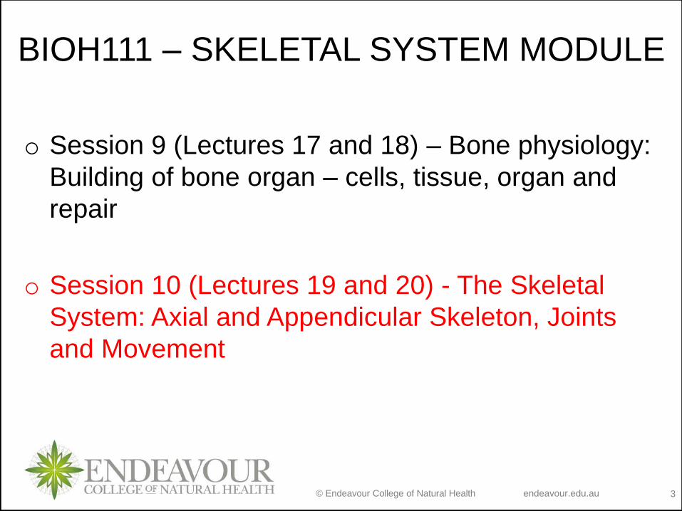
BIOH111 – SKELETAL SYSTEM MODULE
o Session 9 (Lectures 17 and 18) – Bone physiology:
Building of bone organ – cells, tissue, organ and
repair
o Session 10 (Lectures 19 and 20) - The Skeletal
System: Axial and Appendicular Skeleton, Joints
and Movement

BIOH111
Lectures 19 and 20
The Skeletal System
Axial and Appendicular Skeleton
Department of Bioscience
endeavour.edu.au

© Endeavour College of Natural Health endeavour.edu.au 5
OBJECTIVESLecture 19:
Types of bones
Describe the types of bones within the body and relate their shape to their structure
and function
Axial skeleton
Identify parts of axial skeleton and describe structure and function of its parts
Appendicular skeleton
Identify parts of appendicular skeleton and describe structure and function of its
parts
Lecture 20:
Types of joints
Structural and functional classification of joints
Describe the types of joints within the body and relate their shape to their structure
and function
Movement of synovial joints – in tutorial
Describe different movements (gliding, angular, rotation and special movements)
Describe factors affecting movements at synovial joints

© Endeavour College of Natural Health endeavour.edu.au 6
THE SKELETAL SYSTEM
o Axial Skeleton
• lies along longitudinal axis
• 80 bones
• skull, hyoid, ear ossicles,
trunk (vertebrae + ribs +
sternum)
o Appendicular Skeleton
• Extremities (appendages)
• 126 bones
• upper & lower limbs and
pelvic & pectoral girdles
© Endeavour College of Natural Health endeavour.edu.au 7
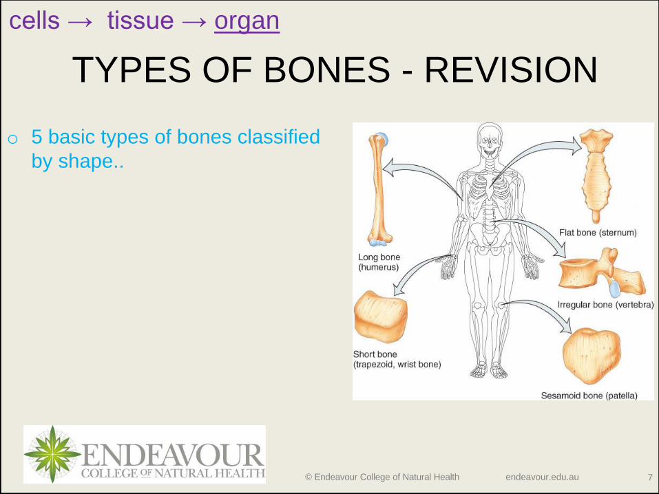
TYPES OF BONES - REVISION
o 5 basic types of bones classified
by shape..
cells → tissue → organ

© Endeavour College of Natural Health endeavour.edu.au 8
BONE SURFACE MARKINGS
Two major types of surface markings:
1. Depressions and openings -
participate in joints or allow the
passage of soft tissue; e.g.
foramen, fossa and sulcus
2. Processes - are projections or
outgrowths that either help form
joints or serve as attachment
points for connective tissue; e.g.
head, condyle and tuberosity

© Endeavour College of Natural Health endeavour.edu.au 9
AXIAL SKELETON
o 80 bones lie along longitudinal
axis
• Skull - cranial (8) and facial (14)
• Hyoid (1)
• Ear ossicles (6)
• Vertebrae (26)
• Ribs (24)
• Sternum (1)
o Function: protection of organs

© Endeavour College of Natural Health endeavour.edu.au 10
SKULL
o Forms the large cranial cavity and smaller
cavities, including the nasal cavity and
orbits (eye sockets). General features:
• Sutures
• Paranasal sinuses
• Moveable bones of the skull: ear ossicles
and mandible
o Composed of 22 bones: 8 cranial bones
(cranium) and 14 facial bones (face)

© Endeavour College of Natural Health endeavour.edu.au 11
CRANIAL BONES
Frontal
Sphenoid
Ethmoid
Temporal (2)
Parietal (2)
Occipital
Function: protect brain, house ear ossicles and are muscle attachment for jaw,
neck & facial muscles

© Endeavour College of Natural Health endeavour.edu.au 12
Function: protect delicate sense organs (smell, taste, vision); support
entrances to digestive and respiratory systems (mouth and nose)
FACIAL BONES
© Endeavour College of Natural Health endeavour.edu.au 13
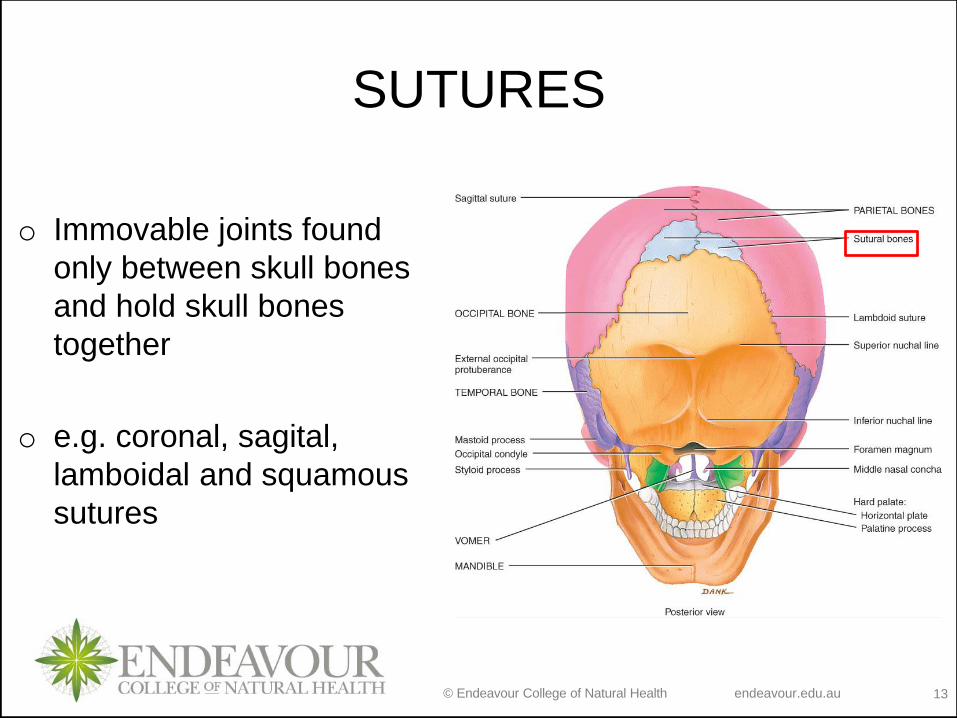
SUTURES
o Immovable joints found
only between skull bones
and hold skull bones
together
o e.g. coronal, sagital,
lamboidal and squamous
sutures

© Endeavour College of Natural Health endeavour.edu.au 14
PARANASAL SINUSES
o Cavities in specific skull bones lined by mucous membranes that
communicate with the nasal cavity; frontal, sphenoid, ethmoid and
maxillae
o Function: lighten the skull and serve as resonating chambers for
speech
o Sinusitis – inflammation of the paranasal sinuses membranes due
to infection or allergy

© Endeavour College of Natural Health endeavour.edu.au 15
FONTANELSo Only found in infants
o Non-ossified; dense connective
tissue filled spaces between the
cranial bones
o Functions: modification of infant
skull size and shape and permits
rapid growth of the brain during
infancy
o Major fontanels: anterior, posterior,
anterolaterals, and posterolaterals

© Endeavour College of Natural Health endeavour.edu.au 16
HYOID BONE
o U-shaped single bone;
o Articulates with no other bone of the body and is suspended by ligament and muscle
o Function: supports the tongue & provides attachment for tongue, neck and pharyngeal muscles
© Endeavour College of Natural Health endeavour.edu.au 17
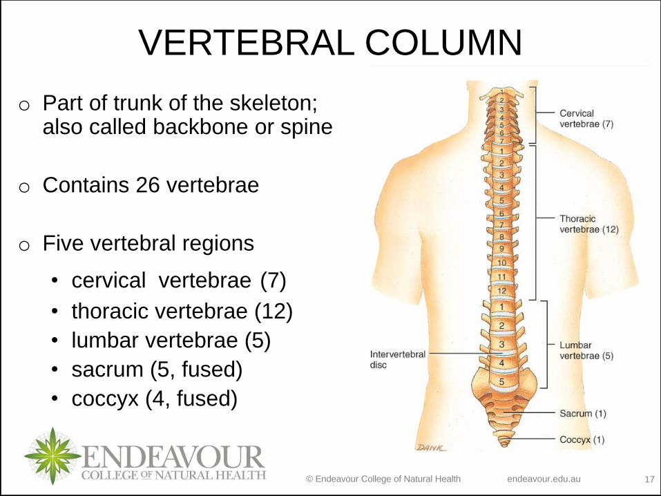
VERTEBRAL COLUMN
o Part of trunk of the skeleton; also called backbone or spine
o Contains 26 vertebrae
o Five vertebral regions
• cervical vertebrae (7)
• thoracic vertebrae (12)
• lumbar vertebrae (5)
• sacrum (5, fused)
• coccyx (4, fused)
© Endeavour College of Natural Health endeavour.edu.au 18
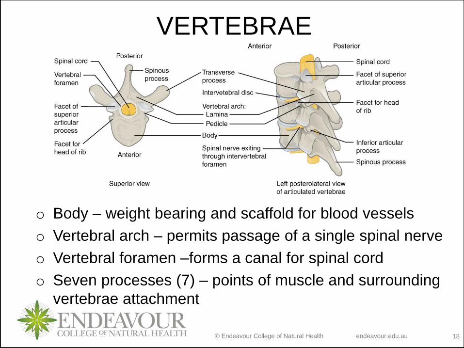
VERTEBRAE
o Body – weight bearing and scaffold for blood vessels
o Vertebral arch – permits passage of a single spinal nerve
o Vertebral foramen –forms a canal for spinal cord
o Seven processes (7) – points of muscle and surrounding
vertebrae attachment

© Endeavour College of Natural Health endeavour.edu.au 19
INTERVERTEBRAL DISCS
o Structure: Fibrocartilaginous ring with a pulpy center; between adjacent vertebrae absorbs vertical shock
o Function: permit various movements of the vertebral column

© Endeavour College of Natural Health endeavour.edu.au 20
CERVICAL REGION
o 7 cervical vertebrae
o Atlas - first cervical vertebra;
supports the skull
o Axis - second cervical
vertebra; permits side-to-side
rotation of the head
o Third to sixth - typical cervical
vertebrae.
o Prominens - seventh cervical
vertebra; base of the neck

© Endeavour College of Natural Health endeavour.edu.au 21
THORACIC VERTEBRAE (T1-T12)
o 12 thoracic vertebrae
o Larger and stronger then cervical
vertebrae
o Function: articulate with the ribs
via transverse process

© Endeavour College of Natural Health endeavour.edu.au 22
LUMBAR REGION
o 5 lumbar vertebrae
o Largest and strongest
vertebrae in the column
o Function: attachment of
large back muscles and
support of body weight
© Endeavour College of Natural Health endeavour.edu.au 23
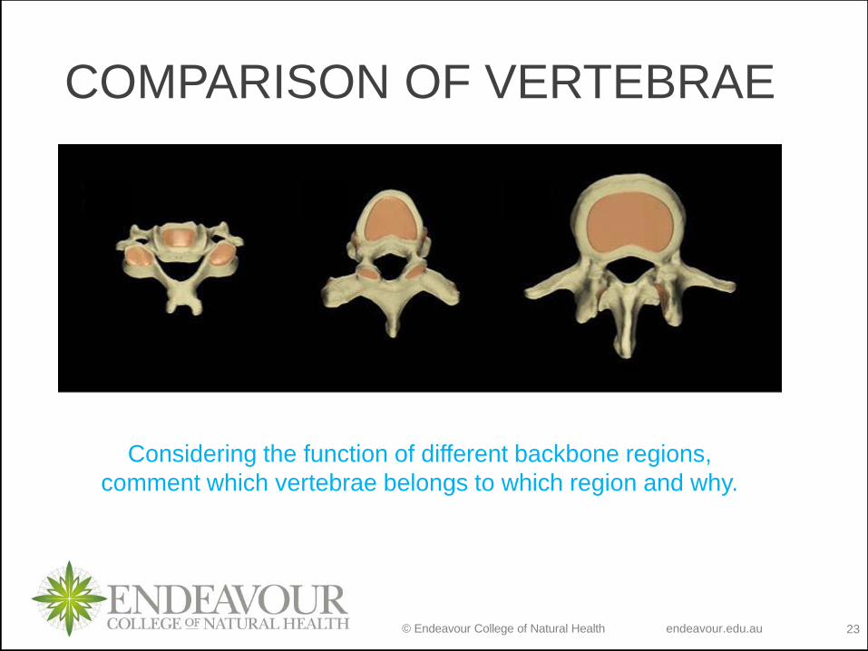
COMPARISON OF VERTEBRAE
Considering the function of different backbone regions,
comment which vertebrae belongs to which region and why.
© Endeavour College of Natural Health endeavour.edu.au 24
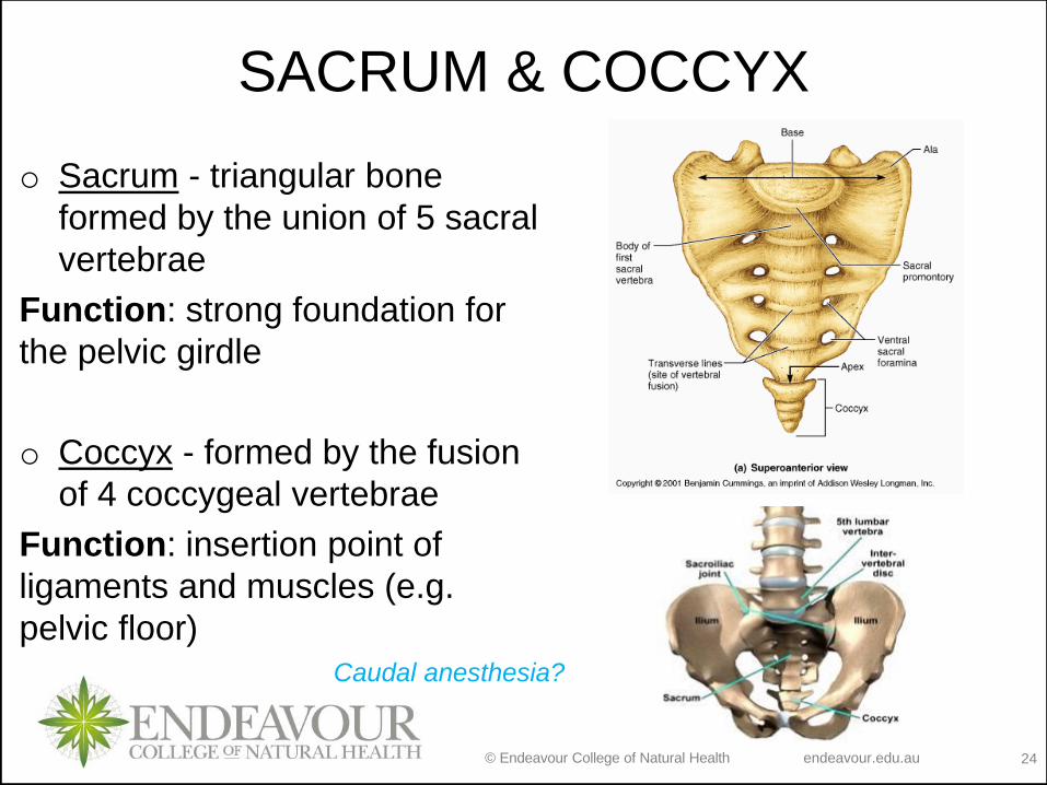
SACRUM & COCCYX
o Sacrum - triangular bone
formed by the union of 5 sacral
vertebrae
Function: strong foundation for
the pelvic girdle
o Coccyx - formed by the fusion
of 4 coccygeal vertebrae
Function: insertion point of
ligaments and muscles (e.g.
pelvic floor)
Caudal anesthesia?
© Endeavour College of Natural Health endeavour.edu.au 25
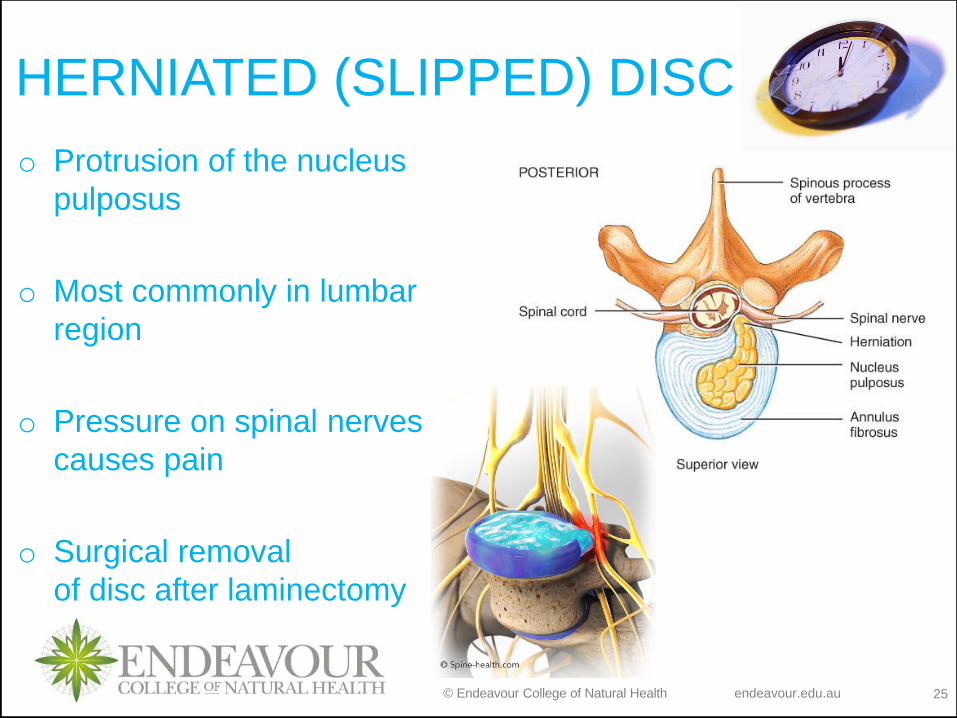
HERNIATED (SLIPPED) DISC
o Protrusion of the nucleus
pulposus
o Most commonly in lumbar
region
o Pressure on spinal nerves
causes pain
o Surgical removal
of disc after laminectomy

© Endeavour College of Natural Health endeavour.edu.au 26
NORMAL CURVES OF THE VERTEBRAL
COLUMN
o In adult, 4 normal vertebral
curves: cervical and lumbar
(anteriorly convex) and thoracic
and sacral (anteriorly concave)
o In the fetus, there is only a single anteriorly concave curve
• Primary curves: thoracic and sacral; formed during fetal development
• Secondary curves: cervical (formed at 4 months) and lumbar (formed at 1 year)
© Endeavour College of Natural Health endeavour.edu.au 27
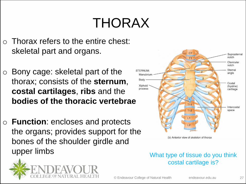
THORAX
o Thorax refers to the entire chest:
skeletal part and organs.
o Bony cage: skeletal part of the
thorax; consists of the sternum,
costal cartilages, ribs and the
bodies of the thoracic vertebrae
o Function: encloses and protects
the organs; provides support for the
bones of the shoulder girdle and
upper limbsWhat type of tissue do you think
costal cartilage is?

© Endeavour College of Natural Health endeavour.edu.au 28
STERNUMo Located on the anterior midline
of the thoracic wall
o Manubrium
• 1st & 2nd ribs
• clavicular notch
o Body
• costal cartilages of 2-10 ribs
o Xiphoid
• ossifies by 40
• CPR position
• abdominal mm
o Sternal puncture
• biopsy

© Endeavour College of Natural Health endeavour.edu.au 29
RIBS
o 12 pairs of ribs: pairs 1-7 are
true ribs; pairs 8-12 are false
ribs (pairs 11 and 12 are false
floating ribs).
o Function: structural support to
the sides of the thoracic cavity

© Endeavour College of Natural Health endeavour.edu.au 30
REVISION
Read Disorders: Homeostatic Imbalances (Tortora, p 227)
and define following terms: scoliosis, kyphosis, lordosis and
spina bifida. Think about:
- cellular and tissue level organisation if the skeleton that
may be affected
- think about the causes and possible treatments of these
disorders within your particular interest.

© Endeavour College of Natural Health endeavour.edu.au 31
APPENDICULAR SKELETON
o Includes 126 bones of the upper
and lower extremities and the
shoulder and hip girdles.
• Pectoral girdle
• Upper limbs
• Pelvic girdle
• Lower limbs
o Function: facilitation of
movement
© Endeavour College of Natural Health endeavour.edu.au 32
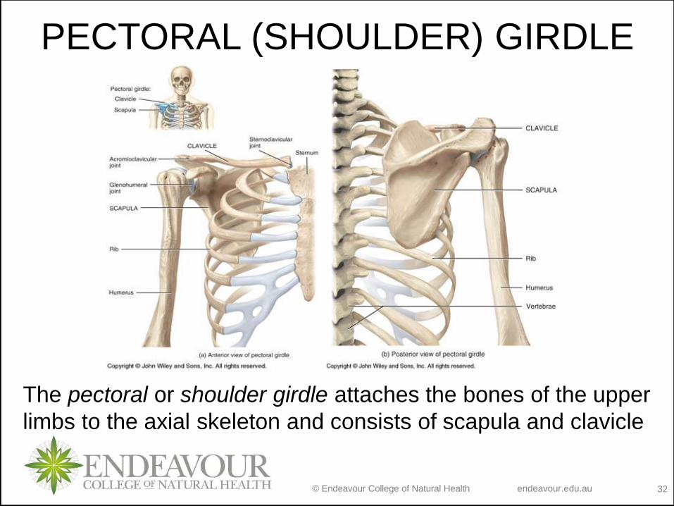
PECTORAL (SHOULDER) GIRDLE
The pectoral or shoulder girdle attaches the bones of the upper
limbs to the axial skeleton and consists of scapula and clavicle
© Endeavour College of Natural Health endeavour.edu.au 33
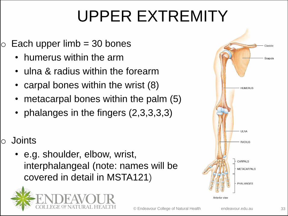
UPPER EXTREMITY
o Each upper limb = 30 bones
• humerus within the arm
• ulna & radius within the forearm
• carpal bones within the wrist (8)
• metacarpal bones within the palm (5)
• phalanges in the fingers (2,3,3,3,3)
o Joints
• e.g. shoulder, elbow, wrist,
interphalangeal (note: names will be
covered in detail in MSTA121)
© Endeavour College of Natural Health endeavour.edu.au 34
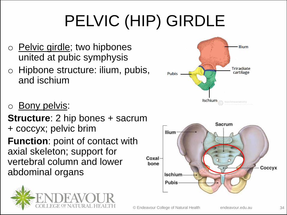
PELVIC (HIP) GIRDLE
o Pelvic girdle; two hipbones united at pubic symphysis
o Hipbone structure: ilium, pubis, and ischium
o Bony pelvis:
Structure: 2 hip bones + sacrum + coccyx; pelvic brim
Function: point of contact with axial skeleton; support for vertebral column and lower abdominal organs

© Endeavour College of Natural Health endeavour.edu.au 35
FALSE AND TRUE PELVIS
o False: greater cavity; encloses
parts of lower abdomen
including uterus, ovaries and
uterine tubes (♀)
o True: surrounds pelvic cavity
that includes vagina and uterus
(♀) or prostate (♂); pelvic inlet
and pelvic outlet; pelvic axis
o Pelvimetry

© Endeavour College of Natural Health endeavour.edu.au 36
FEMALE MALE
Pelvis:
♀ wider & shallower
Pelvic brim:
♀ wider and more oval
Pubic arch:
♀ >90 degrees
True pelvis:
♀ larger pelvic inlet &
outlet
♀ more space
Why are these
differences
important?

© Endeavour College of Natural Health endeavour.edu.au 37
LOWER EXTREMITY
o Each lower limb = 30 bones
• femur and patella (thigh)
• tibia & fibula (leg)
• tarsal bones (foot)
• metatarsals (forefoot)
• phalanges (toes)
o Joints
• e.g. hip, knee, ankle, proximal &
distal tibiofibular (note: names will
be covered in detail in MSTA121)

© Endeavour College of Natural Health endeavour.edu.au 38
FEMUR (THIGHBONE)
o Largest, heaviest and strongest
bone of the body
o It articulates with the hip bone
and the tibia.
o Function: muscle attachments
to aid movement

© Endeavour College of Natural Health endeavour.edu.au 39
PATELLA (KNEECAP)
o Sesamoid bone located anterior to the knee joint.
o Functions: increases the leverage of the tendon of the
quadriceps femoris muscle; maintains the position of the
tendon when the knee is bent; protects the knee joint

© Endeavour College of Natural Health endeavour.edu.au 40
TIBIA AND FIBULA
o Tibia or shinbone is the larger,
medial, weight-bearing bone of
the leg.
o The fibula is parallel and
lateral to the tibia.
© Endeavour College of Natural Health endeavour.edu.au 41
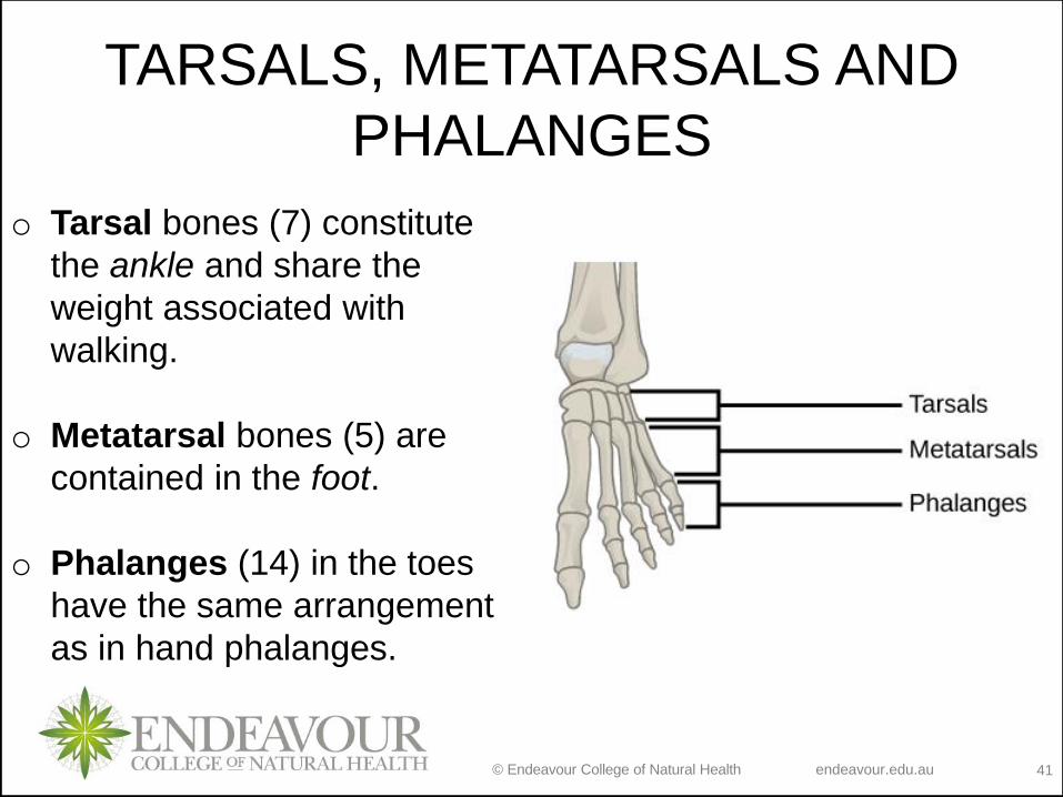
TARSALS, METATARSALS AND
PHALANGES
o Tarsal bones (7) constitute
the ankle and share the
weight associated with
walking.
o Metatarsal bones (5) are
contained in the foot.
o Phalanges (14) in the toes
have the same arrangement
as in hand phalanges.

© Endeavour College of Natural Health endeavour.edu.au 42
ARCHES OF THE FOOT
o Two non-rigid longitudinal arches along each side of foot and
one transverse arch
o Function: distribute body weight over foot; yield & spring back
when weight is lifted

© Endeavour College of Natural Health endeavour.edu.au 43
o Flatfoot
• weakened ligaments
allow bones of
medial arch to drop
o Claw foot
• medial arch is too
elevated
o Hip fracture
• 1/2 million/year in US
• osteoporosis
• arthroplasty
Clinical applications:

© Endeavour College of Natural Health endeavour.edu.au 44
OBJECTIVESLecture 19:
Types of bones
Describe the types of bones within the body and relate their shape to their structure
and function
Axial skeleton
Identify parts of axial skeleton and describe structure and function of its parts
Appendicular skeleton
Identify parts of appendicular skeleton and describe structure and function of its
parts
Lecture 20:
Types of joints
Structural and functional classification of joints
Describe the types of joints within the body and relate their shape to their structure
and function
Movement of synovial joints – in tutorial
Describe different movements (gliding, angular, rotation and special movements)
Describe factors affecting movements at synovial joints

© Endeavour College of Natural Health endeavour.edu.au 45
JOINTS
o Joints hold bones together but
permit movement
o Point of contact
• between 2 bones
• between cartilage and bone
• between teeth and bones
o Arthrology = study of joints
o Kinesiology = study of motion

© Endeavour College of Natural Health endeavour.edu.au 46
CLASSIFICATION OF JOINTSo Structural classification is based on the presence or absence
of a synovial cavity and type of connecting tissue:
• Fibrous – no synovial cavity; dense fibrous connective tissue; little
to no movement; 3 structural types
• Cartilaginous – no synovial cavity; fibrocartilage or hyaline
cartilage; little to no movement; 2 structural types
• Synovial – contain synovial cavity; hyaline cartilage called articular
cartilage; free movement; 6 structural types
o Functional classification based upon movement:
• Synarthrosis - immovable
• Amphiarthrosis - slightly movable
• Diarthrosis - freely movable

© Endeavour College of Natural Health endeavour.edu.au 47
FIBROUS JOINT TYPES
o Sutures:
• unite bones of the skull
• Synarthrosis joints
• Function: shock absorption –add strength and decrease chance of bone fractures
© Endeavour College of Natural Health endeavour.edu.au 48
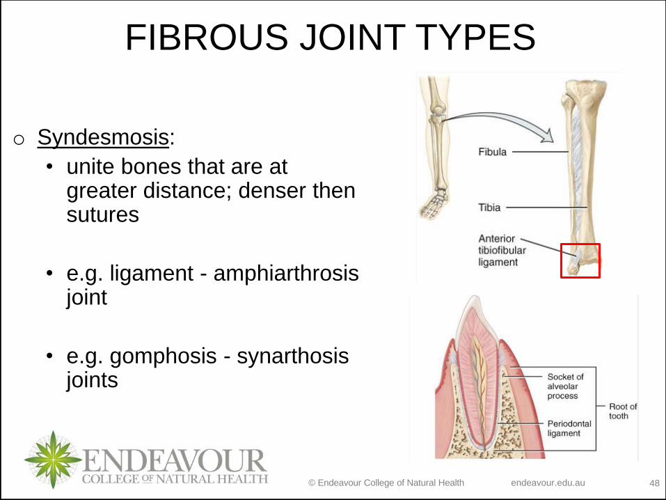
FIBROUS JOINT TYPES
o Syndesmosis:
• unite bones that are at greater distance; denser then sutures
• e.g. ligament - amphiarthrosisjoint
• e.g. gomphosis - synarthosisjoints

© Endeavour College of Natural Health endeavour.edu.au 49
FIBROUS JOINT TYPES
o Interosseous membrane:
• unite neighboring long bones; denser then syndesmosis
• Amphiarthrosis joint
• e.g. between fibula and tibia or between radius and ulna

© Endeavour College of Natural Health endeavour.edu.au 50
CARTILAGINOUS JOINT TYPES
o Synchondrosis:
• Hyaline cartilage
• Synarthrosis joint
• e.g. epiphyseal plate or joints
between ribs and sternum
• Function: permits growth
What do you already know about the epiphyseal plate?
© Endeavour College of Natural Health endeavour.edu.au 51
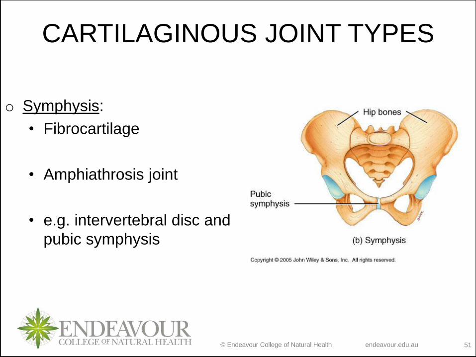
CARTILAGINOUS JOINT TYPES
o Symphysis:
• Fibrocartilage
• Amphiathrosis joint
• e.g. intervertebral disc and
pubic symphysis
© Endeavour College of Natural Health endeavour.edu.au 52
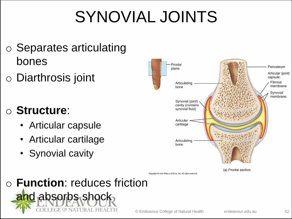
SYNOVIAL JOINTS
o Separates articulating
bones
o Diarthrosis joint
o Structure:
• Articular capsule
• Articular cartilage
• Synovial cavity
o Function: reduces friction
and absorbs shock
© Endeavour College of Natural Health endeavour.edu.au 53
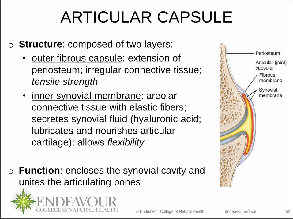
ARTICULAR CAPSULE
o Structure: composed of two layers:
• outer fibrous capsule: extension of
periosteum; irregular connective tissue;
tensile strength
• inner synovial membrane: areolar
connective tissue with elastic fibers;
secretes synovial fluid (hyaluronic acid;
lubricates and nourishes articular
cartilage); allows flexibility
o Function: encloses the synovial cavity and
unites the articulating bones
© Endeavour College of Natural Health endeavour.edu.au 54
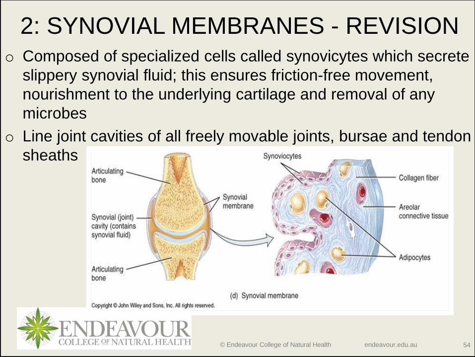
2: SYNOVIAL MEMBRANES - REVISIONo Composed of specialized cells called synovicytes which secrete
slippery synovial fluid; this ensures friction-free movement,
nourishment to the underlying cartilage and removal of any
microbes
o Line joint cavities of all freely movable joints, bursae and tendon
sheaths

© Endeavour College of Natural Health endeavour.edu.au 55
SPECIAL FEATURES OF SYNOVIAL
JOINTS
o Accessory ligaments
• Fibres of dense regular
connective tissue; resist
strains; extracapsular and
intracapsular ligaments
o Articular fat pads
• Accumulation of adipose tissue
in some synovial joints; e.g.
knee pad

© Endeavour College of Natural Health endeavour.edu.au 56
SPECIAL FEATURES OF SYNOVIAL
JOINTSo Articular disc or meniscus
• pads of fibrocartilage
attached around edges to
capsule; allow 2 bones of
different shape to fit tightly;
increase stability of knee -
torn cartilage
o Bursa
• not strictly part of synovial
joint; saclike structures
between structures (e.g.
skin/bone or tendon/bone or
ligament/bone); alleviate
points of possible friction
© Endeavour College of Natural Health endeavour.edu.au 57
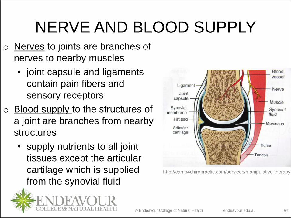
NERVE AND BLOOD SUPPLYo Nerves to joints are branches of
nerves to nearby muscles
• joint capsule and ligaments
contain pain fibers and
sensory receptors
o Blood supply to the structures of
a joint are branches from nearby
structures
• supply nutrients to all joint
tissues except the articular
cartilage which is supplied
from the synovial fluidhttp://camp4chiropractic.com/services/manipulative-therapy

© Endeavour College of Natural Health endeavour.edu.au 58
o Planar:
• bi-axial; bone surfaces are flat or
slightly curved
• side-to-side and back-and-forth gliding
movements; rotation prevented by
ligaments
• e.g. intercarpal joints
o Hinge:
• Uniaxial; convex/concave fit
• Flexion, extension and
hyperextension movements
• e.g. knee, elbow, interphalangeal joint
SYNOVIAL JOINT TYPES

© Endeavour College of Natural Health endeavour.edu.au 59
SYNOVIAL JOINT TYPESo Pivot:
• Monoaxial (allows only rotation around
longitudinal axis); rounded surface of bone
articulates with ring formed by second
bone & ligament
• Supination, pronation; turning head side-
to-side (NO)
• e.g. proximal radioulnar joint
o Ellipsoidal (Condyloid):
• Bi-axial; oval-shaped projection fits into
oval depression
• flex/extend or abduct/adduct
• e.g. wrist and metacarpophalangeal joints
© Endeavour College of Natural Health endeavour.edu.au 60
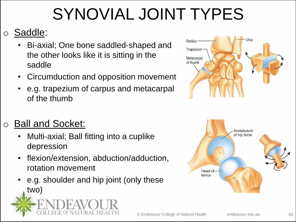
SYNOVIAL JOINT TYPESo Saddle:
• Bi-axial; One bone saddled-shaped and
the other looks like it is sitting in the
saddle
• Circumduction and opposition movement
• e.g. trapezium of carpus and metacarpal
of the thumb
o Ball and Socket:
• Multi-axial; Ball fitting into a cuplike
depression
• flexion/extension, abduction/adduction,
rotation movement
• e.g. shoulder and hip joint (only these
two)

© Endeavour College of Natural Health endeavour.edu.au 62
REVISIONRead Disorders: Homeostatic Imbalances (Tortora, p 287)
and define following terms: rheumatism, osteoarthritis,
rheumatoid arthritis, gouty arthritis, lyme disease, aging
and strain/sprain. Then ask:
- How each or some affect the joints and what is the
overall effect these conditions have on the
bones/joints/movement. How is this different between
the conditions?
- Think about the causes and possible treatments of these
disorders within your particular interest.
Non assessable
© Endeavour College of Natural Health endeavour.edu.au 63
MOVEMENT – GO TO TUTORIAL
Definition: an action of a body or a body part that is described
in a range of terms that indicate form of motion, direction of
motion or relationship of motion between two body parts.
Movement at synovial joints are grouped in 4 categories:
1. Gliding
2. Angular
3. Rotation
4. Special
© Endeavour College of Natural Health endeavour.edu.au 64
RANGE OF MOTION AT SYNOVIAL
JOINTS
Range of motion – range through which the bones of a joint
can be moved; measured in degrees of a circle. Factors that
affect ROM:
o Structure and shape of the articulating bone
o Strength and tautness of the joint ligaments
o Arrangement and tension of the muscles
o Contact of soft parts
o Hormones
o DisuseRead section 9.7 and discuss with your peers
any examples you may have for each.
© Endeavour College of Natural Health endeavour.edu.au 65
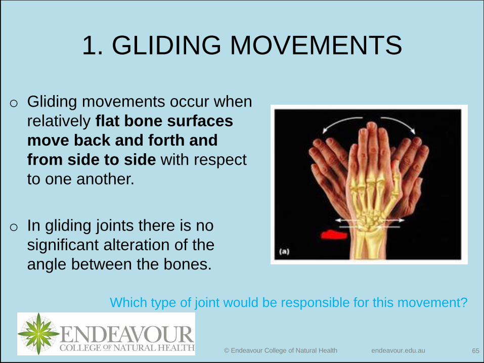
1. GLIDING MOVEMENTS
o Gliding movements occur when
relatively flat bone surfaces
move back and forth and
from side to side with respect
to one another.
o In gliding joints there is no
significant alteration of the
angle between the bones.
Which type of joint would be responsible for this movement?
© Endeavour College of Natural Health endeavour.edu.au 66
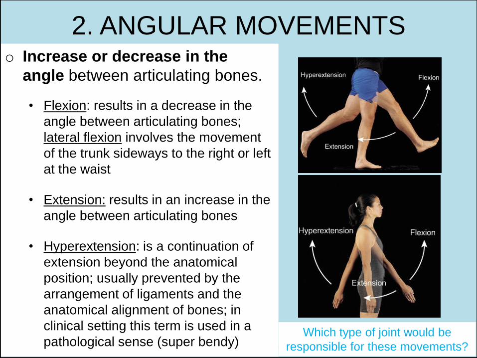
2. ANGULAR MOVEMENTSo Increase or decrease in the
angle between articulating bones.
• Flexion: results in a decrease in the
angle between articulating bones;
lateral flexion involves the movement
of the trunk sideways to the right or left
at the waist
• Extension: results in an increase in the
angle between articulating bones
• Hyperextension: is a continuation of
extension beyond the anatomical
position; usually prevented by the
arrangement of ligaments and the
anatomical alignment of bones; in
clinical setting this term is used in a
pathological sense (super bendy)Which type of joint would be
responsible for these movements?

© Endeavour College of Natural Health endeavour.edu.au 67
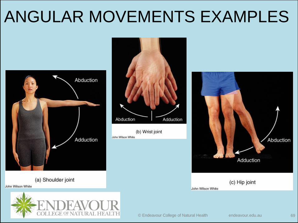
ANGULAR MOVEMENTS EXAMPLES

© Endeavour College of Natural Health endeavour.edu.au 68
• Abduction: movement of a bone
away from the midline
• Adduction: movement of a bone
toward the midline
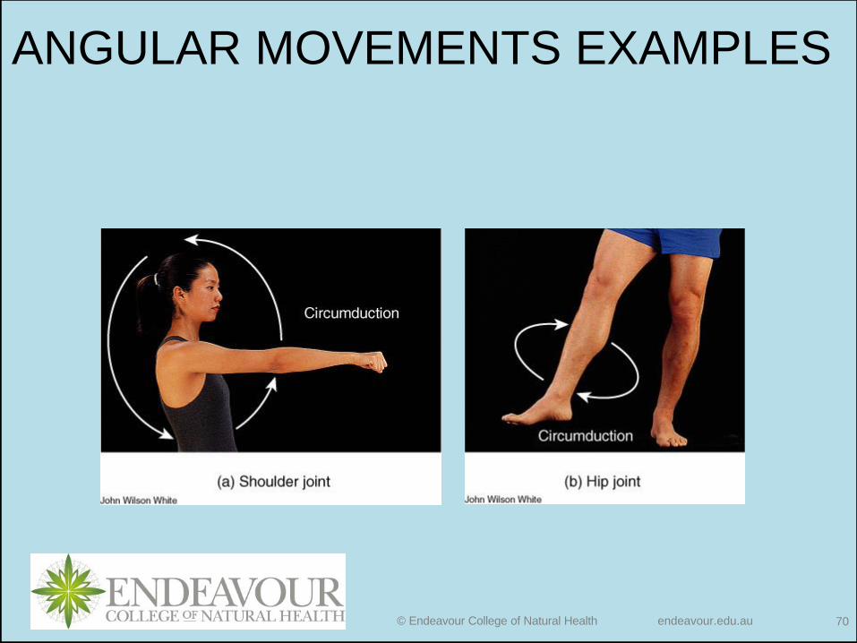
• Circumduction: movement of the
distal end of a part of the body in
a circle; occurs as a result of a
continuous sequence of flexion,
abduction, extension, and
adduction.
2. ANGULAR MOVEMENTS
Which type of joint would be responsible for this movement?
© Endeavour College of Natural Health endeavour.edu.au 69
ANGULAR MOVEMENTS EXAMPLES
© Endeavour College of Natural Health endeavour.edu.au 70
ANGULAR MOVEMENTS EXAMPLES

© Endeavour College of Natural Health endeavour.edu.au 71
o Bone revolves around its
own longitudinal axis
• Medial rotation: anterior
surface of a bone of the limb
is turned toward the
midline
• Lateral rotation: anterior
surface of a bone of the limb
is turned away from the
midline
3. ROTATION MOVEMENTS
© Endeavour College of Natural Health endeavour.edu.au 72
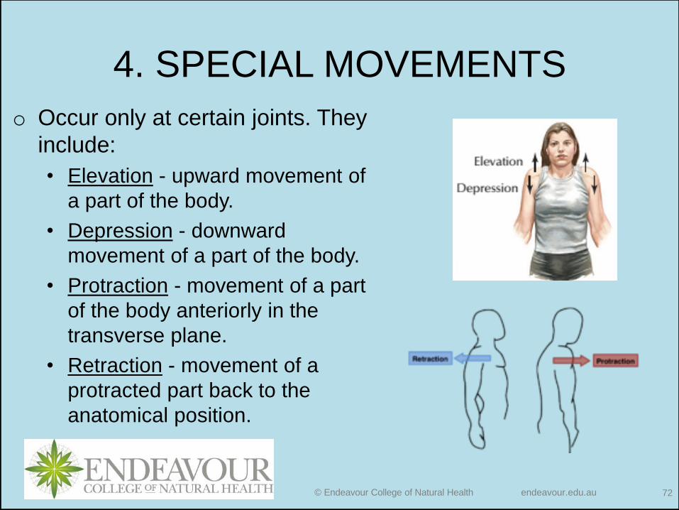
4. SPECIAL MOVEMENTS
o Occur only at certain joints. They
include:
• Elevation - upward movement of
a part of the body.
• Depression - downward
movement of a part of the body.
• Protraction - movement of a part
of the body anteriorly in the
transverse plane.
• Retraction - movement of a
protracted part back to the
anatomical position.

© Endeavour College of Natural Health endeavour.edu.au 73
4. SPECIAL MOVEMENTS
• Inversion - movement of the solesmedially at the intertarsal joints so that they face away from each other.
• Eversion - movement of the soleslaterally at the intertarsal joints so that they face away from each other.
• Dorsiflexion - bending of the foot at the ankle in the direction of the superior surface.
• Plantar flexion - bending of the foot at the ankle in the direction of the plantar surface.
© Endeavour College of Natural Health endeavour.edu.au 74
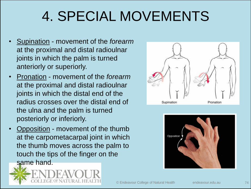
4. SPECIAL MOVEMENTS
• Supination - movement of the forearm
at the proximal and distal radioulnar
joints in which the palm is turned
anteriorly or superiorly.
• Pronation - movement of the forearm
at the proximal and distal radioulnar
joints in which the distal end of the
radius crosses over the distal end of
the ulna and the palm is turned
posteriorly or inferiorly.
• Opposition - movement of the thumb
at the carpometacarpal joint in which
the thumb moves across the palm to
touch the tips of the finger on the
same hand.

![BIOH111 [SESSION 1] Tutorial LANGUAGE OF · PDF fileBIOH111 [SESSION 1] Tutorial – LANGUAGE OF ANATOMY, CELLULAR ORGANISATION Learning Outcome(s) ...](https://static.fdocuments.net/doc/165x107/5aaf13de7f8b9a6b308ce26f/bioh111-session-1-tutorial-language-of-session-1-tutorial-language-of.jpg)


![BIOH111 [SESSION 5] Tutorial- CONNECTIVE & EPITHELIAL TISSUES · © Endeavour College of Natural Health e6fa289111474cda8e33df96da35d56d Last updated on 30-Jan-18 Page 1 of 13 BIOH111](https://static.fdocuments.net/doc/165x107/5b1c02f87f8b9a37258f5652/bioh111-session-5-tutorial-connective-epithelial-tissues-endeavour-college.jpg)


