Biofilm model systems and quantification toolstcoenye/iuap/Brackman.pdf · Biofilm model systems...
58
Biofilm model systems and quantification tools Gilles Brackman Lab of Pharmaceutical Microbiology, Ghent University, Ghent, Belgium; [email protected] 1
Transcript of Biofilm model systems and quantification toolstcoenye/iuap/Brackman.pdf · Biofilm model systems...

Biofilm model systems and
quantification tools
Gilles Brackman
Lab of Pharmaceutical Microbiology,
Ghent University, Ghent, Belgium; [email protected]
1

What are biofilms?
• Biofilms are sessile communities of
– prokaryotic and/or eukaryotic cells,
– attached to a substratum or interface or to each other,
– embedded in a matrix composed, at least partially, of self-produced extracellular material,
– exhibit an altered phenotype compared to planktonic cells (Donlan and Costerton, 2002)
2

Biofilms
CBE, MSU 3

Biofilms are everywhere
4

Biofilm models
Closed Batch Static
Open Flow
Dynamic
In vitro
In vivo Invertebrate Non-mammalian
vertebrate mammalian
Ex vivo
Microcosm
Microcosm
5

Batch systems vs flow systems • Closed system
• Mixing is optional
• Cont. flow stirred tank reactor (CFSTR) • Perfect mixing • Steady state
• Plug flow reactor (PFR)
• Mixing only in radial direction
Coenye and Nelis, 2010 J. Microbiol Methods
Co
nce
ntr
atio
n
Co
nce
ntr
atio
n
Co
nce
ntr
atio
n
6

In vitro batch systems
• Most commonly used • microtiter plate (MTP)
Step 1 • Adhesion step
• 0,5-4h
Step 2 • Remove PL cells
• Rinsing step
Step 3
• BF formation
• Fresh medium
• 20-48-….h
7

In vitro batch systems • MTP: biofilms formed on the bottom and walls of the plate
Brackman G., 2008 unpublished data
B. cenocepacia LMG 18828 (24h)
8

In vitro batch systems • MTP: biofilms formed on glass bottom
Udine et al., 2013, PloS ONE
B. cenocepacia LMG 16656 (WT and QS mutants) (24h; Live/Dead staining)
9

In vitro batch systems • MTP: biofilms formed on surface placed in MTP
• Medical grade silicone
• Orthopedic implant
• Catheters
• Contact lenses
• Hydroxyapatite disks
• Molars/teeth
• Coated materials
• ….
LPM, UGent
10

In vitro batch systems
• Minimum biofilm eradication concentration assay:
• The Calgary device (Ceri et al, 1999)
11

In vitro batch systems
• Minimum biofilm eradication concentration assay:
• The Calgary device (Ceri et al, 1999)
12

In vitro batch systems
• Minimum biofilm eradication concentration assay:
• The Calgary device (Ceri et al, 1999)
13 Harrison et al., 2006
Biol. Proced Online

14
In vitro Flow systems

In vitro flow systems: CFSTR
• Centers for disease control (CDC) biofilm reactor
Polyethylene Lid
“Water jacket”
1L vessel 8 Polypropylene coupon holders
Coupons
Magnetic stir
Media “IN” Air-vents
Media “OUT”
15

In vitro flow systems: CFSTR
• CDC biofilm reactor
set-up of the CDC biofilm reactor LPM, UGent. 16
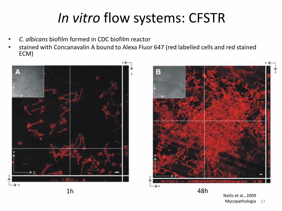
In vitro flow systems: CFSTR
• C. albicans biofilm formed in CDC biofilm reactor • stained with Concanavalin A bound to Alexa Fluor 647 (red labelled cells and red stained
ECM)
Nailis et al., 2009 Mycopathologia
1h 48h
17

In vitro flow systems: CFSTR
• CDC biofilm reactor
LPM, UGent
18

In vitro flow systems: CFSTR
• Other CFSTR systems
Rotating disk reactor Constant depth film
fermentor
Pratten, J. 2007 Curr. Protoc. Microbiol.
Biofilm annular reactor
19

In vitro flow systems: PFR
• Simple design of Flow cell set-up
Weiss Nielsen. 2011. J. Vis. Exp.
20

In vitro flow systems: PFR
• Flow cells
Transmission FC
Coupon evaluation FC
Capillary FC
21

In vitro flow systems: PFR • Flow cells: time-lapse view of biofilm treatment under flow conditions
Lorenz L, Buckingham-Meyer K, Pitts B, 2012, CBE, MSU
22

In vitro flow systems: PFR • Microfluidic devices
Benoit et al., 2010 Appl. Environ. Microbiol. 23

In vitro flow systems: PFR • P. fluorescence biofilm development in microfluidic device
Benoit et al., 2010 Appl. Environ. Microbiol.
Flow direction
24

In vitro flow systems: PFR
• Modified Robbins device (MRD)
McBain A.J., 2009 Adv. Appl. MIcrobiol
25

In vitro flow systems: PFR
• Modified Robbins device (MRD)
set-up of the MRD BF model LPM, UGent.
26

In vitro flow systems: PFR
• Modified Robbins device (MRD)
LPM, UGent. 27

In vitro flow systems: PFR
• Modifications on the MRD design
Garcia et al., 2010. J. Microbiol. Meth. 28

In vitro flow systems: PFR
• Implemented in industrial environments:
29

In vitro flow systems: PFR
• (Colony-)Drip flow reactor (DFR)
Goeres et al., 2009, Nature Protocols McBain A.J., 2009. Adv. Appl Microbiol.
Method E2647-08 Annual Book of ASTM Standards
30

In vitro flow systems: PFR
• (Colony-)Drip flow reactor
Goeres et al., 2009, Nature Protocols Method E2647-08 Annual Book of ASTM Standards 31

In vitro flow systems: PFR
• Polymicrobial wound biofilm in a C-DFR
Woods et al, 2012. J. Appl. Microbiol. 32

Simple in vitro flow systems
Uppuluri and Lopez-Ribot, 2010. Virulence.
Biofilms Hypertextbook P. Stoodley and J. Lennox
33

34
Ex vivo/Microcosms: Specific Biofilm models

• Lubbock chronic wound pathogenic biofilm model (LCWPB)
• Rapid (24h)
• Multispecies (Sa, Pa, Ef)
• Macro/Microscopically resembles in vivo wound biofilms
Sun et al., 2008. Wound Repair Regen.
Microcosms: Wound Biofilm model
35

• Collagen matrix with simulated wound fluid
• P. aeruginosa
• S. aureus
•Microscopically resembles in vivo wound biofilms
Werthen et al., 2010. APMIS.
Microcosms: Wound Biofilm model
In vitro In vivo 36

Ex vivo biofilm models
• Reconstituted Human Epithelia (RHE)
• Human sinosal epithelial, skin, molars,…
• Microvascular endothelial cells (HMEC-1 Cells)
• HeLa cells
• CF-derived bronchial cells
• U2OS osteosarcoma cells
•…..
37

Overview
MTP/Batch: Flow systems:
Less labor intensive More labor intensive
No specialized equipment Require specialized
equipment/skills
Relatively cheap more expensive
multiplexing Limitation/run
≠ in vivo situation? Fluid Flow ≠ in vivo situation?
Microcosm/cell-culture-based model 38
39
In vivo (non-)mammalian Biofilm models

In vivo non-mammalian biofilm models
Lebeaux et al, 2013 Pathogens
40

In vivo non-mammalian biofilm models
Lebeaux et al, 2013 Pathogens
41

In vivo non-mammalian biofilm models
Brackman G., 2013, unpublished data
42
Caenorhabditis elegans

Caenorhabditis elegans
Atkinson et al. 2011 PLoS Path.
In vivo non-mammalian biofilm models
43
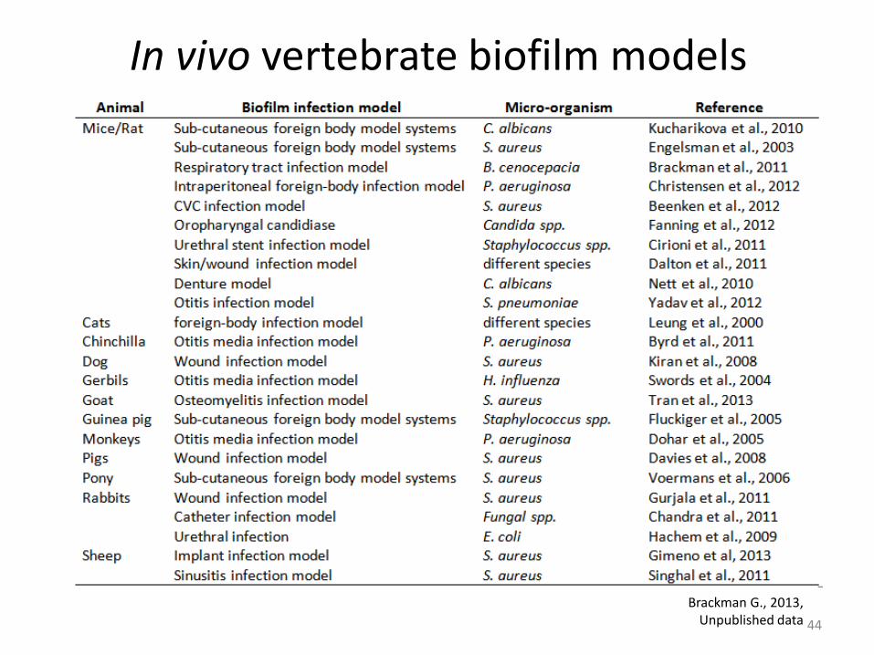
In vivo vertebrate biofilm models
Brackman G., 2013, Unpublished data 44

In vivo vertebrate biofilm models
Rat sub-cutaneous foreign body model
Van Wijngaarden et al. 1999 , AAC Ricicova et al., 2010, Microbiology
45

Invertebrate vs vertebrate
Relatively cheap High throughput Ethical concerns?
Natural
pathology?
Natural pathology?
Limited
Larger= higher
costs
46

Why is it important?
Smith et al. 2013 Infection and Immunity.
Based on Nailis et al. 2010 BMC Microbiology.
Susceptibility (Sa) Gene expression (Ca)
Importance of selecting a model that closely fits the research question in a certain research frame
47

Quantification tools
Direct quantification
Plating
Counting the colonies
Indirect quantification
Staining Labelling
Intensity ≈ # cells
48

Direct quantification
• Absolute cell numbers • Mixed BF: Possible to use selective
conditions • Isolates are available for further
research
• Time consuming & labor-intensive • Time to results is organism dependent
(>18h) • VBNC are not accounted for • Artificial situation might hinder growth
49

Great plate count anomaly
Lewis et al. 2010 ASM Microbeblog Staley and Konopka, 1985 Ann. Rev. Microbiol
• Often observed in biofilms (although
systematic studies are lacking!) • If you plate pieces of the biofilm:
• no recovery
• If you first disperse the biofilm: • recovery is much better
• Importance of dispersing the biofilm
• vortexing and sonication vs scraping
50

Indirect quantification Staining
• Crystal violet (living & dead cells, part of the matrix)
• DMMB (matrix in some organisms; particularly useful for staphylococci)
• Calcofluor white (EPS stain, N-acetylglucosamine)
• SYTO9 (living & dead cells, DNA in the matrix)
• FDA, XTT/MTT/…, CellTiter Blue/Resazurin/Alamar Blue (metabolically active
cells)
… and many more!
51

Resazurin staining
52
O
N
H
OHOH
hydroresorufin

Indirect quantification Luminescent (LuxCDABE) S. aureus
Plaut et al., 2013 Plos ONE
53

Indirect quantification
Brackman et al., 2013, Unpublished data.
• Fast, high-throughput • Equipment • What are you measuring?
CTB fluorescence signal of a S. aureus Mu50 Biofilm after treatment
54
• Optimization is required
0.00E+00
5.00E+04
1.00E+05
1.50E+05
2.00E+05
2.50E+05
3.00E+05
3.50E+05
4.00E+05
untreated CTRL AB(1) AB(2)
flu
ore
sce
nce
(cp
m)
0.5h
1h
95 %
60 %
6 %
84 %

Indirect quantification
Brackman et al., 2013, J. Appl Microbiol.
Correlation between S. aureus pMV158gfp fluorescence signal and total CFU harvested from the biofilms.
• Detection limit for quantification
55
• Mixed biofilms?

Combined method: PMA-qPCR
• Streptococcus spp. • P. gingivalis • A. actinomycetemcomitans • F. nucleatum • V. parvula, • P. intermedia • L. monocytogenes • …..
Sanchez et al., 2013, J. Periodont. Res. Yasunaga et al., 2013, BMC Microbiol
56

Take home messages
• Model systems are essential to study microbial biofilms • In combination with various quantification approaches they allow to mimic
in vivo situations in vitro • Importance of selecting a Biofilm model/quantification method that
closely fits the research question in a certain research frame • Selection based on
• Research question, Resources, level of the research, Preferences,…
• Caution extrapolating results from one model/method to another
57