Biofeedback, Vol 9, No. 2 - Fischell Department of Bioengineering
16
A NEWSLETTER FOR ALUMNI AND FRIENDS OF THE FISCHELL DEPARTMENT OF BIOENGINEERING AT THE A. JAMES CLARK SCHOOL OF ENGINEERING, UNIVERSITY OF MARYLAND, COLLEGE PARK. www.bioe.umd.edu BIOF E EDBACK THE FISCHELL DEPARTMENT of BIOENGINEERING A. JAMES CLARK SCHOOL of ENGINEERING VOL. 9, No. 2 Finding ways to see, posi- tion, measure, and accurately manipulate nanoscale objects is an ongoing challenge for re- searchers developing the next generation of ultra-compact electronics, sensors and opti- cal devices. Even the most advanced conventional microscopes are limited by diffrac- tion of the short- est wavelength of visible light, about 400 nanometers, rendering them unable to produce images or measure- ments of objects that are signifi- cantly smaller than this threshold. Researchers attempt to solve this problem by using “reporting probes.” A near-field scanning optical microscope (NSOM), for example, is equipped with a probe attached to a fine mechanical tip that can scan a nanoscale object and create an image based on the electromagnetic field it generates. But NSOMs are complex, delicate and expensive pieces of equipment, and the presence of the tip disturbs the interaction between the probe and the sample, distorting the image. A study published in Nature Communications by University of Maryland (UMD) researchers, including Fischell Department of Bioengineering (BioE) professor Benjamin Shapiro, describes a novel technique for imaging far below the diffraction limit by using a particle that is much smaller than the wavelength of light as an optical probe. The particle is manipulated with high precision using an inexpensive microfluidic device. The breakthrough has enabled the researchers to capture nanoscale measurements with a spatial accuracy of 12 nanometers. Quantum Dots: Nanoscopic Spotlights in a Microscopic River A quantum dot is a 3–6 nanometer-sized, semiconducting particle about 25 times the diameter of a single atom. At room temperature, quantum dots can emit single photons of light that can be tuned to a IN THIS ISSUE: 2 FACULTY NEWS 5 ENTREPRENEURSHIP 6 STUDENT NEWS: UNDERGRADUATE 7 STUDENT AWARDS 7 RECENT DISSERTATIONS 10 STUDENT NEWS: GRADUATE 12 2013 CAPSTONE PROJECTS 14 EDUCATION NEWS 14 ALUMNI NEWS 15 CHAIR’S MESSAGE continues pg. 2 Researchers Achieve Breakthrough in Nanoprecision Imaging PROBING WITH A SINGLE QUANTUM DOT (QD). (A) IMAGE OF THE MICROFLUIDIC DEVICE. FLUID MOTION IS ACTUATED THROUGH THE FOUR CHANNELS TO POSITION PARTICLES SUSPENDED WITHIN THE CONTROL REGION. (B) A QD IS POSITIONED ALONG A TRAJEC- TORY TO PROBE THE REGION AROUND AN IMMOBILIZED AgNW. COUPLING TO THE WIRE IS MEASURED USING BOTH THE RADIATION FROM THE WIRE END AND THE DOT’S EMIS- SION LIFETIME.
Transcript of Biofeedback, Vol 9, No. 2 - Fischell Department of Bioengineering

A NEWSLETTER FOR ALUMNI
AND FRIENDS OF THE FISCHELL
DEPARTMENT OF BIOENGINEERING
AT THE A. JAMES CLARK SCHOOL
OF ENGINEERING, UNIVERSITY OF
MARYLAND, COLLEGE PARK.
www.bioe.umd.edu
BIOFEEDBACKTHE FISCHELL DEPARTMENT of BIOENGINEERING
A. JAMES CLARK SCHOOL of ENGINEERING
VOL. 9, No. 2
Finding ways to see, posi-tion, measure, and accurately manipulate nanoscale objects is an ongoing challenge for re-searchers developing the next generation of ultra-compact electronics, sensors and opti-cal devices. Even the most advanced conventional microscopes are limited by diffrac-tion of the short-est wavelength of visible light, about 400 nanometers, rendering them unable to produce images or measure-ments of objects that are signifi-cantly smaller than this threshold.
Researchers attempt to solve this problem by using “reporting probes.” A near-field scanning optical microscope (NSOM), for example, is equipped with a probe attached to a fine mechanical tip that can scan a nanoscale object and create an image based on the electromagnetic field it generates. But NSOMs are complex, delicate and expensive pieces of equipment, and the presence of the tip disturbs the interaction between the probe and the sample, distorting the image.
A study published in Nature Communications by University of Maryland (UMD) researchers, including Fischell Department of Bioengineering (BioE) professor Benjamin Shapiro, describes a novel technique
for imaging far below the diffraction limit by
using a particle that is much smaller than the wavelength of light as an optical probe. The particle is manipulated with high precision using an inexpensive microfluidic device. The breakthrough has enabled the researchers to capture nanoscale
measurements with a spatial accuracy of 12 nanometers.
Quantum Dots: Nanoscopic Spotlights in a Microscopic River
A quantum dot is a 3–6 nanometer-sized, semiconducting particle about 25 times the diameter of a single atom. At room temperature, quantum dots can emit single photons of light that can be tuned to a
IN THIS ISSUE:
2 FACULTY NEWS
5 ENTREPRENEURSHIP
6 STUDENT NEWS: UNDERGRADUATE
7 STUDENT AWARDS
7 RECENT DISSERTATIONS
10 STUDENT NEWS: GRADUATE
12 2013 CAPSTONE PROJECTS
14 EDUCATION NEWS
14 ALUMNI NEWS
15 CHAIR’S MESSAGE
continues pg. 2
Researchers Achieve Breakthrough in Nanoprecision Imaging
PROBING WITH A SINGLE QUANTUM DOT (QD). (A) IMAGE OF THE MICROFLUIDIC DEVICE. FLUID MOTION IS ACTUATED THROUGH THE FOUR CHANNELS TO POSITION PARTICLES SUSPENDED WITHIN THE CONTROL REGION. (B) A QD IS POSITIONED ALONG A TRAJEC-TORY TO PROBE THE REGION AROUND AN IMMOBILIZED AgNW. COUPLING TO THE WIRE IS MEASURED USING BOTH THE RADIATION FROM THE WIRE END AND THE DOT’S EMIS-SION LIFETIME.

BIOFEEDBACK VOL. 9 NO. 2
2
desired wavelength. This makes them ideal probes for examining nanostructures smaller than the visible light threshold. Positioned close to a nanoscale object, the quantum dot becomes a sort of spotlight that amplifies what the microscope alone cannot see.
The problem? It’s hard to capture and scan a single quantum dot over another nanoscale object.
The UMD team’s solution lies in a microfluidic device that manipulates and positions quantum dots using precision flow control. A computer algorithm analyzes the dots dispersed inside, selecting one to be the reporting probe. As the microfluidic device creates a fluid flow, the targeted dot begins to move. An image-guided feedback process continually tracks the dot’s location and adjusts the flow accordingly. For example, if the dot is observed to be to the northwest of its desired location, a southeast flow is created to move it into place.
This technique gives researchers the ability to manipulate a single dot precisely, guiding it quickly to desired locations, and holding it in each position with nanometer accuracy so it can be used to scan objects. The dot’s response to each scanned object is measured, providing information about the object’s electromagnetic fields with nanoscale resolution. Since nothing mechanical touches the quantum dot or affects its interaction with the objects it scans, the images produced are distortion-free, clean and sharp.
A Superior, Less Expensive Technique
“In other particle manipulation techniques—for example laser tweezers—the force applied to a particle scales with its volume,” explains Shapiro, one of the paper’s co-authors. “But the viscous forces that the fluid flow applies scale with the diameter of the particle. At the nanoscale, fluid flow has a greater effect on the particle than competing techniques, allowing us to move, guide and immobilize the quantum dot more easily and accurately.”
In addition to its technical superiority, the new nanoscale manipulation system is far less expensive than near-field scanning optical microscopy, which requires equipment that costs hundreds of thousands of dollars.
“The new technique is more versatile, easier to implement, and more accurate by an order of magnitude than conventional NSOM,” says Shapiro’s colleague, Department of Electrical and Computer Engineering and Institute for Research in Electronics and Applied Physics professor Edo Waks. “Basically, we can take a microscope, add a disposable microfluidic device, and beat the capabilities of an NSOM at a fraction of the cost and complexity.
“An undergraduate could build the basic two-channel microfluidic device used in the process, using standard soft-fabrication techniques, in less than an hour for under $50,” he adds.
The UMD team is hoping to package all of the necessary system components into an inexpensive add-on product for microscopes.
Support for the Research
Support for this research has been provided by the Defense Advanced Research Projects Agency (DARPA) and the Office of Naval Research (ONR). The project evolved from early research supported by Shapiro’s 2004 National Science Foundation grant.
In addition to Shapiro and Waks, research team members and co-authors include: Professor John Fourkas (Department of Chemistry & Biochemistry and Institute for Physical Science & Technology), Ph.D. stu-dents Chad Ropp (Electrical and Computer Engineering) and Zachary Cummins (BIOE), and alumnus Sanghee Nah (Ph.D. ’12, Chemistry).
For more information, see:
“Nanoscale imaging and spontaneous emission control with a single nano-positioned quantum dot,” Nature Communications, DOI 10.1038/ncomms2477, published online February 5, 2013.
researchNEWS continued from cover story
JAY JOINS BIOE FACULTY
The Fischell Department of Bioengineering and the A. James Clark School of Engineering are pleased to welcome the newest member of their faculty, Assistant Professor Steven M. Jay.
Jay, who earned his Ph.D. from Yale in 2009, specializes in vascular biology
and bioengineering, tissue and protein engineering, and therapeutic vascularization, all with an emphasis on clinical translation. He will direct the Vascular Pharmacoengineering and Biotherapeutics Laboratory, which will focus on developing novel drug delivery systems, enhancing tissue engineering techniques, and understanding the quantitative biology of interactions between drug carriers and cells.
Prior to joining the University of Maryland and the Clark School, Jay was a postdoctoral fellow in the Department of Cardiovascular Medicine at Brigham and Women’s Hospital and the Harvard Medical School in Cambridge, Mass., where he designed new proteins to promote diabetic wound healing. The proteins interact with specific receptors on the surfaces of the endothelial cells that line the inside of blood vessels, stimulating their growth and development. The presence of a healthier, more robust network of blood vessels in the bed of a non-healing wound should accelerate or enhance the healing process.
Jay is the recipient of a Pathway to Independence Award from the National Institutes of Health (NIH). In addition to funding his mentored fellowship, the prestigious award includes a three-year, $735,000 grant he will use to establish his research group and continue his work on the wound healing project at the Clark School. His other past honors and awards include a 2012 Innovation in Biotechnology Award from American Association of
facultyNEWS
STEVEN M. JAY

A. JAMES CLARK SCHOOL of ENGINEERING GLENN L. MARTIN INSTITUTE OF TECHNOLOGY
3
Pharmaceutical Scientists and a 2011 Department of Defense Breast Cancer Postdoctoral Fellowship.
Jay was drawn to the Clark School’s facilities and its relationships with local federal labs and biotech companies, but it was BioE’s strong reputation and unique environment that convinced him he’d found the right place to launch his academic career.
“The [BioE] faculty have diverse training and bring many different perspectives to everything they do, which I think is a real strength and will help keep the department ahead of the curve,” he says. “Many faculty members are engaged and enthusiastic about making UMD the best place it can be and providing the best opportunities possible for all of our [students]. It was really refreshing to see that attitude and I look forward to contributing to it myself.”
Jay will teach his first course, BIOE 340: Modeling Physiological Systems, in Spring 2014, and plans to introduce a new course in pharmaceutical engineering.
SHAPIRO PROMOTED
The Fischell Department of Bioengineering (BioE) and the Institute for Systems Research (ISR) extend their congratulations to Benjamin Shapiro, who was elevated to the rank of Professor effective July 1, 2013.
For Shapiro, it’s been an unusual journey, one he sometimes finds himself explaining when asked how and why someone who started out as an aerospace engineer ended up a bioengineer, moving cells around lab-on-a-chip devices and devising new drug delivery techniques.
The answer lies in liquid. As an undergraduate, he specialized in aeronautic fluid dynamics. He pursued an interest in control systems as a graduate student, but the focus was still on jet engines. When he joined the Clark School’s Department of Aerospace Engineering and ISR in 2000, his thoughts returned to fluid dynamics, but at a different scale.
“I had just started becoming interested in microfluidics, and fluid flows on small
scales,” he explains. “As I became serious about it, I realized that most of the high-impact applications were in areas like chemical testing, manipulating cells, and DNA separation–there were far fewer applications in jet engines and spacecraft. All of the problems I studied were leading me toward chemistry and biology, and eventually clinical topics. By the time I took my sabbatical at the National Institutes of Health and the National Institute for Standards and Technology in 2009, I was doing almost exclusively biological and medical research. At that point I thought, you know, it’s getting more and more challenging to explain to people why I’m in the aerospace department.” By the time he returned in 2010, he had transferred to BioE.
In the end, making the risky, career-changing move was worth it. “It’s fantastic,” he says, adding that he appreciates his growing connections with the doctors, start-up companies, the NIH and FDA, and with his colleagues on the bioengineering faculty. “There’s no question that being surrounded by people who are doing synergistic things is a tremendous benefit. It amplifies my work.”
As the director of the Control of Miniaturized Systems for Mechatronic, Biological and Clinical Applications Laboratory his research is focused on all aspects of control of medical devices and drug targeting, from initial determination of the dominant physics, to model development, control problem statement and algorithm design, to experimental verification. In recent years, he has applied his expertise in flow control systems and microelectromechanical systems (MEMS) to a diverse group of research projects, including the development of a new technique for drug delivery to the inner and middle ear, the
facultyNEWS
use of flow control and quantum dots to achieve nanoprecise imaging and fabrication (see cover story), neonatal ventilator safety, defect-free colloidal crystal assembly for optoelectronic metamaterials, and controlled delivery of chemotherapy to deep tissue tumors using magnetic nanoparticles. He is also the co-editor of and contributor to a book titled Feedback Control from MEMS to Atoms. In 2012 he delivered a plenary talk at the International Conference on Manipulation, Manufacturing and Measurement on the Nanoscale, and he recently attended his first Gordon Research Conference.
Shapiro is one of a growing number of BioE faculty who have taken department benefactor and inventor Robert E. Fischell’s ideals of entrepreneurship, clinical translation, and interdisciplinary research to heart. Commercialization efforts are underway for some of the technologies he and his colleagues have developed.
“We’re really making a push to get the stuff that we’re doing here in the lab out to patients,” he says. “We want to make sure that the technology we’re developing will be in hospitals and clinics.”
Shapiro, who received his Ph.D. from the California Institute of Technology in
2000, also holds an appointment with the Maryland NanoCenter, and is
affiliated with the Applied Math and Scientific Computation Program. He is the recipi-ent of a 2003 NSF CAREER award and a 2009 Fulbright Scholarship. He has filed 20 patents based on his research,
two of which were awarded 1st and 3rd places
in the university’s annual Office of Technology Commer-cialization’s Invention of the Year Competition.

BIOFEEDBACK VOL. 9 NO. 2
4
appointment is with Wuhan’s School of Resource and Environmental Science.
Founded in 1893, Wuhan University is the country’s third most important center of education and research. The school maintains partnerships and collaborations with over 350 schools around the world. Over 50,000 students attend classes on its campus, located in the largest city in central China.
Payne is engaged in an ongoing collaboration with Wuhan University School of Resource and Environmental Science professor Xiao-Wen Shi. Prior to joining Wuhan’s faculty, Shi spent 10 years working with Payne, BioE professor and chair William E. Bentley, and BioE affiliate professors Reza Ghodssi (director, Institute for Systems Research) and Gary Rubloff (director, Maryland NanoCenter) at the University of Maryland. Their partnership, the Biochip Collaborative, resulted in UMD’s emergence as a world leader in the use of biofabrication–nanoscale construction using materials and mechanisms from biology–to create interfaces between biological and electronic components.
At Wuhan, Payne and Shi are continuing their work with chitosan, a renewable, biode-gradable, and a stimuli-responsive bipolymer derived from crabs and shrimp that responds to imposed electrical signals.
Payne was officially honored with the appointment at a ceremony held in March. Wuhan Committee Chair Xiao-Rong Chen and Vice President Qing-Yun Du presided over the event, at which they recognized
Payne’s research accomplishments in creating high performance materials from renewable natural resources. Du says he hopes the new relationship will continue to strengthen the ties and scientific collaborations between Wuhan University and the University of Maryland.
found everywhere from plants, to soil, to skin and hair. Their properties vary and they serve many purposes, including facilitating intercellular communication in plants and
animals, wound healing, and disinfection, and are present as aromas or flavors in foods.
Though abundant in nature, phenolics remain relatively unstudied. Bentley and Payne are
among the few scientists who have acquired a deeper understanding
of their antioxidant and electrical properties, thanks to a novel characterization technique developed by Payne
Group Research Associate Dr. Eunkyoung Kim. As part of
the Maryland Biochip Collaborative, the professors and their groups are exploring the use of phenolics as interfaces between biological and electronic components in sensors and biomedical devices.
For more information on the Bentley and Payne Groups’ work with phenolics, see: Eunkyoung Kim, Tanya Gordonov, Yi Liu, William E. Bentley, and Gregory F. Payne. “Reverse Engineering To Suggest Biologically Relevant Redox Activities of Phenolic Materials.” ACS Chem. Biol. 2013, 8, 716−724.
For Bentley and Payne’s Perspectives column, see Science 12 July 2013: 136-137. (DOI:10.1126/science.1241562)
PAYNE HONORED WITH GUEST PROFESSORSHIP
BioE professor Gregory Payne (joint, Institute for Bioscience and Biotechnology Research) has received a Guest Professorship at Wuhan University, one of China’s top ten educational institutions. The three-year
SCIENCE GETS BENTLEY AND PAYNE’S PERSPECTIVE ON SELF-ASSEMBLY
BioE and Institute for Bioscience & Biotechnology Research professors William E. Bentley and Gregory F. Payne shared their thoughts on phenolics, chemical compounds which could be used to fabricate new biomaterials with advanced functionality, in the July 12 issue of Science. The pair was invited to author a column for the journal’s Perspectives section, in which experts comment on the papers presented in each issue.
The piece, titled “Nature’s Other Self-Assemblers,” reviews new research that suggests phenolics could be the next big thing in the field of self-assembling biological materials. Phenolics are a class of molecules that are abundant in nature,
WUHAN UNIVERSITY VICE PRESIDENT QING-YUN DU (LEFT) PRESENTING PROFESSOR GREGORY PAYNE WITH A GUEST PROFESSORSHIP AT A CEREMONY ON MARCH 7.
FISHER CO-EDITS BOOK ON TISSUE ENGINEERING
BioE professor and associate chair
John Fisher is the co-editor of and
contributor to a new book titled
Tissue Engineering: Principles and Practices, available from
CRC Press.
The book’s
three sections,
“Fundamentals,”
“Enabling
Technologies,” and
“Applications” are
designed to guide
readers through
the field, covering
the latest opinions
and research on
topics including
nanobiomaterials,
stem cells,
biomimetics,
cartilage and
dental tissue, gene
therapy, artificial organs, and cellular,
vascular and neural engineering. The
book also provides information on
the materials and techniques used
in regenerative medicine, such as
scaffolding, drug delivery systems,
and bioreactors. Contributors have
been drawn from clinical practice,
academia and industry.
facultyNEWS continued

A. JAMES CLARK SCHOOL of ENGINEERING GLENN L. MARTIN INSTITUTE OF TECHNOLOGY
5
Remedium was previously awarded a $150,000 SBIR Phase I grant from the NSF, two Maryland Industrial Partnerships (MIPS) grants totaling $206,000, a $140,000 Maryland Proof of Concept Alliance grant, a $75,000 Maryland Technology Development Corporation Maryland Technology Transfer Fund grant, and a $200,000 Maryland Biotechnology Center Translational Research Award. In 2009, it received the University of Maryland’s Outstanding Invention of the Year Award in the Life Sciences division from the Office of Technology Commercialization.
The young company has been highly successful in business plan competitions, winning first prize and $25,000 in the Oak Ridge National Laboratory’s 2010 Global Venture Challenge; the “Most Promising Security Idea” award and $10,000 in the 2009 4th Annual Global Security Challenge; $8000 in the Maryland Technology Enterprise Institute’s 2007 $50K Business Plan Competition; and $5,000 in the Invest Maryland Challenge.
Remedium has six patents pending related to the Hemogrip platform. Its products, which also include surgical sprays and bandages, are designed to be used by surgeons, soldiers, EMTs, or even unskilled helpers, in locations ranging from the operating room to the battlefield to emergency situations.
DIAGNOSTIC anSERS TAKES 3RD AND UMD PRIZE AT CUPID’S CUP FINALS
Diagnostic anSERS, the Fischell Department of Bioengineering-based startup company founded by graduate students Eric Hoppmann and Sean Virgile, both advised by BioE and Institute for Systems Research assistant professor Ian White, took 3rd place in the final round of the Cupid’s Cup, the national business competition held by the Dingman Center for Entrepreneurship at the University of Maryland’s Robert H. Smith School of Business. The company also won the UMD Prize, awarded to the startup utilizing the most campus resources. The competition was founded by UMD alumnus Kevin Plank, CEO of Under Armour.
The company received a total of $10,000, which they will use to scale up manufacturing for a product release this fall. Diagnostic anSERS uses a novel ink jet printing process to fabricate inexpensive substrates for surface enhanced Raman spectroscopy (SERS), a high-end molecular fingerprinting technique usually confined to a lab.
Diagnostic anSERS says its product will make advanced sensor technology available to more people, both in the lab and in the field, because it uses off-the-shelf technology and other readily available components. The printed sensors, which can be produced on-demand and on location by anyone who has had some basic instruction, are used to detect trace amounts of explosives, toxins and narcotics.
“Our $4 piece of paper is able to perform better than our $105 competitor,” says Virgile.
COMPANY FOUNDED IN CLARK SCHOOL AWARDED $500K TO TEST HEMORRHAGE-HALTING FOAM
Story courtesy of and based on the original by Mtech.
Remedium Technologies Inc., a medical device company founded by Clark School alumni and faculty developing innovative products to control severe hemorrhaging, was awarded a $500,000 Small Business Innovation Research (SBIR) grant from the
National Science Foundation. The award will fund testing of the company’s sprayable foam, which is designed to rapidly halt bleeding caused by large and deep traumatic injuries.
In collaboration with Massachusetts General Hospital and the University of Maryland, Remedium will complete pre-clinical trials to evaluate the safety and efficacy of Hemogrip™ in controlling non-compressible hemorrhaging, bleeding that cannot be slowed or stopped using direct pressure. Hemogrip is a high-pressure, sprayable foam that can expand into an injured body cavity, adhering to tissue and stopping bleeding within minutes. There are currently no hemostatic products avail-able for the treatment of non-compressible wounds, which account for 85 percent of hemorrhage-related deaths.
The grant will also support additional product research by the Clark School’s Complex Fluids and Nanomaterials Group, directed by Remedium co-founder Professor Srinivasa Raghavan (Department of Chemical and Biomolecular Engineering).
“Remedium is honored to be recognized for its product development progress with this important Phase II funding from the National Science Foundation,” says Matthew Dowling (Ph.D. ’10), CEO and co-founder of Remedium. “We are enthusiastic in approaching pre-clinical trials with a product we see as critical in addressing one of the big-gest unmet needs in trauma medicine today.”
Hemogrip’s life-saving technology is based on chitosan—a natural biopolymer found in the exoskeleton of shrimp, crabs, and other crustaceans. Chitosan is unique as a natural material because it is biocompatible, anti-microbial, and highly durable under a wide range of environmental conditions. When applied to wounds, Hemogrip creates a nanoscale, three-dimensional mesh, rapidly coagulating blood and staunching blood loss.
The Hemogrip foam is dispensed from a handheld, lightweight canister. It can be removed quickly and easily without damaging tissue, and since it is based on chitosan—the second most abundant biopolymer on earth—it is also inexpensive.
entrePRENEURSHIP
FISCHELL DEPARTMENT OF BIOENGINEERING GRADUATE STUDENTS AND DIAGNOSTIC anSERS COFOUNDERS ERIC HOPPMANN (LEFT) AND SEAN VIRGILE (RIGHT).
MATTHEW DOWLING

BIOFEEDBACK VOL. 9 NO. 2
6
BIOE UNDERGRAD WINS PYON SU FELLOWSHIP
BioE rising senior Brian Goodall received the University of Maryland’s Pyon Su Fellowship to support his participation in an engineering student exchange program at the Pohang University of Science and Technology (POSTECH) in South Korea.
The 5 million won (about $4600) award is named in honor of the nation’s first Korean college student, Pyon Su, a UMD alumnus who graduated in 1891 from what was then the Maryland Agricultural College. The Pyon Su Fellowship supports students who wish to study abroad at Korean universities.
Goodall was inspired to study abroad in Asia after a trip to China. He decided to pursue opportunities in Korea because he has friends there, and he chose POSTECH for its strengths in science, engineering, math and robotics.
three-dimensional constructs that closely mimic real tissue or organs by laser printing hydrogels that contain multiple layers of cells, growth factors, and vascular networks. While the new technology has great potential to replace damaged organs and heal burns, Isser explains, it is also controversial because of concerns that it could unnaturally extend human life, turn body parts into commodities, or distort how “human” we are. Isser agreed the concerns were valid and should be addressed, but that “preventing the field from advancing could be seen as akin to allowing innocent patients to suffer or die.”
Mitchell’s second place essay, “Ethics of Human Enhancement in Bioengineering,” explored the ethical differences between curative and preventive medicine and enhancements that may unnaturally increase human performance and lifespan. What is considered an “enhancement,” she wrote, may be tempered by cultural evolution. For example, advancements such as prosthetic limbs, joint replacements, organ transplants, and vaccinations are now widely accepted medical practices rather than viewed as unnatural enhancements. Mitchell argued that we should not apologize for our achievements in medicine, nor should we fear the ethical reviews of these achievements that inform the public and allow for acceptance or rejection. Change is natural, she concluded, and it is acceptable to pursue solutions our vulnerabilities.
Dziki’s third place essay, “The Bioethics of Stem Cell Research,” took on one of society’s most controversial biomedical debates, weighing the potential to heal injury and illness against safety and ethical concerns. “I believe that stem cell research is morally justified because of its potential to teach us about disease, and to help us develop drugs and cell-based therapies to treat so many currently incurable diseases,” she wrote. “However, I believe stem cell research must be practiced in a way that is responsible.”
FOR THE EXPANDED VERSION OF THIS
STORY, VISIT: ter.ps/bioethics13
BIOE UNDERGRADS SWEEP BIOETHICS ESSAY CONTEST
BioE freshmen (now sophomores) Kristina Dziki, Ariel Isser, and Renee Mitchell swept the top three spots in the Institute of Biological Engineering’s (IBE) annual bioethics essay contest. The students presented their essays as semifinalists at the IBE’s annual conference in March 2013, where the winners were decided. All three were students in Lecturer Idalis Villanueva’s BIOE 120: Biology for Engineers class.
In the past six years, Clark School bioengineering majors from the University of Maryland have represented 21 of the 29 total finalists, scoring three first place, three second place, and five third place wins, as well as numerous Honorable Mentions.
Isser took first place for his essay, “Bio-Printing: Extending Life at What Expense?” Bio-printing, an emerging practice within the field of tissue engineering, creates
THIS FALL, WE’LL WELCOME 107 FRESHMEN, BRINGING OUR TOTAL UNDERGRADUATE ENROLLMENT TO OVER 440. ALMOST HALF ARE WOMEN.
The quality of the students in our rapidly-growing program never ceases to
amaze us. The members of the incoming Class of 2017 have an average GPA of
4.28 and an average combined SAT score of 1396.
With each passing year, our undergraduates win more scholarships, fellowships,
competitions and awards (see pp. 8-9); become more involved in research; and
produce increasingly sophisticated and patentable prototypes in their senior
Capstone Design course (see pp. 12-13). Some co-author papers in peer-reviewed
journals before they graduate. Many are accepted into medical school, graduate
school, or M.D./Ph.D programs.
In spring 2013 their accomplishments were so numerous we couldn’t fit every
story—or even all of the details—into this issue of Biofeedback. Look for the
ter.ps links in this section (and throughout the issue), which will guide you to more
information online. For example:
• Senior Ben Bulka won the Outstanding ASPIRE Student Research Award for
his efforts to understand how stem cells might be used to treat degeneration
of the spine: ter.ps/bulkaaspire
• Sophomore Joan Zhang was part of a team that won UMD’s “Code for
Community Challenge” for web site and mobile app development:
ter.ps/zhangcode
You can learn more about our undergraduate program on our newly-redesigned
web site at bioe.umd.edu/undergraduate.
studentNEWS: UNDERGRADUATE
7
Congratulations to the following students, who were recognized at
the Clark School’s 2012-2013 Honors and Awards Ceremony held
this spring. They have all demonstrated outstanding academic
and research performance, and have made contributions to the
Department and field. Complete award citations are available on our
web site at: ter.ps/bioeawards13
PROFESSIONAL SOCIETY AWARDS
• The Institute of Biological Engineering’s (IBE) Annual National Bioethics Essay Contest (see story, p. 6):
Ariel Isser: First Place Renee Mitchell: Second PlaceKristina Dziki: Third Place
DEPARTMENT AWARDS
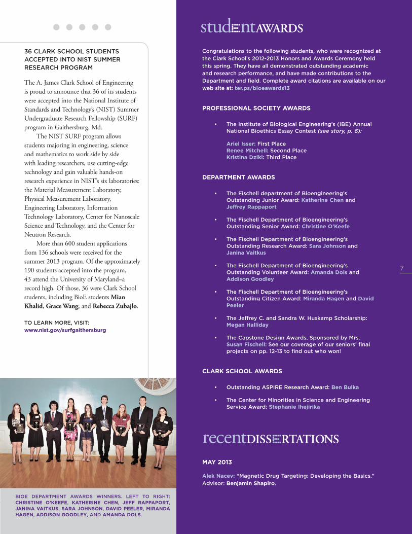
• The Fischell department of Bioengineering’s Outstanding Junior Award: Katherine Chen and Jeffrey Rappaport
• The Fischell Department of Bioengineering’s Outstanding Senior Award: Christine O’Keefe
• The Fischell Department of Bioengineering’s Outstanding Research Award: Sara Johnson and Janina Vaitkus
• The Fischell Department of Bioengineering’s Outstanding Volunteer Award: Amanda Dols and Addison Goodley
• The Fischell Department of Bioengineering’s Outstanding Citizen Award: Miranda Hagen and David Peeler
• The Jeffrey C. and Sandra W. Huskamp Scholarship: Megan Halliday
• The Capstone Design Awards, Sponsored by Mrs. Susan Fischell: See our coverage of our seniors’ final projects on pp. 12-13 to find out who won!
CLARK SCHOOL AWARDS
• Outstanding ASPIRE Research Award: Ben Bulka
• The Center for Minorities in Science and Engineering Service Award: Stephanie Ihejirika
recentDISSERTATIONS
MAY 2013
Alek Nacev: “Magnetic Drug Targeting: Developing the Basics.”
Advisor: Benjamin Shapiro.
studEntAWARDS
36 CLARK SCHOOL STUDENTS ACCEPTED INTO NIST SUMMER RESEARCH PROGRAM
The A. James Clark School of Engineering is proud to announce that 36 of its students were accepted into the National Institute of Standards and Technology’s (NIST) Summer Undergraduate Research Fellowship (SURF) program in Gaithersburg, Md.
The NIST SURF program allows students majoring in engineering, science and mathematics to work side by side with leading researchers, use cutting-edge technology and gain valuable hands-on research experience in NIST’s six laboratories: the Material Measurement Laboratory, Physical Measurement Laboratory, Engineering Laboratory, Information Technology Laboratory, Center for Nanoscale Science and Technology, and the Center for Neutron Research.
More than 600 student applications from 136 schools were received for the summer 2013 program. Of the approximately 190 students accepted into the program, 43 attend the University of Maryland–a record high. Of those, 36 were Clark School students, including BioE students Mian Khalid, Grace Wang, and Rebecca Zubajlo.
TO LEARN MORE, VISIT:
www.nist.gov/surfgaithersburg
BIOE DEPARTMENT AWARDS WINNERS. LEFT TO RIGHT: CHRISTINE O’KEEFE, KATHERINE CHEN, JEFF RAPPAPORT, JANINA VAITKUS, SARA JOHNSON, DAVID PEELER, MIRANDA HAGEN, ADDISON GOODLEY, AND AMANDA DOLS.

BIOFEEDBACK VOL. 9 NO. 2
8
MARYAM MUKHAMEDOVA: SEARCHING FOR NEW TREATMENTS FOR MS
Multiple sclerosis (MS), an autoimmune disease with no known cure, affects hundreds of thousands of people in the United States alone.* MS causes the immune system to unpredictably attack a nerve-insulating protein called myelin as if it were a foreign invader, disrupting communications throughout the central nervous system.
Although drugs that suppress the disease’s faulty response to myelin are avail-able, they also often suppress the healthy activities of the immune system, leav-ing patients vulnerable to other ill-nesses. Many of the drugs are also hy-drophobic, making them difficult to be dissolved, dispersed and absorbed in the human body, which is 60-70 percent water.
Rising senior Maryam Mukhamedova, a member of BioE assistant professor Christopher Jewell’s research group, is part of a team developing biomaterials that could help treat autoimmune disorders and infectious diseases by fine-tuning and controlling the body’s natural immune system response.
Mukhamedova is studying a biomaterial designed deliver insoluble drugs more effec-tively while selectively promoting immuno-logical tolerance. The system could eventually help treat diseases like MS, in which stopping the specific attacks on myelin without affect-ing healthy immune system responses would provide an effective course of treatment.
Mukhamedova says the use of biomaterials to treat autoimmune disorders has great potential, and she is excited to be part of the field now, in its early stages. “If
HHMI FELLOWSHIPS
The competitive HHMI Fellowship program, co-sponsored by HHMI and the University of Maryland’s College of Computer, Mathematical, and Natural Sciences, funds the research activities of undergraduates interested in pursuing careers in medical, biological or life sciences. The program’s goals are to immerse students in the investigative process, increase their aptitude for research, and provide them with the opportunity to collaborate directly with a faculty mentor.
JEFF RAPPAPORT: SAFE AND EFFECTIVE TARGETED DRUG DELIVERY SYSTEMS
Jeff Rappaport joined BioE professor Silvia Muro’s group in Spring 2012 after discover-ing his love for cell biology. His work sup-ports her lab’s overall mission to investigate, design, and improve treatments of rare, inherited diseases.
The Muro Group has been developing new strategies for the safe and effective delivery of therapeutics to specific kinds of cells, particularly by taking advantage of cells’ natural ability to ingest molecules from their surroundings and transport them across biological barriers, processes known as endocytosis and transcytosis, respectively.
Rappaport’s project focuses on the treat-ment of patients suffering from lipidosis, a group of genetic disorders that limit endocy-tosis and allow the buildup of lipids (fats) in cells, ultimately resulting in multiple organ dysfunction and failure. Treatment options for a number of these disorders, he says, cur-rently range from limited to non-existent.
Rappaport hopes to overcome one of the barriers to treatment, the diminished endocytic activity, by enhancing the delivery of lipid-digesting medicinal enzymes to patients that lack them. Preliminary results suggest that new compounds developed by Muro Group collaborators can revive endocytosis in diseased cells, allowing the drug delivery system pioneered by Muro to enter the cells and treat the underlying disorder. If successful, a treatment plan that combines the two groups’ innovations will both help revive endocytic activity and successfully deliver the enzymes the cells need to break down the excess lipids.
“I will grow cells in the laboratory, make them artificially sick, treat them with these compounds and with fluorescent markers for endocytosis, and then analyze the therapeutic effect using fluorescence microscopy,” Rappaport explains. “Hopefully, this will open up new avenues of treatment for patients with deadly genetic disorders.”
Prior to his current project, Rappaport worked with BioE graduate student and fellow Muro Group member Janet Hsu on strategies for delivering therapeutics across the blood/brain barrier. In the summer of 2012, he studied cellular motor proteins at the National Institutes of Health.
BIOE SENIOR AND HHMI UNDERGRADUATE RESEARCH FELLOW JEFF RAPPAPORT ON ASSIGNMENT WITH ENGINEERS WITHOUT BORDERS, INSTALLING SOLAR PANELS IN BURKINA FASO.
studentNEWS: UNDERGRADUATE IMPACTUNDERGRADS MAKE BIG CONTRIBUTIONS TO BIOMEDICAL RESEARCH
Students don’t have to wait until graduate school to get involved in research. Many of the
Fischell Department of Bioengineering’s undergraduates take advantage of opportunities
to work in department or affiliated laboratories, gaining valuable experience that can help
them hone their interests and give them an edge when applying for jobs or to graduate
programs. This year, three of our undergraduate researchers received Howard Hughes
Medical Institute (HHMI) Undergraduate Research Fellowships that supported their work on
projects that could have a significant impact on human health. A fourth—a past HHMI Fellow
himself—was named one of the University of Mayland’s six Undergraduate Researchers of
the Year in recognition of his three years of tissue engineering research.
MARYAM MUKHAMEDOVA

A. JAMES CLARK SCHOOL of ENGINEERING GLENN L. MARTIN INSTITUTE OF TECHNOLOGY
9
all goes well, I will be able to add just a little piece to a very big puzzle of autoimmune disease and that itself will give me a feeling of exuberance.”
Mukhamedova was already an experienced researcher when she joined to the Jewell Research Lab. In 2011-2012, she worked for Department of Biology professor Sergei Sukharev on a project in which she recorded current and ion flow into and out of cells. In the spring and summer of 2012, she investigated amylin aggregation and its effect on diabetes under the guidance of George Washington university professor Aleksander Jeremic.
She says that it was important to her to find her niche during her undergraduate studies to ensure she’d end up in a career she loves. Her research experiences have helped her do just that. After earning her B.S in bio-engineering, Mukhamedova plans to attend a graduate immunology program.
* “Multiple Sclerosis: Hope Through Research.” National Institute of Neurological Disorders and Stroke, National Institutes of Health
KENNY ROSENBERG: MORE EFFECTIVE TREATMENTS FOR OSTEOARTHRITIS
While words “regenerative medicine” are most often encountered in the context of a doctor’s office or a hospital, Kenny Rosenberg (B.S. ’13) has already gotten his start in the discipline. As a member of BioE associate professor and associate chair Adam Hsieh’s Orthopaedic Mechanobiology Lab, he worked to understand how more effective treatments for osteoarthritis could be developed.
Rosenberg, who was mentored by Hsieh and BioE graduate student Julianne Twomey (see related story, p. 10), studied how stem cell response could be engineered to improve treatments for degenerative joint disorders, such as osteoarthritis and degenerative disc disease. “Wear and tear” on cartilage and fibrocartilage tissues causes those who suffer from these diseases lose the cushioning, shock absorption, and flexibility in their joints, resulting in inflammation and pain. The
tissue damage affects both chondrocytes (cartilage cells) and the extracellular matrix, the structural proteins manufactured by chondrocytes.
The implantation of human mesenchymal stem cells (hMSCs) at damaged intervertebral disc or car-
tilage sites has been proposed as a potential therapy because they are capable of becoming adult chondrocytes.
“Unfortunately,” says Rosenberg, “this technique has had little success because these [mesenchymal stem] cells have difficulty overcoming the microenvironment of the damaged tissue.”
Rosenberg, Twomey and Hsieh hoped to overcome this problem by gaining a better understanding of the cells’ mechanotransduction machinery, the means by which they sense their environment and respond by converting external mechanical signals into biochemical ones. The hope is that implanted cells can be engineered to respond to their damaged environment in a way that will be therapeutic.
Rosenberg examined the role of the pericellular matrix (PCM), the coating found immediately surrounding the hMSCs as they differentiate from stem cells to chondrocytes.
“The PCM had previously been shown to buffer the biomechanical stimuli of the cell and is [therefore] likely to play an important role in mechanotransduction,” he explains. Understanding how the PCM affects the hMSCs’ response to the mechanical stress they experience could enable bio- and tissue engineers to create modified cells capable of surviving implantation into areas of damaged cartilage, so they can successfully transform and rebuild what has been lost.
Rosenberg was involved in research throughout his undergraduate career, including serving an internship at the National Institutes of Health’s National Institute on Aging. His goal is to pursue M.D. and Ph.D. degrees.
THOMPSON: UNDERGRADUATE RESEARCHER OF THE YEAR
Joshua Thompson (B.S. ’13) was named one of the University of Maryland’s six 2013 Undergraduate Researchers of the Year by the Maryland Center for Undergraduate Research.
Thompson, who was selected from a highly competitive group of nominees working in diverse fields throughout the university, was recognized for his accomplishments in tissue engineering. He had been a member of BioE professor and associate chair John Fisher’s Tissue Engineering and Biomaterials Laboratory since the summer of 2010.
“In addition to his outstanding research performance,” says Fisher, “Josh is known as a friendly and attentive worker who is ready to assist colleagues. He continually demonstrates his ability to quickly understand new concepts and master experimental techniques. He approaches his work with a high level of dedication...{and he is] an excellent collaborator. I can confidently state that he ranks as one of the most gifted, qualified, and dedicated undergraduate researchers I have mentored.”
Thompson distinguished himself in undergraduate research throughout his time at the Clark School.
In the fall of 2012, he was named one of the UMD’s Philip Merrill Presidential Scholars. A past HHMI fellowship supported his study of a polymer with applications in bone tissue engineering, and a HHMI International Research Program Grant funded a semester abroad at the University of Sydney, where he studied regenerative medicine and biomaterials. In 2011, he received the Maryland Technology Enterprise Institute’s Outstanding ASPIRE Research Award for his study of the efficacy of porous scaffolds as gene delivery devices for skeletal muscle regeneration. He subsequently co-authored a paper on the work that was published in Pharmaceutical Research, one of the top pharmacology journals.
Thompson now attends the Georgetown University School of Medicine.
KENNY ROSENBERG

BIOFEEDBACK VOL. 9 NO. 2
10
and interact with their micromechanical environment needs to be understood.
“When human mesenchymal stem cells become specialized chondrocytes during a process called chondrogenesis, they create a thin pericellular matrix, a coating around themselves consisting mainly of two proteins, type VI collagen and decorin,” she explains. “The matrix has a dual role as both a biomechanical and biochemical transducer that converts mechanical stimuli into biochemical signals. It controls the amount of mechanical load that deforms the chondrocyte as well as its intracellular signaling response, and modulates the assembly and aggregation of other proteins. My aim is to examine the roles of type VI collagen and decorin in the chondrocyte response to mechanical loads.”
By inserting a piece of genetic material called small hairpin RNA (shRNA) into the mesenchymal stem cells’ genome, Twomey has been able to interrupt the molecular signals responsible for producing type VI collagen and decorin during chondrogenesis. The targeted removal of the proteins from the pericellular matrix’s structure, and subsequent observation of the cells’ behavior, shed light on how they develop and function under different mechanical stress conditions. Her research will demonstrate how this novel approach can be used to engineer mesenchymal stem cells toward a desired functional response, present a molecular analysis of mechanosignaling, and provide insight into overcoming practical issues encountered in tissue engineering and regenerative medicine.
Twomey has mentored two undergradu-ate members of Hsieh’s research group, Ben Bulka and Kenny Rosenberg (see related story, p. 9), both of whom contributed to related research in which they studied the short- and long-term effects of mechanical stimulation and molecular signaling.
TWOMEY, WANG WIN WYLIE FELLOWSHIPS TO SUPPORT THEIR WORK IN TISSUE ENGINEERING
BioE graduate students Julianne Twomey and Martha Wang have been awarded University of Maryland Ann G. Wylie Dissertation Fellowships. Created for students who are in the final stages of writing their dissertations, the fellowships include a stipend of $10,000, candidacy tuition remission and financial assistance toward the cost of health insurance. Twomey and Wang have also been recognized for their work as educators: in 2012, they each received a Center for Teaching Excellence Distinguished Teaching Assistant Award from the university.
JUIANNE TWOMEY: POTENTIAL THERAPIES FOR TISSUE REGENERATION
Twomey, who earned her B.S. in mechanical engineering with a specialization in biomedical engineering from the University of Delaware in 2008, conducts her research in advisor and BioE professor Adam Hsieh’s Orthopaedic Mechanobiology Lab, where she explores the feasibility of stem cell therapies for the regeneration of compressive
load bearing tissues, such as cartilage and intervertebral discs.
While adult human mesenchy-mal stem cells can be prompted to differentiate into new, healthy chon-drocytes (cartilage cells), their use in patients suffering from conditions such as osteoarthritis and degenerative disc disease has been
hampered by the inability to control their response to the mechanical environment into which they are transplanted.
Twomey is investigating how stem cells can be genetically engineered to respond in the desired, therapeutic way to their mechanical environment. In order to accomplish this, she says, the way cells “feel”
MARTHA WANG: TREATMENTS FOR LOAD-BEARING BONE DEFECTS
Wang, who earned her B.S. in chemical engineering and biomedical engineering from Carnegie Mellon University in 2004, conducts her research in advisor and BioE professor John Fisher’s
Tissue Engineering and Biomaterials Laboratory. There, she works with a novel biomaterial called (poly(propylene fumarate) (PPF) that can be used to heal large gaps and serious breaks in load-bearing bones, such as in the legs.
Wang’s research uses 3D printed PPF structures that may be designed to fit precisely into voids left by missing bone. These patches serve as scaffolds, or support structures, for
the cultivation of adult stem cells. The stem cells are differentiated into bone cells within a bioreactor and loaded into the PPF scaffolds, which may then be implanted at a defect site. There, the stem cells continue to differentiate into bone cells, multiply and create a new bone matrix. Over time, the scaffold safely degrades as new, healthy bone takes its place, until the gap is filled and the wound is healed.
“My research is exciting [because] it combines many facets of tissue engineering–polymer engineering, 3D printing, bioreactors and stem cells–all into one project,” says Wang.
Wang is a mentor as well as a researcher. Over the past three years she has worked with multiple undergraduates, including 2013 UMD Undergraduate Researcher of the Year Joshua Thompson (See related story, p. 9) and Charlotte Vorwald, who she says have played significant roles in the success of the lab’s research.
Wang also won one of the university’s first All-S.T.A.R. (Scholarship, Teaching, Administration, Research) Fellowships, which provides a $10,000 stipend to graduate students who demonstrate both
JULIANNE TWOMEY
MARTHA WANG
studentNEWS: GRADUATE FELLOWSHIPS

A. JAMES CLARK SCHOOL of ENGINEERING GLENN L. MARTIN INSTITUTE OF TECHNOLOGY
11potency, and safety of MSCs in cellular therapies, with implications for other types of stem cells and cell-based products. Over 130 participants attended from six different universities, two FDA centers, three additional federal agencies (NIH, NIST, DoD), and several companies.
Most recently, in September, the center hosted its second M-CERSI Day at UMB. The all-day event’s open morning program featured guest speakers including Jason Connor from biomedical statistical consulting group Berry Consultants; Sanjay Sehgal of Aexelar Regulatory Experts, Inc., a consortium of biopharmaceutical consultants specializing in development, scale-up and regulatory compliance; and FDA experts Shashi Amur, Charles Cooper, and Robert Lionberger, who spoke on topics including biomarker qualification for drug development, the new drug application process, and generic drug initiatives. Afternoon sessions focused on the center’s ongoing projects and feedback from M-CERSI’s Advisory Panel and Industrial Consortia members.
TO LEARN MORE, VISIT: cersi.umd.edu
outstanding scholarship and service as a teaching assistant, research assistant, or administrative assistant.
After earning her Ph.D., Wang would like to serve as an industry liaison between manufacturing, research and regulatory departments in the biotech field.
FILLING THE GAPS: MAKING BETTER BONE REGENERATION A REALITY
When a bone is so badly damaged it cannot heal on its own, surgeons may turn to autografts (bone taken from elsewhere in the patient’s body) or allografts (bone taken from a donor) to fill in large gaps and replace missing pieces. These procedures have their own risks: autografts result in a second, painful surgery site that could become infected, and allografts could trigger an immune response or transmit disease.
Tissue engineering techniques that regenerate bone from the patient’s own mesenchymal stem cells seem to be an ideal solution: after differentiating (maturing) into osteoblasts (bone cells), the cells could be implanted in the body in a supportive, biocompatible scaffold. Over time, new bone would grow and fill in breaks, gaps and chips. However, the larger the injury, the more difficult the process becomes.
BioE graduate student Bao-Ngoc Nguyen (B.S. ’11, bioengineering) is part of team out to make large-scale bone regeneration a clinical reality, and her efforts have earned her a 2013 Graduate Research Fellowship from the National Science Foundation (NSF). The three-year award funds her work in the BioE professor and associate chair John Fisher’s Tissue Engineering and Biomaterials Laboratory.
The problem with larger injuries, Nguyen says, is ensuring the osteoblasts have an adequate blood supply once implanted.
In the lab, the cells are grown in a device Fisher and his group invented, the tubular perfusion system (TPS), which provides them with a supply of oxygen and nutrients. In the body, bone cells on the outside of an implanted scaffold can get the oxygen they need from nearby blood vessels, but by the time those blood vessels grow into the new tissue, it’s too late for the cells at the center.
Nguyen contributes to an ongoing effort to co-culture endothelial cells, which grow into blood vessels, with the stem cells so a new vascular network can begin to take root within the developing bone tissue. The hope is that once implanted, the engineered blood vessels will begin to grow and spread, quickly linking up with the surrounding network, bringing vital support to the osteoblasts. Nguyen mimics the body’s environment as closely as possible by seeding 3D scaffolds with both kinds of cells and culturing them in the TPS, which has been shown to enhance the growth of engineered tissue over other techniques.
Nguyen says she’s excited to be working on the project because of its relevancy and great potential to be translated into
real world application. And, she adds, it lays the groundwork for other lifesaving procedures.
“Determining a technique of vascularizing any tissue, not just bone, would be a huge step for tissue engineering applications and allow lab-grown organs to reach a clinical stage,” she says. “Because I can see the end result of my research having a tremendous effect on the field of tissue engineering, I
am all the more motivated to work hard and reach my research goals.”
After earning her Ph.D., Nguyen is interested in putting her scientific knowledge to work in the field of science policy.
BAO NGUYEN
ANDORKO WINS BIOENGINEERING CONFERENCE POSTER AWARD
BioE graduate student James Imre Andorko IV, advised by BioE assistant professor Christopher Jewell, won the Masters Division of the 2013 Northeastern Bioengineering Conference (NEBEC) poster competition held in Syracuse, N.Y. in April.
TO LEARN MORE, VISIT:
ter.ps/andorkonebec
HOPPMANN WINS GRID DIVISION FOR PRESENTATION ON DIPSTICK SENSORS
BioE graduate student Eric Hoppmann, advised by Assistant Professor Ian White (BioE and Institute for Systems Research), won the “Pushing the Boundaries of Science” division at the University of Maryland’s 2013 Graduate Research and Interaction Day (GRID).
TO LEARN MORE, VISIT: ter.ps/hoppmanngrid
inBRIEF
M-CERSI UPDATE continued from back cover

BIOFEEDBACK VOL. 9 NO. 2
12
Safer stays and more effective recoveries for patients in intensive care units, better diagnostics for eye and skin conditions, improved surgical tools, a calibration system for radiation therapy, and a baby mattress capable of combating sudden infant death syndrome were among the 15 new products on display at the Capstone II finale in May 2013, where seniors presented the final projects of their undergraduate careers.
Students, mentors, and guests were invited to visit project exhibits, see demonstrations of biomedical device prototypes, interact with Capstone team members, and learn more about each project’s goals, challenges, and results. The event also included the Capstone Design Competition, created and sponsored by Mrs. Susan Fischell. We’re pleased to present the winners of this year’s competition here. For descriptions of all of the projects, please visit ter.ps/bioecap2013.
This year’s panel of judges included:
• BioE Professor Emeritus Art Johnson;
• John W. Karanian of the U.S. Food and Drug Administration’s Laboratory of Cardiovascular and Interventional Therapeutics at the Center for Devices and Radiological Health;
• Brian Lipford, Vice President of medical device prototyping and development firm Key Tech;
• Adjunct professor Jafar Vossoughi; and
• Kaiming Ye, the National Science Foundation’s Biomedical Engineering Program Director.
Our seniors would like to thank their professors and mentors (listed with their Capstone teams), lab staff including Melvin Hill and Gary Seibel, the BioE administrative staff, our judges, and our friends in outside academia and industry for the advice and supplies they donated that helped these projects succeed.
FIRST PLACE
TEAM 6: SYSTEM FOR MONITORING ENDOTRACHEAL TUBES WITH RFID TECHNOLOGY (SMERT)
Megan Halliday, Christine O’Keefe, Rajat Shivacharan, Janina Vaitkus, and Jeremy Zuckerberg
Advisors: Dr. Jeffrey D. Hasday, M.D. (Department of Pulmonology and Critical Care, University of Maryland Medical Center) and Professor Yang Tao (BioE)
Patients in respiratory distress or under general anesthesia require intubation, the insertion of an endotracheal tube that ensures a clear path for air to reach the lungs. Millions of intuba-tions are performed each year, and all require one or more x-rays to ensure correct place-ment of the tube and to monitor for dis-placement. Team 6’s goal was to create “a novel, portable and inexpensive device to continuously and non-invasively monitor endotra-cheal tube position.” Their design incorporates a radio frequency identification (RFID) tag that emits a signal, which is received by an external device. Signal processing software, which takes natural chest motion into account, calculates the change in distance the signal has traveled over time. If the tube has moved more than 2cm from its original position, an alarm alerts hospital staff. The team, which has filed a provisional patent on its device, says that because the idea could be applied to many other medical devices that rely on correct positioning, it could have a sub-stantial, far-reaching impact on patient care.
SECOND PLACE
TEAM 12: IDENTIFYING SKIN CONDITIONS USING MACHINE VISION
Nguyen La, Michael Powell, Dylan Reisinger, Donna Motabar, and Shannon White
Advisors: Dr. Paul Bigeleisen, M.D. (Department of Anesthesiology, University of Maryland Medical Center) and Professor Yang Tao (BioE)
Skin problems are common, and vary in severity from a simple rashes to cancerous lesions. Most people visit their primary care physicians first, either out of habit or because they cannot see a specialist without a referral, but research has revealed that dermatologists
are 41% more accurate in their diagnoses. Team 12’s goal was to design a device primary care physicians can use to increase the accuracy of diagnoses of skin problems in their offices. Their solution consists
of two components. The first is a modified digital camera and calibration target used to capture images of skin under consistent lighting, zoom and exposure conditions. The second is machine vision software that, after automatically adjusting the photos’ size, color and perspective, analyzes the skin problems in them using the “ABCD method” of detecting cancerous lesions: It is able to recognize Asymmetry, irregular Borders, uneven Color, and Diameters greater than 6mm.
2013 Capstone Projects
TEAM 12’S PROTOTYPE.
1ST: TEAM 6 WITH PROTOTYPE.
2ND PLACE: TEAM 12.
13
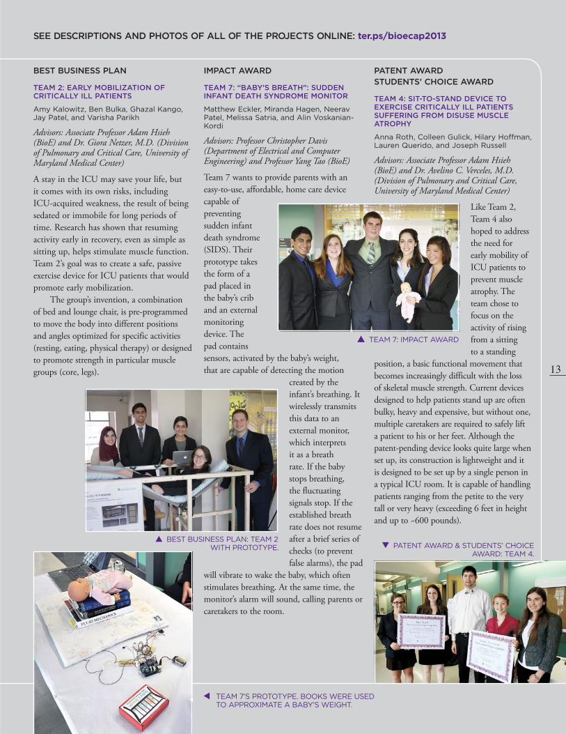
IMPACT AWARD
TEAM 7: “BABY’S BREATH”: SUDDEN INFANT DEATH SYNDROME MONITOR
Matthew Eckler, Miranda Hagen, Neerav Patel, Melissa Satria, and Alin Voskanian-Kordi
Advisors: Professor Christopher Davis (Department of Electrical and Computer Engineering) and Professor Yang Tao (BioE)
Team 7 wants to provide parents with an easy-to-use, affordable, home care device capable of preventing sudden infant death syndrome (SIDS). Their prototype takes the form of a pad placed in the baby’s crib and an external monitoring device. The pad contains sensors, activated by the baby’s weight, that are capable of detecting the motion
created by the infant’s breathing. It wirelessly transmits this data to an external monitor, which interprets it as a breath rate. If the baby stops breathing, the fluctuating signals stop. If the established breath rate does not resume after a brief series of checks (to prevent false alarms), the pad
will vibrate to wake the baby, which often stimulates breathing. At the same time, the monitor’s alarm will sound, calling parents or caretakers to the room.
PATENT AWARDSTUDENTS’ CHOICE AWARD
TEAM 4: SIT-TO-STAND DEVICE TO EXERCISE CRITICALLY ILL PATIENTS SUFFERING FROM DISUSE MUSCLE ATROPHY
Anna Roth, Colleen Gulick, Hilary Hoffman, Lauren Querido, and Joseph Russell
Advisors: Associate Professor Adam Hsieh (BioE) and Dr. Avelino C. Verceles, M.D. (Division of Pulmonary and Critical Care, University of Maryland Medical Center)
Like Team 2, Team 4 also hoped to address the need for early mobility of ICU patients to prevent muscle atrophy. The team chose to focus on the activity of rising from a sitting to a standing
position, a basic functional movement that becomes increasingly difficult with the loss of skeletal muscle strength. Current devices designed to help patients stand up are often bulky, heavy and expensive, but without one, multiple caretakers are required to safely lift a patient to his or her feet. Although the patent-pending device looks quite large when set up, its construction is lightweight and it is designed to be set up by a single person in a typical ICU room. It is capable of handling patients ranging from the petite to the very tall or very heavy (exceeding 6 feet in height and up to ~600 pounds).
BEST BUSINESS PLAN
TEAM 2: EARLY MOBILIZATION OF CRITICALLY ILL PATIENTS
Amy Kalowitz, Ben Bulka, Ghazal Kango, Jay Patel, and Varisha Parikh
Advisors: Associate Professor Adam Hsieh (BioE) and Dr. Giora Netzer, M.D. (Division of Pulmonary and Critical Care, University of Maryland Medical Center)
A stay in the ICU may save your life, but it comes with its own risks, including ICU-acquired weakness, the result of being sedated or immobile for long periods of time. Research has shown that resuming activity early in recovery, even as simple as sitting up, helps stimulate muscle function. Team 2’s goal was to create a safe, passive exercise device for ICU patients that would promote early mobilization.
The group’s invention, a combination of bed and lounge chair, is pre-programmed to move the body into different positions and angles optimized for specific activities (resting, eating, physical therapy) or designed to promote strength in particular muscle groups (core, legs).
SEE DESCRIPTIONS AND PHOTOS OF ALL OF THE PROJECTS ONLINE: ter.ps/bioecap2013
BEST BUSINESS PLAN: TEAM 2 WITH PROTOTYPE.
TEAM 7’S PROTOTYPE. BOOKS WERE USED TO APPROXIMATE A BABY’S WEIGHT.
PATENT AWARD & STUDENTS’ CHOICE AWARD: TEAM 4.
TEAM 7: IMPACT AWARD
14
BIOFEEDBACK VOL. 9 NO. 2
ALUMNUS RECOGNIZED FOR WORK WITH TEACH FOR AMERICA
BioE alumnus David Lai (B.S. ’12) was recently a finalist in Maryland governor Martin O’Malley’s first #MDForward contest, which invited Marylanders to share their stories about how they’re
“moving Maryland forward.” Lai, a 7th grade science teacher at Thomas Johnson Middle School in Lanham, wrote about how he works to inspire his young scholars to become the “doctors, lawyers, scientists, and entrepreneurs [of ] the state’s future,” community leaders, and good citizens.
BIOE COURSE A MODEL FOR NATIONAL ENERGY CURRICULUM
What started as a University of Maryland (UMD) engineering course for non-science majors has become the inspiration and pilot for a new, national “Energy 101” curriculum offered by the Department of Energy (DOE).
BioE and College of Education research associate professor Leigh Abts has become a leader in the national conversation on advancing science, technology, engineering and math (STEM) education initiatives. In 2013, he co-designed and taught a new course, BIOE 289A: Designing a Sustainable World, with former BioE lecturer Idalis Villaneuva. The course encourages students of all majors to contribute their own solutions to critical sustainability issues.
After an introduction to existing and emerging technologies, students were tasked with proposing feasible inventions that would increase sustainability in everyday life. Along the way, they learned about the engineering design process. Their final
projects included concepts such as green gyms powered by energy produced from exercise equipment, photosynthetic solar panels disguised as trees, and a waste-processing toilet that saves water and reduces the burden on sewage treatment plants.
Abts then partnered with the DOE to create a new energy and sustainability curriculum, based on BIOE 289A, which could be used at colleges and universities around the country to develop a new generation of energy-savvy leaders.
The result, Energy 101, is the product of the collaborative efforts of UMD, the Association of Public and Land-grant Universities, the DOE, and the Environment and Energy Study Institute. The unique, interdisciplinary course, comprised of group projects and educational modules, is designed to challenge students to systematically explore the technological, economical, and societal issues behind sound energy policies and decisions, and to teach them to apply that knowledge to their professional and personal lives.
Abts is also part of the effort to get kids interested in science before they get to college. In June 2013, the National Science Foundation invited him to speak at a Congressional briefing on STEM education, co-sponsored by American Society of Mechanical Engineers and Discover magazine. “Harnessing the Power of Engineering to Improve STEM Education in K-12 Schools” was the most recent in a series of events designed to inform legislators and policy makers about STEM education issues of relevance to the American public. Abts’ presentation focused on the importance of engineering design projects to help students develop creative thinking and problem solving skills.
FOR MORE INFORMATION AND VIDEO, SEE: ter.ps/discoverstem
educationNEWS
DAVID LAI
Part of the first generation of his family to attend college, Lai decided to postpone graduate studies or a career in medical device design to join Teach for America. The pro-gram works to eliminate inequities in the nation’s public education system by provid-ing schools in low-income communities with the teachers they need to provide an excellent education for all children.
INSIDE A BURN: ALUMNUS CONTINUES DIAGNOSTIC WORK HE BEGAN AS AN UNDERGRADUATE
When patients arrive at a hospital with burn injuries, doctors diagnose and form a treatment plan based on what they see, but not all burns fit neatly into the familiar categories of first, second and third degree. A type of second degree burn known as a deep partial thickness burn, which extends partway though the second layer of skin known as the dermis, could either heal
on its own or fully penetrate, developing into a third degree burn. Doctors rely on their experience to predict whether surgical intervention, such as a skin graft or removal of damaged tissue, is required.
BioE alumnus Nick Prindeze (B.S. ’12), a research associate at MedStar Washington Hospital Center’s Burn and Surgical Research Laboratory, is part of a team developing an imaging system capable of assessing deep partial thickness burns in ways the human eye cannot by evaluating changes in temperature deep inside the damaged tissue. The information it provides could help doctors make more informed treatment decisions and eliminate unnecessary surgery.
Prindeze and his classmates created a prototype of the system for their Capstone Design project during their senior year. The device uses an imaging technique called active dynamic thermography (ADT) to measure the ability of an area of skin
alumniNEWS

chair’sMESSAGE
15
A. JAMES CLARK SCHOOL of ENGINEERING GLENN L. MARTIN INSTITUTE OF TECHNOLOGY
5). Diagnostic anSERS, founded by current
graduate students Eric Hoppmann and
Sean Virgile, manufactures SERS substrates
using a novel inkjet printing system.
They’re currently working with Ocean
Optics, the leading supplier of miniature
spectrometers, to create the first truly
portable SERS system.
These are just a few highlights you’ve hopefully discovered in this edition of Biofeedback. There is much more on our web site at bioe.umd.edu/news. We hope you share our enthusiasm and might find time to engage with our students, staff, and faculty as we move the Fischell/Terp bioengineering community onward and upward! If you have a free afternoon, contact us and ask to stop by so we can show you what we mean in person.
With Best Regards,
William E. Bentley Robert E. Fischell Distinguished Professor and Chair
THE IMPACT OF OUR DEPART-MENT’S WORK IS SPREADING BEYOND OUR LABS AND OUR CAMPUS.
On the education
front, Professor
Gregory Payne has
been appointed
a Guest Professor
at Wuhan University, one of China’s top
10 educational institutions (see p. 4).
Professor Leigh Abts’ course BIOE 289A:
Designing a Sustainable World, serves as
a model for the Department of Energy’s
national “Energy 101” curriculum, which
universities can use to teach students about
fundamental energy principles and current
energy challenges (see p. 14). And alumnus
David Lai (B.S. ’12) is inspiring young minds
as a member of the Teach for America
program (see p. 14).
Our students are creating an increasing
number of patentable devices in their
senior Capstone Design course—often
in collaboration with doctors from local
hospitals who are seeking solutions to real
challenges they face. One recent Capstone
project led Nick Prindeze (B.S. ’12) to a
full-time position at MedStar Washington
Hospital’s Burn and Surgical Research
Laboratory, where he continues the work he
began as an undergraduate (see p. 14).
Despite being only two years old, our FDA-
sponsored Maryland Center of Excellence
in Regulatory Science and Innovation has
hosted or cosponsored an impressive
schedule of workshops, conferences, and
courses. Participants and speakers have
come from the AIMBE, industry, the DoD,
the FDA, the NIH, and NIST (see back cover).
In entrepreneurship news, Remedium
Technologies, the company co-founded by
former Fischell Fellow Matt Dowling (Ph.D.
’10), has partnered with Massachusetts
General Hospital and the University of
Maryland to complete pre-clinical trials of
its Hemogrip™ foam, which is designed to
control non-compressible hemorrhaging
caused by deep traumatic injuries (see p.
WILLIAM E. BENTLEY
to dissipate heat after being damaged by a burn. The deeper the burn and more serious the damage, the longer it takes for an equilibrium temperature to return to the different layers of skin. These changes in temperature–thermal gradients–are translated into a visual map showing doctors exactly where tissue is necrotic (dead, and requiring removal), static (damaged, but possibly repairable), or hyperaemic (capable of complete recovery with proper care).
Prindeze got his start at MedStar Washington Hospital Center as a volunteer during his junior year. During his senior year he became a part time employee, and was advised on his Capstone Design project by Dr. Jeffrey Shupp, director of the Burn and Surgical Research Laboratory. He joined the lab as full-time employee when he graduated. What began as an undergraduate research opportunity that inspired his final project is now the beginning of a career in
developing better ways to approach prob-lems in skin graft healing, scar reduction, infection and blood coagulation control, and electrical injuries.
“The laboratory is a basic, translational research facility run by burn surgeons and general surgery residents,” Prindeze explains. “We conduct contracted drug, treatment, and clinical studies, as well as develop and perform our own preclinical experiments.”
These in-house experiments have included the further development of the device he designed while a student at the Clark School. Prindeze is joined in his efforts by colleagues at the burn center and by some of his former Capstone group members. In April, the team was invited to present its results at the 45th Annual Meeting of the American Burn Association in Palm Springs, Calif., where it took first place in the conference’s poster competition. A manuscript about the technology was
accepted for publication in the Journal of Burn Care and Research and is scheduled appear in print in January. Future work will include refining and expanding the ADT system’s accuracy, including exploring its potential to identify more severe burns within larger burn areas. In time, Prindeze hopes to see the release of a direct-to-user product capable of evaluating burn wounds in clinical settings.
For more information, see:
N. J. Prindeze, P. Fathi, M. J. Mino, N. A. Mauskar, L. T. Moffatt, and J. W. Shupp. “Active Dynamic Thermography for Burn Wound Depth Detection.” ABA 45th Annual Meeting, April 23-26, 2013, Palm Springs, Calif.
NICK PRINDEZE

The Fischell Department of Bioengineering2330 Jeong H. Kim Engineering BuildingUniversity of MarylandCollege Park, MD 20742-2835
BIOFEEDBACK is published for alumni and friends of The Fischell Department of Bioengineering at the A. James Clark School of Engineering, University of Maryland.
Alumni news and comments are welcome! Please contact us at: Fischell Department of Bioengineering, 2330 Jeong. H. Kim Engineering Building College Park, MD, 20742 (301) 405-7426 / [email protected] http://www.bioe.umd.edu
Department Chair: Dr. William Bentley Editor: Faye Levine
It’s been a busy and productive year for M-CERSI, Maryland’s Center of Excellence in Regulatory Science and Innovation. This FDA-funded collaborative partnership between the University of Maryland, College Park (UMD), the University of Maryland, Baltimore (UMB) and the FDA focuses on the science that underpins the ways drugs and medical devices are regulated.
In March, M-CERSI co-sponsored the Third AIMBE/NIH Workshop on Validation and Qualification of New In Vitro Tools and Models for the Pre-clinical Drug Discovery Process, hosted by the American Institute for Medical and Biological Engineering and
ABOUT THE COVER IMAGE
LYMPH NODES ARE THE TISSUES THAT COORDINATE IMMUNE RESPONSE. OUR COVER IMAGE WAS CREATED FROM IMMUNOFLUORESCENT HISTOLOGY SECTIONS OF LYMPH NODES, WHERE THE DARKER AREAS ARE T CELL ZONES AND THE LIGHTER AREAS ARE B CELL ZONES. DURING INFECTION OR VACCINATION, THESE ZONES BECOME INTERMINGLED TO GENERATE CELLS AND ANTIBODIES THAT MIGRATE OUT OF THE LYMPH NODE AND COMBAT OR PROTECT AGAINST INFECTION. THE JEWELL LAB IS USING NEW DELIVERY TOOLS TO UNDERSTAND HOW BIOMATERIAL VACCINE CARRIERS IMPACT LYMPH NODE SIGNALING, AND HOW THESE INTERACTIONS CAN BE EXPLOITED TO DEVELOP BIOMATERIAL-BASED VACCINES THAT BETTER TREAT INFECTIOUS DISEASE OR AUTOIMMUNE DISORDERS.
the National Institutes of Health. These workshops discuss tools that are designed to augment or replace existing models, especially animal testing. This particular event focused on “animal-on-a-chip” devices, which mimic the fluids, cells, tissues, and organs of humans and animals, allowing physiological microsystems to be investigated in vitro.
In April, BioE professor Keith Herold and M-CERSI co-PI/School of Pharmacy professor James Polli organized a Degree Programs in Regulatory Science and Engineering workshop. The event brought experts on regulatory science academic programs from the FDA and around the
country to campus. It served as a forum for discussions on current and future educational programs and their core competencies, including UMD/UMB’s recently-launched certificate and M.S. programs.
In May, M-CERSI hosted the Workshop on Improved Characterization of Mesenchymal Stem Cells (MSC) for Clinical Trials, co-sponsored by the FDA and the UMB School of Medicine’s Department of Orthopaedics and Center for Stem Cell Biology & Regenerative Medicine. It assessed MSC characterization and discussed methods that can be used to predict the quality,
continues pg. 11
M-CERSI update