Biocorrosion and Aseptic Loosening of Metal …...Biocorrosion and Aseptic Loosening of Metal...
82
Biocorrosion and Aseptic Loosening of Metal Implants: Novel Pathophysiological Mechanisms Dr. Dieter Cadosch, MD This thesis is presented for the degree of Doctor of Philosophy At The University of Western Australia School of Anatomy and Human Biology 2011
Transcript of Biocorrosion and Aseptic Loosening of Metal …...Biocorrosion and Aseptic Loosening of Metal...

Biocorrosion and Aseptic Loosening of Metal Implants:
Novel Pathophysiological Mechanisms
Dr. Dieter Cadosch, MD
This thesis is presented for the degree of
Doctor of Philosophy At
The University of Western Australia
School of Anatomy and Human Biology
2011

This thesis is dedicated to my loving and wonderful parents,
My loved daughters Lia Charleen and Zoë Alina
&
Anne
“By seeking and blundering we learn”
Johann Wolfgang von Goethe
II

Abstract Biocorrosion and Aseptic Loosening of Metal Implants: Novel Pathophysiological Mechanisms
Metal orthopaedic devices exposed to a physiological environment remain prone to
corrosion by several mechanisms, leading to the production of significant amounts of
non-biocompatible wear particles and metal ions. Beside the well-known mechanical
wear and electrochemical redox reactions, metal implants undergo corrosion through
biological activities. The results presented in this thesis suggest that mature
osteoclasts are able to enhance corrosion of titanium and stainless steel implants
and contribute to the release of relevant amounts of corresponding metal ions into
the peri-implant tissues and the systemic blood circulation. In addition to the
increased osteolytic activity caused by wear particles, the results detailed in this
thesis strongly suggests that released titanium ions in the 4+ oxidation state (Ti(IV))
contribute to the pathophysiological mechanism of aseptic loosening by stimulating
both the bone metabolism and immune system through a series of direct and indirect
pathways. Aseptic loosening is believed to be caused by an increased osteolytic
activity at the bone-implant interface leading to loss of stability and ultimately implant
failure. Titanium (IV) ions are able to directly induce the differentiation of osteoclast
precursors toward mature and functional osteoclasts in ~20% of individuals.
Additionally, Ti(IV) ions stimulate the secretion of pro-inflammatory cytokines that are
known to enhance osteoclast recruitment, differentiation, activation and survival.
More evidence is linking the immune system to the bone and to its involvement in the
pathophysiology of aseptic loosening. Titanium (IV) ions influence phenotype and
function of T-lymphocytes, resulting in activation of a CD69+ and CCR4+ T-
lymphocyte and secretion of receptor activator of NF-κB ligand (RANK-L).
To date, revision surgery remains the only option for the treatment of a failed
orthopaedic implant caused by aseptic loosening. These results represent an
important step towards a better understanding of the complex pathophysiological
mechanism of aseptic loosening in patients with titanium implants, and may open
new research perspectives in this field which could offer innovative therapeutic
options and reduce the rate of aseptic loosening.
III

Table of Contents
Abstract III
Table of Contents IV
Publications Arising VI
Talks VII
Posters VIII
Prizes IX
Acknowledgments X
Statement of Candidate Contribution XI
Abbreviations XII
Preface XIII
Chapter 01 | Background and Literature Review 1
Introduction
Biocorrosion
Osteoimmunology: Links between Immunology and Bone System
Chapter 02 | Research Questions 5
Aims
Chapter 03 9
Uptake and intracellular distribution of various metal ions in monocyte-derived
dendritic cells detected by Newport Green DCF diacetate ester
Chapter 04 16
Bio-Corrosion of Stainless Steel by Osteoclasts - In vitro Evidence
Chapter 05 23
Biocorrosion and uptake of titanium by human osteoclasts
Chapter 06 31
Titanium IV ions induced human osteoclast differentiation and
enhanced bone resorption in vitro
IV

Chapter 07 40
Titanium induced production of chemokines CCL17/TARC and CCL22/MDC
in human osteoclasts and osteoblasts
Chapter 08 50
Titanium uptake, induction of RANK-L expression, and enhanced proliferation of
human T-lymphocytes
Chapter 09 | Discussion 58
Cellular Mechanisms of Corrosion
Effects of Metal Ions on Bone Metabolisms
Effects of Titanium Ions on Osteoimmunology
Summary and Outlook
Bibliography 63
Appendix 67
Poster 1
Poster 2
V

Publications Arising The following publications originated from this research project and have been published in peer-reviewed journals. *This manuscript is still under review at the time of thesis submission. Chapter 3: Uptake and intracellular distribution of various metal ions in
human monocyte-derived dendritic cells detected by Newport Green DCF diacetate ester. Cadosch D, Meagher J, Gautschi OP, Filgueira L (2009). J Neurosci Methods. Mar 30;178(1):182-7.
Chapter 4: Biocorrosion of stainless steel by osteoclasts –
in vitro evidence. Cadosch D, Chan E, Gautschi OP, Simmen HP, Filgueira L (2009). J Orthop Res. Jul;27(7):841-6.
Chapter 5: Biocorrosion and uptake of titanium by human osteoclasts.
Cadosch D, Al-Mushaiqri MS, Gautschi OP, Meagher J, Simmen HP, Filgueira L (2010). J Biomed Mater Res A. Dec 15;95(4):1004-10.
Chapter 6: Titanium IV ions induced human osteoclast differentiation and
enhanced bone resorption in vitro. Cadosch D, Chan E, Gautschi OP, Meagher J, Filgueira L (2009). J Biomed Mater Res A. Oct;91(1):29-36.
Chapter 7: Titanium induced production of chemokines CCL17/TARC and
CCL22/MDC in human osteoclasts and osteoblasts. Cadosch D, Gautschi OP, Chan E, Simmen HP, Filgueira L (2010). J Biomed Mater Res A. Feb;92(2):475-83.
Chapter 8: Titanium uptake, induction of RANK-L expression, and
enhanced proliferation of human T-lymphocytes. Cadosch D, Sutanto M, Chan E, Mhawi A, Gautschi OP, von Katterfeld B, Simmen HP, Filgueira L (2010). J Orthop Res. Mar;28(3):341-7.
Chapter 1 & 9: Metal is not inert: role of metal ions released by biocorrosion in
aseptic loosening-current concepts. Cadosch D, Chan E, Gautschi OP, Filgueira L (2009). J Biomed Mater Res A. Dec 15;91(4):1252-62.
Influence of metal ions on human lymphocytes and the generation of titanium specificT-lymphocytes. Chan E, Cadosch D, Gautschi OP, Sprengel K, Filgueira L (2011). J Appl Biomater Biomech. Accepted April 2011.
*Pharmacological blocking of osteoclastic biocorrosion of surgical stainless steel. Lionetto S, Little JA, Heymann D, Filgueira L, Cadosch D (2011).
VI

Talks The thesis results have been presented at the following national and international meetings: 6/2008 Immune reactivity to metal implants; an in vitro investigation.
Australian Society for Medical Research: WA Scientific Symposium
Perth, WA, Australia
8/2008 Biocorrosion of implants and metal sensitivity reactions.
Australian Orthopaedic Association: Annual Scientific Meeting
Crawley, WA, Australia
10/2008 Titanium induced osteoclast recruitment and activation resulting
in enhanced bone resorption: a human in vitro Study.
American College of Surgeons: 94th Annual Clinical Congress
San Francisco, CA, USA
6/2009 Titanium IV Ions Induced Human Osteoclast Differentiation and
Enhanced Bone Resorption in vitro.
96. Jahreskongress der Schweizerischen Gesellschaft für Chirurgie
Montreaux, VD, Switzerland
9/2009 Osteoclastic Corrosion of Stainless Steel and Titanium Implants
22nd European Conference on Biomaterials.
The annual conference of the European Society for Biomaterials
Lausanne, VD, Switzerland
10/2009 Titanium uptake and induction of TGF-beta and RANK-L
expression in human T-lymphocytes.
American College of Surgeons: 95th Annual Clinical Congress
Chicago, IL, USA
10/2009 Increased expression of pro-inflammatory and osteogenic
cytokines by osteoclasts cultured on surgical stainless steel.
American College of Surgeons: 95th Annual Clinical Congress
Chicago, IL, USA
VII

Posters The thesis results have been presented as posters at the following national and international meetings. Selected posters are listed in the appendix. 9/2008 Titanium (IV) ions induced human osteoclast differentiation and
enhanced bone resorption in vitro.
American Association for the Surgery of Trauma
67th Annual Meeting, Maui, Hawaii, USA
10/2008 Titanium ions induced bone resorption due to osteoclast
recruitment and activation: a human in vitro study.
Symposium on Biotechnology in Musculoskeletal Repair
2nd International Symposium, Lausanne, VD, Switzerland
1/2009 Immune reactivity to metal implants: an in vitro investigation.
2nd Singapore Symposium for Immunology, Singapore
5/2010 Titanium IV Uptake, Induction of RANK-L Expression and
Enhanced Proliferation of Human T-Lymphocytes
97. Jahreskongress der Schweizerischen Gesellschaft für Chirurgie
Interlaken, BE, Switzerland
VIII

Prizes The results presented in this thesis have been awarded with the following prices: Scholarship for International Research Fees (SIRF)
University of Western Australia, Crawley, Australia, 2008 - 2011
Excellence in Research Award in Orthopaedic Surgery 2008 American College of
Surgeons
American College of Surgeons, San Francisco, CA, USA, 2008
Travel Award University of Western Australia
University of Western Australia, Crawley, Australia, 2009
IX

X
Acknowledgements Though my name is printed on the cover of this thesis, this work would not have been
possible without the support of many people. It is a great pleasure to thank those
who made this thesis possible. To my loving and wonderful parents: Thank you for
always believing in me and giving me the freedom and the opportunity to pursue my
interests and dreams. I am heartily thankful to my supervisor and mentor Professor
Luis Filgueira. Thank you for your encouragement, guidance and the opportunity to
work at the School of Anatomy and Human Biology at the University of Western
Australia, but especially for your friendship. The friendly and supportive atmosphere
inherent to the whole School of Anatomy and Human Biology contributed essentially
to the final outcome of my studies. I would like to make a special reference to the
entire School of Anatomy and Human Biology. I want to thank my external
supervisor, Professor Hans-Peter Simmen for his generous support, and Professor
Marco Decurtins for his encouragement to undertake a PhD study in Western
Australia. I am particularly grateful to Tricia Knox, who was always so kind to edit all
my manuscripts. I am sincerely thankful to Dr. Oliver P. Gautschi, who has always
been of great support and a dear friend. Finally, I want to thank all of my friends who
made life in Perth so enjoyable, especially Urban M. and his lovely family; you made
me feel at home. For their hospitality and advice I would like to thank Professor Allan
P. and Carole Skirving.

State of Candidate Contribution The work presented in this thesis results from an external PhD study at the School of
Anatomy and Human Biology, University of Western Australia and the Clinic of
Trauma Surgery, University of Zurich. The experiments detailed in this thesis have
been performed at the School of Anatomy and Human Biology, University of Western
Australia under the supervision of Professor Luis Filgueira, and at the Clinic of
Trauma Surgery at the University Hospital of Zurich under the supervision of
Professor Hans-Peter Simmen. Contributions and collaborations with other
colleagues are mentioned accordingly and listed as co-authorships in the published
papers.
Dr. Dieter Cadosch
MD, PhD Candidate
Prof. Hans-Peter Simmen Prof. Luis Filgueira
MD, External Supervisor MD, Supervisor
XI

Abbreviations
Al Aluminium AL Aseptic loosening CCL22 Macrophage- derived chemokine CCL17 Thymus and activation- regulated chemokine Co Cobalt Cr Chromium EFTEM Energy-filtered electron microscopy ELISA Enzyme-linked Immunosorbent Assay Fe Iron IFN-γ Interferon-gamma IL Interleukin M-CSF Macrophage colony- stimulating factor Mn Manganese
Mo Molybdenum Ni Nickel OC Osteoclast/s PCR Polymerase chain
reactions PHA Phytohemagglutinin RANK-L Receptor activator of NF-κB ligand Ti Titanium Ti(IV) Titanium ions in 4+
oxidation state TNF-α Tumour necrosis
factor-alpha TRAP Tartrate-resistant
acid phosphatise V Vanadium Zr Zirconium
XII

Preface
To date, the pathophysiological mechanisms of biocorrosion and aseptic loosening of
metal implants remain not fully understood. Whether cellular mechanisms are able to
enhance biocorrosion of metal implants remains controversial. Furthermore, the
effects of metal ions released by biocorrosion on the immune system and bone
metabolisms remain under-studied. This thesis presents research into understanding
the effects of mature osteoclasts on biocorrosion as well as titanium ions on the
recruitment, differentiation and activation of human osteoclasts.
Chapter 02 gives an overview on the current knowledge on biocorrosion and aseptic
loosening of orthopaedic implants. From this, the hypothesis of enhanced
biocorrosion by osteoclasts and the effects of metal ions on bone metabolisms and
immune system are postulated. A brief outline of the methodology employed to
investigate the hypothesis is also described.
With the aim of understanding cellular biocorrosion and the effects of titanium ions on
osteoclastogenesis, a range of investigations were carried out. Six papers (published
in peer-reviews journals) investigating cellular biocorrosion of stainless steel and
titanium alloys (Chapter 04 and 05), the effects of titanium ions on bone metabolism
(Chapter 06 and 07), and on the immune system (Chapter 08) are presented. Each
paper is self-contained and presented in its entirety in the published format. A short
introduction highlighting the aims and the key results of the paper are provided at the
beginning of each chapter. The discussion attempts to unify the results presented by
the individual papers and to incorporate the possible pathways involving titanium ions
into the pathophysiological mechanism of aseptic loosening.
This journey of discovery inflamed my desire to uncover more of the complex
pathophysiology of aseptic loosening and metal ions. I hope that this thesis will allow
you to re-live the exciting journey of scientific research.
Dr. Dieter Cadosch, MD, PhD Candidate
The University of Western Australia
XIII

Chapter 01
Background Introduction
Metal implants are essential therapeutic tools for the treatment of bone fractures and
joint replacements. The first metal hip prosthesis was implanted in 1956 under great
engineering and medical efforts. In the meantime, total joint arthroplasty has been
performed routinely in over a million patients worldwide every year. The constant
aging of our population will contribute to an increasing number of patients with
implanted metal devices (1). As long as tissue engineering and biodegradable bone
substitutes do not lead to products that will be applicable in clinical routine, prosthetic
and osteosynthetic devices, made of metal and metal alloys, will remain
indispensable in orthopaedic and trauma surgery. Most patients tolerate metal
implants well; however, complications resulting from inflammatory and immune
reactions to metals have been well documented in up to 10% of patients, with the
incidence continuing to increase (2-8).
Many different metals and metal alloys have been used for joint replacement, internal
fixation of bone fractures, and after osteotomy. The metal implants predominately
used in contemporary surgery are made of commercially pure titanium, titanium
alloys (e.g., Ti6Al7Nb, TiAl6V4), and medical grade stainless steel (SS316L). The
employed metallic biomaterials are composed of a variety of metals including
aluminium (Al), chromium (Cr), cobalt (Co), nickel (Ni), molybdenum (Mo), vanadium
(V), titanium (Ti), and iron (Fe). Stainless steel SS316L, for example, contains Cr
(16–18%), Ni (10–14%), and various trace metals (5%) balanced with Fe (9,10). Over
recent decades, significant developments have taken place to provide suitable
metallic biomaterials with optimal biofunctionality, such as stability and a surface
texture allowing cellular adhesion, combined with excellent biocompatibility of low
intrinsic toxicity, inflammatory activation, and immunogenicity (11). However, their
permanent tendency to corrode when implanted into a physiological environment
remains a serious concern (12-14). Several case reports and studies have indicated
that metal implants undergo corrosion inside the human body by various
mechanisms including mechanical wear, biological activity, and mechanically
accelerated electrochemical processes (15-16). Thus, significant amounts of metallic
- 1 -

particles are released into the tissues surrounding metal implants and elevated
concentrations of metal ions have been measured in clinically retrieved capsular and
peri-prosthetic tissues, as well as in distant organs (liver, spleen, and lymph nodes)
and body fluids (serum and urine) in total hip arthroplasty patients (17-20). Implants
used for osteosynthesis have to withstand completely different forces than implants
used for joint replacements. Thus, large implant-derived particles (in the nanometer
range) are produced exclusively by the mechanical wear process in articular coupling
of prostheses and are not present in patients with osteosynthesis implants. However,
both osteosynthesis and joint replacement implants are exposed to biological activity
and electrochemical processes.
A wide range of molecular and cellular interplays have been demonstrated between
the released biocorrosion products and the human organism, involving the immune
and skeletal system. There is increasing clinical and research-based evidence that a
relevant percentage of patients with metal implants may develop metal
hypersensitivity and severe inflammatory side effects, leading to aseptic loosening
(AL) of the implant and even systemic reactions (21-24). The phagocytosis of metal
wear particles by tissue macrophages induces production of pro-inflammatory
cytokines that enhance osteolytic activity at the implant-bone interface (22, 25-27).
Although many studies have investigated the role of metal wear particles in
osteoclastogenesis and AL, little is known about the direct effects of metal ions
released by biocorrosion. At the bone metal interface, metal ions may directly interact
with bone cells contributing to AL by accelerating osteoclastic bone resorption and/or
inhibiting the function of osteoblasts. Various studies have demonstrated that
nontoxic concentrations of metal ions affect the differentiation and function of
osteoblastic cells in vitro (28,29). Furthermore, once released into the systemic blood
circulation, the metal ions bind to serum proteins and form haptens or hapten-like
complexes, which are considered to be relevant antigens recognized by T-
lymphocytes and candidates for eliciting hypersensitivity reactions (18,30). Exposed
to metal–protein complexes, T-lymphocytes proliferate and differentiate to produce
soluble factors such as interleukin (IL)-6, IL-1a/b, and tumour necrosis factor-alpha
(TNF-α) that trigger a particular immune response and activate osteoclastogenesis
(31–33). The extent of this immune response upon implantation of metallic devices
depends predominantly on the individual immune reactivity and on material
characteristics (34).
- 2 -

Biocorrosion
To date, it is well recognized that corrosion of metallic materials implanted in the
human body is an inevitable deteriorating reaction leading to the release of
undesirable metal ions/corrosion products, which are not biocompatible. In the
chemist’s view, corrosion is the visible destruction of a metal caused by interactions
with its environment, which may cause rupture of a structure or loss of function, for
example, breakage of an orthopaedic implant. This aspect is no longer important for
modern metals/alloys in surgery as material loss due to any corrosive attack is
minimal and does not usually compromise the stability of the implant (16,35).
However, despite the great progress in providing corrosion resistant metallic
biomaterials, in a physiological environment corrosion remains a slow and continuous
process, which leads to the release of significant amounts of metal ions and other
corrosion products. For instance, the corrosion of a stainless steel implant releases
Fe, Cr, and Ni ions; corrosion of Ti and Ti alloys release Ti (mostly at the 4+ oxidation
state; Ti(IV)), V and Al ions (36-48). Dissolved metal ions can accumulate in the
tissue, surrounding metal implants or can be released into the systemic blood
circulation and transported to distal organs (17–20). Indeed, significantly higher
concentrations of metal ions have been observed in the body fluids of patients with
stainless steel implants 10–13 years after primary hip arthroplasty when compared
with individuals without implants. This included concentrations of Ni in blood of ~0.51
μg/L, in plasma of ~0.26 μg/L, and in urine of ~2.26 μg/L, and plasma Cr levels of
~0.19 μg/L (14). Similarly, Ti concentrations of ~135.57 μg/L were measured in
patients with Ti-6Al-4V total knee replacements after 57 months (49). The amount of
metal ions released depends on the quality of the surgical procedure and on the
function of the implant. Leopold et al. measured a threefold increase in Ti levels in
patients with well-functioning Ti-alloy joint replacements, while patients with failed
implants showed as much as a 50-fold increase in serum concentrations when
compared with individuals without implants (49). A further study reported ~1 μg/L of
Ti in serum of patients with stable prosthesis; however, the concentrations increased
up to ~4 μg/L in the case of failed implants (51). In a physiological environment,
metallic biomaterials are known to undergo corrosion through a variety of
mechanisms. The most commonly observed forms of corrosion include mechanical
wear processes (e.g., in the articular coupling of prostheses leading to the formation
of wear particles) and electrochemical corrosion (11,22,35). As tissue fluids enter into
- 3 -
contact with the metal surface, corrosion occurs via an electrochemical redox
reaction, in which oxidation (electron loss of the metal) is coupled with reduction
(electron gain of electrolyte components) (35). In terms of how a biological
environment can affect corrosion, it is believed that proteins can influence the
electrochemical behaviour of implant metals/alloys. However, overall results are not
conclusive and the exact effect of proteins on corrosion is still the topic of much
debate (52). Some authors demonstrate that proteins can enhance the propagation
of charges between implant and surrounding fluids while others suggest a reduced
charge transfer by proteins (53–56).
Osteoimmunology: Links between Immunology and Bone System
Recently, published evidence has shown that the immune system is closely
connected to the development of bone cells. This might not be surprising,
considering that the production of immune cells of hematopoietic origin comes from
the bone marrow and that improper bone development leads to a deficient bone
marrow size and function (84). In addition, OC, the only and essential bone resorbing
cells, derive from the same precursor cells as macrophages and dendritic cells,
professional antigen-presenting cells (70). This linkage plays also a major role in
rheumatoid arthritis, cancerous bone metastasis, and other destructive bone
disorders as indicated by an increasing amount of published studies (69,85–87).
There is increasing evidence indicating that inflammatory immune responses
influence bone metabolism. A variety of T-lymphocyte derived factors and cytokines,
including RANK-L and TNF-α, have been shown to directly and indirectly promote
OC activity and inhibit osteoblast function (72,88–93). Other T-lymphocyte-derived
cytokines, including interferon-gamma (IFN-γ) and IL-4, have been shown to
decrease OC activity and thereby decrease bone remodelling and probably delay
bone fracture healing (94,95). Increased levels of IL-10, a cytokine produced by
regulatory T-lymphocytes, have been measured in patients with orthopaedic implants
(96). However, despite increasing evidence supporting the linkage between the
immune system and bone cells, the role of the immune system in the systemic and
peri-implant tissue responses, characterized by increased osteolysis and implant
failure (AL), remains poorly understood with several studies reporting conflicting
results regarding their actual involvement.
- 4 -

Chapter 02 Research Questions and Aims
The project described in this thesis was based on the following hypothesis:
1: Mature Osteoclasts are a Major Cause of Biocorrosion of Metal Implants
To date, it is well recognized that corrosion of metallic materials implanted in the
human body is an inevitable deteriorating reaction leading to the release of
undesirable metal ions and other corrosion products, which are not bio-compatible. In
the chemist’s view, corrosion is the visible destruction of a metal caused by
interactions with its environment, which may cause rupture of a structure or loss of
function, e.g. breakage of an orthopaedic implant. This aspect is no longer important
for modern metals/alloys in surgery as material loss due to any corrosive attack is
minimal and does not usually compromise the stability of the implant (17,37). Despite
the great progress in providing corrosion resistant metallic biomaterials, they remain
prone to corrosion in a physiological environment through a variety of mechanisms.
The most commonly observed include mechanical wear processes (e.g. in the
articular coupling of prostheses leading to the formation of wear particles) and
physiochemical corrosion (electrochemical redox reaction) (14,21,37). Beside the
mechanical and electrochemical aspect of corrosion, we hypothesise that mature
human OC are able to directly corrode the metal surface (e.g. pure Ti) and release
corresponding metal ions. We propose that this process may take place at the bone-
implant interface, representing an additional mechanism of metal corrosion in vivo
and contributing to the levels of metal ions measured in the periprosthetic tissues and
serum from total arthroplasty patients. Previous investigations have demonstrated
that Ti is relatively inert and resistant to corrosion with low water solubility (in the
range of 1 M concentration) at physiologic pH and low reactivity with biomolecules
in an aqueous environment (11). However, Ti concentrations up to ~136 μg/L were
measured in patients with Ti-6Al-4V total knee replacements (15). These levels are
much higher than would be expected given the “corrosion resistant” quality of Ti
demonstrated by in vitro analysis, and leads to our hypothesis that additional
mechanisms must be involved. Additionally, the amount of metal ions released
depends on the function of the implant. Leopold et al. measured a three-fold increase
- 5 -

in Ti levels in patients with well-functioning Ti-alloy joint replacements, while patients
with failed implants showed as much as a 50-fold increase in serum concentrations
when compared with individuals without implants (52). A further study reported ~1
μg/L of Ti in serum of patients with stable prosthesis; however the concentrations
increased up to ~4 μg/L in the case of failed implants surrounded by osteolytic
lesions (53). Given that the most frequent cause of implant failure is AL, it is
reasonable to assume that the OC responsible for the osteolytic lesions may also be
responsible for the increased levels of metal ions measured in patients with failed
implants.
2: Titanium Ions released by Biocorrosion Enhance Osteolysis
Recently great effort has been directed towards understanding the
pathophysiological cascade of events initiated by biocorrosion products, resulting in
periprosthetic bone loss and ultimately aseptic loosening. The mechanical wear
process in articular coupling of prostheses is responsible for severe inflammatory
reactions and bone resorption, which has been recognized as one of the primary
biological mechanism leading to periprosthetic osteolysis (23). Phagocytosis of Ti
wear particles by macrophages induces their activation, producing mediators that
enhance OC formation (27). However, the potential role of Ti ions released by
biocorrosion from the implant surface must also be considered.
Normal bone maintenance relies on the balance between bone formation and bone
resorption (77). Thus the net bone loss at the metal-tissue interface occurs because
of an increased bone resorption or reduced bone formation (77). Previous studies
have demonstrated that non-toxic concentrations of metal ions affect the
differentiation and function of osteoblastic cells in vitro (30). Although there have
been several studies investigating the effect of metal ions on bone formation, very
little is known about their effects on bone resorption (29,30). Increased bone
resorption can be due to an increase of one or more of the following events:
recruitment of OC precursors from the blood circulation at the bone-implant interface,
their differentiation into mature multi-nucleated cells, their functional activation, and
finally their survival. Our hypothesis was that Ti ions may increase recruitment of OC
precursors (due to increased production of chemotactic cytokines) from the systemic
blood circulation to the peri-implant tissues and enhance their subsequent
- 6 -

differentiation into mature OC. This hypothesis was supported by several animal
studies demonstrating that OC precursors in tissues surrounding subcutaneously
implanted wear particle differentiate into mature OC (65-67). Additionally, more
recently, particulate wear debris has been shown to induce chemokine expression in
macrophages, fibroblasts and osteoblasts, including IL-8, monocyte chemoattractant
protein-1, macrophage inflammatory protein 1, and eotaxin (68-71).
3: Titanium Ions affect the Phenotype and Function of human T lymphocytes
Dermal hypersensitivity to metal is common, with up to 20% of Caucasians being
sensitive to Ni (107). Immune reactions to dermal contact and ingestion of metals,
manifested as skin conditions such as eczema, urticaria, erythema and pruritis, are
believed to be of a type IV cell mediated hypersensitivity (12). The earliest case of an
allergic manifestation towards an orthopaedic implant was reported by Foussereau et
al., when a patient presented with an eczematous rash over a stainless steel fracture
plate (108). Beside the clinical observations of metal hypersensitivity observed in
some patients with orthopaedic implants, strengthened by the alleviation of
symptoms after the removal of the causative metal implant, evidence for the
involvement of the immune system comes from several histological studies of
retrieved peri-implant tissues (109). These studies have shown the infiltration of
lymphocytes, monocytes, dendritic cells, macrophages and mast cells into the peri-
implant tissues at various time points after metal implantation. Furthermore, there is
increasing evidence indicating that inflammatory immune responses influence bone
metabolism. A variety of T-lymphocyte derived factors and cytokines, including
RANK-L and TNF-α, have been shown to directly and indirectly promote OC activity
and inhibit osteoblast function (97,98). However, little is known about the metabolic
effects of Ti ions on human T-lymphocytes and its antigenicity, since the majority of
studies have focused on other cells (such as macrophages) and other metal ions
(such as Ni) (142). This study tested the hypothesis that Ti ions released by
biocorrosion, will be taken up by immune cells and cause alterations in T-
lymphocytes phenotype and function including: expression of surface markers,
proliferation, activation and regulation of cytokine expression and secretion.
- 7 -

Aims
The aims of this study were first, to investigate whether human monocytes are able
to grow on metal implants (such as Ti and stainless steel) and differentiate into
mature and functional OC. Second, to test the hypothesis that mature OC are able to
directly corrode the metal surface and release corresponding metal ions into their
environment. Third, to investigate the effects of the released Ti ions on bone
resorption, including OC recruitment, differentiation and activation, and finally, to
investigate their effects on T-lymphocytes, including expression of surface markers,
proliferation, activation and regulation of cytokine expression.
Methodology
The methods used in the presented studies for qualitative and quantitative analysis
have already been well documented and described for use in areas of toxicology,
immunology and morphological studies. They include: (1) Isolation of peripheral
blood cells, (2) in vitro cell cultures of human monocytes, OC and lymphocytes from
blood of healthy donors, (3) Functional assays on dentine slices, (4) Electron
Microscopy, Scanning Electron Microscopy and Energy-Filtered Electron Microscopy
(EFTEM), (5) Flow Cytometry, (6) Proliferation Assays, (7) Cytometric Bead Assays
for cytokines measurements, (8) Enzyme-linked Immunosorbent Assay (ELISA) for
Chemokines Measurements, (9) quantitative reverse transcription polymerase chain
reactions (PCR), (10) Atomic emission spectrometry. All these methods are
described in the corresponding papers (Chapters 4-8). Additionally, a novel method
using Newport GreenTM DCF diacetate ester to fluorescently label intracellular
protein metal complexes was developed (Chapter 3).
- 8 -

- 9 -
Chapter 03 Uptake and intracellular distribution of various metal ions in human monocyte-derived dendritic cells detected by Newport Green DCF diacetate ester The attempt to visualise intracellular protein metal complexes in biomedical research
has currently been limited due to the lack of adequate metal detection methods and
the unavailability of probes for such molecular structures. This first project aimed to
develop a novel detection method to overcome these limitations and enable the
planned research in the field of metals and metal ions. Newport GreenTM DCF
diacetate ester is a cell permeant acetate ester, which becomes fluorescent after
hydrolysis. This molecule is initially uncharged, allowing it to pass through cell
membranes. Once in the cell, it is hydrolysed and becomes charged, hindering its
escape from the cell and allowing it to bind charged protein metal complexes, which
then become fluorescent. In this study, we exposed cultured human monocyte-
derived dendritic cells to a variety of metal ions (including Ti with different oxidation
state: Ti(III) and Ti(IV)) with the aim of having the cells take up and process protein
metal complexes. Newport GreenTM DCF diacetate ester was used to fluorescently
label intracellular protein metal complexes. Flow cytometry analysis and confocal
imaging showed specific staining for monocyte-derived dendritic cells exposed to Al,
Cr, Ni, Ti and Zr ions. The intensity of staining varied between ion types, whereby
Ti(III) resulted in the brightest fluorescence signal. Aluminium, Cr(III), Ni, Ti(IV) and
Zr(IV) were also clearly detectable. Incorporating a new method of fluorescence
metal detection using both, flow cytometry and confocal laser microscopy, this first
study outlined the capacity of Newport GreenTM DCF diacetate ester to fluorescently
stain a variety of intracellular metal ion protein complexes (including Ti) as well as
unveiling the morphology of intracellular protein-bound metals in a monocyte-derived
dendritic cells culture setting. Additional experiments clearly demonstrated that this
protocol is suitable for investigating other cell lines including T-lymphocytes and OC.

Journal of Neuroscience Methods 178 (2009) 182–187
Contents lists available at ScienceDirect
Journal of Neuroscience Methods
journa l homepage: www.e lsev ier .com/ locate / jneumeth
Uptake and intracellular distribution of various metal ions inhuman monocyte-derived dendritic cells detected byNewport GreenTM DCF diacetate ester
Dieter Cadosch ∗, James Meagher, Oliver P. Gautschi, Luis FilgueiraSchool of Anatomy and Human Biology, University of Western Australia, 35 Stirling Highway, Crawley, WA 6009, Australia
a r t i c l e i n f o
Article history:Received 29 May 2008Received in revised form 4 December 2008Accepted 6 December 2008
Keywords:Newport GreenMetal protein complexMetal ionsTitaniumDendritic cellFlow cytometryConfocal microscopy
a b s t r a c t
Background: The attempt to visualise intracellular protein metal complexes has currently been difficultdue to the unavailability of probes for such molecular structures. Newport GreenTM DCF diacetate esteris a cell permeant acetate ester, which becomes fluorescent after hydrolysis. This molecule is initiallyuncharged, allowing it to pass through cell membranes. Once in the cell, it is hydrolysed and becomescharged, hindering its escape from the cell and allowing it to bind charged protein metal complexes,which then become fluorescent.Methods: In this study, we exposed cultured human monocyte-derived dendritic cells (mDC) to a varietyof metal ions with the aim of having the cells take up and process protein metal complexes. NewportGreenTM DCF diacetate ester was used to fluorescently label intracellular protein metal complexes.Results: Flow cytometry analysis and confocal imaging showed specific staining for mDC exposed toaluminium, chromium, nickel, titanium and zirconium ions. The intensity of staining varied between ion
types, whereby Ti(III) resulted in the brightest fluorescence signal. Aluminium, Cr(III), Ni, Ti(IV) and Zr(IV)were also clearly detectable.Conclusion: For the first time, intracellular metal ion protein complexes undergoing cellular processinged in
1
pAt(d2aiitt2
aiM
0d
were successfully visualis
. Introduction
Humans are exposed to metals on a daily basis due to anthro-ogenic activities in their metals containing natural environment.s metals are used for the production of daily used commodi-
ies, potentially everyone is exposed to metals in Western societiesNestle et al., 2002). Metals enter the human body through theigestive and the respiratory system or through the skin (Hostynek,003; Kusaka et al., 2001; Sunderman, 2001). Additionally, nearlyll medical disciplines apply an increasing number of metalmplants, most notably in orthopaedic and trauma surgery. There isncreasing evidence that all metals in contact with biological sys-ems corrode and release metal ions into surrounding tissues andhe systemic blood circulation (Jacobs et al., 1998; Steens et al.,006; Tezer et al., 2005).
It is known that certain metals are essential for the structurend function of specific proteins in most cells and tissues, includ-ng the nervous system, e.g. iron (Fe) and zinc (Zn) (Insel et al., 2008;
urakami and Hirano, 2008). Those metals at low concentrations,
∗ Corresponding author. Tel.: +61 8 6488 3647; fax: +61 8 6488 1051.E-mail address: [email protected] (D. Cadosch).
165-0270/$ – see front matter © 2008 Elsevier B.V. All rights reserved.oi:10.1016/j.jneumeth.2008.12.008
human mDC using flow cytometry and confocal microscopy.© 2008 Elsevier B.V. All rights reserved.
of determined oxidation state and of specific bioorganic formula-tion are needed for adequate function of the human body and fora healthy life. However, the same metals at higher concentrations,of different oxidation states and formulations may be very toxic(Burger et al., 2003; Bush, 2008; Rensing and Maier, 2003; Thomasand Jankovic, 2004). The oxidation state of metal ions may deter-mine whether the metal acts as a physiologically required element,as an allergen or as a toxic molecule. Chromium (Cr) is probably thebest-known example beside nickel (Ni), where Cr(VI) acts as a toxicion, and Cr(III) is an antigenic ion causing inflammatory and allergicreactions (Artik et al., 1999; Shrivastava et al., 2002). To date, it isnot well understood why differences in the oxidation state result insuch diverse responses, and whether or how metal ions may changetheir oxidation state in vivo.
Metals may influence the nervous system by directly interferingwith biochemical and physiological activities, or by inducing andsustaining inflammatory and damaging reactions (Bush, 2008; Inselet al., 2008; Murakami and Hirano, 2008; Thomas and Jankovic,
2004). Sudden, acute intoxication with high metal concentrationscauses usually significantly correlated, unequivocal neurotoxicsymptoms, as described for Cr, cobalt (Co) and lead (Pb) (Gobba,2003; Halatek et al., 2008; Kumar, 2001; Pecze et al., 2005; Songet al., 2008). Little is known about long-term exposure to lower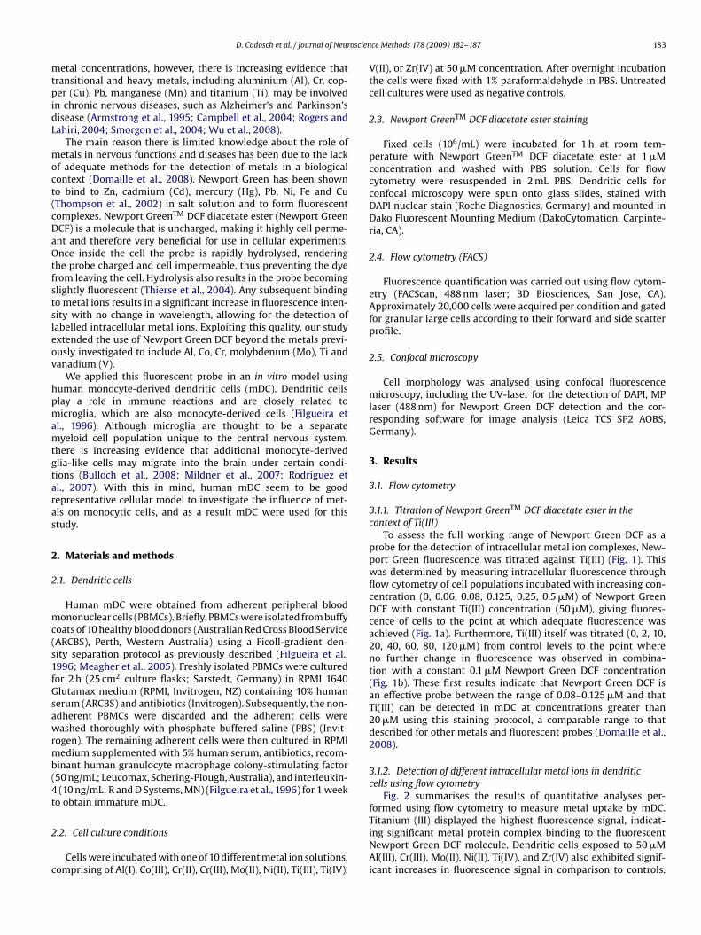
oscien
mtpidL
moct(cDaOtfstsleov
hpmamtgtaras
2
2
mc(s1fGsawrmb(4t
2
c
D. Cadosch et al. / Journal of Neur
etal concentrations, however, there is increasing evidence thatransitional and heavy metals, including aluminium (Al), Cr, cop-er (Cu), Pb, manganese (Mn) and titanium (Ti), may be involved
n chronic nervous diseases, such as Alzheimer’s and Parkinson’sisease (Armstrong et al., 1995; Campbell et al., 2004; Rogers andahiri, 2004; Smorgon et al., 2004; Wu et al., 2008).
The main reason there is limited knowledge about the role ofetals in nervous functions and diseases has been due to the lack
f adequate methods for the detection of metals in a biologicalontext (Domaille et al., 2008). Newport Green has been showno bind to Zn, cadmium (Cd), mercury (Hg), Pb, Ni, Fe and CuThompson et al., 2002) in salt solution and to form fluorescentomplexes. Newport GreenTM DCF diacetate ester (Newport GreenCF) is a molecule that is uncharged, making it highly cell perme-nt and therefore very beneficial for use in cellular experiments.nce inside the cell the probe is rapidly hydrolysed, rendering
he probe charged and cell impermeable, thus preventing the dyerom leaving the cell. Hydrolysis also results in the probe becominglightly fluorescent (Thierse et al., 2004). Any subsequent bindingo metal ions results in a significant increase in fluorescence inten-ity with no change in wavelength, allowing for the detection ofabelled intracellular metal ions. Exploiting this quality, our studyxtended the use of Newport Green DCF beyond the metals previ-usly investigated to include Al, Co, Cr, molybdenum (Mo), Ti andanadium (V).
We applied this fluorescent probe in an in vitro model usinguman monocyte-derived dendritic cells (mDC). Dendritic cellslay a role in immune reactions and are closely related toicroglia, which are also monocyte-derived cells (Filgueira et
l., 1996). Although microglia are thought to be a separateyeloid cell population unique to the central nervous system,
here is increasing evidence that additional monocyte-derivedlia-like cells may migrate into the brain under certain condi-ions (Bulloch et al., 2008; Mildner et al., 2007; Rodriguez etl., 2007). With this in mind, human mDC seem to be goodepresentative cellular model to investigate the influence of met-ls on monocytic cells, and as a result mDC were used for thistudy.
. Materials and methods
.1. Dendritic cells
Human mDC were obtained from adherent peripheral bloodononuclear cells (PBMCs). Briefly, PBMCs were isolated from buffy
oats of 10 healthy blood donors (Australian Red Cross Blood ServiceARCBS), Perth, Western Australia) using a Ficoll-gradient den-ity separation protocol as previously described (Filgueira et al.,996; Meagher et al., 2005). Freshly isolated PBMCs were culturedor 2 h (25 cm2 culture flasks; Sarstedt, Germany) in RPMI 1640lutamax medium (RPMI, Invitrogen, NZ) containing 10% humanerum (ARCBS) and antibiotics (Invitrogen). Subsequently, the non-dherent PBMCs were discarded and the adherent cells wereashed thoroughly with phosphate buffered saline (PBS) (Invit-
ogen). The remaining adherent cells were then cultured in RPMIedium supplemented with 5% human serum, antibiotics, recom-
inant human granulocyte macrophage colony-stimulating factor50 ng/mL; Leucomax, Schering-Plough, Australia), and interleukin-(10 ng/mL; R and D Systems, MN) (Filgueira et al., 1996) for 1 week
o obtain immature mDC.
.2. Cell culture conditions
Cells were incubated with one of 10 different metal ion solutions,omprising of Al(I), Co(III), Cr(II), Cr(III), Mo(II), Ni(II), Ti(III), Ti(IV),
ce Methods 178 (2009) 182–187 183
V(II), or Zr(IV) at 50 �M concentration. After overnight incubationthe cells were fixed with 1% paraformaldehyde in PBS. Untreatedcell cultures were used as negative controls.
2.3. Newport GreenTM DCF diacetate ester staining
Fixed cells (106/mL) were incubated for 1 h at room tem-perature with Newport GreenTM DCF diacetate ester at 1 �Mconcentration and washed with PBS solution. Cells for flowcytometry were resuspended in 2 mL PBS. Dendritic cells forconfocal microscopy were spun onto glass slides, stained withDAPI nuclear stain (Roche Diagnostics, Germany) and mounted inDako Fluorescent Mounting Medium (DakoCytomation, Carpinte-ria, CA).
2.4. Flow cytometry (FACS)
Fluorescence quantification was carried out using flow cytom-etry (FACScan, 488 nm laser; BD Biosciences, San Jose, CA).Approximately 20,000 cells were acquired per condition and gatedfor granular large cells according to their forward and side scatterprofile.
2.5. Confocal microscopy
Cell morphology was analysed using confocal fluorescencemicroscopy, including the UV-laser for the detection of DAPI, MPlaser (488 nm) for Newport Green DCF detection and the cor-responding software for image analysis (Leica TCS SP2 AOBS,Germany).
3. Results
3.1. Flow cytometry
3.1.1. Titration of Newport GreenTM DCF diacetate ester in thecontext of Ti(III)
To assess the full working range of Newport Green DCF as aprobe for the detection of intracellular metal ion complexes, New-port Green fluorescence was titrated against Ti(III) (Fig. 1). Thiswas determined by measuring intracellular fluorescence throughflow cytometry of cell populations incubated with increasing con-centration (0, 0.06, 0.08, 0.125, 0.25, 0.5 �M) of Newport GreenDCF with constant Ti(III) concentration (50 �M), giving fluores-cence of cells to the point at which adequate fluorescence wasachieved (Fig. 1a). Furthermore, Ti(III) itself was titrated (0, 2, 10,20, 40, 60, 80, 120 �M) from control levels to the point whereno further change in fluorescence was observed in combina-tion with a constant 0.1 �M Newport Green DCF concentration(Fig. 1b). These first results indicate that Newport Green DCF isan effective probe between the range of 0.08–0.125 �M and thatTi(III) can be detected in mDC at concentrations greater than20 �M using this staining protocol, a comparable range to thatdescribed for other metals and fluorescent probes (Domaille et al.,2008).
3.1.2. Detection of different intracellular metal ions in dendriticcells using flow cytometry
Fig. 2 summarises the results of quantitative analyses per-formed using flow cytometry to measure metal uptake by mDC.
Titanium (III) displayed the highest fluorescence signal, indicat-ing significant metal protein complex binding to the fluorescentNewport Green DCF molecule. Dendritic cells exposed to 50 �MAl(III), Cr(III), Mo(II), Ni(II), Ti(IV), and Zr(IV) also exhibited signif-icant increases in fluorescence signal in comparison to controls.
184 D. Cadosch et al. / Journal of Neuroscience Methods 178 (2009) 182–187
F wporto n of 1l ions t
IicbHmb
FC
ig. 1. Titration of Newport Green (a) and Ti(III) (b). Increasing concentration of Nef 50 �M; and Ti(III) (0, 2, 10, 20, 40, 60, 80, 120 �M) using a constant concentratioine corresponds to 0 �M. The curves line up according to the increasing concentrat
n contrast, cells exposed to Cr(II), Co(III), and V(II) showed min-mal or no increase in fluorescence signal when compared toontrol cells. These results indicate that Newport Green DCF can
e reliably used to detect and measure metal ion uptake by cells.owever, not surprisingly, different metals, oxidation states andetal ion concentrations all affect the fluorescence signal that cane detected.
ig. 2. Detection of various metal ions in dendritic cells using Newport Green DCF stain ao (b), Cr(II) (c), Cr(III) (d), Mo (e), Ni (f), Ti(III) (g), Ti(IV) (h), V (i) or Zr (k) before stained
Green DCF (0, 0.06, 0.08, 0.125, 0.25, 0.5 �M) using a constant Ti(III) concentration�M Newport Green DCF results in an increasing fluorescence intensity. The black
o the right.
3.2. Microscopy
3.2.1. Detection of intracellular metal ions in dendritic cells using
confocal microscopyFlow cytometry data were further confirmed with confocalmicroscopy. The microscopy analysis provided additional relevantinformation about the subcellular distribution of the metal ions
nd flow cytometry. Cells were incubated with no metal (faint line) or 50 �M Al (a),with Newport Green DCF (bold line).

D. Cadosch et al. / Journal of Neuroscience Methods 178 (2009) 182–187 185
F ence oG etatiov
(acflintgm
ig. 3. Confocal fluorescence microscopy. Dendritic cells were cultured in the presreen DCF. A blue nuclear stain (DAPI) was used in images a, b, c and e. (For interprersion of the article.)
Fig. 3). For that purpose, mDC were incubated with Cr(III), Ti(III)nd Ti(IV), stained with Newport Green DCF and prepared foronfocal microscopic analysis. Confocal images showed intenseuorescence throughout the cytoplasm and nucleolus of mDC
ncubated with Ti(III). Comparable fluorescence intensities wereot observed in control cells. Similar results were confirmed byransmission electron microscopy (data not shown). This sug-ests that Ti(III) ions may bind to proteins of the surface cellembrane, cytoskeletal proteins and ribonucleoproteins found
f Ti(III) (a and b), Ti(IV) (c), Cr(III) (d) or no metal (e) before stained with Newportn of the references to colour in this figure legend, the reader is referred to the web
in the nucleolus. Distinct staining of different regions for DAPIand Newport Green DCF indicates that Ti(III) does not bind tothe same nuclear DNA compounds labelled by DAPI. In addi-tion to Ti(III), confocal imaging also showed specific staining for
Cr(III) and Ti(IV) however neither of these fluorescent signalswere as intense as Ti(III), indicating that these metal ions do notbind to cellular proteins with greater affinity as that of Ti(III).There was no observed nucleolus staining for metals other thanTi(III).
1 oscien
4
bOrvcmactcua
moieNlHiirotwtmn2W
ilpt1oisids
tilmcHaosaabem
fdst
86 D. Cadosch et al. / Journal of Neur
. Discussion
Biomedical research in the context of metals and metal ions haseen limited due to a lack of adequate metal detection methods.nly recently, there has been an increasing panel of fluorescent
eagents available that can be used effectively for the detection of aariety of metals (Domaille et al., 2008). Newport Green, a fluores-ent chelator molecule, is able to bind specifically to intracellularetal protein complexes (Thompson et al., 2002). Incorporatingnew method of fluorescence metal detection using both flow
ytometry and confocal laser microscopy, this study has outlinedhe capacity of Newport GreenTM DCF diacetate ester to fluores-ently stain a variety of metal ion protein complexes as well asnveiling the morphology of intracellular protein-bound metals inn in vitro mDC culture setting.
Chelator molecules with affinities for a variety of metal ionsay interact with metals native to the cell, thus resulting in flu-
rescence information, which does not correspond to the metal ofnterest. Cellular Zn(II) and Cu(II) are the main metals that could bexpected to interfere with cellular fluorescence levels when usingewport Green DCF (Thierse et al., 2004). Consequently, some low
evel background staining of untreated control cells was observed.owever, incubation of the cells in the presence of increasing metal
on concentrations and Newport Green DCF concentrations resultedn significant fluorescence onset values from titration data. The fluo-escence signal of metal ion treated cells, particularly in the contextf Ti(III), sufficiently increased to a level where we could be surehat any background fluorescence from native metal ion complexesas essentially insignificant. In addition, a significant increase of
he fluorescent signal was seen for Al, Cr(III), Mo, Ni and Ti(IV), alletals that have been reported to be either neurotoxic or nomi-
ated to play a role in chronic nervous diseases (Campbell et al.,004; Halatek et al., 2008; Smorgon et al., 2004; Song et al., 2008;ang et al., 2008; Wu et al., 2008).It is also important to consider the toxicity of the various metal
ons when interpreting flow cytometric results of this nature. Pub-ished studies have shown that concentrations of 1% titaniumarticles, eluting Ti ions, kill immune cells (Pioletti et al., 1999) andhat some aqueous Ti, such as TiCl4, is cytotoxic (Yamamoto et al.,998). Our flow cytometry observations suggest that the morphol-gy of the mDC changed due to exposure to Ti(III) and other metallicons, whereby the cells became larger and more mature (data nothown). Cell gating was adjusted to compensate for this changen morphology where possible. However, cell death due to toxicityid not appear to significantly affect the results in this experimentaletting.
Wendt-Larsen et al. (2001) have shown that cells concen-rate metal ions into granules when extracellular metal ion levelsncrease. Our study confirms that observation, as with increasedevels of Ti(III), Newport Green DCF staining displayed a granular
orphology. It is possible that the metal ions are taken up by theell and then concentrated in the endo-lysosomal compartment.owever, the Newport Green DCF stain not only shows granularreas of higher fluorescence intensity but also a general dispersionf fluorescence throughout the cell cytoplasm. This indicates pos-ible diffusion of Ti(III) ions throughout the cytoplasm, cell matrixnd nuclear structures. However, the fact that Ti(III) does not bindnywhere within the nucleus other than the nucleolus confirmseliefs that Ti(III) will readily complex with certain proteins (Hallabt al., 2000, 2001), but also suggests that Ti(III) does not bind to DNAolecules.
The confocal images produced by using this new stain show dif-erences in the distribution and intensity of fluorescence betweenifferent metal types as well as between different oxidationtates of the same metal. Consequently, one has to postulatehat mDC may process metal ions of different oxidation states in
ce Methods 178 (2009) 182–187
different ways resulting in a diverse metal-dependent immuneresponse.
By combining flow cytometry with confocal microscopy, ourstudy demonstrates that Newport GreenTM DCF diacetate ester canbe used to fluorescently label metal ion protein complexes for avariety of different metal ions. Furthermore, our study highlightsthat cellular proteins demonstrate different binding affinities fordifferent metal ion types of the same metal, with for example Ti(III)appearing to have the higher binding affinity with this particularacetate ester compared with Ti(IV). It remains unclear whether thebinding affinity of the protein metal complex to the fluorescentdiacetate molecule is responsible for the difference or whether itis an indication of binding affinity between the metal ion and thecellular proteins alone. Nevertheless, this study will help to furtherunderstand how the mDC process and present metal ions.
Finally, Newport Green DCF is an excellent fluorescent probe forAl(III), Cr(III), Mo(II), Ni(II), Ti(III), Ti(IV) and Zr(IV) in the context ofcells and can be incorporated into procedures involving both flowcytometry and confocal fluorescence microscopy. Ongoing experi-ments have clearly demonstrated that this protocol is suitable forinvestigating tissue sections and other cell lines (T-lymphocytes,neuronal PC12 cells and osteoclastic cells), and that it will help theunderstanding of further pathophysiological mechanisms involvingmetal ions.
Acknowledgements
We thank John Kuo, John Murphy, Paul Rigby and Kathryn Heel(Centre for Microscopy, Characterisation and Analysis, University ofWestern Australia), and Guy Ben-Ary (Image Acquisition and Anal-ysis Facility, School of Anatomy and Human Biology) for excellenttechnical support and advice. We would also like to acknowledgeTrish Knox for her editorial support. This study was supported bythe NIH grant 1 R01 GM072726-01A1.
References
Armstrong RA, Winsper SJ, Blair JA. Hypothesis: is Alzheimer’s disease a metal-induced immune disorder? Neurodegeneration 1995;4:107–11.
Artik S, von Vultee C, Gleichmann E, Schwarz T, Griem P. Nickel allergy in mice:enhanced sensitization capacity of nickel at higher oxidation states. J Immunol1999;163:1143–52.
Bulloch K, Miller MM, Gal-Thot J, Milner TA, Gottfried-Blackmore A, Waters EM,et al. CD11c/EYFP transgene illuminates a discrete network of dendritic cellswithin the embryonic, neonatal, adult, and injured mouse brain. J Comp Neurol2008;508:687–710.
Burger J, Diaz-Barriga F, Marafante E, Pounds J, Robson M. Methodologies to examinethe importance of host factors in bioavailability of metals. Ecotoxicol Environ Saf2003;56:20–31.
Bush AI. Drug development based on the metals hypothesis of Alzheimer’s disease.J Alzheimers Dis 2008;15:223–40.
Campbell A, Becaria A, Lahiri DK, Sharman K, Bondy SC. Chronic exposure to alu-minum in drinking water increases inflammatory parameters selectively in thebrain. J Neurosci Res 2004;75:565–72.
Domaille DW, Que EL, Chang CJ. Synthetic fluorescent sensors for studying the cellbiology of metals. Nat Chem Biol 2008;4:168–75.
Filgueira L, Nestlé FO, Rittig M, Joller HI, Groscurth P. Human dendritic cells phago-cytose and process Borrelia burgdorferi. J Immunol 1996;157:2998–3005.
Gobba F. Occupational exposure to chemicals and sensory organs: a neglectedresearch field. Neurotoxicology 2003;24:675–91.
Halatek T, Sinczuk-Walczak H, Rydzynski K. Early neurotoxic effects of inhala-tion exposure to aluminum and/or manganese assessed by serum levels ofphospholipid-binding Clara cells protein. J Environ Sci Health A: Tox HazardSubst Environ Eng 2008;43:118–24.
Hallab NJ, Mikecz K, Jacobs JJA. Triple assay technique for the evaluation of metal-induced, delayed-type hypersensitivity responses in patients with or receivingtotal joint arthroplasty. J Biomed Mater Res 2000;53:480–9.
Hallab NJ, Mikecz K, Vermes C, Skipor A, Jacobs JJ. Differential lymphocyte reactivity
to serum-derived metal–protein complexes produced from cobalt-based andtitanium-based implant alloy degradation. J Biomed Mater Res 2001;56:427–36.Hostynek JJ. Factors determining percutaneous metal absorption. Food Chem Toxicol2003;41:327–45.
Insel BJ, Schaefer CA, McKeague IW, Susser ES, Brown AS. Maternal iron deficiencyand the risk of schizophrenia in offspring. Arch Gen Psychiatry 2008;65:1136–44.

oscien
J
K
K
M
M
M
N
P
P
R
R
R
D. Cadosch et al. / Journal of Neur
acobs JJ, Skipor AK, Patterson LM, Hallab NJ, Paprosky WG, Black J, et al.Metal release in patients who have had a primary total hip arthroplasty. Aprospective, controlled, longitudinal study. J Bone Joint Surg Am 1998;80:1447–58.
umar S. Acute toxicity of aluminium chloride, acephate, and their coexposure inmale Wistar rat. Int J Toxicol 2001;20:219–23.
usaka Y, Sato K, Suganuma N, Hosoda Y. Metal-induced lung disease: lessons fromJapan’s experience. J Occup Health 2001;43:1–23.
eagher J, Zellweger R, Filgueira L. Functional dissociation of the basolateral tran-scytotic compartment from the apical phago-lysosomal compartment in humanosteoclasts. J Histochem Cytochem 2005;53:665–70.
ildner A, Schmidt H, Nitsche M, Merkler D, Hanisch UK, Mack M, et al. Microgliain the adult brain arise from Ly-6ChiCCR2+ monocytes only under defined hostconditions. Nat Neurosci 2007;10:1544–53.
urakami M, Hirano T. Intracellular zinc homeostasis and zinc signaling. Cancer Sci2008;99:1515–22.
estle FO, Speidel H, Speidel MO. Metallurgy: high nickel release from 1- and 2-eurocoins. Nature 2002;419:132.
ecze L, Papp A, Nagymjteyi L, Desi I. Effect of acute administration of certain heavymetals and their combinations on the spontaneous and evoked cortical activityin rats. Environ Toxicol Pharmacol 2005;19:775–84.
ioletti DP, Takei H, Kwon SY, Wood D, Sung KL. The cytotoxic effect of tita-nium particles phagocytosed by osteoblasts. J Biomed Mater Res 1999;46:399–407.
ensing C, Maier RM. Issues underlying use of biosensors to measure metal bioavail-
ability. Ecotoxicol Environ Saf 2003;56:140–7.odriguez M, Alvarez-Erviti L, Blesa FJ, Rodriguez-Oroz MC, Arina A, Melero I, et al.Bone-marrow-derived cell differentiation into microglia: a study in a progressivemouse model of Parkinson’s disease. Neurobiol Dis 2007;28:316–25.
ogers JT, Lahiri DK. Metal and inflammatory targets for Alzheimer’s disease. CurrDrug Targets 2004;5:535–51.
ce Methods 178 (2009) 182–187 187
Shrivastava R, Upreti RK, Seth PK, Chaturvedi UC. Effects of chromium on the immunesystem. FEMS Immunol Med Microbiol 2002;34:1–7.
Smorgon C, Mari E, Atti AR, Dalla Nora E, Zamboni PF, Calzoni F, et al. Trace elementsand cognitive impairment: an elderly cohort study. Arch Gerontol Geriatr Suppl2004;9:393–402.
Song Y, Xue Y, Liu X, Wang P, Liu L. Effects of acute exposure to aluminum onblood–brain barrier and the protection of zinc. Neurosci Lett 2008;445:42–6.
Steens W, von Foerster G, Katzer A. Severe cobalt poisoning with loss of sight afterceramic–metal pairing in a hip—a case report. Acta Orthop 2006;77:830–2.
Sunderman FW. Nasal toxicity, carcinogenicity, and olfactory uptake of metals. AnnClin Lab Sci 2001;31:3–24.
Tezer M, Kuzgun U, Hamzaoglu A, Ozturk C, Kabukcuoglu F, Sirvanci M.Intraspinal metalloma resulting in late paraparesis. Arch Orthop Trauma Surg2005;125:417–21.
Thierse HJ, Moulon C, Allespach Y, Zimmermann B, Doetze A, Kuppig S,et al. Metal–protein complex-mediated transport and delivery of Ni2+ toTCR/MHC contact sites in nickel-specific human T cell activation. J Immunol2004;172:1926–34.
Thomas M, Jankovic J. Neurodegenerative disease and iron storage in the brain. CurrOpin Neurol 2004;17:437–42.
Thompson RB, Peterson D, Mahoney W, Cramer M, Maliwal BP, Suh SW, et al. Fluo-rescent zinc indicators for neurobiology. J Neurosci Methods 2002;118:63–75.
Wang Y, Kang XJ, Mu SM. Nanometer titanium dioxide and its toxicology progress.Chinese J Pharmacol Toxicol 2008;22:77–80.
Wendt-Larsen J, Abraham-Peskir JV, Medenwaldt R. Al(3+) and Zn(2+)-induced
structural changes and localization in a fully-hydrated live eukaryotic cellularmodel. Cell Biol Int 2001;25:521–30.Wu WH, Sun X, Yu YP, Hu J, Zhao L, Liu Q, et al. TiO2 nanoparticles promote beta-amyloid fibrillation in vitro. Biochem Biophys Res Commun 2008;373:315–8.
Yamamoto A, Honma R, Sumita M. Cytotoxicity evaluation of 43 metal salts usingmurine fibroblasts and osteoblastic cells. J Biomed Mater Res 1998;39:331–40.
Chapter 04 Biocorrosion of stainless steel by osteoclasts - In vitro evidence Most metals in contact with biological systems undergo corrosion by an
electrochemical process. This study investigated whether human OC are able to
grow on stainless steel and enhance corrosion of the metal alloy leading to an
increased release of corresponding metal ions, which may cause inflammatory
reactions and activate the immune system. Scanning electron microscopy analysis
demonstrated long-term viable OC cultures and evident resorption features on the
surface of stainless steel discs on which OC were cultured for 21 days. The findings
were confirmed by atomic emission spectrometry investigations showing significantly
increased levels of Cr, Ni, and Mn in the supernatant of OC cultures. Furthermore,
significant levels of pro-inflammatory cytokines IL-1b, IL-6, and TNF-α, which are
considered to be major mediators of osteolysis, were revealed in the same cultures
by cytometric bead array analysis. Within the present study, it was shown that human
OC precursors are able to grow and differentiate towards mature OC on stainless
steel. The mature cells are able to directly corrode the metal surface and release
corresponding metal ions, which induce the secretion of pro-inflammatory cytokines
that are known to enhance OC differentiation, activation, and survival. Enhanced
corrosion and the subsequently released metal ions may therefore result in enhanced
osteolytic lesions in the peri-prosthetic bone, contributing to the AP of the implant.
- 16 -

Bio-Corrosion of Stainless Steel by Osteoclasts—In Vitro Evidence
Dieter Cadosch,1,2 Erwin Chan,1 Oliver P. Gautschi,1,2 Hans-Peter Simmen,3 Luis Filgueira1
1School of Anatomy and Human Biology, University of Western Australia, Crawley, Australia, 2Department of Orthopaedic and Trauma Surgery,Royal Perth Hospital, Perth, Australia, 3Division of Trauma Surgery, University Hospital Zurich, Zurich, Switzerland
Received 22 August 2008; accepted 18 November 2008
Published online 22 December 2008 in Wiley InterScience (www.interscience.wiley.com). DOI 10.1002/jor.20831
ABSTRACT: Most metals in contact with biological systems undergo corrosion by an electrochemical process. This study investigatedwhether human osteoclasts (OC) are able to grow on stainless steel (SS) and directly corrode the metal alloy leading to the formation ofcorresponding metal ions, which may cause inflammatory reactions and activate the immune system. Scanning electron microscopy analysisdemonstrated long-term viable OC cultures and evident resorption features on the surface of SS discs on which OC were cultured for 21 days.The findings were confirmed by atomic emission spectrometry investigations showing significantly increased levels of chromium, nickel, andmanganese in the supernatant of OC cultures. Furthermore, significant levels of pro-inflammatory cytokines IL-1b, IL-6, and TNF-a, whichare considered to be major mediators of osteolysis, were revealed in the same cultures by cytometric bead array analysis. Within the presentstudy, it was shown that human osteoclast precursors are able to grow and differentiate towards mature OC on SS. The mature cells are ableto directly corrode the metal surface and release corresponding metal ions, which induce the secretion of pro-inflammatory cytokines thatare known to enhance osteoclast differentiation, activation, and survival. Enhanced corrosion and the subsequently released metal ionsmay therefore result in enhanced osteolytic lesions in the peri-prosthetic bone, contributing to the aseptic loosening of the implant.
� 2008 Orthopaedic Research Society. Published by Wiley Periodicals, Inc. J Orthop Res 27:841–846, 2009
Keywords: osteoclast; bio-corrosion; stainless steel; osteolysis; TNF-a
Over the last decades, significant developments havebeen taking place to provide suitable metal alloys forimplants with optimal mechanical characteristics andgood biocompatibility with minimal adverse host tissuereactions.1 However, their permanent tendency tocorrode when exposed to a physiological environmentremains a serious concern.2–4 Elevated concentrationsof metal ions have been measured in clinically retrievedperi-prosthetic tissues, distal organs, and body fluids intotal hip arthroplasty patients.5–7 The biology behindcorrosion inside the human body and the distribution ofthe metal ions throughout the organism is not fullyunderstood. It has been suggested that extracellularbody fluids have corrosive proprieties and contain metalbinding proteins.2,3 Furthermore, it has been debatedwhether osteoclasts (OC) are able to corrode pure metalwhen entering into contact with metal surfaces. How-ever, direct evidence of metal corrosion by OC has beenmissing to date.
Osteoclasts are highly specialized multinucleatedcells, which are capable of bone resorption. They areformed by the fusion of marrow-derived mononuclearprecursors, which circulate in the peripheral blood.Osteoclastic differentiation from hematopoietic andcirculating monocytes occurs in the presence of macro-phage-colony stimulating factor (M-CSF) and the recep-tor activator of NF-kB ligand (RANK-L) which areexpressed on osteoblastic cells.8 Mature OC displaydistinct morphological characteristics, such as multiplenuclei and a basal ruffled border surrounded by amembrane stabilizing the extent ring of actin filaments,and express cathepsin K and tartate-resistant acidphosphatase.9–11
The presence of implant-derived wear debris areknown to be responsible for severe inflammatory andhypersensitivity responses due to the release of pro-inflammatory cytokines that lead to increased osteolyticactivity at the bone–implant interface.12–15 We havepreviously demonstrated that metal ions (such astitanium) directly induce the differentiation of osteoclastprecursors towards mature OC, able of effective boneresorption.16 Furthermore, once released into the sys-temic blood circulation, the metal ions bind to serumproteins and form haptens or hapten-like complexes,which are considered to be relevant antigens recognizedby T lymphocytes and candidates for eliciting hyper-sensitivity reactions.5,17 Exposed to metal–protein com-plexes, T lymphocytes proliferate and differentiate toproduce effectors such as interleukin (IL)-6, IL-1a/b,interferon-gamma (IFN-g) and tumor necrosis factor-alpha (TNF-a) that trigger a particular immuneresponse.18–20
To date, surgical stainless steel 316L (SS) is one of themost frequently used biomaterials because of a favorablecombination of strength, good fabrication properties, lowinherent toxicity, minimal reactivity with biomolecules,and cost efficiency when compared with other metallicimplant materials.1,21 The aims of this study were firstly,to investigate whether human monocytes are able togrow on SS implants and differentiate into mature OC.Secondly, to test the hypothesis that mature OC areable to directly corrode SS and release correspondingmetal ions into the peri-implant environment, whichmay subsequently induce inflammatory reactions.
METHODSMetal DiscsSurgical SS [316L medical grade (UNS S31673) 17–19 Cr, 13-15 Ni, 2.25–3.0 Mo, max. 2.0 Mn, max 0.030 C, max 0.75 Si,max 0.025 P, max 0.010 S, max 0.10 N, max 0.50 Cu] discs were
JOURNAL OF ORTHOPAEDIC RESEARCH JULY 2009 841
Correspondence to: Dieter Cadosch (T: þ61 8 6488 3647; F: þ61 86488 1051; E-mail: [email protected])
� 2008 Orthopaedic Research Society. Published by Wiley Periodicals, Inc.

kindly provided by the RMS Foundation, Bettlach, Switzer-land. The polished test discs were 13 mm in diameter, 1 mmthick, and fitted into the wells of 24-well plates (Sarstedt,Nuernbrecht, Germany). Before use, the discs were washed inethanol, rinsed in distilled water, and steam sterilized.
Isolation of Peripheral Blood Monocytic CellsHuman monocytic cells (MC) were obtained from adherentperipheral blood mononuclear cells (PBMCs) as previouslydescribed.7 Briefly, PBMCs were isolated from buffy coats ofsix healthy blood donors [Australian Red Cross Blood Service(ARCBS), Perth, Australia] through Ficoll-gradient centri-fugation (Amersham Biosciences, Uppsala, Sweden). ThePBMCs were cultured (378C, humidified, 5% CO2) in 25 cm2
tissue culture flasks (Sarstedt) in RPMI-1640 glutamaxmedium (RPMI) (Gibco/Invitrogen, Auckland, New Zealand),supplemented with 10% human serum (ARCBS) and 1%antibiotics (10,000 units/mL penicillin G sodium, 10,000 mg/mL streptomycin sulfate, and 25 mg/mL Amphotericin B,Gibco) (standard medium). After 1 h in culture, the non-adherent PBMCs were discarded and the adherent PBMCsconsisting of 95% of MC cells were washed twice with 0.1 Mphosphate buffered saline (PBS) pH 7.2 (Gibco). The collectedcells were resuspended in RPMI medium and counted in ahemocytometer. Ethical approval for using human blood cellsfor this study was granted by the Ethics Committee of theUniversity of Western Australia and the ARCBS.
Cell Culture Conditions on Stainless Steel Discsand CoverslipsThe isolated MC were seeded either onto the SS discs (106 cells/disc) or glass coverslips (106 cells/coverslips) (12 mm diame-ter), resulting in a nonconfluent cell layer, placed into 24-welltissue culture plates (Sarstedt). After overnight incubation(378C, humidified, 5% CO2), the cultures were replenishedwith 1,500 mL of standard medium. In order to generate OC,half of the cell cultures were supplemented with osteoclastdifferentiation cytokines (10 ng/mL recombinant humanRANK-L and 10 ng/mL recombinant human M-CSF; R&D,Minneapolis, MN).10 Half of the cultures were left as MC. Asnegative controls, SS discs and glass coverslips were incubatedin standard culture medium only. After every incubation week,the conditioned supernatant was collected, pooled, frozen at�208C, and replaced with fresh standard medium. After21 days, the supernatant was collected and the cells fixedwith 2.5% EM grade glutaraldehyde (Electron MicroscopyScience, Washington, PA) in culture medium before processedfurther as described below. Six specimens were cultured pereach condition, and all experiments were carried out intriplicate.
Scanning Electron MicroscopyScanning electron microscopy (SEM) analysis were performedin order to assess whether MC were able to grow anddifferentiate on SS. The fixed cells adherent on the metaldiscs and glass coverslips (one/condition) were dehydratedthrough graded ethanol, critical point dried (BAL-TEC,Balzers, Liechtenstein) and mounted on SEM stubs (AgarScientific Ltd., Stansted, United Kingdom) for 4 nm platinumcoating before examination by SEM. Additionally, functionalevidence of osteoclastic corrosion was determined. For thatpurpose, after 3 weeks, incubation discs and coverslips onwhich MC and OC had been cultured (one/experiment), as well
as negative controls incubated without cells in standardmedium, were washed vigorously with distilled water and leftovernight in 0.25% ammonium hydroxide to remove theadherent cells. After rinsing and ultrasonication for 10 min,the discs and coverslips were air dried and mounted on SEMstubs. Subsequently, the entire surface was investigated bySEM. Documentation and analysis of the specimens wasperformed with a high resolution field emission SEM [Zeiss1555 VP-FESEM, 0.1-30 kV, variable pressure (up to 133 Pa,dry only), with BSE, CL, Oxford Instruments EDS and In LensSE detector capabilities, Oberkochen, Germany; available atthe Centre for Microscopy, Characterization and Analysis(CMCA), University of Western Australia, Perth, Australia].
Atomic Emission SpectrometryThe amount of bio-corrosion was quantified by measuring theconcentrations of chromium (Cr), nickel (Ni), manganese (Mn),and molybdenum (Mo) released into the culture supernatantcollected every 7 days as described above. Ion level detectionwas carried out using atomic emission spectrometry [Induc-tively Coupled Plasma-Optical (Atomic) Emission Spectrom-eter, Varian VISTA Axial, Mulgrave, Australia; available atMurdoch University, Perth, Australia]. The detection limitswere 0.01 mg/L for Cr, 0.002 mg/L for Mn, and 0.04 mg/L for Niand Mo.
Cytometric Bead Array AnalysisThe release of IL-1b, IL-6, and TNF-a from MC and OCcultured on metal discs and glass into the culture medium(collected as described above) was evaluated using a com-mercially available Cytometric Bead Array (CBA) kit (BDBiosciences, San Jose, CA). Fluorescence intensity wasmeasured with FACSCanto II Flow Cytometer (BD Bioscien-ces; available at CMCA) and quantified from a calibrationcurve using FCAP Array v1.0 Software (Softflow Technologies,New Brighton, MN). All assays were performed according tothe manufacturers’ instructions.
Calculations and Statistical AnalysisData were analyzed using SPSS for Windows (version 15.0;SPSS Inc., Chicago, IL). Independent-samples t-tests wereconducted in order to determine if mean ion concentrationswere significantly greater than minimal detection limits. Aone-way ANOVA was used to test mean differences in secretedcytokines in the various cell cultures. A p-value of <0.05 wasconsidered statistically significant.
RESULTSThe aim of this study was to investigate whether humanOC are able to directly corrode SS and release metal ionswhen entering in contact with metal surfaces. The veryfirst step was to evaluate the ability of MC and in vitrogenerated OC to grow on medical grade SS. For thatpurpose, the cells were cultured for 21 days either onglass coverslips or SS discs before being processed forSEM. The experiments demonstrated that human MCand OC grow as well on polished SS surfaces as theygrow on glass and culture dish (Fig. 1A,B). Nomorphological differences were detected in the cellscultured on metal discs when compared to the cultureson glass.
842 CADOSCH ET AL.
JOURNAL OF ORTHOPAEDIC RESEARCH JULY 2009

Evidence and Quantification of Bio-CorrosionThe ability of human OC to directly corrode SS wasinvestigated by documenting erosion lacunae on metaldiscs and by measuring the concentration of metal ionsreleased into the culture supernatant.
Detection of Bio-Corrosion Features Using ScanningElectron MicroscopyAfter 21-days incubation, the cell layers were removedfrom the metal discs and coverslips, and the bio-corrosion features documented by SEM. Figure 1Cshows representative images of resorption pits on SSdiscs on which OC had been cultured. As expected,no corrosion features were detected on metal discs onwhich MC had been cultured and on control discsincubated in standard culture medium only (Fig. 1D).No signs of erosion were detected on glass coverslips onwhich osteoclasts were cultured (data not shown).
Measurement of Metal Ions in Culture Supernatant byAtomic Emission SpectrometryThe amount of bio-corrosion was quantified by measur-ing the levels of metal ions released into the culturesupernatant after every incubation week. Significantlyincreased concentrations of Cr, Ni, and Mn weredetected in the supernatant of OC cultured on SS discswhen compared to the levels measured in the super-natant of MC cultured on SS discs and control discs leftin culture medium alone ( p< 0.01) (Fig. 2). Spectrom-etry analysis revealed concentrations of Mo belowdetection limits (0.04 mg/L) in OC supernatant. How-ever, no differences in ion concentrations were meas-ured between the different collection points in thesupernatant of OC cultures on SS ( p> 0.05) (data notshown). The levels of the different metal released by OCinto the supernatant were congruent with the percent-
age of the corresponding metal contained in the metalalloy. All measurements were below detection limits inthe culture supernatant of MC cultures on SS discs.Furthermore, our analysis indicates that the discswere corrosion resistant when left in standard culturemedium over 3 weeks, as no metal ions were detected inthe medium.
Cytokine MeasurementsIn order to assess the effects of the metal ions releasedinto the supernatant on the MC and OC cultures, theamount of IL-1b, IL-6, and TNF-a secreted into theculture medium were measured. Analysis revealedsignificantly increased concentrations of IL-1b, IL-6,and TNF-a in the supernatant of OC cultured on metaldiscs when compared with MC cultures on SS and OCcultured on glass coverslips ( p< 0.05) (Fig. 3). Analysisrevealed no significant differences in all cytokinemeasurements in the supernatant of MC and OCcultured on glass coverslips ( p>0.05).
DISCUSSIONDespite the enormous progress in providing suitablemetallic biomaterials with minimal bio-reactivity andrejection by the body, metal alloys in contact withbiological systems remain prone to corrosion. Thedetection of resorption pits on the surface of SS discs,on which OC were cultured and the significant levels ofcorresponding ions measured in culture supernatants,deliver first evidence for our hypothesis that maturehuman OC are able to directly corrode metal andsubsequently release metal ions into the cellularenvironment.
In tissue fluids, corrosion occurs at the metal surfaceas an electrochemical redox reaction, in which oxidationis coupled with reduction. In terms of how a biologicalenvironment can affect corrosion, it is believed thatproteins can influence the electrochemical behavior ofimplant metals/alloys. However, the overall resultsare not conclusive and the exact effect of proteins on
Figure 1. SEM images showing human in vitro generatedosteoclasts growing on glass coverslips (A) and on stainless steeldiscs (B). (C) Representative image of resorption pits after 21-daysincubation of human osteoclasts on stainless steel discs (inset showsmagnified resorption pits). (D) Surface detail of metal disc left for21 days in standard culture medium (note that the entire discsurface was investigated and no resorption pits were detected).
Figure 2. Histogram showing the significantly higher meanconcentrations (n¼27, �SD) of Ni, Cr, and Mn released into thesupernatantof human osteoclasts cultured over 21 days on stainlesssteel discs when compared to the levels measured in the super-natant of monocytes cultured on metal discs (below detection limits)and control discs (below detection limits). Mo concentrations inosteoclast cultures were below detection limits. *p<0.01.
BIO-CORROSION OF STAINLESS STEEL 843
JOURNAL OF ORTHOPAEDIC RESEARCH JULY 2009

corrosion is still the topic of much debate.22 Some authorsdemonstrate that proteins can enhance the propagationof charges between implant and surrounding fluids whileother publications suggest a reduced charge transfer byproteins.23–25 In addition, it has been debated whethermature OC are able to corrode the metal surface, butevidence has yet been missing. Our results clearlyindicate that osteoclast precursors are able to grow anddifferentiate on SS, as well as to directly corrode themetal surface as demonstrated by resorption pits on themetal discs. Moreover, the corresponding significantlevels of metal ions measured in the supernatant of OCcultures support the observed resorption features andsuggest that OC release the metal ions into theirenvironment. It is reasonable to assume that this processmay take place at the bone–implant interface, repre-senting an additional mechanism of metal corrosionin vivo and contributing to the levels of metal ionsmeasured in the peri-prosthetic tissues and in the serumfrom total arthroplasty patients.
The longevity of prosthetic implants depends on anumber of independent factors that are determined bythe surgical techniques, the implant, and the host’sreaction to the implant. To date, the formation of
osteolytic lesions, impairing the maintenance of a stableinterface between the implant and the bone remains theleading cause of failure of total joint arthroplasty.26 Peri-prosthetic osteolysis and subsequent aseptic looseningultimately develops in approximately 20% of patientswith reported failure rates of 13% for the femoralcomponent and 34% for the acetabular component.27,28
Several studies have shown the role of metal wearparticles in the initiation and development of asepticloosening by inducing an inflammatory response thatleads to an increased osteolytic activity at the implant–bone interface.12, 29–31 It is well recognized that activatedmacrophages, in response to titanium wear particles,produce pro-inflammatory cytokines, such as TNF-a,IL-6, and IL-1a/b.32–34 TNF-a acts directly on osteoclastprecursors, whereas IL-6 and IL-1a/b act indirectly byincreasing the expression of RANK-L and M-CSFby osteoblasts.35 In the present study, significantlyincreased levels of IL-1b, IL-6, and TNF-a were meas-ured in the supernatant of OC cultured on SS discs.These results are in line with several previous studiesshowing an increased release of bone resorptive cyto-kines IL-1b, IL-6, and TNF-a by cells of the monocyte/macrophage lineages in aseptic loosening.20,34,36 Theenhanced secretion of pro-inflammatory cytokines bythe metal ions (Cr, Ni, Mn) described here indicatesa possible additional mechanism to the macrophage-mediated foreign body reaction induced by the phagocy-tosis of small wear debris, which may contribute to theinflammatory reactions leading to an increased osteo-lytic activity reported in patients with aseptic loosening.
In addition to the interactions mentioned above,metals may also influence the immune system by directlyinterfering with biochemical and physiological activ-ities.37 More importantly, once released into the systemiccirculation, most metal ions have the ability to formcomplexes with native proteins; such metallo-organiccomplexes can induce allergy or may act as allergens inthe body.5,38–40 Metals known as sensitizers are nickelfollowed by cobalt and chromium.2 Such sensitizers arepresent in SS and potentially released by bio-corrosion.They are presented by dendritic cells, the most potentantigen-presenting cells, and subsequently recognizedby specific T-lymphocytes, thereby resulting in metal-specific immune responses including systemic auto-immune-like diseases and hypersensitivity reactions.These immune responses most typically manifest asskin lesions such as hives, eczema, redness, itching, andasthma-like symptoms.2
A metal in living tissue is prone to corrosion dueto electrochemical reactions.4 In addition to the well-investigated electrochemical aspect of corrosion in aphysiological system, this study demonstrates thathuman OC grow as well on SS as they grow on glass,and are able to directly corrode the metal surface andsubsequently release metal ions. Furthermore, theseresults indicate that the released metal ions may causeinflammatory reactions by inducing the secretion of pro-inflammatory cytokines IL-1b, IL-6, and TNF-a, which in
Figure 3. Bar chart showing the expression levels (�SD) of TNF-a, IL-1b, and IL-6 in the supernatant of monocytes and in vitrogenerated osteoclasts cultured on stainless steel discs at 3 weeks ofincubation. The amount of cytokines released was normalized to theconcentrations secreted by the cells (monocytes and osteoclasts)from the same individual incubated during the same period of timeon glass coverslips and averaged (n¼32). *p<0.05.
844 CADOSCH ET AL.
JOURNAL OF ORTHOPAEDIC RESEARCH JULY 2009

turn may enhance osteoclastic activity at the bone–metal interface.
Prosthetic and osteosynthetic devices made of metalalloys will remain indispensable in orthopedic andtrauma surgery. Therefore, the elucidation of theinteractions between metal surface and the surroundingtissues remains of clinical relevance. Implant topogra-phy modulates osteoclastic corrosion thus inducing therelease of metal ions which may mediate aseptic loosen-ing. Activation of cells from the macrophage lineage hasbeen reported to be dependent on the topography and, toa certain extent, to the chemistry of the metal implant.31
Our results may help developing new metal alloys and/orimplant surface topographies, which may reduce osteo-clastic corrosion without compromising the bonding ofthe peri-implant tissue with the metal indispensable forthe implant stability. Further studies are required inorder to outline a complete picture of the interactionbetween metal implant and bone cells, and the molecularpathways, which are involved in their activation throughreleased metal ions.
CONCLUSIONSWithin the present study, it was shown that humanosteoclast precursors were able to grow and differ-entiate towards mature OC on surgical SS. The maturecells were able to directly corrode the surface of SSand released corresponding metal ions into theirenvironment. The released ions induced the secretionof pro-inflammatory cytokines, which may lead to theformation of osteolytic lesions in the peri-prostheticbone, contributing to the loosening of the implant.
ACKNOWLEDGMENTSThis study was supported by the RMS Foundation, Bettlach,Switzerland. The authors gratefully acknowledge the Centrefor Microscopy, Characterization and Analysis (CMCA) for theuse of their facilities, as well as S. Parkinson for his excellentassistance with the SEM analysis. We thank T. Knox fromPathWest Laboratory Medicine WA for her excellent assis-tance, and B. von Katterfeld for the statistical analysis. AtomicEmission Spectrometry Analysis was done at the Marine andFreshwater Research Laboratory, Murdoch University, Perth,Australia.
REFERENCES1. Long M, Rack HJ. 1998. Titanium alloys in total joint
replacement—a materials science perspective. Biomaterials19:1621–1639.
2. Hallab NJ, Anderson S, Stafford T, et al. 2005. Lymphocyteresponses in patients with total hip arthroplasty. J OrthopRes 23:384–391.
3. Jacobs JJ, Hallab NJ, Skipor AK, et al. 2003. Metaldegradation products: a cause for concern in metal-metalbearings? Clin Orthop Relat Res 417:139–147.
4. Tezer M, Kuzgun U, Hamzaoglu A, et al. 2005. Intraspinalmetalloma resulting in late paraparesis. Arch Orthop TraumaSurg 125:417–421.
5. Jacobs JJ, Skipor AK, Patterson LM, et al. 1998. Metal releasein patients who have had a primary total hip arthroplasty. A
prospective, controlled, longitudinal study. J Bone Joint Surg[Am] 80:1447–1458.
6. Savarino L, Granchi D, Ciapetti G, et al. 2002. Ion release inpatients with metal-on-metal hip bearings in total jointreplacement: a comparison with metal-on-polyethylene bear-ings. J Biomed Mater Res 63:467–474.
7. Urban RM, Jacobs JJ, Tomlinson MJ, et al. 2000. Dissem-ination of wear particles to the liver, spleen, and abdominallymph nodes of patients with hip or knee replacement. J BoneJoint Surg [Am] 82:457–476.
8. Lacey DL, Timms E, Tan HL, et al. 1998. Osteoprotegerinligand is a cytokine that regulates osteoclast differentiationand activation. Cell 93:165–176.
9. Akisaka T, Yoshida H, Inoue S, et al. 2001. Organization ofcytoskeletal F-actin, G-actin, and gelsolin in the adhesionstructures in cultured osteoclast. J Bone Miner Res 16:1248–1255.
10. Filgueira L. 2004. Fluorescence-based staining for tartrate-resistant acidic phosphatase (TRAP) in osteoclasts combinedwith other fluorescent dyes and protocols. J HistochemCytochem 52:411–414.
11. Walsh NC, Cahill M, Carninci P, et al. 2003. Multiple tissue-specific promoters control expression of the murine tartrate-resistant acid phosphatase gene. Gene 307:111–123.
12. Bauer TW. 2002. Particles and periimplant bone resorption.Clin Orthop Relat Res 405:138–143.
13. Graves SE, Davidson D, Ingerson L, et al. 2004. TheAustralian Orthopaedic Association National Joint Replace-ment Registry. Med J Aust 180:S31–S34.
14. Harris WH. 1995. The problem is osteolysis. Clin Orthop RelatRes 311:46–53.
15. Voggenreiter G, Leiting S, Brauer H, et al. 2003. Immuno-inflammatory tissue reaction to stainless-steel and titaniumplates used for internal fixation of long bones. Biomaterials24:247–254.
16. Cadosch D, Chan E, Gautschi OP, et al. 2008. Titanium IVions induced human osteoclast differentiation and enhancedbone resorption in vitro. J Biomed Mater Res A (in press).
17. Martin SF. 2004. T lymphocyte-mediated immune responsesto chemical haptens and metal ions: implications for allergicand autoimmune disease. Int Arch Allergy Immunol 134:186–198.
18. Hallab NJ, Caicedo M, Finnegan A, et al. 2008. Th1 typelymphocyte reactivity to metals in patients with total hiparthroplasty. J Orthop Surg 3:6.
19. Streich NA, Breusch SJ, Schneider U. 2003. Serum levels ofinterleukin 6 (IL-6), granulocyte-macrophage colony-stimu-lating factor (GM-CSF) and elastase in aseptic prostheticloosening. Int Orthop 27:267–271.
20. Taki N, Tatro JM, Lowe R, et al. 2007. Comparison of the rolesof IL-1, IL-6, and TNFalpha in cell culture and murine modelsof aseptic loosening. Bone 40:1276–1283.
21. Singh R, Dahotre NB. 2007. Corrosion degradation andprevention by surface modification of biometallic materials.J Mater Sci Mater Med 18:725–751.
22. Yan Y, Neville A, Dowson D. 2006. Understanding the role ofcorrosion in the degradation of metal-on-metal implants. ProcInst Mech Eng [H] 220:173–181.
23. Khan MA, Williams RL, Williams DF. 1999. Conjointcorrosion and wear in titanium alloys. Biomaterials 20:765–772.
24. Williams RL, Brown SA, Merritt K. 1988. Electrochemicalstudies on the influence of proteins on the corrosion of implantalloys. Biomaterials 9:181–186.
25. Zhu J, Xu N, Zhang C. 1999. Characteristics of coppercorrosion in simulated uterine fluid in the presence of protein.Adv Contracept 15:179–190.
BIO-CORROSION OF STAINLESS STEEL 845
JOURNAL OF ORTHOPAEDIC RESEARCH JULY 2009

26. Harris WH. 1994. Osteolysis and particle disease in hipreplacement. A. review. Acta Orthop Scand 65:113–123.
27. Aspenberg P, Van der Vis H. 1998. Migration, particles, andfluid pressure. A. discussion of causes of prosthetic loosening.Clin Orthop Relat Res 352:75–80.
28. Keener JD, Callaghan JJ, Goetz DD, et al. 2003. Twenty-five-year results after Charnley total hip arthroplasty in patientsless than fifty years old: a concise follow-up of a previousreport. J Bone Joint Surg [Am] 85-A:1066–1072.
29. Bi Y, Van De Motter RR, Ragab AA, et al. 2001. Titaniumparticles stimulate bone resorption by inducing differentiationof murine osteoclasts. J Bone Joint Surg [Am] 83-A:501–508.
30. Matthews JB, Green TR, Stone MH, et al. 2001. Comparisonof the response of three human monocytic cell lines tochallenge with polyethylene particles of known size and dose.J Mater Sci Mater Med 12:249–258.
31. Sommer B, Felix R, Sprecher C, et al. 2005. Wear particles andsurface topographies are modulators of osteoclastogenesisin vitro. J Biomed Mater Res A 72:67–76.
32. Jiranek WA, Machado M, Jasty M, et al. 1993. Production ofcytokines around loosened cemented acetabular components.Analysis with immunohistochemical techniques and in situhybridization. J Bone Joint Surg [Am] 75:863–879.
33. Kim KJ, Rubash HE, Wilson SC, et al. 1993. A histologic andbiochemical comparison of the interface tissues in cementless
and cemented hip prostheses. Clin Orthop Relat Res 287:142–152.
34. Stea S, Visentin M, Granchi D, et al. 2000. Cytokines andosteolysis around total hip prostheses. Cytokine 12:1575–1579.
35. Ragab AA, Nalepka JL, Bi Y, et al. 2002. Cytokinessynergistically induce osteoclast differentiation: support byimmortalized or normal calvarial cells. Am J Physiol CellPhysiol 283:C679–C687.
36. Goodman SB, Huie P, Song Y, et al. 1998. Cellular profile andcytokine production at prosthetic interfaces. Study of tissuesretrieved from revised hip and knee replacements. J BoneJoint Surg [Br] 80:531–539.
37. Shrivastava R, Upreti RK, Seth PK, et al. 2002. Effects ofchromium on the immune system. FEMS Immunol MedMicrobiol 34:1–7.
38. Budinger L, Hertl M. 2000. Immunologic mechanisms inhypersensitivity reactions to metal ions: an overview. Allergy55:108–115.
39. Swiontkowski MF, Agel J, Schwappach J, et al. 2001.Cutaneous metal sensitivity in patients with orthopaedicinjuries. J Orthop Trauma 15:86–89.
40. Valentine-Thon E, Schiwara HW. 2003. Validity of MELISAfor metal sensitivity testing. Neuro Endocrinol Lett 24:57–64.
846 CADOSCH ET AL.
JOURNAL OF ORTHOPAEDIC RESEARCH JULY 2009

Chapter 05 Biocorrosion and uptake of titanium by human osteoclasts As tissue fluids enter into contact with the metal surface, corrosion occurs via an
electrochemical redox reaction. In terms of how a biological environment can affect
corrosion, it is believed that proteins can influence the electrochemical behaviour of
implant metals/alloys. Besides the electrochemical aspect of corrosion, it has been
debated whether cellular mechanisms are able to corrode the metal surface. This
study investigated whether human OC are able to grow on Ti and Al, and have
corrosive potency leading to an increased release of corresponding metal ions.
Scanning electron microscopy analysis demonstrated long-term viable OC cultures
on the surface of Ti and Al foils. Atomic emission spectrometry investigations showed
significantly increased levels of Al ions in the supernatant of OC cultured on Al;
however, all measurements in the supernatants of cell cultures on Ti were below
detection limits. Despite this, confocal microscopy analysis with Newport Green DCF
diacetate ester staining depicted intense fluorescence throughout the cytoplasm and
nucleolus of OC cultured on Ti foils. Comparable fluorescence intensities were not
observed in monocytes and control cells cultured on glass. It must be assumed that
Ti ions bind with a strong affinity to phosphorylated intracellular proteins.
Consequently, Ti was not released into the culture supernatant, explaining the no
detectable Ti levels. Titanium–protein complexes may be eventually released into the
extracellular space once the OC die and break down. This study demonstrated that
human OC precursors are able to grow and differentiate toward mature cells on Ti.
Furthermore, it suggests that mature OC enhance corrosion of Ti implants, most
likely by removing the protecting metal oxide layer from the metal surface, and take
up corresponding Ti ions.
- 23 -

Biocorrosion and uptake of titanium by human osteoclasts
Dieter Cadosch,1,2† Mohamed S. Al-Mushaiqri,2* Oliver P. Gautschi,2* James Meagher,2
Hans-Peter Simmen,1 Luis Filgueira2
1Division of Trauma Surgery, University Hospital Zurich, Zurich, Switzerland2School of Anatomy and Human Biology, University of Western Australia, Crawley, Australia
Received 11 September 2009; revised 6 May 2010; accepted 7 June 2010
Published online 24 September 2010 in Wiley Online Library (wileyonlinelibrary.com). DOI: 10.1002/jbm.a.32914
Abstract: All metals in contact with a biological system
undergo corrosion through an electrochemical redox reac-
tion. This study investigated whether human osteoclasts (OC)
are able to grow on titanium and aluminum, and directly cor-
rode the metals leading to the release of corresponding
metal ions, which are believed to cause inflammatory reac-
tions and activate osteoclastic differentiation. Scanning elec-
tron microscopy analysis demonstrated long-term viable OC
cultures on the surface of titanium and aluminum foils.
Atomic emission spectrometry investigations showed signifi-
cantly increased levels of aluminum in the supernatant of OC
cultured on aluminum; however, all measurements in the
supernatants of cell cultures on titanium were below detec-
tion limits. Despite this, confocal microscopy analysis with
Newport Green DCF diacetate ester staining depicted intense
fluorescence throughout the cytoplasm and nucleolus of OC
cultured on titanium foils. Comparable fluorescence inten-
sities were not observed in monocytes and control cells cul-
tured on glass. The present study demonstrated that human
osteoclast precursors are able to grow and differentiate to-
ward mature OC on titanium and aluminum. Furthermore, it
established that the mature cells are able to directly corrode
the metal surface and take up corresponding metal ions,
which subsequently may be released and thereby induce the
formation of osteolytic lesions in the periprosthetic bone,
contributing to the loosening of the implant. VC 2010 Wiley
Periodicals, Inc. J Biomed Mater Res Part A: 95A: 1004–1010, 2010.
Key Words: osteoclast, biocorrosion, titanium, aluminum,
osteolysis, Newport Green
INTRODUCTION
Over the past decades, significant developments have beentaking place to provide suitable metals and metal alloys forprosthetic and osteosynthetic implants with optimal me-chanical characteristics and good biocompatibility with min-imal adverse reactions to host tissue.1 Among other metals,pure titanium and titanium alloys, often containing alumi-num and vanadium, are frequently used biomaterials in den-tal, orthopedic, and trauma surgery because of a favorablecombination of strength, good fabrication properties, lowinherent toxicity, and minimal reactivity with biomoleculeswhen compared with other metallic biomaterials.1,2 How-ever, despite the immense progress in providing suitablemetallic biomaterials, their tendency to corrode whenexposed to a physiological environment remains a seriousconcern to the surgical and biomaterial community.2–4 Ele-vated concentrations of metal ions (including titanium) havebeen measured in clinically retrieved capsular and peripros-thetic tissues as well as in distal organs (liver, spleen, andlymph nodes) and body fluids (serum and urine) of totalhip arthroplasty patients.5–7 The biology behind corrosioninside the human body and the distribution of the metalions throughout the organism is not yet fully understood. Ithas been suggested that extracellular body fluids, including
blood serum, have corrosive proprieties and contain metalbinding proteins.3,4 Furthermore, it has long been debatedwhether osteoclasts (OC) are able to corrode orthopedicimplants when entering in contact with the metal surfaces.Using a human in vitro model, we have recently demon-strated that human OC are able to corrode surgical stainlesssteel.8 However, direct evidence of titanium corrosion by OChas not yet been demonstrated. In a water-based environ-ment, titanium and aluminum form thin oxide layers ontheir surface, protecting them from further electrochemicalcorrosion. At physiological pH, the water solubility for alu-minum and titanium ions is in the range of micromolar con-centrations.9 However, in the presence of certain anionsfound in biological fluids, such as phosphates, titaniumforms complexes with lower water solubility, resulting inconcentrations below detectable levels. Thus, we haverecently established a fluorescent method for the detectionof complex, intracellular metal ions, including titanium,using Newport Green DCF diacetate ester.10
OC are highly specialized, often multinucleated cells,which are uniquely capable of bone resorption.11 They dif-ferentiate from marrow-derived mononuclear precursors,which circulate in the CD14þ monocyte fraction of peri-pheral blood, in the presence of macrophage-colony
*These authors contributed equally to this work and should both be considered as second author.†Dieter Cadosch was awarded a Scholarship for International Fees Research (SIRS) by the University of Western Australia. The research was
carried out while the author was a SIRS scholar.
Correspondence to: D. Cadosch; e-mail: [email protected]
1004 VC 2010 WILEY PERIODICALS, INC.

stimulating factor (M-CSF) and the receptor activator of NF-jB ligand (RANK-L), which is expressed by osteoblasts andother bone-related stromal cells.12,13 Mature OC are oftendescribed as multinucleated cells, displaying additional dis-tinct morphological characteristics, such as a ruffled basalborder surrounded by a membrane-stabilizing extensivering of actin filaments, and expressing cathepsin K and tar-tate-resistant acid phosphatase.14–16 However, also mono-nucleated OC can already display the distinct morphological,molecular, and functional properties of mature cells.17,18 Inrecent years, we have established a reliable in vitro modelfor the generation of human OC using human monocytesisolated from peripheral blood and cultured in the presenceof osteoclastic cytokines (M-CSF and RANK-L).8,15,19–21 Thiswell-characterized in vitro model has been used throughoutthis study.
The aims of this study were as follows: first, to investi-gate whether human monocytes are able to grow on alumi-num and titanium and to differentiate into mature OC; andsecond, to test the hypothesis that mature OC are able todirectly corrode pure titanium and aluminum and releasecorresponding metal ions into the peri-implant environment.Aluminum was included as an easy-to-handle and inexpen-sive basic metal foil model; however, titanium was the mainfocus of this study.
MATERIALS AND METHODS
Metal foilsTitanium foils were purchased from EURO Titan, UK, andwere 5 mm in diameter. Commercially available aluminumfoil (Capral Aluminum, NSW, Australia) was wrapped overglass coverslips for easy handling. Before use, the foils werewashed in ethanol, rinsed in distilled water, and steamsterilized.
Isolation of peripheral blood monocytic cellsEthical approval for the use of human blood cells wasgranted by the Ethics Committee of the University of West-ern Australia and the Australian Red Cross Blood Service(ARCBS), Perth, WA, Australia. Human monocytic cells (MC)were obtained from adherent peripheral blood mononuclearcells (PBMCs). Briefly, PBMCs were isolated from buffy coatsof 10 healthy blood donors (ARCBS) through Ficoll-gradientcentrifugation (Amersham Biosciences, Uppsala, Sweden) aspreviously described.21,22 The PBMCs were cultured (37�C,humidified, 5% CO2) in 25 cm2 tissue culture flasks (Sar-stedt) in RPMI-1640 Glutamax medium (RPMI) (Gibco/Invi-trogen, Auckland, NZ), and supplemented with 10% humanserum (ARCBS) and 1% antibiotics (Gibco) (standard me-dium). After 1 h, the nonadherent PBMCs were discarded,and the adherent PBMCs consisting of MC cells werewashed twice with 0.1M phosphate-buffered saline (PBS)pH 7.2 (Gibco). The collected cells were resuspended inRPMI medium and counted manually in a hemocytometer.
Adherent cell cultures on metal foils and coverslipsThe isolated MC were seeded either onto titanium, alumi-num foils (106 cells/foil) or glass coverslips (12-mm diame-
ter) placed into 24-well tissue culture plates (Sarstedt,Nuernbrecht, Germany). After overnight incubation (37�C,humidified, 5% CO2), the cultures were replenished with1500 mL of standard medium. To generate OC, one-half ofthe cell cultures were supplemented with osteoclast differ-entiation cytokines (10 ng/mL recombinant human RANK-Land 10 ng/mL recombinant human M-CSF, R&D, Minneapo-lis, MN) as previously described.15,19 One-half of the cul-tures were left as MC with standard culture medium onlyand no cytokines added. As negative controls, metal foilsand glass coverslips were incubated in standard culturemedium only. The conditioned supernatant was collectedweekly, pooled, frozen at �20�C, and replaced with 1500 mLfresh standard medium. After 21 days, the supernatant wascollected and the cells fixed with 2.5% EM grade glutaralde-hyde (Electron Microscopy Science, Washington, PA) in culturemedium for scanning electron microscopy (SEM). The samplesdesigned for confocal microscopy with Newport Green DCFdiacetate ester staining were fixed with 1% paraformaldehydein PBS before processed further as described later. Six speci-mens were cultured for each condition and all experimentswere carried out in triplicate.
Atomic emission spectrometryThe amount of biocorrosion was quantified by measuringthe concentrations of titanium and aluminum ions releasedinto the corresponding culture supernatant. Ion levels weremeasured using atomic emission spectrometry [InductivelyCoupled Plasma-Optical (Atomic) Emission Spectrometer,Varian VISTA Axial, Mulgrave, Australia; available at Mur-doch University, Perth, WA, Australia]. The detection limitfor both titanium and aluminum was 0.01 mg/L.
Scanning electron microscopyScanning electron microscopy analysis was performed toassess whether MC were able to grow and differentiate ontitanium and aluminum foil. The glutaraldehyde fixed cellsadherent on the foils and glass coverslips were dehydratedthrough graded ethanol, critical point dried (BAL-TEC,Balzers, Liechtenstein) and mounted on SEM stubs (AgarScientific, Stansted, UK) for 4-nm platinum coating beforeexamination by SEM. Documentation and analysis of thespecimens was performed with a high resolution fieldemission SEM [Zeiss 1555 VP-FESEM, 0.1–30 kV, variablepressure (up to 133 Pa, dry only), with BSE, CL, OxfordInstruments EDS and In Lens SE detector capabilities,Oberkochen, Germany; available at the Centre for Microscopy,Characterization and Analysis, University of Western Australia,Perth, WA, Australia].
Fluorescent tartrate-resistant acidic phosphatase andActin StainingTo further characterize the OC cultures, fluorescent tartrate-resistant acidic phosphatase (TRAP) and actin staining wasperformed. The cells, fixed with 1% paraformaldehyde inPBS, were stained for TRAP activity using a fluorescence-based protocol.15 Briefly, the cells were incubated for 15minutes with ELF97 substrate (20 mM, E6569, Molecular
ORIGINAL ARTICLE
JOURNAL OF BIOMEDICAL MATERIALS RESEARCH A | 15 DEC 2010 VOL 95A, ISSUE 4 1005

Probes/Invitrogen, Eugene, OR) in 110 mM acetate buffer(pH 5.2), containing 1.1 mM sodium nitrite, and 7.4 mM tar-trate (all from Sigma-Aldrich). The nuclei were stained withDAPI (40,6-diamidine-20-phenylindole dihydrochloride, 10ng/mL, Roche Diagnostics, Mannheim, Germany). The basalactin ring was stained with AlexaFluor546-labelled phalloi-din (0.2 U/mL in PBS). After mounting (Dako FluorescentMounting Medium, Dako Cytomation, Carpinteria, CA, USA),the samples were analyzed and documented using confocalmicroscopy.
Newport Green DCF diacetate ester stainingTo investigate the ability of OC to corrode titanium and takeup corresponding ions, paraformaldehyde (1% in PBS) fixedcells adherent to the metal foils and glass coverslips wereincubated for 1 h at room temperature with Newport GreenDCF diacetate ester at 1 lM concentration (MolecularProbes/Invitrogen) and washed with PBS.10 Subsequently,the cells were stained with DAPI nuclear stain and mountedin Dako Fluorescent Mounting Medium (DakoCytomation)for confocal microscopy.
Confocal microscopyCell morphology was analyzed using multiphoton confocalfluorescence microscopy, including the UV-laser for detec-tion of DAPI, multiphoton laser for ELF97, phalliodin Alexa-Fluor546 and Newport Green DCF detection and the corre-sponding software for image analysis (Leica TCS SP2 AOBS,Germany).
Calculations and statistical analysisData were analyzed using SPSS for Windows (version 15.0;SPSS Inc., Chicago, IL). Independent sample t-tests wereconducted to determine whether mean ion concentrationswere significantly greater than minimal detection limits. Ap-value of less than 0.05 was considered statisticallysignificant.
RESULTS
This study aimed to investigate whether human OC are ableto directly corrode titanium and aluminum, and release cor-responding metal ions when in contact with metal surfaces.The first step was to evaluate the ability of MC and in vitrogenerated OC to attach, grow on titanium and aluminum.For that purpose, the cells were cultured for 21 days eitheron glass coverslips or on metal foils before processing forSEM and fluorescence microscopy. The experiments demon-strated that human MC and OC attach and grow as well ontitanium and aluminum surfaces, as they grow on glass andculture dishes (Fig. 1). The OC cultured on metal foilsshowed a more vacuolated membrane and were smallerthan control cells cultured on glass (averaged cell diameter21 6 4 lm vs. 28 6 7 lm, p > 0.05). No differences in celldensity were detected between the different cell cultures(p > 0.05). Additionally, OC were stained for TRAP activityand localization of the basal actin ring using confocal mi-croscopy methods (Fig. 2), confirming the osteoclastic prop-
erties of the cells cultured in the presence of osteoclasticcytokines (M-CSF and RANK-L).
Measurement of metal ions in culture supernatant byatomic emission spectrometryThe first finding to provide evidence and quantification ofdirect corrosion of titanium and aluminum by human OCwas realized by measuring the concentration of metal ionsreleased into the corresponding culture supernatant afterevery week of incubation. Increased concentrations of alu-minum were detected in the supernatant of OC cultured onaluminum foils when compared with the levels measured inthe supernatant of MC cultured on aluminum foils and con-trol foils left in culture medium without cells (p < 0.01)
FIGURE 1. Representative SEM images showing human OC growing
on glass (A), titanium foil (B), and aluminum foil (C) after 21 days of
incubation.
1006 CADOSCH ET AL. BIOCORROSION AND UPTAKE OF TITANIUM

(Table I). No differences in ion concentrations were meas-ured between the different collection points (each incuba-tion week) in the supernatant of OC cultures (p > 0.05)(data not shown). Surprisingly, all measurements werebelow detection limits in the culture supernatant of OC andMC cultured on titanium foils as well as in the medium inwhich the foils were left during the same period of time.
Detection of intracellular titanium ions in OCusing confocal microscopyTo demonstrate corrosion of titanium and uptake of corre-sponding ions, the cells cultured on titanium foils werestained using Newport Green DCF diacetate ester and ana-lyzed using confocal microscopy. Images showed intensefluorescence signals in OC cultured on titanium foils [Fig.3(D,E)], when compared with fluorescence intensities in OCand MC cultured on glass [Fig. 3(A–C)]. The microscopyanalysis provided additional relevant information about thesubcellular distribution of the titanium ions in OC culturedon titanium foils, showing intense partially granular andpartially diffuse fluorescence throughout the cytoplasm anddistinct fluorescence of the nuclear heterochromatin. Thissuggests that titanium ions bind to cytoplasmic and nuclearstructures and are therefore not released into the culturesupernatant. The fluorescent Newport Green DCF signal forcells cultured on aluminum was similar to the control levels(data not shown).
DISCUSSION
Despite the remarkable progress in providing suitable me-tallic biomaterials such as titanium and titanium alloys,with minimal bioreactivity and rejection by the body, metalimplants in contact with biological systems remain prone tocorrosion. The aim of this study was to investigate the cor-rosive potency of human OC on titanium and aluminum. Forthat purpose, in vitro generated OC using a well-establishedmodel, and corresponding cultured monocytes were used.Aluminum was used as an inexpensive easy-to-handle metalmodel, whereas titanium was the main focus of thisresearch.
In tissue fluids, corrosion occurs at the metal surfaceas an electrochemical redox reaction, in which oxidation
FIGURE 2. Multiphoton confocal fluorescence microscopy of TRAP (A; green color) and actin (B; red color) stain on OC cultured for 21 days
(Nuclei are stained in blue with DAPI). Arrow 1 indicates the well-documented basal actin ring in a monocucleated osteoclastic cell. Arrow 2 indi-
cates a multinucleated osteoclast in close contact with two other cells, possibly undergoing additional fusion and formation of a cell with more
nuclei. [Color figure can be viewed in the online issue, which is available at wileyonlinelibrary.com.]
TABLE I. Measurement of Aluminium Concentrations
Culture conditionsAluminum
concentrations (mg/L)
Monocytes on glass 0.032 (60.004)Osteoclasts on glass 0.078 (60.024)Monocytes on aluminum 0.162 (60.125)Osteoclasts on aluminum 9.500* (60.384)
Measurement of aluminium concentrations (6SD) in the culture su-
pernatant using atomic emission spectrometry after 21 days incubation.
Osteoclasts cultured on aluminium foils released significantly higher
concentrations when compared with monocyte cultures. Note all meas-
urements of titanium concentrations were below detection limits.
*p < 0.01.
FIGURE 3. Multiphoton confocal fluorescence microscopy image (op-
tical sections through the ‘‘nuclear level’’) showing monocytes cul-
tured for 21 days on glass (A) or titanium foil (B), as well as OC on
glass (C) or on titanium foil (D and E). Note the staining of intracellu-
lar titanium complexes with Newport Green DCF diacetate ester,
detected only in OC cultured on titanium foil, in a granular and dif-
fuse distribution throughout the cytoplasm, as well as in the hetero-
chromatin of the nucleus (D and E; green color). Nuclei are stained in
blue with DAPI. Note the multiple nuclei in the cells on (D) and (E).
[Color figure can be viewed in the online issue, which is available at
wileyonlinelibrary.com.]
ORIGINAL ARTICLE
JOURNAL OF BIOMEDICAL MATERIALS RESEARCH A | 15 DEC 2010 VOL 95A, ISSUE 4 1007

(electron loss of the metal) is coupled with reduction (elec-tron gain of electrolyte components).23 In terms of how abiological environment can affect corrosion, it is believedthat proteins can influence the electrochemical behavior ofimplant metals/alloys. However, the overall results are notconclusive and the exact effect of proteins on corrosion isstill the topic of much debate.24–28 Beside the electrochemi-cal aspect of corrosion in a physiological environment, ithas been debated whether mature OC themselves are ableto corrode the metal surface.
Using fluorescence microscopy analysis, we have con-firmed that MC isolated from peripheral blood used forthese experiments can differentiate into TRAP positive, mul-tinucleated OC displaying a distinct basal actin ring. Addi-tionally, we have previously demonstrated that the humanOC generated in vitro using the same protocol, are func-tional cells able to resorb dentin and express cathepsin K,as well as a basal acidic compartment.19–21 Using this invitro OC model, our results indicate that osteoclast precur-sors are able to grow and differentiate on titanium and alu-minum, as well as to directly corrode the metal surface asdemonstrated by the release of aluminum ions into the cul-ture supernatant and by the intracellular detection of tita-nium. It is reasonable to assume that a similar process maytake place at the bone–implant interface and represent anadditional mechanism of metal corrosion in vivo that con-tributes to the levels of metal ions (including titanium)measured in the periprosthetic tissues, in distal organs andin the serum from total arthroplasty patients.5–7
Both aluminum and titanium corrode easily in a water-based environment. However, a thin oxide layer is quicklyformed on the surface, protecting the metal from furtherelectrochemical corrosion.9,29 In a physiological context, itmust be considered that functional OC are able to generatean environment more prone to corrosion, by secreting pro-tons into the resorption lacunae.18 The low pH in theresorption compartment with the high concentration of Hþmight destabilize the oxide layer, leading to free metal ionson the surface.9,29 Removal of the metal oxide layer willsubsequently expose new metal to additional corrosionenhanced by the acidic conditions in the osteoclastic resorp-tion pit. Once solubilized in the osteoclastic pit, the metalions are most likely taken up by the OC into the TRAP-containing transcytotic compartment for further process-ing.21,30,31 Usually, resorbed material is eventually releasedby the OC into the extracellular space. This process happensin the case of aluminum, as shown in this study and forother metal ions such as cobalt and chromium as previouslydemonstrated.8 However, certain metal ions may bind to cel-lular structures and form stable complexes that remain inthe cells. This is certainly the case for titanium ions thatbind to cytoplasmic and nuclear structures, as depicted byconfocal fluorescence microscopy. Consequently, titaniumwas not released into the culture supernatant, explainingthe nondetectable titanium levels in our cultures. It is rea-sonable to assume that titanium ions bind to phosphoryl-ated molecules, including phosphorylated intracellular pro-teins.32–35 This hypothesis is further supported in previous
experiments by our group using element-specific electronmicroscopy methods, such as energy-filtered transmissionelectron microscopy, showing the strong affinity of titaniumions for phosphorylated cellular structure. By complexingwith phosphorylated cellular structures, which often corre-spond to active functional states of enzymes or signalingproteins, titanium may interfere with signaling pathways.Additionally, the attachment of titanium to membrane phos-pholipids may change its fluidity and thereby alter cellularfunctions such as migration, protein secretion and the abil-ity to respond to stimuli as shown with other metals suchas aluminum and nickel.36 Titanium–protein complexes maybe eventually released into the extracellular space, and con-ceivably into the systemic circulation in patients, once theOC die and break down. However, additional studies arerequired to show how titanium is translocated from theimplant site into the surrounding tissue and how it accumu-lates in distant organs.
Aseptic loosening of metal joint implants remains a signif-icant problem in orthopedic surgery. More than 10% ofpatients suffer from the condition within 20 years of aprimary hip arthroplasty, with the incidence continuing toincrease.37 Aseptic loosening is suggested to result from anincreased osteolytic activity at the bone-implant interface,which ultimately leads to loss of fixation.38–42 Several studieshave demonstrated the role of titanium wear particles (in thenanometer range) in the initiation and development of asep-tic loosening by inducing an inflammatory response charac-terized by an enhanced production of proinflammatory cyto-kines, such as TNF-a, IL-6, and IL-1a/b, which ultimatelylead to an increased osteolytic activity.38,43–47 In addition topathophysiological mechanisms involving wear particles, wehave previously demonstrated that titanium ions directlyinduce differentiation of osteoclast precursors toward matureOC capable of effective bone resorption.19 Titanium ions arealso supposed to play a role in the recruitment of osteoclastprecursors by inducing the expression of specific cytokines.20
The osteoclastic corrosion described here is likely to contrib-ute to the pathophysiological mechanism mentioned earlierby releasing intracellular titanium ions after cell death.
Summarizing, this study demonstrates that human OCgrow as well on titanium and aluminum as they grow onglass and culture dishes. Human OC are able to directly cor-rode the metal surface and take up corresponding metalions. Aluminum ions are released by OC into the surround-ing extracellular space, whereas titanium ions remain in theOC, as they appear to bind to cellular structures, probablyphosphorylated proteins, phospholipids, and nucleotides.Following cell death, the released titanium ions are likely tocontribute to the pathophysiological mechanisms of asepticloosening by inducing osteoclastic differentiation. Further-more, once titanium–protein complexes are released intothe blood circulation they may act as antigens and triggerinflammatory immune reactions.5,48
CONCLUSIONS
Within the present study, it was confirmed that humanosteoclast precursors were able to grow and differentiate
1008 CADOSCH ET AL. BIOCORROSION AND UPTAKE OF TITANIUM

toward mature OC on aluminum and titanium. The maturecells were able to directly corrode the surface of the metalsand take up metal ions. Similar processes may take place invivo, leading to increased biocorrosion, and release of metalions into the surrounding tissue and into the systemic bloodcirculation.
ACKNOWLEDGMENT
The authors thank T. Knox for her outstanding assistance andeditorial advice.
REFERENCES1. Long M, Rack HJ. Titanium alloys in total joint replacement—A
materials science perspective. Biomaterials 1998;19:1621–1639.
2. Singh R, Dahotre NB. Corrosion degradation and prevention by
surface modification of biometallic materials. J Mater Sci Mater
Med 2007;18:725–751.
3. Hallab NJ, Anderson S, Stafford T, Glant T, Jacobs JJ. Lympho-
cyte responses in patients with total hip arthroplasty. J Orthop
Res 2005;23:384–391.
4. Jacobs JJ, Hallab NJ, Skipor AK, Urban RM. Metal degradation
products: A cause for concern in metal-metal bearings? Clin
Orthop Relat Res 2003;417:139–147.
5. Jacobs JJ, Skipor AK, Patterson LM, Hallab NJ, Paprosky WG,
Black J, Galante JO. Metal release in patients who have had a pri-
mary total hip arthroplasty. A prospective, controlled, longitudinal
study. J Bone Joint Surg Am 1998;80:1447–1458.
6. Savarino L, Granchi D, Ciapetti G, Cenni E, Nardi Pantoli A, Rotini
R, Veronesi CA, Baldini N, Giunti A. Ion release in patients with
metal-on-metal hip bearings in total joint replacement: A compar-
ison with metal-on-polyethylene bearings. J Biomed Mater Res
2002;63:467–474.
7. Urban RM, Jacobs JJ, Tomlinson MJ, Gavrilovic J, Black J,
Peoc’h M. Dissemination of wear particles to the liver, spleen,
and abdominal lymph nodes of patients with hip or knee replace-
ment. J Bone Joint Surg Am 2000;82:457–476.
8. Cadosch D, Chan E, Gautschi OP, Simmen HP, Filgueira L. Bio-
corrosion of stainless steel by osteoclasts–in vitro evidence. J
Orthop Res 2009;27:841–846.
9. Tengvall P, Lundstrom I. Physico-chemical considerations of tita-
nium as a biomaterial. Clin Mater 1992;9:115–134.
10. Cadosch D, Meagher J, Gautschi OP, Filgueira L. Uptake and in-
tracellular distribution of various metal ions in human monocyte-
derived dendritic cells detected by Newport Green DCF diacetate
ester. J Neurosci Methods 2009;178:182–187.
11. Vaananen HK, Zhao H, Mulari M, Halleen JM. The cell biology of
osteoclast function. J Cell Sci 2000;113:377–381.
12. Fujikawa Y, Quinn JM, Sabokbar A, McGee JO, Athanasou NA.
The human osteoclast precursor circulates in the monocyte frac-
tion. Endocrinology 1996;137:4058–4060.
13. Lacey DL, Timms E, Tan HL, Kelley MJ, Dunstan CR, Burgess T,
Elliott R, Colombero A, Elliott G, Scully S, Hsu H, Sullivan J, Haw-
kins N, Davy E, Capparelli C, Eli A, Qian YX, Kaufman S, Sarosi I,
Shalhoub V, Senaldi G, Guo J, Delaney J, Boyle WJ. Osteoprote-
gerin ligand is a cytokine that regulates osteoclast differentiation
and activation. Cell 1998;93:165–176.
14. Akisaka T, Yoshida H, Inoue S, Shimizu K. Organization of cytos-
keletal F-actin. G-actin, and gelsolin in the adhesion structures in
cultured osteoclast. J Bone Miner Res 2001;16:1248–1255.
15. Filgueira L. Fluorescence-based staining for tartrate-resistant
acidic phosphatase (TRAP) in osteoclasts combined with other
fluorescent dyes and protocols. J Histochem Cytochem 2004;52:
411–414.
16. Walsh NC, Cahill M, Carninci P, Kawai J, Okazaki Y, Hayashizaki
Y, Hume DA, Cassady Al. Multiple tissue-specific promoters con-
trol expression of the murine tartrate-resistant acid phosphatase
gene. Gene 2003;307:111–123.
17. Ishii M, Saeki Y. Osteoclast cell fusion: mechanisms and mole-
cules. Mod Rheumatol 2008;18:220–227.
18. Vaananen HK, Laitala-Leinonen T. Osteoclast lineage and func-
tion. Arch Biochem Biophys 2008;473:132–138.
19. Cadosch D, Chan E, Gautschl OP, Meagher J, Zeilwager R, Fil-
gueira L. Titanium IV ions induced human osteoclast differentia-
tion and enhanced bone resorption in virto. J Biomed Mater Res
A 2009;91:29–36.
20. Cadosch D, Gautschi OP, Chan E, Simmen HP, Filgueira L. Tita-
nium induced production of chemokines CCL17/TARC and CCL22/
MDC in human osteoclasts and osteoblasts. J Biomed Mater Res
A 2010;92:475–483.
21. Meagher J, Zeilweger R, Filgueira L. Functional dissociation of
the basolateral transcytotic compartment from the apical phago-
lysosomal compartment in human osteoclasts. J Histochem Cyto-
chem 2005;53:665–670.
22. Filgueira L, Nestle FO, Rittig M, Joller HI, Groscurth P. Human
dendritic cells phagocytose and process Borrella burgdorferi.
J Immunol 1996;157:2998–3005.
23. Steinemann SG. Metal implants and surface reactions. Injury
1996;27:SC16–SC22.
24. Contu F, Elsener B, Bohni H. Electrochemical behavior of CoCrMo
alloy in the active state in acidic and alkaline buffered solution.
J Electrochem Soc 2003;150:B419–B424.
25. Khan MA, Williams RL, Williams DF. Conjoint corrosion and wear
in titanium alloys. Biomaterials 1999;20:765–772.
26. Williams RL, Brown SA, Merritt K. Electrochemical studies on the
influence of proteins on the corrosion of implant alloys. Biomate-
rials 1988;9:181–186.
27. Yan Y, Neville A, Dowson D. Understanding the role of corrosion
in the degradation of metal-on-metal implants. Proc Inst Mech
Eng [H] 2006;220:173–181.
28. Zhu J, Xu N, Zhang C. Characteristics of copper corrosion in
simulated uterine fluid in the presence of protein. Adv Contracept
1999;15:179–190.
29. Roelofs F, Vogelsberger W. Dissolution kinetics of nanodispersed
gamma-alumina in aqueous solution at different pH: Unusual ki-
netic size effect and formation of a new phase. J Colloid Interface
Sci 2006;303:450–459.
30. Nesbitt SA, Horton MA. Trafficking of matrix collagens through
bone-resorbing osteoclasts. Science 1997;276:266–269.
31. Salo J, Lehenkari P, Mulari M, Metsikko K, Vaananen HK. Re-
moval of osteoclast bone resorption products by transcytosis. Sci-
ence 1997;276:270–273.
32. Chen CT, Chen YC. Fe3O4/TiO2 core/shell nanoparticles as affinity
probes for the analysis of phosphopeptides using TiO2 surface-
assisted laser desorption/ionization mass spectrometry. Anal
Chem 2005;77:5912–5919.
33. Ikeguchi Y, Nakamura H. Determination of organic phosphates by
column-switching high performance anion-exchange chromatog-
raphy using on-line preconcentration on titania. Anal Sci 1997;13:
479–483.
34. Ikeguchi Y, Nakamura H. Selective enrichment of phospholipids
by titania. Anal Sci 2000;16:541–543.
35. Kawahara M, Nakamura H, Nakajima T. Group separation of ribo-
nucleosides and deoxyribonucleosides on a new ceramic titania
column. Anal Sci 1989;5:763–764.
36. Schuster GS, Caughman GB. Alterations of cell lipids by metal
salts. J Biomed Mater Res A 2004;70:347–353.
37. Harris WH. Osteolysis and particle disease in hip replacement. A
review. Acta Orthop Scand 1994;65:113–123.
38. Bauer TW. Particles and periimplant bone resorption. Clin Orthop
Relat Res 2002;405:138–143.
39. Graves SE, Davidson D, Ingerson L, Ryan P, Griffith EC, McDer-
mott BF, McElroy HJ, Pratt NL. The Australian Orthopaedic
Association National Joint Replacement Registry. Med J Aust
2004;180:S31–S34.
40. Harris WH. The problem is osteolysis. Clin Orthop Relat Res 1995;
311:46–53.
41. Lalor PA, Revell PA, Gray AB, Wright S, Railton GT, Freeman MA.
Sensitivity to titanium. A cause of implant failure? J Bone Joint
Surg Br 1991;73:25–28.
ORIGINAL ARTICLE
JOURNAL OF BIOMEDICAL MATERIALS RESEARCH A | 15 DEC 2010 VOL 95A, ISSUE 4 1009

42. Voggenreiter G, Leiting S, Brauer H, Leiting P, Majetschak M,
Bardenheuer M, Obertacke U. Immuno-inflammatory tissue reaction
to stainless-steel and titanium plates used for internal fixation of
long bones. Biomaterials 2003;24:247–254.
43. Bi Y, Van De Motter RR, Ragab AA, Goldberg VM, Anderson JM,
Greenfield EM. Titanium particles stimulate bone resorption by
inducing differentiation of murine osteoclasts. J Bone Joint Surg
Am 2001;83A:501–508.
44. Jiranek WA, Machado M, Jasty M, Jevsevar D, Wolfe HJ, Goldr-
ing SR, Goldberg MJ, Harris WH. Production of cytokines around
loosened cemented acetabular components. Analysis with immu-
nohistochemical techniques and in situ hybridization. J Bone
Joint Surg Am 1993;75:863–879.
45. Ragab AA, Nalepka JL, Bi Y, Greenfield EM. Cytokines synergisti-
cally induce osteoclast differentiation: support by immortalized
or normal calvarial cells. Am J Physiol Cell Physiol 2002;283:
C679–C687.
46. Sommer B, Felix R, Sprecher C, Leunig M, Ganz R, Hofstetter W.
Wear particles and surface topographies are modulators of osteo-
clastogenesis in vitro. J Biomed Mater Res A 2005;72:67–76.
47. Stea S, Visentin M, Granchi D, Ciapetti G, Donati ME, Sudanese
A, Zanotti C, Toni A. Cytokines and osteolysis around total hip
prostheses. Cytokine 2000;12:1575–1579.
48. Martin SF. T lymphocyte-mediated immune responses to chemi-
cal haptens and metal ions: Implications for allergic and autoim-
mune disease. Inter Arch Allergy Immunol 2004;134:186–198.
1010 CADOSCH ET AL. BIOCORROSION AND UPTAKE OF TITANIUM

Chapter 06 Titanium IV ions induced human osteoclast differentiation and enhanced bone resorption in vitro The sum of all aspects of biocorrosion, including the mechanism presented in
Chapter 04 and 05, leads to the release of Ti ions (predominantly Ti in the 4+
oxidation state: Ti(IV)) from surgical implants, with concentrations in the range of 1
μM in tissue and blood. The aim of this study was to investigate the influence of
Ti(IV) ions on differentiation and function of OC and their precursor cells in vitro.
Human monocytes and in vitro generated OC were exposed to 1 μM Ti(IV) ions for
10 days. Thereafter, OC differentiation, activation, and function were evaluated.
Transcription of specific OC genes was measured using quantitative reverse PCR),
which showed increased expression of tartrate-resistant acid phosphatase (TRAP) in
Ti(IV)-treated monocytes from ~20% of the patients. Detection and quantification of
intracellular TRAP activity using phosphatase substrate ELF97 as a fluorescent
substrate revealed a significant increase of TRAP-positive cells in Ti(IV)-treated
monocytes. In addition, the OC phenotype of the TRAP-positive cells was
investigated through functional assays by measuring lacunar resorption of dentine.
Ti(IV)-treated monocytes became functional bone resorbing cells, significantly
increasing their osteo-resorptive activity to similar levels as OC in vitro. These results
suggest that Ti(IV) ions directly (i.e. disconnected from OC differentiation cytokines)
induce differentiation of OC precursors toward mature and functional OC in ~20% of
individuals.
- 31 -

Titanium IV ions induced human osteoclast differentiationand enhanced bone resorption in vitro
Dieter Cadosch,1,2 Erwin Chan,1 Oliver P. Gautschi,1,2 James Meagher,1 Rene Zellweger,2 Luis Filgueira11School of Anatomy and Human Biology, University of Western Australia, Crawley, Australia2Department of Orthopaedic and Trauma Surgery, Royal Perth Hospital, Perth, Australia
Received 10 January 2008; revised 4 May 2008; accepted 3 June 2008Published online 5 August 2008 in Wiley InterScience (www.interscience.wiley.com). DOI: 10.1002/jbm.a.32183
Abstract: There is increasing evidence that titanium (Ti)ions are released from orthopedic implants, with concentra-tions in the range of 1 lM in tissue and blood, and mayplay a role in aseptic loosening of orthopedic implants. Thisstudy investigated whether Ti(IV) ions induce differentia-tion of monocytic osteoclast precursors into osteo-resorptivemultinucleated cells and influence the activation and func-tion of in vitro generated osteoclasts. Human monocytesand in vitro generated osteoclasts were exposed to 1 lMTi(IV) ions for 10 days. Thereafter, osteoclast differentiation,activation, and function were evaluated. Transcription ofspecific osteoclastic genes was measured using quantitativereverse transcription polymerase chain reactions, whichshowed increased expression of tartrate-resistant acid phos-phatase (TRAP) in �20% of Ti(IV)-treated monocytes.
Detection and quantification of intracellular TRAP activityusing ELF97 as a fluorescent substrate revealed a significantincrease of TRAP-positive cells in Ti(IV)-treated monocytes.Additionally, as demonstrated on dentin slide cultures,Ti(IV)-treated monocytes became functional bone resorbingcells, significantly increasing their osteo-resorptive activityto similar levels as osteoclasts in vitro. These results suggestthat Ti(IV) ions released by biocorrosion from orthopedicimplants induce differentiation of monocytes towardmature, functional osteoclasts, which may well contributethe pathomechanism of aseptic loosening. � 2008 WileyPeriodicals, Inc. J Biomed Mater Res 91A: 29–36, 2009
Key words: titanium; osteoclast; monocyte; tartrate-resist-ant acid phosphatase; ELF97
INTRODUCTION
Failure of orthopedic joint implants is a significantproblem in orthopedic surgery, leading to pain, lossof function, and ultimately, revision surgery. Failurecan be caused by various factors such as infection,fracture, mechanical failure, or poor surgical tech-nique. However, the most frequent cause of implantfailure is aseptic loosening, which is thought tooccur in over 10% of cases within 20 years of aprimary hip arthroplasty.1 Aseptic loosening issuggested to occur through an increase of osteolyticactivity at the bone–implant interface, leading to lossof fixation and potentially difficult revision sur-gery.2,3 To date, the pathophysiological mechanismof increased osteolysis in the presence of metalimplants is still not fully understood.
Several studies have shown that the net loss ofbone at the tissue–implant interface occurs due toincreased bone resorption by enhanced osteoclasticactivity and/or a decrease in bone formation byosteoblasts. The mechanical wear process in articularcoupling of prostheses is responsible for severeinflammatory reactions and bone resorption, whichhas been recognized as the primary biological mech-anism leading to periprosthetic osteolysis.4 Phagocy-tosis of titanium wear particles by macrophagesinduces their activation, producing mediators thatenhance osteoclast formation.5 However, the poten-tial role of titanium ions released by biocorrosionfrom the implant surface must also be considered.
Titanium as a biomaterial has been extensivelystudied and is frequently used in restorative surgery.Pure titanium (Ti) and several titanium alloys, suchas TiAl6V4, Ti318, and Ti350, are commonly used.After implantation, all metal surfaces are immedi-ately surrounded by a layer of extracellular fluid andproteins. Subsequently, a dense passive oxide film isformed with titanium at the Ti(IV) oxidation state.Ti(IV)O2 provides the benefits of low inherent toxic-ity, low water solubility (in the range of 1 lM con-centration), and a low reactivity with biomolecules
Correspondence to: D. Cadosch; e-mail: [email protected] grant sponsor: National Institutes of Health;
contract grant number: GM072726Contract grant sponsor: AO Foundation; contract grant
number: 05Z34
� 2008 Wiley Periodicals, Inc.

in an aqueous environment.6 However, despite theserelatively inert qualities, titanium has been shown tobe released into surrounding tissues of orthopedicimplants by various mechanisms including wear,corrosion, biological activity, and mechanically accel-erated electrochemical processes such as stress, fret-ting, and fatigue corrosion.7 Indeed, elevated concen-trations of titanium and other metal ions have beenmeasured in clinically retrieved capsular, peripros-thetic tissues, distal organs (liver, spleen, and lymphnodes), and body fluids (serum and urine) in totalhip arthroplasty patients.8,9 Jacobs et al. have meas-ured titanium concentrations up to 11.17 ng/mL inthe serum of patients with titanium alloy prosthesesat 36 months postoperatively.10 Dorr et al. reported ti-tanium levels in the fibrous membrane encapsulatingtitanium-alloy implants up to 22 ng/mL (�0.5 lM).11
Therefore, it can be hypothesized that metal ions maycontribute to aseptic loosening by accelerating osteo-clastic bone resorption and/or inhibiting the functionof osteoblasts. Previous studies have demonstratedthat nontoxic concentrations of metal ions affect thedifferentiation and function of osteoblastic cells invitro.12 Although there have been several studiesinvestigating the effect of metal ions on cell metabo-lism, relatively few have investigated their effects onbone cells. Of those investigating bone cells, mostfocused on osteoblastic cells.12,13 To date, little isknown about the effects of metal ions on humanosteoclast differentiation and function.
Osteoclasts are highly specialized multinucleatedcells, which are uniquely capable of bone resorp-tion.14 They are formed by the fusion of marrow-derived mononuclear precursors which circulate inthe CD14þ monocyte fraction of peripheral blood.15
Osteoclastic differentiation from hematopoietic andcirculating monocytes occurs in the presence of mac-rophage-colony stimulating factor (M-CSF) and thereceptor activator of NF-jB ligand (RANK-L) whichis expressed by osteoblasts and other bone-relatedstromal cells.16 Mature osteoclasts display distinctcharacteristics. First, they have specific morphologi-cal qualities, such as multiple nuclei and a basalruffled border surrounded by a membrane stabiliz-ing the extent ring of actin filaments.17 Second, thereis a specific expression of cathepsin K (CATK),which is found within the lysosomal granules. Last,they express tartrate-resistant acid phosphatase(TRAP), which is regarded as an osteoclastic marker,although there are other cells of the macrophage/dendritic cell lineage that express TRAP under cer-tain conditions.18,19
The aim of this study was to test the hypothesisthat Ti(IV) ions, which represent the majority andmost stable form of titanium ions released by biocor-rosion from titanium implants, induce osteoclastdifferentiation in blood monocytes to become osteo-
resorptive cells. For this purpose, TRAP expressionand activity was assessed.19–21 In addition, the boneresorptive function of the cells was evaluated byusing dentin slide cultures.
MATERIALS AND METHODS
Isolation of peripheral blood monocytic cells
Blood samples of 22 individuals were used for this study.Every sample was examined independently throughout allstudy assays. Human monocytes were obtained from ad-herent peripheral blood mononuclear cells (PBMCs).Briefly, PBMCs were isolated from buffy coats [AustralianRed Cross Blood Service (ARCBS), Perth, WA, Australia]through Ficoll-gradient centrifugation (Amersham Bioscien-ces, Uppsala, Sweden). The PBMCs were cultured (378C,humidified, 5% CO2) in 25-cm2 tissue culture flasks (Sar-stedt, Nuernbrecht, Germany) in RPMI-1640 Glutamaxmedium (RPMI; Gibco/Invitrogen, Auckland, NZ), supple-mented with 10% human serum (HS) (ARCBS) and 1% anti-biotics (10,000 U/mL penicillin G sodium, 10,000 lg/mLstreptomycin sulphate, and 25 lg/mL amphotericin B in0.85% saline; Gibco). After 1 h in culture, the nonadherentPBMCs were discarded and the adherent PBMCs consistingof monocytic cells were washed twice with 0.1M phos-phate-buffered saline (PBS), pH 7.2 (Gibco).
Cell culture conditions
Human osteoclasts were generated in vitro from the ad-herent PBMCs.22 Briefly, the cells (�1.5 to 2 3 106 cells/flask) were cultured in 5 mL of RPMI supplemented with5% HS and 1% antibiotics (standard culture medium) for24 h. Subsequently, the supernatant was removed and thecell cultures rinsed twice with PBS before adding freshmedium. Thereafter, the adherent PBMCs were used onone hand as monocytes. On the other hand, osteoclastswere generated in vitro by culturing the adherent PBMC inthe presence of cytokines (10 ng/mL recombinant humanRANK-L and 10 ng/mL recombinant human M-CSF, R&D,Minneapolis, MN). The monocytes and the in vitro gener-ated osteoclasts were subsequently treated with 100 nM to100 lM TiCl4 (Fluka, Switzerland; atomic spectrometrystandard purity) before processed further as describedlater. Ethics approval for using human blood cells for thisstudy was granted by the Ethics Committee of the Univer-sity of Western Australia and the ARCBS.
Real-time reverse transcription PCR
Cells were collected after 10 days in culture under thedifferent conditions described above. Total RNA wasextracted from cells using Ultraspec1 RNA (Biotecx Labo-ratories, South Loop East, Houston, TX). mRNA wasreverse-transcribed to cDNA using oligo(dT)18 primer andSuperScript III (Invitrogen Life Technologies). cDNA wasamplified by PCR in a thermal cycler (RotorGene 3000,
30 CADOSCH ET AL.
Journal of Biomedical Materials Research Part A

Corbett Research, Mortlake, Australia) using a PlatinumPCR Supermix (Invitrogen) and the following primer pairs:50-CTG GCT GAT GGT GCC ACC CCT G-30 (þ) and 50-CTC TCA GGC TGC AGG CTG AGG-30 (2) for TRAP; 50-CCC GAA GGG AAA CAA GCA-30 (þ) and 50-GCC TGTACC TGT ACA GCA-30 (2) for CATK; and 50-GCC AAGGTC ATC CAT GAC AAC TTT GG-30 (þ) and 50-GCCTGC TTC ACC ACC TTC TTG ATG TC-30 (2) for glyceral-dehyde-3-phosphate dehydrogenase (GAPDH). The ampli-fication conditions were denaturation at 958C for 30 s(10 min for the first cycle), annealing at 538C for 30 s, andextension at 728C for 30 s (5 min for the last cycle) for40 cycles for all osteoclast markers and for GAPDH. Speci-ficity of amplification products was assessed according tothe melting curves and through separation by electropho-resis on 2% agarose gels, stained with ethidium bromide(Sigma-Aldrich). The measurements of gene expression ofuntreated monocytes were used for baseline calculation.The other conditions were calculated according to the rela-tive expression levels, using RotorGene 3000 Software(Corbett Research, Mortlake, Australia).
Detection of TRAP in monocytes and osteoclastswith ELF97 and flow cytometry (FACS)
After 10 days in culture under the different conditionsdescribed earlier, the cells were fixed in culture mediumcontaining 0.5% paraformaldehyde. The phosphatase sub-strate ELF97 (Molecular Probes, Eugene, OR) with a corre-sponding protocol for cells in suspension was used for thedetection of endogenous TRAP activity in monocytes andosteoclasts as previously described by our group.19 Briefly,the fixed cells were washed twice with distilled water andsubsequently incubated with 200 lM ELF97 in 110 mM ac-etate buffer staining solution (pH 5.2, 1.1 mM sodium ni-trate, 7.4 mM tartrate) for 20 min. Additionally, the cellswere characterized for expression of surface markers usingfluorescence-labeled mouse monoclonal antibodies bindingspecifically to human HLA-DR (phycoerythrin fluorescentlabeled; Becton Dickinson Biosciences, San Jose, CA) andCD45 (PerCP fluorescent labeled; Becton Dickinson Bio-sciences) according to the manufacturers’ instructions.Unstained controls and isotype antibodies were used toaccount for autofluoresence and background signal. Thesamples were analyzed in a FACSVantage Cell Sorter(Becton Dickinson Biosciences) equipped with a UV laser.ELF97 was excited at 350 nm and the TRAP related signalcollected with a 530/30 band pass filter (515–545 nm). ThePE and PerCP fluorochromes were excited with the488 nm laser with the signals collected by a 575/26 bp fil-ter and 675/20 bp filter, respectively. Monocytes andosteoclasts were gated according to their forward (FSC)and side scatter (SSC) profile. Approximately 10,000 gatedcells were analyzed. The data were further analyzed andprepared on Flowjo 8.5.3 (Treestar, Ashland, OR).
Bone resorption tests on dentin slides
Functional evidence of osteoclastic function and bone re-sorptive activity was determined by a lacunar resorption
assay system using cell cultures on dentin slides.23 Forthat purpose, whale dentine slides (diameter 15 mm, thick-ness 0.5 mm) were cut from a sperm whale tooth, pur-chased from Kaempf (Osborne Park, Western Australia)(collected prior to 1972, before ban on whale hunting inAustralia). Adherent cells (4 3 105 cells/well) were cul-tured on dentine slides in six-well multiwell plates (Sar-stedt, Nuernbrecht, Germany). The cells were incubated(378C, humidified, 5% CO2) overnight before the additionof cytokines and TiCl4 as described earlier. After 21 daysin culture, dentin slides were washed vigorously in waterand left overnight in 0.25% ammonium hydroxide toremove all cellular material in order to assess the extent ofbone resorption by osteoclasts. Subsequently, the dentinslides were stained with 1% toluidine blue in 1% sodiumborate for 10 min. The stained dentin slides showing theresorption features were documented with a NikonInverted Microscope Eclipse TE 300 (Nikon Instruments,Melville, NY) and a Nikon CCD digital camera. The entiresurface of the dentine was examined for evidence of lacu-nar bone resorption. The images were analyzed usingDXM 1200F-ACT-1 image processing software (Nikon).
Resorption pits were predominantly observed as indi-vidual small pits or, very rarely, as multilocular areas,stained with toluidine blue. As such, a resorption pit wasdefined as a dark-blue excavation of bone surface with aclear rim of unchanged and unstained original surfacebetween neighboring excavations. The extent of boneresorption was determined by calculating the total planarsurface area of discrete resorption pits and by the numberof pits formed. The diameters of 20 pits/slide were meas-ured and their mean used to calculate the surface of thecounted pits. Pit numbers were counted by a single-blinded observer for all experiments.
Statistical analysis
Data were analyzed using SPSS for Windows (version15.0; SPSS, Chicago, IL). A one-way analysis of variance(ANOVA) was used to test mean differences in expressionof TRAP, CATK, and GAPDH, as well as mean differencesin the number of resorption pits and surface area acrossthe various cell cultures. Bonferroni post hoc tests wereused to determine which conditions showed significantdifferences in gene expressions as well as resorption sur-face area and pits. A p-value of less than 0.05 was consid-ered statistically significant.
RESULTS
The aim of this study was to investigate the influ-ence of Ti(IV) ions on differentiation and function ofosteoclasts and their precursor cells. The very firststep was to evaluate the optimal and relevant tita-nium concentration. For this purpose, the monocytesand the in vitro generated osteoclasts were treatedwith different TiCl4 concentrations from 100 nM to100 lM over 10 days. These first experiments
TITANIUM IV INDUCED HUMAN OSTEOCLAST DIFFERENTIATION 31
Journal of Biomedical Materials Research Part A

revealed that a 100 nM concentration had only aminor effect on TRAP expression (1.7 and 2.4 rela-tive increase of gene expression for treated mono-cytes and osteoclasts respectively). A 1 lM concen-tration, which corresponds to the concentrationmeasured in patients with implant loosening, hadthe strongest effect on osteoclastic activity and celldifferentiation (5.0 and 7.1 relative increase of TRAPexpression for treated monocytes and osteoclastsrespectively).10,11 Higher concentrations showed atoxic effect on cell cultures (0.7 and 0.4 relativedecreased expression of GAPDH for 10 and 100 lMconcentrations, respectively). Subsequently, a 1 lMconcentration was chosen as standard condition forall experiments.
Effect of Ti(IV) on expression ofosteoclastic markers
After 10 days incubation, the effects of Ti(IV) onosteoclastic activity and differentiation were assessedby measuring expression of mRNA for TRAP andCATK, osteoclastic markers, in monocytes andin vitro generated osteoclasts from 22 individuals.CATK, which is found in the lysosomal granules,and TRAP are both considered specific osteoclastmarkers.18,19 There were two different patterns ofgene expression among the 22 tested healthy blooddonors. Five donors (22.73%) showed a ‘‘responsive’’pattern characterized by increased expression ofTRAP mRNA and to a lesser extent of CATK mRNAdue to Ti(IV) treatment (p < 0.001). The gene expres-sion was not affected by Ti(IV) in the blood samplesof the other 17 (77.27%) individuals (nonresponsivepattern). Table I shows the combined data of theeffect of Ti(IV) on the gene expression of TRAP andCATK in the different culture conditions all tested‘‘responsive’’ individuals. A one-way ANOVArevealed a statistically significant difference of TRAPand CATK expression between the different cell cul-tures (F(3,56) 5 528.64, p < 0.001 for TRAP and F(3,56)5 280.94, p < 0.001 for CATK). None of the groupswere significantly different in their expression of thehousekeeping gene GAPDH (F(3,56) 5 2.74, p > 0.05).
FACS analysis for TRAP activity in monocytesand osteoclasts
Flow cytometry was used to measure HLA-DRand CD45 expression on the cells and to evaluateTRAP activity. As expected, after 10 days incubation,untreated monocytes expressed high levels of HLA-DR, while all cells developing toward osteoclastsdownregulated HLA-DR expression. All cells main-tained a high level of CD45 expression.22 The effectsof Ti(IV) on the osteoclastic activity and differentia-
tion were also evaluated by measuring the enzy-matic activity of TRAP using ELF97 as a substrateafter 10 days of incubation.19–21 As expected, expres-sion of TRAP activity was not detected in untreatedcultured monocytes. Analysis of in vitro generatedosteoclasts showed TRAP activity at very high levelsfor �43% of the total cells, at lower levels for themajority of cells. The results of TRAP activity, asmeasured by flow cytometry, matched the mRNAexpression analysis, showing the same individualsbeing ‘‘responsive’’ or ‘‘nonresponsive,’’ respectively.In the ‘‘responsive’’ individuals, the Ti(IV)-treatedmonocytes showed an upregulation of TRAP activ-ity, amounting to �67% of cells showing very highTRAP activity. On the other hand, Ti(IV) induced aslight decrease of TRAP activity in the in vitro gener-ated osteoclasts. Figure 1 shows representative FACSresults of up regulated TRAP activity in Ti(IV)-treated cells obtained from a ‘‘responsive’’ donor.
Bone resorptive function
Having revealed that Ti(IV) induces differentiationand maturation of monocytes toward osteoclasts in‘‘responsive’’ individuals, the resorptive function ofthe cells was assessed on dentin slides. For this pur-pose, the cells were cultured on dentin slides for21 days and the extent of lacunar resorption was mea-sured. Dentin resorption was assessed for the numberof resorption pits formed and for the total area ofresorbed dentin surface. Figure 2 shows representa-tive images of resorption pits for the ‘‘responsive’’individuals tested. As expected, untreated monocytesdisplayed only few resorption pits (32 6 5.4 pits/mm2) and a negligible resorbed dentin area (0.35%).In vitro generated osteoclasts revealed an average of822 (626.5) resorption pits/mm2 with a correspond-ing resorption area of 9.08% (p < 0.001). Ti(IV) treat-ment of monocytes resulted in a significantlyincreased number of resorption pits (670 6 21.2 pits/mm2) and resorption area, up to 7.23% of the totaldentine surface when compared with untreatedmonocytes (p < 0.001). Ti(IV) treatment of osteoclasts
TABLE IMeasurements of TRAP, CATK, and GAPDH Expression
TRAP CATK GAPDH
Monocytes 1 6 0.00a 1 6 0.00a 1 6 0.00Osteoclasts 8.13 6 0.19a 3.09 6 0.10a 1.11 6 0.06Monocytes þ Ti(IV) 5.25 6 0.15a 6.25 6 0.16a 1.17 6 0.05Osteoclasts þ Ti(IV) 7.25 6 0.14a 4.83 6 0.19a 0.99 6 0.08
The measurements of TRAP, CATK, and GAPDHexpression by monocytes were used for baseline calcula-tion (5 1). The other conditions were calculated accordingto the relative increase of expression levels (�SE).
ap < 0.05.
32 CADOSCH ET AL.
Journal of Biomedical Materials Research Part A

showed a slight, although significantly decreasednumber of resorption pits (552 6 15.0 pits/mm2)and total resorption area (6.26%) compared withuntreated osteoclasts (p < 0.001). Quantitative analy-sis of resorbed areas is depicted in Figure 3.
DISCUSSION
Periprosthetic osteolysis is the most relevant factorin aseptic loosening of orthopedic implants. The netbone loss at the metal–tissue interface occurs becauseof one or all of the following events: increasedrecruitment of osteoclast precursors from the bloodcirculation at the bone-implant interface, enhanceddifferentiation and functional activation of osteoclastprecursors into mature multinucleated cells, andfinally longer survival of osteoclasts.24 Several stud-ies have shown the role of wear particles in inducingaseptic loosening of orthopedic implants.4,5,25 Experi-
ments in murine models have shown the inductionof osteoclast differentiation as the cause of boneresorption in the presence of titanium particles.26 Itis well recognized that activated macrophages inperiprosthetic tissue, in response to titanium wearparticles, produce inflammatory cytokines such astumor necrosis factor-a (TNFa), interleukin-6 (IL-6),and interleukin-1a/b (IL-1a/b) that stimulate osteol-ysis.27–29 Although many studies have investigatedthe role of metal wear particles in osteoclastogenesisand aseptic loosening, little is known about the effectof metal ions released by biocorrosion. Our studysuggests that in patients with titanium-containingimplants and periprosthetic osteolysis, Ti(IV) ionsinduce the differentiation of osteoclast precursors,resulting in TRAPþ cells capable of bone resorption.On the other hand, Ti(IV) ions were revealed to haveonly a minor inhibiting effect on the osteoclastic ac-tivity of in vitro generated osteoclasts.
Osteoclasts are derived from hematopoietic pro-genitors of the monocyte-macrophage lineage.30
Figure 1. Expression of TRAP measured by flow cytometric detection of ELF97. The shaded region represents the controlstaining. The continuous line represents the TRAP activity in the stained cells (X-axis 5 fluorescent intensity; Y-axis 5percentage of cells). Note the increased fluorescent intensity for Ti(IV) treatment indicating increased TRAP activity.
TITANIUM IV INDUCED HUMAN OSTEOCLAST DIFFERENTIATION 33
Journal of Biomedical Materials Research Part A

Osteoblasts or bone marrow-derived stromal cellsexpressing RANK-L and M-CSF are involved inosteoclastogenesis through a mechanism involvingcell-to-cell contact with osteoclast precursors.16,31
Our study found a slightly inhibitory effect of Ti(IV)on the osteoclastic capacity of in vitro generatedosteoclasts, which have differentiated under the con-trol of osteoblast-derived factors, such as M-CSF andRANK-L. The effect of the addition of Ti(IV) on theosteoclasts was a decrease of TRAPþ cells and areduction of their resorptive capacity by �30% whencompared with untreated osteoclasts. These observa-tions are in accordance with previously publishedstudies. Nichols et al. showed a decrease in resorp-tion ability and formation of rat bone-marrow–derived osteoclasts when exposed to titanium ions.32
Under the physiological control of osteoblasts, osteo-clasts are responsible for regulated bone resorption,which is an obligatory event not only during bonegrowth but also during bone remodeling and frac-ture healing. These results suggest that Ti(IV) maydisconnect the osteoclast function from the osteoblas-tic control mechanisms while still exhibiting relevantbone resorption.
Primarily, our study indicates that Ti(IV) ionsinduce monocytes to differentiate toward osteoclasts.Under the influence of Ti(IV) the expression of tran-scripts encoding TRAP, and its enzymatic activitywere found to be clearly upregulated in monocytescocultured with Ti(IV) ions. In addition, the osteo-clast phenotype of the TRAPþ cells was investigated
Figure 2. Effects of Ti(IV) on bone resorption. The images show a representative example of resorption pits (dark spots)on dentine slides after 21 days incubation at different culture conditions. (A) Untreated monocytes. (B) Monocytes þTi(IV). (C) Untreated osteoclasts. (D) Osteoclasts þ Ti(IV).
Figure 3. Histogram representing the mean area (percent-age of total area 6 SE) of resorption on dentine slides after21 days in culture under different conditions (a,bp < 0.001).Note the significant increase of dentin resorption bytitanium-treated monocytes.
34 CADOSCH ET AL.
Journal of Biomedical Materials Research Part A

through functional assays by measuring lacunarresorption of dentine. The results of the functionaltests were in line with the changes in the formationof TRAPþ cells and gene expression previouslyobserved, revealing an increased osteolytic activityamong the monocytes incubated with Ti(IV). Giventhe advances in the understanding of osteoclastogen-esis within the last few years, it is possible to specu-late on the detailed mechanisms involved in thestimulation of osteoclast differentiation by Ti(IV)ions. Usually, an increased osteoclastic differentiationis primarily due to an increased production ofRANK-L by the osteoblastic cells.30,33–35 However, theprocess described in our study is a direct effect ofTi(IV) ions on the osteoclast precursor cells and dis-connected from the physiological control of osteo-blast-derived factors, such as M-CSF and RANK-L.Several studies have demonstrated the strong affinityof titanium ions for organic phosphates suggestingthat Ti(IV) may bind to phosphorylated proteins, lip-ids, or nucleosides.36,37 It is tempting to speculate thatTi(IV) binds to phosphorylated proteins of yet un-identified signal transduction pathways and/or hasan effect on gene expression by binding to specificnucleotides, which then promotes the differentiationof monocytes toward mature functional osteoclasts.This is likely to occur in periprosthetic tissues, where,because of biocorrosion, titanium ion concentrationsare known to be elevated, as discussed earlier.
Of great interest was the finding that only �20%of the tested healthy blood donors showed aresponse to Ti(IV), leaving the remaining 80% with-out a response to Ti(IV) exposure. This fact was vali-dated by matching results between gene expression,protein synthesis, and functional tests within thetested individuals. It is important to note that clinicalobservations of aseptic loosening report a similarpercentage (10–15%) of patients being affected byperiprosthetic osteolysis after primary arthroplasty.1
Biological diversities between individuals may leadto molecular variations in the signaling pathwaysinvolved in the reaction to Ti(IV) resulting in a tita-nium ‘‘compatibility’’ or ‘‘incompatibility.’’
In summary, we have found that Ti(IV) ionsinduce the differentiation of mononuclear osteoclastprecursors and influence the osteoclastic activity in�20% of individuals. It is reasonable to furtherhypothesize that Ti(IV) ions could also play a role inthe recruitment and survival of monocytes andosteoclasts. Further studies are required to outline acomplete picture of the interaction between metalions and bone cells, and the molecular pathwayswhich are involved in their activation through Ti(IV)ions. The compounding effect of our demonstrationthat Ti(IV) ions are able to increase osteolytic activityin vitro by acting directly on precursors of boneresorbing cells, in addition to the recognized role of
wear particles, indicates that titanium ions are mostlikely to contribute to aseptic loosening. This in turnopens new research perspectives in this field, whichcould offer innovative therapeutic options, such as apossible use of an inhibitor specific to titanium ionsfor the prevention of periprosthetic osteolysis.Finally, studies investigating the effects of specificmetal ions on individual bone metabolism wouldhelp in selecting suitable metals for the orthopedicimplant alloys for each individual patient.
CONCLUSIONS
This study provides strong support for the hy-pothesis that titanium ions induce differentiation ofhuman precursor cells toward active osteoclasts in�20% of individuals. Conversely, titanium ions mayhave only a minor effect on osteoclasts that have dif-ferentiated under the physiological control of osteo-blast-derived factors such as M-CSF and RANK-L.
The authors thank C. Motteram and B. von Katterfeldfor the statistical analysis, as well as G. Cozens and G.Ben-Ary for their excellent technical assistance and advice.The authors gratefully acknowledge K. Heel of Centre forMicroscopy, Characterization and Analysis (CMCA) forher excellent assistance with the cytometric analysis.
References
1. Harris WH. Osteolysis and particle disease in hip replace-ment. A review. Acta Orthop Scand 1994;65:113–123.
2. Graves SE, Davidson D, Ingerson L, Ryan P, Griffith EC,McDermott BF, McElroy HJ, Pratt NL. The Australian Ortho-pedic Association National Joint Replacement Registry. Med JAust 2004;180:S31–S34.
3. Harris WH. The problem is osteolysis. Clin Orthop Relat Res1995;311:46–53.
4. Bauer TW. Particles and periimplant bone resorption. ClinOrthop Relat Res 2002;405:138–143.
5. Matthews JB, Green TR, Stone MH, Wroblewski BM, Fisher J,Ingham E. Comparison of the response of three human mono-cytic cell lines to challenge with polyethylene particles ofknown size and dose. J Mater Sci Mater Med 2001;12:249–258.
6. Tengvall P, Lundstrom I. Physico-chemical considerations oftitanium as a biomaterial. Clin Mater 1992;9:115–134.
7. Okazaki Y, Gotoh E, Manabe T, Kobayashi K. Comparison ofmetal concentrations in rat tibia tissues with various metallicimplants. Biomaterials 2004;25:5913–5920.
8. Savarino L, Granchi D, Ciapetti G, Cenni E, Nardi Pantoli A,Rotini R, Veronesi CA, Baldini N, Giunti A. Ion release inpatients with metal-on-metal hip bearings in total joint replace-ment: a comparison with metal-on-polyethylene bearings.J Biomed Mater Res 2002;63:467–474.
9. Urban RM, Jacobs JJ, Tomlinson MS, Gavrilovic J, Black J,Peoc’h M. Dissemination of wear particles to the liver, spleen,and abdominal lymph nodes of patients with hip or kneereplacement. J Bone Joint Surg Am 2000;82:457–476.
10. Jacobs JJ, Skipor AK, Patterson LM, Hallab NJ, Paprosky WG,Black J, Galante JO. Metal release in patients who have had a
TITANIUM IV INDUCED HUMAN OSTEOCLAST DIFFERENTIATION 35
Journal of Biomedical Materials Research Part A

primary total hip arthroplasty. A prospective, controlled, lon-gitudinal study. J Bone Joint Surg Am 1998;80:1447–1458.
11. Dorr LD, Bloebaum R, Emmanuel J, Medrum R. Histologic,biochemical, and ion analysis of tissue and fluids retrievedduring total hip arthroplastry. Clin Orthop Relat Res 1990;261:82–95.
12. Thompson GJ, Puleo DA. Effects of sublethal metal ion con-centrations on osteogenic cells derived from bone marrowstromal cells. J Appl Biomater 1995:6;249–258.
13. Fanti P, Kindy MS, Mohapatra S, Klein J, Colombo G, Mal-luche HH. Dose-dependent effects of aluminium on osteocal-cin synthesis in osteoblast-like ROS 17/2 cells in culture. AmJ Physiol 1992;263:E1113–E1118.
14. Vaananen HK, Zhao H, Mulari M, Halleen JM. The cell biol-ogy of osteoclast function. J Cell Sci 2000;113:377–381.
15. Fujikawa Y, Quinn JMW, Sabokbar A, McGee JO, AthanasouNA. The human mononuclear osteoclast precursors circulatesin the monocytes fraction. Endocrinology 1996;137:4058–4060.
16. Lacey DL, Timms E, Tan HL, Kelley MJ, Dunstan CR, BurgessT, Elliott R, Colombero A, Elliott G, Scully S, Hsu H, SullivanJ, Hawkins N, Davy E, Capparelli C, Eli A, Qian YX, KaufmanS, Sarosi I, Shalhoub V, Senaldi G, Guo J, Delaney J, Boyle WJ.Osteoprogeterin ligand is a cytokine that regulates osteoclastdifferentiation and activation. Cell 1998;93:165–176.
17. Akisaka T, Yoshida H, Inoue S, Shimuzu K. Organisation ofcytoskeletal F-actin, G-actin, and gelsolin in the adhesionstructure in cultured osteoclasts. J Bone Miner Res 2001;16:1248–1255.
18. Walsh NC, Cahill M, Carinici P, Kawai J, Okazaki Y, Haya-shizaki Y, Hume DA, Cassady AI. Multiple tissue-specificpromoters control expression of the murine tartrate-resistantacid phosphatase gene. Gene 2003;307:111–123.
19. Filgueira L. Fluorescence-based staining for tartrate-resistantacidic phosphatase (TRAP) in osteoclasts combined withother fluorescent dyes and protocols. J Histochem Cytochem2004;52:411–414.
20. Telford WG, Cox WG, Stiner D, Singer VL, Doty SB. Detec-tion of endogenous alkaline phosphatase activity in intactcells by flow cytometry using the fluorogenic ELF-97 phos-phatase substrate. Cytometry 1999;37:314–319.
21. Benito GE, Sanchez ML, del Pino-Montes J, Calvo JJ, Menen-dez P, Garcıa-Marcos MA, Osdoby P, Orfao A. A new cyto-metric method for the immunophenotypic characterization ofbone-derived human osteoclasts. Cytometry 2002;50:261–266.
22. Meagher J, Zellweger R, Filgueira L. Functional dissociationof the basolateral transcytotic compartment from the apicalphago-lysosomal compartment in human osteoclasts. J Histo-chem Cytochem 2005;53:665–670.
23. Domon T, Yamazaki Y, Fukui A, Ohnishi Y, Takahashi S,Yamomoto T, Wakita M. Three-dimensional distribution of
the clear zone of migrating osteoclasts on dentin slices invitro. Tissue Cell 2002;34:326–336.
24. Greenfield EM, Bi Y, Ragab AA, Goldberg VM, Van De Mot-ter RR. The role of osteoclast differentiation in aseptic loosen-ing. J Orthop Res 2002;20:1–8.
25. Sommer B, Felix R, Sprecher C, Leunig M, Ganz R, HofstetterW. Wear particles and surface topographies are modulatorsof osteoclastogenesis in vitro. J Biomed Mater Res A 2005;72:67–76.
26. Bi Y, Van De Motter RR, Ragab AA, Goldberg VM, AndersonJM, Greenfield EM. Titanium particles stimulate bone resorp-tion by inducing differentiation of murine osteoclasts. J BoneJoint Surg Am 2001;83:501–508.
27. Jiranek WA, Machado M, Jasty M, Jevsevar D, Wolfe HJ,Goldring SR, Goldberg MJ, Harris WH. Production of cyto-kines around loosened cemented acetabular components.Analysis with immunohistochemical techniques and in situhybridization. J Bone Joint Surg Am 1993;75:863–879.
28. Kim KJ, Rubash HE, Wilson SC, D’Antonio JA, McClain EJ.A histologic and biochemical comparison of the interface tis-sues in cementless and cemented hip prostheses. Clin OrthopRelat Res 1993;287:142–152.
29. Stea S, Visentin M, Granchi D, Ciapetti G, Donati ME, Suda-nese A, Zanotti C, Toni A. Cytokines and osteolysis aroundtotal hip prostheses. Cytokine 2000;12:1575–1579.
30. Suda T, Takahashi N, Martin TJ. Modulation of osteoclast dif-ferentiation. Endocr Rev 1992;13:66–80.
31. Udagawa N. Mechanisms involved in bone resorption. Bio-gerontology 2002;3:79–83.
32. Nichols KG, Puleo DA. Effect of metal ions on the formationand function of osteoclastic cells in vitro. J Biomed Mater Res1997;35:265–271.
33. Ragab AA, Nalepka JL, Bi Y, Greenfield EM Cytokines syn-ergistically induce osteoclast differentiation: Support byimmortalized or normal calvarial cells. Am J Physiol CellPhysiol 2002;283:C679–C687.
34. Haynes DR, Crotti TN, Potter AE, Loric M, Atkins GJ, HowieDW, Findlay DM. The osteoclastogenic molecules RANKLand RANK are associated with periprosthetic osteolysis.J Bone Joint Surg Br 2001;83:902–911.
35. Greenfield EM, Bi Y, Miyauchi A. Regulation of osteoclastactivity. Life Sci 1999;65:1087–1102.
36. Ikeguchi Y, Nakamura H. Determination of organic phos-phates by column-switching high performance anion-exchange chromatography using on-line preconcentration ontitania. Anal Sci 1997;13:479–483.
37. Chen CT, Chen YC. Fe3O4/TiO2 core/shell nanoparticles asaffinity probes for the analysis of phosphopeptides usingTiO2 surface-assisted laser desorption/ionization mass spec-trometry. Anal Chem 2005;77:5912–5919.
36 CADOSCH ET AL.
Journal of Biomedical Materials Research Part A
Chapter 07 Titanium induced production of chemokines CCL17/TARC and CCL22/MDC in human osteoclasts and osteoblasts The most relevant aspect of AL is ultimately the net bone loss at the metal-tissue
interface. Beside enhanced differentiation and functional activation into mature OC
(including the effects of Ti(IV)) and their longer survival, this may occur because of
an increased recruitment of OC precursors to the peri-implant tissues. This study
investigated whether Ti(IV) induces expression of chemokines and cytokines that are
important in osteoclastogenesis in human OC and osteoblasts. Incubation of those
cells with 1 μM Ti(IV) significantly up-regulated expression of CCL17/TARC (thymus
and activation-regulated chemokine), CCL22/MDC (macrophage-derived
chemokine), RANK-L and macrophage colony-stimulating factor (M-CSF) and pro-
inflammatory cytokines as determined by quantitative real-time PCR and ELISA
assays. Additionally, flow cytometry was used to show Ti(IV) related increased
expression of CCR4, the cognate receptor for CCL17 and CCL22 in challenged OC
precursors. These results strongly suggest that Ti(IV) ions play a role in the
recruitment of OC precursors to the bone-implant interface by increasing CCL17 and
CCL22 expression and by up-regulating their cognate receptor. Moreover the
increased expression of RANK-L and M-CSF by osteoblasts together with increased
levels of pro-inflammatory cytokines may enhance OC differentiation and activity.
- 40 -

Titanium induced production of chemokines CCL17/TARCand CCL22/MDC in human osteoclasts and osteoblasts
Dieter Cadosch,1,2 Oliver P. Gautschi,1,2 Erwin Chan,1 Hans-Peter Simmen,3 Luis Filgueira11School of Anatomy and Human Biology, University of Western Australia, Crawley, Western Australia, Australia2Department of Orthopaedic and Trauma Surgery, Royal Perth Hospital, Perth, Western Australia, Australia3Division of Trauma Surgery, University Hospital Zurich, Zurich, Switzerland
Received 23 August 2008; revised 26 October 2008; accepted 13 November 2008Published online 9 February 2009 in Wiley InterScience (www.interscience.wiley.com). DOI: 10.1002/jbm.a.32390
Abstract: There is increasing evidence that titanium(Ti(IV)) ions are released from orthopedic implants and playa role in aseptic loosening. This study aimed to investigatewhether titanium induces expression of chemokines andcytokines that are important in osteoclastogenesis in humanosteoclasts and osteoblasts. Incubation of those cells with1 lM Ti(IV) significantly upregulated expression of CCL17/TARC and CCL22/MDC, RANK-L, M-CSF and pro-inflam-matory cytokines as determined by quantitative real-timePCR and ELISA assays. Additionally, flow cytometry wasused to show Ti(IV) related increased expression of CCR4,the cognate receptor for CCL17 and CCL22 in challenged
osteoclast precursors. These results strongly suggest thatTi(IV) ions play a role in the recruitment of osteoclast pre-cursors to the bone-implant interface by increasing CCL17and CCL22 expression and by upregulating their cognate re-ceptor. Moreover the increased expression of RANK-L andM-CSF by osteoblasts together with increased levels of pro-inflammatory cytokines may enhance osteoclast differentia-tion and activity, and subsequently contribute to the patho-mechanism of aseptic loosening. � 2009 Wiley Periodicals,Inc. J Biomed Mater Res 92A: 475–483, 2010
Key words: titanium; osteoclast; osteoblast; CCL17; CCL22
INTRODUCTION
The most frequent cause of joint implant failure inorthopedic surgery is aseptic loosening, which isthought to occur in over 10% of cases within 20years of a primary hip arthroplasty.1 Over the lastdecade, considerable effort has been directed toinvestigate the effects of the metal particles pro-duced by the mechanical wear process occurring inthe articular coupling of prostheses.2–6 The presenceof wear particles is responsible for a severe inflam-matory reaction because of the release of proinflam-matory cytokines by activated macrophages thatfavor differentiation of osteoclast precursors intomature, multinucleated osteoclasts capable of effi-cient bone resorption.5,7 In addition, osteoblastsstimulated with wear particles increase their produc-tion of cytokines, such as receptor activator of NF-jB
ligand (RANK-L) and macrophage colony-stimulat-ing factor (M-CSF) that enhance osteoclast differen-tiation and activation.7 However, to date, the patho-physiological mechanism of increased osteolysis inthe presence of metal implants remains not fullyunderstood. In addition to the wear particles-medi-ated reactions, the potential role of metal ions (suchas titanium) released by biocorrosion from theimplant surface must also be considered in the con-text of aseptic loosening.
Pure titanium and several titanium alloys are com-monly used in trauma and orthopedic surgery. Tita-nium provides the benefits of low inherent toxicity,low water solubility at physiologic pH (in the rangeof 1 lM concentration), and low reactivity with bio-molecules in an aqueous environment.8 Despitethese relatively inert qualities, titanium has beenshown to be released into surrounding tissues ofmetal implants by various mechanisms includingwear, corrosion, and biological activity.9 Indeed, ele-vated concentrations of titanium have been meas-ured in clinically retrieved capsular and peripros-thetic tissues, as well as distal organs (liver, spleen,and lymph nodes) and body fluids (serum andurine) in total hip arthroplasty patients.10,11 In 1990,Dorr et al. reported titanium levels in the fibrous
Correspondence to: D. Cadosch; e-mail: [email protected] grant sponsor: National Institute of Health;
contract grant number: 5R01GM072726-02Contract grant sponsor: AO Foundation (Switzerland);
contract grant number: 05Z34
� 2009 Wiley Periodicals, Inc.

membrane encapsulating titanium-alloys implantsup to 22 ng/mL.12 In 1998, Jacobs et al. have meas-ured titanium concentrations up to 11.17 ng/mL inthe serum of patients with titanium-alloy prosthesesat 36 months postoperatively.13
Titanium ions may contribute to implant looseningby affecting the formation of bone surrounding theimplant and/or increasing local osteoclastic boneresorption. With respect to the latter, we have previ-ously demonstrated that titanium ions (in a þ4 oxi-dation state, Ti(IV)), in concentrations similar tothose measured in retrieval studies, induced the dif-ferentiation of osteoclast precursors toward matureand functional osteoclasts.14 Additional past studieshave demonstrated that nontoxic concentrations ofmetal ions affect the differentiation and function ofosteoblastic cells in vitro.6 Despite these studiesrelaying the effect of metal ions on cell differentia-tion and their activation, the effects of metal ionsreleased from orthopedic implants are not com-pletely characterized. Thus, it is reasonable to furtherhypothesize that Ti(IV) could also play a role in theactivation and recruitment of osteoclast precursorsfrom the systemic blood circulation to the bone-implant interface by inducing cytokine and chemo-kine production.
Chemokines are a family of low-molecular weight(6–14 kDa) cytokines, which primarily inducedirected migration of hematopoietic cells throughinteractions with a group of seven transmembrane Gprotein-coupled receptors.15 CCL17/TARC (thymusand activation-regulated chemokine) and CCL22/MDC (macrophage-derived chemokine) are the tworecognized ligands for the chemokine receptor CCR4and are known to be mainly produced by cell line-ages closely related to osteoclasts such as dendriticcells.16 This study investigated whether Ti(IV) ions,potentially released by biocorrosion from titaniumimplants, induce synthesis and release of chemo-kines CCL17 and CCL22 and cytokines, such asRANK-L and M-CSF, by human osteoblasts andin vitro-generated osteoclasts. Furthermore, the effectof Ti(IV) on the expression of the chemokine recep-tor CCR4 in human osteoclast precursors was inves-tigated using flow cytometry.
MATERIALS AND METHODS
Cells
Ethical approval for using human blood cells for thisstudy was granted by the Ethics Committee of the Univer-sity of Western Australia and the Australian Red CrossBlood Service (ARCBS). Blood samples collected from 19healthy individuals were used for this study. Every samplewas examined independently throughout all study assays.
Human monocytes were obtained from adherent periph-eral blood mononuclear cells (PBMCs). Briefly, PBMCswere isolated from buffy coats (ARCBS, Perth, WA, Aus-tralia) through Ficoll-gradient centrifugation (AmershamBiosciences, Uppsala, Sweden). The PBMCs were cultured(378C, humidified, 5% CO2) in 25-cm2 tissue culture flasks(Sarstedt, Nuernbrecht, Germany) in RPMI-1640 Glutamaxmedium (RPMI, Gibco/Invitrogen, Auckland, NZ), supple-mented with 10% human serum (ARCBS) and 1% antibiot-ics (10,000 U/mL penicillin G sodium, 10,000 lg/mL strep-tomycin sulfate, and 25 lg/mL amphotericin B, Gibco)(standard culture medium). After 1 h in culture, the non-adherent PBMCs were discarded, and the adherent PBMCsconsisting of monocytic cells were washed twice with0.1M phosphate buffered saline (PBS) pH 7.2 (Gibco).
The conditionally immortalized human fetal osteoblasticcell line hFOB1.19 (hFOB), obtained from the AmericanType Culture Collection (ATCC; Manassas, VA), was usedas a human osteoblastic cell model. The cells were culturedaccording to ATCC protocols in 25-cm2 tissue cultureflasks (Sarstedt) containing D-MEM/F-12 medium (Gibco)supplemented with 15% (v/v) fetal calf serum (JRH Bio-sciences, Lenexa, KS) and 1% antibiotics (Gibco) at 348C(humidified, 5% CO2). Twenty-four hours prior to in vitroincubations, flasks containing hFOB cells were washedtwice with PBS and reincubated with standard medium.
Cell culture conditions
Human osteoclasts were generated in vitro from the ad-herent PBMCs as previously described.14,17 Briefly, the iso-lated cells were cultured in 5-mL standard culture mediumfor 24 h. Subsequently, the supernatant was removed andthe cell cultures rinsed twice with PBS before adding freshmedium. Thereafter, a set of the adherent PBMCs wereused as monocytes. Osteoclasts were generated in vitro byculturing the alternative set of adherent PBMCs in thepresence of cytokines (10 ng/mL recombinant humanRANK-L and 10 ng/mL recombinant human M-CSF, R&DSystems, Minneapolis, MN).
Monocytes, in vitro-generated osteoclasts (�1.5–2 3 106
cells/flask), and hFOB cells (�1–2 3 105 cells/flask, sub-confluent) were subsequently treated with 1 lM TiCl4(Fluka, Switzerland; atomic spectrometry standard purity).An incubation period of 10 days was chosen to allow themonocytes to differentiate into mature osteoclasts and toinvestigate the longer-term effects of Ti(IV) on cell cul-tures. Our previous experiments revealed that a 1 lM con-centration was the most appropriate condition having thestrongest effect on cellular activity and differentiation.14
Untreated cell cultures (incubated with standard medium)were used as negative control. The cultured cells andsupernatants were collected after 10 days of incubation.The cells were processed immediately as described later,and the supernatant was stored at 2208C until use.
Reverse transcription and real-time PCR
Total RNA was extracted from cells using Ultraspec1
RNA (Biotecx Laboratories, South Loop East, Houston).
476 CADOSCH ET AL.
Journal of Biomedical Materials Research Part A

mRNA was reverse-transcribed to cDNA using oligo(dT)18primer and SuperScript III (Invitrogen Life Technologies).cDNA was amplified by PCR in a thermal cycler (Rotor-Gene 3000, Corbett Research, Mortlake, Australia) using aPlatinum PCR Supermix (Invitrogen) and the primer pairslisted in Table I. The amplification conditions were denatu-ration at 958C for 30 s (10 min for the first cycle), anneal-ing at 538C for 30 s, and extension at 728C for 30 s (5 minfor the last cycle) for 40 cycles. Specificity of amplificationproducts was assessed according to the melting curvesand through separation by electrophoresis on 2% agarosegels, stained with ethidium bromide (Sigma-Aldrich). Themeasurements of gene expression by untreated cells wereused for baseline calculation. The other conditions werecalculated according to the relative expression levels, usingRotorGene 3000 Software (Corbett Research, Mortlake,Australia).
ELISA assays
Commercially available ELISA reagents were employedfor the determination of chemokines (CCL17, CCL22) andM-CSF released from monocytes, in vitro-generated osteo-clasts, and hFOB cells into the culture medium (Quanti-kine1, R&D Systems). All procedures were performedfollowing the manufacturer’s instructions. Photometricmeasurements were conducted at an absorbance of 450 nmand recorded using a microplate reader (Multiskan RC,Labsystems OY, Helsinki, Finland). The detection limits ofthe immunoassays were 7 pg/mL for CCL17, 62.5 pg/mLfor CCL22, and 9 pg/mL for M-CSF.
Cytometric bead array analysis
The secretion of IL-1a/b, IL-6, RANK-L, and tumor ne-crosis factor-alpha (TNF-a) into the culture medium wasevaluated using a commercially available Cytometric BeadArray (CBA) kit (BD Biosciences, San Jose, CA). Fluores-
cence intensity was measured with FACSCanto II Flow Cy-tometer (BD Biosciences) and quantified from a calibrationcurve using FCAP Array v1.0 Software (Softflow Technolo-gies, New Brighton, MN). All assays were performedaccording to the manufacturer’s instructions.
Flow cytometry
Flow cytometry (FACS) was used to investigate theinfluence of Ti(IV) on the expression of the chemokinereceptors CCR4, CCR6, and CCR7 in human mononuclearosteoclast precursors. Adherent PBMCs (5 3 105/well)were incubated in the presence of 1 lM Ti(IV) for 2, 4 and6 h. Cell culture and staining were carried out in roundbottom 96-well plates (Sarstedt). Human anti-mouse mono-clonal antibodies specific for chemokine receptors CCR4-PE, CCR6-FITC, and CCR7-APC (R&D Systems) wereused. Unstained controls and isotype antibodies were usedto account for autofluoresence, background signal, andunspecific antibody binding. After staining, the cells werefixed with 0.5% paraformaldehyde. Fluorescent quantifica-tion was performed with FACSCanto II Flow Cytometerusing FACS DIVA Software (BD Biosciences). All eventswere recorded and stopped at 30,000 gated events. Mono-cytes were gated according to their forward and side scat-ter profile. The recorded data were analyzed using Flowjo8.5.3 Software (Treestar, Ashland, OR).
Calculations and statistical analysis
Data were analyzed using SPSS for Windows (version15.0; SPSS, Chicago, IL). A one-way ANOVA was used totest mean differences in expression of genes and secretedcytokines and chemokines in the various cell cultures. Bon-ferroni post hoc tests were used to determine which condi-tions showed significant differences in gene expressions aswell as released proteins. A p-value of less than 0.05 wasconsidered statistically significant.
TABLE IChemokine and Cytokine Specific Primers Sequence
PrimersSequence(50 ? 30)
CCL17 (TARC) Sense CTG CTC TGC TTC TGG GGA CAntisense TGT TTG GCT TTG GGG TCT GC
CCL22 (MDC) Sense GGT CCC TAT GGT GCC AAT GAntisense TTA TCA AAA CAA CGC CAG GC
RANK-L Sense CCT ACG CAC AAG GCG AAG ATG CAntisense CGT AGA CCA CGA TGA TGT CGC C
M-CSF Sense CTG ACC AGC TCA GAG AGAAntisense CTC ATC AAT GTG CAG GA
TNF-a Sense CCA TGA GCA CTG AAA GCA TGAAntisense TCA CAG GGC AAT GAT CCC AAA GTA CTG CCC
IL-1 Sense CCT ACG CAC AAG GCG AAG ATG CAntisense CGT AGA CCA CGA TGA TGT CGC C
IL-6 Sense TTCGGT CCA GTT GCC TTC TCCAntisense GGT TGG GTC AGG GGT GGT TAT T
GAPDH Sense GCC AAG GTC ATC CAT GAC AAC TTT GGAntisense GCC TGC TTC ACC ACC TTC TTG ATG TC
TITANIUM INDUCED PRODUCTION OF CHEMOKINES CCL17 AND CCL22 477
Journal of Biomedical Materials Research Part A

RESULTS
Effect of titanium (IV) on chemokines andcytokines expression
After 10 days incubation, the influence of Ti(IV)on chemoattractant activity was assessed by meas-uring the mRNA expression of CCL17 and CCL22 inmonocytes, in vitro-generated osteoclasts, and hFOBcells. Additionally, the effect of Ti(IV) on osteoclastdifferentiation and activation was investigated bydetermining the expression of RANK-L, M-CSF,TNF-a, IL-1a/b, and IL-6. Table II shows the com-bined data of gene expression of chemokines andcytokines in the different cell cultures. Interestingly,analysis revealed slightly decreased chemokine lev-els in monocytes exposed to Ti(IV) (p > 0.05),whereas statistically increased expression of CCL17and CCL22 was measured in Ti(IV)-challengedosteoclasts and hFOB cells (p < 0.05). Increasedexpression of TNF-a was revealed in Ti(IV)-treatedosteoclasts when compared with untreated cells (p <0.05). Statistically increased levels of RANK-L andM-CSF were detected in hFOB cells, because of themetal challenge (p < 0.05). None of the culture con-ditions was significantly different neither in theirexpression of the housekeeping gene GAPDH nor inIL-1a/b and IL-6 expression (p > 0.05).
Measurement of chemokines and cytokines inculture media
Chemokines and cytokine concentrations in thesupernatants of monocytes, in vitro-generated osteo-clasts, and hFOB cells treated with or without Ti(IV)were obtained after 10 days of incubation, usingELISA and CBA assays. The measured chemokinesand cytokines were normalized to the individual(untreated cells incubated with standard mediumfrom the same individual) and averaged. Figure 1shows the concentrations of CCL17 and CCL22released within the various culture conditions. The
PCR results correlate with the increased levels ofCCL17 and CCL22 in the culture supernatants ofosteoclasts and hFOB cells incubated with Ti(IV) (p< 0.001). Similarly, increased levels of TNF-a werereleased by osteoclasts challenged with Ti(IV) (p <0.001) (Fig. 2). The production of M-CSF and RANK-L by hFOB cells in response to Ti(IV) was signifi-cantly elevated compared with untreated cells (p <0.001) (Fig. 3). High levels of IL-6 and converselylow levels of IL-1a/b were measured in monocyte,osteoclast, and hFOB cell cultures. However, Ti(IV)exposure did not influence their expression (data notshown).
Chemokine receptor expression in mononuclearosteoclast precursors
Flow cytometry was used to evaluate the effect ofTi(IV) on chemokine receptor CCR4, CCR6, andCCR7 expression in mononuclear osteoclast precur-sors. Untreated cells expressed similar levels ofCCR4, CCR6, and CCR7 (data not shown). Ti(IV)treatment induced no changes in the expression ofCCR6 and CCR7, whereas increasing levels of CCR4over time were exhibited in cells exposed to Ti(IV)ions when compared with untreated mononuclearosteoclast precursors (Fig. 4).
DISCUSSION
Aseptic loosening of orthopedic implants mayoccur because of multiple reasons. The most relevantfactor is ultimately the net bone loss at the metal-tis-sue interface, which occurs because of one or moreof the following events: (1) increased recruitment ofosteoclast precursors from the systemic blood circu-lation, (2) enhanced differentiation and functionalactivation into mature multinucleated cells, and (3)longer survival of osteoclasts.18 In the past decade,several studies using murine and human cell cul-tures models have demonstrated the role of metal
TABLE IIQuantitative Reverse Transcription Polymerase Chain Reaction Measuring the Effect of 1 lM Ti(IV) Ions on
Chemokines and Cytokines Gene Expression
CCL17 CCL22 RANK-L M-CSF TNF-a IL-1 IL-6 GAPDH
Untreated cultures 1 1 1 1 1 1 1 1Monocytes þ Ti(IV) 0.95 6 0.2 0.89 6 0.2 0.88 6 0.3 0.92 6 0.1 1.05 6 0.2 1.06 6 0.1 1.02 6 0.1 1.04 6 0.1Osteoclasts þ Ti(IV) 1.97 6 0.2* 2.23 6 0.3* 1.13 6 0.4 1.09 6 0.1 1.53 6 0.3* 1.06 6 0.1 1.03 6 0.1 1.04 6 0.1hFOB þ Ti(IV) 2.18 6 0.1* 1.65 6 0.3* 1.75 6 0.3* 1.65 6 0.2* 0.94 6 0.1 0.98 6 0.1 0.96 6 0.1 1.03 6 0.1
The measurements of specific mRNA expression by corresponding untreated cells (monocytes, in vitro-generated osteo-clasts, and hFOB cells) were used for baseline calculation (51). The other conditions were calculated according to the rela-tive increase of expression levels (6SE), p < 0.05.
478 CADOSCH ET AL.
Journal of Biomedical Materials Research Part A

wear particles in inducing a severe inflammatoryreaction through the release of proinflammatorycytokines by activated macrophages and osteoblasts,hence enhancing osteoclastogenesis.2,5,19,20 By usinghuman cell cultures and functional bone resorptionassays, we have previously demonstrated that Ti(IV)ions released by biocorrosion induce differentiationof mononuclear osteoclast precursors into functionalosteoclasts.14 This study further demonstrates theenhanced synthesis and secretion of RANK-L and
M-CSF by hFOB cells and TNF-a by osteoclasts,when exposed to Ti(IV) levels correspondent withthe levels measured in the periprosthetic tissues andserum of patients with total joint replacement.12,13
It is well recognized that activated macrophages,in response to titanium wear particles, produceproinflammatory cytokines, such as TNF-a, IL-6, andIL-1a/b.21–23 TNF-a acts directly on osteoclast pre-cursors, whereas IL-6 and IL-1a/b act indirectly byincreasing the expression of RANK-L and M-CSF by
Figure 1. Histogram showing the mean expression levels (n 5 19, 6SD) of CCL17 (a) and CCL22 (b) in the supernatantof monocytes, in vitro-generated osteoclasts, and hFOB cells after 10 days of incubation with and without 1 lM Ti(IV). Theamount of chemokines release was normalized to the individual (unchallenged cells from the same individual incubatedwith standard medium) and averaged. *p < 0.001.
Figure 2. Mean concentrations (n 5 19, 6SD) of TNF-areleased into the culture supernatant of monocytes andin vitro-generated osteoclasts incubated with and without1 lM Ti(IV). *p < 0.001.
Figure 3. Bar chart showing the mean concentrations (n5 19, 6SD) of M-CSF and RANK-L produced by hFOBcells after 10 days incubation with and without 1 lMTi(IV) ions. *p < 0.001.
TITANIUM INDUCED PRODUCTION OF CHEMOKINES CCL17 AND CCL22 479
Journal of Biomedical Materials Research Part A

osteoblasts, which in turn, directly affect osteoclastprecursors through a mechanism involving cell-to-cell contact.7 The latter is part of the couplingbetween bone resorption and bone formation, whichis essential not only during bone growth but alsoduring bone remodeling and fracture healing. In thisstudy, IL-1a/b and IL-6 did not appear to play animportant role as their expression was not affectedby Ti(IV) challenge. However, we found that Ti(IV)had a significant effect on the production of TNF-aby in vitro-generated osteoclasts, which have differ-entiated under the control of osteoblast-derived fac-tors, such as M-CSF and RANK-L. As osteoclasts areresponsible for regulated bone resorption, whileunder the physiological control of osteoblasts, theseresults suggest that Ti(IV) may disconnect the osteo-clast function from the osteoblastic control mecha-nisms, shifting the balance toward bone resorptionvia an autocrine/paracrine pathway. Moreover, theresults revealed a significant increased production ofRANK-L and M-CSF by Ti(IV)-treated hFOB. Adirect effect of Ti(IV) ions on the cytokine produc-tion by hFOB is indicated, and it is reasonable toassume that this process may represent an additionalpathway, which may activate focal bone resorptionleading to implant failure.
Given that an increase in osteoclast differentiationacts most likely in concert with increased recruit-ment of osteoclast precursors to tissues containinghigh levels of metal ions, we investigated the effectsof Ti(IV) ions on the chemoattractant ability of bonecells. Most importantly, recent studies demonstratedthat osteoclasts express CCL22 upon activation byRANK-L.24,25 CCL17/TARC and CCL22/MDC arethe two recognized ligands for CCR4.26 This studydemonstrated that Ti(IV) enhanced the synthesis andsecretion of CCL17 and CCL22 in osteoclasts andhFOB cells. Furthermore, an increased expression ofCCR4 in Ti(IV)-challenged osteoclast precursors wasdetected using flow cytometric analysis. Althoughthere have been major advances in understanding
the mechanisms by which the differentiation andactivation of osteoclast precursors is induced, little isknown about the factors which regulate their recruit-ment. In other tissues, chemokines play a major rolein these processes inducing cytoskeletal rearrange-ment, adhesion and directional migration of leuko-cytes, macrophages, and monocytes during homeo-static and inflammatory conditions.27–29 CCL22 hasbeen shown to be expressed by activated macro-phages and mature dendritic cells, whereas CCL17has been shown to be secreted by keratinocytes andendothelial cells.26 In this study, we demonstratedthat mononuclear osteoclast precursors upregulatedCCR4 expression when treated with titanium con-centration similar to those measured in vivo. Theincreased expression of CCL17 and CCL22 ligandsby Ti(IV)-challenged osteoclasts and hFOB cells, asreported here, strongly suggests the pivotal role ofthese chemokines in the recruitment and arrest ofCCR4 expressing osteoclast progenitors to the metal-bone interface. The osteoclast precursors recruited tothe periprosthetic tissues are, as discussed earlier,subsequently exposed to high concentrations of vari-ous cytokines, which synergistically may stimulateosteoclast differentiation and activation.
Although CCR4, also known as skin-homing re-ceptor, is preferentially expressed by Th2 cells,recent publications have demonstrated that CCR4 isalso expressed by Th17 cells.30,31 Circulating CCR4positive T cells may be arrested to the periprosthetictissues through increased concentrations of CCL17and CCL22 by ‘‘Ti(IV)-activated’’ osteoclasts andosteoblasts. This hypothesis is supported by micro-scopic analysis of specimens obtained from revisionsurgery of failed hip replacements, which revealed avaried composition of the pseudomembrane includ-ing up to 26% of lymphocytes.32 Th17 cells haverecently been shown to be a specialized subset ofT cells involved in inflammatory conditions likeautoimmunity, gut inflammation, and rheumatoidarthritis.33 Aseptic loosening has often been closely
Figure 4. Representative flow cytometric detection of the expression of the chemokine receptor CCR4 by mononuclearosteoclast precursors after 2, 4, and 6 h incubation with and without Ti(IV). Note the over time increased fluorescent inten-sity of CCR4 in Ti(IV) challenged cells. (X-axis, fluorescent intensity; Y-axis, percentage of cells).
480 CADOSCH ET AL.
Journal of Biomedical Materials Research Part A

compared with rheumatoid arthritis in terms of pro-gression, joint/bone destruction, (osteolysis) as wellas involving similar factors such as RANK-L.34 Th17cells link the immune system and bone as they pro-duce both IL-17 and RANK-L in parallel.35 Besidethe osteogenic effect discussed earlier, RANK-L alsoaffects dendritic cells by accelerating autoimmuneand inflammatory conditions through production ofmore proinflammatory cytokines.36,37 Most interest-ingly, it has recently been shown that IL-17 has dualfunctions; causing an inflammatory reaction and anenhanced osteolysis by inducing RANK-L produc-tion in osteoblasts resulting in a loss of nuclear fac-tor-kappa-B/osteprogenin balance.35 In periodontaldiseases, both B and T lymphocytes are the primarysource of RANK-L, which leads to bone loss.38
Beside the direct effects of Ti(IV) ions stimulatingosteoblasts to produce osteoclastogenesis factors(RANK-L and M-CSF) and osteoclasts to expressTNF-a, an increased RANK-L production throughrecruited CCR4 positive T cells may represent otherpossibilities to be considered in the pathomechanismof periimplant osteolysis.
Collectively, these results imply a series of novelpathways through which Ti(IV) ions may contributeto the phenomenon of aseptic loosening in patientswith titanium implants. By incorporating our resultsin the light of current understanding, we postulate ona possible mechanism involving Ti(IV) ions in thepathomechanism of aseptic loosening (Fig. 5). First,Ti(IV) may enhance the expression of CCR4 receptorsamong circulating osteoclast precursors. Second, CCR4positive cells including osteoclast precursors, Th2 andTh17 cells, are subsequently recruited to the bone-implant interface through increased levels of cell sur-face CCL17 and CCL22 ligands. Third, the increasedlevels of RANK-L, M-CSF, and proinflammatory cyto-kines in the periprosthetic tissues through a directeffect of Ti(IV) on osteoblasts and osteoclasts, increaseosteoclast differentiation and activation of local andrecruited cells. Finally, the recruited and activated Tcells (including Th17) release additional proinflamma-tory cytokines (IL-17 and RANK-L), which resultantlyincrease osteoclast differentiation, activation, andinflammation leading to the beginning of a viciouscycle involving amplification of responses.
Figure 5. Postulated pathway involving Ti(IV) ions in the pathomechanism of aseptic loosening. [Color figure can beviewed in the online issue, which is available at www.interscience.wiley.com.]
TITANIUM INDUCED PRODUCTION OF CHEMOKINES CCL17 AND CCL22 481
Journal of Biomedical Materials Research Part A

In summary, this study shows for the first timethat Ti(IV) ions directly enhance expression ofRANK-L and M-CSF in osteoblastic cells. In addi-tion, Ti(IV) directly increases TNF-a expression inosteoclasts. Furthermore, concentrations of Ti(IV),similar to the levels measured in the serum ofpatients with total joint replacement, upregulate theexpression of the chemoattractant cytokines CCL17and CCL22 in osteoclasts and hFOB cells, and theircognate receptor CCR4 in osteoclast precursors. Theseresults strongly suggest the pivotal role of titaniumions in the recruitment of osteoclast precursors andCCR4 positive cells to the periprosthetic tissue and inenhancing osteoclast differentiation and activation.Collectively, these findings indicate the contributionof Ti(IV) through a series of direct and indirect path-ways to the pathomechanism of aseptic loosening inpatients with orthopedic implants. This in turn opensnew research perspectives in this field which couldoffer new therapeutic options, such as a possibleblocking or complexing agent for titanium ions pre-venting periprosthetic osteolysis. Finally, studiesinvestigating the effects of specific metal ions on indi-vidual bone metabolism would assist in selecting suit-able metals for the orthopedic implant alloys for eachindividual patient. The pathomechanism of asepticloosening appears to be very complex, and furtherstudies are required to outline a complete picture ofthe interaction between metal and bone cells andtheir molecular pathways, which are involved in theactivation through metal ions such as Ti(IV).
CONCLUSIONS
This study provides strong support for the hy-pothesis that titanium ions, released by biocorrosionfrom orthopedic implants, play a role in the recruit-ment of osteoclast precursors to the bone-implantinterface by inducing expression and secretion of thechemokines CCL17 and CCL22 and upregulation ofthe CCR4 receptor. Additionally, Ti(IV) directlyincreased the expression of cytokines such asRANK-L, M-CSF, and TNF-a, which in turn enhancedifferentiation and activation of the recruited cells,and therefore contribute to the pathomechanism ofaseptic loosening.
The authors thank T. Knox (PathWest Laboratory Medi-cine WA) for her excellent assistance and G. Cozens (Uni-versity of Western Australia) for the outstanding technicalsupport and advice.
References
1. Harris WH. Osteolysis and particle disease in hip replace-ment. A review. Acta Orthop Scand 1994;65:113–123.
2. Bauer TW. Particles and periimplant bone resorption. ClinOrthop Relat Res 2002;405:138–143.
3. Fanti P, Kindy MS, Mohapatra S, Klein J, Colombo G, Mal-luche HH. Dose-dependent effects of aluminum on osteocal-cin synthesis in osteoblast-like ROS 17/2 cells in culture. AmJ Physiol 1992;263:E1113–E1118.
4. Granchi D, Amato I, Battistelli L, Ciapetti G, Pagani S, AvnetS, Baldini N, Giunti A. Molecular basis of osteoclastogenesisinduced by osteoblasts exposed to wear particles. Biomateri-als 2005;26:2371–2379.
5. Matthews JB, Green TR, Stone MH, Wroblewski BM, Fisher J,Ingham E. Comparison of the response of three humanmonocytic cell lines to challenge with polyethylene particlesof known size and dose. J Mater Sci Mater Med 2001;12:249–258.
6. Thompson GJ, Puleo DA. Effects of sublethal metal ion con-centrations on osteogenic cells derived from bone marrowstromal cells. J Appl Biomater 1995;6:249–258.
7. Ragab AA, Nalepka JL, Bi Y, Greenfield EM. Cytokines syn-ergistically induce osteoclast differentiation: Support byimmortalized or normal calvarial cells. Am J Physiol CellPhysiol 2002;283:C679–C687.
8. Tengvall P, Lundstrom I. Physico-chemical considerations oftitanium as a biomaterial. Clin Mater 1992;9:115–134.
9. Okazaki Y, Gotoh E, Manabe T, Kobayashi K. Comparison ofmetal concentrations in rat tibia tissues with various metallicimplants. Biomaterials 2004;25:5913–5920.
10. Savarino L, Granchi D, Ciapetti G, Cenni E, Nardi Pantoli A,Rotini R, Veronesi CA, Baldini N, Giunti A. Ion release inpatients with metal-on-metal hip bearings in total jointreplacement: A comparison with metal-on-polyethylene bear-ings. J Biomed Mater Res 2002;63:467–474.
11. Urban RM, Jacobs JJ, Tomlinson MJ, Gavrilovic J, Black J,Peoc’h M. Dissemination of wear particles to the liver, spleen,and abdominal lymph nodes of patients with hip or kneereplacement. J Bone Joint Surg Am 2000;82:457–476.
12. Dorr LD, Bloebaum R, Emmanual J, Meldrum R. Histologic,biochemical, and ion analysis of tissue and fluids retrievedduring total hip arthroplasty. Clin Orthop Relat Res1990;261:82–95.
13. Jacobs JJ, Skipor AK, Patterson LM, Hallab NJ, Paprosky WG,Black J, Galante JO. Metal release in patients who have had aprimary total hip arthroplasty. A prospective, controlled, lon-gitudinal study. J Bone Joint Surg Am 1998;80:1447–1458.
14. Cadosch D, Chan E, Gautschi OP, Meagher J, Zellweger R,Filgueira L. Titanium IV ions induced human osteoclast dif-ferentiation and enhanced bone resorption in vitro. J BiomedMater Res A 2009;91:29–36.
15. Yoshie O, Imai T, Nomiyama H. Novel lymphocyte-specificCC chemokines and their receptors. J Leukoc Biol 1997;62:634–644.
16. Radstake TR, van der Voort R, ten Brummelhuis M, de WaalMalefijt M, Looman M, Figdor CG, van den Berg WB, BarreraP, Adema GJ. Increased expression of CCL18, CCL19, andCCL17 by dendritic cells from patients with rheumatoid ar-thritis, and regulation by Fc g receptors. Ann Rheum Dis2005;64:359–367.
17. Meagher J, Zellweger R, Filgueira L. Functional dissociationof the basolateral transcytotic compartment from the apicalphago-lysosomal compartment in human osteoclasts. J Histo-chem Cytochem 2005;53:665–670.
18. Greenfield EM, Bi Y, Ragab AA, Goldberg VM, Van De Mot-ter RR. The role of osteoclast differentiation in aseptic loosen-ing. J Orthop Res 2002;20:1–8.
19. Bi Y, Van De Motter RR, Ragab AA, Goldberg VM, Anderson
JM, Greenfield EM. Titanium particles stimulate bone resorp-
tion by inducing differentiation of murine osteoclasts. J Bone
Joint Surg Am 2001;83:501–508.
482 CADOSCH ET AL.
Journal of Biomedical Materials Research Part A

20. Sommer B, Felix R, Sprecher C, Leunig M, Ganz R, Hofstetter
W. Wear particles and surface topographies are modulators
of osteoclastogenesis in vitro. J Biomed Mater Res A 2005;
72:67–76.
21. Jiranek WA, Machado M, Jasty M, Jevsevar D, Wolfe HJ,Goldring SR, Goldberg MJ, Harris WH. Production of cyto-kines around loosened cemented acetabular components.Analysis with immunohistochemical techniques and in situhybridization. J Bone Joint Surg Am 1993;75:863–879.
22. Kim KJ, Rubash HE, Wilson SC, D’Antonio JA, McClain EJ.
A histologic and biochemical comparison of the interface tis-
sues in cementless and cemented hip prostheses. Clin Orthop
Relat Res 1993;287:142–152.23. Stea S, Visentin M, Granchi D, Ciapetti G, Donati ME, Suda-
nese A, Zanotti C, Toni A. Cytokines and osteolysis aroundtotal hip prostheses. Cytokine 2000;12:1575–1579.
24. Lean JM, Murphy C, Fuller K, Chambers TJ. CCL9/MIP-1gamma and its receptor CCR1 are the major chemokineligand/receptor species expressed by osteoclasts. J Cell Bio-chem 2002;87:386–393.
25. Nakamura ES, Koizumi K, Kobayashi M, Saitoh Y, Arita Y,Nakayama T, Sakurai H, Yoshie O, Saiki I. RANKL-inducedCCL22/macrophage-derived chemokine produced fromosteoclasts potentially promotes the bone metastasis of lungcancer expressing its receptor CCR4. Clin Exp Metastasis2006;23:9–18.
26. Ferenczi K, Fuhlbrigge RC, Pinkus J, Pinkus GS, Kupper TS.Increased CCR4 expression in cutaneous T cell lymphoma. JInvest Dermatol 2002;119:1405–1410.
27. Campbell JJ, Butcher EC. Chemokines in tissue-specific andmicroenvironment-specific lymphocyte homing. Curr OpinImmunol 2000;12:336–341.
28. Rossi D, Zlotnik A. The biology of chemokines and theirreceptors. Annu Rev Immunol 2000;18:217–242.
29. Zlotnik A, Yoshie O. Chemokines: A new classificationsystem and their role in immunity. Immunity 2000;12:121–127.
30. Acosta-Rodriguez EV, Rivino L, Geginat J, Jarrossay D, Gat-torno M, Lanzavecchia A, Sallusto F, Napolitani G. Surface phe-notype and antigenic specificity of human interleukin 17-pro-ducing T helper memory cells. Nat Immunol 2007;8:639–646.
31. Baatar D, Olkhanud P, Sumitomo K, Taub D, Gress R, Bira-gyn A. Human peripheral blood T regulatory cells (Tregs),functionally primed CCR4þ Tregs and unprimed CCR4-Tregs, regulate effector T cells using FasL. J Immunol 2007;178:4891–4900.
32. Wooley PH, Nasser S, Fitzgerald RH. The immune responseto implant materials in humans. Clin Orthop Relat Res 1996;326:63–70.
33. Schmidt-Weber CB, Akdis M, Akdis CA. TH17 cells in thebig picture of immunology. J Allergy Clin Immunol 2007;120:247–254.
34. Goldring SR, Schiller AL, Roelke M, Rourke CM, O’Neil DA,Harris WH. The synovial-like membrane at the bone-cementinterface in loose total hip replacements and its proposedrole in bone lysis. J Bone Joint Surg Am 1983;65:575–584.
35. Sato K, Suematsu A, Okamoto K, Yamaguchi A, Morishita Y,Kadono Y, Tanaka S, Kodama T, Akira S, Iwakura Y, Cua DJ,Takayanagi H. Th17 functions as an osteoclastogenic helper Tcell subset that links T cell activation and bone destruction.J Exp Med 2006;203:2673–2682.
36. Bachmann MF, Wong BR, Josien R, Steinman RM, OxeniusA, Choi Y. TRANCE, a tumor necrosis factor family member
critical for CD40 ligand-independent T helper cell activation.J Exp Med 1999;189:1025–1031.
37. Fouque-Aubert A, Chapurlat R. Influence of RANKL inhibi-
tion on immune system in the treatment of bone diseases.Joint Bone Spine 2008;75:5–10.
38. Kawai T, Matsuyama T, Hosokawa Y, Makihira S, Seki M,
Karimbux NY, Goncalves RB, Valverde P, Dibart S, Li YP,
Miranda LA, Ernst CW, Izumi Y, Taubman MA. B and T
lymphocytes are the primary sources of RANKL in the bone
resorptive lesion of periodontal disease. Am J Pathol
2006;169:987–998.
TITANIUM INDUCED PRODUCTION OF CHEMOKINES CCL17 AND CCL22 483
Journal of Biomedical Materials Research Part A

Chapter 08 Titanium uptake, induction of RANK-L expression, and enhanced proliferation of human T-lymphocytes
Recently, published evidence has shown that the immune system is closely
connected to the development of bone cells. A variety of T-lymphocyte derived
factors and cytokines, including RANK-L and TNF-α, have been shown to directly
and indirectly promote OC activity and inhibit osteoblast function. The aim of this
study was to investigate Ti(IV) uptake by human T-lymphocytes and its effects on
phenotype and proliferation. Freshly isolated human non-adherent peripheral blood
mononuclear cells, were exposed to Ti(IV). Bioavailability and distribution of Ti(IV) in
T-lymphocytes was determined by EFTEM. The effects of Ti(IV) challenge on non-
activated and phytohemagglutinin (PHA)-activated cells were assessed by flow
cytometric analysis of surface markers, RANK-L production, and proliferation assays.
EFTEM colocalized Ti(IV) with phosphorus in the nucleus, ribosomes, cytoplasmic
membranes, and the surface membrane of T-lymphocytes. Ti(IV) increased
significantly the expression of CD69, CCR4, and RANK-L in a concentration-
dependent manner. Titanium enters T-lymphocytes through a currently unknown
mechanism and binds to phosphorus-rich cell structures. Titanium influences
phenotype and function of T-lymphocytes, resulting in activation of a CD69+ and
CCR4+ T-lymphocyte and secretion of RANK-L.
- 40 -

Titanium Uptake, Induction of RANK-L Expression, and EnhancedProliferation of Human T-Lymphocytes
Dieter Cadosch,1,2,3 Michael Sutanto,1 Erwin Chan,1 Amir Mhawi,1 Oliver P. Gautschi,1,2 Brilliana von Katterfeld,1
Hans-Peter Simmen,3 Luis Filgueira1
1School of Anatomy and Human Biology, University of Western Australia, 35 Stirling Highway, Crawley, Australia 6009, 2Department of Orthopaedicand Trauma Surgery, Royal Perth Hospital, Perth, Australia, 3Division of Trauma Surgery, University Hospital Zurich, Zurich, Switzerland
Received 3 June 2009; accepted 27 August 2009
Published online 6 October 2009 in Wiley InterScience (www.interscience.wiley.com). DOI 10.1002/jor.21013
ABSTRACT: There is increasing evidence that titanium ions are released from orthopedic implants by biocorrosion. Theaim of this study wasto investigate titanium uptake by human T-lymphocytes and its effects on phenotype and proliferation. Freshly isolated human nonadherentperipheral blood mononuclear cells (NA-PBMC), were exposed to TiCl4 [Ti(IV)]. Bioavailability and distribution of Ti(IV) in T-lymphocyteswas determined by energy-filtered electron microscopy (EFTEM). The effects of Ti(IV) challenge on nonactivated and PHA-activated cellswere assessed by flow cytometric analysis of surface markers, RANK-L production, and proliferation assays. EFTEM colocalized Ti(IV) withphosphorus in the nucleus, ribosomes, cytoplasmic membranes, and the surface membrane of T-lymphocytes. Ti(IV) increased significantlythe expression of CD69, CCR4, and RANK-L in a concentration-dependent manner. Titanium enters T-lymphocytes through a currentlyunknown mechanism and binds to phosphorus-rich cell structures. Titanium influences phenotype and function of T-lymphocytes, resultingin activation of a CD69þ and CCR4þ T-lymphocyte population and secretion of RANK-L. These results strongly suggest the involvement oftitanium ions challenged T-lymphocytes in the complex pathophysiological mechanisms of aseptic loosening of orthopedic implants. � 2009
Orthopaedic Research Society. Published by Wiley Periodicals, Inc. J Orthop Res 28:341–347, 2010
Keywords: titamium; RANK-L; T-lymphocytes; EFTEM; proliferation
For several decades, total joint replacements have beenhailed as a great success in the treatment of arthritis-related joint degeneration, with more than a millionpatients worldwide undergoing primary arthroplastyeach year. Most patients tolerate orthopedic metalimplants well; however, complications resulting fromimmune reactivity to metals have been well docu-mented.1–6 Dermal hypersensitivity to metal is com-mon, with up to 20% of Caucasians being sensitive tonickel.7 Immune reactions to dermal contact andingestion of metals, manifested as skin conditionssuch as eczema, urticaria, erythema, and pruritis, arebelieved to be of a type IV cell-mediated hypersensi-tivity.8 Currently, the role of T-lymphocytes in thesystemic and peri-implant tissue responses, charac-terized by increased osteolysis and implant failure(aseptic loosening) in patients with metallic orthopedicdevices, remains poorly understood, with several con-flicting studies regarding their actual involvement.9
Pure titanium and titanium alloys are commonly usedbiomaterials in trauma and orthopedic surgery (wristand acetabular prosthesis of a total hip arthroplasty).10
Titanium provides the benefits of low water solubility atphysiological pH, low inherent toxicity, and low reac-tivity with biomolecules in an aqueous environment.11
Despite these relatively inert qualities, titanium hasbeen shown to be released in its most stable Ti(IV)oxidation state at micromolar concentrations into thetissues surrounding metal implants by various mecha-nisms including wear, corrosion, and biological activ-ity.12 Once released into the systemic circulation, themetal ions bind to serum proteins and form haptens orhapten-like complexes, which can be transported to and
accumulate in distal organs.13 Indeed, homogenates ofremote organs and clinically retrieved tissues frompatients with titanium alloy total hip arthroplastyhave indicated that significantly increased titaniumconcentrations can occur in the liver, kidney, spleen,and lymph nodes. In patients with titanium implants(used for osteosynthesis and prosthetic), infiltration ofperi-implant tissues with macrophages and T-lympho-cytes, along with minor skin sensitivity has beenshown.14,15 Despite this, little is known about themechanisms of aseptic loosening of titanium implantsand what role T-lymphocytes may play in a titanium-related inflammatory process in joints and bone.
This study investigated whether Ti(IV) ions, whichrepresent the main and most stable form of oxidatedtitanium,11 are taken up by immune cells and whetherthey cause alterations in T-lymphocyte proliferation,phenotype, and expression of the receptor activator ofNF-kB ligand (RANK-L).
MATERIALS AND METHODSIsolation of Nonadherent Peripheral BloodMononuclear CellsEthical approval for the use of human blood cells was granted bythe Ethics Committee of the University of Western Australia(agreement number RA/4/1/1069) and the Australian Red CrossBlood Service (ARCBS), Perth, WA, Australia (agreementnumber 06-03WA-25).
Blood samples were obtained from 18 healthy donors withoutany metallic implants. Every sample was examined independ-ently throughout all study conditions. Human nonadherentperipheral blood mononuclear cells (NA-PBMC) were used as asource for T-lymphocytes. Previous studies have shown that T-lymphocytes make up more than 70% in freshly isolated NA-PBMC, the remaining population being monocytes, dendriticcells and B-lymphocytes.16 Usually, the numbers of T-lympho-cytes increase in culture to more than 95% under specificstimulatory conditions. Briefly, PBMC were isolated from buffy
JOURNAL OF ORTHOPAEDIC RESEARCH MARCH 2010 341
Correspondence to: Dieter Cadosch (T: þ61 8 6488 3647; F: þ61 86488 1051; E-mail: [email protected])
� 2009 Orthopaedic Research Society. Published by Wiley Periodicals, Inc.

coats (ARCBS) through Ficoll-gradient centrifugation (Amer-sham Biosciences, Uppsala, Sweden). The PBMC were cultured(378C, humidified, 5% CO2) in 25 cm2 tissue culture flasks(Sarstedt, Nuernbrecht, Germany) in RPMI-1640 Glutamaxmedium (RPMI; Gibco/Invitrogen, Auckland, NZ), supple-mented with 5% human serum (ARCBS) and antibiotics(10,000 units/mL Penicillin G sodium, 10,000 mg/mL Strepto-mycin sulphate and 25 mg/mL Amphotericin B, Gibco) (standardmedium) to remove the adherent monocyte cells. After 1 h inculture, the NA-PBMC were collected and counted manuallyusing a Double Neubauer chamber.
Determination of Cell ViabilityCell viability was determined using Trypan blue exclusionbefore being processed further, as follows. Twenty microlitersof MulticelTM (0.5% trypan blue, Nalgene, TRACE, Australia)was added to 100 mL of NA-PBMC, which had been incubatedfor 24 h in standard medium supplemented with andwithout 100 mM TiCl4 (Sigma/Fluka, Buchs, Switzerland,atomic spectrometry standard purity). Thereafter, the cellswere counted and the percentage of nonstained viable cellscalculated. Using the above described standard culturemedium and the described range of Ti(IV) concentrations,there was no titanium precipitate seen in these experiments.
Energy-Filtered Transmission Electron Microscopy (EFTEM) andElectron Energy Loss Spectrometry (EELS)To investigate Ti(IV) uptake by T-lymphocytes and theircellular distribution, viable NA-PBMC (4� 107/condition)were incubated in standard medium supplemented with100 mM Ti(IV). After 3 days in culture the cells were collected,washed once with 0.1 M phosphate buffered saline pH 7.2(Gibco) and immediately fixed in RPMI medium with 2.5% EMgrade glutaraldehyde (Electron Microscopy Science, Washing-ton, PA, USA). The glutaraldehyde-fixed cells were washed withculture medium and resuspended into 70% ethanol before beingtransferred into hard grade LR White resin (London ResinCompany, London, UK) for overnight incubation at roomtemperature. After 24 h, the medium was replaced by freshresin and the cells reincubated for 1 h at room temperature. Thecells were then embedded in size 2 gelatin capsules andpolymerized at 608C for at least 24 h. Ultrathin sections of30 nm were cut on a Leica Ultracut UCT ultramicrotome (Leica,North Ryde, NSW, Australia), collected on 1000-mesh coppergrids (ProSciTech, Thuringowa, QLD, Australia), and coatedwith a carbon layer (5 nm) to stabilize the sections before theywere analyzed.
The sections were examined using a 300 kV JEOL 3000Ffield-emission TEM (JEOL, Tokyo, Japan) equipped with apostcolumn Gatan Imaging Filter, a TV-rate camera, and aMulti-Scan digital camera (Gatan, Inc., Pleasanton, CA, USA).A 150 mm condenser and a 60 mm objective aperture was usedfor imaging the elements of interest. The selection of theappropriate cellular region was achieved using the TV-rateretractable camera and energy filtered imaging at an energyloss selected to give good structural contrast. For this purpose,the inelastic contrast imaging was done using settings of energylosses between 100 to 150 eV and an energy selecting slit widthof 20 eV. The conventional three windows method was used forthe elemental mapping. For titanium, the two backgroundwindows (pre-edge 1 and pre-edge 2), centered at E¼ 428and E¼ 436 eV, respectively, had been carefully selectedto minimize interference from the overlap with the extendedtail of the nitrogen-specific edge. The titanium-specific window
(post-edge) was centered at E¼ 464 eV (L2,3-titanium ionizationedge). An energy selecting slit width of 8 eV was used. Todetermine the intracellular distribution of titanium relative tothe naturally existing cellular elements, the same cellularregion that was mapped for titanium, was also mapped forcarbon, chlorine, nitrogen, oxygen, phosphorus, and sulphur.All images (256� 256 pixels) were recorded at a nominalmagnification of 10,000�with a CCD camera using the viewingand imaging software ImageJ (National Institutes of Health,Bethesda, MD, USA).
Electron energy loss spectra (EELS) were recorded from thesame cellular region used for elemental mapping. The spectrumand energy dispersion was selected such that only nitrogen,titanium, and oxygen ionization edges were visible in thespectrum.
Proliferation AssaysThe influence of Ti(IV) on cell proliferation was assessed byimmunocolorimetric assay. Briefly, 5� 105 NA-PBMC wereseeded in round bottom 96-well plates (Sarstedt) with 200 mL ofstandard medium per well. TiCl4 was added at a concentrationof 100 mM, and then serially diluted to a final concentrationof 3.125 mM in quadruplets. All proliferation assayswere consequently carried out in duplicate, with and withoutphytohemagglutinin (PHA) (0.1 mg/mL) (L4144, Sigma,St. Louis, MO, USA) activation.
After 24 h, 100 mL of supernatant was removed and freshmedium containing the corresponding concentrations of Ti(IV)ions was added. Samples from the same assay condition werepooled and stored at�808C until processed further for RANK-Lmeasurements as described below. Cell proliferation wasmeasured after 5 days incubation using a cell proliferationenzyme-linked immunosorbent assay (ELISA) 5-bromo-20-deoxyuridine (BrdU) colorimetric kit (Roche, Mannheim,Germany), according to the manufacturers’ instructions. Theplates were read and recorded using a scanning multi-wellspectrophotometer (Multiskan RC, Labsystems OY, Helsinki,Finland) at an absorbance of 405 nm. Data were expressed asthe percentage of absorbance relative to untreated (standardmedium) controls.
Flow Cytometry (FACS)Flow cytometry was used to investigate the influence of Ti(IV)on the expression of specific cellular surface markers. NA-PBMC (5� 105/well) were cultured with and without PHAactivation in the presence of 1 mM or 10 mM Ti(IV) for 24 h. Cellculture and staining were carried out in round-bottom 96-wellplates (Sarstedt). Human specific antimouse monoclonal anti-bodies for lymphocyte specific surface markers were used;CD69-Alexa488 (Clone FN50, Biolegend, Inc., San Diego,CA, USA), CD26-PE (Clone BA5b, Biolegend), CD45-PerCP,CD4-PeCy7, CD25-APC, CD8-PerCP, CD3-APCCy7 (BD Phar-mingen, San Jose, CA, USA), CCR6-FITC, CCR7-APC, andCCR4-PE (RD Systems, Minneapolis, MN, USA). Unstainedcontrols and isotype antibodies were used to account forautofluoresence and background signal. After staining, thecells were fixed with 0.5% paraformaldehyde. Fluorescentquantification was performed with FACSCanto II FlowCytometer using FACS DIVA Software (BD Biosciences). Allevents were recorded and stopped at 30,000 gated events.Lymphocytes were gated according to their forward scatter(FSC) and side scatter (SSC) profile. Subsequently, theT-lymphocytes were selected from the gated lymphocytepopulation by double positive expression of CD3 and CD4,
342 CADOSCH ET AL.
JOURNAL OF ORTHOPAEDIC RESEARCH MARCH 2010

and further investigated for their expression of the specificsurface markers mentioned above. The recorded data wereanalyzed using Flowjo 8.5.3 Software (Treestar Inc., Ashland,OR, USA).
Cytometric Bead Array (CBA) AnalysisThe release of RANK-L into the culture medium (collected asdescribed above) was evaluated after 24 h of incubation withTi(IV) using a Single Plex kit (Linco Research, St. Charles,MO, USA) on a Luminex Plate Reader (Luminex, Menlo Park,CA, USA). The assay was performed according to themanufacturers’ instructions. The amount of cytokine releasedwas normalized to the individual (unchallenged cells from thesame individual) and averaged.
Calculations and Statistical AnalysisData were analyzed using SPSS for Windows (version 15.0;SPSS Inc., Chicago, IL, USA). A one-way analysis of variance(ANOVA) was used to test mean differences in proliferationrates across the various cell cultures. Bonferroni post hoc testswere used to determine which conditions showed significantdifferences in gene expression and proliferation rates. Ap-value of <0.05 was considered statistically significant.
RESULTSThe aim of this study was to investigate the influence ofTi(IV) at different micromolar concentrations (between1 and 100 mM) on the function of human T-lymphocytesin vitro. The very first step was to determine the relativetoxicity of Ti(IV) and the upper limits of challenge dose.For that purpose, NA-PBMC were treated with 100 mMTiCl4 for 24 h. These first experiments revealed that atleast 95% of the cells were viable, showing no traces oftrypan blue within the cells. Additionally, no morpho-logical changes were observed at light microscopic level.
As a result, Ti(IV) concentrations up to 100 mM wereconsidered suitable for all experiments.
Uptake and Distribution of Titanium (IV) in T CellsAfter 3 days incubation with 100 mM Ti(IV), thepresence and distribution of Ti(IV) within theT-lymphocytes was investigated using EELS andEFTEM analysis. The treated cells showed an absorp-tion peak around 464 eV in EELS, corresponding to theexistence of titanium atoms (Fig. 1). EFTEM resultsrevealed the presence of titanium in the nucleus,ribosomes, and in the surface and vesicular membranes,constantly colocalized with phosphorus, suggestingcomplexing of titanium with the phosphate group ofnucleotides and phospholipids, and possibly with phos-phorylated proteins (Fig. 2).
Influence of Titanium (IV) on NA-PBMC ProliferationFirst, we were interested in investigating the influenceof titanium on nonactivated T-lymphocytes. After 5 daysincubation, the immunocolorimetric proliferation assayrevealed two distinct spontaneous proliferation pat-terns among the Ti(IV) treated NA-PBMC of the testeddonors (n¼ 18). In a subset of 11 subjects (61%), Ti(IV)exposure at low micromolar concentrations demon-strated a slight yet significantly increased cell prolifer-ation compared with nontreated cells. With increasingtitanium concentrations, there were correspondingand significant decreases in the proliferation rates(‘‘nonresponder’’), even to below the proliferation rateof untreated cells above 25 mM (p<0.05) (Fig. 3a).Conversely, the NA-PBMC of the remaining sevensubjects (39%) demonstrated a continuous, Ti(IV)
Figure 1. EELS histogram. EELS his-togram of an activated T-lymphocyte after24 h incubation with 100 mM Ti(IV). Thehistogram shows an absorption peak at464 eV, which is specific for titanium,providing strong evidence for titaniumavailability.
TITANIUM UPTAKE IN HUMAN T-LYMPHOCYTES 343
JOURNAL OF ORTHOPAEDIC RESEARCH MARCH 2010

concentration-dependent increase in the proliferativeresponse (‘‘responder’’) to the metal challenge (p< 0.05)(Fig. 3b). The measurements were run in triplicate forall blood donors and presented comparable results atall times. Retrospective sample size calculationsindicated that there were sufficient sample numbersfor statistical comparisons between assay and groupvariables.
Second, we wanted to know about the influenceof titanium on the proliferation of PHA-activatedT-lymphocytes (Fig. 3c). These experiments reveal-ed a concentration-dependent, enhanced proliferativeresponse to titanium challenge with a maximum ofaverage activity at a 6.25 mM concentration (p< 0.05).Interestingly, in contrast to the two different prolifera-tion patterns (‘‘nonresponder’’ vs. ‘‘responder’’) noted inthe nonactivated cell cultures, Ti(IV) did not producesignificant variations in the proliferation pattern ofPHA-activated NA-PBMC among the tested blooddonors (p¼0.89). A paired t-test confirmed higher meanproliferation rates of PHA-activated cells challengedwith Ti(IV) concentrations between 6.25 and 50 mM,and at all concentrations when compared to treatedNA-PBMC cells of the ‘‘responder’’ and ‘‘nonresponder’’group, respectively (p<0.05).
Effect of Titanium (IV) on Surface Marker ExpressionNonactivated and PHA-activated cells incubated withTi(IV) were tested for phenotypic changes. Ti(IV) treat-ment did not change the expression of CD45, CD3, andCD4 (data not shown). The expression of the activationmarker CD26 was not changed by Ti(IV) treatment inCD3þCD4þ cells (Fig. 4). However, further analysisrevealed a minor increase of CD25 and a major increaseof CD69 expression in PHA-activated CD3þCD4þT-lymphocytes exposed to Ti(IV). Additionally, expres-sion of chemokine receptors CCR6 and CCR7 was at avery low level in CD3þCD4þ cells, independent ofthe treatment. Interestingly, CCR4 levels increased inCD3þCD4þ T-lymphocytes exposed to Ti(IV), especiallyin PHA-activated cells.
Figure 2. EFTEM images. EFTEM images of different elemental maps at 10,000� magnification of activated T-lymphocytes after 24 hincubation with 100mM Ti(IV): nitrogen (a), phosphorous (b), titanium (c). Image of overlayed titanium (red) map and phosphorus (blue) mapbinding to phosphorus-containing structures (in the white oval) such as DNA in the nucleus, ribosomes in the cytoplasm, and phospholipids ofthe plasma membranes (d).
Figure 3. Proliferation rates. Mean proliferation rates of restingT cells in 61% of individuals (n¼11) were found to be inhibited byTi(IV) at high concentrations (a), while the remainder (n¼7)showed enhanced proliferation (b). Proliferation rates of PHA-activated cells were found to be higher than those of naıve cellcultures, and did not reveal different response patterns within thetested individuals (c). Activated cells exhibited concentration-dependent decreased proliferation with increasing Ti(IV) concen-trations from 6.25 mM (c). The proliferation rates were calculated asthe percentage of the negative control [proliferation rate ofnonactivated NA-PBMC incubated with standard medium contain-ing 0% Ti(IV)]. *p<0.05.
344 CADOSCH ET AL.
JOURNAL OF ORTHOPAEDIC RESEARCH MARCH 2010

Measurement of RANK-L in Culture MediaInterestingly, significantly increased levels of RANK-Lwere measured with increasing metal concentrations inthe supernatant of PHA-activated cells, when comparedto metal unchallenged cells (p<0.001 for RANK-L atconcentrations �50 mM) (Fig. 5). There was a Ti(IV)concentration-dependent, but statistically nonsigni-ficant RANK-L production by nonactivated cells at lowlevels (<10 pg/mL).
DISCUSSIONAseptic loosening of metal joint implants remains asignificant problem in orthopedic surgery.10 Over 10% ofpatients suffer from the condition within 20 years of aprimary hip arthroplasty, with the incidence continuingto increase.17 Aseptic loosening is suggested to resultfrom inflammatory and hypersensitivity responses tometals, which lead to increased osteolytic activity at thebone–implant interface and ultimately loss of fixa-
tion.5,6,14,15,18–20 However, little is known about theeffects of Ti(IV) ions on human T-lymphocytes, becausemost published studies have focused on other cells (suchas macrophages, osteoblasts, and osteoclasts) and othermetal ions.21
It is well recognized that titanium degradationproducts including Ti(IV) ions are released by biocorro-sion and accumulate in lymphoid tissues.11,13,14,22,23
However, this is the first study using EFTEM analysis,which is recognized as the gold standard in identifyingtitanium availability in biological specimens,24 to dem-onstrate the ability of human T-lymphocytes to take upTi(IV). EFTEM results indicate a strong Ti(IV) affinityfor phosphorus-containing molecules such as the euchro-matin in the nucleus, ribosomes in the cytoplasm, andphospholipids in the cell membrane. The strong phos-phate binding property for titanium has been discoveredand described previously, and applications for enrich-ment and molecular characterization of phosphorylated
Figure 4. Flow cytometry. Representativeflow cytometric detection of the expression ofactivation markers (CD69, CD26, and CD25)and chemokine receptors (CCR4, CCR6, andCCR7) by CD3þ and CD4þ T lymphocytesexposed to different treatments (PHA þ/�,Ti(IV) þ/�) after 24-h incubation. Note theincreased fluorescent intensity of CD69 andCCR4 in Ti(IV) challenged T lymphocytes.(X-axis¼fluorescent intensity; Y-axis¼percentage of cells).
Figure 5. Expression of RANK-L. Histogramshowing the expression levels of RANK-L in thesupernatant of PHA-activated and nonactivatedNA-PBMC from ‘‘responders’’ (n¼5) after 24 hof incubation with different Ti(IV) concentra-tions (0, 12.5, 50, and 100 mM). The amount ofcytokine release was normalized to the indi-vidual (unchallenged cells from the same indi-vidual) and averaged. Note the significantincrease in RANK-L production in PHA-acti-vated cells treated with 50 and 100 mM Ti(IV).*p<0.05.
TITANIUM UPTAKE IN HUMAN T-LYMPHOCYTES 345
JOURNAL OF ORTHOPAEDIC RESEARCH MARCH 2010

molecules have been abundantly established.25 Ti(IV)may bind to phosphorylated intracellular proteins,which often correspond to active functional states ofenzymes or signaling proteins and interfere with signal-ing pathways. Additionally, it is reasonable to assumethat the attachment of Ti(IV) to membrane phospholi-pids may change its fluidity and thereby alter cellularfunctions such as migration, protein secretion, and theability to respond to stimuli as shown with other metalssuch as aluminum and nickel.26 Despite demonstratingthe availability of titanium in T-lymphocytes, themechanisms by which Ti(IV) ions enter the cells shouldbe further investigated in the future.
As Ti(IV) ions were shown to be nontoxic and taken upby the cells, we investigated the effects of Ti(IV) ions onactivation of T-lymphocytes. Our results indicate thatpreactivated T-lymphocytes are further activated byTi(IV) and increase the expression of CD25 slightly andCD69 significantly. CD69 is involved in cell activationand signaling.27 In patients with Co–Cr hip prosthesis,Granchi et al.28 also noted the expression of CD69 byCD3þ cells. Increase in CD69 by Ti(IV) may enhance ordeviate cell activation. However, the molecular mecha-nisms resulting in increased expression of activationmarkers by Ti(IV) have to be further investigated infuture experiments.
Of great interest was the finding that Ti(IV) inducedtwo distinct proliferation patterns among nonactivatedcells of tested healthy subjects. Approximately 61% ofblood donors (designated as ‘‘nonresponder’’) showed adecrease in spontaneous proliferation rates with increas-ing Ti(IV) concentrations, while the remaining 39%(designated as ‘‘responder’’) revealed a significantlyincreased proliferative response to the metal challenge.Previous studies have indicated either an enhanced ordecreased proliferation of T-lymphocytes exposed totitanium.29,30 However, earlier publications investi-gated a small number of subjects (n¼ 8), and this is thefirst study demonstrating both, a ‘‘responder’’ and a‘‘nonresponder’’ pattern within the same group of testedindividuals. This may indicate an individual dependentimmune reactivity to Ti(IV) among approximately 39% ofindividuals, present before any contact with titanium.Preexisting titanium-specific T-lymphocytes may beresponsible for the metal reactivity in the ‘‘responder’’subjects as suggested by lymphocyte transformationassays.31 However, it is not yet possible to determine aclear relationship between the lymphocyte reactivityassays and the clinical outcome of patients withtitanium implants. Further prospective studies correlat-ing in vitro test results and implant performance arenecessary to determine the clinical screening capacity ofsuch assays and draw more accurate conclusions.
Our study clearly shows that titanium inducessecretion of RANK-L in activated T-lymphocytes.RANK-L plays a crucial role in the activation, matura-tion, and function of osteoclasts—the only bone-resorbing cells.32 Secretion of RANK-L in a boneenvironment will result in bone resorption. Activation
of T-lymphocytes in peri-implant tissues can occur in thecontext of autoimmune diseases such as rheumatoidarthritis and infections.10 Additionally, there is evidencethat titanium itself may become antigenic and induceactivation of specific T-lymphocytes.33 In the presence oftitanium ions that are released from an orthopedicimplant, activated T-lymphocytes may be a major sourceof RANK-L, responsible for enhanced osteoclast activa-tion, and resulting in aseptic loosening.
CONCLUSIONSTitanium (IV) enters T-lymphocytes through a currentlyunknown mechanism and binds to phosphorus-contain-ing molecules. Most likely, this intracellular bindingchanges phenotype and function of T-lymphocytes. Thosechanges may well be involved in the inflammatorypathways and bone resorption in the presence of anorthopedic implant.
ACKNOWLEDGMENTSWe thank C. Motteram for the statistical analysis, as well as T.Knox and G. Cozens for their excellent assistance. We alsogratefully acknowledge the Centre for Microscopy, Charac-terization, and Analysis (CMCA), University of WesternAustralia, for the use of their facilities. This study has beensupported by the National Institutes of Health GrantGM072726 and the AO Foundation Grant 05Z34.
REFERENCES1. Carlsson AS, Magnusson B, Moller H. 1980. Metal sensitivity
in patients with metal-to-plastic total hip arthroplasties. ActaOrthop Scand 51:57–62.
2. Kubba R, Taylor JS, Marks KE. 1981. Cutaneous complica-tions of orthopedic implants. A two-year prospective study.Arch Dermatol 117:554–560.
3. Mayor MB, Merritt K, Brown SA. 1980. Metal allergy and thesurgical patient. Am J Surg 139:477–479.
4. Merritt K, Rodrigo JJ. 1996. Immune response to syntheticmaterials. Sensitization of patients receiving orthopaedicimplants. Clin Orthop Relat Res 326:71–79.
5. Thomas P, Bandl WD, Maier S, et al. 2006. Hypersensitivity totitanium osteosynthesis with impaired fracture healing,eczema, and T-cell hyperresponsiveness in vitro: case reportand review of the literature. Contact Dermatitis 55:199–202.
6. Thomas P, Thomsen M. 2008. [Allergy diagnostics in implantintolerance]. Orthopade 37:131–135.
7. Rustemeyer T, von Blomberg BM, van Hoogstraten IM, et al.2004. Analysis of effector and regulatory immune reactivity tonickel. Clin Exp Allergy 34:1458–1466.
8. Hallab NJ, Anderson S, Stafford T, et al. 2005. Lymphocyteresponses in patients with total hip arthroplasty. J OrthopRes 23:384–391.
9. Looney RJ, Schwarz EM, Boyd A, et al. 2006. Periprostehticosteolysis: an immunologist’s update. Curr Opin Rheumatol18:80–87.
10. Radmer S, Andresen R, Sparmann M. 2003. Total wristarthroplasty in patients with rheumatoid arthritis. J HandSurg Am 28:789–794.
11. Tengvall P, Lundstrom I. 1992. Physico-chemical considera-tions of titanium as a biomaterial. Clin Mater 9:115–134.
12. Okazaki Y, Gotoh E, Manabe T, et al. 2004. Comparison ofmetal concentrations in rat tibia tissues with various metallicimplants. Biomaterials 25:5913–5920.
346 CADOSCH ET AL.
JOURNAL OF ORTHOPAEDIC RESEARCH MARCH 2010

13. Jacobs JJ, Skipor AK, Patterson LM, et al. 1998. Metal releasein patients who have had a primary total hip arthroplasty. Aprospective, controlled, longitudinal study. J Bone Joint SurgAm 80:1447–1458.
14. Lalor PA, Revell PA, Gray AB, et al. 1991. Sensitivity totitanium. A cause of implant failure? J Bone Joint Surg Br73:25–28.
15. Voggenreiter G, Leiting S, Brauer H, et al. 2003. Immuno-inflammatory tissue reaction to stainless-steel and titaniumplates used for internal fixation of long bones. Biomaterials24:247–254.
16. Vollenweider I, Moser R, Groscurth P. 1994. Development offour donor-specific phenotypes in human long-term lympho-kine-activated killer cell cultures. Cancer Immunol Immun-other 39:305–312.
17. Harris WH. 1994. Osteolysis and particle disease inhip replacement. A review. Acta Orthop Scand 65:113–123.
18. Bauer TW. 2002. Particles and periimplant bone resorption.Clin Orthop Relat Res 405:138–143.
19. Graves SE, Davidson D, Ingerson L, et al. 2004. TheAustralian Orthopaedic Association National Joint Replace-ment Registry. Med J Aust 180:S31–34.
20. Harris WH. 1995. The problem is osteolysis. Clin Orthop RelatRes 311:46–53.
21. Au A, Ha J, Hernandez M, et al. 2006. Nickel and vanadiummetal ions induce apoptosis of T-lymphocyte Jurkat cells.J Biomed Mater Res A 79:512–521.
22. Dorr LD, Bloebaum R, Emmanual J, et al. 1990. Histologic,biochemical, and ion analysis of tissue and fluids retrievedduring total hip arthroplasty. Clin Orthop Relat Res 261:82–95.
23. Urban RM, Jacobs JJ, Tomlinson MJ, et al. 2000. Dissem-ination of wear particles to the liver, spleen, and abdominallymph nodes of patients with hip or knee replacement. J BoneJoint Surg Am 82:457–476.
24. Valadares LF, do Carmo Braganca F, da Silva CA, et al. 2007.Low-energy-loss EFTEM imaging of thick particles andaggregates. J Colloid Interface Sci 309:140–148.
25. Ikeguchi Y, Nakmura H. 2000. Selective enrichment ofphospholipids by titania. Anal Sci 16:541–543.
26. Schuster GS, Caughman GB. 2004. Alterations of cell lipids bymetal salts. J Biomed Mater Res A 70:347–353.
27. Sancho D, Gomez M, Sanchez-Madrid F. 2005. CD69 is animmunoregulatory molecule induced following activation.Trends Immunol 26:136–140.
28. Granchi D, Ciapetti G, Savarino L, et al. 2000. Expression ofthe CD69 activation antigen on lymphocytes of patients withhip prosthesis. Biomaterials 21:2059–2065.
29. Hallab NJ, Caicedo M, Finnegan A, et al. 2008. Th1 typelymphocyte reactivity to metals in patients with total hiparthroplasty. J Orthop Surg 3:6.
30. Kohilas K, Lyons M, Lofthouse R, et al. 1999. Effect ofprosthetic titanium wear debris on mitogen-induced monocyteand lymphoid activation. J Biomed Mater Res 47:95–103.
31. Stejskal VD, Danersund A, Lindvall A, et al. 1999. Metal-specific lymphocytes: biomarkers of sensitivity in man.Neuroendocrinol Lett 20:289–298.
32. Kong YY, Feige U, Sarosi I, et al. Activated T cells regulatebone loss and joint destruction in adjuvant arthritis throughosteoprotegerin ligand. Nature 402:304–309.
33. Chan E, Mhawi A, Clode P, et al. 2009. Effects of titanium(IV)ions on human monocyte-derived dentritic cells. Metallomics1:166–174.
TITANIUM UPTAKE IN HUMAN T-LYMPHOCYTES 347
JOURNAL OF ORTHOPAEDIC RESEARCH MARCH 2010

Chapter 09 Discussion Cellular Mechanisms of Corrosion
Besides the electrochemical aspect of corrosion, it has been debated whether mature
OC are able to corrode the metal surface. The results presented in this thesis
(Chapter 04 and 05) demonstrate that OC precursors are able to grow and
differentiate on surgical stainless steel, Al, and Ti, and to directly corrode the metal
surface as demonstrated by resorption pits on the metal surface and the release of
corresponding metal ions into culture media. Both Al and Ti corrode easily in a water
based environment. However, a thin oxide layer is quickly formed on the surface,
protecting the metal from further electrochemical corrosion (11,103). In a
physiological context, it must be considered that functional OC are able to generate
an environment more prone to corrosion, by secreting protons into the resorption
lacunae. The low pH in the resorption compartment with the high concentration of H+
might destabilize the oxide layer, leading to free metal ions on the surface (11,103).
Removal of the metal oxide layer will subsequently expose new metal to additional
corrosion enhanced by the acidic conditions in the osteoclastic resorption pit. Once
solubilized in the osteoclastic pit, the metal ions are most likely taken up by the OC
into the TRAP-containing transcytotic compartment for further processing (104,105).
Usually, resorbed material including metal ions is eventually released by the OC into
the extracellular space. This happens in the case for several metals such as Al, Co
and Cr as shown in Chapter 04 and 05. However, certain metal ions may bind to
cellular structures and form stable complexes that remain in the OC. This is certainly
the case for Ti ions that bind to cytoplasmic and nuclear structures, as depicted by
confocal fluorescence microscopy. However, Ti–protein complexes may be released
into the extracellular space once the OC die and break down. We proposed that this
process may take place at the bone-implant interface, representing an additional
mechanism of metal corrosion in vivo and contributing to the levels of metal ions
measured in the periprosthetic tissues and serum from total arthroplasty patients.
Effects of Metal Ions on Bone Metabolism
Total joint replacement has been a great success with improvements in the surgical
techniques, skeletal fixation, prophylaxis against infections, and fatigue strength of
- 58 -

the component. However, the formation of periprosthetic osteolytic lesions, impairing
the maintenance of a stable interface between the implant and the bone develops in
~15–20% of all patients within 20 years of a primary hip arthroplasty with reported
failure rates of 13% for the femoral component and 34% for the acetabular
component (57–60). Recently, great effort has been directed toward understanding
the pathophysiological cascade of events initiated by biocorrosion products, resulting
in periprosthetic bone loss and ultimately AL. Normal bone maintenance relies on the
balance between bone formation and bone resorption. The net bone loss at the
metal-tissue interface occurs because of an increased bone resorption or reduced
bone formation. Increased bone resorption can be due to an increase of one or more
of the following events: recruitment of OC precursors from the blood circulation at the
bone-implant interface, their differentiation into mature multinucleated cells, their
functional activation, and finally their survival. Increased recruitment of OC
precursors and their subsequent differentiation into mature OC may be involved in
the pathomechanisms of AL. It is reasonable to assume that some of the OC
precursors recruited to the peri-implant tissues differentiate into mature OC. In line
with this hypothesis, several animal studies have demonstrated that OC precursors in
tissues surrounding subcutaneously implanted wear particle differentiate into mature
OC (61–63). It must be assumed that the increased recruitment of OC precursors is
due to increased concentrations of chemotactic cytokines induced by wear particles
and released metal ions. In fact, more recently, particulate wear debris
(predominantly polyethylene) has been shown to induce chemokine expression in
macrophages, fibroblasts, and osteoblasts, including IL-8, monocyte chemoattractant
protein-1, macrophage inflammatory protein 1, and eotaxin (64–67). The in vitro
experiments detailed in Chapter 07 suggest that Ti(IV) ions enhance the synthesis
and secretion of CCL17/TARC and CCL22/MDC in mature OC. Most importantly,
recent studies have demonstrated that OC also express CCL22/MDC upon activation
by RANK-L (68,69). Taken together, these studies lead to the conclusion that
recruitment of OC precursors is an important mechanism by which wear particles and
possibly metal ions stimulate osteolysis in patients with orthopaedic implants.
Osteoclasts are formed by the fusion of bone marrow-derived mononuclear
precursors, which circulate in the CD14+ monocyte fraction of peripheral blood (70).
Osteoclastic differentiation from hematopoietic and circulating monocytes is a
multistep event that occurs in the presence of M-CSF and RANK-L expressed by
- 59 -

osteoblasts and other bone-related stromal cells. The process includes precursor
proliferation, commitment to the OC lineage, expression of OC specific genes such
as TRAP, and finally fusion into multinucleated giant cells (71). Several studies have
shown, in both animal and human in vitro models, that periprosthetic osteolysis is
primarily driven by an increased OC differentiation and activity. A similar increased
presence of mature OC has been reported in patients with AL (73). In addition,
synovial fluid of patients with loose implants contains elevated levels of TRAP,
presumably due to secretion of this enzyme by the increased number of OC (74,75).
It is well recognized that phagocytosis of metallic wear particles by macrophages
induces their activation, producing mediators that enhance osteoclastogenesis (26).
The release of proinflammatory cytokines, including TNF-α, IL-6, and IL-1α/β by
activated macrophages has been recognized as one of the primary cellular
mechanisms responsible for increased OC differentiation (76–78). These cytokines
act synergistically enhancing differentiation of OC precursors into mature OC able to
efficiently resorb the peri-implant bone (79,80). TNF-α acts directly on OC
precursors, whereas IL-6 and IL-1α/β act indirectly by increasing the expression of
RANK-L and M-CSF by osteoblasts, which in turn, directly drives osteoclastogenesis
through a mechanism involving cell-to-cell contact (80). The latter is part of the
coupling between bone resorption and formation, which is essential not only during
bone growth but also during bone remodelling and fracture healing. In addition to
activated macrophages exposed to wear particles, the potential role of metal ions
released by biocorrosion from the implant surface must also be considered. The
results presented in Chapter 06 suggest that Ti(IV) ions directly induce the
differentiation of OC precursors toward mature OC in ~20% of individuals. These
results suggest also that Ti(IV) may disconnect the OC function from the osteoblastic
control mechanisms, while still exhibiting relevant bone resorbing capacity.
Taken together these results and the studies discussed above suggest that metal
wear and ions (e.g. Ti(IV)) induce increased differentiation and activation of OC
precursors leading to a large number of mature OC able to effectively resorb the
bone surrounding metal implants, but also potentially enhance biocorrosion of the
implant, resulting in more ion release and the development of a vicious cycle. In
addition to an enhanced recruitment and functional maturation of OC precursors, it is
also reasonable to assume that cytokines released in response to biocorrosion
products (including metal ions) increase OC survival. This hypothesis is supported by
- 60 -

studies showing that several bone resorptive cytokines increase OC survival (81,82).
For example, IL-1 increases OC survival by activating RANK and thereby inhibiting
apoptosis (83). Greenfield et al. found that conditioned media from marrow cells
incubated with Ti particles slightly increased OC survival (72). The results presented
in Chapter 04 showed an increased expression of IL-1 in the supernatant of OC
cultured on surgical stainless steel and Ti foils. The previous studies along with the
results presented in this thesis suggest that and increased OC survival may further
enhance the complex pathomechanism of increased osteolysis in AL.
Effects of Released Titanium Ions on Osteoimmunology
Dermal hypersensitivity to metal is common, with up to 20% of Caucasians being
sensitive to Ni (97). Immune reactions to dermal contact and ingestion of metals,
manifested as skin conditions such as eczema, urticaria, erythema, and pruritis are
believed to be of a type IV cell mediated hypersensitivity (12). The earliest case of an
allergic manifestation towards an orthopaedic implant was reported by Foussereau et
al., when a patient presented with an eczematous rash over a stainless steel fracture
plate (98). Besides the clinical observations of metal hypersensitivity observed in
some patients with orthopaedic implants, strengthened by the alleviation of
symptoms after the removal of the causative metal implant, evidence for the
involvement of the immune system comes from several histological studies of
retrieved peri-implant tissues (99). These studies have shown the infiltration of
lymphocytes, monocytes, dendritic cells, macrophages, and mast cells into the peri-
implant tissues at various time points after metal implantation. Immunohistochemical
analysis of peri-implant tissues revealed high levels of immune specific markers
correlating with an inflammatory response, including CD3+ T-lymphocytes, CD4+
cells, and cells with abundant MHC class II (HLA-DR) expression (99–101). Another
indicator of immune activity is the finding of increased cytokine levels in the serum of
patients with orthopaedic implants. In that respect, cytokines able to induce bone
resorption such as IL-1β, TNF-α, IL-6, and macrophage-granulocyte colony-
stimulating factor (GM-CSF) have been of particular interest, and elevated serum
cytokine levels have been reported in patients with total hip prosthesis (102). The
results presented in Chapter 08 showed that Ti(IV) induces secretion of RANK-L in
activated T-lymphocytes. RANK-L plays a crucial role in the activation, maturation,
and function of OC. Secretion of RANK-L in a bone environment will result in bone
- 61 -

resorption. Activation of T-lymphocytes in peri-implant tissues can occur in the
context of autoimmune diseases such as rheumatoid arthritis and infections.
Additionally, there is evidence that Ti itself may become antigenic and induce
activation of specific T-lymphocytes (106). In the presence of Ti(IV) ions that are
released from an orthopaedic implant, activated T-lymphocytes may be a major
source of RANK-L, responsible for enhanced OC activation, and contributing to AL.
Summary and Outlook
The constant aging of our population will contribute to an increasing number of
patients with implanted metal devices. The results of this work represent an important
step toward understanding the complex pathophysiology of AL and its prevention. In
addition to the recognized role of wear particles, these results indicate that Ti(IV) ions
are most likely to contribute to AL by enhancing OC differentiation, activation and
recruitment. This work also links the immune system to the bone and to its
involvement in AL. Titanium (IV) enters T-lymphocytes through a currently unknown
mechanism and binds to phosphorus-containing molecules. Most likely, this
intracellular binding changes phenotype and function of T-lymphocytes. Those
changes may well be involved in the inflammatory pathways and bone resorption in
the context of AL. To date, revision surgery remains the only option for the treatment
of failed implants caused by AL. Preoperative studies investigating the effects of
specific metal ions on individual bone metabolism and immune system would help in
selecting suitable alloys for each patient and subsequently reduce the occurrence of
AL. Ongoing studies aim to develop such tests and to investigate whether OC
biocorrosion can be blocked pharmacologically.
- 62 -

Bibliography
1. Thull R. Scientific aspects of raw materials in medicine. Naturwissenschaften 1994;81:481–488. 2. Carlsson AS, Magnusson B, Moller H. Metal sensitivity in patients with metal-to-plastic total hip arthroplasties. Acta Orthop Scand 1980;51:57–62. 3. Harris WH. Osteolysis and particle disease in hip replacement. A review. Acta Orthop Scand 1994;65:113–123. 4. Kubba R, Taylor JS, Marks KE. Cutaneous complications of orthopedic implants. A two-year prospective study. Arch Dermatol 1981;117:554–560. 5. Mayor MB, Merritt K, Brown SA. Metal allergy and the surgical patient. Am J Surg 1980;139:477–479. 6. Merritt K, Rodrigo JJ. Immune response to synthetic materials. Sensitization of patients receiving orthopaedic implants. Clin Orthop Relat Res 1996;71–79. 7. Thomas P, Bandl WD, Maier S, Summer B, Przybilla B. Hypersensitivity to titanium osteosynthesis with impaired fracture healing, eczema, and T-cell hyperresponsiveness in vitro: Case report and review of the literature. Contact Derm 2006;55:199–202. 8. Thomas P, Thomsen M. Allergy diagnostics in implant intolerance. Orthopade 2008;37:131–135. 9. Disegi JA, Wyss H. Implant materials for fracture fixation: A clinical perspective. Orthopedics 1989;12:75–79. 10. Pohler O, Strauman F. Characteristics of the stainless steel ASIF/AO implants. Waldeburg, Switzerland: Swiss Association for the study of Internal Fixation; 1975. 11. Tengvall P, Lundstrom I. Physico-chemical considerations of titanium as a biomaterial. Clin Mater 1992;9:115–134. 12. Hallab NJ, Anderson S, Stafford T, Glant T, Jacobs JJ. Lymphocyte responses in patients with total hip arthroplasty. J Orthop Res 2005;23:384–391. 13. Jacobs JJ, Hallab NJ, Skipor AK, Urban RM. Metal degradation products: A cause for concern in metal-metal bearings? Clin Orthop Relat Res 2003;139–147. 14. Singh R, Dahotre NB. Corrosion degradation and prevention by surface modification of biometallic materials. J Mater Sci Mater Med 2007;18:725–751. 15. Okazaki Y, Gotoh E, Manabe T, Kobayashi K. Comparison of metal concentrations in rat tibia tissues with various metallic implants. Biomaterials 2004;25:5913–5920. 16. Tezer M, Kuzgun U, Hamzaoglu A, Ozturk C, Kabukcuoglu F, Sirvanci M. Intraspinal metalloma resulting in late paraparesis. Arch Orthop Trauma Surg 2005;125:417–421. 17. Dorr LD, Bloebaum R, Emmanual J, Meldrum R. Histologic, biochemical, and ion analysis of tissue and fluids retrieved during total hip arthroplasty. Clin Orthop Relat Res 1990; 82–95. 18. Jacobs JJ, Skipor AK, Patterson LM, Hallab NJ, Paprosky WG, Black J, Galante JO. Metal release in patients who have had a primary total hip arthroplasty. A prospective, controlled, longitudinal study. J Bone Joint Surg Am 1998;80: 1447–1458. 19. Savarino L, Granchi D, Ciapetti G, Cenni E, Nardi Pantoli A, Rotini R, Veronesi CA, Baldini N, Giunti A. Ion release in patients with metal-on-metal hip bearings in total joint replacement: A comparison with metal-on-polyethylene bearings. J Biomed Mater Res 2002;63:467–474. 20. Urban RM, Jacobs JJ, Tomlinson MJ, Gavrilovic J, Black J, Peoc’h M. Dissemination of wear particles to the liver, spleen, and abdominal lymph nodes of patients with hip or knee replacement. J Bone Joint Surg Am 2000;82:457–476. 21. Al-Saffar N, Revell PA. Pathology of the bone-implant interfaces. J Long Term Eff Med Implants 1999;9:319–347. 22. Bauer TW. Particles and periimplant bone resorption. Clin Orthop Relat Res 2002;138–143. 23. Korkusuz F, Senkoylu A. Hard tissue-implant interactions-1: New concepts on bone and metal interactions. Joint Dis Relat Surg 2003;14:51–57. 24. Voggenreiter G, Leiting S, Brauer H, Leiting P, Majetschak M, Bardenheuer M, Obertacke U. Immuno-inflammatory tissue reaction to stainless-steel and titanium plates used for internal fixation of long bones. Biomaterials 2003;24:247–254. 25. Bi Y, Van De Motter RR, Ragab AA, Goldberg VM, Anderson JM, Greenfield EM. Titanium particles stimulate bone resorption by inducing differentiation of murine osteoclasts. J Bone Joint Surg Am 2001;83:501–508. 26. Matthews JB, Green TR, Stone MH, Wroblewski BM, Fisher J, Ingham E. Comparison of the response of three human monocytic cell lines to challenge with polyethylene particles of known size and dose. J Mater Sci Mater Med 2001;12:249–258.
- 63 -

27. Sommer B, Felix R, Sprecher C, Leunig M, Ganz R, Hofstetter W. Wear particles and surface topographies are modulators of osteoclastogenesis in vitro. J Biomed Mater Res A 2005;72:67–76. 28. Fanti P, Kindy MS, Mohapatra S, Klein J, Colombo G, Malluche HH. Dose-dependent effects of aluminum on osteocalcin synthesis in osteoblast-like ROS 17/2 cells in culture. Am J Physiol 1992;263:E1113–E1118. 29. Thompson GJ, Puleo DA. Effects of sublethal metal ion concentrations on osteogenic cells derived from bone marrow stromal cells. J Appl Biomater 1995;6:249–258. 30. Martin SF. T lymphocyte-mediated immune responses to chemical haptens and metal ions: Implications for allergic and autoimmune disease. Int Arch Allergy Immunol 2004; 134:186–198. 31. Hallab NJ, Caicedo M, Finnegan A, Jacobs JJ. Th1 type lymphocyte reactivity to metals in patients with total hip arthroplasty. J Orthop Surg 2008;3:6. 32. Streich NA, Breusch SJ, Schneider U. Serum levels of interleukin 6 (IL-6), granulocyte-macrophage colony-stimulating factor (GM-CSF) and elastase in aseptic prosthetic loosening. Int Orthop 2003;27:267–271. 33. Taki N, Tatro JM, Lowe R, Goldberg VM, Greenfield EM. Comparison of the roles of IL-1, IL-6, and TNFalpha in cell culture and murine models of aseptic loosening. Bone 2007; 40:1276–1283. 34. Thomas P. Allergic reactions to implant materials. Orthopade 2003;32:60–64. 35. Steinemann SG. Metal implants and surface reactions. Injury 1996;27:(Suppl 3)SC16–SC22. 36. Angle CR. Organ-specific therapeutic intervention. In: Goyer RA, Klaassen CD, Waalkes MP, editors. Metal Toxicology. New York: Academic Press; 1995. p 71–110. 37. Bundy KJ. Corrosion and other electrochemical aspects of biomaterials. Crit Rev Biomed Eng 1994;22:139–251. 38. Escalas F, Galante J, Rostoker W, Coogan PH. MP35N: A corrosion resistant, high strength alloy for orthopedic surgical impants: Bio-assay results. J Biomed Mater Res 1975; 9:303–313. 39. Gawkrodger DJ. Nickel sensitivity and the implantation of orthopaedic prostheses. Contact Derm 1993;28:257–259. 40. Hallab NJ, Jacobs JJ, Skipor A, Black J, Mikecz K, Galante JO. Systemic metal-protein binding associated with total joint replacement arthroplasty. J Biomed Mater Res 2000;49: 353–361. 41. Laing PG. Compatibility of biomaterials. Orthop Clin North Am 1973;4:249–273. 42. Lalor PA, Revell PA, Gray AB, Wright S, Railton GT, Freeman MA. Sensitivity to titanium. A cause implant failure? J Bone Joint Surg Br 1991;73:25–28. 43. Liden C, Wahlberg JE, Mariachi HI. Skin. In: Goyer RA, Klaassen CD, Waalkes MP, editors. Metal Toxicology. New York: Academic Press; 1995. p 447–464. 44. Long M, Rack HJ. Titanium alloys in total joint replacement—A materials science perspective. Biomaterials 1998;19: 1621–1639. 45. Mears DC. The use of dissimilar metals in surgery. J Biomed Mater Res 1975;9:133–148. 46. Okazaki Y, Rao S, Ito Y, Tateishi T. Corrosion resistance, mechanical properties, corrosion fatigue strength and cytocompatibility of new Ti alloys without Al and V. Biomaterials 1998;19:1197–1215. 47. Parker AW, Drez D, Jacobs JJ. Titanium dermatitis after failure of a metal-backed patellas. Am J Knee Surg 1993;6: 129–131. 48. Scales JT. Black staining around titanium alloy prostheses—An orthopaedic enigma. J Bone Joint Surg Br 1991;73: 534–536. 49. Okazaki Y, Gotoh E. Comparison of metal release from various metallic biomaterials in vitro. Biomaterials 2005;26: 11–21. 50. Leopold SS, Berger RA, Patterson L, Skipor AK, Urban RM, Jacobs JJ. Serum titanium level for diagnosis of a failed, metal-backed patellar component. J Arthroplasty 2000;15: 938–943. 51. Jacobs JJ, Skipor AK, Black J, Urban R, Galante JO. Release and excretion of metal in patients who have a total hipreplacement component made of titanium-base alloy. J Bone Joint Surg Am 1991;73:1475–1486. 52. Yan Y, Neville A, Dowson D. Understanding the role of corrosion in the degradation of metal-on-metal implants. Proc Inst Mech Eng H 2006;220:173–181. 53. Contu F, Elsener B, Bo¨hni H. Electrochemical behavior of CoCrMo alloy in the active state in acidic and alkaline buffered solution. J Electrochem Soc 2003;150:B419–B424. 54. Khan MA, Williams RL, Williams DF. Conjoint corrosion and wear in titanium alloys. Biomaterials 1999;20:765–772. 55. Williams RL, Brown SA, Merritt K. Electrochemical studies on the influence of proteins on the corrosion of implant alloys. Biomaterials 1988;9:181–186. 56. Zhu J, Xu N, Zhang C. Characteristics of copper corrosion in simulated uterine fluid in the presence of protein. Adv Contracept 1999;15:179–190. 57. Agarwal S. Osteolysis-basic science, incidence and diagnosis. Curr Orthop 2004;18:220–231. 58. Aspenberg P, Van Der Vis H. Migration, particles, and fluid pressure. A discussion of causes of prosthetic loosening. Clin Orthop Relat Res 1998;75–80.
- 64 -

59. Harris WH. The problem is osteolysis. Clin Orthop Relat Res 1995;46–53. 60. Keener JD, Callaghan JJ, Goetz DD, Pederson DR, Sullivan PM, Johnston RC. Twenty-five-year results after Charnley total hip arthroplasty in patients less than fifty years old: A concise follow-up of a previous report. J Bone Joint Surg Am 2003;85:1066–1072. 61. Pandey R, Quinn J, Joyner C, Murray DW, Triffitt JT, Athanasou NA. Arthroplasty implant biomaterial particle associated macrophages differentiate into lacunar bone resorbing cells. Ann Rheum Dis 1996;55:388–395. 62. Quinn J, Joyner C, Triffitt JT, Athanasou NA. Polymethylmethacrylate-induced inflammatory macrophages resorb bone. J Bone Joint Surg Br 1992;74:652–658. 63. Sabokbar A, Fujikawa Y, Neale S, Murray DW, Athanasou NA. Human arthroplasty derived macrophages differentiate into osteoclastic bone resorbing cells. Ann Rheum Dis 1997;56:414–420. 64. Cho DR, Shanbhag AS, Hong CY, Baran GR, Goldring SR. The role of adsorbed endotoxin in particle-induced stimulation of cytokine release. J Orthop Res 2002;20:704–713. 65. Frokjaer J, Deleuran B, Lind M, Overgaard S, Soballe K, Bunger C. Polyethylene particles stimulate monocyte chemotactic and activating factor production in synovial mononuclear cells in vivo. An immunohistochemical study in rabbits. Acta Orthop Scand 1995;66:303–307. 66. Ishiguro N, Kojima T, Ito T, Saga S, Anma H, Kurokouchi K, Iwahori Y, Iwase T, Iwata H. Macrophage activation and migration in interface tissue around loosening total hip arthroplasty components. J Biomed Mater Res 1997;35:399–406. 67. Nakashima Y, Sun DH, Trindade MC, Chun LE, Song Y, Goodman SB, Schurman DJ, Maloney WJ, Smith RL. Induction of macrophage C-C chemokine expression by titanium alloy and bone cement particles. J Bone Joint Surg Br 1999;81:155–162. 68. Lean JM, Murphy C, Fuller K, Chambers TJ. CCL9/MIP-1gamma and its receptor CCR1 are the major chemokines ligand/receptor species expressed by osteoclasts. J Cell Biochem 2002;87:386–393. 69. Nakamura ES, Koizumi K, Kobayashi M, Saitoh Y, Arita Y, Nakayama T, Sakurai H, Yoshie O, Saiki I. RANKL-induced CCL22/macrophage-derived chemokine produced from osteoclasts potentially promotes the bone metastasis of lung cancer expressing its receptor CCR4. Clin ExpMetastasis 2006;23:9–18. 70. Fujikawa Y, Quinn JM, Sabokbar A, Mcgee JO, Athanasou NA. The human osteoclast precursor circulates in the monocyte fraction. Endocrinology 1996;137:4058–4060. 71. Lacey DL, Timms E, Tan HL, Kelley MJ, Dunstan CR, Burgess T, Elliott R, Colombero A, Elliott G, Scully S, Hsu H, Sullivan J, Hawkins N, Davy E, Capparelli C, Eli A, Qian YX, Kaufman S, Sarosi I, Shalhoub V, Senaldi G, Guo J, Delaney J, Boyle WJ. Osteoprotegerin ligand is a cytokine that regulates osteoclast differentiation and activation. Cell 1998;93:165–176. 72. Greenfield EM, Bi Y, Ragab AA, Goldberg VM, Van De Motter RR. The role of osteoclast differentiation in aseptic loosening. J Orthop Res 2002;20:1–8. 73. Kadoya Y, Revell PA, Al-Saffar N, Kobayashi A, Scott G, Freeman MA. Bone formation and bone resorption in failed total joint arthroplasties: Histomorphometric analysis with histochemical and immunohistochemical technique. J Orthop Res 1996;14:473–482. 74. Kim KJ, Hijikata H, Itoh T, Kumegawa M. Joint fluid from patients with failed total hip arthroplasty stimulates pit formation by mouse osteoclasts on dentin slices. J Biomed Mater Res 1998;43:234–240. 75. Kovacik MW, Gradisar IA, Haprian JJ, Alexander TS. Osteolytic indicators found in total knee arthroplasty synovial fluid aspirates. Clin Orthop Relat Res 2000;186–194. 76. Jiranek WA, Machado M, Jasty M, Jevsevar D, Wolfe HJ, Goldring SR, Goldberg MJ, Harris WH. Production of cytokines around loosened cemented acetabular components. Analysis with immunohistochemical techniques and in situ hybridization. J Bone Joint Surg Am 1993;75:863–879. 77. Kim KJ, Rubash HE, Wilson SC, D’antonio JA, Mcclain EJ. A histologic and biochemical comparison of the interface tissues in cementless and cemented hip prostheses. Clin Orthop Relat Res 1993;142–152. 78. Stea S, Visentin M, Granchi D, Ciapetti G, Donati ME, Sudanese A, Zanotti C, Toni A. Cytokines and osteolysis around total hip prostheses. Cytokine 2000;12:1575–1579. 79. Lowe R, Nalepka JL, Van De Motter RR, Goldberg VM, Bi Y, Greenfield EM. Stimulation of osteoclast differentiation by wear debris depends on synergistic interactions between IL-1alpha, IL-1beta, IL-6, and TNF-αlpha. Trans Orthop Res Soc 2001;26:959. 80. Ragab AA, Nalepka JL, Bi Y, Greenfield EM. Cytokines synergistically induce osteoclast differentiation: Support by immortalized or normal calvarial cells. Am J Physiol Cell Physiol 2002;283:C679–C687. 81. Greenfield EM, Bi Y, Miyauchi A. Regulation of osteoclast activity. Life Sci 1999;65:1087–1102. 82. Suda T, Takahashi N, Martin TJ. Modulation of osteoclast differentiation. Endocr Rev 1992;13:66–80.
- 65 -

83. Jimi E, Nakamura I, Ikebe T, Akiyama S, Takahashi N, Suda T. Activation of NF-kappaB is involved in the survival of osteoclasts promoted by interleukin-1. J Biol Chem 1998;273: 8799–8805. 84. Aguila HL, Rowe DW. Skeletal development, bone remodeling, and hematopoiesis. Immunol Rev 2005;208:7–18. 85. Fouque-Aubert A, Chapurlat R. Influence of RANKL inhibition on immune system in the treatment of bone diseases. Joint Bone Spine 2008;75:5–10. 86. Sato K, Suematsu A, Okamoto K, Yamaguchi A, Morishita Y, Kadono Y, Tanaka S, Kodama T, Akira S, Iwakura Y, Cua DJ, Takayanagi H. Th17 functions as an osteoclastogenic helper T cell subset that links T cell activation and bone destruction. J Exp Med 2006;203:2673–2682. 87. Schmidt-Weber CB, Akdis M, Akdis CA. TH17 cells in the big picture of immunology. J Allergy Clin Immunol 2007; 120:247–254. 88. Granchi D, Ciapetti G, Stea S, Savarino L, Filippini F, Sudanese A, Zinghi G, Montanaro L. Cytokine release in mononuclear cells of patients with Co-Cr hip prosthesis. Biomaterials 1999;20:1079–1086. 89. Hallab NJ, Anderson S, Caicedo M, Skipor A, Campbell P, Jacobs JJ. Immune responses correlate with serum-metal in metal-on-metal hip arthroplasty. J Arthroplasty 2004;19: 88–93. 90. Huang W, O’keefe RJ, Schwarz EM. Exposure to receptoractivator of NFkappaB ligand renders pre-osteoclasts resistant to IFN-gamma by inducing terminal differentiation. Arthritis Res Ther 2003;5:R49–R59. 91. Ingham E, Fisher J. The role of macrophages in osteolysis of total joint replacement. Biomaterials 2005;26:1271–1286. 92. Ritchlin CT, Haas-Smith SA, Li P, Hicks DG, Schwarz EM. Mechanisms of TNF-αlpha- and RANKL-mediated osteoclastogenesis and bone resorption in psoriatic arthritis. J Clin Invest 2003;111:821–831. 93. Romas E, Gillespie MT, Martin TJ. Involvement of receptor activator of NFkappaB ligand and tumor necrosis factoralpha in bone destruction in rheumatoid arthritis. Bone 2002;30:340–346. 94. Moreno JL, Kaczmarek M, Keegan AD, Tondravi M. IL-4 suppresses osteoclast development and mature osteoclast function by a STAT6-dependent mechanism: Irreversible inhibition of the differentiation program activated by RANKL. Blood 2003;102:1078–1086. 95. Takayanagi H, Ogasawara K, Hida S, Chiba T, Murata S, Sato K, Takaoka A, Yokochi T, Oda H, Tanaka K, Nakamura K, Taniguchi T. T-cell-mediated regulation of osteoclastogenesis by signalling cross-talk between RANKL and IFN-gamma. Nature 2000;408:600–605. 96. Sypniewska G, Lis K, Bilinski PJ. Bone turnover markers and cytokines in joint fluid: Analyses in 10 patients with loose hip prosthesis and 39 with coxarthrosis. Acta Orthop Scand 2002;73:518–522. 97. Rustemeyer T, Von Blomberg BM, Van Hoogstraten IM, Bruynzeel DP, Scheper RJ. Analysis of effector and regulatory immune reactivity to nickel. Clin Exp Allergy 2004; 34:1458–1466. 98. Foussereau J, Laugier P. Allergic eczemas from metallic foreign bodies. Trans St Johns Hosp Dermatol Soc 1966;52:220– 225. 99. Katou F, Andoh N, Motegi K, Nagura H. Immuno-inflammatory responses in the tissue adjacent to titanium miniplates used in the treatment of mandibular fractures. J Craniomaxillofac Surg 1996;24:155–162. 100. Perry MJ, Mortuza FY, Ponsford FM, Elson CJ, Atkins RM. Analysis of cell types and mediator production from tissues around loosening joint implants. Br J Rheumatol 1995;34: 1127–1134. 101. Torgersen S, Moe G, Jonsson R. Immunocompetent cells adjacent to stainless steel and titanium miniplates and screws. Eur J Oral Sci 1995;103:46–54. 102. Granchi D, Verri E, Ciapetti G, Stea S, Savarino L, Sudanese A, Mieti M, Rotini R, Dallari D, Zinghi G, Montanaro L. Bone-resorbing cytokines in serum of patients with aseptic loosening of hip prostheses. J Bone Joint Surg Br 1998; 80:912–917. 103. Roelofs F, Vogelsberger W. Dissolution kinetics of nanodispersed gamma-alumina in aqueous solution at different pH: Unusual kinetic size effect and formation of a new phase. J Colloid Interface Sci 2006;303:450–459. 104. Nesbitt SA, Horton MA. Trafficking of matrix collagens through bone-resorbing osteoclasts. Science 1997;276:266–269. 105. Salo J, Lehenkari P, Mulari M, Metsikko K, Vaananen HK. Removal of osteoclast bone resorption products by transcytosis. Science 1997;276:270–273. 106. Chan E, Mhawi A, Clode P, et al. 2009. Effects of titanium(IV) ions on human monocyte-derived dentritic cells. Metallomics 1:166–174. 107. Au A, Ha J, Hernandez M, Polotsky A, Hungerford DS, Frondoza CG. Nickel and vanadium metal ions induce apoptosis of T-lymphocyte Jurkat cells. J Biomed Mater Res A. 2006 Dec 1;79(3):512-21.
- 66 -

- 67 -
Appendix

Ti22
Titanium Induced Bone Resorption Due to Osteoclast Recruitment and Activation
D. Cadosch1,2, E. Chan1, OP. Gautschi1,2, J. Meagher2, AP. Skirving1, HP. Simmen3 and L. Filgueira21Department of Orthopaedic and Trauma Surgery, Royal Perth Hospital, Perth, Western Australia
2School of Anatomy and Human Biology, University of Western Australia, Australia 3Klinik für Unfallchirurgie, Universitätsspital Zürich, Switzerland
BackgroundThere is increasing evidence that titanium (Ti(IV)) ions are released from orthopedic implants and play a role in aseptic loosening. Our aims were to
investigate, whether Ti(IV) ions induce recruitment and differentiation of monocytes into osteo-
resorptive
multinucleated cells, and influence the activation and function of in vitro
generated osteoclasts.
Materials and MethodsHuman monocytes
and in vitro
generated osteoclasts were exposed to 1µM Ti(IV) ions for ten days.
Thereafter, transcription of specific osteoclast genes, including tartrate-resistant acid phosphatase
(TRAP) and cathepsin
K (CATK), and chemokines
(CCL17/TARC & CCL22/MDC) were measured
using semi-quantitative PCR and ELISA analysis.
The effects of Ti(IV) on osteoclastic activity and differentiation were also evaluated by measuring the enzymatic activity of TRAP using ELF97.
Additionally, functional evidence of osteoclastic activity was determined by a lacunar
resorption assay system using cell cultures on dentin slides.
ResultsAfter Ti(IV) treatment monocytes
from five donors (22.7%) showed an increased expression of TRAP
and CATK.
Detection and quantification of
intracellular TRAP activity revealed a significant
increase of TRAP-positive cells in treated monocytes
(Fig. 1). Ti(IV) treated monocytes
became functional bone resorbing
cells, increasing significantly their
osteo-resorptive
activity (Fig. 2&3). Additionally
, Ti(IV) treated osteoclasts showed significantl
y increased CCL17 and CCL22 expression and
secretion (Fig. 4).
Conclusions This study provides strong support for the hypothesis that Ti(IV) ions, released by bio-corrosion from metal implants, induce differentiation of monocytes towards mature, functional osteoclasts in
approximately 20% of individuals. Additionally, Ti (IV) ions activate the expression and secretion of chemokines in mature osteoclasts. These results suggest enhanced
recruitment of osteoclast precursors from the blood circulation and induced osteoclastic differentiation, which may well contribute to the pathomechanism
of aseptic loosening.
Monocytes+Ti
Osteoclasts+Ti
Monocytes
OsteoclastsFigure 2:
Resorption pits (dark spots) depicting the effects of Ti(IV) on bone resorption on dentine slides after 21 days incubation.
Figure 1:
Expression of TRAP by Flow Cytometric
detection of ELF97. The red line represents TRAP activity in the cells, the shaded region, the control staining. (X-axis=fluorescent intensity; Y-axis=% of cells).
Figure 3:
Mean area (±
SD) of resorption on dentine slides after 21 days in culture. *p<0.001
Figure 4:
Expression levels (±SD) of CCL17 and CCL22 in the culture supernatant after ten days of incubation. *p<0.001.
CCL22CCL17
0
250
500
750
1000
0
2000
4000
6000
8000pg
/mL
pg/m
L
**
+T
itani
um (
IV)
+T
itani
um (
IV)
+T
itani
um (
IV)
+T
itani
um (
IV)
Monocytes Osteoclasts Monocytes Osteoclasts0
2
4
6
8
Reso
rptio
n A
rea
ofTo
tal S
urfa
ce A
rea
(%)
**
+T
itani
um (
IV)
+T
itani
um (
IV)
Monocytes Osteoclasts
*
MonocyteT Cell
Up-regulation CCR4
Peri-Implant Tissues
Endothelium
Circulation
Ti22
⑂
⑂
⑂⑂
Dissemination Biocorrosion
CCL17/22
Recruitment
Osteolysis
Aseptic Loosening
TNF
RANK
RANK-LM-CSF
Osteoclasts
RANK-L
RANK
⑂ ⑂
Osteoblast
⑂⑂ ⑂
Monocyte
TRAP
Monocytes
Osteoclasts
Untreated +Ti(IV)
Postulated pathway involving Ti(IV) ions in the pathomechanism
of aseptic loosening (including data from previous studies, Cadosch et al.
2008).

Titanium IV Uptake, Induction of RANK-L Expressionand Enhanced Proliferation of Human T-Lymphocytes
Dieter Cadosch, MD, PhDc1,2,3, Michael Sutanto1, Erwin Chan, PhD1,Amir Mhawi, PhD1, Hans-Peter Simmen, MD3, Luis Filgueira, MD1
1School of Anatomy and Human Biology, University of Western Australia, Australia 2Department of Surgery, Kantonsspital Winterthur, Winterthur, Switzerland3Division of Trauma Surgery, University Hospital Zurich, Zurich, Switzerland
BackgroundThere is increasing evidence that titanium ions are released from orthopaedic implants by biocorrosion and play a role in aseptic loosening. The aim of this study was to investigate titanium uptake by human T-lymphocytes and its effects on phenotype and proliferation.
ConclusionsTitanium enters T-lymphocytes through a currently unknown mechanism and binds to phosphorus-rich cell structures. Titanium influences phenotype and function of T-lymphocytes, resulting in activation of a CD69+ and CCR4+ T-lymphocyte population and secretion of RANK-L. These results strongly suggest the involvement of titanium ions challenged T-lymphocytes in the complex pathophysiological mechanisms of aseptic loosening of orthopaedic implants.
Materials and MethodsFreshly isolated human non-adherent peripheral blood mononuclear cells (NA-PBMC), were exposed to TiCl4 (Ti(IV)). Bioavailability and distribution of Ti(IV) in
T-lymphocytes was determined by energy-
filtered electron microscopy (EFTEM). The effects of Ti(IV) challenge on non-activated and
phytohemagglutinin-activated (PHA) (0.1 mg/mL) cells were assessed by flow cytometric analysis of surface markers, RANK-L production and proliferation assays (BrdU).
ResultsEFTEM showed Ti(IV) uptake by T-lymphocytes and co-localized Ti(IV) with phosphorus in the
nucleus, ribosomes, cytoplasmic
membranes and the surface membrane of T-lymphocytes (Fig. 1). Ti(IV) induced two distinct proliferation patterns among the resting (non-activated) NA-PBMC of the tested subjects. Approximately 61% showed a decrease in proliferation with increasing Ti(IV) concentrations, while the remaining 39% revealed a significant proliferative response to the metal challenge (Fig 2). Proliferation rates of PHA-activated cells were found to be higher than those of resting cell cultures, and did not reveal different response patterns within the tested individuals (data not shown). Ti(IV) increased significantly the expression of CD69, CCR4 and RANK-L in a concentration dependent manner (Fig. 3 & 4).
Figure 1:
Energy-filtered electron microscopy images of different elemental maps at 10’000 X
magnification of activated T-lymphocytes after 24 hours incubation with 100 μM
Ti(IV): nitrogen (A), phosphorous (B), titanium (C). Image of overlayed
titanium (red) map and phosphorus (blue) map binding to phosphorus-containing structures (yellow arrows) such as DNA in the nucleus, ribosomes in the cytoplasm and phospholipids of the plasma membranes (D). N =
nucleus, C = cytoplasma.
0
0.1
0.2
0.3
0.4
0.5
3.125 6.25 12.5 25 50 100
*
*
*
A: Nitrogen Map B: Phosphorus Map
N N
D: Titanium & Phosphorus MapC: Titanium Map
C
N N
C
CC
Titanium IV Concentrations (μM)
Mea
n p
rolif
erat
ion
rat
es
Ti22
MonocyteT Cell
Up-regulation CCR4
Peri-Implant Tissues
Endothelium
Circulation
Ti22
⑂
⑂
⑂⑂
Dissemination Biocorrosion
CCL17/22
Recruitment
Osteolysis
Aseptic Loosening
TNF
RANK
RANK-LM-CSF
Osteoclasts
RANK-L
RANK
⑂ ⑂
Osteoblast
⑂⑂
⑂
Monocyte
Activation Marker CD69 Chemokine Receptor CCR4
A
B
C
D
A
B
C
D
Figure 3: Representative Flow Cytometric detection of the expression of activation
marker CD69 and chemokine receptor CCR4 by CD3+ and CD4+ T-lymphocytes exposed to different treatments (PHA +/-, Ti(IV) +/-) after 24 hours incubation. Note the increased fluorescent intensity of CD69 and CCR4 in Ti(IV) challenged T lymphocytes (D). (X-axis=fluorescent intensity; Y-axis=percentage of cells). A: untreated T-lymphocytes; B: 0.01M Ti(IV); C: PHA-activated; D: PHA-activated and 0.01M Ti(IV.
Figure 4:
Histogram showing the expression levels of RANK-L (±SD) in the supernatant of non-
activated NA-PBMC after 24 hours of incubation with increasing Ti(IV) concentrations. The amount of cytokine release was normalized to the individual (unchallenged cells from the same individual) and averaged. *p<0.05.
RANK-L
*
pg
/mL
200
180
160
140
120
100
80
60
40
20
*
0 μM 12.5 μM 50 μM 100 μM
Postulated pathway involving Ti(IV) ions in the pathophysiological mechanism of aseptic loosening (including data from previous studies: Cadosch D et al.
2008, 2009 and 2010).
Correspondence: [email protected]
or
Figure 2:
Mean proliferation rates (±SD) of resting (non-activated) T-lymphocytes in 61% of
individuals (n=21) were found to be inhibited by Ti(IV) at high concentrations (data not shown), while the remainder 39% showed enhanced proliferation. The proliferation rates were calculated as the percentage of the negative control (proliferation rate of non-activated NA-PBMC incubated with standard medium containing 0% Ti(IV). *p<0.05.