Sachio Kawai - InTech - Open Science Open Minds | InTechOpen
Chapter 6
© 2012 Mohamed, licensee InTech. This is an open access chapter distributed under the terms of the Creative Commons Attribution License (http://creativecommons.org/licenses/by/3.0), which permits unrestricted use, distribution, and reproduction in any medium, provided the original work is properly cited.
Biocomposite Materials
Khaled R. Mohamed
Additional information is available at the end of the chapter
http://dx.doi.org/10.5772/48302
1. Introduction
Composite materials may be restricted to emphasize those materials that contain a continuous matrix constituent that binds together and provides form to an array of a stronger, stiffer reinforcement constituent. The resulting composite material has a balance of structural properties that is superior to either constituent material alone. Combining the advantages of inorganic and organic components such as HA/organic composites that show good biocompatibility and favorable bonding ability with surrounding host tissues inherent from HA. Besides, the problems associated with HA ceramic, such as its intrinsic brittleness, poor formability and migration of HA particles from the implanted sites, can be circumvented by the integration of HA ceramic with biopolymers. In order to achieve controlled bioactivity and biodegradability, polymer-ceramic composites have been proposed. The composites of ceramics with natural degradable polymers have attracted much interest as bone filler. Several particle composites based on degradable biopolymers such as chitosan and gelatin with inorganic powders, were developed as bone filler.
2. Hydroxyapatite (HA)
Hydroxyapatite (HA) (Ca10 (PO4)6(OH)2, is widely used in musculoskeletal procedures due to its chemical and crystallographic similarity to the carbonated apatite in human bones and teeth (Suchanek and Yoshimura, 1998). While sintered HA can be machined and used in pre-fabricated forms, several formulations of calcium phosphate cements can be molded as pastes and harden in situ. HA is the main component of teeth and bones in vertebrates. Good mechanical properties with superior biocompatibility of sintered HA make it well preferred bone and tooth implant material (Kim et al., 2008). Calcium phosphates, especially HA, are excellent candidates for bone repair and regeneration and have been used in bone tissue engineering for two decades. Although HA is bioactive and osteoconductive, its mechanical properties are inadequate, making it unable to be used as a load bearing implant.
Composites and Their Applications 114
2.1. Properties
HA power (HAp), a major inorganic component of bone, has been used extensively for biomedical implant applications and bone regeneration due to its bioactive, biodegradable and osteoconductive properties (Gomez-Vega et al., 2000). HA is available in market in many forms like solids blocks, micro-porous blocks and as granules (Murgan and Ramakrishna, 2005). Nano-hydroxyapatite (n-HA) has been proven to be of great biological efficacy. nHA precipitates may have higher solubility and therefore affect the biological responses. It was able to promote the attachment and growth of human osteoblast-like cells (Huang et al., 2004). Clinical trials have shown that HA cement is both biocompatible and resistant to infection and that the HA coating improves the success rate of implants. It has also been demonstrated that HA ceramics support mesenchymal stem cell (MSC) attachment, proliferation, and differentiation (Zhao et al., 2006). Nano scaled HA with extraordinary properties such as high surface area to volume ratio and ultra fine structure similar to that of biological apatite, which is of great effect on cell-biomaterial interaction, has been reported to be used for the treatment of bone defects, and it could bond to living bone in implanted areas (Liuyun et al., 2008). HAp has been used in bone regeneration and as a substitute of bone and teeth because it is a biocompatible, bioactive, non-inflammatory, non-toxic, osteoconductive and non-immunogenic material (Grande et al., 2009).
Instability of the particulate nHA is often encountered when the particles are mixed with saline or patient’s blood and hence migrate from the implanted site into surrounding tissues and causing damage to health tissue (Miyamato and Shikawa, 1998). Also, HA ceramic is difficult to shape in specific forms required for bone substitution, due to its hardness and brittleness. Therefore, a composites of HAp and organic polymers have become of great interest to compensate the weak mechanical points of HAp (Furukawa et al., 2000). Mohamed and Mostafa, (2008) reported that HA has low fracture toughness, hardness and brittleness, therefore, HA cannot serve as a bulk implant material under the high physiological loading conditions traditionally associated with implants.
Since the natural bone is a composite mainly consisted of nano-sized, needle-like HAp crystals and collagen fibers, many efforts have been made to modify HAp by polymers, such as poly-lactic acid (Kasuga et al., 2001), chitosan (Viala et al., 1998), polyethylene (Wang and Bonfield, 2001) to compensate the weak mechanical points of HAp. Yamaguchi et al., (2003) reported that chitosan is flexible and has a high resistance upon heating due to the intra-molecular hydrogen bonds formed between hydroxyl and amino groups. A composite biomaterial of HAp and chitosan is, therefore, expected to show an increased osteoconductivity and biodegradation together with a sufficient mechanical strength for orthopedic use. A major disadvantage of current orthopedic implant materials such as sintered hydroxyapatite is that they exist in a hardened form, requiring the surgeon to drill the surgical site around the implant or to carve the graft to the desired shape. This can lead to increases in bone loss, trauma, and surgical time. Hence, the moldable, self-setting calcium phosphate cement (CPC)-chitosan composite is desirable for dental, craniofacial and orthopedic repairs, especially where shaping and contouring for esthetics are needed. The
Biocomposite Materials 115
CPC powder can be mixed with the chitosan liquid to form a paste that can be applied in surgery via minimally invasive techniques such as injection, with fast-setting and anti-washout capabilities to form a scaffold in situ (Moreau and Xu, 2009).
2.2. Applications
Bone defects that are generated by tumor resection, trauma, and congenital abnormality have been clinically treated by the implantation of bioceramics or autogenous and allogenous bone grafts. Although autografting is a popular procedure for reconstructive surgery, it has several disadvantages, such as the shortage of donor supply, the persistence of pain, the nerve damage, fracture, and cosmetic disability at the donor site. On the other hand, there are no donor site problems for allografting, while allografting has some clinical risks including disease transmission and immunological reaction (Takahashi et al., 2005). HA has been incorporated into a wide variety of biomedical devices including dental implants, coatings on Ti based hip implants, biodegradable scaffolds, and other types of orthopedic implants (Wilson and Hullb, 2007). Synthetic HA, is a bioactive material that is chemically similar to biological apatite HA, has been used as a bioactive phase in the composites, coating on metal implants, and granular filler for direct incorporation into human tissues (Rehman et al., 1995). The characteristics of an ideal ceramic composite for bone tissue engineering should comply to the following parameters:(1) A biodegradability for bone remodeling, (2) A macroporosity for in-growth into the composite, (3) Mechanical stability/ease of handling, (4) Osteoconductivity to guide bone around/inside the implant, (5) Carrier for growth factors/cells. The use of these materials for tissue engineering purposes is still explored. Most researchers are aware that HA has low resorbability of sintered Ca P-ceramics. Because of the positive influence of ceramics on cell differentiation/proliferation, it is not surprising that bone forming cells are introduced into these ceramics to speed-up tissue in-growth. The surface of sintered ceramics is chemically stable and therefore a good substrate for seeding cells. In the field of bone tissue engineering most scaffolds are ceramic or ceramic-derivatives. Especially HA-based calcium phosphate compound is regarded as high-potential scaffolds due to their osteoconductive properties. Next to the scaffold material, in bone tissue engineering, cells and growth factors are also introduced to speed-up tissue in-growth. In most ceramic scaffolds, however, difficulties arise when cells are added, most probably due to a limited supply of nutrition at the inside of the implant or less than optimal cell-cell interactions. Growth factors like bone morphogenic protein-2 (BMP-2), transforming growth factor (TGF-β), basic fibroblast growth factor (b-FGF) and vascular endothelial growth factor (VEGF) are commonly introduced into these scaffolds due to their osteoinductive properties and vascularization (Habrakenb et al., (2007). HA has a composition and structure very close to natural bone mineral and therefore has been considered to be the ideal material to build bone tissue engineering scaffold due to its osteoconductivity and osteoinductivity (Wang et al., 2007). Some in vitro studies demonstrated that nano-phase HA (67 nm grain size) significantly enhanced osteoblast adhesion and strikingly inhibited competitive fibroblast adhesion
Composites and Their Applications 116
compared to conventional, 179 nm grain size HA, after just 4 h of culture. Researchers believe they know why they have elucidated the highest adsorption of vitronectin (a protein well known to promote osteoblast adhesion) on nanophase ceramics, which may explain the subsequent enhanced osteoblast adhesion on these materials. In addition, enhanced osteoclast-like cell functions (such as the synthesis of tartrate-resistant acid phosphatase (TRAP) and the formation of resorption pits) have also been observed on nano-HA compared to conventional HA, nano-porous or nano-fibrous polymer matrices can be fabricated via electrospinning, phase separation, particulate leaching, chemical etching and 3-D printing techniques (Zhang et al., 2008).
3. Chitosan polymer (C)
3.1. Structure
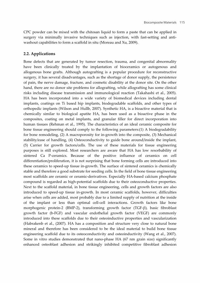
Brimacombe and Webber, (1964) reported that the chitosan consists of repeating units of beta (1-4) 2-amino-2-deoxy-D-glucopyranon (D-glucosamine) (Fig.1). The primary unit of chitin is 2-acetamido-2-deoxy-D-glucose, while that of chitosan is 2-amino-2-deoxy-D-glucose with beta, 1-4 glucosidic linkages (Muzarelli, 1985). Chitosan is a natural polysaccharide derived from chitin by its deacetylation (Dang and Leong, 2006). Chitosan shares a number of chemical and structural similarities with collagen. These similarities form the basis for the development of HAp/chitosan nano-composites for use in bone tissue engineering (Wilson et al., 2007
Figure 1. Structure of chitosan
3.2. Origin
The history of chitosan dates back to the 19 th century, when Rouget, 1859 discussed the deacetylated form of chitosan. Chitin, the source material for chitosan, is one of the most abundant organic materials, being second only to cellulose in the amount produced annually by biosynthesis. It is an important constituent of the exoskeleton in animals, especially in crustacean, molluscs and insects. It is also the principal fibrillar polymer in the cell wall of certain fungi (Eugene and Lee, 2003). Chitosan is one of the most abundant naturally occurring polysaccharides, primarily obtained as a sub-product of seafood, containing amino and hydroxyl groups (Muzarelli, 1985 and Manjubala et al., 2006).
Biocomposite Materials 117
3.3. Properties
3.3.1. Physical and chemical properties
The size of chitosan molecule plays an important role in drug delivery system where the release rate was reported to be inversely proportional to the molecular weight of the reservoir. Chitosan is insoluble in water, alkali and many organic solvents but soluble in many dilute aqueous solutions of organic acids, of which most commonly used are formic acid and acetic acid. Tomihata and Ikada, (1997) concluded that molecular weight of chitosan affect the crystal size and morphological character of its cast film. Gorbunoff, (1984) concluded that the chemical interaction between NH3+ and HPO42- control the adsorption of chitosan on octacalcium phosphate (OCP) crystal surface which is similar to the binding of protein to the surface of apatite crystals. Chitosan permeability increased in the amorphous region of the membrane (Brine, 1984). Chitosan forms a polycation in aqueous acid solutions like acetic acid and hydrochloric acid via protonation of amine functions (Muzarelli, 1985).
Blair et al., (1987) reported that chitosan prepared from crab shell chitin has a lower molecular weight than that prepared from prawn shells and tensile strength versus elongation decrease at the break with prolonged treatment in alkali solutions and both increase proportional to higher molecular weight. Also, crystallinity of membrane increased with decreasing molecular weight of chitosan and it is hydrophilic i.e it contains a large number of OH groups which are easy to combine with another group and form new bond (Ogawa et al., 1992). The larger molecular weight of chitosan used, the higher tensile strength and higher tensile elongation of membrane were obtained (Chen and Hwa, 1995). The degree of deacetylation affects the physical properties of chitosan membrane and it does not change during ultrasonic degradation on chitosan. The molecular weight of the prepared chitosan depends on the severity of the deacetylation process.
Chitosan is flexible and has a high resistance upon heating due to the intra-molecular hydrogen bonds formed between hydroxyl and amino groups (Lee et al., 1999). The degradation rate is inversely related to the degree of crystallinity which is controlled mainly by the degree of deacetylation (DD). Highly deacetylated forms (85 %) exhibit relatively a low degradation rate and may take several months in vivo, whereas, the forms with lower DD degrade more rapidly. The degradation rates also inherently affect both the mechanical and solubility properties (Kamiyama et al., 1999).
The cationic nature of chitosan also allows for pH-dependent electrostatic interactions with anionic glycosaminoglycans (GAG) and proteoglycans distributed widely throughout the body and other negatively charged species. This property is one of the important elements for tissue engineering applications because numbers of cytokines/growth factors are known to be bound and modulated by GAG including heparin and heparin sulfate (Nishikawa et al., 2000). Chitosan is easy to handle for its resistive nature to heating due to intra-molecular hydrogen bonds between hydroxyl and amino groups (Itoh et al., 2003). Chitosan is a very versatile biopolymer with film, fiber, and micro/nanoparticle forming properties. It is biocompatible, biodegradable and non-toxic (Yilmaz, 2004). Deacetylation of chitin with a
Composites and Their Applications 118
degree of deacetylation more than 50 % gives chitosan, which is soluble in organic acids such as acetic or formic acid, and has been more widely used than chitin as films, membranes, fibres and particles ( Kang et al., 2006). Chitosan has three types of reactive functional groups, an amino group as well as both primary and secondary hydroxyl groups at the C(2), C(3), and C(6) positions, respectively. These groups allow modification of chitosan like graft copolymerization for specific applications, which can produce various useful scaffolds for tissue engineering applications. The chemical nature of chitosan in turn provides many possibilities for covalent and ionic modifications that allow extensive adjustment of mechanical and biological properties (Kim et al., 2008). When the temperature is 30°C, the crystallinity of chitosan membrane is relatively high and its crystal particles are much small, which makes the mechanical properties poor. When the temperature is as high as 90°C, the temperature will cause the change of chitosan properties and the chitosan membrane is nearly not a crystal structure at this moment, resulting in the fall of mechanical properties (Xianmiao et al., 2009).
3.3.2. Biological properties
Chitosan potentiates the differentiation of osteoprogenitor cells and may facilitate the formation of bone. Rao and Sharma, (1997) concluded that chitosan is an ideal non-toxic biopolymer and the cell binding and cell activating properties of chitosan play a crucial role in its potential action. Chitosan degrades in the body to non-harmful and non-toxic compounds and has been used in various fields such as nutrition, metal recovery and biomaterials (Muzzarelli et al., 2001). It has gained much attention as a biomaterial in diverse tissue engineering applications due to its low cost, large-scale availability, anti-microbial activity, and biocompatibility (Khora and Limb, 2003). Chitosan was suggested as an alternative polymer for use in orthopedic applications to provide temporary mechanical support to the regeneration of bone cell in-growth due to its good biocompatible (Khora and Limb, 2003), non-toxic, biodegradable and inherent wound healing characteristics (Eugene and Lee 2003). Chitosan had been used in various forms such as zero dimension microsphers, two-dimension membrane, three-dimension pin or rod (Hu et al., 2003). Therefore, much attention has been paid to chitosan-based biomedical materials, for instance, as a drug delivery carrier or a wound-healing agent. Chitosan is structurally similar to glycosaminoglycan (GAG) and has many desirable properties as tissue engineering scaffolds (Kuma, et al., 2004). It was reported as being neither antigenic in mamalian test system nor thrombogenic and chitosan reported to improve hemostasis, decreased fibroplasias with enhanced tissue organization as well as normal bone formation (Mohamed, 2004). Chitosan marginally supports biological activity of diverse cell types (Sarasam and Madihally, 2005).
A number of natural and synthetic polymers have been studied for overcoming the weak points as bone substitutes. Chitosan has been found in a broad spectrum of applications along with unique biological properties including biocompatibility, biodegradability to harmless products, non-toxicity, physiological inertness, remarkable affinity to proteins, antibacterial, haemostatic, fungi-static, anti-tumoral and anti-cholesteremic properties (Kim

Biocomposite Materials 119
et al., 2008). Chitosan has been shown to degrade in vivo, which is mainly by enzymatic hydrolysis. The degradability of a scaffold plays a crucial role on the long-term performance of tissue-engineered cell/material construct because it affect many cellular process, including cell growth, tissue regeneration, and host response. If a scaffold is used for tissue engineering of skeletal system, degradation of the scaffold biomaterial should be relatively slow, as it has to maintain the mechanical strength until tissue regeneration is almost completed. Lysozyme is the primary enzyme responsible for in vivo degradation of chitosan, which appears to target acetylated residues (Kim et al., 2008).
3.4. Applications
Chitosan of biopolymer have been used as blood coagulant, in artificial kidney membrane, digestive sutures, hypercholesterolemic agents, media for the slow release of drugs and hemostatic agent (Mohamed, 2004). It is a good candidate for biomedical applications such as for wound healing, vaccine delivery, as well as tissue regeneration (Yilmaz, 2004). It has been extensively investigated in biotechnological, biomedical, and environmental fields (Dang and Leong, 2006). Chitosan polymer is used in dentistry, because it prevents the formation of plaque and tooth decay. Since chitosan can regenerate the connective tissue that covers the teeth near the gums, it offers possibilities for treating periodontal diseases such as gingivitis and periodontitis (Elizalde-Pen et al., 2007).
3.4.1. Membrane
Aiba et al., (1986) used chitosan in membrane separation, chemical engineering, medicine and biotechnology areas. It was also found that the water adsorption and the mechanical properties of fibroin membrane were improved by blending chitosan (Chen et al., 1998).
3.4.2. Skin
Muzzarelli et al., (1988) used chitosan as artificial skin substitute and they reported that no adverse effect after implantation in tissue. In general, these materials have been found to evoke a minimal foreign body reaction, with little or no fibrous encapsulation. It observed the typical course of healing with formation of normal granulation tissue, often with accelerated angiogenesis (Suh and Matthew, 2000). Also, chitosan has many advantages for wound healing such as hemostasis, accelerating the tissue regeneration and stimulating the fibroblast synthesis of collagen (Mi et al., 2001). Chitosan possesses the properties favorable for promoting rapid dermal regeneration and accelerate wound healing suitable for applications extending from simple wound coverings to sophisticated artificial skin matrices (Kim et al., 2008).
3.4.3. Bone substitutes
Sapelli et al., (1986) used chitosan powder to promote healing of periodontal pockets, palatal wounds and extraction sites. Malette et al., (1986) proved enhanced leg bone regeneration in
Composites and Their Applications 120
dogs using chitosan. It was reported to accelerate wound healing and was applied for bone wound repair in dogs (Borah et al., 1992) as well as bone growth in critical size metacarpal fibular defects. Klokkevold et al., (1992) concluded that chitosan solution may enhance the formation of bone. Later, it was reported to improve osseous healing of defects in femoral coundyl of sheep and stimulated cell proliferation and organized the hystoarchitectural tissue structure (Muzzarelli et al., 1994). Chitosan has been also extensively used in bone tissue engineering since after exploring its capacity to promote growth and mineral rich matrix deposition by osteoblasts in culture. Also, chitosan is biocompatible (additional minimizes local inflammation), biodegradable, and can be molded into porous structures (allows osteoconduction) (Martino et al., 2005 and Kim et al., 2008).
3.4.4. Drug delivery system (DDS)
Chitosan as an inert and hydrophilic material, its gel is suitable for application as matrices for enzyme/cell immobilization (Roberts, 1992) and for separation processes (Li et al., 1992). Felt et al., (1998) discussed the use of chitosan to manufacture sustained release systems deliverable by other routes such as nasal, ophthalmic, transdermal and implantable devices. Tarsi et al., (1998) suggested that low molecular weight chitosan may be very interesting as potential antidental caries agents. Chitosan has been reported to enhance drug delivery across the nasal or mucosal layer without damage (van der Lubben et al., 2001). The selectivity of membrane is a critical parameter in membrane separation and several factors are affecting its selectivity such as pore size, thus it is very suitable for the use as DDS and in artificial kidney (Mohamed, 2004). The cationic properties of chitosan offer valuable properties for drug delivery systems, gene delivery systems, and tissue engineering, that is, the formation of ion complexes between chitosan and anionic drugs or DNA can be used as a delivery vehicle (Kim et al., 2007).
3.4.5. Anti-bacterial
The experiments of antibacterial activity of chitosan-graft-polyethylene terephthalate (PET) against S. aureus showed a high growth inhibition in the range of 75–86% and still maintained a 48–58% bacterial growth inhibition after laundering (Hu et al., 2003). Chitosan and chito-oligosaccharides grafted membranes showed antibacterial activity against Escherichia coli, Pseudomonas aeruginosa, methicilin-resistant Staphylococcus aureus (MRSA), and S. aureus (Hu et al., 2003). Chitosan is a biomaterial with antiseptic property and the influence of the release or positive migration of protonated glucosamine fractions from the biopolymer into the microbial culture is the responsible event for the antimicrobial performance of the biopolymer (Beherei et al., 2009).
3.4.6. In-vitro application
Chitosan and collagen have intrinsic properties that support growth and differentiation of osteoblasts. Collagen was combined with chitosan and cross-linked to improve the biological stability and strength of chitosan–collagen composite sponges to reach the demand of an
Biocomposite Materials 121
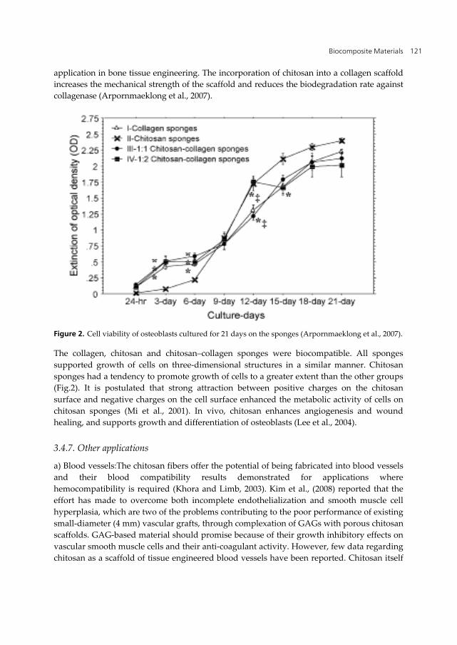
application in bone tissue engineering. The incorporation of chitosan into a collagen scaffold increases the mechanical strength of the scaffold and reduces the biodegradation rate against collagenase (Arpornmaeklong et al., 2007).
Figure 2. Cell viability of osteoblasts cultured for 21 days on the sponges (Arpornmaeklong et al., 2007).
The collagen, chitosan and chitosan–collagen sponges were biocompatible. All sponges supported growth of cells on three-dimensional structures in a similar manner. Chitosan sponges had a tendency to promote growth of cells to a greater extent than the other groups (Fig.2). It is postulated that strong attraction between positive charges on the chitosan surface and negative charges on the cell surface enhanced the metabolic activity of cells on chitosan sponges (Mi et al., 2001). In vivo, chitosan enhances angiogenesis and wound healing, and supports growth and differentiation of osteoblasts (Lee et al., 2004).
3.4.7. Other applications
a) Blood vessels:The chitosan fibers offer the potential of being fabricated into blood vessels and their blood compatibility results demonstrated for applications where hemocompatibility is required (Khora and Limb, 2003). Kim et al., (2008) reported that the effort has made to overcome both incomplete endothelialization and smooth muscle cell hyperplasia, which are two of the problems contributing to the poor performance of existing small-diameter (4 mm) vascular grafts, through complexation of GAGs with porous chitosan scaffolds. GAG-based material should promise because of their growth inhibitory effects on vascular smooth muscle cells and their anti-coagulant activity. However, few data regarding chitosan as a scaffold of tissue engineered blood vessels have been reported. Chitosan itself
Composites and Their Applications 122
was documented to promote migration of endothelial cells and fibroblasts so as to accelerate wound healing. b) Nerve: Chitosan has been studied as a candidate material for nerve regeneration due to its properties such as antitumor, antibacterial activity, biodegradability and biocompatibility. Neurons that were cultured on the chitosan membrane can grow well and that chitosan tube can greatly promote the repair of the peripheral nervous system, also the chitosan fibers supported the adhesion, migration and proliferation of Schwann cells (SCs), which provide a similar guide for regenerating axons to Büngner bands in the nervous system (Yuan et al., 2004). c) Liver: Chitosan as a promising biomaterial can be applied in liver tissue engineering due to its various properties such as its structure that is similar to glycosamineglycans (GAGs), which are components of the liver extracellular matrix (ECM) (Li et al., 2003). d) Cartilages: Chitosan is one of the most abundant polysaccharides and thus shares some bioactivities with various glycosaminoglycans and hyaluronic acid present in articular cartilage (Suh and Matthew, 2000). Lu et al., 1999 has demonstrated that the chitosan solution injected into the knee articular cavity of rats lead to a significant increase in the density of chondrocytes in the knee articular cartilage, indicating that chitosan could be potentially beneficial to the wound healing of articular cartilage (Lee et al., 2004).
4. Bone structure
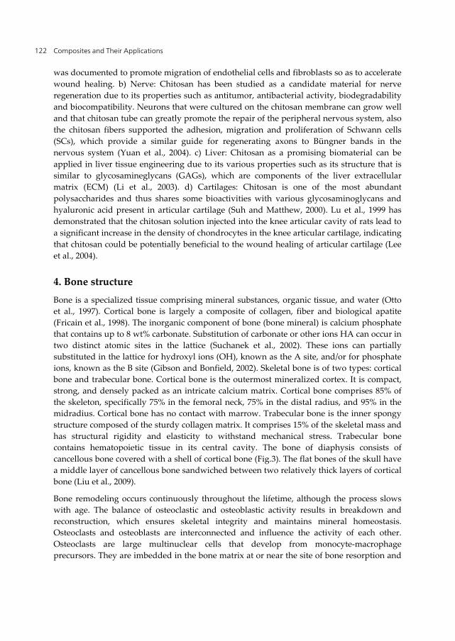
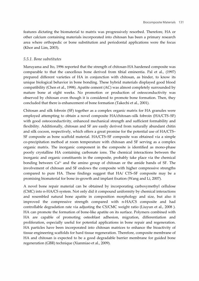
Bone is a specialized tissue comprising mineral substances, organic tissue, and water (Otto et al., 1997). Cortical bone is largely a composite of collagen, fiber and biological apatite (Fricain et al., 1998). The inorganic component of bone (bone mineral) is calcium phosphate that contains up to 8 wt% carbonate. Substitution of carbonate or other ions HA can occur in two distinct atomic sites in the lattice (Suchanek et al., 2002). These ions can partially substituted in the lattice for hydroxyl ions (OH), known as the A site, and/or for phosphate ions, known as the B site (Gibson and Bonfield, 2002). Skeletal bone is of two types: cortical bone and trabecular bone. Cortical bone is the outermost mineralized cortex. It is compact, strong, and densely packed as an intricate calcium matrix. Cortical bone comprises 85% of the skeleton, specifically 75% in the femoral neck, 75% in the distal radius, and 95% in the midradius. Cortical bone has no contact with marrow. Trabecular bone is the inner spongy structure composed of the sturdy collagen matrix. It comprises 15% of the skeletal mass and has structural rigidity and elasticity to withstand mechanical stress. Trabecular bone contains hematopoietic tissue in its central cavity. The bone of diaphysis consists of cancellous bone covered with a shell of cortical bone (Fig.3). The flat bones of the skull have a middle layer of cancellous bone sandwiched between two relatively thick layers of cortical bone (Liu et al., 2009).
Bone remodeling occurs continuously throughout the lifetime, although the process slows with age. The balance of osteoclastic and osteoblastic activity results in breakdown and reconstruction, which ensures skeletal integrity and maintains mineral homeostasis. Osteoclasts and osteoblasts are interconnected and influence the activity of each other. Osteoclasts are large multinuclear cells that develop from monocyte-macrophage precursors. They are imbedded in the bone matrix at or near the site of bone resorption and
Biocomposite Materials 123
dissolve first the calcium and then the organic matrix of the bone. Osteoblasts arise from mesenchymal cells and are found layered over the bone. They deposit calcium into the matrix that is building up cortical bone and produce collagen and other proteins to synthesize the bone matrix. Osteoclasts that are in close proximity can increase sensitivity of osteoblasts to growth factors. When a bone is broken there is usually bleeding into the space between the bone and the periosteum. This produces a hematoma, swelling due to blood. The osteoblasts, bone-producing cells, near the hematoma invade it along with small blood vessels from the bone. In a short time, the hematoma is replaced by bone tissue produced by the osteoblasts. In general, this bone is cancellous, but the added bone makes the broken junction much thicker than it was. The swollen bone is called the callus. A remarkable process follows. Where there is strain, the newly formed cancellous bone condenses into compact bone. Where strain is absent, the cancellous bone disappears through the activity of osteoclasts, bone destroying cells. This process seems to determine the development of the skeleton in normal growth (Roodman, 2004).
Figure 3. Bone structure
5. Hydroxyapatite/chitosan biocomposites: Introduction
For the increase of bioactivity and mechanical property, some composites of polymer and bioactive ceramics have been developed for bone tissue engineering. These composites fulfill the mechanical properties required for their function as skeleton, teeth and cells of organisms. Among these composites, HAp/polymer composites have attracted much attention since such composites may have osteconductivity due to the presence of HAp, which has a similar chemical composition and structure to the mineral phase of human bones and hard tissues. Thus, HA/polymer composite scaffolds are of interest for biomedical applications (Jin et al.,
Composites and Their Applications 124
2008). Kikuchi et al., (2004) prepared a self organized HA-collagen nano-composite by a biomimetic co-precipitation method. It was reported that the composite had similar microstructure to native bone and showed osteoclastic resorption and good osteoconductivity. However, a major concern over HA-collagen composite is the high cost of collagen, which limits its clinical application in healing bone defect to its insouciant formability and flexibility. Polymers such as chitosan have a higher degradation rate than bioceramics. Incorporation of HA into a chitosan polymer matrix has been shown to increase osteoconductivity and biodegradability with significant enhancement of mechanical strength (Yamaguchi et al., 2001). Chitosan’s primary attractive features including its biocompatibility, biodegradability, flexibility, adhesiveness and anti-infectivity, make it as a feasible wound healing agent and an ideal polymeric matrix for HA ceramic (Rusu et al., 2005).
5.1. Structure
The free amino groups of chitosan (C-NH2) was protonated to C-NH3+, when chitosan was dissolved in acetic acid (HAc) solution, which was shown as follows:
2 3C NH HAc C NH +Ac pH 4.2
The presence of calcium and phosphate ions in chitosan solutions leads to formation of HA\chitosan composites through electrostatic interactions between C-NH3+ and Ca2+ and\or PO43- ions to form C-Ca and C-PO4 complexes. There is also an interaction between OH of chitosan and OH of HA via hydrogen bond (Hu et al., 2004).
5.2. Properties
Chitosan can be utilized in combination with other bioactive inorganic ceramics, especially HA to further enhance tissue regenerative efficacy and osteoconductivity. Incorporation of HA with chitosan, the mineral component of bone, could improve the bioactivity and the bone bonding ability of the chitosan/HA composites (Wang et al., 2002). Chitosan just plays in a role of adhesive to dissolve the problem of difficulty of HA specific shape and migration of HA powder when implanted. HA/chitosan nano-composites are prepared by the precipitation. It is proposed that the nano-structure of HA/chitosan composite will have the best biomedical properties in the biomaterials applications (Chen et al., 2002). Also, it has been demonstrated that chitosan-hydroxyapatite composite induces osteoconductivity in osseous defects and could act as drug vehicle. It is important to be able to load these composites with short-time life and controlled action anti-inflammatory to reduce or eliminate undesirable inflammatory processes (Larena et al., 2004).
It is desirable to develop a composite material with favorable properties of chitosan and HA. The designed composites are expected to have an optimal mechanical performance and a controllable degradation rate as well as eminent bioactivity and this will be of great importance for bone remodeling and growth (Zhang et al., 2005). It must be emphasized at this point that the successful design of a bone substitute material requires an appreciation of
Biocomposite Materials 125
the structure of bone. Thus, the use of a hybrid composite that makes up of chitosan and calcium phosphate resembles the morphology and properties of natural bone. This may be one-way to solve the problem of calcium phosphates brittleness, besides possessing good biocompatibility, high bioactivity and great bone-bonding properties (Ding, 2007). The mechanical strength of the chitosan/calcium phosphate composite fiber with core-shell structure increased with an increased concentration of chitosan solution (Matsuda et al., 2004). Among the composites studied, the 30/70 chitosan/n HA exhibits the maximum value of compressive strength, about 120 MPa, which is strong enough to be used in load-bearing sites of bone tissue. In contrast the compressive strength of pure HA compact prepared by the similar method has been reported as 6.5 MPa about one twentieth of the maximum value of the composite. In general, the proper stress transfer occurring between the reinforcement and the matrix governs the mechanical characteristics of filled polymers. The chemical and mechanical interlocking between n-HA and chitosan are accounts for the efficient stress-transfer in the composite system. Besides, the interactions such as hydrogen bonding and chelation between the two phases, also contribute to the good mechanical properties of chitosan/n-HA composite (Zhang et al., 2005). The biodegradable composites based on chitosan and calcium phosphate have been prepared using a simple mixing and heating method. The detrimental effects of the simulated physiological environment on mechanical properties of the hybrid composites resulted in the significantly decrease in strength and modulus. The chitosan/calcium phosphate composites containing 10 wt/v % might an optimal material in terms of initial strength and degradation behavior. Although susceptibility to solution attack, this type of chitosan/calcium phosphate composites with high initial strength might be acceptable for use in bone tissue repair (Ding, 2007).
Three-dimensional biodegradable chitosan/nHA composite scaffolds were characterized by superior mechanical, physicochemical, and biological properties compared to pure chitosan scaffolds for bone tissue engineering. The nanocomposite scaffolds were characterized by a highly porous structure and the pore size was similar for scaffolds with varying n-HA content. The nano-composite scaffolds exhibited greater compression modulus, slower degradation rate and reduced water uptake, but the water retention ability was similar to that of pure chitosan scaffolds. Favorable biological response of pre-osteoblast on nanocomposite scaffolds included improved cell adhesion, higher proliferation, and well spreading morphology in relation to pure chitosan scaffold (Thein-Han and Misra, 2009).
Mechanical properties of biocomposites: hydroxyapatite/chitosan (HA/CS) nano-composite rods were reinforced via a covalently cross-linking method. The bending strength and bending modulus of the cross-linked HA/CS (5/100, wt/wt) rods could arrive at 178 MPa and 5.2 GPa, respectively, increased by 107% and 52.9% compared with uncross-linked HA/CS (5/100, wt/wt) rods (Takagi et al., 2003). The presence of HA-DBM filler into the grafted chitosan copolymer matrix resulted in compressive strength properties are quite close to those of cancellous bone (2–12 MPa). This result is due to effect of the presence of demineralized bone matrix (DBM) powder and pMMA having bone cement formation within this composite (Mohamed et al., 2007). The E-modulus and compressive strength for
Composites and Their Applications 126
the three composites HA/, 90%HA-10Ti/, and 70%HA-30%Ti/grafted chitosan copolymer composites recorded comparable values compared to the cancellous bone. Therefore, the presence of HA filler or HA filler containing titania content up to 30% into the copolymer resulted in compressive strength properties that are quite close to those of cancellous bone (2–12 MPa) (Mohamed et a., 2008).
Collagen/apatite composite membranes exhibited significantly improved mechanical properties compared with their pure collagen equivalent; their mechanical properties were still lower than those of natural bone (Teng et al., 2009). It was notified that hardness of calcium pyrophosphate (CPP)/ chitosan composite (66.80) was increased compared to chitosan copolymer (60.88) and the hardness of CPP/chitosan-grafted composite (68.23) was also increased compared to chitosan–gelatin copolymer (84.12) proving the polymer/filler interaction and adhesion. Also, the compressive strength of CPP/chitosan composite (6.53 MPa) was increased compared to chitosan copolymer (5.11 MPa). These values of compressive strength were comparable to those of human cancellous bone (Kokubo et al., 2003). As a result, CPP filler powder into the chitosan copolymer matrix containing chitosan or chitosan–gelatin polymer resulted in more effective reinforcement of the composite, then, stiffer composite (El-Kady et al., 2009). The CPC–chitosan composites were more stable in water than conventional calcium phosphate cement (CPC). They did not disintegrate even when placed in water immediately after mixing. The CPC–chitosan paste hardened within 10 min in all cases. The authors demonstrated that CPC–chitosan composites are stable in a wet environment and have acceptable mechanical strengths for clinical applications (Wang et al., 2010).
5.3. Preparation
Although powder ceramics remain the form of choice for filling small irregular defects, the therapeutic effect of the filling implant was lost by migration of particles from the defect site. Furthermore, it was difficult to be handled and kept in place compactly for convenient fabrication and operation of block-type ceramics (Lin et al., 1998). Thus, it is necessary to mix a suitable binder with the granular material to overcome these problems. Presently, the approaches to obtain chitosan/hydroxyapatite (HAp) composite materials are based either on mixing or co-precipitation methods. Yamaguchi et al., (2001) have developed one of the co-precipitation methods that lead to a type of chitosan/HAp composites. In this approach, the composite was co-precipitated in one step, by dropping a chitosan solution containing phosphoric acid into a calcium hydroxide suspension. Other approaches employ either the biomineralization of chitosan in a solid form (especially as membranes) in simulated body fluids (SBF) (Beppu and Santana, 2001) or by mixing of a chitosan solution with different calcium phosphate fillers followed by their precipitation as hydrogel composite. The use of these approaches leads to incorporation of inorganic fillers into the structure of composites (Schwarz and Epple, 1998), either as nano-sized or micro-sized particles.
Different preparation methods of HAp/chitosan composites have been reported, such as mechanical mixing of HAp powder in a chitosan solution, coating of HAp particles onto a
Biocomposite Materials 127
chitosan sheet, coating of HAp crystals onto a tendon chitosan (Yamaguchi et al., 2003) and a co-precipitation method. The chitosan/HAp hybrid fiber has also been reported by Chung and Korean, (2002). However, these materials were shown in the macroscopically homogeneous. The conventional method to fabricate chitosan/HA composite is that HA powder was mixed with chitosan, dissolved in 2% acetic acid solution, then the mixture was impressed into mold, finally was freezing-dried to make sponge composite. Surface modification and polyblend methods can be used to change the physicochemical properties of chitosan–gelatin membranes or scaffolds by incorporating hyaluronic acid. Adding hyaluronic acid can improve the mechanical, biological and anti-degradation properties of the membranes or the scaffolds (Mao et al., 2003).
Hydroxyapatite (HAp) was prepared by precipitation method, while the biphasic hydroxyapatite/tricalcium phosphate (HA/B-TCP) was prepared by heating the prepared HAp at 900°C for 5 hours in air. To improve bioactivity both HAp and HAp/TCP fillers were loaded onto chitosan grafted with two monomers, hydroxyethylmethacrylate (HEMA) and methylmethacrylate (MMA) during copolymerization process (Hashem and Mohamed, 2007). Also, biocomposites containing HA-DBM mixture powder loaded onto the copolymer matrix containing the grafted chitosan with poly methylmethacrylate (pMMA) and its derivative during copolymerization were fabricated (Mohamed et al, 2007).
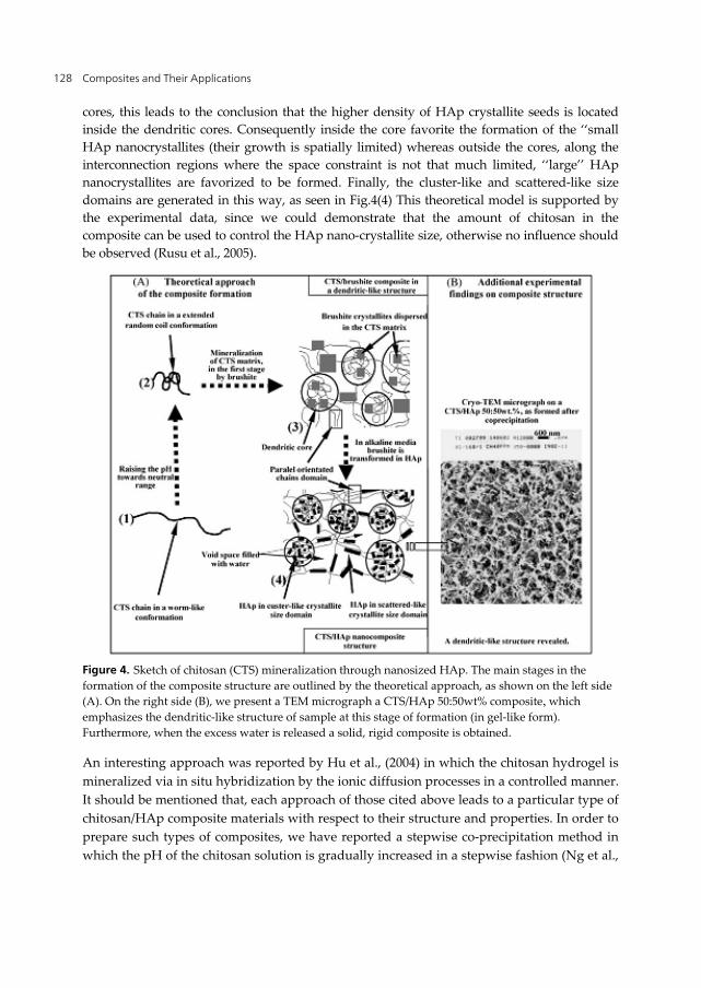
The chitosan mineralization in the case of using a stepwise co-precipitation approach involves the following stages: First, chitosan chains change their conformation as a function of environmental parameters, such as pH. Starting from extended conformations such as worm-like, as seen in Fig.4 (1), adopted at low pH (until 3–3.5), chitosan turns to the more compact conformations such as extended random coil Fig.4(2) and even to more compacted random coil conformations, at higher pH values (from 3.5 until 6). Between 5.5 (the pH at which brushite is precipitated) and 6.5–6.7 (the pH at which chitosan is precipitated) is the pH range that leads to formation of an interconnected three-dimensional net work between chitosan and brushite that can be approximated as a dendritic-like structure, as shown in Fig.4(3). This structural model is characterized by highly dense irregular shaped cores which are linked by the chitosan bridging segments. In the dendritic core, the chitosan chains are randomly packed as amorphous regions while in the bridging regions more extended conformations are found, in some parts parallel oriented chains domains are presented. Since the dendritic core is in the range between 200 and 600nm hence it is formed by the many compact random coil chitosan units that are approaching one another chitosan chain segments can interact with the CaPs phases (i.e. seeds of HAp already formed at pH 5.5 and identified by its XRD pattern, brushite and some other ACP). In this way, they achieve of so called ‘‘anchoring regions’’ by different specific interactions such as ion-dipole or/and through the complexation of Ca with chitosan. Considering that a dendritic-like structure is formed in the earlier stage of composite formation, one can explain the complex bimodal distribution of HAp nano-crystallites in the chitosan matrix. Inside the dendritic core, the chitosan chains density is much greater than outside of them, shown as inter-connection blob regions. We assume that the probability to find a certain number of HAp crystallite seeds per a chitosan chain unit is the same, inside and outside the dendritic core regions. Since the chitosan chain density is much greater inside the
Composites and Their Applications 128
cores, this leads to the conclusion that the higher density of HAp crystallite seeds is located inside the dendritic cores. Consequently inside the core favorite the formation of the ‘‘small HAp nanocrystallites (their growth is spatially limited) whereas outside the cores, along the interconnection regions where the space constraint is not that much limited, ‘‘large’’ HAp nanocrystallites are favorized to be formed. Finally, the cluster-like and scattered-like size domains are generated in this way, as seen in Fig.4(4) This theoretical model is supported by the experimental data, since we could demonstrate that the amount of chitosan in the composite can be used to control the HAp nano-crystallite size, otherwise no influence should be observed (Rusu et al., 2005).
Figure 4. Sketch of chitosan (CTS) mineralization through nanosized HAp. The main stages in the formation of the composite structure are outlined by the theoretical approach, as shown on the left side (A). On the right side (B), we present a TEM micrograph a CTS/HAp 50:50wt% composite, which emphasizes the dendritic-like structure of sample at this stage of formation (in gel-like form). Furthermore, when the excess water is released a solid, rigid composite is obtained.
An interesting approach was reported by Hu et al., (2004) in which the chitosan hydrogel is mineralized via in situ hybridization by the ionic diffusion processes in a controlled manner. It should be mentioned that, each approach of those cited above leads to a particular type of chitosan/HAp composite materials with respect to their structure and properties. In order to prepare such types of composites, we have reported a stepwise co-precipitation method in which the pH of the chitosan solution is gradually increased in a stepwise fashion (Ng et al.,
Biocomposite Materials 129
2008). Biodegradable hydroxyapatite/chitosan-gelatin polymeric biocomposites were fabricated by using HA powder and HA filler containing titania powder (10 and 30%) with a chitosan and gelatin grafted co-polymeric matrix during copolymerization process (Mohamed and Mostafa, 2008). Preparation of a model HAp/CTS (30:70 in mass ratio) nanocomposite nanofibers using a two-step method, which involves firstly preparing HAp/CTS nanocomposites by a co-precipitation synthesis approach and then fabricating the resultant HAp/CTS nanocomposites, aided with a fiber-forming additive–ultrahigh molecular weight poly(ethylene oxide), into nanofibers via the electrospinning process (Zhang et al., 2008).
5.4. Characterization
The FT-IR spectra show that the two characteristic bands of amide I (1655 cm-1) and amide II (1599 cm-1) for chitosan shift to lower wavenumber after being compounded, which suggests that interaction must take place between chitosan and n-HA, including hydrogen bonds between -NH2 and-OH of n-HA as well as the chelation between -NH2 and Ca++. The more shift of these bands to lower wave number, the stronger the hydrogen bonds between these groups and also the stronger the interaction between these molecules (Zhang et al., 2005). The XRD pattern of precipitated HA shows un-differentiated broad peaks with poor crystallinity around the characteristic region. However, the crystallographic structure of precipitated HA nano-crystals is more identical with natural bone mineral (biological apatite). Hence, the prepared HA nano-crystallites in this investigation have more similarity with natural bone mineral in terms of degree of crystallinity and structural morphology. The calcined HA exhibited all the characteristic diffracted peaks of stoichiometric HA with higher degree of crystallinity. Rising in the calcination temperature shapes the diffracted peaks more sharper, which is a good sign for the improvement of crystallinity of precipitated HA. The obtained results did not show any peaks corresponding to calcium carbonate and calcium oxide and hence suggesting that the ingredients were reacted completely and produced a homogeneous HA. The XRD analysis of HA/chitosan composites proved that, a broad peak assigned to chitosan at 20° becomes wider and weaker with increase of n-HA. It suggests that the addition of n-HA obviously affects the crystallinity of chitosan. The characteristic peaks at 25.8° (002) and 39.6° (310) are used to calculate the n-HA crystal sizes (Xianmiao et al., 2009). The TGA mass loss of HA\C composite increased from 3.3–6.5 mass% as the chitosan concentration increased from 0–2.5 mass%. The amount of chitosan that adsorbed on HA was 2.8–3.1 mass% based on Carbon–Hydrogen–Nitrogen (CHN) analysis. The specific surface area of HA increased after aging in chitosan acetate gel solutions and attained a high value of 160 m2/g in comparison to 85 m2/g for untreated HA (Wilson and Hull, 2008).
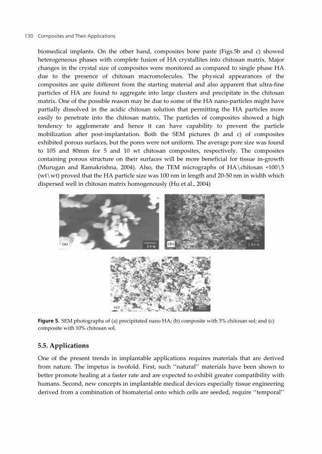
The SEM micrograph of precipitated HA (Fig.5a) exhibited nano-sized crystals with almost uniform particles size. The HA particles prepared were not only stoichiometric but also mono-dispersive and roughly particles were not fused together with other crystals. It can be inferred that majority of the particles were of single crystals, regular shape and cleaner contours with no agglomeration which are highly beneficial for coating of nano HA onto
Composites and Their Applications 130
biomedical implants. On the other hand, composites bone paste (Figs.5b and c) showed heterogeneous phases with complete fusion of HA crystallites into chitosan matrix. Major changes in the crystal size of composites were monitored as compared to single phase HA due to the presence of chitosan macromolecules. The physical appearances of the composites are quite different from the starting material and also apparent that ultra-fine particles of HA are found to aggregate into large clusters and precipitate in the chitosan matrix. One of the possible reason may be due to some of the HA nano-particles might have partially dissolved in the acidic chitosan solution that permitting the HA particles more easily to penetrate into the chitosan matrix. The particles of composites showed a high tendency to agglomerate and hence it can have capability to prevent the particle mobilization after post-implantation. Both the SEM pictures (b and c) of composites exhibited porous surfaces, but the pores were not uniform. The average pore size was found to 105 and 80mm for 5 and 10 wt chitosan composites, respectively. The composites containing porous structure on their surfaces will be more beneficial for tissue in-growth (Murugan and Ramakrishna, 2004). Also, the TEM micrographs of HA\chitosan =100\5 (wt\wt) proved that the HA particle size was 100 nm in length and 20-50 nm in width which dispersed well in chitosan matrix homogenously (Hu et al., 2004)
Figure 5. SEM photographs of (a) precipitated nano HA; (b) composite with 5% chitosan sol; and (c) composite with 10% chitosan sol.
5.5. Applications
One of the present trends in implantable applications requires materials that are derived from nature. The impetus is twofold. First, such ‘‘natural’’ materials have been shown to better promote healing at a faster rate and are expected to exhibit greater compatibility with humans. Second, new concepts in implantable medical devices especially tissue engineering derived from a combination of biomaterial onto which cells are seeded, require ‘‘temporal’’
Biocomposite Materials 131
features dictating the biomaterial to matrix was progressively resorbed. Therefore, HA or other calcium containing materials incorporated into chitosan has been a primary research area where orthopedic or bone substitution and periodontal applications were the focus (Khor and Lim, 2003).
5.5.1. Bone substitutes
Maruyama and Ito, 1996 reported that the strength of chitosan-HA hardened composite was comparable to that the cancellous bone derived from tibial eminentia. Pal et al., (1997) prepared different varieties of HA in conjunction with chitosan, as binder, to know its unique biological behavior in bone bonding. These hybrid materials displayed good blood compatibility (Chen et al., 1998). Apatite cement (AC) was almost completely surrounded by mature bone at eight weeks. No promotion or production of osteoconductivity was observed by chitosan even though it is considered to promote bone formation. Then, they concluded that there is enhancement of bone formation (Takechi et al., 2001).
Chitosan and silk fobroin (SF) together as a complex organic matrix for HA granules were employed attempting to obtain a novel composite HA/chitosan–silk fobroin (HA/CTS–SF) with good osteoconductivity, enhanced mechanical strength and sufficient formability and flexibility. Additionally, chitosan and SF are easily derived from naturally abundant chitin and silk cocoon, respectively, which offers a great promise for the potential use of HA/CTS–SF composite as bone scaffold material. HA/CTS–SF composite was obtained via a simple co-precipitation method at room temperature with chitosan and SF serving as a complex organic matrix. The inorganic component in the composite is identified as mono-phase poorly crystalline HA containing carbonate ions. The chemical interactions between the inorganic and organic constituents in the composite, probably take place via the chemical bonding between Ca2+ and the amino group of chitosan or the amide bands of SF. The involvement of chitosan and SF endows the composite with higher compressive strengths compared to pure HA. These findings suggest that HA/ CTS–SF composite may be a promising biomaterial for bone in-growth and implant fixation (Wang and Li, 2007).
A novel bone repair material can be obtained by incorporating carboxymethyl cellulose (CMC) into n-HA/CS system. Not only did it compound uniformity by chemical interactions and resembled natural bone apatite in composition morphology and size, but also it improved the compressive strength compared with n-HA/CS composite and had controllable degradation rate via adjusting the CS/CMC weight ratio (Liuyun et al., 2008 ). HA can promote the formation of bone-like apatite on its surface. Polymers combined with HA are capable of promoting osteoblast adhesion, migration, differentiation and proliferation, especially useful for potential applications in bone repair and regeneration. HA particles have been incorporated into chitosan matrices to enhance the bioactivity of tissue engineering scaffolds for hard tissue regeneration. Therefore, composite membrane of HA and chitosan is expected to be a good degradable barrier membrane for guided bone regeneration (GBR) technique (Xianmiao et al., 2009).
Composites and Their Applications 132
5.5.2. Bone tissue engineering
The scaffold is a key component of tissue engineering (Langer and Vacanti, 1993). The study of inorganic crystal assembly in or on an organic polymer matrix is an important focus of bio-mineralization to produce nano composites, which can mimic natural bone. The 3D macro porous scaffolds play an important role in the formation of new tissues and provide a temporary scaffold to guide new tissue in-growth and regeneration (Nikalson and Langer, 1997). Chitosan has been proposed to serve as a non-protein matrix for 3D tissue growth. Chitosan could provide the biological primer for cell-tissue proliferation and reconstruction. One of the most promising features of chitosan is its excellent ability to be processed into porous structures for use in cell transplantation and tissue regeneration. In tissue engineering, the porous structure of chitosan provides a scaffold for bone cells to grow in and seed new bone regeneration. For rapid cell growth, the scaffold must have optimal micro architecture such as pore size, shape and specific surface area (Madihally and Matthew, 1999).
In bone tissue engineering, the biodegradable substitutes act as a temporary skeleton inserted into the defective sites of skeleton or lost bone sites, in order to support and stimulate bone tissue regeneration while they gradually degrade and are replaced by new bone tissue (Service, 2000). Chitosan-based scaffolds possess some special properties for use in tissue engineering. The major goal in fabricating scaffolds for bone tissue engineering is to accurately control pore size and porosity. Porous chitosan structures can be formed by freezing and lyophilizing chitosan–acetic acid solutions in suitable moulds (Chow and Khor, 2000). Bone regeneration research needed to deal with various clinical bone diseases such as bone infections, bone tumors and bone loss by trauma (Braddock et al., 2001). To combine the osteoconductivity of calcium phosphate and good biodegradability of polymers, composites have been developed for bone tissue engineering either by directly mixing the components or by a biomimetic approach (Wei and Ma, 2004). Polymer-ceramic composite scaffolds are expected to mimic natural bone, in the way that natural bone is also a composite of inorganic compounds (calcium phosphates especially substituted carbonated hydroxyapatite) and organic compounds (collagen, protein matrix, etc.). The hydroxyapatite–chitosan–alginate porous network has been reported and demonstrated to be suitable for bone tissue engineering applications using osteoblast cells (Zhao et al., 2003).
Research advances in bone regeneration in tissue engineering have focused on the development of three dimensional (3D) porous scaffolds that can serve as a support, reinforce and in some cases organize the tissue regeneration or replacement in a natural way (Sachlos et al., 2003). Several studies have been focused on chitosan–calcium phosphates (CP) composites for this purpose in bone tissue engineering. Beta-tricalcium phosphate (β-TCP) and hydroxyapatite (HA) of CP bioceramics are excellent candidates for bone repair and regeneration because of their similarity in chemical composition with inorganic components of bone (Zhang et al., 2003). Tissue engineering is regarded as an ultimately ideal medical treatment for diseases that have been too difficult to be cured by existing methods. This biomedical engineering is designed to repair injured body parts and restore
Biocomposite Materials 133
their functions by using laboratory-grown tissues, materials and artificial implants. For regeneration of failed tissues, this biomedical engineering utilizes three fundamental tools: living cell, signal molecules, and scaffold. The choice of chitosan as a tissue support material is governed among others by multiple ways by which its biological, physical and chemical properties can be controlled and engineered under mild conditions (Krajewska, 2005). The 3D macro porous scaffolds play an important role in the formation of new tissues and provide a temporary scaffold to guide new tissue in-growth and regeneration. The fabrication of biodegradable and osteoconductive scaffolds with a 3D interconnected porous network has been a formidable challenge. The feasibility of producing cost effective organic–inorganic scaffolds for tissue engineering to mimic bone by the diffusion method was performed. The porous structure of chitosan scaffold was homogeneously mineralized using this technique of apatite formation at room temperature. The mineralized scaffolds were found to be non-cytotoxic and better for cell proliferation and growth, as indicated by the enzyme activity and protein levels, than un-mineralized scaffold. This suggested that it could be used for further osteoconductivity studies. A biodegradable matrix with sufficient mechanical strength, optimized architecture and suitable degradation rate, which could finally be replaced by newly formed bone, is most desirable (Manjubala et al., 2006). Although the chitosan based composite biomaterials need to improve their mechanical properties for bone tissue engineering, no doubt that chitosan is a promising candidate scaffold material in clinical practice due to the worthiest ability to bind anionic molecules such as growth factors, GAG and DNA. Especially, the ability to link chitosan to DNA may render this material a good potential as a substrate for gene activated matrices in gene therapy application in orthopedics (Kim et al., 2008).
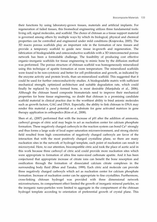
Shen et al., (2007) performed that with the increase of pH after the addition of ammonia, carboxyl groups of citric acid may begin to act as nucleation center for calcium phosphate formation. These negatively charged carboxyls in the reaction system can bond Ca2+ strongly and thus forms a large scale of local super saturation microenvironment, and strong electric field resulted from high concentration of negatively charged carboxyls are favor of the interaction that with the most positively charged crystalline plane, so there are many nucleation sites in the network of hydrogel template, each point of nucleation can result in microcrystal. Here, to our attention, biocompatible citric acid took the place of acetic acid in this work because three carboxyl of citric acid could provide more nucleation sites which were appropriate to formation of ultra fine nano-sized carbonate apatite. And it has been conjectured that appropriate increase of citrate ions can benefit the bone resorption and ossification through the formation of dissociated calcium citrate complexes in the surrounding body fluid (Rhee and Tanaka, 1999). Each citric acid molecule can provide three negatively charged carboxyls which act as nucleation center for calcium phosphate formation. Increase of nucleation center can be appropriate to fine crystallites. Furthermore, cross-linking chitosan hydrogel was provided with three dimensional network microstructures, its compartment effect limited the growth of inorganic mineral particles, so the inorganic nano-particles were limited to aggregate in the compartment of the chitosan hydrogel template according to orientation of preferential growth of crystal plane. This
Composites and Their Applications 134
multiple-order template effect based on multiple-point nucleation of citric acid and compartment of hydrogel network had a very obvious mediation in the formation process of homogeneous composites (Shen et al., 2007) (Fig.6). In bone tissue engineering, the biodegradable scaffold is a temporary template introduced at the defective site or lost bone to initiate bone tissue regeneration, while it gradually degrades and is replaced by newly formed bone tissue. Finally, an ideal scaffold is characterized by excellent biocompatibility, controllable biodegradability, cytocompatibility, suitable microstructure (pore size and porosity) and mechanical properties. Additionally, it must be capable of promoting cell adhesion and retaining the metabolic functions of attached cells (Thein-Han and Misra, 2009).
Figure 6. The scheme of formation of homogeneous chitosan/carbonate apatite composite and 3D nanocomposite scaffold (Shen et al., 2007).
5.5.3. In vitro applications
The process of apatite formation on the bioactive materials in living body could be reproduced in simulated body fluid (SBF), which means that in vivo bone bioactivity of a material can be predicted by assessing apatite formation on its surface in SBF. They confirmed that there are two types of material which inserted into living body. One of them
Biocomposite Materials 135
was able to have apatite form on its surface in SBF, and consequently has apatite produced on its surface in the living body, and bonds to living bone through this apatite layer. The other type was directly bond to living bone without the formation of apatite on their surfaces, so examination of apatite formation on the surface of a material in SBF is useful for predicting the in vivo bone bioactivity of the material, not only qualitatively but also quantitatively (Kokubo and Takadama, 2006).
5.5.3.1. In simulated body fluid (SBF)
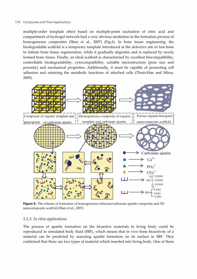
From SEM photos (Fig.7), it can be known that chitosan in the chitosan /nHA composite gradually degraded during the soaking in SBF solution, which resulted in plenty of macro-and micropores on the surface of and inside the specimens. At the same time, a lot of tiny apatite crystals deposited on the surface of the specimens, and till the 8th week, a thin layer of bone-like apatite, being highly bioactive was formed. At the first 4 weeks, the degradation rate of chitosan was higher than the deposition rate of apatite on the surface of specimens, which corresponding to a continuous increase of the rate of weight loss. After that, the deposition of apatite is prior to the degradation of chitosan, so the rate of weight loss decreased. This was also confirmed by the rate of water adsorption with the degradation of chitosan during the specimen’s soaking in SBF solution, a more sponge-like structure was formed, which can hold more water. However, with more apatite crystals deposition, some of these pores were filled or covered, so water adsorption decreased (Zhang et al., 2005). Kong et al., (2006) reported that chitosan/nano-hydroxyapatite composite scaffolds analysis showed that after incubation in simulated body fluid on both of the scaffolds (the apatite-coated composite scaffolds and apatite-coated chitosan scaffolds), carbonate hydroxyapatite was formed. With increasing nano-hydroxyapatite content in the composite, the quantity of the apatite formed on the scaffolds increased. Compared with pure chitosan, the composite with nano-hydroxyapatite could form apatite more readily during the biomimetic process, which suggests that the composite possessed better mineralization activity (Kong et al., 2006).
Figure 7. The SEM images of chitosan/nHA composites after soaking in SBF solutions for (a) 0 week, (b) 1week, (c) 4 weeks and (d) 8 weeks, Magn. × 400.
The swelling properties and degradation behavior proved the stability of hydroxyapatite-titania/chitosan-gelatin polymeric biocomposites into the media. In-vitro test behavior confirmed that the prepared composite enhanced the deposition of Ca++ and P ions onto the surface that is in the favor of the formation of apatite layer. FT-IR and SEM-EDAX of
Composites and Their Applications 136
copolymer and three composites post-immersion verified the formation of spherical apatite particles onto the copolymer surface; therefore, it was expected to enhance the apatite nucleation onto the filler composite surface especially hydroxyapatite-titania/chitosan-gelatin (AK1) composite containing 10% content of titania (Mohamed and Mostafa, 2008).
5.5.3.2. Bone Tissue engineering (Scaffold)
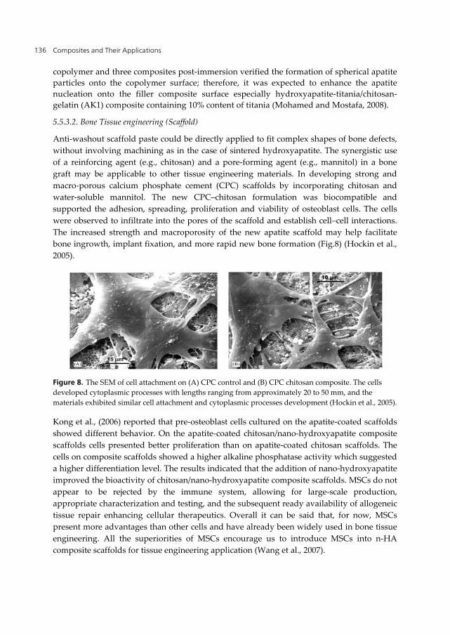
Anti-washout scaffold paste could be directly applied to fit complex shapes of bone defects, without involving machining as in the case of sintered hydroxyapatite. The synergistic use of a reinforcing agent (e.g., chitosan) and a pore-forming agent (e.g., mannitol) in a bone graft may be applicable to other tissue engineering materials. In developing strong and macro-porous calcium phosphate cement (CPC) scaffolds by incorporating chitosan and water-soluble mannitol. The new CPC–chitosan formulation was biocompatible and supported the adhesion, spreading, proliferation and viability of osteoblast cells. The cells were observed to infiltrate into the pores of the scaffold and establish cell–cell interactions. The increased strength and macroporosity of the new apatite scaffold may help facilitate bone ingrowth, implant fixation, and more rapid new bone formation (Fig.8) (Hockin et al., 2005).
Figure 8. The SEM of cell attachment on (A) CPC control and (B) CPC chitosan composite. The cells developed cytoplasmic processes with lengths ranging from approximately 20 to 50 mm, and the materials exhibited similar cell attachment and cytoplasmic processes development (Hockin et al., 2005).
Kong et al., (2006) reported that pre-osteoblast cells cultured on the apatite-coated scaffolds showed different behavior. On the apatite-coated chitosan/nano-hydroxyapatite composite scaffolds cells presented better proliferation than on apatite-coated chitosan scaffolds. The cells on composite scaffolds showed a higher alkaline phosphatase activity which suggested a higher differentiation level. The results indicated that the addition of nano-hydroxyapatite improved the bioactivity of chitosan/nano-hydroxyapatite composite scaffolds. MSCs do not appear to be rejected by the immune system, allowing for large-scale production, appropriate characterization and testing, and the subsequent ready availability of allogeneic tissue repair enhancing cellular therapeutics. Overall it can be said that, for now, MSCs present more advantages than other cells and have already been widely used in bone tissue engineering. All the superiorities of MSCs encourage us to introduce MSCs into n-HA composite scaffolds for tissue engineering application (Wang et al., 2007).
Biocomposite Materials 137
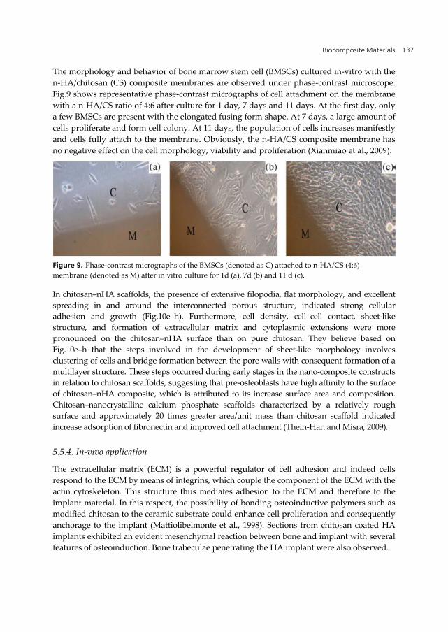
The morphology and behavior of bone marrow stem cell (BMSCs) cultured in-vitro with the n-HA/chitosan (CS) composite membranes are observed under phase-contrast microscope. Fig.9 shows representative phase-contrast micrographs of cell attachment on the membrane with a n-HA/CS ratio of 4:6 after culture for 1 day, 7 days and 11 days. At the first day, only a few BMSCs are present with the elongated fusing form shape. At 7 days, a large amount of cells proliferate and form cell colony. At 11 days, the population of cells increases manifestly and cells fully attach to the membrane. Obviously, the n-HA/CS composite membrane has no negative effect on the cell morphology, viability and proliferation (Xianmiao et al., 2009).
Figure 9. Phase-contrast micrographs of the BMSCs (denoted as C) attached to n-HA/CS (4:6) membrane (denoted as M) after in vitro culture for 1d (a), 7d (b) and 11 d (c).
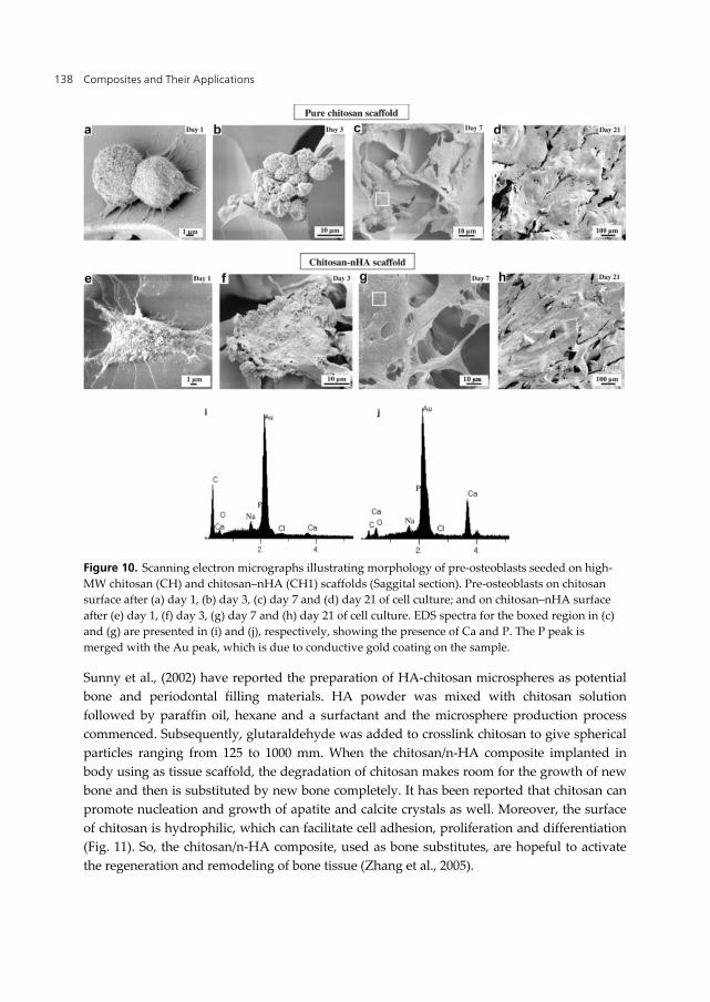
In chitosan–nHA scaffolds, the presence of extensive filopodia, flat morphology, and excellent spreading in and around the interconnected porous structure, indicated strong cellular adhesion and growth (Fig.10e–h). Furthermore, cell density, cell–cell contact, sheet-like structure, and formation of extracellular matrix and cytoplasmic extensions were more pronounced on the chitosan–nHA surface than on pure chitosan. They believe based on Fig.10e–h that the steps involved in the development of sheet-like morphology involves clustering of cells and bridge formation between the pore walls with consequent formation of a multilayer structure. These steps occurred during early stages in the nano-composite constructs in relation to chitosan scaffolds, suggesting that pre-osteoblasts have high affinity to the surface of chitosan–nHA composite, which is attributed to its increase surface area and composition. Chitosan–nanocrystalline calcium phosphate scaffolds characterized by a relatively rough surface and approximately 20 times greater area/unit mass than chitosan scaffold indicated increase adsorption of fibronectin and improved cell attachment (Thein-Han and Misra, 2009).
5.5.4. In-vivo application
The extracellular matrix (ECM) is a powerful regulator of cell adhesion and indeed cells respond to the ECM by means of integrins, which couple the component of the ECM with the actin cytoskeleton. This structure thus mediates adhesion to the ECM and therefore to the implant material. In this respect, the possibility of bonding osteoinductive polymers such as modified chitosan to the ceramic substrate could enhance cell proliferation and consequently anchorage to the implant (Mattiolibelmonte et al., 1998). Sections from chitosan coated HA implants exhibited an evident mesenchymal reaction between bone and implant with several features of osteoinduction. Bone trabeculae penetrating the HA implant were also observed.
Composites and Their Applications 138
Figure 10. Scanning electron micrographs illustrating morphology of pre-osteoblasts seeded on high-MW chitosan (CH) and chitosan–nHA (CH1) scaffolds (Saggital section). Pre-osteoblasts on chitosan surface after (a) day 1, (b) day 3, (c) day 7 and (d) day 21 of cell culture; and on chitosan–nHA surface after (e) day 1, (f) day 3, (g) day 7 and (h) day 21 of cell culture. EDS spectra for the boxed region in (c) and (g) are presented in (i) and (j), respectively, showing the presence of Ca and P. The P peak is merged with the Au peak, which is due to conductive gold coating on the sample.
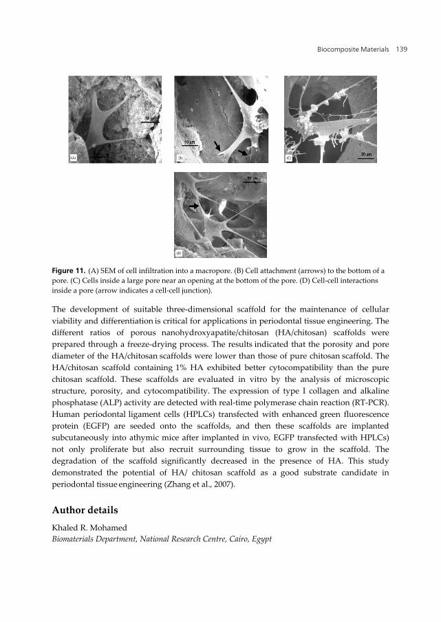
Sunny et al., (2002) have reported the preparation of HA-chitosan microspheres as potential bone and periodontal filling materials. HA powder was mixed with chitosan solution followed by paraffin oil, hexane and a surfactant and the microsphere production process commenced. Subsequently, glutaraldehyde was added to crosslink chitosan to give spherical particles ranging from 125 to 1000 mm. When the chitosan/n-HA composite implanted in body using as tissue scaffold, the degradation of chitosan makes room for the growth of new bone and then is substituted by new bone completely. It has been reported that chitosan can promote nucleation and growth of apatite and calcite crystals as well. Moreover, the surface of chitosan is hydrophilic, which can facilitate cell adhesion, proliferation and differentiation (Fig. 11). So, the chitosan/n-HA composite, used as bone substitutes, are hopeful to activate the regeneration and remodeling of bone tissue (Zhang et al., 2005).
Biocomposite Materials 139
Figure 11. (A) SEM of cell infiltration into a macropore. (B) Cell attachment (arrows) to the bottom of a pore. (C) Cells inside a large pore near an opening at the bottom of the pore. (D) Cell-cell interactions inside a pore (arrow indicates a cell-cell junction).
The development of suitable three-dimensional scaffold for the maintenance of cellular viability and differentiation is critical for applications in periodontal tissue engineering. The different ratios of porous nanohydroxyapatite/chitosan (HA/chitosan) scaffolds were prepared through a freeze-drying process. The results indicated that the porosity and pore diameter of the HA/chitosan scaffolds were lower than those of pure chitosan scaffold. The HA/chitosan scaffold containing 1% HA exhibited better cytocompatibility than the pure chitosan scaffold. These scaffolds are evaluated in vitro by the analysis of microscopic structure, porosity, and cytocompatibility. The expression of type I collagen and alkaline
phosphatase (ALP) activity are detected with real-time polymerase chain reaction (RT-PCR). Human periodontal ligament cells (HPLCs) transfected with enhanced green fluorescence protein (EGFP) are seeded onto the scaffolds, and then these scaffolds are implanted
subcutaneously into athymic mice after implanted in vivo, EGFP transfected with HPLCs) not only proliferate but also recruit surrounding tissue to grow in the scaffold. The degradation of the scaffold significantly decreased in the presence of HA. This study demonstrated the potential of HA/ chitosan scaffold as a good substrate candidate in periodontal tissue engineering (Zhang et al., 2007).
Author details
Khaled R. Mohamed Biomaterials Department, National Research Centre, Cairo, Egypt
Composites and Their Applications 140
6. References
Aiba I., Izume S., Minoura M.N. and Fujiwara Y. In chitin in Nature and Technology Ed. R.A.A. Muzzarelli, C. Jeuniaux and G.M. Goodey, Plenum Press, New York, (1986) 396-398.
Arpornmaeklong P., Suwatwirote N., Pripatnanont P., Oungbho K. Growth and differentiation of mouse osteoblasts on chitosan–collagen sponges. Int. J. Oral Maxillofac. Surg. 36 (2007) 328–337.
Beherei H.H., Mohamed K.R., Mahmoud A. I. Egyptian J. of Chemistry, xxx (2009) xxx-xxx. Under press.
Beppu M.M. and Santana C.C. In vitro biomineralization of chitosan. Key Eng Mater (2001) 192–195:31–4.
Blair H. S., Guthrie J., Law T. and Turkington p. Chitosan and modified membranes. App. Polymer. Sci., Vol. 33 (1987) 641 – 656.
Borah G., Scott G. and Wortham K. Bone induction by chitosan in endochondral bones of the extremities” Brine, C., Sanford P.A., Zikakis JP, 5th Int. Conf. Chitin and Chitosan , 1991, Princeton , N.J, London : Elsevier Applied Science, (1992) 47- 53.
Braddock M., Houston P., Campbell C., Aschroft P. Born again bone: tissue engineering for bone repair. News Physiol Sci 16 (2001) 208–13.
Brine C.J. Introduction: Chitin: Accomplishments and Perspectives. In Zikakis JP, ed chitin, chitosan and related enzymes. London: Academic Press (1984).
Chen H., Tian, X. and Zou, H. Preparation and blood compatibility of new silica-chitosan hybrid biomaterials. Artif. Cells Blood Substit. Immobil. Biotechnol., 26(4) (1998) 431-6.
Chen F., Wang Z., Lin C. Preparation and characterization of nano-sized hydroxyapatite particles and hydroxyapatite/chitosan nano-composite for use in biomedical materials Materials Letters 57 (2002) 858–861.
Chen R.H. and Hwa, H.D. Effect of molecular weight of chitosan with the same degree of deacetylation on the thermal, mechanical, and permeability properties of the prepared membrane. J. Carbohydrate Polymers, 29 (1995) 353-358.
Chow K.S. and Khor E. Novel fabrication of open-pore chitin matrixes. Biomacromolecules 1 (2000) 61–7.
Chung Y.S. and Korean J. Fiber Soc. 39 (5) (2002) 532– 536. Dang, J. M.; Leong, K. W. Adv. Drug Delivery Rev. 58 (2006) 487–499. Ding S. Biodegradation behavior of chitosan/calcium phosphate composites. J. Non-
Crystalline Solids 353 (2007) 2367–2373. Elizalde-Pen E.A., Flores-Ramirez N., Luna-Barcenas G., Va´squez-Garcı S.R., Ara´mbula-
Villa G., Garcı´a-Gaita B., Rutiaga-Quinones J.G., Gonza´lez-Herna´ndeze J. Synthesis and characterization of chitosan-g-glycidyl methacrylate with methyl methacrylate. European Polymer Journal 43 (2007) 3963–3969.
Eugene K. and Lee Y.L. Implantable applications of chitin and chitosan. Biomaterials 24 (2003) 339-49.
Biocomposite Materials 141
El-Kady A.M., Mohamed K.R., El-Bassyouni G.T, Fabrication, characterization and bioactivity evaluation of calcium pyrophosphate/ polymeric biocomposites. Ceramics International 35 (2009) 2933–2942.
Felt O., Buri P. and Gurny R. Chitosan: a unique polysaccharide for drug delivery. J. Drug Dev. Ind. Pharm, 24 (11) (1998) 979- 93.
Fricain J.C., Bareille R., Ulysse F., Dupuy B., Amedee J. J. Biomed. Mater.Res. 42 (1998) 96-102.
Furukawa T., Matsusue Y., Yasunaga T., Shikinami Y., Okuno M., Nakamura T., Biomaterials 21 (2000) 889.
Gibson I.R. and Bonfield W. J. Biomed. Mater. Res. 59 (2002) 697-708. Gomez-Vega J.M., Saiz E., et al., Biomaterials 21 (2000) 105. Gorbunoff J. M. J. Interaction of proteins with hydroxyapatite. Anal. Biochem., 136 (1984)
425-445. Grande C. J, Torres F. G, Gomez C. M, Bano M. C. Nanocomposites of bacterial
cellulose/hydroxyapatite3 for biomedical application. Acta Biomaterialia xxx (2009) xxx–xxx.
Habraken W.J.E.M., Wolke J.G.C., Jansen J.A. Ceramic composites as matrices and scaffolds for drug delivery in tissue engineering, Advanced Drug Delivery Reviews 59 (2007) 234–248.
Hashem A.H and Mohamed K.R. Chitosan graft copolymer-HA/DBM biocomposites: Preparation, characterization and in-vitro evaluation Chitosan graft copolymer-HA/DBM biocomposites: Preparation, characterization and in-vitro evaluation. Egyptian J. of Chemistry, 50 (5) (2007) 625-644.
Hockin H., Xua K., Carl G., Simon Jrb. Fast setting calcium phosphate-chitosan scaffold: Mechanical properties, biocompatibility and Biomaterials 26 (2005) 1337–1348.
Hockin H.K., Xua C.G., Simon J. Fast setting calcium phosphate–chitosan scaffold: mechanical properties and biocompatibility. Biomaterials 26 (2005)1337–1348.
Hu Q., Li B., Wang M., Shen J. Preparation and characterization of biodegradable chitosan/hydroxyapatite nanocomposite rods via in situ hybridization: a potential material as internal fixation of bone fracture. Biomaterials 25(5) (2004) 779–85
Hu Q.L., Qian XZ., Li BQ., Shen J.C. Study on chitosan rods prepared by in situ precipitation method, Chinese. J Chem Univ 24(3) (2003) 528-31.
Hu S. G., Jou C. H., & Yang M. C. J. Applied Polymer Science 88 (12) (2003) 2797–2803. Huang J., Best S.M., Bonfieu W., Brooks R.A., et al. In vitro assessment of the biological
response to nano-size Hydroxyapatite. J Mater Sci-Mater Med 15 (2004) 441–5. Itoh S., Yamaguchi I., Suzuki M., Ichinose S., Takakuda K., Kobayashi H., Shinomiyag K.,
Tanaka J. Hydroxyapatite-coated tendon chitosan tubes with adsorbed laminin peptides facilitate nerve regeneration In vivo Brain Research 993 (2003) 111 – 123.
Kamiyama K., Onishi H., Machida Y. Biodisposition characteristics of N-succinyl–chitosan and glycol–chitosan in normal and tumor bearing mice. Biol Pharm Bull 22(2) (1999)179–86.
Kang H., Cai Y and Liu P. Synthesis, characterization and thermal sensitivity of chitosan-based graft copolymers. Carbohydrate Research 341 (2006) 2851–2857.
Composites and Their Applications 142
Kasuga T., Ota Y., et al., Biomaterials 22 (2001) 19. Khora E. and Limb L.Y. Implantable applications of chitin and chitosan Biomaterials 24
(2003) 2339–2349. Kikuchi, M., Matsumoto H. N., Yamada T., Koyama Y., Takakuda K., Tanaka J.
Glutaraldhyde cross-linked hydroxyapatite/collagen self-organization nanocomposites. Biomaterials, 25 (2004) 63–69.
Kim D. G., Jeong Y. I., Nah J. W. J. Appl. Polym. Sci. 105 (2007) 3246–3254. Kim S.B., Y J Kim Y.J., Yoon T. L., Park S.A., Cho I. H., Kim E.J, Kim I.A., Shin J.W. The
characteristics of a hydroxyapatite–chitosan–PMMA bone cement Biomaterials 25 (2004) 5715–5723.
Kim Y., Seo S., Moon H., Yoo M., Park I., Kim B., Cho C. Chitosan and its derivatives for tissue engineering applications. Biotechnology Advances 26 (2008) 1-21
Kokubo T., Takadama H. How useful is SBF in predicting in vivo bone bioactivity? Biomater 27(15) (2006) 2907–15.
Kokubo T., Kim H., Kawashita M., Novel bioactive materials with different mechanical properties, Biomaterials 24 (2003) 2161–2175.
Kong L., Gao Y., Lu G., Gong Y., Zhao N., Zhang X. A study on the bioactivity of chitosan/nanohydroxyapatite composite scaffolds for bone tissue engineering. European Polymer Journal 42 (2006) 3171–3179.
Krajewska B. Membrane-based processes performed with use of chitin/chitosan materials. Sep Purif Technol 41 (2005) 305–12.
Langer R. and Vacanti J.P. Tissue engineering. Science 260 (1993) 920–6. Larena A., Caceres D. A., Vicario C., Fuentes A. Release of a chitosan–hydroxyapatite
composite loaded with ibuprofen and acetyl-salicylic acid submitted to different sterilization treatments. Applied Surface Science 238 (2004) 518–522
Lee S.B., Kim Y.H., Chong M.S., Lee Y.M. Preparation and characteristics of hybrid scaffolds composed of beta-chitin and collagen. Biomaterials 25 (2004)2309-2317.
Lee Y.L., Khor E., Ling C.E., J. Biomed. Mater. Res. 48 (1999) 111. Li G., Dunn E.T., Grandmaison E.W. and Goosen, M. F.A. Applications and properties of
chitosan. J. Bioact. Compat. Polyss, 7 (1992) 370-397. Li J., Pan J., Zhang L., Guo X., Yu Y. Culture of primary rat hepatocytes within porous
chitosan scaffolds. J Biomed Mater Res A 67 (2003a) 938-43 Li X., TsushimaY., Morimoto M., Saimoto H., OkamotoY., Minami S., et al. Biological
activity of chitosan–sugar hybrids: specific interaction with lectin. Polym Adv Technol 11 (2000) 176–9.
Lin F.H., Yao C.H., Sun J.S., Liu H.C., Huang C.W. Biological effects and cytotoxicity of the composite composed by tricalcium phosphate and glutaraldehyde cross-linked gelatin. Biomaterials 19 (1998) 905–17.
Liu Z., Han J. and Czernuszka T. Gradient collagen/nano-hydroxyapatite composite scaffold: Development and characterization. Acta Biomaterialia 5 (2009) 661–669.
Liuyun J, Yubao L, Li Z, Jianguo L. Preparation and properties of a novel bone repair composite: nano-hydroxyapatite/chitosan/carboxymethyl cellulose. J Mater Sci: Mater Med (2008) 19:981–987.
Biocomposite Materials 143
Lu J.X., Prudhommeaux F., Meunier A., Sedel L., Guillemin G. Effects of chitosan on rat knee cartilages. Biomaterials 20 (1999) 1937–44.
Madihally S.V. and Matthew H.W.T. Porous chitosan scaffolds for tissue engineering. Biomaterials 20 (1999)1133–42.
Malette W.G., Quigley H.J., Adickes E.D. Chitin in nature and Technology Muzzarelli R, Jeuniauxc, Gooday, G.W, eds: Chitosan effect in vascular surgery Tissue culture and Tissue regenerations, New York: Plenum press, (1988) 435 - 442.
Manjubala I., Scheler S., Bossert J, Jandt K.D. Mineralization of chitosan scaffolds with nano-apatite formation by double diffusion technique. Acta Biomaterialia 2 (2006) 75–84.
Mao J.S., liu H. F., Yin Y. J., Yao K.D. The properties of chitosan–gelatin membranes and scaffolds modified with hyaluronic acid by different methods. J. Biomaterials 24 (2003) 1621–1629.
Martino A.D., Sittinger M., Risbud M.V. Chitosan: a versatile biopolymer for orthopaedic tissue-engineering. Biomaterials 26 (2005) 5983–90.
Matsuda A., Ikoma T., Kobayashi H., Tanaka J. Mater. Sci. Eng. C 24 (2004) 723. Mattiolibelmonte M., De Benedittis A., Muzzarelli R. A. A., Mengucci P., et al Bioactivity modulation of bioactive materials in view of their application in osteoporotic
patients. J Mat. Sci: Mat. In Med. 9 (1998) 485-492. Mi F.L., Shyu S.S., Wu Y.B., Lee S.T., Shyong J.Y., Huang R.N., Biomaterials 22 (2) (2001) 165. Miyamato Y. and Shikawa K.I. Basic properties of calcium phosphate cement containing
atelocollagen in its liquid or powder phases. Biomaterials 19 (1998) 707–15). Mohamed K.R. Preparation of ceramic/ceramic and/or ceramic/biopolymer composites in
the system of “ Al2O3 – CaO – P2O5” and their characterization as bioceramic” Ph.D, Biophysics Dept., Faculty of Science, Cairo University.
Mohamed K.R and Mostafa A.A. Preparation and bioactivity evaluation of hydroxyapatite-titania/chitosan-gelatin polymeric biocomposites J. Materials Science and Engineering C, 28 (2008) 1087–1099.
Mohamed K.R., El Bassyouni G.E., Beheri H.H. Chitosan graft copolymer-HA/DBM biocomposites: Preparation, characterization and in-vitro evaluation. J. Applied Polymers Scienc, 105 (2007) 2553-2563.
Moreau J L. and Xu H.H.K. Mesenchymal stem cell proliferation and differentiation on an injectable calcium phosphate-chitosan composite scaffold. Biomaterials, xxx (2009) 1–8.
Murgan R. and Ramakrishna S. Crystallographic study of hydroxyapatite bioceramics derived from various sources: Cryst Growth Des 5 (2005)111-2).
Murugan R and Ramakrishna S. Bioresorbable composite bone paste using polysaccharide based nano hydroxyapatite. Biomaterials 25 (2004) 3829–3835
Muzarelli R.A. In: Aspinall GOA, editor. The polysaccharides. New York, NY: Academic Press; (1985) 417–25.
Muzzarelli R., Baldassarre V., Conti F., Ferrara P., Biagini G., Gazzanelli G. and Vasi V. Biological activity of chitosan : Ultrastructural study. Biomaterials, 9 (3) (1988) 247-52.
Muzzarelli R.A., Mattioli-Belmonte M., Tietz C., et al., Stimulatory effect on bone formation exerted by a modified chitosan. Biomat. J., 15 (13) (1994) 1075-1081.
Muzzarelli R.A.A., Biagini G., et al., Carbohydr. Polym. 45 (2001) 35.
Composites and Their Applications 144
Ng C H., Rusu V M., Peter M.G. Formation of chitosan hydroxyapatite composites in the presence of different organic acids. Adv Chitin Sci;7, (2008).
Nikalson L.E. and Langer R.S. Advances in tissue engineering of blood vessels and other tissues. Trans Immunol 5 (1997) 303–6.
Nishikawa H., Ueno A., Nishikawa S., Kido J., Ohishi M., Inoue H., et al. Sulfated glycosaminoglycan synthesis and its regulation by transforming growth factor-beta in rat clonal dental pulp cells. J Endod 26 (2000)169–7.
Ogawa K., Yui, T. and Miu M. J. Bioscince. Biotech. Biochem., 56 (6) (1992) 858. Otto A. W., Klau J.W., Johnson J .M.,George S. M. Thin solid films 292 (1-2) (1997) 135-144. Rao S.B. and Sharma, C.P. Use of chitosan as a biomaterial: Studies on its safety and
hemostatic potential” Biomed Mat. Res., Vol. 34 (1) (1997) 21-28, Jan. Rehman I., Smith R., Hench, L.L., Bonfield W. J. Biomed. Mater. Res. 29 (1995) 1287-1294. Rhee S.H. and Tanaka J. Effect of citric acid on the nucleation of hydroxyapatite in a
simulated body fluid. Biomaterials 20 (1999) 2155–60. Roberts G.A.F., Chitin Chemistry, macmillan (1992) Roodman G.D. Mechanisms of bone metastasis. NEJM. 350 (2004)1655-1664. Rouget, (1859) : Book of chitin , Edited by Muzzarelli, R.A.A., (1977), Ancona, Italy, 60100,
Pergamon Press. Rusu V. M., Ng C. H., Wilke M., Tiersch B., Fratzl P., Peter, M. G. Size-controlled
hydroxyapatite nanoparticles as self-organized organic–inorganic composite materials. Biomaterials, 26 (2005) 5414–5426.
Sachlos E., Reis N., Amsley C., Derby B., Czernuska J.T. Novel collagen scaffolds with predefined internal morphology made by solid free form fabrication. Biomaterials 4 (2003)1487–97.
Sapelli P.L., Baldassare V., Muzzarelli R.A.A. and Emanuelli M. Chitosan in dentistry. Chitin in Nature and Technology, (1986) 507-512.
Sarasam A. and Madihally S.V. Characterization of chitosan-polycaprolactone blends for tissue engineering applications. Biomaterials 26 (2005) 5500–8.
Schwarz K. and Epple M. Biomimetic crystallisation of apatite in a porous polymer matrix. Chem Eur J. 4(10) (1998) 1898–903.
Service R.F. Tissue engineers build new bone. Science 289 (2000)1498–500. Shen X., Tong H., Jiang T., Zhu Z., Wan P., Hu J., Composites Science and Technology 67
(2007) 2238–2245 Suchanek W.L., Shuk P., Byrappa K., Riman R.E.,TenHuisen K.S., Janas V.F. Biomaterials 23
(2002) 699-710. Suchanek W. and Yoshimura M., Processing and properties of hydroxyapatite-based
biomaterials for use as hard tissue replacement implants. J Mater Res 13 (1998) 94–117. Suh J.K.F and Matthew H.W.T., Application of chitosan-based polysaccharide biomaterials
in cartilage tissue engineering: a review. Biomaterials 21(24) (2000) 2589–98. Sunny M.C., Ramesh P.,Varma H.K. Microstructured microspheres of hydroxyapatite
ceramic. J Mater Sci Mater Med 13 (2002) 623–32.
Biocomposite Materials 145
Takahashi Y., Yamamoto M., Tabata Y. Enhanced osteoinduction by controlled release of bone morphogenetic protein-2 from biodegradable sponge composed of gelatin and β-tricalcium phosphate. Biomaterials 26 (2005) 4856–5.
Takagi S., Chow L., Hirayama S., Eichmiller F., Properties of elastomeric calcium phosphate cement–chitosan composites, Dental Materials, 19 (8) (2003) 797-804.
Takechi M., Ishikawa K., Miyamoto Y., Nagayama M., Suzuki K. Tissue responses to anti-washout apatite cement using chitosan when implanted in the rat tibia JMat.Sci. In Medicine 12, (2001)597-602
Tarsi R., Corbin B., Pruzzo C. and Muzzarelli R.A. Effect of low molecular weight chitosan on the adhesive properties of oral streptococci. Oral Microbial Immunol., 13 (4) (1998) 217-24.
Teng S., Lee E., Yoon B., Shin D., Kim H., Oh J. Chitosan/nano-hydroxyapatite composite membranes via dynamic filtration for guided bone regeneration. J. Biomed. Mater. Res. Part A, 88A(3) (2009) 569–580.
Thein-Han W.W and Misra R.D.K. Biomimetic chitosan-nanohydroxyapatite composite scaffolds for bone tissue engineering. Acta Biomaterialia, 5(4), (2009) 1182-1197.
Tomihata K. and Ikada Y. In-vitro and in-vivo degradation of films of chitin and its deacetylated derivatives. J. Biomater., 18 (7) (1997) 567 – 75.
Van Der Lubben I. M., Verhoef J. C., Borchard G., Junginger H. E. Adv. Drug Delivery Rev. 52 (2001)136–144.
Viala S., Freche M., Lacout J.L., Ann. Chim. Sci. Mater. 23 (1998) 69. Wang H., Li Y., Zuo Y., Li J., Ma S., Cheng L. Biocompatibility and osteogenesis of
biomimetic nano-hydroxyapatite/polyamide composite scaffolds for bone tissue engineering. Biomaterials 28 (2007) 3338–3348.
Wang L. and Li C., Preparation and physicochemical properties of a novel hydroxyapatite/chitosan–silk fobroin composite, Carbohydrate Polymers 68 (2007) 740–745.
Wang M. and Bonfield W. Biomaterials 22 (2001)1311. Wang X., Ma J., Wang Y., He B. Bone repair in radii and tibias of rabbits with
phosphorylated chitosan reinforced calcium phosphate cements. Biomaterials 23 (2002) 4167–76.
Wang Z and Hu Q, Preparation and properties of three-dimensional hydroxyapatite/chitosan nano-composite rods. Biomed. Mater. (2010) 5 045007 doi:10.1088/1748-6041/5/4/045007.
Wei G. and Ma P.X. Structure and properties of nano-hydroxyapatite/ polymer composite scaffolds for bone tissue engineering. Biomaterials 25 (2004) 4749–57.
Wilson O.C., and Hull J.R. et al., (2008): "Surface modification of nanophase hydroxyapatite with chitosan" Materials Science and Engineering C 28 , 434–437.
Xianmiao C., Yubao L., Yi Z., Li Z., Jidong L., and Huanan W., "Properties and in vitro biological evaluation of nano-hydroxyapatite/chitosan membranes for bone guided regeneration" Materials Science and Engineering C 29, (2009) 29–35.
Yamaguchi I., Iizuka S., Osaka A., Monma H., Tanaka J. The effect of citric acid addition on chitosan/hydroxyapatite composites. Colloids and Surfaces A: Physicochem. Eng. Aspects 214 (2003) 111 -118.
Yamaguchi I., Itoh S., Suzuki M., Osaka A., Tanaka J., Biomaterials 24 (2003) 3285– 3292.
Composites and Their Applications 146
Yamaguchi I., Tokuchi K., Fukuzaki H., Koyama Y., Takakuda K., Monma H., et al. Preparation and microstructure analysis of chitosan/hydroxyapatite nanocomposites. J of Biomedical Materials Research, 55 (2001) 20–27.
Yilmaz E. Adv. Exp. Med. Biol. 553 (2004) 59. Yuan Y., Zhang P., Yang Y., Wang X., Gu X. The interaction of Schwann cells with chitosan
membranes and fibers in vitro. Biomaterial.25 (8) (2004) 4273. Zhang Y., Ni M., Zhang M., Ratner B. Calcium phosphate chitosan composite scaffolds for
bone tissue engineering. Tissue Eng 9 (2005) 337-45. Zhang Y.F. ,Cheng X.R. , Chen Y., Shi B., Chen X-H . , Xu D-X., Ke J. Three dimensional
Nanohydroxyapatite/chitosan scaffold as potential tissue engineered periodontal tissue J of Biomaterials Applications, Vol. 21, No. 4, ( 2007) 333-349
Zhang Y., Venugopal J. R., Adel El-Turki , Ramakrishna S., Bo Su , Lim C. T. Electrospun biomimetic nano-composite nano-fibers of hydroxyapatite/chitosan for bone tissue engineering. Biomaterials 29 (2008) 4314-4322.
Zhao A. F., Graysona W. L., Maa T., Bunnellb B., Luc W. W. Effects of hydroxyapatite in 3-D chitosan–gelatin polymer network on human mesenchymal stem cell construct development Biomaterials 27 (2006) 1859–1867.
Zhao F., Yin Y., Lu W.W., Leong C., Zhang W., Zhang J., et al. Preparation and histological evaluation of biomimetic three-dimensional hydroxyapatite/chitosan–gelatin network composite scaffolds. Biomaterials 23 (2003) 3227–34.