
Biocompatible Colloidal Suspensions Based on Magnetic Iron ... · by transmission electron...
18
ORIGINAL RESEARCH published: 28 March 2017 doi: 10.3389/fphar.2017.00154 Frontiers in Pharmacology | www.frontiersin.org 1 March 2017 | Volume 8 | Article 154 Edited by: Daniele Tibullo, University of Catania, Italy Reviewed by: Michail I. Gladyshev, Institute of Biophysics of Siberian Branch of Russian Academy of Sciences, Russia Anisoara Cimpean, University of Bucharest, Romania *Correspondence: Iulia Pinzaru [email protected] Cosmin Cîtu [email protected] † These authors have contributed equally to this work. Specialty section: This article was submitted to Experimental Pharmacology and Drug Discovery, a section of the journal Frontiers in Pharmacology Received: 23 November 2016 Accepted: 10 March 2017 Published: 28 March 2017 Citation: Coricovac D-E, Moac ˘ a E-A, Pinzaru I, Cîtu C, Soica C, Mihali C-V, P˘ acurariu C, Tutelyan VA, Tsatsakis A and Dehelean C-A (2017) Biocompatible Colloidal Suspensions Based on Magnetic Iron Oxide Nanoparticles: Synthesis, Characterization and Toxicological Profile. Front. Pharmacol. 8:154. doi: 10.3389/fphar.2017.00154 Biocompatible Colloidal Suspensions Based on Magnetic Iron Oxide Nanoparticles: Synthesis, Characterization and Toxicological Profile Dorina-Elena Coricovac 1† , Elena-Alina Moac ˘ a 1† , Iulia Pinzaru 1 *, Cosmin Cîtu 2 *, Codruta Soica 1 , Ciprian-Valentin Mihali 3 , Cornelia P ˘ acurariu 4 , Victor A. Tutelyan 5 , Aristidis Tsatsakis 6 and Cristina-Adriana Dehelean 1 1 Faculty of Pharmacy, “Victor Babecs” University of Medicine and Pharmacy, Timi¸ soara, Romania, 2 Faculty of Medicine, “Victor Babe ¸ s” University of Medicine and Pharmacy, Timi¸ soara, Romania, 3 “George Emil Palade” Electron Microscopy Center, Institute of Life Sciences, “Vasile Goldi¸ s” Western University of Arad, Arad, Romania, 4 Faculty of Industrial Chemistry and Environmental Engineering, Politehnica University of Timi ¸ soara, Timi ¸ soara, Romania, 5 Federal Research Centre of Nutrition, Biotechnology and Food Safety, Moscow, Russia, 6 Department of Forensic Sciences and Toxicology, Faculty of Medicine, University of Crete, Crete, Greece The use of magnetic iron oxide nanoparticles in biomedicine has evolved intensely in the recent years due to the multiple applications of these nanomaterials, mainly in domains like cancer. The aim of the present study was: (i) to develop biocompatible colloidal suspensions based on magnetic iron oxide nanoparticles as future theranostic tools for skin pathology and (ii) to test their effects in vitro on human keratinocytes (HaCat cells) and in vivo by employing an animal model of acute dermal toxicity. Biocompatible colloidal suspensions were obtained by coating the magnetic iron oxide nanoparticles resulted during the solution combustion synthesis with a double layer of oleic acid, as innovative procedure in increasing bioavailability. The colloidal suspensions were characterized in terms of dynamic light scattering (DLS) and transmission electron microscopy (TEM). The in vitro effects of these suspensions were tested by means of Alamar blue assay and the noxious effects at skin level were measured using non-invasive methods. The in vitro results indicated a lack of toxicity on normal human cells induced by the iron oxide nanoparticles colloidal suspensions after an exposure of 24 h to different concentrations (5, 10, and 25 μg·mL −1 ). The dermal acute toxicity test showed that the topical applications of the colloidal suspensions on female and male SKH-1 hairless mice were not associated with significant changes in the quality of barrier skin function. Keywords: bioavailability, skin barrier, toxicity, magnetic iron oxide nanoparticles, colloidal suspensions, solution combustion synthesis Abbreviations: AOA, antioxidant activity; CIE, Commission Internationale de L’Eclairage; DBET, particle diameter from BET; DLS, dynamic light scattering; DMEM, Dulbecco’s modified Eagle Medium; DPPH, 1,1-Diphenyl-2-picryl-hydrazyl; DXRD, crystallite size; FBS, Foetal Bovine Serum; HaCat cells, human keratinocytes cell line; Hc, coercivity; Mr, remanent magnetization; MRI, magnetic resonance imaging; Ms, saturation magnetization; OA, oleic acid; PBS, phosphate saline buffer; SBET, specific surface area; TEM, transmission electron microscopy; i.v., intravenously administration.
-
Upload
phunghuong -
Category
Documents
-
view
217 -
download
0
Transcript of Biocompatible Colloidal Suspensions Based on Magnetic Iron ... · by transmission electron...

ORIGINAL RESEARCHpublished: 28 March 2017
doi: 10.3389/fphar.2017.00154
Frontiers in Pharmacology | www.frontiersin.org 1 March 2017 | Volume 8 | Article 154
Edited by:
Daniele Tibullo,
University of Catania, Italy
Reviewed by:
Michail I. Gladyshev,
Institute of Biophysics of Siberian
Branch of Russian Academy of
Sciences, Russia
Anisoara Cimpean,
University of Bucharest, Romania
*Correspondence:
Iulia Pinzaru
Cosmin Cîtu
†These authors have contributed
equally to this work.
Specialty section:
This article was submitted to
Experimental Pharmacology and Drug
Discovery,
a section of the journal
Frontiers in Pharmacology
Received: 23 November 2016
Accepted: 10 March 2017
Published: 28 March 2017
Citation:
Coricovac D-E, Moaca E-A, Pinzaru I,
Cîtu C, Soica C, Mihali C-V,
Pacurariu C, Tutelyan VA, Tsatsakis A
and Dehelean C-A (2017)
Biocompatible Colloidal Suspensions
Based on Magnetic Iron Oxide
Nanoparticles: Synthesis,
Characterization and Toxicological
Profile. Front. Pharmacol. 8:154.
doi: 10.3389/fphar.2017.00154
Biocompatible Colloidal SuspensionsBased on Magnetic Iron OxideNanoparticles: Synthesis,Characterization and ToxicologicalProfileDorina-Elena Coricovac 1†, Elena-Alina Moaca 1†, Iulia Pinzaru 1*, Cosmin Cîtu 2*,
Codruta Soica 1, Ciprian-Valentin Mihali 3, Cornelia Pacurariu 4, Victor A. Tutelyan 5,
Aristidis Tsatsakis 6 and Cristina-Adriana Dehelean 1
1 Faculty of Pharmacy, “Victor Babecs” University of Medicine and Pharmacy, Timisoara, Romania, 2 Faculty of Medicine,
“Victor Babes” University of Medicine and Pharmacy, Timisoara, Romania, 3 “George Emil Palade” Electron Microscopy
Center, Institute of Life Sciences, “Vasile Goldis” Western University of Arad, Arad, Romania, 4 Faculty of Industrial Chemistry
and Environmental Engineering, Politehnica University of Timisoara, Timisoara, Romania, 5 Federal Research Centre of
Nutrition, Biotechnology and Food Safety, Moscow, Russia, 6Department of Forensic Sciences and Toxicology, Faculty of
Medicine, University of Crete, Crete, Greece
The use of magnetic iron oxide nanoparticles in biomedicine has evolved intensely in the
recent years due to the multiple applications of these nanomaterials, mainly in domains
like cancer. The aim of the present study was: (i) to develop biocompatible colloidal
suspensions based on magnetic iron oxide nanoparticles as future theranostic tools for
skin pathology and (ii) to test their effects in vitro on human keratinocytes (HaCat cells) and
in vivo by employing an animal model of acute dermal toxicity. Biocompatible colloidal
suspensions were obtained by coating the magnetic iron oxide nanoparticles resulted
during the solution combustion synthesis with a double layer of oleic acid, as innovative
procedure in increasing bioavailability. The colloidal suspensions were characterized in
terms of dynamic light scattering (DLS) and transmission electron microscopy (TEM).
The in vitro effects of these suspensions were tested by means of Alamar blue assay
and the noxious effects at skin level were measured using non-invasive methods.
The in vitro results indicated a lack of toxicity on normal human cells induced by the
iron oxide nanoparticles colloidal suspensions after an exposure of 24 h to different
concentrations (5, 10, and 25 µg·mL−1). The dermal acute toxicity test showed that
the topical applications of the colloidal suspensions on female and male SKH-1 hairless
mice were not associated with significant changes in the quality of barrier skin function.
Keywords: bioavailability, skin barrier, toxicity, magnetic iron oxide nanoparticles, colloidal suspensions, solution
combustion synthesis
Abbreviations: AOA, antioxidant activity; CIE, Commission Internationale de L’Eclairage; DBET, particle diameter fromBET; DLS, dynamic light scattering; DMEM, Dulbecco’s modified Eagle Medium; DPPH, 1,1-Diphenyl-2-picryl-hydrazyl;DXRD, crystallite size; FBS, Foetal Bovine Serum; HaCat cells, human keratinocytes cell line; Hc, coercivity; Mr, remanentmagnetization; MRI, magnetic resonance imaging; Ms, saturationmagnetization; OA, oleic acid; PBS, phosphate saline buffer;SBET, specific surface area; TEM, transmission electron microscopy; i.v., intravenously administration.

Coricovac et al. Iron Oxide Nanoparticles: Toxicological Profile
GRAPHICAL ABSTRACT
INTRODUCTION
The field of nanotechnology has known a considerable progressin recent years, mainly regarding its involvement in differentbiomedicine applications. There were recorded extensive effortsto develop effective nanomaterials as diagnostic, prevention andtreatment tools for cancer and other disorders, such as infectious,cardiovascular, and central nervous system diseases (Zdrojewiczet al., 2015; Radomska et al., 2016; Torres-Sangiao et al., 2016).The interest for the liaison between nanoparticles and medicinecould be quantified by the number of articles enlisted in thePubMed database—over 130,000 at the moment that this articlewas written (November 2016), number acquired after a search for“nanoparticles.”
From the great number of nanoparticle types, magneticnanoparticles attracted an increased attention due to theirpotential use in multiple medicine branches, such as: magneticresonance imaging, thermal therapy, cell labeling, drug, and genetargeted delivery, in vitro diagnosis, immunoassays, nucleic acidconcentration (reviewed in Gobbo et al., 2015; Medeiros et al.,2015). The group of magnetic nanoparticles includes: pure metals(iron, nickel, and cobalt), metal alloys or metal oxides (Condeet al., 2014; Soares et al., 2016). Iron oxide nanoparticles are thehighest ranked nanomaterials in medicine due to their uniquephysico-chemical properties (superparamagnetism) and theirestablished biocompatibility and stability in aqueous solutions(Medeiros et al., 2015; Soares et al., 2016, reviewed in Valdiglesiaset al., 2016). Iron oxide nanoparticles were employed for multipleclinical applications, including: magnetic resonance imaging(MRI) as contrast agents, drug carrier platforms for anticanceragents, magnetic cell separation, in high-gradient magnetic fieldseparations, treatment of retinal detachment, bio-catalysis, andprotein purification (Shete et al., 2015; Tran et al., 2015).Besides this, bioavailability of orally introduced Fe (III) oxide
nanoparticles (in form of hematite) was characterized in maleWistar rats (Raspopov et al., 2011). Nanoparticles of ferric oxidewith mean size 13.4 nm were capable to restore iron deposits ofanimals impaired as a result of iron-deficient diet consumption.
A plus of the iron oxide nanoparticles consists in the factthat this type of nanoparticles are not charged or aggregated atphysiological pH due to their isoelectric point—7; aggregatednanoparticles being easily detected by the immune cells andcleared from the organism before they could fulfill theirassignment (Tran et al., 2015).
For biomedical uses are considered adequate only themagnetic nanoparticles that comply with the followingrequirements: superparamagnetic properties at roomtemperature, a wide saturation magnetization, a size in the rangeof 20 nm for in vivo administration and to be biocompatible(Shete et al., 2015). In order to convert the pure magneticnanoparticles into biocompatible dispersions it was proposedthe use of different polymers as capping agents or surfactants,like: starch, chitosan, dextran, oleic acid, polyethylene glycol, etc.(Józefczak et al., 2012; Medeiros et al., 2015; Shete et al., 2015).
For the synthesis of iron magnetic nanoparticles wasproposed an array of methods, including: mechanical grinding,arc discharge, high temperature decomposition of organicprecursors, chemical co-precipitation, laser ablation, gasdeposition, electron beam lithography, reverse micelles method,hydrothermal method, microemulsion, solvothermal method,solution combustion synthesis, etc. (reviewed in Gupta andGupta, 2005; Sun et al., 2007; Li et al., 2010; Ianos et al., 2012;Velusamy et al., 2016). Among all these methods, solutioncombustion synthesis was proved to be a promising alternativefor the preparation of a considerable number of metal oxidenanopowders with multiple benefits: short preparation time, lowenergy consumption, cheap starting materials, self-sustainedreaction, high effectiveness, simple procedure and low cost
Frontiers in Pharmacology | www.frontiersin.org 2 March 2017 | Volume 8 | Article 154

Coricovac et al. Iron Oxide Nanoparticles: Toxicological Profile
apparatuses and suitability for mass production (Ianos et al.,2012; Huang et al., 2016).
In the present study, biocompatible colloidal suspensions ofmagnetic iron oxide nanoparticles coated with oleic acid wereprepared and characterized. There were investigated the effectsof these nanoparticles both in vitro on normal cell lines —human keratinocytes (HaCat cells) and in vivo by evaluating theacute dermal toxicity after topical application of the colloidalsuspensions.
MATERIALS AND METHODS
MaterialsChemicals and ReagentsThe reagents used for the synthesis of Fe3O4 (magnetite)and γ-Fe2O3 (maghemite) nanoparticles and of the colloidalsuspensions were: Fe(NO3)3·9H2O (Roth)—as oxidizing agent,C6H8O7·H2O (Silal Trading) and D-(+)-C6H12O6 (Riedel deHaën)—as fuels and oleic acid—C18H34O2 (Merck 65–88%)—as surfactant. The cell culture media—Dulbecco’s modified EagleMedium with high glucose and the other chemicals used forcell culture: Fetal Bovine Serum (FBS), penicillin/streptomycin,PBS (phosphate saline buffer), Trypsin/EDTA, Trypan Blue,and Alamar blue solutions were bought from Sigma Aldrich,Germany. The ethanol was purchased from Chimreactiv-Bucharest, Romania. All chemicals were of the highest grade ofpurity commercially available.
CellsThe cell line used in this study was of human origin: humankeratinocytes—HaCat cells (was offered by the University ofDebrecen, Hungary). The cell line was kept in standardconditions before culture (liquid nitrogen).
AnimalsThe animals used for the acute dermal toxicity test were femaleand male SKH-1 hairless mice purchased from Charles RiverLaboratories (Budapest, Hungary) and kept in the universityanimal facility.
MethodsSynthesis of Fe3O4 NanoparticlesThe magnetite and maghemite nanoparticles used for thepreparation of colloidal suspensions were synthesized using anew version of the solution combustion synthesis (Ianos et al.,2012, 2014). The aqueous solution containing Fe(NO3)3·9H2Oand C6H8O7·H2O (fuel for the synthesis of magnetite), D-(+)-C6H12O6 (fuel for the synthesis of maghemite) respectively,was heated to 400◦C in the absence of air, in a round bottomflask. As the water evaporated, a smoldering combustion reactionoccurred, leading to the formation of a black powder. Theresulted black powder was hand crushed, washed with warmdistilled water and dried at 80◦C (for samples S1 and S2). Theblack powder resulted by using glucose as fuel was further treatedwith H2O2 in order to remove the residual carbon present on thesurface of particles after combustion reaction (for sample S3).
Preparation of Stable Colloidal SuspensionsThe protocol used for the preparation of the colloidal suspensionsby coating with a double layer of oleic acid, was a modifiedversion of the protocol described by Bica et al. (2007). In brief,the iron oxide nanoparticles prepared by solution combustionsynthesis were sonicated for several hours and then coveredwith a double layer of oleic acid. For the coating process it wasused an excess of oleic acid (2:1 ratio). The residual oleic acidand the other salts were eliminated by decantation and it wasobtained a dispersion of oleic acid double layer coated iron oxidenanoparticles, that was further dissolved in phosphate bufferedsaline (PBS—sample S1) and in distilled water (samples S2 andS3) respectively, leading to stable colloidal suspensions.
Characterization of the Iron Oxide NanoparticlesIron oxide nanoparticles were characterized in terms ofXRD phase composition (Rigaku Ultima IV, CuKα, Tokyo,Japan), specific surface area (nitrogen adsorption-desorption,Micromeritics ASAP 2020, Micromeritics InstrumentCorporation, Norcross, USA) and magnetic properties (VSM880 ADE/DMS magnetometer DMS/ADE Technologies,Massachusetts, USA).
Characterization of the Colloidal SuspensionsThe resulted colloidal suspensions solubilized in PBS (sample S1)and in distilled water (samples S2 and S3) were characterizedby dynamic light scattering—DLS, using a ZetaSizer NanoZSMalvern Instrument (Worcestershire, UK). The morphology andultrastructure of aggregates and nanoparticles were characterizedby transmission electron microscopy (TEM) using a FEI Tecnai12 Biotwin, (Oregon, SUA) electron microscope. The samplesobtained as described in the previous section were sonicated for15 min. After the sonication, 1 drop from each sample was placedon the copper grid surface. This method was used to determinethe size of the nanoparticles.
Antioxidant Activity of the Iron Oxide Colloidal
SuspensionsThe antioxidant activity of the samples was evaluated by DPPHradical scavenging assay which was originally described by Blois(1985). The solution of DPPH (1 mmol·L−1) was used as astandard reagent. As control, it was prepared a solution ofascorbic acid (from Lach-Ner; 0.167 mmol·L−1) in ethanol 96%.From each sample (S1, S2, S3) there were made three dilutions:1:10; 1:50; 1:100, as presented in Table 1.
A mixture of: 0.5 mL of each dilution, 2 mL ethanol 96%and 0.5 mL DPPH ethanolic solution was analyzed using aT80UV/VIS Spectrophotometer (PG Instruments LtD) at 516 nmfor 20 min.
Antioxidant activity was calculated using the followingformula:
AOA (%) = 100−At516
A(t= 0)516
· 100
Where: AOA= antioxidant activity
At516 = absorbance of the sample (S1, S2, S3) measured at
516 nm at a specific time
Frontiers in Pharmacology | www.frontiersin.org 3 March 2017 | Volume 8 | Article 154

Coricovac et al. Iron Oxide Nanoparticles: Toxicological Profile
TABLE 1 | Samples S1, S2, and S3 concentrations after dilution.
Sample Initial concentration,
Ci (mg·mL−1)
Dilution Final concentration,
Cf (mg·mL−1)
S1 10.4 1:10 1.04
1:50 0.208
1:100 0.104
S2 52 1:10 5.2
1:50 1.04
1:100 0.52
S3 78 1:10 7.8
1:50 1.56
1:100 0.78
A(t= 0)516 = absorbance of the blank solution measured at
516 nm (without test sample− S1, S2, S3).
Cell CultureThe human keratinocytes—HaCat were cultured in Dulbecco’smodified Eagle Medium (DMEM) with high glucose (4.5 g·L−1),15 mM Hepes, and 2 mM L-glutamine, supplemented with 100U·mL−1 penicillin, 100 µg·mL−1 streptomycin, and 10% fetalbovine serum (FBS). The cells were kept in standard conditions: ahumidified atmosphere with 5% CO2 at 37◦C and were passagedevery 2 days. Cell number was determined using the cell countingchamber—Neubauer in the presence of Trypan blue.
Viability Assay—Alamar BlueThe viability was measured using the Alamar blue technique, aclassical assay to measure the cytotoxicity induced by differentagents. The cells (1 x 10⁴/200 µL medium/well) were seeded ina 96-well plate and allowed to attach. After the cells attachedto the plate, were stimulated with different concentrations (5,10, and 25 µg·mL−1) of the 3 colloidal suspensions for 24 h.Before the Alamar blue reagent be added, the medium thatcontained the suspensions was removed and new medium wasadded into each well—200 µL/well (the iron nanoparticles couldinterfere with the spectrophometrical measurement of Alamarblue and the results might have been false positive). At 24 hpost-stimulation, it was added a volume of 20 µL/well of Alamarblue solution (10% of the volume of cell culture medium presentin each well—200 µL). The plates were incubated for 3 h at37◦C, followed by the measurement of the absorbance usingxMarkTMMicroplate Spectrophotometer (Biorad). Cell viabilitywas calculated according to the formula used in one of ourprevious articles (Soica et al., 2014).
Scratch AssayThis assay represents an in vitro wound healing assay andwas used to determine how fast a generated gap in themonolayer can be repopulated by migrating cells, and todefine differences in migration or proliferation under variousexperimental conditions.
The scratch assay protocol was applied according to the datafrom the literature (Jung et al., 2012): 2 × 10⁵ cells/well wereseeded in 12-well culture plates 1 day prior to the experiment.Scratches were performed in a defined area using a small pipettetip (10 µL). Detached cells and medium were removed bywashing with PBS prior to treatment. Cells were observed over aperiod of 24 h. Photos of the scratches were taken at different timepoints (0, 3, and 24 h) using an Optika Microscopes Optikam ProCool 5 and Optika View.
In vivo Acute Dermal ToxicityThe experimental procedures and protocols that were appliedin the present study were in agreement with the EuropeanDirective 2010/63/EU and the National Law 43/2014 regardingthe protection of animals used for scientific purposes. Theexperimental protocol was approved by the Committee forResearch Ethics of “Victor Babes” University of Medicine andPharmacy, Timisoara, Romania. The animals received food andwater ad libitum and were kept in the University animal facilityunder standard conditions [constant temperature of 22.5 ± 2◦Cand relative humidity of 55% ± 5%, 12 h (light)—12 h (dark)cycle]. The animals used in the study were adult female and maleSKH-1 hairless mice (n = 5/group). The acute dermal toxicitywas tested according to the OECD guideline 402. The femalemice were non-pregnant. Before the experiment, the mice wereacclimatized to the laboratory conditions for 2 weeks and weredivided in 3 groups/sex: group 1—mice that received topically thecolloidal suspension of magnetite—S1 (in PBS), group 2—micethat received topically the colloidal suspension of magnetite—S2(in distilled water) and group 3—mice that received topically thecolloidal suspension of maghemite—S3 (in distilled water). Thevolume of colloidal suspension topically administered was 100µL/3 times at an interval of 20 min between applications. Thesevolumes were applied on the first day of experiment and theanimals were monitored for a period of 14 days. The parametersevaluated were: changes in mice weights, behavioral pattern(salivation, tremors, lethargy, sleep, and coma) and changes inskin appearance by measuring the physiological skin parametersvalues.
Non-invasive Skin Parameters MeasurementsThe skin parameters were measured by the means of anon-invasive technique using the equipment from Courage-Khazaka, Germany: an electronic Skin Colorimeter CL 400and the Corneometer R©CM 825. The Skin Colorimeter CL 400principle is based on the tristimulus colorimetry and uses theCommission Internationale de l’Eclairage (CIE) L∗a∗b∗ colorsystem to determine skin color modifications. The color wasexpressed using the parameters L∗a∗b∗, where: L∗ measured skinreflectance or lightness (a gray scale with values ranging from 0to 100 where 0 is black and 100 is white); a∗ measures the coloursaturation from red to green (scale from +60 to −60, wherepositive values indicate varying intensities of red); b∗ measuresthe color saturation from yellow to blue (scale+60 to−60, wherepositive values indicate varying intensities of yellow; Alaluf et al.,2002). The L∗a∗b∗ parameters were expressed as arbitrary units.
Frontiers in Pharmacology | www.frontiersin.org 4 March 2017 | Volume 8 | Article 154
Coricovac et al. Iron Oxide Nanoparticles: Toxicological Profile
TABLE 2 | Characteristics of the colloidal suspensions based on iron oxide nanoparticles.
Powder
no.
The combustible
used in combustion
reaction
The phase
composition
obtained
The density of the
colloidal suspension
(g/cm3)
The density of the
dispersion medium
(g/cm3)
The mass of iron
oxide calculated / ml
suspension (g)
S1 C6H8O7·H2O
Citric acid
Fe3O4 1.0126 PBS—1.0044 0.0104
S2 C6H8O7·H2O
Citric acid
Fe3O4 0.9956 Water—0.9465 0.0520
S3 C6H12O6
Glucose
γ-Fe2O3 1.0097 Water—0.9465 0.078
The hydration of the stratum corneum was determined using theCorneometer R©CM 825 probe.
Statistical AnalysisThe statistical programs and softwares applied in the presentstudy were GraphPad Prism 5 and Origin 8 (OriginLab—Dataanalysis and Graphing Software).
RESULTS
Characterization of the ColloidalSuspensions Based on Iron OxideNanoparticlesThe colloidal suspensions (magnetite—samples S1 and S2 andmaghemite—sample S3) doubled coated with oleic acid wereprepared using the iron oxide nanoparticles obtained duringthe solution combustion synthesis. The coating process occurredafter the solution combustion synthesis using oleic acid in excessas compared to the amount of nanopowder (2:1 ratio).
The main characteristics of the samples used in the presentstudy were presented in Table 2.
The density of the colloidal suspension and the density ofdispersion medium (distilled water or PBS) were measured usinga Portable Density Meter: DMA 35 from Anton Parr at 25◦C.
The volume fraction was calculated with the following formula:
ϕ =ρCS − ρDM
5.2− ρDM
Where: ρCS- the density of coloidal suspensionρDM - the density of dispersion medium5.2- the density of F e3O4
The mass of iron oxide was calculated by multiplication of thevolume fraction of each colloidal suspension with 5.2 and with 1mL of each colloidal suspension used.
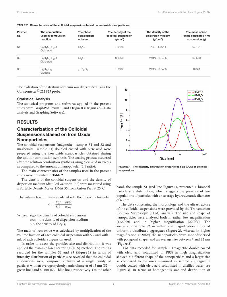
In order to assess the particles size and distribution it wasapplied the dynamic laser scattering (DLS) method. The resultsrecorded for the samples S2 and S3 (Figure 1) in terms ofintensity distribution of particles size revealed that the colloidalsuspensions were composed virtually of a single family ofparticles with an average hydrodynamic diameter of 43 nm (S2—green line) and 80 nm (S3—blue line), respectively. On the other
FIGURE 1 | The intensity distribution of particles size (DLS) of colloidal
suspensions.
hand, the sample S1 (red line Figure 1), presented a bimodalparticle size distribution, which suggests the presence of twopopulations of particles with an average hydrodynamic diameterof 63 nm.
The data concerning the morphology and the ultrastructureof the colloidal suspensions were provided by the TransmissionElectron Microscopy (TEM) analysis. The size and shape ofnanoparticles were analyzed both in rather low magnification(16,500x) and in higher magnification (220Kx). Theanalysis of sample S2 in rather low magnification indicateduniformly distributed aggregates (Figure 2), whereas in highermagnification (220Kx) the nanoparticles were monodispersedwith polygonal shapes and an average size between 7 and 22 nm(Figure 3).
TEM data recorded for sample 1 (magnetite double coatedwith oleic acid solubilized in PBS) in high magnetizationshowed a different shape of the nanoparticles and a larger sizeas compared to the ones measured in sample 2 (magnetitedouble coated with oleic acid solubilized in distilled water; seeFigure 3). In terms of homogenous size and distribution of
Frontiers in Pharmacology | www.frontiersin.org 5 March 2017 | Volume 8 | Article 154

Coricovac et al. Iron Oxide Nanoparticles: Toxicological Profile
FIGURE 2 | TEM micrograph of the sample S2, general aspect of
aggregates in rather low magnification (16,500x).
the nanoparticles, all three samples analyzed presented thesecharacteristics (Figure 3).
According to the measurements (in high magnification) inall three samples (S1, S2, and S3), it could be observed aninverse correlation between the iron oxide concentration in thesample and the nanoparticle size. In the samples with a higherconcentration of iron oxide it was observed a decreased valueof nanoparticle size (7.64 nm–sample S2 and 13.52 nm–sampleS3), whereas in the sample with a lower concentration in Fe3O4
(sample S1), the nanoparticles had an increased size (21.46 nm;see Figure 4).
Antioxidant Activity of the Iron OxidesColloidal SuspensionsThe antioxidant activity of the samples was evaluated by DPPHradical scavenging assay. There were prepared three dilutions foreach sample (see Table 1) and tested for antioxidant activity.
In Figure 5 was presented the antioxidant activity (AOA) ofall three samples of biocompatible magnetic colloidal suspensionbased on iron oxide nanoparticles. The antioxidant activityassessment was carried out for 1,200 s, but we chose to representonly the data recorded until 400 s during this time being detectedmost of the activity (after this time point no significant activitywas detected until the end of experiment).
According to our results, all the samples showed areduced antioxidant activity as compared to the AOA ofascorbic acid, used as positive control. Furthermore, sampleS1 (magnetite—Fe3O4 nanoparticles dispersed in PBS) did notexhibit antioxidant activity for any dilution analyzed. In thecase of sample S3 (maghemite—γ-Fe2O3 nanoparticles dispersedin water), the first dilution analyzed (1:10) did not reveal anantioxidant activity, but at higher dilution the AOA of the sampleachieved a value of ∼20%. This could be explained by the factthat this sample had the highest concentration in γ-Fe2O3 and adilution of 1:10 could be too concentrated to present antioxidantactivity.
FIGURE 3 | TEM micrograph of the samples. High magnification (220Kx).
The details of measurements of nanoparticles size are given in red.
The sample S2 (Fe3O4 nanoparticles dispersed in distilledwater)—contained the same type of nanoparticles (Fe3O4) assample S1, but the differences between S1 and S2 were theamount of Fe3O4 (S1—10.4mg vs. S2—52 mg) and the liquid
Frontiers in Pharmacology | www.frontiersin.org 6 March 2017 | Volume 8 | Article 154

Coricovac et al. Iron Oxide Nanoparticles: Toxicological Profile
carrier (PBS/distilled water); this was the only sample thatshowed antioxidant activity for each dilution. The fact thatthe sample (S2) showed antioxidant activity compared with
FIGURE 4 | The inverse correlation between the iron oxide
concentration in samples and nanoparticle dimensions.
FIGURE 5 | AOA of all three samples of iron oxide colloidal
suspensions, including the dilutions.
the sample S1 could be explained by the liquid carrier—anycompound dispersed in water shows a higher antioxidant activitycompared to the same compound dispersed in other liquidcarrier, and by the amount of Fe3O4 in the sample.
In Figure 6 was presented the AOA of the samples both atinitial and at the final moment. As it could be seen, the AOA(for each dilution) increased in time, this increase being in aninversely proportional relation with the samples concentration.This could be explained by the fact that a colloidal suspensionbased on iron oxides shows antioxidant activity only when theconcentration in Fe3O4 or γ-Fe2O3 was higher and the solidfraction was dispersed in distilled water.
Evaluation of Magnetic ColloidalSuspensions Effects on Cell ViabilityCells viability was expressed as percentage of viable cells (%)related to the control cells (the cells that were stimulated withthe same concentration of solvent—for sample—S1—PBS and forsamples—S2 and S3—distilled sterile water). This parameter wasassessed by the means of Alamar blue technique.
Stimulation of the HaCat cells with different concentrations(5, 10, and 25 µg·mL−1) of samples S1, S2, and S3 colloidalsuspensions for 24 h led to an increase of cell viabilityas compared to control cells, what indicated that the ironnanoparticles did not affect cells viability (Table 3). Similarresults were obtained for a higher concentration (50 µg·mL−1)in the case of all three test samples—S1, S2, and S3 (data notshown).
TABLE 3 | In vitro effect of samples S1, S2, and S3 after 24 h stimulation.
Samples Percentage of viable HaCat cells (%)
PBS/H2O 5 µg·mL−1 10 µg·mL−1 25 µg·mL−1
S1 100 111.47 101.49 109.26
S2 100 118.45 129.15 102.78
S3 100 131.38 128.11 119.67
FIGURE 6 | The AOA of the samples at initial (t = 0 s) and at the final (t = 1,200 s) moment.
Frontiers in Pharmacology | www.frontiersin.org 7 March 2017 | Volume 8 | Article 154

Coricovac et al. Iron Oxide Nanoparticles: Toxicological Profile
FIGURE 7 | The effect of S1 colloidal suspension on HaCat cells migration and proliferation. The cells were stimulated with different concentrations of the
colloidal suspension (C1 = 5, C2 = 10, and C3 = 25 µg·mL−1 ) and were taken photos at 0, 3, and 24 h post-stimulation.
FIGURE 8 | The effect of S2 colloidal suspension on HaCat cells migration and proliferation. The cells were stimulated with different concentrations of the
colloidal suspension (C1 = 5, C2 = 10, and C3 = 25 µg·mL−1 ) and were taken photos at 0, 3, and 24 h post-stimulation.
Effects of Magnetic Colloidal Suspensionson Cell Migration and ProliferationThe effect of the magnetic iron oxide nanoparticles colloidalsuspensions on cell migration and proliferation was assessed bythe means of scratch assay, a wound healing type technique. Afterthe scratches were drawn (when the confluence of the cells wasaround 90%), the cells were stimulated for 24 h with the sameconcentrations tested for the cytotoxic effect. There were takenpictures at different time points (0, 3, and 24 h) in order topursue the impact of the test suspensions on cells migration andproliferation.
As it can be seen in Figure 7, the S1 colloidal suspension didnot affect the normal keratinocytes—HaCat migration, neither
after 3 h, nor after 24 h, moreover, could it be said that the testsuspension had a stimulatory effect on cells proliferation. Thecells were abundant on the plate and well attached.
The Figures 8, 9 showed that the samples S2 and S3 colloidalsuspensions induced a similar effect onHaCat cells migration andproliferation as the one described for sample S1: a stimulatoryeffect, results that were in agreement with the data recorded forthe cytotoxicity test.
In vivo Evaluation of Acute Dermal ToxicityThe in vivo effects of the colloidal suspensions of magnetite andmaghemite coated with a double layer of oleic acid were testedemploying the OECD guideline 402 protocol for acute dermal
Frontiers in Pharmacology | www.frontiersin.org 8 March 2017 | Volume 8 | Article 154

Coricovac et al. Iron Oxide Nanoparticles: Toxicological Profile
FIGURE 9 | The effect of S3 colloidal suspension on HaCat cells migration and proliferation. The cells were stimulated with different concentrations of the
colloidal suspension (C1 = 5, C2 = 10, and C3 = 25 µg·mL−1 ) and were taken photos at 0, 3, and 24 h post-stimulation.
toxicity. The mice used in the study were female and male SKH-1 hairless mice. The body weights of the mice were recordedevery 2 days for 14 days and no significant modifications wereobserved neither in the female groups, nor in male groups.Concerning the behavioral patterns (salivation, lethargy, sleep,and coma), there were observed no such signs in any groupsof mice during the experiment frametime. These data indicatedthat topical applications of iron colloidal suspensions were notassociated with weight loss or interferences at nervous systemlevel.
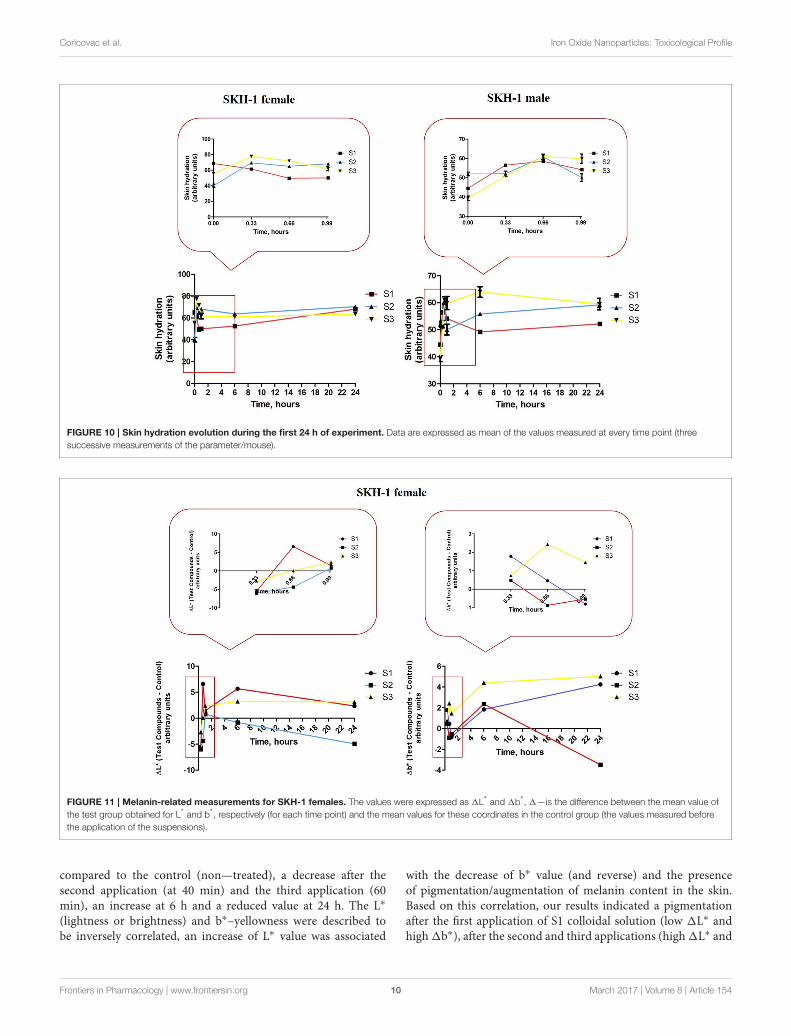
Another parameter that was assessed was the skin appearanceafter topical application of the iron colloidal suspensions. Thechanges in skin appearance were characterized by monitoringthe evolution of skin physiological parameters—skin hydration,melanin content and erythema using a non-invasive technique.The measurements were performed before the application of thecolloidal suspensions (values considered as controls), after eachapplication (at 20, 40, and 60 min), at 6 h, 24 h and in everysecond day until the last day of experiment (day 14). The mostsignificant changes were observed in the first 24 h. Until the endof experiment the values were constant (data not shown).
Topical application of the iron colloidal suspensions led tosome changes in the values of skin hydration both in the femaleand male groups of mice (Figure 10). S1 colloidal suspensionin PBS (red line) led to a decrease in the skin hydration valuein the females group with each application (in the first 1 h),but the measurements at 6 h and 24 h indicated a similar valueto the one measured before the application of the colloidalsuspension (control value) and this value continued in the samerange until the end of experiment. In the case of S2 and S3iron colloidal suspensions in distilled water (blue and yellowlines) it was detected an increase of the parameter in thefirst hour, this hydration status being maintained in the first24 h.
Group 1 of male mice (S1 treated—red line) indicated anincrease of skin hydration value in the first hour (after the 3successive applications), but after 6 h the value of the parameterreturned at the initial value (before topical application) andcontinued in this range until the end of experiment. When wereapplied S2 and S3 iron colloidal suspensions the evolution of skinhydration in the groups of male mice was similar with the oneobserved in the female groups—an increase value in the first hourand this value was maintained in the first 24 h.
Melanin content and the erythema were measured by themeans of tristimulus colorimetry using the skin colorimeter CL400 probe (Courage–Khazaka, Germany).
The iron colloidal suspensions were applied in the posteriorthorax region of the mice (females and males) and the tristimuluscolorimetric L∗a∗b∗ measurements were recorded before eachapplication (in the first hour), at 6 h, 24 h, and every 2 daysuntil the end of the experiment. There were analyzed the valuesfor L∗–skin reflectance (lightness) and b∗–yellowness, associatedwith melanin content, and a∗–redness, an indicator for erythema.
For the females from group 1 that received S1 colloidalsuspension in PBS (red line—Figure 11, left graph) there wereobserved the following aspects: 1L∗ value decreased after thefirst application of the S1 suspension (at 20 min), the secondapplication led to an increase of this parameter (at 40 min) andthe third application was accompanied by a decrease of1L∗ value(at 60 min) as compared to the one recorded after the secondapplication. At 6 h after the first application the 1L∗ value wassimilar with the one recorded after the second application (at 40min), whereas at 24 h the value was in the same range with theone recorded after the third application (at 60 min).
In the case of 1b∗ for the females from group 1 (S1colloidal suspension topically applied—red line, Figure 11, rightgraph) the evolution of this parameter was as follows: thefirst application (at 20 min) led to an increased value as
Frontiers in Pharmacology | www.frontiersin.org 9 March 2017 | Volume 8 | Article 154
Coricovac et al. Iron Oxide Nanoparticles: Toxicological Profile
FIGURE 10 | Skin hydration evolution during the first 24 h of experiment. Data are expressed as mean of the values measured at every time point (three
successive measurements of the parameter/mouse).
FIGURE 11 | Melanin-related measurements for SKH-1 females. The values were expressed as 1L* and 1b*, 1—is the difference between the mean value of
the test group obtained for L* and b*, respectively (for each time point) and the mean values for these coordinates in the control group (the values measured before
the application of the suspensions).
compared to the control (non—treated), a decrease after thesecond application (at 40 min) and the third application (60min), an increase at 6 h and a reduced value at 24 h. The L∗
(lightness or brightness) and b∗–yellowness were described tobe inversely correlated, an increase of L∗ value was associated
with the decrease of b∗ value (and reverse) and the presenceof pigmentation/augmentation of melanin content in the skin.Based on this correlation, our results indicated a pigmentationafter the first application of S1 colloidal solution (low 1L∗ andhigh1b∗), after the second and third applications (high1L∗ and
Frontiers in Pharmacology | www.frontiersin.org 10 March 2017 | Volume 8 | Article 154

Coricovac et al. Iron Oxide Nanoparticles: Toxicological Profile
FIGURE 12 | Melanin-related measurements for SKH-1 males. The values were expressed as 1L* and 1b*, 1—is the difference between the mean value of the
test group obtained for L* and b*, respectively (for each time point) and the mean values for these coordinates in the control group (the values measured before the
application of the suspensions).
low 1b∗) the skin recovered and no pigmentation was recordeduntil the end of experiment.
Group 2 of female mice topically exposed to S2 colloidalsolution—in distilled water presented some variations in the1L∗ values during the experiment, especially in the first 24 h(blue line, Figure 11, left graph): a decrease after the first (at20 min) and second (at 40 min) applications (more significantafter the first application), an increase after the third application(at 60 min) followed by a reduced value at 6 and 24 h. Aninversely evolution was recorded for 1b∗ (blue line, Figure 11,right graph) values in the same group: an increase of the valueafter the first application (at 20 min), a decrease after the second(at 40 min) and third (at 60 min) applications, followed byincreased values at 6 and 24 h. These data showed that applicationof S2 colloidal suspension induced a significant pigmentation/ anaugmentation of melanin content after the first application, theskin tried to recover after the third application (high L∗ and lowb∗), but the pigmentation became more intense after 6 and 24 h,respectively.
The results recorded for group 3 of female mice topicallyexposed to S3 colloidal suspension (maghemite) in distilled waterconcerning 1L∗ parameter (yellow line, Figure 11, left graph)showed: a decrease after the first application (at 20min), followedby an increase after the second and the third applications,increase that continued in the same range at 6 and 24 h, too.In the case of 1b∗ values, for group 3 of females mice wererecorded only positive values, these values presenting a linearcorrelation with 1L∗ values (yellow line, Figure 11, right graph),data that were in contrast with the ones measured for the othertwo suspensions, S1 and S2, an explanation being the fact thatS3 suspensions did not induce any changes in the melanincontent.
In order to quantify the effect induced by the S1, S2 andS3 colloidal suspensions on melanin content in the male micegroups, the parameters L∗ and b∗ were measured. In the firstgroup ofmalemice (topically treated with S1 colloidal suspensionin PBS) was determined the following evolution for 1L∗ values(red line, Figure 12, left graph): all the three applications induceda decrease of 1L∗ (similar negative values), at 4 h after thefirst application the value of 1L∗ became positive reaching amaximum at 6 h and continued in the same range until 24 h.The values recorded for 1b∗ (red line, Figure 12, right graph) inthe same group of mice were in an inverse relationship with 1L∗
after the three applications of the S1 colloidal suspension (low L∗
and high b∗), while the values recorded at 6 and 24 h presentedan ascending trend. These results could be explained as thedevelopment of a temporary pigmentation under the incidenceof the applied suspensions followed by a recession of this statusafter 6 h post-application.
1L∗ values measured in group 2 of male mice—exposed toS2 colloidal suspension in distilled water (blue line, Figure 12,left graph) exhibited the following tendency: a decrease afterthe 3 successive applications (negative values, the highest beingrecorded after the second application). The values at 6 and 24h were closed to the ones calculated after the first and thirdapplications. The other parameter evaluated 1b∗ presented anevolution inversely correlated with the 1L∗ values (low L∗
and high b∗ and reverse; blue line, figure 12, right graph).These results showed that S2 suspension topically applied ledto an elevation of melanin content in the skin of male mice,pigmentation that was conserved during the 24 h.
The results obtained in the case of group 3 of male miceexposed to S3 colloidal suspension in distilled water (maghemite;yellow line, Figure 12, left and right graphs) were similar with
Frontiers in Pharmacology | www.frontiersin.org 11 March 2017 | Volume 8 | Article 154

Coricovac et al. Iron Oxide Nanoparticles: Toxicological Profile
FIGURE 13 | Erythema-related measurements. The values were expressed as 1a*, where 1—is the difference between the mean value of the test group
obtained for a* (for each time point) and the mean value for this coordinate in the control group (the values measured before the application of the suspensions).
the ones obtained for group 3 of female mice showing a directlycorrelation between 1L∗ and 1b∗, data that could be explainedas no impact of this suspension—S3 on melanin content.
Another skin parameter measured in order to characterizethe changes in the skin color post-application of the testsuspensions was 1a∗—redness or the green-red chromaticitycoordinate related to erythema occurrence.1a∗ and1L∗ are alsointerrelated in a linear inversely relationship, the lower the L∗
value is, the higher the a∗ value should be and also the degreeof redness and erythema.
For group 1 of female mice (exposed to S1 suspension) thevalues of 1a∗ that respected the inverse correlation with 1L∗
were only the values recorded after the second and the thirdapplications (at 40 and 60 min), the others being in a directcorrelation. The data could indicate that the erythema occurredstarting with the second application and in time after 6 and 24h the reaction faded and was retracted (red line, Figure 13, leftgraph).
The values calculated for the second group of female mice(exposed to S2 suspension) presented a similar tendency as theone observed to group 1, but the first signs of erythema weredetected after the first application and the skin recovered after6 h (blue line, Figure 13, left graph).
In group 3 of female mice (exposed to S3 suspension), therelationship between1a∗ and1L∗ was a linear directly one, whatcould be associated with a lack of effect on this parameter (yellowline, Figure 13, left graph).
There were also observed some variations of the valuescalculated for 1a∗ in the groups of male mice as compared tocontrol values that were measured before the application of thesuspensions.
The first group of mice (exposed to S1 suspension) presenteda direct correlation between 1a∗ and 1L∗ (high a∗ and high L∗)
what could be explained as the absence of an impact on erythemavalues (red line, Figure 13, right graph). In the second group ofmale mice (exposed to S2 suspension), there were detected somesigns of erythema after the first and the third applications (higha∗ and low L∗), but the erythema attenuated in time (at 6 h; blueline, Figure 13, right graph).
The male mice from group 3 (exposed to S3 suspension)presented signs of erythema starting with the first applicationand the signs were still visible after 24 h. These data could beexplained by the inverse correlation between1a∗ and1L∗ valuescalculated in this group (yellow line, Figure 13, right graph).
DISCUSSIONS
Iron oxide nanoparticles were found in different polymorphicphases, as: α, β, γ, δ, and ε—Fe2O3, maghemite (γ—Fe2O3) andmagnetite (Fe3O4) being the two compounds mostly employedin biomedical applications. Magnetite and maghemite representthe first choice as magnetic nanoparticles for biomedical uses dueto their unique features, such as: superparamagnetic propertiesper se–exhibit magnetism only in the presence of an externalmagnetic field, a single magnetic domain with a large constantmagnetic moment and low toxicity (Muthukumaran and Philip,2016; Velusamy et al., 2016). Magnetite was described to exceedthe maghemite from the point of view of magnetic susceptibilityand saturationmagnetization (Muthukumaran and Philip, 2016).
Magnetite (Fe3O4) presents a cubic crystal structure of inversespinel that has the Fe2+ cations located in the octahedralsite (site B—surrounded by 6 oxygen anions) and the Fe3+
cations distributed evenly between the tetrahedral (site A—surrounded by 4 oxygen anions) and the B site (Li et al., 2010).This distribution of the cations is responsible for the totalmagnetization of the molecule (Marinca et al., 2016). Besides
Frontiers in Pharmacology | www.frontiersin.org 12 March 2017 | Volume 8 | Article 154

Coricovac et al. Iron Oxide Nanoparticles: Toxicological Profile
the metallic nanoparticles used as valuable tools to enhancethe effectiveness of the current therapies and to increase thecompliance of the patient to the treatments, there were describedother types of nanoparticles engineered in this direction, suchas: polymeric colloids, liposomes, solid lipid nanoparticles,cyclodextrins, and others (Kuskov et al., 2010; Medeiros et al.,2015; Soica et al., 2016).
The method used to synthesize iron oxide nanoparticlesis considered a key factor for the future applications of thenanoparticles since the electrical, optical andmagnetic features ofthese items are correlated to their size, parameter that is assignedduring this process (Li et al., 2010). Other parameters that shouldbe strictly controlled in the synthesis of iron oxide nanoparticlesare shape, uniformity, crystallinity, and crystal structure (Jianget al., 2014).
There were described some inconveniences concerning themethods used for the synthesis of iron oxide nanoparticles (poormonodispersity, irregular shape of the particles; Bloemen et al.,2012). Finding the proper method to obtain magnetic ironoxide nanoparticles with a reduced number of inconveniencesrelated to the physico-chemical properties of the nanoparticles, interms of stability, biocompatibility, the proper size and shape forbiomedical uses, represented a real challenge for the researchers.A method that complies with most of these requirementswas proved to be the solution combustion method. Solutioncombustion synthesis was described as a simple, universal andlow time consuming method that could be applied for thepreparation of a variety of nanosize materials. This type ofprocedure it’s based on a self-sustained reaction that requiresthe presence of oxidants (homogenous solutions) and fuels(like: urea, glycine, hydrazides, etc.). The resultant products ofthis procedure are nanosize oxide materials, but this methodalso delivers a homogeneous concentration of trace amounts ofrare-earth impurity ions in a single step. Solution combustionsynthesis proved to be an efficient method for the procurement ofdifferent metal nanopowders, such as: Ni, Zn, Cd, Al, Ti, Cu, Fe,etc. (reviewed in Aruna and Mukasyan, 2008; Ianos et al., 2012,2014; Huang et al., 2016).
In the present study, it was applied the solution combustionsynthesis in order to obtain the magnetic iron oxidenanoparticles. The major advantage of the combustion methodis that the final reaction product is obtained directly aftercombustion, without subsequent calcinations, thereforethe energy consumption is reduced. Phase composition,morphology, and reactivity of the synthesized powders areestablished based on the application of specific requirementsand controlled by synthesis conditions. In a study developedby Gupta and Wells it was shown the selective behavior ofthe magnetic iron oxide nanoparticles obtained by the meansof combustion synthesis (toxic for tumor, but protective withnormal cells), these results offering a new perspective on thepotential use of these magnetic nanoparticles in cancer therapy(Gupta and Wells, 2004).
Ianos and co-workers proposed an adapted protocol for thesolution combustion synthesis in order to obtain nano-scalediron oxide nanoparticles (magnetite and maghemite; Ianos et al.,2012, 2014). For the synthesis of the iron oxide nanoparticles
analyzed in this study it was applied the protocol described byIanos et al. By employing the same method, the results that wereobtained in terms of physico-chemical properties are similar withthe ones showed by Ianos et al. (2012; 2014), as follows: formagnetite (DXRD—crystallite size = 18; SBET—specific surfacearea = 56; DBET—particle diameter from BET = 21; Ms—saturation magnetization = 57.7; Mr—remanent magnetization= 4.5; Hc—coercivity = 5.2) and for maghemite (DXRD—crystallite size = 5; SBET—specific surface area = 149; DBET—particle diameter from BET = 8; Ms—saturation magnetization= 41.5; Mr—remanent magnetization = 0.7; Hc—coercivity =
1). Considering the magnetic properties of the resulted powders,it was observed that the saturation magnetization of the samplesS1, S2 prepared by combustion synthesis was slightly higherthan that of the sample S3 which after combustion was washedwith oxygenated water in order to remove the carbon from themagnetic nanoparticles surface. At the same time, the remanentmagnetization and the coercivity of combustion synthesizedmagnetic nanoparticles were very close to the superparamagneticbehavior. The specific surface area of sample S3 was the highest,which mean that the particles are very small, fact that was alsoobserved from the BET diameter. This aspect indicated that theseparticles, obtained by combustion method, having narrow sizedistribution with a size range of <50 nm, can be successfullyused for intravenous administration (i.v.), not only as magneticresonance imaging (MRI) contrast agents, vectors for gene anddrug delivery, or as agents for hyperthermia therapy (Boyer et al.,2010; Estelrich et al., 2015).
One of the problems encountered in the synthesis of ironoxide nanoparticles is the aggregation process characteristic forthe nanoscale particles with a large surface-to-volume ratio. Thesolution proposed for this matter was the use of stabilizing agentsor coating agents that adhere to the surface of the nanoparticlesand offer a spatial isolation leading to the achievement ofmonodisperse nanoparticles (Li et al., 2010). To avoid suchagglomeration and coagulation of the nanoparticles, they arecoated with specific surfactants that present in their structurea hydrophobic element and a polar group. The hydrophobicelement is adsorbed on the surface of the nanoparticles whereasthe polar group enwidens into the water solution and protectsthe nanoparticle against agglomeration, process that is known asfunctionalization of the nanoparticle (Medeiros et al., 2015).
To obtain biocompatible colloidal suspensions of iron oxidenanoparticles, in this study it was proposed as coating agent afatty acid—the oleic acid. Oleic acid (OA) is frequently usedas a capping agent for the iron oxides nanoparticles, becauseit can form a dense protective monolayer, which is stronglybonded to the surface of nanoparticles, thus being producedmonodisperse and uniform nanoparticles. If the iron oxidenanoparticles are monolayer—coated with OA, these will bedispersible only in organic solvents thereby limiting their usefor biomedical applications (Patil et al., 2014). A single layerof OA is adequate for loading different hydrophobic drugs,but is not biocompatible and appropriate for medical use dueto the presence of hydrophobic surfaces with a large surfacearea that might lead to considerable particles size (aggregationand big clusters formation) and recognition by the immune
Frontiers in Pharmacology | www.frontiersin.org 13 March 2017 | Volume 8 | Article 154

Coricovac et al. Iron Oxide Nanoparticles: Toxicological Profile
cells and clearance. In order to obtain biocompatible iron oxidenanoparticle suspensions is required the addition of a hydrophilicsurfmer to the oleic acid monolayer used for functionalization ofthe nanoparticles (Tran et al., 2015).
The novelty of the method that was proposed to preparebiocompatible colloidal suspensions of iron oxide nanoparticlesin the present study consisted in coating with double layer ofOA the iron oxide nanoparticles resulted during the solutioncombustion synthesis, method that was not described in theliterature, not to our knowledge. The coating process wasconducted in compliance with the protocol described byBica et al. (2007) with several modifications and there wereobtained OA double coated iron oxide nanoparticles that werecharacterized in terms of DLS and TEM analyses.
The values that were measured for the particles sizedimensions of the three samples analyzed (S1—63 nm, S2—43nm, S3—80 nm—Figure 1) by the means of DLS indicate thatthe coated nanoparticles have a very narrow size distributionrange with superparamagnetic behavior at room temperature.The hydrodynamic diameter of the coated nanoparticles is higheras compared to the values measured for the pure/uncoatednanoparticles, and this could be a confirmation of thecoating process. Furthermore, the nanoparticles are stable inPBS/distilled water at neutral pH. Based on these data, it could besaid that the colloidal suspensions of the OA double coated ironoxide nanoparticles obtained are suitable for in vitro and in vivoapplications, their dimensions being in the range established forbiomedical domain, an average particle’s diameter smaller than100 nm and the polydispersity 0.1 (Shete et al., 2015; Medeiroset al., 2015).
According to the TEM measurements and micrographs(Figure 3) concerning the morphology and ultrastructure of thecolloidal suspensions, the results confirmed that the preparediron oxide particles were of nanosize, similar results beingdescribed by Buzea et al. (2007). The values recorded for thesize of the nanoparticles from all the samples (see Figure 4)are in the range of 7–22 nm, what could indicate the presenceof high polydispersity, results that are in accordance with thedata obtained from DLS measurements. It is known that DLSmeasures the size of the aggregates not of a single iron oxideparticle and this could be an explanation for the differencesbetween the values of average particle diameter measured byDLS technique and the ones recorded by TEM. Our results arein agreement with the data from the literature (Li et al., 2010;Medeiros et al., 2015; Shete et al., 2015).
A considerable number of studies used oleic acid inmonolayeras coating agent for different nanoparticles engineered fortargeted drug delivery and release and the results showed thatthis fatty acid played a crucial role in controlling the shape,monodispersity and thermal stability of the nanoparticles (Liet al., 2010; Tran et al., 2015; Muthukumaran and Philip, 2016;Velusamy et al., 2016). Jiang et al. proved that the shape andsize of the magnetic iron nanoparticles synthetized by high-temperature decomposition was influenced by the concentrationof oleic acid used as coating agent: the higher the concentrationof OA, the wider the particle size and of irregular shapes (Jianget al., 2014). Similar results regarding the impact of oleic acid
on particle size and shape dependent on concentration of acidused, were described by Soares et al. in a study published in2015 (Soares et al., 2015). The group of Marinca proposed anovel method for the synthesis of magnetite nanoparticle coatedwith oleic acid for biomedical purposes, which proved to be acombination between the ceramic method and wet mechanicalmilling and the results obtained highlighted the dual role ofoleic acid as surfactant and to prevent the coalescence of thenanoparticles. Moreover, OA coating limits the contamination ofthe resulted powder with iron and forbids the reaction betweenmagnetite and iron (Marinca et al., 2016).
The bond between the first layer of oleic acid and theiron oxide nanoparticles could be explained by the interactionbetween the carboxyl group from the oleic acid structure andthe surface of the iron oxide, probably through a coordinationof the iron atoms and both oxygen from the carboxyl group. Thehydrocarbon tail of the oleic acid remains free to interact withthe second surfactant—the surfmer (the active surfactant) andleads to a solubilization of the coated iron oxide nanoparticlesin organic solvents (Medeiros et al., 2015; Shete et al., 2015). Byadding another layer of oleic acid to the iron oxide nanoparticlescoated with a first layer of oleic acid, it was achieved an aqueoussuspension of iron oxide nanoparticles with a long stability (morethan 3 months; Lan et al., 2007; Li et al., 2010).
The group of Ingram obtained magnetite nanoparticles by co-precipitation method, that were further coated with a doublelayer of oleic acid what provided them colloidal stability in waterat pH = 7–10 and proved to be efficient to stabilize the oil-wateremulsions with applicability in magnetic imaging and sensingapplications (Ingram et al., 2010). Our results are in concordancewith the data existent in the literature, since we obtained stablewater dispersible suspensions of OA double coated iron oxidenanoparticles.
Concerning the antioxidant activity assessment, it wasobserved that for an amount of 52mg of Fe3O4 (sample S2)the AOA obtained was 20% for the dilution 1:100. This valueis similar to the one obtained in the case of using 72mg γ-Fe2O3. Paul et al. investigated the antioxidant properties of ironoxide particles of different sizes and showed that the free radicalscavenging efficiency of bare iron oxide nanoparticles (α-Fe2O3)was found to be almost 50% for DPPH by 200mg of iron oxidenanoparticles (Paul et al., 2009). Bhattacharya et al. found a veryhigh free radical scavenging activity of 89% by using 10mg of α-Fe2O3/C nanocomposites. This can be explained by the electrontransfer from the nanocomposite system toward the free radicallocated at the nitrogen atom in DPPH (Bhattacharya et al., 2014).
A matter of great concern for the use of nanoparticlesin biomedical field is considered the incomplete toxicologicalprofile of these nanomaterials, the toxicity data being somehowcontroversial. A considerable number of studies were conductedin order to verify the toxicity induced by the nanoparticles, bothin vitro and in vivo, but also in the other domains that usenanotechnology/nanoparticles (agriculture, food sector; Balmuriet al., 2016; Bostan et al., 2016; Neagu et al., 2016; Piperigkouet al., 2016; Valdiglesias et al., 2016).
One of the objectives suggested in this study consisted in thein vitro and in vivo toxicological evaluation of the biocompatible
Frontiers in Pharmacology | www.frontiersin.org 14 March 2017 | Volume 8 | Article 154

Coricovac et al. Iron Oxide Nanoparticles: Toxicological Profile
colloidal suspensions obtained in order to establish their safetyprofile.
Taking into consideration the data that were obtained in thesection of physico-chemical characterization of the novel OAdouble-layered iron oxide nanoparticles colloidal suspensions,it was tested the effect of these suspensions in vitro on humankeratinocytes (HaCat cell line) viability. The test was performedusing Alamar blue assay. This assay relies on the capacity ofthe metabolically active cells (living cells) to reduce resazurin,the active compound from Alamar blue solution in order toquantitatively measure the number of viable normal or cancercells after stimulation with test compounds (Riss et al., 2016).Our results presented in Table 3 indicated, in the case of allthree colloidal suspensions (magnetite and maghemite oleic aciddouble coated nanoparticles), a lack of toxicity induced by theiron oxide nanoparticles colloidal suspensions after an exposureof 24 h at the concentrations used (5, 10, and 25 µg·mL−1).
Similar results were obtained by Naseroleslami et al.when tested different concentrations (in the range of 25–800 µg·mL−1) of PEGylated superparamagnetic iron oxidenanoparticles obtained by co-precipitation method on human-derived amniotic membrane stem cells, the cell viability atthe lowest concentration (25 µg·mL−1) being 99.96 ± 0.05%independent of exposure time (24, 48, and 72 h; Naseroleslamiet al., 2016). In a recent study developed by Joris and co-workers, concerning the safety profile of iron oxide nanoparticlesin neural cells of different origin (human and mouse) and type(stem cells, progenitor cell line and cancer cell line), it wasshown that iron oxide nanoparticles coated with an amphiphilicpolymer—poly(isobutylene-alt-maleic anhydride)-graft-dodecyl(PMA) induced the lowest loss of cell viability as compared togold and silver nanoparticles. The highest susceptibility to acutetoxicity was observed in the case of human neural stem cellsfollowed by the mouse stem cells, whereas the least susceptibilitywas recorded for cancer cells (Joris et al., 2016). Shete et al.tested both uncoated and coatedmagnetite nanoparticles on L929cell line (mouse fibroblasts) and it was observed a very lowcytotoxicity even after a period of 48 h stimulation with differentconcentrations (0.1, 0.5, 1.0, 1.5, and 2.0 mg·mL−1; Shete et al.,2015).
Pongrac et al. demonstrated that poly(L-lysine)-coatedmaghemite nanoparticles after a stimulation of 48 h led to a cellviability and proliferation around 80% at a concentration of 0.2mg·mL−1 while a concentration similar (0.03 mg·mL−1) with thehighest concentration used in the present study (25 µg·mL−1)was responsible for around 5% dead neural stem cells—NSC(Pongrac et al., 2016), results that indicated a reduced degree oftoxicity induced by the iron nanoparticles.
There were also published data that affirmed the toxicityinduced by the iron oxide nanoparticles, the noxious effects beingattributed to the difference in particles size and shape, the doseused for stimulation, and the cell type (reviewed in Valdiglesiaset al., 2016).
Another parameter tested in order to verify the in vitro effectsof the colloidal suspensions of magnetite and maghemite wastheir impact on cells migration and proliferation. According toour results (Figures 7, 8, 9), the suspensions had no effect on
HaCat cells migration at all the concentrations tested, nor after3 h, neither after 24 h stimulation. Our data are in agreementwith the data presented by Muhammad et al. who showedthat stimulation with SPIO (superparamagnetic iron oxide)nanoparticles of adipose-derived mouse stem cells and bonemarrow-derivedmouse stem cells did not impaired cell migration(Muhammad et al., 2015).
The in vitro results that were obtained could be considereda toxicological profile for the coating agent, in this case oleicacid, since the iron oxide nanoparticles were not degraded in thetimeframe of the cytotoxicity assay—24 h.
Since the colloidal suspensions that were obtained in thepresent study, represent an element of originality (no biologicalstudies were performed to the best of our knowledge on ironoxide nanoparticles obtained by solution combustion synthesisand coated with a double layer of oleic acid), to test their effectsin vivo became mandatorily.
In this study it was conducted a test of dermal acute toxicityof the colloidal suspensions on female and male SKH-1 hairlessmice, toxicity that was verified after topical applications of thesuspensions. It was decided to perform this kind of toxicity testbased on the fact that the preliminary data obtained will representthe fundamental basis for further in vivo studies regarding theformulation of topical forms using iron oxide nanoparticles ascarriers for agents effective in different skin pathologies. Our datashowed that topical applications of S1, S2, and S3 suspensions hadno effect on mice body weight or on behavioral patterns.
In order to evaluate the effects induced by these colloidalsuspensions at skin level, there were measured and analyzedseveral physiological skin parameters, including: skin hydration,melanin content and erythema. The method applied wasa non-invasive assay, using the equipment from Courage-Khazaka Electronics GmbH: Corneometer R©CM 825 and SkinColorimeter CL 400.
Topical application of S1 colloidal suspension in PBS led to adecrease of skin hydration in the females group, but the initialvalues of the parameter were achieved during the first 24 h post-application (red line, Figure 10, left graph). These modificationswere not detected in the male mice group that suffered the sameprotocol (red line, Figure 10, right graph). The S2 and S3 aqueoussuspensions induced an increase of skin hydration in both femaleand male mice groups, hydration that was maintained during thefirst 24 h (blue and yellow lines, Figure 8).
The differences between female and male mice concerningthe skin hydration recorded in the present experiment can beexplained by the sex disparities in skin structure: the malespresent a lower pH value, a higher sebum content and skinhydration values, and also a higher susceptibility to develop skinpathologies, including cancer (Boelsma et al., 2003; Deheleanet al., 2016).
The tristimulus colorimetry method was applied in orderto verify the changes of skin color associated to the topicalapplication of test suspensions. This type of method is commonlyused in dermato-cosmetic research, the parameters investigated,melanin content and erythema being indicators of skin barrierintegrity and sensitivity after contact with different substances,drugs or vehicles (Matias et al., 2015). This technique offers
Frontiers in Pharmacology | www.frontiersin.org 15 March 2017 | Volume 8 | Article 154

Coricovac et al. Iron Oxide Nanoparticles: Toxicological Profile
quantitative measurements of skin color expressed with the helpof a 3-digit output L∗, a∗ and b∗—arbitrary values, system thatis recognized by the International Commission of Illumination(Commission Internationale de L’Eclairage—CIE; Alaluf et al.,2002; Tzung et al., 2009). L∗ is known as lightness, brightness,or level of darkness and can take values in the interval 0 and100, the value 0—is black and 100 is white and is associated withthe melanin content from the external skin layers. a∗ can reachnegative or positive values between −60 and + 60, from greento red, this marker being known as redness and is considered anindicator for the presence of the erythema (the higher the valuesis, the erythema is more intense). b∗—yellowness, the coordinatethat is related to melanin content from the skin, can be expressedas negative or positive values from −60 (blue) to +60 (yellow;Alaluf et al., 2002; Tzung et al., 2009; Yang et al., 2016).
There was described an inverse correlation between the L∗
and a∗ and b∗ values. The darkest type of skin presents a lowL∗ value and high values for a∗ and b∗ (Alaluf et al., 2002).Our results indicated that in the group 1 of female mice wasobserved a pigmentation after the first application of S1, whereasthe erythema signs were detected only after the second and thirdapplication. Both pigmentation and erythema faded until the endof 24 h. These data showed that application of S1 suspension onfemale mice skin led to some changes of skin parameter, but thechange was not significant in terms of disturbing the integrity ofskin barrier function.
In the case of the second group of female mice—exposed toS2 suspension it was detected a pigmentation that lasted for 24 hwhile the erythema signs were recorded after the first application,but after 6 h the erythema values were decreased. These datescould be explained by the fact that S2 suspension was betterabsorbed in the skin being an aqueous solution, what led to apigmentation for 24 h. The S3 suspension had no impact in thegroup of female mice neither on the parameter related to melanincontent (L∗ and b∗), nor on the a∗—the coordinate related toerythema.
The results recorded in the groups of male mice wereas follows: S1—application was associated with a temporarypigmentation after the applications of the suspension (<6 h),but no sign of erythema was recorded; S2—application led to anincrease of melanin content value that was maintained constantin the 24 h and it was observed a slight erythema after theapplication (only for 6 h) and S3—application had no impact onmelanin content, but there were recorded erythema signs afterthe topical applications.
The changes observed in the skin parameters values showeda slight disturbance associated to the topical applications of theiron oxide nanoparticles, but these changes were temporary andshould not be interpreted as signs of toxicity.
On account of our group expertise in the field of skinpathologies (Dehelean et al., 2013, 2016; Gheorgheosu et al.,2013; Soica et al., 2014; Danciu et al., 2015), the present studycould be seen as a first step in the synthesis and characterizationof the magnetic iron nanoparticles double coated with oleicacid as future carrier platforms for transdermal drug delivery inskin malignancies (melanoma and non-melanoma skin cancers,auto-immune diseases—epidermolysis bullosa acquisita). In thisregard was evaluated the toxicological profile of the colloidal
suspensions on human keratinocytes cell line and by applying thedermal acute test to SKH-1 hairless mice.
The transdermal drug delivery offers several advantages ascompared to the other routes of administration (oral andintravenous): a controlled release of the drug, avoidance of thefirst hepatic metabolism and a higher patient compliance byreducing the pain associated to i.v. administration. Oleic acid isa FDA recognized agent for increasing skin permeation and itis frequently applied in different commercial formulations. Themechanism of action of oleic acid consists in: interaction with thelipid content of the stratum corneum what leads to the changes inthe lipid bilayer, characterized by the apparition of some poolsresponsible for the defects that appear in the permeability and itis facilitated the entry of different molecules into the profoundskin layers (Shah et al., 2012).
All these data support the idea of oleic acid double coatediron oxide nanoparticles as promising carrier platforms for drugdelivery in skin disorders.
CONCLUSIONS
In the current study were obtained biocompatible colloidalsuspensions based on iron oxide nanoparticles prepared by themeans of solution combustion synthesis and coated with a doublelayer of oleic acid. TEM and DLS analyses confirmed theirnanosize features (an average particle diameter around 7–22nm) what makes them suitable for biomedical applications, inaddition to their high stability and solubility in aqueous solutions.Magnetite solubilized in distilled water (sample 2) exhibited anantioxidant activity at the concentrations tested. The in vitroevaluations of the colloidal suspensions tested indicated a lack oftoxicity on human keratinocytes cell viability, proliferation, andmigration. The in vivo acute dermal toxicity test revealed somechanges in the values of physiological skin parameters, but notsignificant as to interfere with the skin barrier function. Thesedata offer valuable information for future studies regarding theuse of iron oxide nanoparticles as carrier platforms for drugdelivery in skin pathology.
AUTHOR CONTRIBUTIONS
EM, IP, and CP—effectuated the synthesis of the iron oxidenanoparticles and preparation of the colloidal suspensions,the physico-chemical characterization, analysis of the data anddrafting the work. DC and CD—contribution at the conceptionof the study, performed the in vitro and the in vivo tests, analysisand interpretation of the data acquired, drafting the work andprepared the manuscript for submission. CM—performed TEMassay, acquired and analyzed the data, drafting the work. CS, CC,VT, and AT—elaboration of the final version of the manuscript,correction of the language, analysis of the data and revisedcritically the work.
FUNDING
This work was supported by a grant of the Romanian NationalAuthority for Scientific Research and Innovation, CNCS –UEFISCDI, project number PN-II-RU-TE-2014-4-2842.
Frontiers in Pharmacology | www.frontiersin.org 16 March 2017 | Volume 8 | Article 154

Coricovac et al. Iron Oxide Nanoparticles: Toxicological Profile
REFERENCES
Alaluf, S., Atkins, D., Barrett, K., Blount, M., Carter, N., and Heath, A. (2002).The impact of epidermal melanin on objective measurements of humanskin colour. Pigment Cell Res. 15, 119–126. doi: 10.1034/j.1600-0749.2002.1o072.x
Aruna, S. T., and Mukasyan, A. S. (2008). Combustion synthesisand nanomaterials. Curr. Opin. Solid State Mater. Sci. 12, 44–50.doi: 10.1016/j.cossms.2008.12.002
Balmuri, S. R., Selvaraj, U., Kumar, V. V., Anthony, S. P., Tsatsakis, A. M.,Golokhvast, K. S., et al. (2016). Effect of surfactant in mitigating cadmiumoxide nanoparticle toxicity: implications for mitigating cadmium toxicity inenvironment. Environ. Res. 152, 141–149. doi: 10.1016/j.envres.2016.10.005
Bhattacharya, K., Gogoi, B., Buragohain, A. K., and Deb, P. (2014).Fe2O3/C nanocomposites having distinctive antioxidant activity andhemolysis prevention efficiency. Mater. Sci. Eng. C 42, 595–600.doi: 10.1016/j.msec.2014.05.062
Bica, D., Vekas, L., Avdeev, M. V., Marinica, O., Socoliuc, V., Balasoiu,M., et al. (2007). Sterically stabilized water based magnetic fluids:synthesis, structure, and properties. J. Magn. Magn. Mater. 311, 17–21.doi: 10.1016/j.jmmm.2006.11.158
Bloemen, M., Brullot, W., Luong, T. T., Geukens, N., Gils, A., and Verbiest,T. (2012). Improved functionalization of oleic acid-coated iron oxidenanoparticles for biomedical applications. J Nanopart. Res. 14:1100.doi: 10.1007/s11051-012-1100-5
Blois, M. S. (1985). Antioxidant determinations by the use of a stable free radical.Nature 181, 1199–1200. doi: 10.1038/1811199a0
Boelsma, E., van de Vijver, L. P., Goldbohm, R. A., Klöpping-Ketelaars, I. A.,Hendriks, H. F., and Roza, L. (2003). Human skin condition and its associationswith nutrient concentrations in serum and diet. Am. J. Clin. Nutr. 77:348e355.
Bostan, H. B., Rezaee, R., Valokala, M. G., Tsarouhas, K., Golokhvast, K., Tsatsakis,A. M., et al. (2016). Cardiotoxicity of nano-particles. Life Sci. 165, 91–99.doi: 10.1016/j.lfs.2016.09.017
Boyer, C., Whittaker, M. R., Bulmus, V., Liu, J., and Davis, T. P. (2010). The designand utility of polymer stabilized iron oxide nanoparticles for nanomedicineapplications. NPG Asia Mater 2, 23–30. doi: 10.1038/asiamat.2010.6
Buzea, C., Pacheco Blandino, I. I., and Robbie, K. (2007). Nanomaterialsand nanoparticles: sources and toxicity. Biointerphases 2, MR17–MR172.doi: 10.1116/1.2815690
Conde, J., Dias, J. T., Grazú, V., Moros, M., Baptista, P. V., and de laFuente, J. M. (2014). Revisiting 30 years of biofunctionalization and surfacechemistry of inorganic nanoparticles for nanomedicine. Front. Chem. 2:48.doi: 10.3389/fchem.2014.00048
Soica, C., Oprean, C., Borcan, F., Danciu, C., Trandafirescu, C., Coricovac,D., et al. (2014). The synergistic biologic activity of oleanolic and ursolicacids in complex with hydroxypropyl-γ-cyclodextrin.Molecules 19, 4924–4940.doi: 10.3390/molecules19044924
Danciu, C., Oprean, C., Coricovac, D. E., Andreea, C., Cimpean, A., Radeke, H.,et al. (2015). Behaviour of four different B16 murine melanoma cell sublines:C57BL/6J skin. Int. J. Exp. Pathol. 96, 73–80. doi: 10.1111/iep.12114
Dehelean, C. A., Feflea, S., Gheorgheosu, D., Ganta, S., Cimpean, A. M., Muntean,D., et al. (2013). Anti-angiogenic and anti-cancer evaluation of betulinnanoemulsion in chicken chorioallantoic membrane and skin carcinoma inBalb/c mice. J. Biomed. Nanotechnol. 9, 577–589. doi: 10.1166/jbn.2013.1563
Dehelean, C. A., Soica, C., Pinzaru, I., Coricovac, D., Danciu, C., Pavel, I., et al.(2016). Sex differences and pathology status correlated to the toxicity of somecommon carcinogens in experimental skin carcinoma. Food Chem. Toxicol. 95,149–158. doi: 10.1016/j.fct.2016.07.007
Estelrich, J., Escribano, E., Queralt, J., and Busquets, M. A. (2015). Ironoxide nanoparticles for magnetically-guided and magnetically-responsive drugdelivery. Int. J. Mol. Sci. 16, 8070–8101. doi: 10.3390/ijms16048070
Gheorgheosu, D., Jung, M., Ören, B., Schmid, T., Dehelean, C., Muntean, D., et al.(2013). Betulinic acid suppresses NGAL-induced epithelial-to-mesenchymaltransition in melanoma. Biol. Chem. 394, 773–781. doi: 10.1515/hsz-2013-0106
Gobbo, O. L., Sjaastad, K., Radomski, M. W., Volkov, Y., and Prina-Mello,A. (2015). Magnetic nanoparticles in cancer theranostics. Theranostics 5,1249–1263. doi: 10.7150/thno.11544
Gupta, A. K., and Gupta, M. (2005). Synthesis and surface engineering of ironoxide nanoparticles for biomedical applications. Biomaterials 26, 3995–4021.doi: 10.1016/j.biomaterials.2004.10.012
Gupta, A. K., and Wells, S. (2004). Surface-modified superparamagneticnanoparticles for drug delivery: preparation, characterization, and cytotoxicitystudies. IEEE Trans. Nanobiosci. 3, 66–73. doi: 10.1109/TNB.2003.820277
Huang, M., Qin, M., Cao, Z., Jia, B., Chen, P., Wu, H., et al. (2016). Magneticiron nanoparticles prepared by solution combustion synthesis and hydrogenreduction. Chem. Phys. Lett. 657, 33–38. doi: 10.1016/j.cplett.2016.05.043
Ianos, R., Taculescu, A., Pacurariu, C., and Lazau, I. (2012). Solution combustionsynthesis and characterization of magnetite, Fe3O4, nanopowders, J. Am.
Ceram. Soc. 95, 2236–2240. doi: 10.1111/j.1551-2916.2012.05159.xIanos, R., Taculescu (Moaca), E. A., Pacurariu, C., and Niznansky, D. (2014). δ-
Fe2O3 nanoparticles prepared by combustion synthesis, followed by chemicaloxidation of residual carbon with H2O2. Mater. Chem. Phys. 148, 705–711.doi: 10.1016/j.matchemphys.2014.08.038
Ingram, D. R., Kotsmar, C., Yoon, K. Y., Shao, S., Huh, C., Bryant, S. L., et al. (2010).Superparamagnetic nanoclusters coated with oleic acid bilayers for stabilizationof emulsions of water and oil at low concentration. J. Colloid Interface Sci. 351,225–232. doi: 10.1016/j.jcis.2010.06.048
Jiang, F. Y., Li, X., Zhu, Y., and Tang, Z. (2014). Synthesis and magneticcharacterizations of uniform iron oxide nanoparticles. Physica B 443, 1–5.doi: 10.1016/j.physb.2014.03.009
Joris, F., Valdepérez, D., Pelaz, B., Soenen, S. J., Manshian, B. B., Parak, W.J., et al. (2016). The impact of species and cell type on the nanosafetyprofile of iron oxide nanoparticles in neural cells. J. Nanobiotechnol. 14:69.doi: 10.1186/s12951-016-0220-y
Józefczak, A., Hornowski, T., Skumiel, A., Závišová, V., Koneracká, M.,Tomašovicová, N., et al. (2012). Effect of the molecular weight of poly(ethyleneglycol) on the properties of biocompatible magnetic fluids. Int. J. Thermophys.
33, 640–652. doi: 10.1007/s10765-011-1061-4Jung, M., Weigert, A., Tausendschön, A., Mora, J., Oren, B., Sola, A., et al. (2012).
Interleukin-10-induced neutrophil gelatinase-associated lipocalin productionin macrophages with consequences for tumor growth. Mol. Cell. Biol. 32,3938–3948. doi: 10.1128/MCB.00413-12
Kuskov, A. N., Voskresenskaya, A. A., Goryachaya, A. V., Shtilman, M.I., Spandidos, D. A., Rizos, A. K., et al. (2010). Amphiphilic poly-N-vinylpyrrolidone nanoparticles as carriers for non-steroidal anti-inflammatorydrugs: characterization and in vitro controlled release of indomethacin. Int. J.Mol. Med. 26, 85–94. doi: 10.3892/ijmm_00000438
Lan, Q., Liu, C., Yang, F., Liu, S., Xu, J., and Sun, D. (2007). Synthesisof bilayer oleic acid-coated Fe3O4 nanoparticles and their application inpH-responsive Pickering emulsions. J. Colloid Interface Sci. 310, 260–269.doi: 10.1016/j.jcis.2007.01.081
Li, D., Jiang, D., Chen, M., Xie, J., Wu, Y., Dang, S., et al. (2010). An easyfabrication of monodisperse oleic acid-coated Fe3O4 nanoparticles.Mater. Lett.
64, 2462–2464. doi: 10.1016/j.matlet.2010.08.025Marinca, T. E., Chicinas, H. F., Neamtu, B. V., Isnard, O., Pascuta, P.,
Lupu, N., et al. (2016). Mechanosynthesis, structural, thermal, and magneticcharacteristics of oleic acid coated Fe3O4 nanoparticles. Mater. Chem. Phys.
171, 336–345. doi: 10.1016/j.matchemphys.2016.01.025Matias, A. R., Ferreira, M., Costa, P., and Neto, P. (2015). Skin colour, skin redness
and melanin biometric measurements: comparison study between Antera R©
3D, Mexameter( R©) and Colorimeter R© . Skin Res. Technol. 21, 346–362.doi: 10.1111/srt.12199
Medeiros, S. F., Filizzola, J. O. C., Fonseca, V. F. M., Oliveira, P. F. M., Silva, T.M., Elaissari, A., et al. (2015). Synthesis and characterization of stable aqueousdispersion of functionalized double-coated iron oxide nanoparticles. Mater.
Lett. 160, 522–525. doi: 10.1016/j.matlet.2015.08.026Muhammad, G., Jablonska, A., Rose, L., Walczak, P., and Janowski, M. (2015).
Effect of MRI tags: SPIO nanoparticles and 19F nanoemulsion on variouspopulations of mouse mesenchymal stem cells. Acta Neurobiol. Exp. (Wars) 75,144–59.
Muthukumaran, T., and Philip, J. (2016). Effect of phosphate andoleic acid capping on structure, magnetic properties and thermalstability of iron oxide nanoparticles. J. Alloys Compd. 689, 959–968.doi: 10.1016/j.jallcom.2016.08.067
Frontiers in Pharmacology | www.frontiersin.org 17 March 2017 | Volume 8 | Article 154

Coricovac et al. Iron Oxide Nanoparticles: Toxicological Profile
Naseroleslami, M., Parivar, K., Khoei, S., and Aboutaleb, N. (2016). Magneticresonance imaging of human-derived amniotic membrane stem cells usingpegylated superparamagnetic iron oxide nanoparticles. Cell J. 18, 332–339.doi: 10.22074/cellj.2016.4560
Neagu, M., Piperigkou, Z., Karamanou, K., Engin, A. B., Docea, A. O., Constantin,C., et al. (2016). Protein bio-corona: critical issue in immune nanotoxicology.Arch. Toxicol. 1–18. doi: 10.1007/s00204-016-1797-5
Patil, R. M., Shete, P. B., Thorat, N. D., Otari, S. V., Barick, K. C., Prasad, A.I., et al. (2014). Non-aqueous to aqueous phase transfer of oleic acid coatediron oxide nanoparticles for hyperthermia application. RSC Adv. 4, 4515–4522.doi: 10.1039/C3RA44644A
Paul, S., Saikia, J. P., Samdarshi, S. K., and Konwar, B. K. (2009). Investigationof antioxidant property of iron oxide particles by 1′-1′ diphenylpicryl-hydrazyle (DPPH) method. J. Magn. Magn. Mater. 321, 3621–3623.doi: 10.1016/j.jmmm.2009.07.004
Piperigkou, Z., Karamanou, K., Engin, A. B., Gialeli, C., Docea, A. O., Vynios, D.H., et al. (2016). Emerging aspects of nanotoxicology in health and disease:from agriculture and food sector to cancer therapeutics. Food Chem. Toxicol.
91, 42–57. doi: 10.1016/j.fct.2016.03.003Pongrac, I. M., Dobrivojevic, M., Ahmed, L. B., Babic, M., Šlouf, M., Horák,
D., et al. (2016). Improved biocompatibility and efficient labeling of neuralstem cells with poly(L-lysine)-coated maghemite nanoparticles. Beilstein J.
Nanotechnol. 7, 926–936. doi: 10.3762/bjnano.7.84Radomska, A., Leszczyszyn, J., and Radomski, M. W. (2016). The
nanopharmacology and nanotoxicology of nanomaterials: new opportunitiesand challenges. Adv. Clin. Exp. Med. 25, 151–162. doi: 10.17219/acem/60879
Raspopov, R. V., Trushina, É. N., Gmoshinskiı, I. V., and Khotimchenko, S. A.(2011). Bioavailability of nanoparticles of ferric oxide when used in nutrition.Experimental results in rats. Vopr. Pitan. 80, 25–30.
Riss, T. L., Moravec, R. A., Niles, A. L., Duellman, S., Benink, H. A., WorzellaT. J., et al. (2016). “Cell viability assays,” in Assay Guidance Manual, eds G. S.Sittampalam, N. P. Coussens, K. Brimacombe, A. Grossman,M. Arkin, D. Auld,C. Austin, J. Baell, B. Bejcek, T. D. Y. Chung, J. L. Dahlin, V. Devanaryan, T. L.Foley, M. Glicksman, M. D. Hall, J. V. Hass, J. Inglese, P. W. Iversen, M. Lal-Nag, Z. Li, J. McGee, O. McManus, T. Riss, O. J. Trask Jr., J. R. Weidner, M.Xia, and X. Xu (Bethesda, MD: Eli Lilly & Company and the National Centerfor Advancing Translational Sciences), 262–291.
Shah, P. P., Desai, P. R., and Singh, M. (2012). Effect of oleic acid modifiedpolymeric bilayered nanoparticles on percutaneous delivery of spantide II andketoprofen. J. Control. Release 158, 336–345. doi: 10.1016/j.jconrel.2011.11.016
Shete, P. B., Patil, R. M., Tiwale, B. M., and Pawar, S. H. (2015). Water dispersibleoleic acid-coated Fe3O4 nanoparticles for biomedical applications. J. Magn.
Magn. Mater. 377, 406–410. doi: 10.1016/j.jmmm.2014.10.137Soares, P. I., Lochte, F., Echeverria, C., Pereira, L. C., Coutinho, J. T.,
Ferreira, I. M., et al. (2015). Thermal and magnetic properties ofiron oxide colloids: influence of surfactants. Nanotechnology 26:425704.doi: 10.1088/0957-4484/26/42/425704
Soares, P. I. P., Laia, C. A. T., Carvalho, A., Pereira, L. C. J., Coutinho, J. T.,Ferreira, I. M. M., et al. (2016). Iron oxide nanoparticles stabilized witha bilayer
of oleic acid formagnetic hyperthermia and MRI applications. Appl. Surf. Sci.383, 240–247. doi: 10.1016/j.apsusc.2016.04.181
Soica, C., Coricovac, D., Dehelean, C., Pinzaru, I., Mioc, M., Danciu, C.,et al. (2016). Nanocarriers as tools in delivering active compounds forimmune system related pathologies. Recent. Pat. Nanotechnol. 10, 128–145.doi: 10.2174/1872210510999160427113345
Soica, C., Danciu, C., Savoiu-Balint, G., Borcan, F., Ambrus, R., et al. (2014).Betulinic acid in complex with a gamma-cyclodextrin derivative decreasesproliferation and in vivo tumor development of non-metastatic and metastaticB164A5 cells. Int. J. Mol. Sci. 15, 8235–8255. doi: 10.3390/ijms15058235
Sun, J., Zhou, S., Hou, P., Yang, Y., Weng, J., Li, X., et al. (2007). Synthesis andcharacterization of biocompatible Fe3O4 nanoparticles. J. Biomed. Mater. Res.
A. 80, 333–341. doi: 10.1002/jbm.a.30909Torres-Sangiao, E., Holban, A. M., and Gestal, M. C. (2016). Advanced
nanobiomaterials: vaccines, diagnosis and treatment of infectious diseases.molecules. 21:E867. doi: 10.3390/molecules21070867
Tran, T. T. D., Vo, T. V., and Tran, P. H. L. (2015). Design of iron oxidenanoparticles decorated oleicacid and bovine serum albumin for drug delivery.Chem. Eng. Res. Des. 94, 112–118. doi: 10.1016/j.cherd.2014.12.016
Tzung, T. Y., Yang, C. Y., Huang, Y. C., and Kao, F. J. (2009). Colorimetry providesa rapid objective measurement of de novo hair growth rate in mice. Skin Res.
Technol. 15, 459–463. doi: 10.1111/j.1600-0846.2009.00390.xValdiglesias, V., Fernández-Bertólez, N., Kiliç, G., Costa, C., Costa, S.,
Fraga, S., et al. (2016). Are iron oxide nanoparticles safe? Currentknowledge and future perspectives. J. Trace. Elem. Med. Biol. 38, 53–63.doi: 10.1016/j.jtemb.2016.03.017
Velusamy, P., Chia-Hung, S., Shritama, A., Venkat Kumar, G., Jeyanthi, V., andPandian, K. (2016). Synthesis of oleic acid coated iron oxide nanoparticlesand its role in anti-biofilm activity against clinical isolates of bacterialpathogens. J. Taiwan. Inst. Chem. Eng. 59, 450–456. doi: 10.1016/j.jtice.2015.07.018
Yang, Z., Zhong, H., Chen, J., et al. (2016). A genetic mechanism for convergentskin lightening during recent human evolution.Mol. Biol. Evol. 33, 1177–1187.doi: 10.1093/molbev/msw003
Zdrojewicz, Z., Waracki, M., Bugaj, B., Pypno, D., and Cabała, K. (2015). Medicalapplications of nanotechnology. Postepy. Hig. Med. Dosw. 69, 1196–1204.doi: 10.5604/17322693.1177169
Conflict of Interest Statement: The authors declare that the research wasconducted in the absence of any commercial or financial relationships that couldbe construed as a potential conflict of interest.
Copyright © 2017 Coricovac, Moaca, Pinzaru, Cîtu, Soica, Mihali, Pacurariu,
Tutelyan, Tsatsakis and Dehelean. This is an open-access article distributed under
the terms of the Creative Commons Attribution License (CC BY). The use,
distribution or reproduction in other forums is permitted, provided the original
author(s) or licensor are credited and that the original publication in this journal
is cited, in accordance with accepted academic practice. No use, distribution or
reproduction is permitted which does not comply with these terms.
Frontiers in Pharmacology | www.frontiersin.org 18 March 2017 | Volume 8 | Article 154


![The Relativistic Electron Density [1ex] and Electron ... · PDF fileThe Relativistic Electron Density and Electron Correlation Markus Reiher ... Electron density distributions for](https://static.fdocuments.net/doc/165x107/5ab2020e7f8b9aea528d15ec/the-relativistic-electron-density-1ex-and-electron-relativistic-electron-density.jpg)


