Dispersões; Soluções; Colóides; Suspensões. 3 Inês Borralho - [email protected].

Biochimica et Biophysica Acta 1798 (2010) 1547–1555
Contents lists available at ScienceDirect
Biochimica et Biophysica Acta
j ourna l homepage: www.e lsev ie r.com/ locate /bbamem
The specificity of frutalin lectin using biomembrane models
Thatyane M. Nobre a,⁎, Felippe J. Pavinatto b, Márcia R. Cominetti c, Heloísa S. Selistre de-Araújo c,Maria E.D. Zaniquelli d, Leila M. Beltramini a
a Grupo de Biofísica Molecular Sérgio Mascarenhas, IFSC, Universidade de São Paulo, São Carlos, SP, Brazilb Grupo de Polímeros Bernhard Gross, IFSC, Universidade de São Paulo, São Carlos, SP, Brazilc Departamento de Ciências Fisiológicas, Universidade Federal de São Carlos, São Carlos, SP, Brazild Laboratório de Físico-Química de Superfícies e Colóides, Departamento de Química, Faculdade de Filosofia, Ciências e Letras de Ribeirão Preto, Universidade de São Paulo,Ribeirão Preto, SP, Brazil
⁎ Corresponding author. Depto. Física e InformáticaInstituto de Física de São Carlos Av. Trabalhador São-CarlTel.: +55 16 33739875; fax: +55 16 33715381.
E-mail address: [email protected] (T.M. Nobre)
0005-2736/$ – see front matter © 2010 Elsevier B.V. Adoi:10.1016/j.bbamem.2010.03.021
a b s t r a c t
a r t i c l e i n f oArticle history:Received 14 December 2009Received in revised form 9 March 2010Accepted 23 March 2010Available online 2 April 2010
Keywords:LectinFrutalinLangmuir monolayersGlycolipidsDynamic surface elasticityBrewster angle microscopy
Frutalin is a homotetrameric α-D-galactose (D-Gal)-binding lectin that activates natural killer cells in vitro andpromotes leukocyte migration in vivo. Because lectins are potent lymphocyte stimulators, understanding theinteractions that occur between them and cell surfaces can help to the action mechanisms involved in thisprocess. In this paper, we present a detailed investigation of the interactions of frutalin with phospho- andglycolipids using Langmuirmonolayers as biomembranemodels. The results confirm the specificity of frutalin forD-Gal attached to a biomembrane. Adsorption of frutalinwasmore efficient for the galactose polar head lipids, incontrast to the one for sulfated galactose, in which a lag time is observed, indicating a rearrangement of themonolayer to incorporate the protein. Regarding ganglioside GM1 monolayers, lower quantities of the proteinwere adsorbed, probably due to the farther apart position of D-galactose from the interface. Binary mixturescontaining galactocerebroside revealed small domains formed at high lipid packing in the presence of frutalin,suggesting that lectin induces the clusterization and the forming of domains in vitro, which may be a form ofreceptor internalization. This is the first experimental evidence of such lectin effect, and it may be useful tounderstand the mechanism of action of lectins at the molecular level.
Universidade de São Paulo –
ense, 400, São Carlos, SP, Brazil.
.
ll rights reserved.
© 2010 Elsevier B.V. All rights reserved.
1. Introduction
Lectins represent a large class of proteins that specifically bindboth simple and complex carbohydrates, in solution or attached to cellsurfaces, in a reversiblemanner. These proteins are involved in severalcarbohydrate-mediated processes associated with recognition ofmolecules present in membranes, e.g., cell–cell interactions, cellcommunication, cell proliferation and death, or in the extracellularmatrix [1]. Since carbohydrates are themajor components of the outersurface of mammalian cells [2,3] and because these sugars are specificfor each developmental stage of the cells, lectins can play animportant role in cell characterization. Changes in cellular glycosyl-ation are a hallmark of cancer cells, and significant correlations arenow established between some glycosylation patterns and clinicalprognosis [4,5]. Indeed, lectins are able to agglutinate malignantlytransformed cells, but not their normal parental cells [6–8]. Malagoliniet al. [9] reported that apoptotic cell lines from different histologicalorigins, as well as normal human and mice neutrophils becomestrongly reactive with the SNA (Sambucus nigra agglutinin) specific
for Siaα2,6Gal/GalNAc structures. The latter was observed for cellswith different histology, leading to the conclusion that apoptosis andprimary necrosis induce a specific glycosylation change, which isindependent of cell type and nature of the stimulus [10]. Therefore,lectins are pointed out as potential biotechnological tools fordiagnosis and therapy of different types of cancer, where some canbe employed as low toxicity drug delivery systems.
Among the lectins, jacalin-related lectin family (JRL) is a group ofplant lectins that derived their name from jacalin, thefirstmember to beidentified from the seeds of jackfruit (Artocarpus integrifolia). Jacalin iscomposed of four subunitswith amajorα-chain of 133 amino acids anda minor β-chain of 20 amino acids, which exhibited a type-I β-prismfold, comprised of three Greek keys (4-stranded β-sheets) [11]. Lectinsin this family present a high structural homology and sequentialidentity, although with different levels of specificity and affinity forligands. Frutalin is a homotetrameric α-D-galatose lectin belonging tothe (JRL) family derived from Artocarpus incisa [12], with an apparentmolecular mass of 66 kDa. Previous results indicated that this lectin isstable up to 60 °C and very resistant to chemical denaturation.Furthermore, frutalin does not need metals to develop its hemagglu-tinant activity, which is three times higher than jacalin.
In this paper, we investigated the interaction of frutalin with bothnatural membranes of MDA-MB-231 tumor cells and artificial biomem-brane models. The biomembrane models were used to investigate the

1548 T.M. Nobre et al. / Biochimica et Biophysica Acta 1798 (2010) 1547–1555
lectin interaction at molecular level and comprised Langmuir mono-layers containingphospholipids and glycolipids. The ability of frutalin tospecifically bind sugars incorporated in the membrane model washelpful in understanding the initial steps of the macromolecularinteraction of the protein with glycobiomembranes. We have shownfor thefirst time in the in vitromodel that frutalin induces the formationof clusters. Such effect is a consequence of the specific interactionsbetween frutalin and a galactose–lipid.
2. Materials and methods
2.1. Chemicals
Dipalmitoylphosphatidylcholine (DPPC), galactocerebroside (Gal-Cer), sulfate cerebroside (Sul), and the ganglioside GM1 werepurchased from Avanti Polar Lipids and used without purification.The chemical structures of the lipids are presented in Scheme 1.
2.2. Frutalin purification
Frutalin purification was performed as described by Moreira et al.[12]. Briefly, dried seeds from A. incisawere ground and stirred for 6 hin 0.15 M phosphate-buffered solution (PBS), pH 7.4, 1:10 wt./vol., at4 °C. The mixture was centrifuged, 20 min at 2702×g at 4 °C. Thesupernatant was submitted to dialofiltration on YM 3.5 membrane(Millipore) in PBS to half its original volume, and this solution wascalled crude extract. Frutalin was purified on a Sepharose-D-galactosecolumn eluted with 0.2 M PBS D-galactose, and protein concentrationwas determined by the method of Bradford [13].
2.3. Cell adhesion assays
MDA-MB-231 breast tumor cells were obtained from ATCC HTB-26(USA). Frozen cell stocks were inoculated in 75-cm2 cell culture flaskscontaining 10 mL of DMEM supplemented with 10% of FBS, 100 U/mL
Scheme 1. Chemical structure of the used phospholipids and glycolipids: dipalmitoylphosphat(GM1).
penicillin, and 100 μg/mL streptomycin. Cells were cultivated at 37 °Cunder 5% CO2 atmosphere until reaching 90% of confluence. For anoptimal growth, the medium was changed each 2 days. Adhered cellswere detached by incubation for 5 minutes in 5 mL of 0.25% trypsin inPBS (50 mM phosphate buffer, pH 7.4, 0.5 mM EDTA, 150 mM NaCl).Medium and serum were supplied from Cultilab, Campinas, Brazil.
Adhesion assays were performed as previously described [14].Briefly, cell suspensions were adjusted to 5×10⁶ cells/mL in adhesionbuffer (20 mM HEPES, pH 7.4, 150 mM NaCl, 5 mM KCl, 1 mM MnCl2,1 mM MgSO4) and CMFDA (chloromethylfluorescein diacetate) wasadded up to a final concentration of 12.5 μM. Cells were incubated for30 minutes at 37 °C and then washed three times with the adhesionbuffer. The cell suspension was adjusted to 106 cells/mL. Frutalin(10 μg) was previously incubated with D-Gal for 30 min at roomtemperature at different concentrations (1–1000nmol/L). The mix-ture was dispensed into 96-well flat-bottom plates and incubatedovernight at 4 °C. The negative controls used 100 μL of a solution of 1%bovine serum albumin (BSA) in the same buffer. Positive controlswere composed of 100 μL of collagen type I (10 μg per well). Afterincubation, each well was poured out and blocked with 200 μL of BSA100mg/mL in adhesion buffer for 2 hours at room temperature,followed by three wash steps with adhesion buffer. Labeled cellsuspensions were dispensed into the coated wells (100 μL) andincubated for 30 minutes at 37 °C. After washing out the unboundcells, the remaining cells were lysed by the addition of 0.5% Triton X-100. The plates were read using a SpectraMax Gemini XS fluorescenceplate reader (Molecular Devices, Sunnyvale, CA) with a 485-nmexcitation and a 530-nm emission filter. The mean value of eachexperiment (carried out in triplicate) was plotted and normalized inpercentage values of the positive controls.
2.4. Langmuir monolayers
Langmuir monolayers were formed by spreading a lipid solutionover an aqueous subphase containing PBS buffer solution. MilliQ-Plus™
idylcholine (DPPC), galactocerebroside (GalCer), sulfate cerebroside (Sul), and ganglioside
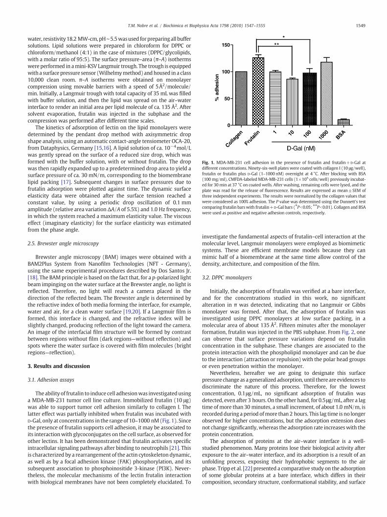
Fig. 1. MDA-MB-231 cell adhesion in the presence of frutalin and frutalin+D-Gal atdifferent concentrations. Ninety-six-well plates were coated with collagen I (10 μg/well),frutalin or frutalin plus D-Gal (1–1000 nM) overnight at 4 °C. After blocking with BSA(100 mg/ml), CMFDA-labeled MDA-MB-231 cells (1×10⁵ cells/well) previously incubat-ed for 30 min at 37 °C on coated wells. After washing, remaining cells were lysed, and theplate was read for the release of fluorescence. Results are expressed as mean±SEM ofthree independent experiments. The results were normalized by the collagen values thatwere considered as 100% adhesion. The P value was determined using the Dunnett's testcomparing frutalin bars with frutalin+D-Gal bars (⁎Pb0.05; ⁎⁎Pb0.01). Collagen and BSAwere used as positive and negative adhesion controls, respectively.
1549T.M. Nobre et al. / Biochimica et Biophysica Acta 1798 (2010) 1547–1555
water, resistivity18.2 MW-cm, pH∼5.5wasused forpreparingall buffersolutions. Lipid solutions were prepared in chloroform for DPPC orchloroform/methanol (4:1) in the case of mixtures (DPPC/glycolipids,with a molar ratio of 95:5). The surface pressure–area (π–A) isothermswere performed in amini-KSV Langmuir trough. The trough is equippedwith a surface pressure sensor (Wilhelmymethod)andhoused in a class10,000 clean room. π–A isotherms were obtained on monolayercompression using movable barriers with a speed of 5Å2/molecule/min. Initially, a Langmuir trough with total capacity of 35 mL was filledwith buffer solution, and then the lipid was spread on the air–waterinterface to render an initial area per lipid molecule of ca. 135 Å2. Aftersolvent evaporation, frutalin was injected in the subphase and thecompression was performed after different time scales.
The kinetics of adsorption of lectin on the lipid monolayers weredetermined by the pendant drop method with axisymmetric dropshape analysis, using an automatic contact-angle tensiometer OCA-20,from Dataphysics, Germany [15,16]. A lipid solution of ca. 10−4mol/Lwas gently spread on the surface of a reduced size drop, which wasformed with the buffer solution, with or without frutalin. The dropwas then rapidly expanded up to a predetermined drop area to yield asurface pressure of ca. 30 mN/m, corresponding to the biomembranelipid packing [17]. Subsequent changes in surface pressures due tofrutalin adsorption were plotted against time. The dynamic surfaceelasticity data were obtained after the surface tension reached aconstant value, by using a periodic drop oscillation of 0.1 mmamplitude (relative area variationΔA/A of 5.5%) and 1.0 Hz frequency,in which the system reached a maximum elasticity value. The viscouseffect (imaginary elasticity) for the surface elasticity was estimatedfrom the phase angle.
2.5. Brewster angle microscopy
Brewster angle microscopy (BAM) images were obtained with aBAM2Plus System from Nanofilm Technologies (NFT - Germany),using the same experimental procedures described by Dos Santos Jr.[18]. The BAMprinciple is based on the fact that, for a p-polarized lightbeam impinging on the water surface at the Brewster angle, no light isreflected. Therefore, no light will reach a camera placed in thedirection of the reflected beam. The Brewster angle is determined bythe refractive index of both media forming the interface, for example,water and air, for a clean water surface [19,20]. If a Langmuir film isformed, this interface is changed, and the refractive index will beslightly changed, producing reflection of the light toward the camera.An image of the interfacial film structure will be formed by contrastbetween regions without film (dark regions—without reflection) andspots where the water surface is covered with film molecules (brightregions—reflection).
3. Results and discussion
3.1. Adhesion assays
The ability of frutalin to induce cell adhesionwas investigatedusinga MDA-MB-231 tumor cell line culture. Immobilized frutalin (10 μg)was able to support tumor cell adhesion similarly to collagen I. Thelatter effect was partially inhibited when frutalin was incubated withD-Gal, only at concentrations in the range of 10–1000 nM(Fig. 1). Sincethe presence of frutalin supports cell adhesion, it may be associated toits interactionwith glycoconjugates on the cell surface, as observed forother lectins. It has been demonstrated that frutalin activates specificintracellular signaling pathways after binding to neutrophils [21]. Thisis characterized by a rearrangement of the actin cytoskeleton dynamic,as well as by a focal adhesion kinase (FAK) phosphorylation, and itssubsequent association to phosphoinositide 3-kinase (PI3K). Never-theless, the molecular mechanisms of the lectin frutalin interactionwith biological membranes have not been completely elucidated. To
investigate the fundamental aspects of frutalin–cell interaction at themolecular level, Langmuir monolayers were employed as biomimeticsystems. These are efficient membrane models because they canmimic half of a biomembrane at the same time allow control of thedensity, architecture, and composition of the film.
3.2. DPPC monolayers
Initially, the adsorption of frutalin was verified at a bare interface,and for the concentrations studied in this work, no significantalteration in π was detected, indicating that no Langmuir or Gibbsmonolayer was formed. After that, the adsorption of frutalin wasinvestigated using DPPC monolayers at low surface packing, in amolecular area of about 135 Å2. Fifteen minutes after the monolayerformation, frutalin was injected in the PBS subphase. From Fig. 2, onecan observe that surface pressure variations depend on frutalinconcentration in the subphase. These changes are associated to theprotein interaction with the phospholipid monolayer and can be dueto the interaction (attraction or repulsion) with the polar head groupsor even penetration within the monolayer.
Nevertheless, hereafter we are going to designate this surfacepressure change as a generalized adsorption, until there are evidences todiscriminate the nature of this process. Therefore, for the lowestconcentration, 0.1μg/mL, no significant adsorption of frutalin wasdetected, even after 3 hours. On the other hand, for 0.5μg/mL, after a lagtime of more than 30 minutes, a small increment, of about 1.0 mN/m, isrecorded during a period ofmore than 2 hours. This lag time is no longerobserved for higher concentrations, but the adsorption extension doesnot change significantly, whereas the adsorption rate increaseswith theprotein concentration.
The adsorption of proteins at the air–water interface is a well-studied phenomenon. Many proteins lose their biological activity afterexposure to the air–water interface, and its adsorption is a result of anunfolding process, exposing their hydrophobic segments to the airphase. Tripp et al. [22] presented a comparative study on the adsorptionof some globular proteins at a bare interface, which differs in theircomposition, secondary structure, conformational stability, and surface

Fig. 2. Adsorption kinetics for frutalin over DPPC monolayers at 135Å2/mol at proteinconcentrations of (■) 0.1, (▲) 0.5, (●) 1.0, and (▼) 2.0μg/mL.
1550 T.M. Nobre et al. / Biochimica et Biophysica Acta 1798 (2010) 1547–1555
distribution of amino acid residues. The authors verified that someproteins, including superoxide dismutase and bovine ribonuclease A,presented a lag time of few minutes before being adsorbed at theinterface. On the other hand, proteins like bovine serumalbumin,whichpresent a low effective surface hydrophobicity, rapidly adsorb at theinterface. Later on, other studies attested this proposition throughspectroscopic methods such as infrared spectroscopy, which showedthat the adsorption of some globular proteins at the air–water interfaceis a result of their partial unfolding [23,24]. Conversely, the samespectroscopic data indicated that some proteins, like horseradishperoxidase, were not denatured when adsorbed on a preformed, low-pressure lipidic monolayer. In this way, analyzing the adsorptionkinetics of frutalin on DPPCmonolayers, the slight increment in surfacepressure, is being initially attributed to a protein–DPPC interaction andnot to some loss of secondary structure of the protein.
Surface pressure versusmolecular area curves for DPPCmonolayerswere performed at different concentrations of frutalin in the subphase.The curves were recorded 30 minutes after protein injection to allowprotein adsorption. Fig. 3 displays an expansion of themonolayer witha small shift of the curve to higher molecular area values. Thisexpansion is dependent on protein concentration, in the same way itwas verified for the adsorption kinetics of frutalin at DPPCmonolayersat low lipid packing (Fig. 2). Taking into account that lectin is veryhydrophilic and stable in solution, the results of kinetics and surfacepressure curves should not be related to the protein unfolding. Oneshould also be aware that the head group of the phospholipid is a
Fig. 3. Surface pressure isotherms forDPPC(■) inbuffer solution, (●) in frutalin0.1μg/mL,and (▲) in frutalin 0.5μg/mL, recorded 30 minutes after protein injection.
zwitterion at pH=5.0, having negative and positive charges comingfrom phosphate (pKa∼1.8) and amine (pKb∼11.0) groups, respec-tively. Since the pI for frutalin is 6.0, there canbe electrostatic repulsioninfluencing the π–A curves, which displayed no changes in the mainphase transition, indicating that the lipid is occupying a larger area butwith no other species present at the interface. It is likely that theincrease in surface pressure observed in the adsorption kinetics plots isalso due to electrostatic repulsion, since the expansion of themonolayer can be interpreted as an increase in the surface pressurevalues for a constant area value. Similar results were formerlyobserved for the enzyme alcohol dehydrogenase (ADH) interactingwith DMPA monolayers, in which electrostatic repulsion delays ADHpenetration in the initial steps, since the enzyme and the phospholipidare both negatively charged [25].
Since frutalin exhibits specificity for sugar, it is expected that thestability of the protein in solution increases by the presence of D-Gal. Acomparison of the adsorption of frutalin on DPPC monolayers in theabsence or presence of D-Gal, its binding sugar, was performed. Uponcomparison of the two curves presented in Fig. 4, one can note that thepresence of D-Gal in the solution promoted a decrease in the effectproduced on the surface pressure. Since the sugar does not interactwith the monolayer, frutalin is preferentially in the subphase,indicating that the lectin is more stable in solution.
3.3. Mixed DPPC/glycolipid monolayers
After investigating the surface activity of the lectin and the influenceof its sugar binding on the interaction with pure DPPC monolayers, weverified theeffect of theprotein onmixedDPPC/glycolipidsmonolayers,to mimic the sugar immobilized on the cell surface. A proportion of 5%was chosen according to the amount of glycolipids found inmammaliancell membranes [26]. As can be seen in Scheme 1, the three glycolipidsemployed have a galactose molecule as a polar head, but withdifferences in the electrically charged group and monosaccharidechain length. The latter allowed us to investigate both the influence ofthe position of galactose in the polar head, using the ganglioside GM1,and theeffect of chargedgroupspresent in the sugarusing theglycolipidSul. The results were compared with GalCer, which presents only agalactose group as polar head.
Initially, to explore the interaction of frutalin with mixed mono-layers, π–A isotherms were measured for mixed films of DPPC–5%GalCer in the absence and presence of frutalin. The curves are presentedin Fig. 5. The π–A isotherm for a DPPC–5% GalCer was more expandedcompared to pureDPPC, since theminimumarea permolecule, which is48 Å2 for pureDPPC, increases to ca. 62 Å2 upon addition of 5% of GalCer.Furthermore, the curve for this mixed film does not present a plateau
Fig. 4. Adsorption kinetics of frutalin 1.0μg/mL over DPPC monolayers with the initialsurface pressureof 0 mN/m, (●) frutalin solutionwithout D-galactose, and (■) 10μg/mLofD-galactose added to the frutalin solution.

1551T.M. Nobre et al. / Biochimica et Biophysica Acta 1798 (2010) 1547–1555
corresponding to the coexistence of liquid-expanded and liquid-condensed phases (LE–LC). Comparing the curves for a pure DPPC filmand for the mixed, we can infer that the mixture is nonideal, inaccordance with previous reports fromMaggio et al. [27,28].
The addition of frutalin (Fig. 5) caused different effects on thepacking of the monolayer. Initially, at higher area values, the lectinpromotes an expansion of the DPPC–5% GalCer mixedmonolayer. Thisbehavior cannot be compared with the effect of the lectin over DPPCmonolayers. In the case of this mixed monolayer (DPPC–5% GalCer),we can observe that the shape of the curve is different in the presenceof frutalin. First, the π–A isotherm does not begin at zero surfacepressure because frutalin presents an induced surface activity, even atlow surface packing. This adsorption resulted in a shift of the isothermto higher molecular areas (expansion) until the surface pressure of ca.25 mN/m is reached, indicating the incorporation of frutalin into themonolayer at the buffer–lipid interface. Second, we can discuss thebehavior of the ca. 25 mN/m surface pressure. Interestingly, aninversion in the initial behavior is noted, resulting in a shift of thecurve to lower-molecular area values. Since this condensationprovokes lower molecular area values than that obtained for a purelipid monolayer, we can speculate about the expulsion of the proteinfrom the interface. However, this ejection seems not to be the onlyphenomena occurring. Some mixed systems reported in literatureresulted in a condensation of the lipid monolayer. Yin and Chang [29]investigated the interfacial behavior of DPPC Langmuir monolayerswith fibrinogen, one of the major plasma proteins, by using infraredreflection–absorption spectroscopy. The authors observed that, forthe mixed DPPC/fibrinogen layer at the interface, hysteresis curvessuggested that the fibrinogen molecules were expelled from theinterface upon compression, apparently because of the presence ofinsoluble DPPC molecules. Furthermore, the desorption of fibrinogenfrom the interface, at high surface pressure values, removed apronounced amount of DPPC. Upon a subsequent expansion, areadsorption of fibrinogen is noted. The expulsion of some moleculesfrom the interface was noted even for insoluble mixed films, such asthe antifungal amphotericin B (AmB) with the lipid dipalmitoylphosphatidyl serine (DPPS). The authors investigated the collapsedregion and noticed the existence of two independent collapses: thefirst correspondent to the ejection of the AmB molecules from thesurface, and the other one to the expulsion of the phospholipid [30].On the other hand, Maniti et al. [31] recently investigated theinteraction of mitochondrial creatine kinase (mtCK) with differentacyl chain length phosphatidylglycerol in Langmuir monolayers andobserved that both surface pressure and compressional modulusshowed a condensation effect for some lipids tested, indicating theformation of a liquid-condensed phase, promoted by the protein. For
Fig. 5. Surface pressure versus area isotherms for a DPPC–5%GalCer Langmuir film formedover a pure PBS buffer subphase (●) and over a subphase of PBS buffer containing 0.1μg/mL of frutalin (■).
our system, we believe that the effects discussed by Maniti et al. arebetter applied, since just a single collapse was observed in ourisotherms.
Fig. 6 presents the π–A isotherms for the mixture of Sul and GM1(in a 5% proportion) with DPPC. The idea was to compare the effect ofthese lipids with the GalCer behavior to investigate parameters as thecharge and the position of the D-Gal immobilized at the interface. Forthe DPPC–5% Sul mixedmonolayer, the LE–LC phase transition is moreevident than that observed in the curve for the DPPC–5% GalCermonolayer, shown in Fig. 5, being more similar to those obtained forpure DPPC. As it can be noted, the addition of Sul shifted the DPPC π–Acurve to lowermolecular areas. This behavior was expected and this isin agreement with similar results obtained by Sun et al. [32], whostudied different Sul/DPPC molar ratios (from 0.2 to 1.0 of Sul) andobserved that by increasing the Sul molar ratio, the plateaucorrespondent to the LE–LC coexistence is shifted to lower molecularareas and higher surface pressures. The authors have also investigatedthe thermodynamics of the mixture and concluded that the system ismiscible and stable, and the negative deviations from the idealitysuggested attractive interactions between the molecules, resulting inthe condensation of the film.
The π–A isotherm recorded for the mixed DPPC–5% GM1 filmindicates that the addition of the ganglioside affects the monolayer inaway that the LE–LC coexistence region is extinct. Furthermore, in ourcase, 5% of GM1 expands the monolayer, since the minimum areashifted from 48 to 62 Å2. It has been reported in the literature that theeffect of GM1 over DPPC monolayers is dependent on the molarfraction between the lipids. Ohta et al. [33] observed that, at pH 1.2,for GM1 values of up to 0.25, a condensation of the monolayer,compared with pure DPPC, occurs. However, above 0.5 of GM1 molarfraction, the monolayer seems to be more expanded.
For both systems (mixed DPPC–5% Sul or GM1), the effect of lectinaddition is similar to DPPC–5% GalCer, in which the condensation ofthe monolayer was observed with the addition of the lectin at highsurface pressures. For the DPPCmixedmonolayer with Sul, we noticedthat besides this condensation, the presence of frutalin induces a less-evident coexistence of the LE–LC region. Interestingly, for themixtures of DPPC with Sul and GM1, the inversion point at ca.25 mN/m is not observed. This means that, for both expanded andcompact films, frutalin is present at the interface. Hence, we canspeculate that the physical immersion of the protein in the lipidmonolayer is deeper, and/or the binding with the galactose unit isstronger, which avoids frutalin expulsion.
For mixed DPPC–Sul and DPPC–GM1 monolayers the change inmolecular areas can be analyzed at the pressures of 10 and 30 mN/m,representing fluid and compact stages, respectively. At the lower
Fig. 6. Surface pressure versus area isotherms for DPPC–5% Sul (●, ○) and DPPC–5%GM1 (■, □) Langmuir monolayers formed over subphases of pure PBS buffer (○, □),and PBS buffer with 0.1μg/mL frutalin dissolved (●, ■).

1552 T.M. Nobre et al. / Biochimica et Biophysica Acta 1798 (2010) 1547–1555
pressure, the area changed from ca. 90.7 Å2 to 83.8 Å2 for the Sul-containing film, and from 84.9 Å2 to 77.3 Å2 for the GM1-containingmonolayer. In both cases, the shift promoted by the presence offrutalin was ca. 7 Å2. However, at 30 mN/m, the displacement ofmolecular areas for the isotherms of monolayers containing Sul occursfrom 56.2 Å2 to 43.3 Å2, and for GM1 system, from 54.6 Å2 to 39.6 Å2,almost two times higher than the values obtained for the fluid phase.
Fig. 7 shows the results of the investigation of the interactionbetween frutalin and mixed monolayers, specifically at a surfacepressure of 30 mN/m. This surface pressure is correspondent to thatfound for a lipidic packing of a natural biomembrane [17]. Adsorptionkinetics was followed by changes in surface pressure with time at aconstant area, using the pendant drop technique, with the axisym-metric drop shape analysis. Upon comparing the timescales of Figs. 4and 7, one may observe that adsorption of frutalin, which takes morethan 1 hour for expanded films, takes place in a few minutes for amonolayer in the condensed state. Moreover, the extension inwhich πhas changed, interpreted as the protein adsorption, is dependent onthe monolayer constitution. The interaction of frutalin promoted aless pronounced increase in surface pressure for a pure DPPCmonolayer, in comparison to monolayers containing glycolipids. Theeffects of frutalin were more evident for the DPPC–5% GalCer system,in which equilibrium was not reached even after 12 minutes, at asurface pressure above 34 mN/m (or ΔπN4 mN/m).
Comparing the adsorption kinetics for frutalin in the three systemscontaining glycoconjugates, at a surface pressure of 30 mN/m, weobserved that only in the case of DPPC–5% GalCer that the curveincreased monotonically, whereas for the other mixtures, theadsorption seems to occur in two steps. However, even for DPPC–GM1 and DPPC–Sul systems, it is possible to perceive that the finalsurface pressure values are different, indicating differences in theirbehavior. Based on the results in Fig. 7, some points should behighlighted: (i) The adsorption of frutalin to DPPC–5% GalCer in asingle step can be attributed to an easiness of the lectin to interactwith the D-Gal group at the interface, since GalCer presents only thismonosaccharide as the polar head. (ii) For DPPC–GM1 and DPPC–Sul,the initial decrease in the surface pressure can be related to apreliminary orientation change of the hydrophobic tails to betteraccommodate the protein molecules. In the case of DPPC–5% GM1,these changes in orientation may be related to the position of the D-Gal in the polar head, since in this glycolipid, the sugar is in a fartherposition from the interface. We can speculate that, for the interactionof frutalin with GM1, the protein does not reach the surface, occurringin such way that the interface is less affected. Such events are notreadily detected in the surface pressure measurements, since theyrequire a lag time to cause changes in the alkyl lipid chain. (iii) For themixed system containing Sul, the initial lag timewas also attributed to
Fig. 7. Adsorption kinetics of frutalin 0.1μg/mL over Langmuir monolayers of (black line)DPPC, (red line)DPPC–5%GalCer, (green line)DPPC–5%Sul, and (blue line)DPPC–5%GM1.
a rearrangement of the lipid monolayer. However, in this case, thechange was promoted by an initial repulsion between the negativecharges from the sulfate groups of the lipid and frutalin. Theconsequent alteration in the lipid packing probably facilitates theinteraction of the protein with the sugar in the glycolipid. Thisbehavior was previously observed for NCAP, an alkaline phosphatasefrom the filamentous fungus Neurospora crassa [34].
3.4. Dilatational surface elasticity
After the system reached the adsorption equilibrium (Fig. 7), thedrop was submitted to a sinusoidal compression–expansion stress todetermine the dilatational surface elasticity of the film. This parameteris very important to verify how frutalin can affect the mechanicalresistance of themonolayer and the fluidity of the film. The results arepresented in Table 1.
In our case, E values are related to the effect of the protein on thestructure of the lipid monolayer, and how its presence can affect thefluidity of themembrane model [35–37]. When E values are higher forthe pure lipid monolayer than in the presence of the protein, we inferthat the presence of the protein affects the lipid packing, turning itmore fluid and indicating that some interactions occurring in theprotein–lipid system are not preserved after the mechanical defor-mation imposed to the surface by the applied frequency. This behaviorindicates a viscoelastic system, and is associated with a phenomena,such as (i) conformational changes of the macromolecule; (ii)orientational changes, mainly of the lipids hydrophobic tails, afterdeformation; (iii) loss of the monolayer for the solution. In all threecases, we consider that the components of the monolayer, in this caselipid and protein, are not interacting in an efficient way, since theinteractions were lost after disturbance. On the other hand, when Evalues obtained in the presence of the protein were higher than thosefor the pure monolayer, we characterize the system in which theinteraction or incorporation of the protein contributes as a morestructured monolayer. In several cases involving proteins and lipids,the protein interacts not only with the lipid polar head but also withthe hydrophobic tails [35], causing the monolayer to be more orientedand compacted. Comparing E values for the pure DPPCmonolayer andthose obtained in the presence of frutalin, one realizes that frutalinreduces the surface elasticity, making the film more fluid. Consideringthis result together with those from adsorption kinetics, one sees thatthe interpretation in terms of slight repulsion of the DPPC moleculesand protein at the film makes sense. In this way, the decrease in Evalues attested that there is no attractive interaction between thelectin and the lipidmonolayer, whichwas also the reason for the smallchange in surface pressure caused by frutalin adsorption (Fig. 2).
The presence of frutalin at the subphase of the mixed monolayersresulted in an increase of E values, indicating a different interactionwiththe lipid monolayer in the presence of glycolipids. Interestingly, theeffect of the lectin is almost the same for these three glycolipids, sincewe can see in Table 1 that frutalin almost duplicates the E value ascompared with the E measured for pure monolayers (absence offrutalin), respectively. This finding is much likely related to the fact thatthe interaction of the lectin with the sugar groups occurs in more thanone step and the position of the sugar groups affects only the initial stepinducing changes in the adsorption kinetics. Since this kind of
Table 1Dilatational surface elasticity values for the studied monolayers formed over a bufferedsubphase in the absence and presence of frutalin.
Film components E in PBS (mN/m) E in frutalin 0.1μg/mL (mN/m)
DPPC 210.9 121.3DPPC–5% GalCer 141.9 280.6DPPC–5% Sul 110.3 223.2DPPC–5% GM1 120.9 234.2
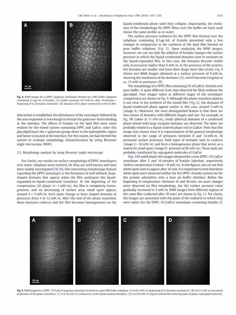
Fig. 8. BAM images for a DPPC Langmuir monolayer formed on a PBS buffer subphasecontaining 0.1μg/mL of frutalin: (A) surface pressure of 9 mN/m—after 30 minutes—beginning of LC domains nucleation; (B) domains of LC phase immersed on the LE film.
1553T.M. Nobre et al. / Biochimica et Biophysica Acta 1798 (2010) 1547–1555
interaction is established, the disturbance of themonolayer followed bythe area expansion is not enough to disrupt the galactose–lectin bindingat the interface. The effects of frutalin on the lipid film were moreevident for the mixed system containing DPPC and GalCer, since thisglycolipid bears the D-galactose group closer to the hydrophobic regionand hence is located at the interface. For this reason,we had elected thissystem to evaluate morphology characterization by using Brewsterangle microscopy (BAM).
3.5. Morphology analysis by using Brewster angle microscopy
For clarity, our results on surface morphology of DPPC monolayersover water subphase were omitted, for they are well known and havebeenwidely investigated [38,39]. One interestingmorphologic featureregarding the DPPC monolayer is the formation of well defined, bean-shaped domains that appear when the film undergoes the liquid-expanded-to-liquid-condensed transition. At the beginning of thecompression (LE phase; π∼1 mN/m), the film is completely homo-geneous, and on decreasing of surface area, small spots appear,around π=3mN/m. Such spots change to bean shaped domains atpressures from 5 to 12 mN/m. After the end of the phase transition,these domains coalesce and the film becomes homogeneous on the
Fig. 9. BAM images for aDPPC–5%GalCer Langmuirmonolayer formedonapurePBSbuffer subpof domains of the phase transition). (C) π of 16 mN/m (coalescence of the liquid-ordered doma
liquid-condensed phase until they collapse. Importantly, the evolu-tion of the morphology for DPPC films over the buffer we have usedshows the same profile as in water.
The surface pressure isotherm for the DPPC film formed over thesubphase containing 0.1μg/mL of frutalin presented only a fewchanges in comparison to the isotherm of the lipid film formed onpure buffer subphase (Fig. 3). Upon analyzing the BAM images,however, we can see that the addition of frutalin changes the surfacepressure in which the liquid-condensed domains start to nucleate onthe liquid-expanded film. In this case, the domains become visibleonly at pressures higher than 9 mN/m. In the presence of the protein,the domains are smaller and have their shape more like circles. Fig. 8shows two BAM images obtained at a surface pressure of 9 mN/m,showing the nucleation of the domains (A), which become irregular atca. 13 mN/m pressures (B).
Themorphology of a DPPC film containing 5% of GalCer, formed overpure buffer, is quite different from that observed for films without theglycolipid. Four images taken at different stages of the monolayercompression are shown in Fig. 9. Although the phase transition plateauis not clear in the isotherm of the mixed film (Fig. 6), the domains ofliquid-condensed phase appear earlier in this case, around 5 mN/m(image A). Moreover, the most distinguished feature is that there aretwo classes of domains with different shapes and size. For example, inFig. 9B (taken at 11 mN/m), small spherical domains of a condensedphase mixed with large irregular domains are observed. The latter areprobably related to a liquid-ordered phase rich in GalCer. Note that thisimage was chosen since it is representative of the general morphologyobserved in the range of pressures between 8 and 14 mN/m. Atincreased surface pressures, both types of domains start to coalesce(image C—16 mN/m) and form a homogeneous phase that serves as amatrix for small spots (imageD—pressure of 20 mN/m). These spots areprobably constituted by segregated molecules of GalCer.
Figs. 10AandBdepict the images obtained for anewDPPC–5%GalCermonolayer after 5 and 16 minutes of frutalin injection, respectively(before compression π initial=0 mN/m). In the figures,we can see thatwhite spots start to appear after 16 min. It is important to note that thesewhite spots were observed neither for the DPPC–frutalin system nor forthe protein adsorption onto a bare air–buffer interface. Before thebeginning of compression—between 16 and 30 min—no more changeswere observed on film morphology, but the surface pressure valuegradually increased to 5 mN/m. BAM images from different regions ofthe same film (collected after 30 min) are shown in Fig. 11. For clarity,the images are associated with the point of the isotherm in which theywere taken. For the DPPC–5% GalCer monolayer containing frutalin, LC
hase. (A)π of 5 mN/m(beginningof LCdomains nucleation). (B)πof 11 mN/m(twokindsins). (D) π of 20 mN/m (liquid-ordered film showing spots of phase-segregatedmaterial).
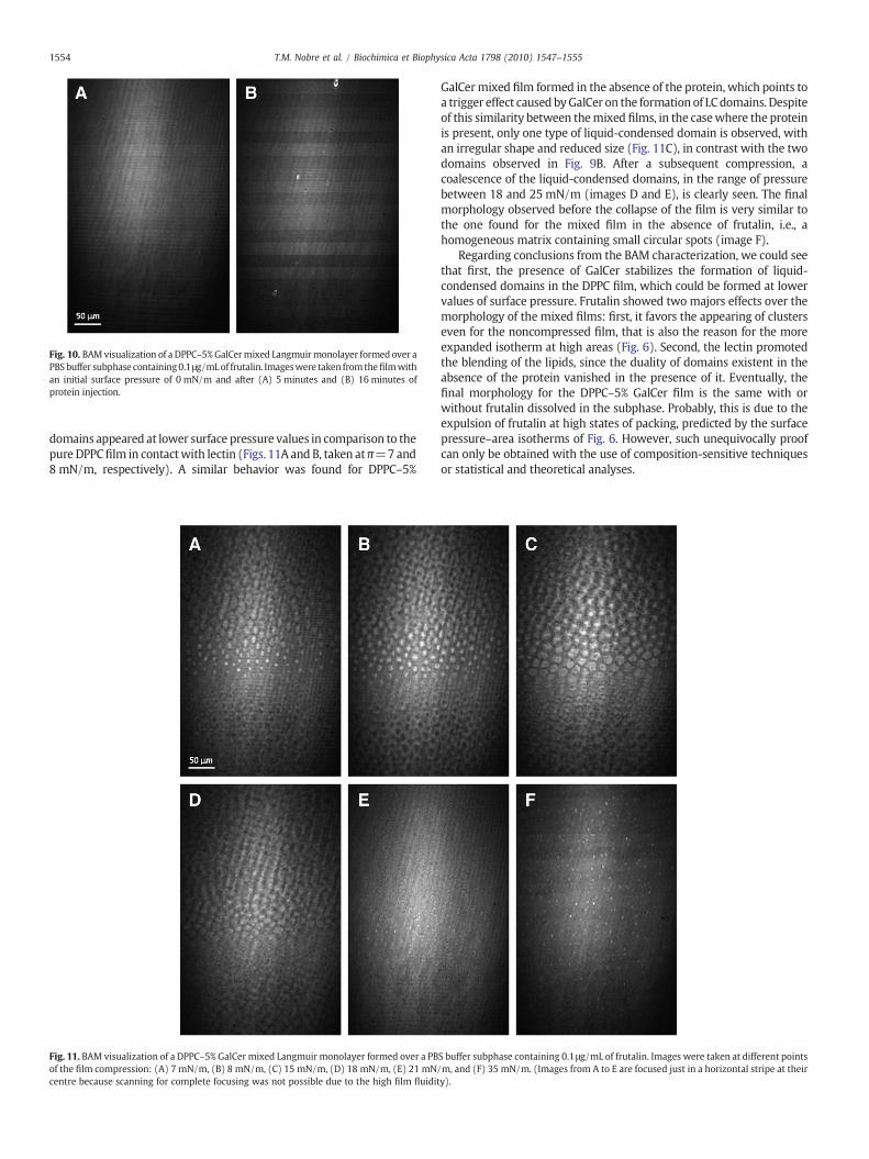
Fig. 10. BAMvisualization of a DPPC–5%GalCermixed Langmuirmonolayer formed over aPBSbuffer subphase containing0.1μg/mLof frutalin. Imageswere taken fromthefilmwithan initial surface pressure of 0 mN/m and after (A) 5 minutes and (B) 16 minutes ofprotein injection.
1554 T.M. Nobre et al. / Biochimica et Biophysica Acta 1798 (2010) 1547–1555
domains appeared at lower surface pressure values in comparison to thepure DPPCfilm in contactwith lectin (Figs. 11A and B, taken at π=7and8 mN/m, respectively). A similar behavior was found for DPPC–5%
Fig. 11. BAM visualization of a DPPC–5% GalCer mixed Langmuir monolayer formed over a PBof the film compression: (A) 7 mN/m, (B) 8 mN/m, (C) 15 mN/m, (D) 18 mN/m, (E) 21 mNcentre because scanning for complete focusing was not possible due to the high film fluidit
GalCermixed film formed in the absence of the protein, which points toa trigger effect caused byGalCer on the formation of LCdomains. Despiteof this similarity between themixed films, in the casewhere the proteinis present, only one type of liquid-condensed domain is observed, withan irregular shape and reduced size (Fig. 11C), in contrast with the twodomains observed in Fig. 9B. After a subsequent compression, acoalescence of the liquid-condensed domains, in the range of pressurebetween 18 and 25mN/m (images D and E), is clearly seen. The finalmorphology observed before the collapse of the film is very similar tothe one found for the mixed film in the absence of frutalin, i.e., ahomogeneous matrix containing small circular spots (image F).
Regarding conclusions from the BAM characterization, we could seethat first, the presence of GalCer stabilizes the formation of liquid-condensed domains in the DPPC film, which could be formed at lowervalues of surface pressure. Frutalin showed two majors effects over themorphology of the mixed films: first, it favors the appearing of clusterseven for the noncompressed film, that is also the reason for the moreexpanded isotherm at high areas (Fig. 6). Second, the lectin promotedthe blending of the lipids, since the duality of domains existent in theabsence of the protein vanished in the presence of it. Eventually, thefinal morphology for the DPPC–5% GalCer film is the same with orwithout frutalin dissolved in the subphase. Probably, this is due to theexpulsion of frutalin at high states of packing, predicted by the surfacepressure–area isotherms of Fig. 6. However, such unequivocally proofcan only be obtained with the use of composition-sensitive techniquesor statistical and theoretical analyses.
S buffer subphase containing 0.1μg/mL of frutalin. Images were taken at different points/m, and (F) 35 mN/m. (Images from A to E are focused just in a horizontal stripe at theiry).
1555T.M. Nobre et al. / Biochimica et Biophysica Acta 1798 (2010) 1547–1555
4. Conclusions
We demonstrated that frutalin can interact with Langmuir mono-layers; however, its incorporation to the film is observed only in thepresence of glycolipids containing D-Gal. Adsorption kinetics anddilatational surface elasticity data indicated that the position of thegalactose and the presence of charged groups affected the initial steps ofthe frutalin interaction, as noticed for GM1 and Sul-containing mono-layers, with a first decrease in the surface pressure. BAM images showedthat frutalin can induce the formation of domains even for thenoncompressed DPPC–GalCer film. Probably, these domains could be“raft-like,” but this is currently under investigation. The observation ofthese microdomains can have important consequences in membraneorganization, since they can represent clustering of possible receptors orevenamechanismof cell internalization,whichremains tobeelucidated.
Since frutalin binds to the cell surface, as demonstrated here forMDA-MB-231 cells, and activates intracellular signaling pathways inneutrophils, the results presented here add new evidences for themolecular mechanism of membrane interaction with lectins. There-fore, the biotechnological applications of frutalin can be expanded to,for example, incorporating such proteins in nanoparticles for targeteddrug delivery.
Acknowledgments
The authors are grateful for the financial support of the Brazilianagencies FAPESP and CNPq.
References
[1] N. Sharon, Lectins:past, presentand future, Biochem.Soc. Trans. 36 (2008)1457–1460.[2] T. Feizi, Demonstration by monoclonal antibodies that carbohydrate structures of
glycoproteins and glycolipids are onco-developmental antigens, Nature 314 (1985)53–57.
[3] S. Hakomori, Aberrant glycosylation in cancer cell membranes as focused onglycolipids overview and perspectives, Cancer Res. 45 (1985) 2405–2414.
[4] Y.J. Kim, A. Varki, Perspective on the significance of altered glycosylation ofglycoproteins in cancer, Glycoconjugate J. 14 (1997) 569–576.
[5] S. Hakomori, Glycosylation defining cancer malignancy: new wine in an oldbottle, Proc. Natl. Acad. Sci. USA 99 (16) (2002) 10231–10233.
[6] M. Burger, A.R. Goldberg, Identification of a tumor-specific determinant ofneoplastic cell surfaces, Proc. Natl. Acad. Sci. USA 57 (1967) 359–366.
[7] M. Inbar, L. Sachs, Interaction of the carbohydrate-binding protein concanavalin Awith normal and transformed cells, Proc. Natl. Acad. Sci. USA 63 (1969) 1418–1425.
[8] B.A. Sela, H. Lis, N. Sharon, L. Sachs, Different locations of carbohydrate-containingsites at the surface membrane of normal and transformed mammalian cells, J.Membr. Biol. 3 (1970) 267–279.
[9] N. Malagolini, M. Chiricolo, M. Marini, F. Dall'Olio, Exposure of α2, 6-sialylatedlactosaminic chains marks apoptotic and necrotic death in different cell types,Glycobiology 19 (2) (2009) 172–181.
[10] S. Taube, J.W. Perry, K. Yetming, S.P. Patel, H. Auble, L. Shu, H.F. Nawar, C.H. Lee, T.D.Connell, J.A. Shayman, C.E. Wobus, Ganglioside-linked terminal sialic acid moietiesonmurine macrophages function as attachment receptors for murine noroviruses, J.Virology 83 (9) (2009) 4092–4101.
[11] R. Sankaranarayanan, K. Sekar, R. Banerjee, V. Sharma, A. Surolia, M. Vijayan, Anovel mode of carbohydrate recognition in jacalin, a Moraceae plant lectin with abeta-prism fold, Nature Struct. Biol. 3 (1996) 596–603.
[12] R.A. Moreira, C.C. Castelo-Branco, A.C.O. Monteiro, R.O. Tavares, L.M. Beltramini,Isolation and partial characterization of a lectin from Artocarpus incisa L. seeds,Phytochemistry 46 (1) (1998) 139–144.
[13] M.M. Bradford, A rapid and sensitive method for the quantitation of microgramquantities of protein using the principle of protein–dye binding, Anal. Biochem.72 (1976) 248–254.
[14] D.H.F. Souza, M.R.C. Iemma, L.L. Ferreira, J.P. Faria, M.L.V. Oliva, R.B. Zingali, S.Niewiarowski, H.S. Selistre de Araújo, The disintegrin-like domain of the snake
venom metalloprotease alternagin inhibits alpha 2-beta-1-integrin-mediated celladhesion, Arch. Biochem. Biophys. 384 (2000) 341–350.
[15] L. Caseli, D.C. Masui, R.P.M. Furriel, F.A. Leone, M.E.D. Zaniquelli, Adsorptionkinetics and dilatational rheological studies for the soluble and anchored forms ofalkaline phosphatase at the air/water interface, J. Braz. Chem. Soc. 16 (5) (2005)969–977.
[16] T.M. Nobre, K. Wong, M.E.D. Zaniquelli, Equilibrium and dynamic aspects ofdodecyltrimethylammonium bromide adsorption at the air/water interface in thepresence of lambda-carrageenan, J. Coll. Interf. Sci. 305 (1) (2007) 142–149.
[17] D. Marsh, Lateral pressure in membranes, Biochim. Biophys. Acta. 1286 (1996)183–223.
[18] D.S. Dos Santos Jr, F.J. Pavinatto, D.T. Balogh, L. Misoguti, O.N. Oliveira Jr, C.R.Mendonça, In situ UV–VIS. Absorbance measurements for Langmuir films of thepoly[4-[2-(methacryloyloxy)-ethyl]ethylamino]-2-chloro-4-nitroazobenzene](HPDR13) azopolymer, J. Coll. Interf. Sci. 276 (2004) 138–142.
[19] D. Höning, D.J. Möbius, Direct visualization of monolayers at the air–waterinterface by Brewster angle microscopy, J. Phys. Chem. 95 (1991) 4590–4592.
[20] S. Hénon, J. Meunier, Microscope at the Brewster angle: direct observation of first-order phase transitions in monolayers, Rev. Sci. Instrum. 62 (1991) 936–939.
[21] A.C. Brando-Lima, R.F. Saldanha-Gama,M.G.M.O.Henriques, A.C.O.Monteiro-Moreira,,R.A. Moreira, C. Barja-Fidalgo, Frutalin, a galactose-binding lectin, induces chemotaxisand rearrangement of actin cytoskeleton in human neutrophils: involvement oftyrosine kinase and phosphoinositide 3-kinase, Toxicol. Appl. Pharm. 208 (2005)145–154.
[22] B.C. Tripp, J.J. Magda, J.D. Andrade, Adsorption of globular–proteins at the air/waterinterface as measured via dynamic surface-tension—concentration-dependence,mass-transfer considerations, and adsorption-kinetics, J. Coll. Interf. Sci. 173 (1)(1995) 16–27.
[23] T.F. Schmidt, L. Caseli, T. Viitala, O.N. Oliveira Jr., Enhanced activity of horseradishperoxidase in Langmuir–Blodgett films of phospholipids, Biochim. Biophys. Acta:Biomembranes 1778 (10) (2008) 2291–2297.
[24] I. Estrela-Lopis, G. Brezesinski, H. Mohwald, Influence of model membrane structureon phospholipase D activity, Phys. Chem. Chem. Phys. 2 (2000) 4600–4604.
[25] L. Caseli, A.C. Perinotto, T. Viitala, V. Zucolotto, O.N. Oliveira Jr, Immobilization ofalcohol dehydrogenase in phospholipid Langmuir–Blodgett films to detectethanol, Langmuir 25 (5) (2009) 3057–3061.
[26] P.L. Yeagle, The Structure of Biological Membranes, CRC Press, Boca Raton, 1991.[27] B. Maggio, Favorable and unfavorable lateral interactions of ceramide, neutral
glycosphingolipids and gangliosides in mixed monolayers, Chem. Phys. Lipids132 (2004) 209–224.
[28] B. Maggio, D.C. Carrer, M.L. Fanani, R.G. Oliveira, C.M. Rosetti, Interfacial behaviorof glycosphingolipids and chemically related sphingolipids, Curr. Opin. Coll. Interf.8 (2004) 448–458.
[29] C.-L. Yin, C.-H. Chang, Infrared spectroscopy analysis of mixed DPPC/fibrinogenlayer behavior at the air/liquid interface under a continuous compression–expansion condition, Langmuir 22 (2006) 6629–6634.
[30] J. Miñones Jr., J. Miñones, J.M. Rodríguez-Patino, O. Conde, E. Iribarnegaray,Miscibility of amphotericin B-dipalmitoyl phosphatidyl serine mixed monolayersspread on the air/water interface, J. Phys. Chem. B 107 (2003) 4189–4195.
[31] O. Maniti, M. Cheniour, O. Marcillat, C. Vial, T. Granjon, Morphology modificationsin negatively charged lipid monolayers upon mitochondrial creatine kinasebinding, Mol. Membr. Biol. 26 (3) (2009) 171–185.
[32] R. Sun, C. Hao, J. Zhang, Y. Chang, C. Niu, A monolayer study on phase behavior andmorphology of binarymixtures of sulfatideswith DPPC andDPPE, Coll. Surf. B 73 (2)(2009) 161–167.
[33] Y. Ohta, S. Yokoyama, H. Sakai, M. Abe, Membrane properties of mixed gangliosideGM1/phosphatidylcholine monolayers, Coll. Surf. B 33 (3–4) (2004) 191–197.
[34] T.M. Nobre, H.S. Silva, R.P.M. Furriel, F.A. Leone, P.B. Miranda, M.E.D. Zaniquelli, Amolecular viewof the interaction between iota-carrageenan and a phospholipidfilmand its role in enzyme immobilization, J. Phys. Chem. B 113 (2009) 7491–7497.
[35] L. Caseli, M.L. Moraes, V. Zucolotto, M. Ferreira, T.M. Nobre, M.E.D. Zaniquelli, U.P.Rodrigues Filho, O.N. Oliveira Jr., Fabrication of phytic acid sensor based on mixedphytase–lipid Langmuir–Blodgett films, Langmuir 22 (2006) 8501–8508.
[36] L. Caseli, F.N. Crespilho, T.M. Nobre, M.E.D. Zaniquelli, V. Zucolotto, O.N. OliveiraJr., Using phospholipid Langmuir and Langmuir–Blodgett films as matrix forurease immobilization, J. Coll. Interf. Sci. 319 (2008) 100–108.
[37] T.F. Schmidt, L. Caseli, T.M. Nobre, M.E.D. Zaniquelli, O.N. Oliveira Jr., Interaction ofhorseradish peroxidase with Langmuir monolayers of phospholipids, Coll. Surf. A:Physicochem. Eng. Asp. 321 (2008) 206–210.
[38] C.W. McConlogue, T.K. Vanderlick, A close look at domain formation in DPPCmonolayers, Langmuir 13 (1997) 7158–7164.
[39] G.A. Lawrie, I.R. Gentle, G.T. Barnes, The structure of mixed monolayer filmsof DPPC and hexadecanol, Colloid Surf. A: Physicochem. Eng. Asp. 171 (2000)217–224.