Biochemistry 1.01 Cell Membrane
8
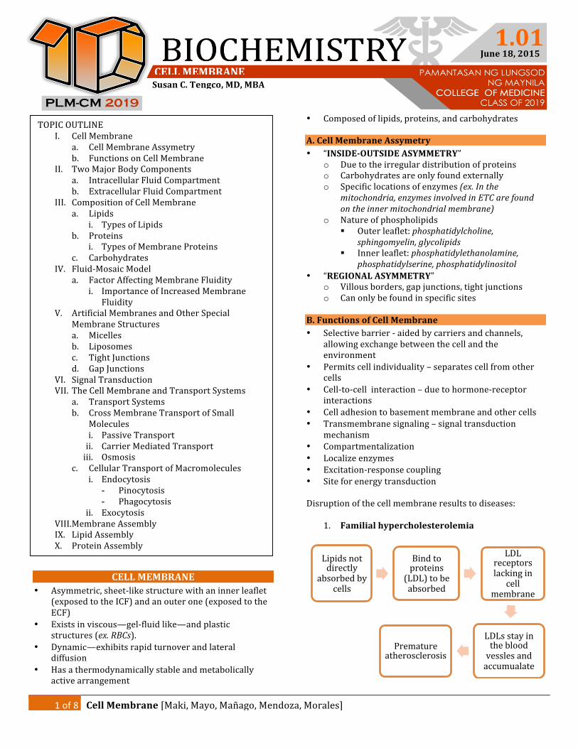
1 of 8 Cell Membrane [Maki, Mayo, Mañago, Mendoza, Morales] CELL MEMBRANE Susan C. Tengco, MD, MBA 1.01 June 18, 2015 CELL MEMBRANE • Asymmetric, sheetlike structure with an inner leaflet (exposed to the ICF) and an outer one (exposed to the ECF) • Exists in viscous—gelfluid like—and plastic structures (ex. RBCs). • Dynamic—exhibits rapid turnover and lateral diffusion • Has a thermodynamically stable and metabolically active arrangement • Composed of lipids, proteins, and carbohydrates A. Cell Membrane Assymetry • “INSIDEOUTSIDE ASYMMETRY” o Due to the irregular distribution of proteins o Carbohydrates are only found externally o Specific locations of enzymes (ex. In the mitochondria, enzymes involved in ETC are found on the inner mitochondrial membrane) o Nature of phospholipids Outer leaflet: phosphatidylcholine, sphingomyelin, glycolipids Inner leaflet: phosphatidylethanolamine, phosphatidylserine, phosphatidylinositol • “REGIONAL ASYMMETRY” o Villous borders, gap junctions, tight junctions o Can only be found in specific sites B. Functions of Cell Membrane • Selective barrier aided by carriers and channels, allowing exchange between the cell and the environment • Permits cell individuality – separates cell from other cells • Celltocell interaction – due to hormonereceptor interactions • Cell adhesion to basement membrane and other cells • Transmembrane signaling – signal transduction mechanism • Compartmentalization • Localize enzymes • Excitationresponse coupling • Site for energy transduction Disruption of the cell membrane results to diseases: 1. Familial hypercholesterolemia Lipids not directly absorbed by cells Bind to proteins (LDL) to be absorbed LDL receptors lacking in cell membrane LDLs stay in the blood vessles and accumualate Premature atherosclerosis TOPIC OUTLINE I. Cell Membrane a. Cell Membrane Assymetry b. Functions on Cell Membrane II. Two Major Body Components a. Intracellular Fluid Compartment b. Extracellular Fluid Compartment III. Composition of Cell Membrane a. Lipids i. Types of Lipids b. Proteins i. Types of Membrane Proteins c. Carbohydrates IV. FluidMosaic Model a. Factor Affecting Membrane Fluidity i. Importance of Increased Membrane Fluidity V. Artificial Membranes and Other Special Membrane Structures a. Micelles b. Liposomes c. Tight Junctions d. Gap Junctions VI. Signal Transduction VII. The Cell Membrane and Transport Systems a. Transport Systems b. Cross Membrane Transport of Small Molecules i. Passive Transport ii. Carrier Mediated Transport iii. Osmosis c. Cellular Transport of Macromolecules i. Endocytosis - Pinocytosis - Phagocytosis ii. Exocytosis VIII. Membrane Assembly IX. Lipid Assembly X. Protein Assembly
-
Upload
april-aram -
Category
Documents
-
view
226 -
download
2
description
trans
Transcript of Biochemistry 1.01 Cell Membrane
1 of 8 Cell Membrane [Maki, Mayo, Mañago, Mendoza, Morales]
CELL MEMBRANE Susan C. Tengco, MD, MBA
1.01 June 18, 2015
CELL MEMBRANE • Asymmetric, sheet-‐like structure with an inner leaflet
(exposed to the ICF) and an outer one (exposed to the ECF)
• Exists in viscous—gel-‐fluid like—and plastic structures (ex. RBCs).
• Dynamic—exhibits rapid turnover and lateral diffusion
• Has a thermodynamically stable and metabolically active arrangement
• Composed of lipids, proteins, and carbohydrates
A. Cell Membrane Assymetry • “INSIDE-‐OUTSIDE ASYMMETRY”
o Due to the irregular distribution of proteins o Carbohydrates are only found externally o Specific locations of enzymes (ex. In the
mitochondria, enzymes involved in ETC are found on the inner mitochondrial membrane)
o Nature of phospholipids § Outer leaflet: phosphatidylcholine,
sphingomyelin, glycolipids § Inner leaflet: phosphatidylethanolamine,
phosphatidylserine, phosphatidylinositol • “REGIONAL ASYMMETRY”
o Villous borders, gap junctions, tight junctions o Can only be found in specific sites
B. Functions of Cell Membrane • Selective barrier -‐ aided by carriers and channels,
allowing exchange between the cell and the environment
• Permits cell individuality – separates cell from other cells
• Cell-‐to-‐cell interaction – due to hormone-‐receptor interactions
• Cell adhesion to basement membrane and other cells • Transmembrane signaling – signal transduction
mechanism • Compartmentalization • Localize enzymes • Excitation-‐response coupling • Site for energy transduction Disruption of the cell membrane results to diseases:
1. Familial hypercholesterolemia
Lipids not directly
absorbed by cells
Bind to proteins (LDL) to be absorbed
LDL receptors lacking in
cell membrane
LDLs stay in the blood vessles and accumualate
Premature atherosclerosis
TOPIC OUTLINE I. Cell Membrane
a. Cell Membrane Assymetry b. Functions on Cell Membrane
II. Two Major Body Components a. Intracellular Fluid Compartment b. Extracellular Fluid Compartment
III. Composition of Cell Membrane a. Lipids
i. Types of Lipids b. Proteins
i. Types of Membrane Proteins c. Carbohydrates
IV. Fluid-‐Mosaic Model a. Factor Affecting Membrane Fluidity
i. Importance of Increased Membrane Fluidity
V. Artificial Membranes and Other Special Membrane Structures a. Micelles b. Liposomes c. Tight Junctions d. Gap Junctions
VI. Signal Transduction VII. The Cell Membrane and Transport Systems
a. Transport Systems b. Cross Membrane Transport of Small
Molecules i. Passive Transport ii. Carrier Mediated Transport iii. Osmosis
c. Cellular Transport of Macromolecules i. Endocytosis
- Pinocytosis - Phagocytosis
ii. Exocytosis VIII. Membrane Assembly IX. Lipid Assembly X. Protein Assembly

2 of 8 Cell Membrane[Maki, Mayo, Mañago, Mendoza, Morales]
Cell Membrane 1.01
2. Congenital Goiter
3. Myocardial Ischemia 4. Acute Pancreatitis
TWO MAJOR BODY COMPONENTS
A. INTRACELLULAR FLUID COMPARTMENT (ICF)
o 2/3 (40%)of total body water o Provides proper environment for cell to:
§ Synthesize, store and utilize energy § Repair itself § Replicate § Perform special function
o Cell housekeeping function o Predominant ions: K+, Mg2+, PO4-‐, proteins;
negatively charged
B. EXTRACELLULAR FLUID COMPARTMENT (ECF) o 1/3 (20%) of total body water o Subdivided into plasma and interstitial fluid o Acts as transport/delivery system of nutrients,
ions, oxygen, hormonesand waste products o Predominant ions: Na+, Ca2+, Cl-‐, glucose
Notes:
The ICF and ECF have different compositions and consistencies Changes in the composition occur from time-‐to-‐time, but will return to normal due to membrane activity
COMPOSITION OF CELL MEMBRANE A. LIPIDS • Provide basic structure; backbone • Amphipathic due to hydrophobic and hydrophilic
parts – attributing to formation of a bilayer • With FA tails
o Saturated FAs – straight tailsàorganized, compact, crystalline membrane
o Unsaturated FAs – kinked tailsàdue to double bond, disorganized, fluid membrane
Three Important Types of Lipids 1. Phospholipids – lipids with Phosphate groups. Lends
to selective permeability of cell membrane as it allows lipophilic substances (e.g O2, CO2, alcohol) to pass through.
Figure 1. Phospholipids
Figure 2. Lipid Bilayer
i. Phosphoglycerides
-‐ most common phospholipid -‐ consist of a glycerol backbone + 2 fatty acid
chains connected via ester linkages + phosphorylated alcohol
-‐ (e.g. ethanolamine, choline, serine, glycerol, or inositol)
-‐ Fatty acids are even-‐numbered (16-‐18 C atoms) which could be saturated or unsaturated
Iodine needs receptors to be absorbed into
cells
Cell membrane lacks Iodine receptors
Iodine is not absorbed
Thyroid hormones are not produced
Pancreas make and keep digestive enzymes in inactive state
Injlammation of pancreas
Cell membrane is disrupted
Enzymes will leak out
Digestion of nearby
structures will occur
Polar head group
Apolar, hydrocarbon tails
Aqueous
Aqueous
Hydrophilic
Hydrophobic
Hydrophilic

3 of 8 Cell Membrane[Maki, Mayo, Mañago, Mendoza, Morales]
Cell Membrane 1.01
-‐ Simplest phosphoglyceride is phosphatidic acid
Figure 3. Phosphoglyceride
ii. Sphingomyelin
Figure 4. Sphingomyelin
-‐ second major class of phospholipid -‐ contains a sphingosine backbone instead of
glycerol -‐ A fatty acid is attached by an amide link to
the amino group of sphingosine = CERAMIDE
-‐ Hydroxyl group of sphingosine is esterified to phosphorylcholine
-‐ Sphingomyelin is prominent in myelin sheath
2. Glycosphingolipids – sugar attached to a ceramide
backbone; found in nerve tissues i. Cerebrosides ii. Gangliosides
3. Sterols
i. Cholesterol -‐ Most common sterol and intercalates with
membrane phospholipids -‐ 27-‐Carbon atom with 4 rings conferring
rigidity
-‐ All parts are hydrophobic except for the hydroxyl group near the polar heads.
-‐ “Moderator molecule” that moderates membrane fluidity
-‐ Increases fluidity if T < Tm* -‐ Decreases fluidity if T > Tm
*Tm – transition temperature; temperature at which cell membrane becomes disorganized
Figure 5. Cholesterol
B. PROTEINS • Amphipathic structures • Determines membrane function • Act as pumps, channels, carriers, receptors, enzymes,
structural components, antigens
Two Types of Membrane Proteins 1. Integral/Transmembrane
-‐ attached directly to phospholipids -‐ require detergents to be removed -‐ amphipathic, globular and spans the bilayer
(transmembrane) several times in certain proteins
-‐ asymmetrically distributed in cell membrane
2. Peripheral -‐ do not interact directly with phospholipids -‐ attached to integral proteins -‐ usually found inside the cell -‐ Some are cytoskeletal proteins (ex. Ankyrin in
RBCs is attached to integral protein Band 3 and anchors spectrin à providing stability to RBCs)
Legend: Phosphorylcholine Sphingosine Fatty Acid

4 of 8 Cell Membrane[Maki, Mayo, Mañago, Mendoza, Morales]
Cell Membrane 1.01
C. CARBOHYDRATES • occur in association with lipids or proteins :
glycolipids or glycoproteins • mostly found on the external membrane surface • functions :
o receptors o antigens o confers negative charge to cell (as glycocalyx)
FLUID-‐MOSAIC MODEL (Singer & Nicholson)
• universally accepted description of membrane
structure • “icebergs” (proteins) floating in a “sea” of
phospholipids • membranes undergo phasic changes from stiff (gel or
crystalline) to fluid state • both lipids and proteins undergo "rapid
redistribution" in the plane of the membrane ("lateral diffusion")
Factors Affecting Membrane Fluidity
1. Lipid composition -‐ longer and more saturated fatty acid chains
exhibit higher transition temperature -‐ unsaturated cis bonds tend to increase membrane
fluidity -‐ presence of cholesterol the moderator molecule
2. Temperature
Transition Temperature (Tm) -‐ temperature at which structure undergoes transition from ordered to disordered state -‐ ↑ temperatures = membrane fluidity increases -‐ ↓ temperatures = hydrophobic side chains
become aligned = stiff structure 3. Role of Cholesterol
-‐ modifies membrane fluidity -‐ at temperatures above Tm, its rigid structure
LIMITS FLUIDITY (condensing effect)
-‐ at temperatures below Tm, it INCREASES FLUIDITY by interfering with the interactions of hydrocarbon tails of fatty acids (induces disorder)
Importance of Increased Membrane Fluidity
1. Permeability to water and other hydrophilic molecule increases
2. Lateral mobility of integral proteins increases* * especially important with proteins involved in transport and receptor proteins 3. Increased protein diffusion – since some proteins are
internalized, allows for faster appearance ARTIFICIAL MEMBRANES AND OTHER SPECIAL
MEMBRANE STRUCTURES
A. Micelle
• are relatively small aggregates of amphipathic
molecules forming a monolayer with : o hydrophobic regions -‐ shielded from H20 o hydrophilic regions -‐ immersed or interact with
H20 • arrangement of different regions depends on the
chemical environment where the micelle is situated • single-‐layer unlike cell membrane • used in detergents • clinical application of micelles :
o are formed when bile acids (which are amphipathic) associate with products of lipid digestion
o bile acids-‐formed micelles assist in the digestion and absorption of fat plus ADEK
B. Liposomes • Vesicles surrounded with lipid bilayer • Consist of phospholipids that are of natural or
synthetic origin • Lipid content can be varied allowing for examination
of varying lipid composition on certain functions (ie., transport)
• In the study of factors that affect protein and enzyme function
• May be used for specific drug delivery and gene therapy

5 of 8 Cell Membrane[Maki, Mayo, Mañago, Mendoza, Morales]
Cell Membrane 1.01
• Considered as possible cancer treatment; manufacturing of lyposomes that deliver drugs specifically to tumor cells
C. Tight Junctions • Located below the apical surface of epithelial cells • Prevents the diffusion of macromolecules between
them • Composed of proteins occludin, claudins • Sites of paracellular transport • Means of attachment • Prevents diffusion of macromolecules • Allows paracellular transport of water (e.g. Na+ K+
ATPase) • Physical connection between cells
D. Gap Junctions
• Low resistance connection between cells • More functional connection • Made of connexons (made of connexins) and are
aligned with another cell • Transports small ions, molecules, and impulses • In heart muscles, they are known as syncytium
E. Lipid Raft • are dynamic areas of the exoplasmic leaflet of the lipid
bilayer enriched in cholesterol, sphingolipids and proteins
• involved in and enhances signal transduction by clustering elements of the signaling systems
SIGNAL TRANSDUCTION
• biochemical signals from hormones, neurotransmitters bind to receptors in the cell membrane
• transmits information to the cytoplasm via these membranes through the generation of signalling molecules : cyclic nucleotides, calcium, diacylglycerol and phosphoinositides
• Hormones and neurotransmitters cannot enter the cell, and thus only attach to receptors found in the cell membrane
• Requires secondary messengers (e.g. cAMP, IP3)
• One hormoneàmultiple effectsàsignal is amplified • Signal transduction will end once GTP is hydrolyzed
back to GDP THE CELL MEMBRANE AND TRANSPORT SYSTEMS • Cell membrane transport systems are very important
because : 1. The cell membrane is SELECTIVE 2. Cell membrane RECEIVES AND TRANSMITS
SIGNALS from other cells and chemicals
Transport Systems

6 of 8 Cell Membrane[Maki, Mayo, Mañago, Mendoza, Morales]
Cell Membrane 1.01
• According to direction of movement:
o UNIPORT -‐ moves ONE TYPE of substance
bidirectionally o COTRANSPORT
§ SYMPORT -‐ moves TWO solutes in the SAME DIRECTION Ex: Na+ and glucose cotransport
§ ANTIPORT -‐ moves TWO solutes in the OPPOSITE DIRECTION Ex : Na+ (in) and Ca++ or H+ (out)
Cross Membrane Transport of Small Molecules
A. Passive Transport • SIMPLE DIFFUSION
o From high to low concentration o No energy required; depends on natural kinetic
energy of molecules o Limited by (1) thermal agitation of molecules, (2)
concentration and electrical gradient, and (3) solubility of solute
o FACTORS AFFECTING SIMPLE DIFFUSION: 1. concentration gradient across membrane 2. electrical potential across membrane 3. permeability coefficient of the substance to the membrane,, lipid solubility
4. pressure difference across membrane 5. thickness of membrane 6. temperature 7. distance 8. number of channels
• ION CHANNELS
o are for water soluble substances (ions) that cannot just simply permeate the membrane
o permeability depends upon size, extent of hydration and charge density of the ion
o there are specific channels for each ion o activity of some channels are regulated by
neurotransmitters o function can be impaired by disease/mutations o channels can be “gated” o ION CHANNEL GATING
§ VOLTAGE GATING - channels open or close in response to
changes in membrane potential - Ex: sodium channels
§ LIGAND GATING - a specific molecule or chemical binds to a
receptor which opens the channel - Ex: binding of Acetylcholine (Ach) to its
receptor opens Na+ channels
• AQUAPORINS o water channels found in certain cells : RBC, distal
tubules and collecting ducts of renal nephrons o are tetrameric membrane proteins o 5 distinct aquaporins : AP-‐1 to AP-‐5 o mutation in AP-‐2 is the cause of nephrogenic
Diabetes Insipidus B. Carrier-‐Mediated Transport • FACILITATED DIFFUSION
o Unilateral transport o Uses a “ping-‐pong” mechanism wherein the
carrier undergoes conformational changes o Pong state = carrier is exposed to high
concentrations of solute

7 of 8 Cell Membrane[Maki, Mayo, Mañago, Mendoza, Morales]
Cell Membrane 1.01
o Ping state = carrier is exposed to a lower concentration of solute
o Will only work if carrier is available o FACTORS AFFECTING FACILITATED DIFFUSION:
1. concentration gradient across membrane 2. amount of carrier available (key control step) 3. rapidity of solute-‐carrier interaction 4. rapidity of conformational change for both the loaded and unloaded carrier
5. presence of certain hormones : Insulin, GH and glucocorticoids
• ACTIVE TRANSPORT
o transport is away from thermodynamic equilibrium (energy requiring)
o Two types: § Primary active transport
- requires energy from light, electron
movement or ATP hydrolysis - energy for this process represents 30
40% of energy expenditure of the cell - Ex: Na+K+ATPase
§ Secondary Active Transport
- Energy is supplied by a concentration
gradient caused by action of primary transport
- Ex. Gluc-‐Na+ transport will only occur after action of Na+K+ATPase
- Primary mechanism of oral rehydration solutions
Legend: -‐ Primary Active Transport -‐ Secondary Active Transport
C. Osmosis
• Net flow of solvent from low solute to high solute
concentration • Requires a semi-‐permeable membrane with respect to
the solvent • High [solute] = High Osmotic Pressure
• OSMOTIC PRESSURE
o minimum pressure required to negate or reverse osmosis.
o force or pressure is applied on the side of the membrane with higher solute concentration to push the solvent back to the area with low solute concentration

8 of 8 Cell Membrane[Maki, Mayo, Mañago, Mendoza, Morales]
Cell Membrane 1.01
Cellular Transport of Macromolecules
A. Endocytosis • uptake of proteins, polysaccharides, and
polynucleotides
PINOCYTOSIS
A. Fluid-‐Phase Pinocytosis
o nonselective o uptake through small vesicles o active process
B. Absorptive Pinocytosis o selective; receptor-‐mediated o involves clathrin-‐coated pits which require Ca to
contract. o Ex: LDL Receptors
*Downregulation – internalization of receptors via absorptive pinocytosis. Occurs when there is continuous exposure of receptors to ligands. PHAGOCYTOSIS • involves ingestion of large particles : whole cells
(bacteria), particles (viruses) and cellular debris • involves only specialized cells : macrophages and
neutrophils • macrophages ingest a large volume of their cell
membrane through this process
B. Exocytosis • is the release of macromolecules to the exterior • signal for initiation is often via a hormone which binds
to cell-‐surface receptors → increased Ca++ • 3 fates of molecules released thru exocytosis :
o attach to cell surface to become peripheral proteins (Ex: antigens)
o may become part of extracellular matrix (Collagen, GAGs)
o may enter ECF and signal other cells (hormones)
EXOCYTOSIS VS. ENDOCYTOSIS
MEMBRANE ASSEMBLY • both lipids and proteins are inserted independently in
membranes • lipids and proteins turnover independently and at
different rates • topogenic sequences (signal N terminal or internal or
stop) are important in determining the structure of proteins in membranes
• final sorting of many membrane proteins occur in the trans golgi
• specific sorting sequences guide proteins to particular organelles (Ex: mannose-‐6-‐PO4 guides hydrolases destined for lysosomes while KDEL [Lys-‐Asp-‐Glu-‐Leu] specify proteins for the ER)
LIPID ASSEMBLY
• enzymes responsible reside in the cisternae of ER • phospholipids self assemble as they are synthesized
into thermodynamically stable bilayers • lipid vesicles migrate and fuse with GA membrane
which in turn fuse with PM
PROTEIN ASSEMBLY • explained by the SIGNAL HYPOTHESIS • requires ER-‐-‐> GA-‐-‐> -‐-‐> PM • there are 2 kinds of proteins :
o those synthesized by membrane bound ribosomes (secreted proteins and integral proteins) that contain a SIGNAL PEPTIDE at their N-‐terminal
o those synthesized by free ribosomes (cytosolic proteins, extrinsic proteins in the inner PM leaflet) that lack signal peptide
“Aim high and always hit the best.”
![[XLS]version 3.0 of the TMF Reference Model · Web view6/16/2015 1 1.01 1 12 1 1.01 2.2000000000000002 2 12 1 1.01 5.0999999999999996 3 12 1 1.01 4 12 1 1.01 5 12 1 1.01 5.6 6 12 1](https://static.fdocuments.net/doc/165x107/5aa34d617f8b9ada698e1317/xlsversion-30-of-the-tmf-reference-model-view6162015-1-101-1-12-1-101-22000000000000002.jpg)


![Lipid assembly into cell membranes - IJSbio.ijs.si/~krizaj/group/Predavanja 2011/Biochemistry Lipids... · membrane lipid asymmetry are found in the red blood cell membrane [3], and](https://static.fdocuments.net/doc/165x107/5e324dd387dca6413522f348/lipid-assembly-into-cell-membranes-krizajgrouppredavanja-2011biochemistry-lipids.jpg)