Biochem2 (1)
35
BIOCHEMICAL TESTING PART TWO
-
Upload
mozelle-faith-nierra -
Category
Documents
-
view
206 -
download
1
Transcript of Biochem2 (1)

BIOCHEMICAL TESTING PART TWO

Distinguishing Enterobacteriaceae
•Many found in the intestines of human or other mammals •Varrying pathogenicity:
•Commensals, opportunists or pathogens•Some found in the environment
Example species: Escherichia coliKlebsiella pneumoniaeCitrobacter freundiiEnterobacter aerogenes
Proteus mirabilisSalmonella typhiShigella dysenteriaeYersinia enterocolitica
•Large family of bacteria•Gram negative rods•Capable of fermenting various sugars

Large amounts of acid from lactose fermentation cause the dyes to precipitate on the colony surface, producing a black center or a “green metallic sheen” (E. coli)
Smaller amounts of acid production result in pink coloration of the growth (E. aerogenes)
Nonfermenting enterics do not produce acid so their colonies remain colorless or take on the color of the media (P. vulgaris)
Previous Experience with Enterobacteriaceae: EMB Media

Triple Sugar Iron (TSI): fermentation of sugars, sulfur reduction
IMViC:Indole: Break down the amino acid TryptophanMethyl Red: Glucose oxidationVoges-Proskauer: Production of neutral end productsCitrate: Citrate fermentation
Urease: Hydrolyzation of Urea
Phenylalanine Deaminase: converts the amino acid phenylalanine to phenylpyruvic acid
Nitrate Reductase: Reduction of nitrate (NO3) to nitrite (NO2)
Biochemical Testing

TRIPLE SUGAR IRON TEST (TSI) Used to differentiate among the different
groups of Enterobacteriaceaebased on their ability to ferment glucose, lactose and/or sucrose
Also differentiates between groups capable of reducing sulfur to hydrogen sulfide
(Sodium Thiosulfate -> Hydrogen sulfide)

TSI Results: Red slant/Red butt = no fermentation Red slant/Yellow butt = only glucose
fermentation Yellow slant/yellow butt = lactose
and/or sucrose fermentation
Dark color: Hydrogen Sulfide producedSodium thiosulfate reduced
P 190

IMViC TESTS A series of four tests consisting of:
Used to differentiate the Enterobacteriaceae
We will look at each test individually
Indole: Break down the amino acid Tryptophan
Methyl Red: Glucose oxidation
Voges-Proskauer: Production of neutral end products
Citrate: Citrate fermentation

Indole Test (SIM: Sulfide,Indole,Motility)
Identifies bacteria capable of producing indole Some bacteria are capable of converting
tryptophan (an amino acid) to indole and pyruvic acid by using the enzyme tryptophanase
Pyruvic acid can be converted to energy or used to synthesize other compounds required by the cell
Tryptophan Indole Ring Pyruvic Acid Ammonia

Procedure:
Obtain 4 SIM Deep tubes Inoculate by the stab method with
the following organisms:E.coli, P.vulgaris, E. aerogenes K. pneumoniae

Indole Test Results:
Motility (if present) can be seen as growth of the bacteria away from the stab line
Sulfur in the media may be reduced to hydrogen sulfide (H2S); this appears as a “blackening” within the media
If indole is produced, upon addition of Kovac’s Reagent (10 drops), a “cherry-red” band forms on the surface of the media

Methyl Red Test Used to determine the ability of a
bacteria to oxidize glucose and produce stable acid end products
Methyl red is a pH indicator (red at pH less than 4.4 and yellow at a pH greater than 6)
The combination medium used for this test is the MR-VP (methyl red/Voges-Proskauer) broth
Acid production: positive methyl redEnd products of neutral pH : positive Voges-Proskauer

Procedure:
Obtain 3 MR-VP broth tubes Inoculate (using a loop) with the
following organisms:E.coli K.pneumoniae E.aerogenes

Results: From the 3 MR-VP broths that you
inoculated, transfer 2 mLs from each and place into 3 separate clean tubes (set aside these aliquots for the VP test)
To the remaining, original tubes that you inoculated add 5 drops of methyl red indicator
A red color indicates that glucose has been oxidized

Methyl Red Test Results:
Methyl red positivetube on the right
Methyl red negative tube on the left
A red color indicates that glucose has been oxidized.

Voges-Proskauer Test Used to determine the ability of
microbes to produce nonacidic or neutral end products
Remember that the MR-VP broth is a combined medium used for two tests—Methyl Red and Voges-Proskauer
You have already inoculated the 3 MR-VP broth tubes from the previous procedure (Methyl Red Test) with E.coli K.pneumoniae E.aerogenes
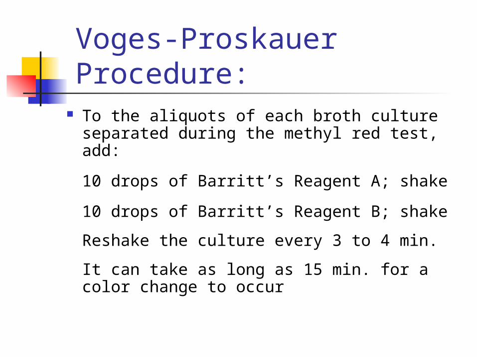
To the aliquots of each broth culture separated during the methyl red test, add:
10 drops of Barritt’s Reagent A; shake
10 drops of Barritt’s Reagent B; shake
Reshake the culture every 3 to 4 min.
It can take as long as 15 min. for a color change to occur
Voges-Proskauer Procedure:
Voges-Proskauer Results:
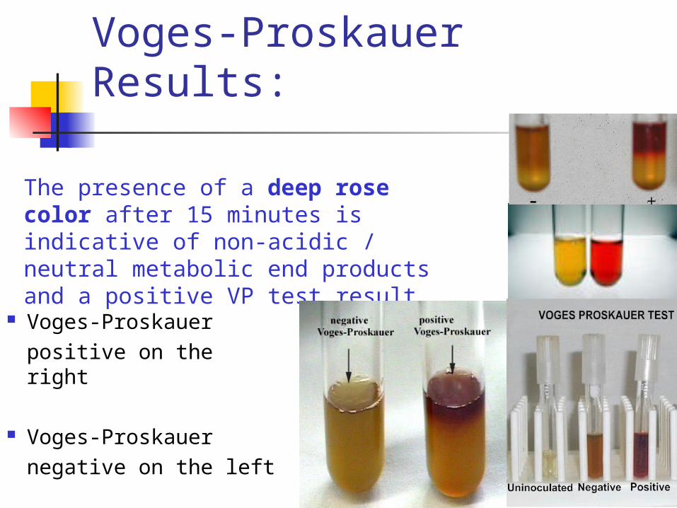
Voges-Proskauerpositive on the right
Voges-Proskauernegative on the left
The presence of a deep rose color after 15 minutes is indicative of non-acidic / neutral metabolic end products and a positive VP test result.

Citrate Utilization Test Used to determine if an organism
is capable of fermenting citrate and using that citrate as its sole carbon source
The ability of an organism to utilize citrate occurs via the enzyme citrase

Procedure: Obtain 3 Simmons Citrate agar slants Inoculate these slants using the stab
and streak method (the same way you inoculated the TSI media using a needle) with the following organisms:
E.coli K.pneumoniae E.aerogenes

Citrate Test Results:
Simmon’s Citrate agar utilizes sodium citrate as its sole carbon source
Bromthymol blue is included as a pH indicator; the medium initially is green
Organisms capable of using citrate as a carbon source turn the media “Prussian blue”.
- + - +

Page 199
Summary of IMViC Reactions

Urease Test Used to differentiate organisms based
on their ability to hydrolyze urea with the enzyme urease
The pH indicator, phenol red, is used to detect the breakdown of urea and the production of ammonia which is used by bacteria to produce amino acids and nucleotides
Procedure:
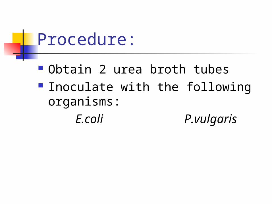
Obtain 2 urea broth tubes Inoculate with the following
organisms:E.coli P.vulgaris

Urease Test Results: Urinary tract pathogens from the genus
Proteus may be distinguished from other enterics
urease Urea + H2O CO2 + H20 + NH3
As the alkaline end products build, phenol red turns from yellowish gold to pink—a positive result

Urease Test Results
Urease positive organism on the right
Urease negative organism on the left
As the alkaline end products build, phenol red turns from yellowish gold to pink—a positive result

Phenylalanine Deaminase Test
Used to identify bacteria possessing the enzyme phenylalanine deaminase
Phenylalanine deaminase converts the amino acid phenylalanine to phenylpyruvic acid + NH3

Procedure:
Obtain 2 phenylalanine agar slants Inoculate (with a loop on the
surface) with the following organisms:
E.coli P.vulgaris
Results Phenylpyruvic acid produced by some
organisms is colorless
After inoculation and incubation, 10% ferric chloride, an oxidizing agent, is added to the surface of the slants
Ferric chloride (FeCl3) reacts with the phenylpyruvic acid (if present) and changes color from yellow to green—a positive result

Phenylalanine Deaminase Results:
Ferric chloride (FeCl3) reacts with the phenylpyruvic acid (if present) and changes color from yellow to green — a positive result
Positive Negative
Positive Negative
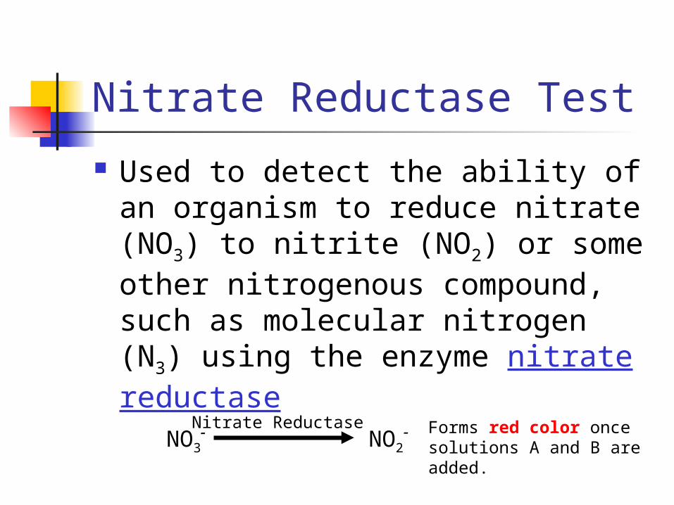
Nitrate Reductase Test
Used to detect the ability of an organism to reduce nitrate (NO3) to nitrite (NO2) or some other nitrogenous compound, such as molecular nitrogen (N3) using the enzyme nitrate reductase
NO3 NO2
Nitrate Reductase- - Forms red color once solutions A and B are added.

Procedure:
Obtain 3 Nitrate broth tubes Inoculate (with a loop) those tubes
with the following organisms:
E.coli A.faecalis P.aeruginosa

Results: (Pay close attention to this test; its one of the hardest test to read)
After inoculation and incubation, the ability of an organism to reduce nitrate to nitrite (or molecular nitrogen) is detected by adding two reagents:
Solution A(sulfanilic acid)Solution B (α-naphthylamine)
If a red color appears after addition of solution A and B, this is considered a positive result
NO3 NO2
Nitrate Reductase- - Forms red color once solutions A and B are added.

Results continued: If there is no color change occurs
after additions of solutions A & B, two possibilities must be considered:
1) nitrates were not reduced by the organism
2) the organism possessed such potent nitrate reductase enzymes that nitrates were reduced beyond nitrites to ammonia or even molecular nitrogen
NO3 NO2
Nitrate Reductase- -NH3
+ (Ammonia)
N2 (Nitrogen Gas)
P 219
NO3 NO3- -
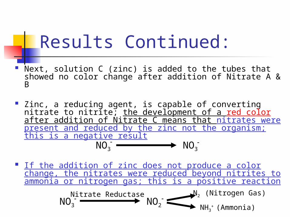
Results Continued: Next, solution C (zinc) is added to the tubes that
showed no color change after addition of Nitrate A & B
Zinc, a reducing agent, is capable of converting nitrate to nitrite; the development of a red color after addition of Nitrate C means that nitrates were present and reduced by the zinc not the organism; this is a negative result
If the addition of zinc does not produce a color change, the nitrates were reduced beyond nitrites to ammonia or nitrogen gas; this is a positive reaction
NO3 NO3- -
NO3 NO2
Nitrate Reductase- -NH3
+ (Ammonia)
N2 (Nitrogen Gas)

Solutions A and B have been added to these tubes
Solution C has been added to these tubes
E. coli - Reductase Positive
NO3 NO2
Nitrate Reductase- -
P. aeruginosaReductase Positive
C. xerosis - Reductase Negative
NO3 NO3- -
NO3 NO2
Nitrate Reductase
- -NH3
+ (Ammonia)
N2 (Nitrogen Gas)
Unreactivetubes


![1 $SU VW (G +LWDFKL +HDOWKFDUH %XVLQHVV 8QLW 1 X ñ 1 … · 2020. 5. 26. · 1 1 1 1 1 x 1 1 , x _ y ] 1 1 1 1 1 1 ¢ 1 1 1 1 1 1 1 1 1 1 1 1 1 1 1 1 1 1 1 1 1 1 1 1 1 1 1 1 1 1](https://static.fdocuments.net/doc/165x107/5fbfc0fcc822f24c4706936b/1-su-vw-g-lwdfkl-hdowkfduh-xvlqhvv-8qlw-1-x-1-2020-5-26-1-1-1-1-1-x.jpg)

![1 ¢ Ù 1 £¢ 1 £ £¢ 1 - Narodowy Bank Polski · 1 à 1 1 1 1 \ 1 1 1 1 ¢ 1 1 £ 1 £ £¢ 1 ¢ 1 ¢ Ù 1 à 1 1 1 ¢ à 1 1 £ ï 1 1. £¿ï° 1 ¢ 1 £ 1 1 1 1 ] 1 1 1 1 ¢](https://static.fdocuments.net/doc/165x107/5fc6757af26c7e63a70a621e/1-1-1-1-narodowy-bank-polski-1-1-1-1-1-1-1-1-1-1-1.jpg)

![[XLS]fmism.univ-guelma.dzfmism.univ-guelma.dz/sites/default/files/le fond... · Web view1 1 1 1 1 1 1 1 1 1 1 1 1 1 1 1 1 1 1 1 1 1 1 1 1 1 1 1 1 1 1 1 1 1 1 1 1 1 1 1 1 1 1 1 1 1](https://static.fdocuments.net/doc/165x107/5b9d17e509d3f2194e8d827e/xlsfmismuniv-fond-web-view1-1-1-1-1-1-1-1-1-1-1-1-1-1-1-1-1-1-1-1-1-1.jpg)