BIOBIO- --- 391039103910 MMMM A S T E R ’’’’ S SS S TTTTH E S I S I N … · BIOBIO- ---...
69
BIO BIO BIO BIO-3910 3910 3910 3910 M ASTER ASTER ASTER ASTER ’ S S S S T HESIS IN HESIS IN HESIS IN HESIS IN BIOLOGY BIOLOGY BIOLOGY BIOLOGY The substrate specificities and physiological function of the Toxoplasma gondii apicoplast phosphate translocator Hanne Risan Johnsen May, 2009 FACULTY OF SCIENCE Department of Biology University of Tromsø
Transcript of BIOBIO- --- 391039103910 MMMM A S T E R ’’’’ S SS S TTTTH E S I S I N … · BIOBIO- ---...

B I OB I OB I OB I O ---- 3 9 1 03 9 1 03 9 1 03 9 1 0
MMMM A S T E RA S T E RA S T E RA S T E R ’’’’ S S S S TTTT H E S I S I N H E S I S I N H E S I S I N H E S I S I N B I O L O G YB I O L O G YB I O L O G YB I O L O G Y
The substrate specificities and physiological function of the Toxoplasma gondii apicoplast phosphate translocator
Hanne Risan Johnsen
May, 2009
FACULTY OF SCIENCE Department of Biology
University of Tromsø

2

3
B I OB I OB I OB I O ---- 3 9 1 03 9 1 03 9 1 03 9 1 0
M A S T E R ’ S T H E S I S I N B I O L O G Y
The substrate specificities and physiological function of the Toxoplasma gondii apicoplast phosphate translocator
Hanne Risan Johnsen
May, 2009

4

5
Table of contents
Acknowledgments ..................................................................................................................... 7
Abbreviations ............................................................................................................................ 8
Abstract ..................................................................................................................................... 9
1. Introduction ........................................................................................................................ 11
1.1 Phylum Apicomplexa ..................................................................................................... 11
1.2 The apicoplast ................................................................................................................. 13
1.3 Plastidic phosphate translocators in plants ..................................................................... 17
1.4 Apicoplast phosphate translocators ................................................................................ 19
1.5 Aim of study ................................................................................................................... 20
2. Materials and Methods ...................................................................................................... 21
2.1 Electroporation ............................................................................................................... 21
2.2 Plasmid isolation ............................................................................................................. 22
2.3 Introduction of DNA into yeast cells .............................................................................. 23
2.4 Plasmid DNA isolation from yeast ................................................................................. 25
2.5 DNA Restriction enzyme digestion ................................................................................ 26
2.6 Polymerase Chain Reaction ............................................................................................ 27
2.7 Agarose gel electrophoresis ............................................................................................ 29
2.8 Yeast growth ................................................................................................................... 30
2.9 Membrane preparation .................................................................................................... 31
2.10 Purification of Phosphatidylcholine ............................................................................. 32
2.11 Preparation of liposomes .............................................................................................. 32
2.12 Transport experiment .................................................................................................... 33
2.13 SDS-PAGE ................................................................................................................... 35
2.14 Silver staining ............................................................................................................... 36
2.15 Western Blot ................................................................................................................. 37
3. Results ................................................................................................................................. 41
3.1 Amplification of cDNA in E.coli ................................................................................... 42
3.2 Introduction of TgPT into the yeast strain INVSc1 ........................................................ 43
3.3 Heterologous expression of TgPT in INVSc1 ................................................................ 44
3.4 Determination of substrate specificities ......................................................................... 46
3.5 Analysis of enzyme kinetics ........................................................................................... 51

6
3.6 Kinetic constants ............................................................................................................. 53
4. Discussion ............................................................................................................................ 57
4.1 The TgPT connects apicoplast and cytosolic metabolic pathways ................................. 57
4.2 Therapeutic implications ................................................................................................ 60
4.3 Conclusion ...................................................................................................................... 61
5. References ........................................................................................................................... 63
Appendix I ............................................................................................................................... 68
Appendix II ............................................................................................................................. 69

7
Acknowledgments
I would like to express my gratitude to my supervisor professor Karsten Fischer who gave me
the opportunity to work on this project and for guiding me throughout the process. Thanks to
all the people at the Department of Biology at the University of Tromsø for support and
friendship. A special thank to Rafal Butowt which I have cooperated with in this project.
Most of the work presented in this thesis was carried out at the AG Flügge lab at Botanisches
Institut, Köln. I would like to give a special thanks to professor Ulf-Ingo Flügge and his
working group for their hospitality. Thanks to Sonja Hetfeld and Diana Hille for guiding me
in the laboratory.
I must acknowledge the kind offer of TgPT cDNA from Dr. Wolfgang Bohne and his group at
the Institute of Medical Microbiology, University of Göttingen.
Lastly, I would like to thank friends and family for their support!

8
Abbreviations
3-PGA 3-phosphoglycerate
ATP Adenosine Triphosphate
Bp Base pairs
cDNA complementary DNA
dH2O distilled water
DOXP 1-deoxy-D-xylulose 5- phosphate
F Farad
FAS Fatty Acid Synthesis
G Gravity in centrifugation
GAPDH Glyceraldehyde 3-phosphate Dehydrogenase
GPI Glucose-6-phsophate Isomerase
GPT Glucose-6-phosphate Translocator
His Histidin
kV kilo Volt
LB Luria Bertani
MVA Mevalonate
NADH Nicotinamide Adenine Dinucleotide
OPPP Oxidative Pentose Phosphate Pathway
PCR Polymerase Chain Reaction
PDH Pyruvate Dehydrogenase Complex
PEP Phosphoenolpyruvate
Pi Inorganic phosphate
PPT Phosphoenolpyruvate/ Phosphate Translocator
pPT plastidic Phosphate Translocator
rpm revolutions per minute
SC Synthetic Complete
SDS-PAGE Sodium Dodecyl Sulfate Polyacrylamide Gel Electrophoresis
TgPT Toxoplasma gondii Phosphate Translocator
TP Triose Phosphates
TPT Triose Phosphate Translocator
XPT Xylulose-5-phosphate Translocator
Xyl5P Xylulose-5-phosphate

9
Abstract
The apicomplexan parasites including the causative agents of malaria (Plasmodium spp.) and
toxoplasmosis (Toxoplasma gondii) are running the risk of developing resistance to existing
drugs. Members of the phylum Apicomplexa harbour a relict plastid, the apicoplast, which
show homology to the chloroplasts of plants and algae. The apicoplast appears to be
indispensable to the parasites, and due to its prokaryotic origin it represents an excellent drug
target. T. gondii possesses a single plastidic membrane transporter (TgPT) that shows
similarity to the phosphate translocators (pPTs) in higher plants which comprises four
subfamilies (TPT, PPT, GPT, XPT). Its physiological function has not been previously
studied, but the TgPT was proposed to comprise a connection between the metabolic
pathways in the apicoplast and the metabolic processes in the cytosol. In this study, the
substrate specificities of the TgPT were determined by heterologous expression of the protein
in yeast and reconstitution of its transport activity in liposomes. The TgPT showed similarities
to both TPTs and PPTs from higher plants combining the transport activities of the two
subfamilies by simultaneously transporting compounds phosphorylated at different C-atoms.
The TgPT accepts inorganic phosphate and PEP equally as substrates, having an even higher
affinity to triose phosphates and 3-PGA. Based on our results, the TgPT likely connect the
cytosolic metabolism with metabolic pathways in the apicoplast by delivering carbon units for
at least two essential biosynthetic pathways, the fatty acid synthesis and the DOXP pathway
for isoprenoid synthesis. Additionally, the TgPT contributes an indirect supply of ATP and
reduction power to the apicoplast. These results have recently been corroborated by Boris
Striepen and his group by showing that a knock-out of the TgPT resulted in rapid death of the
parasite, establishing the transporter as essential for parasite viability and thereby an attractive
drug target.

10

11
1. Introduction
1.1 Phylum Apicomplexa
Apicomplexan parasites represent a large phylum consisting of several thousands of
protozoan species including the causative agents of malaria
(Plasmodium spp.) and
toxoplasmosis (Toxoplasma gondii) (Levine 1988). Because of an emerging resistance in the
apicomplexan parasites to the already existing drugs there is an urgent need for new
treatments as well as new targets in the fight against malaria, toxoplasmosis and other
parasitic diseases (Trape et al. 2002).
Malaria is one of the world’s major diseases found in many tropical and subtropical regions.
Of the four species of Plasmodium that infects humans, Plasmodium falciparum is the most
lethal. The disease is transmitted to humans by infected female mosquitoes that inject saliva
together with an anticoagulant during blood feeding. Saliva from an infected mosquito
contains motile asexual cells, sporozoites, which are transported with the blood to the liver
where they divide asexually and produces daughter cells, merozoites. Released merozoites
invade red blood cells where they enlarge and turn into a ring-formed feeding stage,
trophozoites, which split again to form a multinucleated cell stage, schizonts, that produce
large amounts of new merozoites. When the merozoites are released into the body the host
displays the flu-like symptoms of malaria. The symptoms may appear and disappear in
phases, these cyclic symptoms of malaria are caused by the life cycle of the parasites, as they
develop, mature, reproduce and are once again released into the blood stream to infect new
cells. Some of the merozoites reach the sexual phase and turn into gametocytes that start to
produce gametes only when extracted from the infected human host by a mosquito (Wiesner
2003).

12
T. gondii has a complex lifecycle that alternates between asexually dividing forms and a
sexual phase. The asexual part of the life cycle can take place in any warm-blooded animal,
including humans, causing the disease toxoplasmosis. Humans acquire toxoplasmosis by
contact with infected cat feces or by ingestion of undercooked and raw meat. It is estimated
that about one-third of the world's human population are infected with Toxoplasma, but the
infection is usually asymptomatic in healthy individuals. Toxoplasmosis is the most common
opportunistic infection in immunocompromised patients with AIDS or in transplant patients
(Luft and Remington 1988). In addition, congenital transmission can occur if woman are
infected with Toxoplasma for the first time during pregnancy (Kim and Weiss 2008). The
sexual part of the life cycle takes place only in members of the family Felidae, including wild
and domestic cats. Cats may become infected with T. gondii either by ingestion of infectious
oocysts from the environment or by ingestion of tissue cysts from intermediate hosts as
rodents and birds (Tenter et al. 2000). The transmission of the parasite can be facilitated by its
ability to modify its host's behavior. Infection with T.gondii in rats alters their perception of
cats, in some cases even turning their natural aversion into attraction (Berdoy 2000).
The basic biochemistry, genetics, and subcellular architecture of the apicomplexan species are
prominently similar, although many aspects of disease pathogenesis, host range and life cycle
are not conserved. T. gondii, being the most experimentally tractable, has been used as a
model system for studying many biological aspects of apicomplexan parasites (David et al.
2002, Pfefferkorn et al. 1988). T. gondii provides an attractive experimental system as the
parasite can be grown in many different cell types (Pfefferkorn et al. 1988), and most
organelles can be labeled with fluorescent reporters permitting quantitative analysis in living
cells (Striepen et al. 1998, 2000). Ultrastructural studies have revealed a detailed picture of
the T. gondii cell structure (Hager et al. 1999), and the cells can be stably transformed both by
homologous and non-homologous transformation (Roos et al. 1994).

13
1.2 The apicoplast
Plastids are plant-specific organelles which are able to perform many specialized functions
that are essential for plant growth and development, such as photosynthesis, nitrogen
assimilation, synthesis of amino acids and fatty acids, as well as storage of carbohydrates and
lipids (Bowsher et al. 1992). Even in non-photosynthetic plants and algae the plastids are
indispensable because cells containing plastids have become dependent on certain metabolic
pathways for the production of metabolites that are exported from the organelle (McFadden
and Roos 1999, Ralph et al. 2001).
Members of the phylum Apicomplexa hold a relict plastid, homologous to the chloroplast of
plants and algae. The apicoplast is a four-membrane-bound compartment, distinct from the
mitochondria and other organelles, which was first identified
in T. gondii by in situ
hybridization experiments. The organelle contains a 35-kb circular genome that encodes a
number of genes including RNA polymerase and ribosomal genes (Kohler et al. 1997,
McFadden et al. 1996, Wilson et al. 1996). The sequencing of the full P. falciparum genome
and the identification of an apicoplast-targeting sequence (Foth et al. 2003) allowed
identification of several hundred nuclear-encoded proteins that probably are targeted to this
organelle, including housekeeping enzymes involved in DNA replication, transcription,
translation, protein import, and specific metabolic and transport activities (Gardner et al.
2002).
All plastids have originated from a primary endosymbiosis where photosynthetic prokaryotes
were engulfed by phagotrophic eukaryotes (Cavalier-Smith 1982). This event led to the
development of red and green algae, plants and glaucophytes by the production of a double
membrane bound endosymbiont that subsequently became a chloroplast. A further engulfment
of the primary endosymbionts by other phagotrophic eukaryotes resulted in a lateral transfer
of the plastid into several eukaryotic lineages. Secondary endosymbiosis has occurred several
times and by preservation of the plastid it has given rise to diverse groups of algae and other
eukaryotes (McFadden and Gilson 1995, McFadden and Roos 1999).

14
Both the phylogeny of the apicoplast genome and the structure of the organelle support that
the plastid evolved through secondary endosymbiosis (McFadden and Roos 1999). One of the
characteristics for secondary plastids is the presence of more than two membranes
surrounding the organelle. The apicoplast has four membranes (Illustrated in figure 1) (Kohler
et al. 1997) whereas two are presumed to be derived from the chloroplast, one from the algae
plasma membrane and one from the host cell (Fast et al. 2001).
It is still controversial whether the apicoplast has originated from a green or red alga during
secondary endosymbiosis (Cai 2003, Funes et al. 2002, Kohler et al. 1997) but research of
recent date supports red algal ancestry of the apicomplexan plastid (Coppin et al. 2005, Funes
et al. 2004, Gould et al. 2008, Harper and Keeling 2003, Moore et al. 2008). The storage
polysaccharide amylopectin accumulated in the cytoplasm of T. gondii and the dinoflagellate
Crypthecodinium cohnii is believed to be similar to the starch accumulated by red algae.
There is evidence that this storage polysaccharide is synthesized through a UDP-glucose-
based pathway much simpler than that described for plants. This pathway is very similar to
the one found to be encoded in the recently sequenced genome of the red algae
Cyanidioschyzon merolae and it is distinct from the plant starch pathway (Coppin et al. 2005).
Figure 1: The four membranes surrounding the apicoplast. Show a
transmission electron micrograph of a Plasmodium apicoplast with four
membranes (Figure from Ralph et al. 2004). Secondary plastids have
one or two additional membranes from the engulfment of a primary
plastid-containing eukaryote by a second eukaryote.

15
The apicoplast is essential to the parasites (Fichera and Roos 1997, Sullivan et al. 2000) as
inhibition of its function or loss of the organelle leads to immediate or delayed death due to
failure during invasion of new host cells (He et al. 2001, McFadden and Roos 1999).
Apicoplast-deficient parasites replicate normally in the first infectious cycle but die in the
subsequent host cell, validating the apicoplast as essential for parasite viability (He et al.
2001). This delayed death effect has led to speculations that the apicoplast may be required
for the establishment of a functional parasitophorous vacuole. The parasitophorous vacuole is
a specialized structure inside infected cells where the parasites replicate until host cell lysis
(Lingelbach and Joiner 1998, Suss-Toby et al. 1996).
Identifying the secondary plastid in apicomplexan parasites have profound implications for
development of new treatments because it represents an opportunity to target the parasites
with treatments that are relatively harmless to the mammalian hosts. Recent studies of the
apicomplexan genomes and protein localization together with inhibitor studies have provided
some insight into the metabolism of the apicoplast. The apicoplast has a typical bacterial
housekeeping machinery such as DNA replication, transcription, translation as well as
anabolic pathways for synthesis of fatty acids (Waller et al. 1998), isoprenoid precursors
(Jomaa et al. 1999) and some of the reactions for heme synthesis (Sato et al. 2004). The
bacterial nature of the plastidic pathways makes them interesting as potential drug targets
because they are fundamentally different from the equivalent eukaryotic pathways in their
animal hosts (McFadden and Roos 1999, Ralph et al. 2001).
Enzymes involved in fatty acid synthesis have been localized to the apicoplast (Waller et al.
1998), and in Toxoplasma and Plasmodium the fatty acid biosynthesis system appears to be
essential for parasite survival and virulence (Mazumdar and Striepen 2007, Mazumdar et al.
2006). Fatty acid biosynthesis is defined as the metabolic process by which acetyl coenzyme
A (acetyl-CoA) precursors are converted to long fatty acid chains (Goodman et al. 2007).
Fatty acids play a critical role in cells as metabolic precursors for biological membranes and
energy stores (Waller and McFadden 2005). The apicoplast has a prokaryotic type II fatty acid
synthesis (FAS II) that differs in structure, kinetics and inhibitor susceptibility from the
eukaryotic type I fatty acid synthesis (FAS I) found in the animal hosts (Surolia et al. 2004,
Waller et al. 1998). The presence of a type II pathway for fatty acid biosynthesis in the
apicoplast represents a potential target for parasite-specific inhibitors (Waller et al. 2003).
16
Isoprenoid synthesis is another pathway localized to the apicoplast. The enzymes mediating
the isoprenoid pathway in apicomplexan parasites are distinct from those found in the human
hosts (Jomaa et al. 1999). The common precursor for all isoprenoids, isopentenyl
diphosphate, can be produced by two different biosynthetic routes, either via the mevalonate
(MVA) pathway, or via the 1-deoxy-D-xylulose 5- phosphate (DOXP) pathway. The DOXP
pathway is absent in mammals, but is used by most eubacteria as well as by the plastids of
algae and higher plants. In plants both pathways are present, with the DOXP pathway being
operative in the plastids and the MVA pathway in the cytosol. In green algae the cytosolic
MVA pathway is missing and isoprenoid supply exclusively depends on the plastidic DOXP
pathway (Disch et al. 1998). In P. falciparum the biosynthesis of isoprenoids is achieved by
the DOXP pathway. The enzymes of the DOXP pathway are localized inside the apicoplast
and all the enzymes involved in the pathway represent potential new drug targets (Jomaa et al.
1999, Wiesner and Jomaa 2007). The isoprenoid synthesis in the apicoplast is the only source
of isoprenes in the parasite, and it is considered to serve both the apicoplast and the
mitochondrion with isoprenes (Ralph et al. 2004).
Genomic analysis revealed an apicoplast pathway for synthesis of heme in P. falciparum and
T. gondii. In plants, the tetrapyrrole biosynthesis pathway branches and produces both heme
and chlorophyll. In Plasmodium, the pathway seems to be split between the apicoplast,
mitochondrion and possibly the cytosol. The first part of the pathway takes place in the
mitochondrion and continues in the apicoplast. The following steps seem to be either
mitochondrial or cytosolic, and the pathway probably terminates in the mitochondrion (Ralph
et al. 2004).

17
1.3 Plastidic phosphate translocators in plants
Many of the metabolic pathways establish in plastids depend on precursors from the cytosol
as well as the cytosolic pathways often require products from the chloroplast. All plastids are
surrounded by envelope membranes that restrict the nonspecific diffusion of polar molecules.
In plants four subfamilies of plastidic phosphate translocators (pPTs) that differ in function
and substrate affinity have been identified in the inner envelope membrane of the chloroplast,
the TPTs, PPTs, GPTs and XPTs (Figure 2). The transporters function as antiport systems
where transport in one direction depends on simultaneous transport of another solute in the
opposite direction (Flügge 1999). The TPT is involved in export of carbon from plastids,
while the other transporters are importing metabolites into plastids in form of
phosphoenolpyruvate (PEP), glucose-6-phosphate (Glc6P) and xylulose-5-phosphate (Xyl5P)
(Eicks et al. 2002, Fischer et al. 1997, Kammerer et al. 1998).
The triose phosphate/ phosphate translocator (TPT) mediates export of fixed carbon from the
Calvin Cycle in the form of triose phosphates and 3-PGA from the chloroplast into the cytosol
for use in the biosynthesis of sucrose and amino acids. Phosphate released from these
processes is transported back into the chloroplasts for production of ATP needed in the Calvin
Cycle and for photosynthetic electron transport (Flügge et al. 1989). In C4 -plants
phosphoenolpyruvate (PEP) is exported from the chloroplast through a phosphoenolpyruvate/
phosphate translocator (PPT) and is used as substrate for the PEP carboxylase in the cytosol to
form the four-carbon compound oxaloacetate. Phosphate produced in this reaction is
transported back into the chloroplast. The function of PPTs in C3-plants and nongreen tissues
of C4-plants is to import PEP from the cytosol into the plastid for use in the shikimate
pathway where amino acids and secondary plant products are synthesized. Non-green plastids
are not able to convert 3-PGA into PEP therefore 3-PGA produced from triose phosphates is
exported from the plastid, converted into PEP in the cytosol, and then re-imported (Fischer et
al. 1997). Cytosolically generated hexose phosphates derived from sucrose are transported by
a glucose 6-phosphate translocator (GPT) into the plastids of non-green tissues as a source of
carbon for starch and fatty acid biosynthesis, additionally acting as substrate for the oxidative
pentose phosphate pathway (OPPP) where it is transformed into triose phosphates. Both triose
phosphates and inorganic phosphate released from the starch biosynthesis can be used as
counter substrates for the GPT antiporter (Kammerer et al. 1998). The main function of the
xylulose-5-phosphate translocator (XPT) is the import of xylulose-5-phosphate
(Xul-5-P)

18
produced in the cytosol into plastids to provide the Calvin cycle and the OPPP with carbon.
The XPT subfamily is found only in dicotyledonous plants (Eicks et al. 2002).
Figure 2: The proposed functions of the four plastidic phosphate translocator proteins in plants
(TPTs, PPTs, GPTs and XPTs). The TPT mediates export of carbon from the Calvin cycle in the
form of triose phosphates that can be converted to PEP in the cytosol. PEP is imported into the plastids
via the PPT to provide the substrate for the shikimic acid pathway. The GPT imports Glc-6-P into the
plastids for the syntheses of starch and fatty acids (not shown) and as a substrate for the OPPP. Triose
phosphates can serve as a counter-substrate of either the GPT or the XPT. The XPT provides the
plastids with Xul-5-P, which is produced in the cytosol to provide the Calvin cycle and the OPPP with
carbon (Eicks et al. 2002).

19
1.4 Apicoplast phosphate translocators
Not all plastids perform photosynthesis and the lack of photosynthetic activity is compensated
by feeding metabolites from the cytoplasm into the non-photosynthetic plastids (Fischer and
Weber 2002). The biosynthetic pathways taking place in the apicoplast require mechanisms to
provide the plastid with carbon sources, ATP and reduction power. In order to search for
membrane transporters in the apicomplexan parasites the genome of P. falciparum was
queried using chloroplast pPT protein sequences. Two highly scoring predicted proteins were
identified and tested against the GenBank non-redundant database for similarity to non-
apicomplexan sequences. The first protein (PfAPT1) had highest homology to a chloroplast
triose phosphate translocator, the second (PfAPT2) had highest homology to the
phosphoenolpyruvate translocator of chloroplasts. The same working group subsequently
identified a T. gondii orthologue of PfAPT1, but no T. gondii sequence orthologous to
PfAPT2 has been found to date, using plant and apicomplexan pPTs as queries of the protein
and nucleotide databases (Karnataki et al. 2007). Given that T. gondii possesses only a single
pPT this protein possibly is transporting more than one substrate (Fleige et al. 2007). The pPT
in T. gondii is localized to the envelope membranes in the apicoplast and hence is dubbed
TgPT (T. gondii phosphate translocator). Within the subfamilies, the plant pPTs show > 80 %
identity, while across subfamilies they show about 35 % identity. The apicomplexan pPTs
show a maximum of 32 % identity to the chloroplast pPTs and because specific amino acids
have not been identified that definitively correlate with substrate specificity, it is not possible
to assign specificity of the apicomplexan pPTs on the basis of sequence homology alone.

20
1.5 Aim of study
T. gondii possesses a single plastidic membrane transporter (TgPT) that shows similarity to
the phosphate translocators (pPTs) in higher plants. Higher plants possess up to four pPT
subfamilies with different substrate specificities (Knappe et al. 2003). Given that T. gondii
possesses only one plastidic transporter it was proposed that the TgPT possibly was
transporting more than one substrate (Fleige et al. 2007). Analyses of the apicomplexan
genomes and protein localization have provided first insights into the metabolism of the
apicoplast (Fleige et al. 2007, Jomaa et al. 1999, Ralph et al. 2004). It has been established
that the apicoplast is involved in the synthesis of fatty acids (Waller et al. 1998), isopentenyl
diphosphate (Jomaa et al. 1999) and heme (Sato et al. 2004). The proposed function of the
TgPT has been to comprise a connection between the metabolic pathways in the apicoplast
and the metabolic processes in the cytosol (Fleige et al. 2007, Roos 2002).
The apicoplast is known to be indispensable to the apicomplexan parasites, and due to its
prokaryotic origin the plastid represents an attractive target for the development of parasite-
specific drugs. By determining the substrate specificities of the TgPT its physiological
function will be revealed and thereby give more insight into the metabolism of apicomplexan
parasites. This knowledge can possibly contribute to identification of new drug targets and
treatments against the parasitic diseases.
The substrate specificities of the TgPT could not be deduced from the amino acid sequence
because it does not show sufficient similarity to any of the four subfamilies of higher plant
pPTs. In this thesis the work has been focused at determining the substrate specificities of the
plastidic phosphate translocator identified in T. gondii. To do so transport experiments with
different phosphorylated metabolites were performed by heterologous expression of the TgPT
gene in yeast and reconstitution of its transport activity in liposomes. The substrate
specificities were corroborated by measurements of apparent kinetic constants for the
accepted substrates, and compared with substrate specificities of phosphate translocators in
higher plants and red algae.

21
2. Materials and Methods
In order to determine the substrate specificities of the T.gondii phosphate translocator (TgPT),
the corresponding cDNA were cloned into BamHI/ EcoRI and HindIII/BamHI restriction sites
in the yeast (Saccharomyces cerevisiae) expression vectors pYES-N and pYES-C (Invitrogen,
see appendix) thereby adding a 6x His-tag to the N- and C-terminus of TgPT, respectively.
2.1 Electroporation
Electroporation is a mechanical method used to introduce polar molecules into a host cell
through the cell membrane by electric pulses. A high-voltage electric field is applied to the
cells, producing transient holes in the cell membrane through which plasmid DNA enters
(Calvin and Hanawalt 1988).
LB medium, pH 7
1.0 % Tryptone
0.5 % Yeast extract
1.0 % NaCl
100 µg/ml ampicillin
Electrocompetent Escherichia coli (E.coli) cells (50 µl) were thawed on ice and incubated
with 2 µl of either pYES-TgPT-N or pYES-TgPT-C for 1 min. The bacterial solution was
transferred to a sterile 2 mm electroporation cuvette that was mounted into the Pulse
controller (GenePulser, Bio-Rad) between the anode and cathode. The electroporation
apparatus was set to 2.5 kV (12.5 kV/cm), 25 µF, and the pulse controller to 200 ohm. The
pulse was delivered by pushing in both charging buttons simultaneously and holding until a
short beep was heard. The cuvette was removed and 200 µl S.O.C medium (Invitrogen) was
added immediately. The cells were transferred into a 1.5 ml tube and incubated at 37 °C for 1
h at 200 rpm. Different dilutions of the cells were spread onto selective ampicillin plates and
grown overnight. Successfully transformed cells were picked and transferred to 3 ml LB-
ampicillin medium for overnight growth at 37 °C.

22
2.2 Plasmid isolation
The Birnboim-Doly method for rapid extraction of plasmid DNA from E. coli is based on
selective alkaline denaturation of high molecular weight chromosomal DNA while covalently
closed circular DNA remains double-stranded. E.coli cells containing the plasmid of interest
are lysed with sodium dodecyl sulfate (SDS) and NaOH. Denatured chromosomal DNA
renature and aggregate into an insoluble precipitation when neutralized by acidic sodium
acetate. The high concentration of sodium acetate also causes precipitation of protein-SDS
complexes and high molecular weight RNA. The three major contaminating macro-molecules
can be removed by centrifugation after precipitation with sodium acetate. The plasmid DNA
is recovered from the supernatant by isopropanol precipitation (Birnboim and Doly 1979).
Solution I
50 mM Glucose
25 mM Tris, pH 8
10 mM EDTA, pH 8
Solution II
0.2 M NaOH
1 % (w/v) SDS
High salt solution
3 M Sodium acetate, pH 4.8
From an overnight culture of E.coli, 1.5 ml was centrifuged at 11000 x g for 1.5 min at room
temperature. The supernatant was removed and the pellet was thoroughly resuspended in 100
µl of Solution I. After 5 min incubation on ice 200 µl of Solution II was added and mixed by
gently pipetting three times up and down. The mixture was for a second time incubated on ice
for 5 min. A volume of 150 µl ice-cold High salt solution was added and the tube vortexed
immediately. The solution was centrifuged at 16000 x g for 10 min and the supernatant
transferred to a new tube where it was mixed with 350 µl of isopropanol. It was again
centrifuged at 16000 x g for 10 min. The supernatant was discarded and the pellet washed
with 500 µl 70 % ethanol (-20 °C). It was repelleted at 16000 rpm for 5 min and dried before
resuspended in 20 µl RNase-A (1 mg/ml) and incubated at 37 °C for 30 min. Plasmids were

23
stored at -20 °C until use. The plasmid DNA concentrations after isolation were estimated by
agarose gel electrophoresis with a molecular mass ruler (MassRuler™, Fermentas).
2.3 Introduction of DNA into yeast cells
E.coli is commonly used as a host for the expression of foreign proteins, but because of the
toxicity of the gene product it failed to express the plastidic phosphate translocator proteins
from higher plants. Yeast has therefore previously been used for the expression of functional
plastidic translocators (Flügge 1999). The uracil-auxotrophic yeast strain INVSc1 (Invitrogen)
was transformed with the plasmids pYES-TgPT-N and pYES-TgPT-C by lithium acetate
mediated transformation. The alkali cations from lithium acetate make intact yeast cells
competent to take up DNA by addition of polyethylene glycol (PEG) followed by a heat
shock (Ito et al. 1983). Successfully transformed cells were grown on selective plates.
YPD medium
1 % yeast extract
2 % peptone
2 % dextrose
10x TE-buffer
100 mM Tris, pH 7.5
10 mM EDTA
10x Lithium acetate
1 M Lithium acetate, pH 7.5
PEG 4000 solution
50 % PEG
SC-medium, 1 liter (20 g agar/ liter)
6.7 g SC-medium (Bacto-yeast nitrogen base w/o amino acids)
100 ml Drop out medium (see “yeast growth”)
2 % glucose

24
5 ml of YPD medium was inoculated with a single yeast colony and grown over night at 30
°C. From the saturated culture 1 µl was transferred to 300 ml of YPD medium and grown
overnight at 30 °C. Cells were harvested by centrifuging 5 min at 4000 x g in room
temperature. The pellet was resuspended in 10 ml of sterile water and transferred to a 15 ml
Falcon tube and repelleted for 5 min at 5000 x g in room temperature. The yeast cells were
resuspended in 1.5 ml freshly prepared buffered lithium solution (1 volume 10x TE buffer, 1
volume 10x lithium acetate, 8 volumes sterile water). For each transformation 200 µg carrier
DNA (Single-stranded salmon sperm, Sigma) was mixed with ≤ 5 µg transforming DNA and
200 µl yeast suspension in an eppendorf tube. 1.2 ml freshly prepared PEG solution (8
volumes 50 % PEG, 1 volume 10x TE buffer, 1 volume 10x lithium acetate) was added and
then incubated for 30 min at 30 °C. A heat shock was performed for exactly 15 min in a 42 °C
water bath and cells pelleted by 5 seconds centrifugation in room temperature. The cells were
resuspended in 500 µl 1x TE buffer and different dilutions were spread onto plates lacking
uracil for selective growth of cells containing the plasmid. Incubation was performed at 30 °C
until appearance of transformants (2-4 days). Successfully transformed colonies were
inoculated in 5 ml cultures of selective medium lacking uracil and grown overnight at 200
rpm in 30 °C.

25
2.4 Plasmid DNA isolation from yeast
A rapid method for plasmid DNA isolation from yeast cells is based on breakage of the cells
by vortexing with glass beads in a detergent solution, followed by a separation of nucleic
acids from proteins by phenol/chloroform extraction (Hoffman and Winston 1987).
Breaking buffer
2 % Triton X-100
1 % SDS
100 mM NaCl
10 mM Tris-HCl, pH 8
1 mM EDTA, pH 8
Phenol/chloroform/isoamyl alcohol
25:24:1 (v/v/v) phenol/chloroform/isoamyl alcohol
TE-buffer
10 mM Tris, pH 7.6
1 mM EDTA
Overnight yeast cultures were harvested by centrifuging for 5 min at 1500 x g in room
temperature. The cell pellet was resuspended in 200 µl breaking buffer and transferred to an
eppendorf tube containing 0.3 g glass beads and 200 µl phenol/chloroform/isoamyl alcohol.
The cells were broken by vortexing for 3 min at highest speed. A volume of 200 µl TE-buffer
was added and vortexed briefly before centrifuged 5 min at high speed, room temperature.
The aqueous layer (max 400 µl) was transferred to a clean tube which was filled up with 100
% ethanol and mixed by inversion. The tube was centrifuged again for 10 min at high speed
and the pellet was washed twice with 80 % ethanol. After drying the DNA pellet was
resuspended in 250 µl TE-buffer and stored at -20 °C until use.

26
2.5 DNA Restriction enzyme digestion
After plasmid isolation from E.coli the presence of a correct insert with the expected size
1031 bp, was verified by restriction enzyme digestion. Restriction enzymes or restriction
endonucleases are enzymes isolated from bacteria that recognize and cut specific sequences in
DNA to produce fragments, called restriction fragments. The DNA sample is first digested
with restriction enzymes to generate DNA fragments, and then the different sized fragments
are separated by agarose gel electrophoresis. Plasmids with correct restriction sites will
generate two visible bands of DNA on the gel, and those with altered restriction sites will not
be cut and will generate only a single band (Reece 2004).
In this study, plasmid DNA purified from E.coli by Birnboim-Doly plasmid isolation was
digested to confirm whether the cloning of TgPT into the pYES-N/C vectors had been
successful.
Table 1: Restriction enzymes and buffers used for N-terminal and C-terminal constructs
Construct Restriction enzymes Restriction buffer
N-terminal EcoRI / BamHI EcoRI buffer
C-terminal HindIII / BamHI HindIII buffer
Table 2: Mastermix for restriction enzyme digestion
Reagent Volume
Restriction enzymes 0.5 µl of each
Restriction buffer 1 µl
DNA* 2 µl
dH2O 6 µl
*Plasmid DNA isolated from E.coli
For each reaction, 2 µl plasmid DNA was added to 8 µl of mastermix. The digestion was
carried out for 2 hours at 37°C. The restriction fragments were separated by 1 % agarose gel
electrophoresis and visualized by ethidium bromide staining.

27
2.6 Polymerase Chain Reaction
Polymerase Chain Reaction (PCR) is a method for enzymatic amplification of specific
sequences of DNA. Knowledge of the DNA sequence flanking the sequence of interest is
required in order to produce synthetic complementary primers that are needed to initiate the
amplification process. The PCR reaction goes through three different steps defined by the
critical aspects of time and temperature; denaturation, annealing and elongation, these steps
make up a cycle that is repeated 20-35 times to achieve a satisfying amount of amplified DNA
In the denaturation step, the PCR reaction is heated to a temperature of around 95 °C where
the hydrogen bonds of the double helix are broken resulting in single-stranded molecules to
be used as templates. In the annealing step, the reaction temperature is lowered to around 50-
60°C so that the primers can anneal to the single-stranded DNA template forming short
segments of double-stranded DNA where the polymerase attaches and begins DNA synthesis.
During the extension/elongation step, the DNA polymerase synthesizes new DNA strands
complementary to the DNA template strands (Reece 2004, Wilson and Walker 2000).
To verify correct insertion of the construct into the yeast strain InvSc1 after lithium acetate
mediated transformation, plasmid DNA was isolated from the yeast cells by
phenol/chloroform mediated DNA isolation and used as template for PCR reaction. The TgPT
gene was amplified using the primers Toxo1 (5`-ACTTCTTCGTCCACATCGGC) and Toxo2
(5`-TTTCGCGATGATCTGCTGGC) (Sigma).
Table 3: Standard PCR Mastermix
Reagents Volume
10X buffer 5 µl
Forward primer 1µl
Reverse primer 1 µl
dNTP 1 µl
Taq Polymerase 0,6 µl
dH2O 40,5 µl

28
Table 4: PCR program. * Repeated 35 cycles
Phase Time Temperature
Initial denature 2 min 95 °C
Denature* 30 sec 94 °C
Anneal* 15 sec 55 °C
Elongation* 1 min 72 °C
10 min 72 °C
Cooling ∞ 4 °C
The PCR reactions were performed with a DNA EngineGradient Cycler PTC-200 (MJ Research)
Mastermix (49 µl) and plasmid DNA (1 µl) were mixed in a PCR tube to a total volume of 50
µl. The PCR thermo cycler was programmed according to table 4. The program start with a 2
minutes denaturation at 95 °C, and after 35 cycles the program end with a 10 minutes
elongation followed by a 4 °C unlimited hold. The PCR products were separated by agarose
gel electrophoresis and detected by ethidium bromide staining.

29
2.7 Agarose gel electrophoresis
Agarose gel electrophoresis is a method used to separate nucleic acids; DNA or RNA
molecules by size. This is achieved by moving negatively charged nucleic acid molecules
through an agarose matrix with an electric field (electrophoresis). The negative charge of the
molecules is due to the phosphate backbone they possess. Shorter molecules move faster and
migrate farther than longer ones and by varying the concentration of agarose, one can separate
varying ranges of DNA fragment sizes (Sambrook 2001). The separated DNA fragments are
stained with ethidium bromide for visualization on the gel by UV-light. The binding of
ethidium bromide to DNA results in distortion of the double helix and increases its overall
length. DNA to which ethidium bromide is bound fluoresces under UV-light. The size of the
DNA fragments can be determined by comparing them to DNA-ladders with fragments of
known size (Reece 2004).
1% Agarose gel
0.6 g agarose
60 ml TBE buffer
1 µl ethidium bromide (5 µg/ml)
TBE-buffer
89 mM Tris, pH 7.6
89 mM boric acid,
2 mM EDTA
To make a 1 % gel, 0.6 g agarose was boiled in 60 ml TBE-buffer to dissolve. The liquid
agarose was mixed with 1 µl ethidium bromide, poured into a casting frame with a comb to
make the wells and left to polymerize for 30 min. The gel was then placed in an
electrophoresis tank containing TBE-buffer as the running buffer. Loading buffer (6 x DNA
Loading Dye, Fermentas) was added to the samples before loaded into the wells. In this study,
GeneRuler™ 1 kb DNA Ladder and GeneRuler™ DNA Ladders Mix (Fermentas) were used
as size markers. The gel was run for 60 min at 90 volts and DNA bands visualized by UV
light using a Gel Doc 2000 (BioRad).

30
2.8 Yeast growth
The yeast strain Saccharomyces cerevisiae has several properties which have established it as
an important tool in the expression of foreign proteins for research, industrial or medical use.
Yeast can be grown rapidly on simple media to high cell density and its genetics are more
advanced than any other eukaryote (Romanos et al. 1992). The gene encoding the TgPT
protein is under control of a galactose regulated promoter which is controlled by two
regulatory proteins and the carbon source in the medium. When galactose is present, the two
regulatory proteins interact and activate transcription. In the absence of galactose the
transcriptional activation is blocked, and in the presence of glucose the transcription of the
galactose genes are strongly repressed (Johnston et al. 1994).
SC-medium, 1 liter
6.7 g SC-medium (Bacto-yeast nitrogen base w/o amino acids)
100 ml Drop out medium
50 ml glucose/galactose (40%) Fill up with dH2O
10X Drop out (DO) medium for yeast culture, 1 liter
Isoleucine 300 mg
Valin 1500 mg
Adenine 200 mg
Arginine 200 mg
Histidin 200 mg
Leucine 1000 mg
Lysine 300 mg
Methionine 200 mg
Phenylalanine 500 mg
Threonin 2000 mg
Tryptophan 200 mg Fill up with dH2O
Single colonies of yeast were inoculated into 5 ml of SC-medium containing 2 % glucose and
grown over night at 30°C with shaking at 200 rpm. The overnight culture was transferred into
a 50 ml culture and grown over night at 30°C with shaking at 200 rpm. The cells were
centrifuged at 1500 x g for 5 min at 4°C and the supernatant discarded. Then the cells were

31
resuspended in 2 x 400 ml SC-medium containing 2 % galactose to induce the expression of
mTgPT-N and mTgPT-C, and grown at 30 °C with shaking at 200 rpm. After 8 hours the
induction was terminated by harvesting the cells with centrifugation at 1500 * g for 5 min.
The cells were stored at -20°C until ready to use (Protocol from Invitrogen Catalogue nos.
8252-20).
2.9 Membrane preparation
Exogenous proteins in yeast are targeted to membrane-bound compartments that are readily
isolated for biochemical experiments (Ton and Rao 2004).
1X TE-buffer
10 mM Tris/HCl, pH 7.5
1 mM EDTA
PMSF
100 mM in EtOH
Glass beads (~ 0.5 mm)
Glass beads were incubated with 0.1 M HCl overnight. Then they were washed under running
water for an hour, before adding sterile water and checking the pH. The glass beads were dried
over night using filter paper.
All steps were conducted at 4 °C using precooled media and equipment. After thawing, the
yeast cells were disrupted in 210 µl TE-buffer containing 100 mM protease inhibitor Phenyl-
methyl-sulfonyl-fluoride (PMSF). The cells were transferred to 1.5 ml eppendorf tubes
containing 400 mg glass beads for homogenization by strong shaking in 6 min at 4°C
(TissueLyser II, Qiagen). The cell remains were separated from the membranes by adding 750
µl TE/PMSF and shaking by hand before centrifuging in 45” at 1000 rpm. This was repeated
by transferring the supernatant to new tubes and centrifuging in 45” at 1000 rpm. The
supernatant was transferred to new tubes with part of the lid cut off and centrifuged for 25
min at 45000 rpm (Optima™ L-100 XP Ultracentrifuge). The pellets were frozen in liquid
nitrogen and stored at – 80 °C until use. The protein concentrations were measured by Bio-
Rad (Bradford) Protein Assay, a dye-binding analysis in which a differential color change of a
dye occurs in response to various concentrations of protein.

32
2.10 Purification of Phosphatidylcholine
Phosphatidylcholine (100 g) was dissolved in 320 ml chloroform and separated by adding 1.4
liter of ice-cold acetone by continuous stirring for 1 hour. The lipids were precipitated over
night at 4°C. The precipitate was dissolved in 320 ml diethyl ether and extracted under
vacuum in a rotting flask. When the ether was completely evaporated the purified lipids were
stored at -20 °C until use.
2.11 Preparation of liposomes
Liposomes are artificially constructed vesicles of a lipid bilayer enclosing an aqueous
compartment (Szoka and Papahadjopoulos 1980). The system allows accurate measurement
of solute transport from one compartment to another by reconstituted membrane proteins
(Hanke et al. 1999). The TgPT transport activity was reconstituted into preloaded liposomes
in order to determine the substrate specificities.
Liposome buffer (preloaded liposomes)
100 mM Tricin/KOH, pH 7.6
30 mM K-gluconate
20 mM substrate (Pi, 3-PGA, PEP, TP or Glc6P)
Liposome buffer (empty liposomes)
100 mM Tricin/KOH, pH 7.6
60 mM K-gluconate
PD-10 buffer
100 mM Na-gluconate
50 mM K-gluconate
10 mM Tricin/KOH, pH 7.6
The liposomes were preloaded with exchangeable substrates by adding liposome buffer (1 ml/
kinetic) containing a phosphorylated metabolite to the purified phospholipids (125 mg
phospholipids/ 1 ml liposome buffer). By sonication for some minutes on ice (duty cycle:
20%, output: 5) the lipids were broken into liposomes. The membrane pellets isolated from
yeast cells were dissolved in 100 µl dH2O and the proteins were solubilized from the

33
membranes by adding 7 µl 20 % Triton X-100. The protein solution was added to the
liposomes (50 µl membranes/ 750 µl liposomes), mixed by inversion and frozen in liquid
nitrogen. The frozen liposomes were thawed on ice and sonicated with 30 pulses (duty cycle:
30, output: 5) to integrate the proteins into the membranes (Kasahara and Hinkle 1977). PD-
10 columns (Amersham Biosciences) were equilibrated by washing with 3 x 5 ml PD-10
buffer. The columns contain Sephadex™ G-25 for size exclusion chromatography that allows
separation of liposomes preloaded with substrate from the external solution. For each column
900 µl of liposomes were loaded and eluted with 3 ml PD-10 buffer. When the flow-through
turned white, 1 ml of liposomes was collected in an eppendorf tube.
2.12 Transport experiment
The transport activity of the TgPT protein reconstituted into liposomes is measured as
movement of radiolabelled substrate into the liposomes. Radioactively labeled inorganic
phosphate [32
P] (purchased from Amersham-Pharmacia, Germany) and cold Pi is added to the
surrounding extracellular fluid and will be transported into the liposomes in exchange with
preloaded substrate. The transport rate is quantified at different time points by liquid
scintillation counting (Hanke et al. 1999).
Na-acetate buffer
150 mM Na-acetate
Substrate solution
2 µCi 32
PO4 per kinetic
10 mM KH2PO4
For each kinetic 850 µl of the liposome solution was mixed with 45 µl of substrate solution,
and 200 µl from this was loaded on the columns at time points 15”, 35”, 55” and 75”. To
terminate the transport activity external radioactivity was removed by passing the suspension
over a column preloaded with Dowex AG1-X8 resin (Bio-Rad) to bind charged particles in
the extracellular fluid. The anion exchange columns were prepared from Pasteur pipettes with
cotton and pre-equilibrated with 150 mM Na-acetate buffer. The liposomes were eluted with 2
x 850 µl Na-acetate buffer and collected in scintillation tubes. The radioactivity of the eluate
was determined by liquid scintillation counting.

Figure 3: Experimental setup for transport measurements with the TgPT antiporter.
setup liposomes were preloaded with the substrate of interest and radioactively labeled phosphate [
was used as counter exchange substrate to get an indication which metabolites the TgPT accepts as
substrates. To determine the affinity to
and [32
P] used as counter exchange substrate. The second setup illustrates a competitive binding
experiment used to determine the affinity of
was measured in the presence of the unlabeled substrates. All liposomes used for the competitive
binding experiment were preloaded with 20 mM inorganic phosphate. A:
inhibitors (PEP, 3-PGA, TrioseP, Glc6P), C: inorganic
34
Experimental setup for transport measurements with the TgPT antiporter.
setup liposomes were preloaded with the substrate of interest and radioactively labeled phosphate [
was used as counter exchange substrate to get an indication which metabolites the TgPT accepts as
substrates. To determine the affinity to phosphate (Km) the liposomes were preloaded with phosphate,
P] used as counter exchange substrate. The second setup illustrates a competitive binding
experiment used to determine the affinity of non-radioactively labeled substrates. The binding of
was measured in the presence of the unlabeled substrates. All liposomes used for the competitive
binding experiment were preloaded with 20 mM inorganic phosphate. A: [32
PGA, TrioseP, Glc6P), C: inorganic phosphate.
Experimental setup for transport measurements with the TgPT antiporter. In the first
setup liposomes were preloaded with the substrate of interest and radioactively labeled phosphate [32
P]
was used as counter exchange substrate to get an indication which metabolites the TgPT accepts as
) the liposomes were preloaded with phosphate,
P] used as counter exchange substrate. The second setup illustrates a competitive binding
radioactively labeled substrates. The binding of [32
P]
was measured in the presence of the unlabeled substrates. All liposomes used for the competitive 32
P]-Pi, B: Unlabeled

35
2.13 SDS-PAGE
Sodium dodecyl sulfate polyacrylamide gel electrophoresis (SDS-PAGE) is a method used for
separating proteins according to their electrophoretic mobility. Sodium dodecyl sulphate
(SDS) is an anionic detergent which denatures secondary and non–disulfide–linked tertiary
structures, and applies a negative charge to each protein in proportion to its mass. The
proteins migrate in response to an electrical field through pores in the gel matrix. The
combination of gel pore size and protein charge, size and shape determines the migration rate
of a protein (Reece 2004).
Sample buffer
0.125 M Tris-HCl, pH 6.8
4 % (w/v) SDS
20 % (v/v) Glycerin
10 % (v/v) 2-Mercaptoethanol
0.01 % (w/v) Bromo-phenol blue
10X Laemelli buffer
250 mM Tris
1.92 M Glycine
1 % SDS
Table 5: Reagents and volumes for polyacrylamide gels
Stacking gel, 4 % Separation gel, 12 %
dH2O 2,7 ml 3,4 ml
Acrylamide, 30 % 0,67 ml 4,0 ml
Tris 1,5 M/ 1 M 0,5 ml 2,5 ml
SDS, 10 % 40 µl 90 µl
APS, 10 % 25 µl 60 µl
TEMED 6 µl 40 µl

36
The SDS-PAGE was carried out according to the Laemelli protocol from 1970. The
separating gel was prepared and allowed to polymerize on the bottom of the cast before
pouring the stacking gel on top. Due to the higher acrylamide percentage in the separating gel,
the proteins will be concentrated on the interface between the two gels resulting in increased
protein resolution. After complete polymerization the chamber was assembled and Laemelli
buffer poured into the well. Each membrane pellet were solubilized in 100 µl sample buffer
and loaded onto the gel (5 – 25 µl per/lane). The electrophoresis was carried out at 80 V for 1-
2 hours, using Power Pac 300 (Bio-Rad).
2.14 Silver staining
Silver staining is a sensitive tool used for protein visualization after polyacrylamide gel
electrophoresis with a detection level down to 0.3-10 ng. The protein detection depends on the
binding of silver ions to the amino acid side chains, followed by reduction to free metallic
silver. The protein bands are visualized as spots on the gel where the reduction occurs
(Switzer et al. 1979).
Fixing solution
50 % (v/v) Methanol
12 % (v/v) Acetic acid
0.019 % (v/v) Formaldehyde
Washing solution I
50 % (v/v) Ethanol
Washing solution II
615 mM Sodium Thiosulfate
Staining solution
0.2 % (w/v) Silver nitrate
0.075 % (v/v) Formaldehyde

37
Developing solution
0.57 M Sodium carbonate
0.019 % (v/v) Formaldehyde
17.3 µM Sodium Thiosulfate
Stop solution
50 % (v/v) Methanol
12 % (v/v) Acetic acid
After electrophoresis the gels were fixed over night to restrict protein movement from the gel
matrix and to remove interfering ions and detergent from the gel. The gels were then washed
in 50 % ethanol for 3 x 20 min to remove the remaining detergent ions. To increase the
sensitivity and the contrast of the staining the gels were incubated for 1 min in solution III,
and rinsed three times with dH2O. The gels were then incubated in the staining solution for 20
min. When staining was complete, gels were rinsed in dH2O and moved to developing
solution. The reaction was stopped by adding stop solution when the desired intensity of the
bands was reached.
2.15 Western Blot
Western blotting is a useful method for the identification and quantification of specific
proteins. Proteins are usually separated using SDS-PAGE before transferred to a membrane
where they are probed by antibodies specific to the target protein. Following a blocking step
to prevent any nonspecific binding of antibodies to the surface of the membrane, the
membrane is probed with a primary antibody that attaches to the antigen of interest. After a
washing step, the membrane is incubated with a secondary antibody that is reactive toward the
primary antibody. After probing with secondary antibody, the membrane is washed again and
incubated with an appropriate enzyme substrate (Towbin et al. 1979). One of the substrates
used for protein detection is luminol-based and produce a chemiluminescent signal.
Chemiluminescence is a chemical reaction that produces energy released in the form of light
in the presence of horseradish peroxidase and a peroxide buffer. The intensity of the signal
after development should correlate with the abundance of the antigen on the membrane
(http://www.piercenet.com/files/WBGuide.pdf).

38
In this study, protein samples after membrane preparation were resolved by SDS-PAGE
before they were transferred to a Polyvinylidene Fluoride (PVDF) membrane, where they
were detected by HisProbe™ (Pierce) directed against the polyhistidine tag.
Blocking buffer
4 % (w/v) Milk powder
1X TBS buffer Stored at 4° C
10X TBS buffer
100 mM Tris-HCl, pH 7.5
1.5 M NaCl
TBS-T buffer
3X TBS buffer
0.05 % (v/v) Tween 20
0.2 % (v/v) Triton X-100
10X WBB
20 mM Tris
150 mM Glycine
Cathode buffer
1X WBB
0.1 % SDS
Anode buffer
1X WBB
30 % Methanol
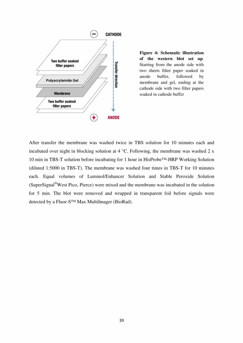
The blot was built from the anode side starting with two sheets of filter paper soaked in anode
buffer. The transfer membrane was incubated in methanol followed by anode buffer, and
placed on top of the filter paper. The gel was soaked in cathode buffer and laid on top of the
membrane, and then covered by two more filter papers soaked in cathode buffer. The system
was closed and current was applied for 1.5 hours, 0.65 mA / cm2
membrane.
39
After transfer the membrane was washed twice in TBS solution for 10 minutes each and
incubated over night in blocking solution at 4 °C. Following, the membrane was washed 2 x
10 min in TBS-T solution before incubating for 1 hour in HisProbe™-HRP Working Solution
(diluted 1:5000 in TBS-T). The membrane was washed four times in TBS-T for 10 minutes
each. Equal volumes of Luminol/Enhancer Solution and Stable Peroxide Solution
(SuperSignal®
West Pico, Pierce) were mixed and the membrane was incubated in the solution
for 5 min. The blot were removed and wrapped in transparent foil before signals were
detected by a Fluor-S™ Max MultiImager (BioRad).
Figure 4: Schematic illustration
of the western blot set up.
Starting from the anode side with
two sheets filter paper soaked in
anode buffer, followed by
membrane and gel, ending at the
cathode side with two filter papers
soaked in cathode buffer

40

41
3. Results
In order to determine the substrate specificities of the T.gondii phosphate translocator (TgPT)
cDNA encoding the transport protein was cloned into the yeast expression vectors p-YES-N
and pYES-C (Invitrogen), resulting in the proteins mTgPT-N and mTgPT-C. After
amplification in E.coli, the plasmids were transformed into the yeast strain INVSc1 for
heterologous expression of the proteins, followed by a reconstitution of their transport
activities in liposomes. Western blot analyses were performed to verify yeast expression of
the TgPT after galactose mediated induction. Total membranes were isolated from yeast cells
and solubilized proteins were integrated into liposome membranes. For antiport membrane
proteins the transport in one direction depends on simultaneous transport of another solute in
the opposite direction. Thus, the liposomes were preloaded with an exchangeable metabolite
and radioactively labeled phosphate, [32
P]-Pi, was used as external counter exchange substrate.
The transport activity for different metabolites was measured using liquid scintillation
counting of the amount [32
P] imported into the liposomes after a given time of transport
activity.
The substrate specificities of the TgPT were corroborated by calculation of the kinetic
constants for the substrates accepted by the transporter. The kinetic data from the transport
experiments were analyzed according to the Michaelis-Menten model to determine apparent
Km and Ki values for the different substrates. For precise calculations of the kinetic constants
the data were converted by taking the reciprocals of the Michaelis-Menten equation and
graphically present the data in a Lineweaver-Burk plot. The substrate specificities of the
TgPT were compared with substrate specificities of phosphate translocators in higher plants
and red algae.

42
3.1 Amplification of cDNA in E.coli
The corresponding cDNA of the TgPT protein was cloned into the yeast expression vectors
pYES-N and pYES-C (Invitrogen), which are 6 x His-tag fusion vectors with a galactose-
inducible promoter. Electrocompetent E.coli cells were transformed with pYES-TgPT-N and
pYES-TgPT-C to amplify the plasmids before transformation into yeast cells. Plasmid DNA
was isolated from E.coli by the Birnboim-Doly method and the presence of correct insert
TgPT gene was confirmed by restriction enzyme digestion with the restriction enzymes
BamHI /EcoRI for the N-terminal fusion and HindIII / BamHI for the C-terminal fusion. The
different sized restriction fragments were separated on a 1 % agarose gel and visualized by
ethidium bromide staining.
The restriction fragments after digestion of the N-terminal fusion vector from three different
plastid isolations are shown in figure 5. Expected band sizes were 1031 bp and 6000 bp.
Figure 5: Gel electrophoresis
showing fragments of pYES-TgPT-
N isolated from E.coli after
restriction enzyme digestion. A
restriction enzyme digestion of pYES-
TgPT-N was performed with the
restriction enzymes BamHI and
EcoRI, followed by an agarose gel
electrophoresis to separate and
visualize the restriction fragments.
Lane 1: GeneRuler™ 1 kb DNA
Ladder (Fermentas), Lane 2-4:
products after restriction enzyme
digestion with BamHI and EcoRI.
Fragments corresponding to sizes 1031
and 6000 bp can be seen in all three
lanes.
The results show that all three digestion reactions gave bands at predicted sizes, (Figure 5,
lane 2-4), confirming that the cloning of TgPT into the pYES-N vector was successful.
Digestion of the C-terminal fusion vector gave similar results (data not shown).

43
3.2 Introduction of TgPT into the yeast strain INVSc1
Plasmids containing the TgPT gene were transformed into competent cells from the uracil-
auxotrophic yeast strain INVSc1 to produce protein required for the transport experiments.
Yeast cells containing the plasmid after transformation were selected for on selective plates
lacking uracil, and several colonies were checked for correct insert by PCR with Toxo1 and
Toxo2. The primers are specific for the TgPT gene and should amplify a region of 470 bp.
Untransformed yeast cells were used as negative controls.
Figure 6: Gel electrophoresis showing PCR products after amplification with primers specific
for the TgPT gene. Plasmid DNA from yeast cells were used as template. Lane 1: GeneRuler™ DNA
Ladders Mix (Fermentas), Lane 2-4: untransformed yeast (negative control), Lane 5-7: yeast
transformed with pYES-TgPT-N. Lane 5-7 show bands at the predicted size, ~470 bp, after
amplification.
Results presented in figure 6 show that the yeast strain INVSc1 contains the pYES-TgPT after
transformation. As expected, negative controls (Figure 6, lane 2-4) show no bands.
Successfully transformed yeast cells were picked and grown in selective liquid cultures (SC-
medium lacking uracil) for further use in transport experiments.

44
3.3 Heterologous expression of TgPT in INVSc1
In order to verify expression of the recombinant TgPT protein in the yeast transformants and
to optimize the induction time, cells were grown in the presence of galactose and harvested at
different time points ranging from 2 to 24 hours after induction. Membrane fractions were
prepared by strong shaking with glass beads and high speed (45000 x g) centrifugation.
Proteins were separated by SDS-PAGE, and western blot analysis with HisProbe™ (Pierce)
directed against the polyhistidine tag verified the galactose-inducible accumulation of the
TgPT protein compared to a control, which maintained the empty expression vector.
Figure 7: Western blot detection of the TgPT protein by HisProbe™ (Pierce). The results verify
expression of the TgPT gene in yeast (Lane 1-2) and are showing an increasing signal with prolonged
exposure to galactose (Lane 4-6). Lane 1-2: TgPT expression after 8 hours induction, Lane 3: empty
vector (negative control), Lane 4: TgPT expression after 2 hours induction, Lane 5: 4 hours induction,
Lane 6: 8 hours induction
The western blot verified protein expression after galactose mediated induction (Figure 7, lane
1-2). As expected, no signal was detected in the protein fraction from cells transformed with
an empty vector (Figure 7, lane 3). There was an increasing expression with time between 2
and 8 hours post-induction (Figure 7, lane 4-6), but no further enhancement of expression
thereafter (data not shown).

45
A number of transformed yeast colonies were screened by western blotting in order to identify
colonies strongly expressing the TgPT. The signal intensity detected on the blot was
compared with amount of protein visualized by silver staining after SDS-PAGE. A strong
signal on the blot together with a corresponding weak signal on the silver stained gel indicates
a high protein expression rate.
Figure 8: Western blot detection of the TgPT protein in different clones by HisProbe™ (Pierce).
Show significant expression of the protein in 6 of 8 colonies. Lane 1-8: TgPT expression in different
colonies after 8 hours galactose induction.
The results presented in figure 8 show similar expression levels in 6 of 8 colonies after
screening. The absent signal in lane 2 and 3 indicate colonies either missing the insert or
having very low expression level.
After analyzing the TgPT expression level of different clones by western blot and silver
staining, one clone was chosen for further experiments. Yeast cells were grown in selective
SC-medium over night and harvested after 8 hours of galactose induction. Total yeast
membrane fractions were isolated and reconstituted into liposomes for transport experiments.

46
3.4 Determination of substrate specificities
The heterologous expression of cDNAs encoding transport proteins in yeast and the
reconstitution of their transport activity in liposomes has been used as method for functional
characterization for several members of the plastid phosphate translocators in plants (Eicks et
al. 2002, Fischer et al. 1997, Kammerer et al. 1998, Loddenkotter et al. 1993). In order to
determine the substrate specificities of the TgPT, liposomes were prepared by sonication of
acetone-washed phosphatidylcholine in a buffered solution containing an exchangeable
substrate for preloading. Proteins were purified from yeast membranes by using Triton X-100
as detergent and incorporated into the liposomes by a freeze/thaw step followed by sonication.
The transport experiments were started by addition of [32
P]-Pi as the external counter
exchange substrate to liposomes containing unlabeled substrate, and terminated at different
time points by passing the liposomes over an anion exchange column. The activity was
determined by liquid scintillation counting.
The phosphate transport activities of both the N- and C-terminal fusion proteins were
determined. Yeast membrane fractions from cells transformed with an empty vector (NTC)
were used as negative control to determine the background transport activity of endogenous
yeast proteins. Additionally, transport with empty liposomes (EN) was performed as control
to confirm that there was no substrate transport into the liposomes unrelated to the TgPT.

47
Figure 9: Determination of the phosphate/phosphate counter exchange activity. Uptake of
inorganic phosphate in the presence and absence of phosphate (10 mM) preloaded to the liposomes.
PN: TgPT with N-terminal fusion of His-tag, PC: TgPT with C-terminal fusion of His-tag, EN: TgPT
reconstituted into empty liposomes, NTC: cells transformed with an empty vector. Counts per minute
(CPM) is the number of atoms in a given quantity of radioactive material that are detected to have
decayed in one minute.
The protein with C-terminal fusion (PC) showed a transport activity that makes up less than
30 % of the activity possessed by the protein with an N-terminal fusion (PN) (Figure 9).
Therefore, only the N-terminal fusion protein was used for further experiments. Virtually no
phosphate uptake occurred in the absence of a suitable counter substrate within the liposomes
(EN), demonstrating the antiport function of the reconstituted translocator. The control
transformed with an empty vector (NTC) to determine background transport activity of
endogenous yeast proteins showed a negligible transport activity.
0
500
1000
1500
2000
2500
3000
3500
15 35 55 75
CP
M
seconds
PN
PC
EN
NTC

48
In order to determine which metabolites are accepted as substrates by the TgPT, liposomes
were preloaded with different metabolites (10 mM) and [32
P]-Pi (10 mM) was used as external
counter exchange substrate. The amount of [32
P] transported into the liposomes in exchange
with the preloaded substrates was measured by liquid scintillation counting after different
length of transport activity.
Figure 10: Determination of substrate affinities by transport experiment. Transport activities
were measured for substrates transported by the TgPT reconstituted into liposomes that had been
preloaded with different metabolites. The graph show amount of [32
P] transported into the liposomes
after 15, 35, 55 and 75 seconds. TP: triose phosphate, PEP: phosphoenolpyruvate, 3-PGA: 3-
phosphoglycerate, Glc6P: glucose-6-phosphate.
After initial experiments with different metabolites preloaded to the liposomes, glucose-6-
phosphate (Figure 10), fructose-1-phosphate and glucose-1-phosphate (data not shown)
obviously cannot be transported by the TgPT, and were not used for further transport
experiments in order to determine the kinetic constants. Evidently, PEP, 3-PGA and inorganic
phosphate are about equally well accepted as countersubstrates by the TgPT, while triose
phosphates display an even higher transport rate.
0
500
1000
1500
2000
2500
3000
3500
4000
4500
15 35 55 75
CP
M
seconds
TP
PEP
3-PGA
Glc6P
Phosphate

49
The transport experiments carried out to determine substrate specificities was repeated in at
least three independent experiments under similar conditions. The transport activity for the
different metabolites was calculated as percentage of the phosphate/phosphate transport
activity, which was set to 100 %, and compared with substrate specificities of pPTs from
higher plants and red algae (Table 6).
Table 6: Substrate specificities of plastidic phosphate translocators from T.gondii (TgPT, this
thesis), red algae (TPT, PPT) (Linka et al. 2008) and higher plants (TPT, PPT, GPT) (Kammerer et al.
1998). The phosphate transport proteins were isolated from yeast cells and reconstituted into
liposomes that had been preloaded with the indicated substrates. Activities are given as percentage of
the Pi/Pi counter exchange activity.
TgPT GsTPT GsPPT SoTPT BoPPT PsGPT
Phosphate 100 100 100 100 100 100
Triose
phosphate
192 ± 50 87 25 92 22 112
3-PGA 167 ± 34 25 22 90 16 50
PEP 99 ± 16 20 90 5 72 20
Glucose-6-P 25 ± 5 25 15 5 2 90
Data for TgPT are summarized as the mean ± SE of at least three independent experiments
Obviously, the TgPT protein accepts inorganic phosphate and PEP equally as substrates,
having an even higher affinity to triose phosphates and 3-PGA (Table 6). The substrate
specificities of pPTs from red algae (Linka et al. 2008) and higher plants (Kammerer et al.
1998) were compared with data from the phosphate translocator in T.gondii (Table 6). Higher
plants possess up to four different pPTs with different substrate specificities (Flügge 1999),
while T.gondii has only one pPT that obviously combine the substrate specificities of both
TPTs and PPTs. Red algae on the other hand possess both TPTs and PPTs that are accepting
triose phosphates and PEP, respectively. The TPT in red algae are lacking the 3-PGA
transport function found in higher plants and T. gondii (Linka et al. 2008).

50
Figure 11: Substrate specificities of the TgPT, TPT and PPT. T.gondii have only one pPT (TgPT)
that combines the substrate specificities of both TPTs and PPTs from higher plants. Data for TPT and
PPT are taken from transport experiments with higher plants (SoTPT, BoPPT) (Kammerer et al.
1998).
The results presented in figure 11 show that the TgPT has substrate specificities similar to
plant TPTs and PPTs, accepting triose phosphates, PEP and 3-PGA as substrates. Glc6P is
only poorly transported by both the TgPT and the plant transporters (Figure 10 and 11), and
when the hexose phosphates have to compete with other phosphorylated substrates under in
vivo conditions they are not transported at all. The transport activity of triose phosphates and
3-PGA are considerable higher for the TgPT than the plant transporters, which can be
explained by the apicoplast relying on an indirect transport of energy and reduction power
carried out by a shuttle of triose phosphate / 3-PGA.
0
50
100
150
200
250
Pi TrioseP 3-PGA PEP Glc6P
Tra
nsp
ort
act
ivit
y %
Substrates
TgPT
TPT
PPT

51
3.5 Analysis of enzyme kinetics
The kinetic data from the transport experiments were analyzed according to the Michaelis-
Menten model to determine apparent Km and Ki values for the substrates accepted by the
TgPT. Km is an indicator of the affinity that an enzyme has for a given substrate. A low Km
value indicates high affinity to the substrate, and an enzyme that recognizes different
substrates will have a different Km for each substrate.
V0 = Vmax [S] / (Km + [S]) Michaelis-Menten equation
The reaction rate (V) increases with increasing substrate concentration, [S], asymptotically
approaching the maximum rate (Vmax). The Michaelis-Menten constant (Km) is equivalent to
the substrate concentration at which the rate of transport is half of Vmax. To calculate the Km
and Vmax in a more precise way, the Michaelis-Menten equation can be converted into a linear
form by taking the reciprocal of both sides of the equation. This is called the Lineweaver-
Burk equation and the method transforms the hyperbolic Michaelis-Menten equation to a
linear equivalent, from which the Vmax and Km parameters can be determined.
1 / V0 = 1 / Vmax + (Km / Vmax) * (1 / [S]) Lineweaver-Burk equation
Many substrates for enzymes or transporters are not available in a radioactive form. Since
they are unlabeled there is no way to directly measure their affinity for the protein. The
affinity of the unlabeled substrate for the protein can be determined indirectly by measuring
its ability to compete with a radioactive labeled substrate for the protein.
Ki = Km [I] / (Km (app) - Km) Competitive inhibition
To measure the effect of an inhibitor the enzyme velocity is measured at a variety of substrate
concentrations in the presence and absence of an inhibitor. In the presence of a competitive
inhibitor it takes a higher substrate concentration to achieve the same velocities that were
reached in its absence. Although Vmax can still be reached if sufficient substrate is available,
one-half Vmax requires a higher [S] than before and as a result the Km value is higher. An
inhibitor constant, Ki, similar to the Km, can be defined and incorporated into the Michaelis-
Menten equation (Motulsky 1999).
.

52
Figure 12: A graphical presentation of the Lineweaver-Burk equation of enzyme kinetics. The
Lineweaver-Burk plot provides a useful graphical method for analysis of the Michaelis-Menten
equation (Lineweaver and Burk 1934).
In a Lineweaver-Burk plot, Km is derived from the negative x-axis intercept and Vmax is
obtained from the y-axis intercept (Figure 12). In the presence of a competitive inhibitor, the
Km value will increase due to the ability of the inhibitor to affect the transport of phosphate.
The Vmax value will not change. From the negative x-axis intercept in presence of the
competitive inhibitor a Ki value for the competing substrate can be calculated (Motulsky
1999).

53
3.6 Kinetic constants
The TgPT appears to combine the substrate specificities of both TPTs and PPTs from higher
plants, accepting inorganic phosphate, PEP, triose phosphates and 3-PGA (Table 6). In order
to support the substrate specificity data the apparent kinetic constants were measured for all
substrates accepted by the TgPT. To determine the apparent Km value for inorganic phosphate
the transport experiments were repeated using liposomes preloaded with 20 mM inorganic
phosphate and different concentrations of [32
P]-Pi as external counter exchange substrate.
0 5 10 15 20 250
100
200
300
[S] (mM)
V (
pm
ol/m
in)
Figure 13: Determination of the Michaelis-Menten constant. The phosphate/phosphate exchange
activity measured at different concentrations of inorganic phosphate. Velocity is given as pmol/min
and the concentration of substrate is given in mM. The transport activity goes up with increasing
substrate concentration. Km is the substrate concentration where V equals one-half Vmax.
The phosphate/phosphate counter exchange activity (Figure 13) was used to determine the
apparent Km value of inorganic phosphate. The data were analyzed by a Lineweaver-Burk plot
(not shown) to obtain more accurate values for Km and Vmax. The Lineweaver-Burke plot
increases the precision by linearizing the data, and provides a useful graphical method for
analysis of the Michaelis-Menten equation. The apparent Km value for phosphate was
determined from several independent experiments.

54
To facilitate detection of the substrate transport activity, Km can only be determined for
radioactively labeled substrates. For the additional substrates transported by the TgPT binding
of a variety of concentrations (0.2 - 20 mM) of inorganic phosphate were measured in absence
and presence of the unlabeled substrates to determine apparent Ki values. All liposomes were
preloaded with 20 mM Pi.
0.0 0.5 1.0 1.50
5000
10000
15000
20000
minutes
CP
M
Figure 14: Determination of inhibitor affinities. Transport activity (CPM) of phosphate in absence
(●) and presence (♦) of triose phosphate (5 mM) as competitive inhibitor. There is a decrease in the
transport rate for phosphate when triose phosphate is present, indicating that triose phosphate is
transported by the TgPT and thereby inhibiting transport of phosphate.
The data presented in figure 14 shows the results from a single experiment with 5 mM triose
phosphate acting as competitive inhibitor on different concentrations (0.2-2 mM) of inorganic
phosphate. With increasing concentrations of substrate the inhibition by triose phosphate
decreases. A Lineweaver-Burk plot of the results (not shown) enables determination of the
apparent Ki value for triose phosphate (shown in table 7).

55
The apparent kinetic constants for triose phosphates, PEP and 3-PGA were determined by
measuring the enzyme velocity at a variety of phosphate concentrations in the presence and
absence of various competitive substrates. The results were analyzed by Lineweaver-Burk
plots to determine the apparent Ki values.
Table 7: The apparent Km and Ki values for various phosphorylated metabolites. The kinetic
constants calculated for phosphate (Km), 3-PGA, triose phosphates and PEP (Ki) by graphical analysis
(Lineweaver-Burk).
Substrate Kinetic constant TgPT
Phosphate Km 1.39 ± 0,28 mM
Triose phosphate Ki 1.63 ± 0,26 mM
3-PGA Ki 1.33 ± 0,49 mM
PEP Ki 1.65 ± 0,52 mM
Data are summarized as the mean ± SE from 4-6 independent experiments
Inorganic phosphate has an apparent Km value of 1.39 mM (Table 7), which is close to the
apparent Km values of the plant transporters that are ranging from 0.8 to 1.1 mM (Fischer et
al. 1997, Kammerer et al. 1998). The apparent constants for competitive inhibition (Ki)
measured for the additional substrates transported by the TgPT are all in the same range as the
Km for phosphate (Table 7), indicating that the transporter is accepting the metabolites equally
and thereby are having similar affinity to all of the substrates. The apparent kinetic constants
validate the information from the substrate specificity experiments (Table 6), and the TgPT
obviously combine the substrate specificities of both TPTs and PPTs from higher plants.

56

57
4. Discussion
4.1 The TgPT connects apicoplast and cytosolic metabolic pathways
T. gondii possesses one plastidic membrane protein (TgPT) located in the apicoplast envelope
membrane that shows similarity to the plant plastid phosphate translocators (Fleige et al.
2007, Karnataki et al. 2007). Based on knowledge about the Plasmodium genome and the
sugar metabolism in T.gondii, the proposed function of the TgPT is to transport metabolites
between the cytosolic glycolysis and metabolic pathways in the apicoplast (Fleige et al. 2007,
Roos 2002). In plants the glycolytic pathways localized in the cytosol and the chloroplasts are
connected by pPTs transporting phosphorylated intermediates of glycolysis across the inner
envelope membrane (Flügge et al. 2003). The T.gondii genome holds all the glycolytic
enzymes as duplicates with the exception of hexokinase and glucose-6-phosphate isomerase
(GPI), which are encoded once in the genome. Whereas the cytosol contains a complete set of
enzymes for glycolysis, at least three glycolytic isoenzymes are evidently targeted to the
apicoplast (Fleige et al. 2007). None of the phosphate translocators from algae and higher
plants have the ability to transport simultaneously compounds phosphorylated at C-atom 2
(e.g. PEP) and C-atom 3 (e.g. triose phosphates and 3-PGA), respectively (Flügge et al.
2003). Higher plants therefore possess up to four different pPT subfamilies with different
substrate specificities (Knappe et al. 2003). Because T. gondii obviously possesses only one
plastidic transporter it was proposed that the TgPT possibly was transporting more than one
substrate (Fleige et al. 2007).
The TgPT is evidently involved in integration of the apicoplast into the cellular metabolism,
and a detection of its physiological functions will increase the knowledge about the
metabolism of Apicomplexa and might enable the development of new drug targets. In order
to determine the role of the apicoplast, the function of the T. gondii membrane translocator
was analyzed. By heterologous expression of the TgPT gene in yeast and reconstitution of its
transport activity in liposomes the substrate specificities of the TgPT were identified. Further,
the results were verified by determination of the kinetic constants, Km and Ki, for substrates
accepted by the transporter. Liposomes were preloaded with the different substrates and [32
P]-
Pi was used as external counter exchange substrate. The transport activities were measured by
liquid scintillation counting of [32
P] imported into the liposomes at different time points after
induction.

58
In this study, we have demonstrated that the phosphate translocator in T. gondii (TgPT) is able
to transport triose phosphates, 3-PGA and PEP, but does not possess any transport activity of
glucose-6-phosphate (Table 6) or other hexose phosphates (data not shown). The TgPT
accepts inorganic phosphate and PEP equally as substrates, having an even higher affinity to
triose phosphates and 3-PGA. The apparent kinetic constants for the substrates accepted by
the TgPT were determined and they were all in the same range as the Km for phosphate (Table
7), supporting the substrate specificity data. The substrate specificities of the phosphate
translocator in T.gondii (this study) was compared with transport activity data from the pPTs
from red algae (Linka et al. 2008) and higher plants (Kammerer et al. 1998) (Table 6). The
TgPT showed similarities to both TPTs and PPTs from higher plants combining the transport
activity of the two transporters by simultaneously transporting compounds phosphorylated at
different C-atoms.
All components needed to shuttle triose phosphates and 3-PGA are expressed in T. gondii,
namely the cytosolic and plastid isoforms of phosphoglycerate kinase (PGK), glyceraldehyde
3-phosphate dehydrogenase (GAPDH) and triose phosphate isomerase (TPI) (Fleige et al.
2007). The shuttle of triose phosphates and 3-PGA results in a net transfer of ATP and NADH
from the cytosol to the apicoplast, which is necessary to provide biosynthetic pathways in the
apicoplast with energy and reduction power since the apicoplast has no photosynthetic
activity (Mazumdar and Striepen 2007). Because apicoplasts lack an oxidative pentose
phosphate pathway (OPPP), the GAPDH reaction, together with the pyruvate dehydrogenase
(PDH) are the only processes producing reducing equivalents (NADH/NADPH) inside the
plastid. The importance of the triose phosphate / 3-PGA shuttle is reflected in the high
transport rate of triose phosphates and 3-PGA compared to the plant transporters (Table 7).
The red algae G. sulphuraria possesses both TPTs and PPTs, but neither of them show any 3-
PGA transport activity (Linka et al. 2008). Since both NADPH and ATP are produced by
photosynthesis in red algae they are most likely not dependent of energy and reduction power
acquired from the triose phosphate / 3-PGA shuttle in the same way as the non-photosynthetic
T.gondii.

The apicoplast, similar to other non
because they lack enolase. 3-
the cytosol, converted into PEP, and then re
pathway and fatty acid synthesis. Inside the apicoplast, PEP can be converted by pyruvate
kinase to pyruvate, which in turn serves as a substrate for the PDH complex to produce
Acetyl-CoA for fatty acid synthesis. The presence of the PDH co
important implications for the Acetyl
only enzyme which can provide the apicoplast with Acetyl
addition, the conversion of pyruvate into Acetyl
power required for fatty acid synthesis in non
Figure 15: Proposed functions of the TgPT.
net transfer of energy (ATP) and reduction power
(Mazumdar and Striepen 2007)
precursors for isoprenoid biosynthesis are produced. 3
exported from the apicoplast, converted into
released and pyruvate is fed into the DOXP pathway and fatty acid synthesis
59
apicoplast, similar to other non-green plastids, is not able to convert 3
-PGA produced from triose phosphates is therefore exported to
the cytosol, converted into PEP, and then re-imported into the apicoplast for u
pathway and fatty acid synthesis. Inside the apicoplast, PEP can be converted by pyruvate
kinase to pyruvate, which in turn serves as a substrate for the PDH complex to produce
for fatty acid synthesis. The presence of the PDH complex in apicoplasts has
important implications for the Acetyl-CoA dependent pathway because it appears to be the
only enzyme which can provide the apicoplast with Acetyl-CoA (Fleige
addition, the conversion of pyruvate into Acetyl-CoA produces NADH, which is reduction
power required for fatty acid synthesis in non-photosynthetic plastids (Goodman
Proposed functions of the TgPT. The shuttle of triose phosphates and 3
and reduction power (NADH) from the cytosol to the apicoplast
(Mazumdar and Striepen 2007). The triose phosphates are fed into the DOXP pathway where
precursors for isoprenoid biosynthesis are produced. 3-PGA produced from triose phosphate is
exported from the apicoplast, converted into PEP in the cytosol and then re-imported. Phosphate is
released and pyruvate is fed into the DOXP pathway and fatty acid synthesis (Fleige
green plastids, is not able to convert 3-PGA into PEP
PGA produced from triose phosphates is therefore exported to
imported into the apicoplast for use in the DOXP
pathway and fatty acid synthesis. Inside the apicoplast, PEP can be converted by pyruvate
kinase to pyruvate, which in turn serves as a substrate for the PDH complex to produce
mplex in apicoplasts has
CoA dependent pathway because it appears to be the
(Fleige et al. 2007). In
CoA produces NADH, which is reduction
(Goodman et al. 2007).
The shuttle of triose phosphates and 3-PGA results in a
from the cytosol to the apicoplast
. The triose phosphates are fed into the DOXP pathway where
PGA produced from triose phosphate is
imported. Phosphate is
(Fleige et al. 2007).

60
Both red algae and T.gondii lack any pPTs transporting Glc6P or other hexose phosphates
(GPTs) (Linka et al. 2008). The GPTs are established only in green algae and higher plants,
indicating that the evolution of GPTs is connected to the evolution of starch metabolism
(Deschamps et al. 2008). In red algae and T.gondii the starch synthesis is still localized in the
cytosol, but a relocation of the starch synthesis into plastids in green algae made the
development of plastid transporters (GPTs) necessary for import of starch precursors (Coppin
et al. 2005). The absence of Glc6P transport activity in T. gondii, in addition to the lack of
starch synthesis, can be explained by the lack of a plastid localized glucose-6-phosphate
isomerase (GPI). If Glc6P were imported into the apicoplast it would not be able to enter the
glycolytic pathway since T.gondii possesses only one GPI, which is located in the cytosol
(Fleige et al. 2007). GPI is an essential enzyme for catabolic glycolysis and anabolic
gluconeogenesis that takes part in both the oxidative pentose phosphate pathway (OPPP) and
starch synthesis in plants (Grauvogel et al. 2007).
4.2 Therapeutic implications
The currently available drugs against diseases caused by the phylum Apicomplexa are often
relatively toxic to the host, as most are based on targeting biochemical pathways which are
shared between host and parasite (Luzzi and Peto 1993, Salako 1984). The discovery of a
relict plastid in apicomplexan parasites (McFadden et al. 1996) followed by the detection of
metabolic pathways within by the apicoplast and most recently the determination of the
substrate specificities of the T. gondii phosphate translocator, revealed a new range of drug
targets for the treatment of apicomplexan diseases (McFadden and Roos 1999, Ralph et al.
2001). By developing treatment directed against any of the metabolic pathways in the
apicoplast the parasitic diseases can be controlled without affecting the hosts.
The antimalarial activities of antibiotics were observed first in the late 1940s when animal
studies and clinical trials demonstrated that chloramphenicol and tetracyclines had
antimalarial properties (Dahl 2008). Treatment with antibiotics has almost no effect on
parasite growth during the first intracellular cycle, whereas the parasites die during reinvasion
of new host cells. This effect seems to be related to the fact that the likely targets of the
antibiotics are primarily essential for replication of the apicoplast, giving rise to apicoplast
deficient daughter cells during intracellular division (Wiesner and Jomaa 2007). The
antibiotics quinolones and fluoroquinolones are believed to inhibit apicoplast-localized

61
prokaryotic DNA gyrases, resulting in an arrest of the plastidic DNA replication. The
antiparasitic activity of rifampicin is most likely linked to inhibition of the apicoplast RNA
polymerase, and numerous anti-bacterial agents are considered to inhibit protein synthesis
inside the apicoplast. A common characteristic of most of these antibiotics is a delayed death
effect where the parasite remains viable for a prolonged time after drug exposure. It was
demonstrated that the plastid genome of T. gondii began to degrade in the cycles after
antibiotic treatment, linking the effects of antibiotics to the apicoplast (Fichera and Roos
1997).
A recent study has demonstrated that although drugs which interfere with the prokaryotic
housekeeping functions of translation, transcription and genome replication lead to apicoplast
loss, they do not kill the parasites rapidly, as they permit other apicoplast biochemical
processes to proceed. However, parasites treated with metabolic inhibitors that target key
enzymes of the apicoplast fatty acid or isoprenoid biosynthetic pathways, have an immediate
effect on the parasite (Ramya et al. 2007). Both pathways are involved in the production and
modification of lipids, and lipid-bound proteins could be required for the parasite-host
interaction or the generation of the parasitophorous vacuole, which are essential for successful
invasion of new host cells (Ralph et al. 2004). During invasion of new host cells, the parasite
forms a parasitophorous vacuole that enables an efficient uptake
of nutrients and protection
against host immune defenses. A breakage of the parasitophorous vacuole membrane (PVM)
causes death to the parasites, but the cause is still unclear (Zhao et al. 2009).
4.3 Conclusion
Based on our results, the TgPT likely connect the cytosolic metabolism with metabolic
pathways in the apicoplast by delivering carbon units for at least two essential biosynthetic
pathways, the fatty acid synthesis and the DOXP pathway for isoprenoid synthesis.
Additionally, the TgPT contributes an indirect supply of ATP and reduction power to the
apicoplast. The pathways for biosynthesis of fatty acids and isoprenoids localized to the
apicoplast are known to be essential for survival of the parasites (Jomaa et al. 1999,
Mazumdar et al. 2006). Thus, the transporter delivering substrates to these pathways can be
expected to be equally vital for the parasite. Recently, it has been established that a knock-out
of the TgPT leads to rapid death of T. gondii (Brooks et al, submitted), confirming that the
phosphate translocator is essential for parasite survival. This knowledge, together with the
62
determination of the TgPT substrate specificities, the T. gondii translocator appears attractive
as potential drug target for treatment of parasitic diseases without any delayed effect.
Further genetic and biochemical analysis of the apicoplast should be performed in order to
reveal more details about the functions of the organelle. In the search for new drug targets a
thoroughly description of the apicoplast metabolism has to be completed. Finding drugs
acting without any delay is important as acute infections need to be controlled immediately.
Furthermore, the delayed death effect can have implications for development of drug
resistance within the apicomplexan parasites (Goodman et al. 2007). The cause of the delayed
death observed following the loss of apicoplast function is still unknown. With the intention
to understand the basis for the delayed death phenotype, pharmacological studies need to be
complemented with molecular studies using techniques for genetic manipulation in
Plasmodium and Toxoplasma (Balu and Adams 2007, Mazumdar et al. 2006).

63
5. References
Balu, B. and Adams, J.H. (2007) Advancements in transfection technologies for
Plasmodium. Int J Parasitol, 37, 1-10.
Berdoy, M., Webster, J. P., Macdonald, D. W. (2000) Fatal attraction in rats infected with
Toxoplasma gondii. Proc Biol Sci, 267, 1591-1594.
Birnboim, H.C. and Doly, J. (1979) A rapid alkaline extraction procedure for screening
recombinant plasmid DNA. Nucl. Acids Res., 7, 1513-1523.
Bowsher, C.G., Boulton, E.L., Rose, J., Nayagam, S. and Emes, M.J. (1992) Reductant for
glutamate synthase in generated by the oxidative pentose phosphate pathway in non-
photosynthetic root plastids. The Plant Journal, 2, 893-898.
Cai, X., Fuller, A. L., McDougald, L. R., Zhu, G. (2003) Apicoplast genome of the
coccidian Eimeria tenella. Gene, 321, 39-46.
Calvin, N.M. and Hanawalt, P.C. (1988) High-efficiency transformation of bacterial cells
by electroporation. J. Bacteriol., 170, 2796-2801.
Cavalier-Smith, T. (1982) The Origins of Plastids. Biological Journal of the Linnean
Society, 17, 289-306.
Coppin, A., Varré, J.-S., Lienard, L., Dauvillée, D., Guérardel, Y., Soyer-Gobillard, M., Buléon, A., Ball, S. and Tomavo, S. (2005) Evolution of Plant-Like Crystalline
Storage Polysaccharide in the Protozoan Parasite Toxoplasma gondii Argues for a Red
Alga Ancestry. Journal of Molecular Evolution, 60, 257-267.
Dahl, E.L., Rosenthal, P. J. (2008) Apicoplast translation, transcription and genome
replication: targets for antimalarial antibiotics. Trends in Parasitology, 24, 279-284.
David, S.R., Michael, J.C., Robert, G.K.D., Martin, F., Omar, S.H., Cynthia, Y.H., Jessica, C.K., Michael, K.S. and Boris, S. (2002) Mining the Plasmodium genome
database to define organellar function: what does the apicoplast do? Philosophical
Transactions of the Royal Society B: Biological Sciences, 357, 35-46.
Deschamps, P., Moreau, H., Worden, A.Z., Dauvillee, D. and Ball, S.G. (2008) Early
Gene Duplication Within Chloroplastida and Its Correspondence With Relocation of
Starch Metabolism to Chloroplasts. Genetics, 178, 2373-2387.
Disch, A., Schwender, J., Muller, C., Lichtenthaler, H.K. and Rohmer, M. (1998)
Distribution of the mevalonate and glyceraldehyde phosphate/pyruvate pathways for
isoprenoid biosynthesis in unicellular algae and the cyanobacterium Synechocystis
PCC 6714. Biochemical Journal, 333, 381-388.
Eicks, M., Maurino, V., Knappe, S., Flugge, U.-I. and Fischer, K. (2002) The Plastidic
Pentose Phosphate Translocator Represents a Link between the Cytosolic and the
Plastidic Pentose Phosphate Pathways in Plants. Plant Physiol., 128, 512-522.
Fast, N.M., Kissinger, J.C., Roos, D.S. and Keeling, P.J. (2001) Nuclear-Encoded, Plastid-
Targeted Genes Suggest a Single Common Origin for Apicomplexan and
Dinoflagellate Plastids. Mol Biol Evol, 18, 418-426.
Fichera, M.E. and Roos, D.S. (1997) A plastid organelle as a drug target in apicomplexan
parasites. Nature, 390, 407-409.
Fischer, K., Kammerer, B., Gutensohn, M., Arbinger, B., Weber, A., Hausler, R.E. and Flugge, U.I. (1997) A New Class of Plastidic Phosphate Translocators: A Putative
Link between Primary and Secondary Metabolism by the
Phosphoenolpyruvate/Phosphate Antiporter. Plant Cell, 9, 453-462.
Fischer, K. and Weber, A. (2002) Transport of carbon in non-green plastids. Trends in Plant
Science, 7, 345-351.
Fleige, T., Fischer, K., Ferguson, D.J.P., Gross, U. and Bohne, W. (2007) Carbohydrate
Metabolism in the Toxoplasma gondii Apicoplast: Localization of Three Glycolytic
64
Isoenzymes, the Single Pyruvate Dehydrogenase Complex, and a Plastid Phosphate
Translocator. Eukaryotic Cell, 6, 984-996.
Flügge, U.-I. (1999) Phosphate translocators in plastids. Annual Review of Plant Physiology
and Plant Molecular Biology, 50, 27-45.
Flügge, U.I., Fischer, K., Gross, A., Sebald, W., Lottspeich, F. and Eckerskorn, C. (1989)
The triose phosphate-3-phosphoglycerate-phosphate translocator from spinach
chloroplasts: nucleotide sequence of a full-length cDNA clone and import of the in
vitro synthesized precursor protein into chloroplasts. EMBO J, 8, 39-46.
Flügge, U.I., Häusler, R.E., Ludewig, F. and Fischer, K. (2003) Functional genomics of
phosphate antiport systems of plastids. Physiologia Plantarum, 118, 475-482.
Foth, B.J., Ralph, S.A., Tonkin, C.J., Struck, N.S., Fraunholz, M., Roos, D.S., Cowman, A.F. and McFadden, G.I. (2003) Dissecting Apicoplast Targeting in the Malaria
Parasite Plasmodium falciparum. Science, 299, 705-708.
Funes, S., Davidson, E., Reyes-Prieto, A., Magallon, S., Herion, P., King, M.P. and Gonzalez-Halphen, D. (2002) A Green Algal Apicoplast Ancestor. Science, 298,
2155.
Funes, S., Reyes-Prieto, A., Pérez-Martínez, X. and González-Halphen, D. (2004) On the
evolutionary origins of apicoplasts: revisiting the rhodophyte vs. chlorophyte
controversy. Microbes and Infection, 6, 305-311.
Gardner, M.J., Hall, N., Fung, E., White, O., Berriman, M., Hyman, R.W., Carlton,
J.M., Pain, A., Nelson, K.E., Bowman, S., Paulsen, I.T., James, K., Eisen, J.A.,
Rutherford, K., Salzberg, S.L., Craig, A., Kyes, S., Chan, M.S., Nene, V.,
Shallom, S.J., Suh, B., Peterson, J., Angiuoli, S., Pertea, M., Allen, J., Selengut, J.,
Haft, D., Mather, M.W., Vaidya, A.B., Martin, D.M., Fairlamb, A.H., Fraunholz,
M.J., Roos, D.S., Ralph, S.A., McFadden, G.I., Cummings, L.M., Subramanian,
G.M., Mungall, C., Venter, J.C., Carucci, D.J., Hoffman, S.L., Newbold, C., Davis, R.W., Fraser, C.M. and Barrell, B. (2002) Genome sequence of the human
malaria parasite Plasmodium falciparum. Nature, 419, 498-511.
Goodman, C.D., Su, V. and McFadden, G.I. (2007) The effects of anti-bacterials on the
malaria parasite Plasmodium falciparum. Mol Biochem Parasitol, 152, 181-191.
Gould, S.B., Waller, R.F. and McFadden, G.I. (2008) Plastid Evolution. Annual Review of
Plant Biology, 59, 491-517.
Grauvogel, C., Brinkmann, H. and Petersen, J. (2007) Evolution of the glucose-6-
phosphate isomerase: the plasticity of primary metabolism in photosynthetic
eukaryotes. Mol Biol Evol, 24, 1611-1621.
Hager, K., Striepen, B., Tilney, L. and Roos, D. (1999) The nuclear envelope serves as an
intermediary between the ER and Golgi complex in the intracellular parasite
Toxoplasma gondii. J Cell Sci, 112, 2631-2638.
Hanke, G., Bowsher, C., N Jones, M., Tetlow, I. and Emes, M. (1999) Review article.
Proteoliposomes and plant transport proteins. J. Exp. Bot., 50, 1715-1726.
Harper, J.T. and Keeling, P.J. (2003) Nucleus-Encoded, Plastid-Targeted Glyceraldehyde-
3-Phosphate Dehydrogenase (GAPDH) Indicates a Single Origin for Chromalveolate
Plastids. Mol Biol Evol, 20, 1730-1735.
He, C.Y., Shaw, M.K., Pletcher, C.H., Striepen, B., Tilney, L.G. and Roos, D.S. (2001) A
plastid segregation defect in the protozoan parasite Toxoplasma gondii. EMBO J, 20,
330-339.
Hoffman, C.S. and Winston, F. (1987) A ten-minute DNA preparation from yeast efficiently
releases autonomous plasmids for transformation of Escherichia coli. Gene, 57, 267-
272.

65
http://www.piercenet.com/files/WBGuide.pdf Chemiluminescent Western Blotting Technical
Guide and Protocols.
Ito, H., Fukuda, Y., Murata, K. and Kimura, A. (1983) Transformation of intact yeast cells
treated with alkali cations. J. Bacteriol., 153, 163-168.
Johnston, M., Flick, J.S. and Pexton, T. (1994) Multiple mechanisms provide rapid and
stringent glucose repression of GAL gene expression in Saccharomyces cerevisiae.
Mol. Cell. Biol., 14, 3834-3841.
Jomaa, H., Wiesner, J., Sanderbrand, S., Altincicek, B., Weidemeyer, C., Hintz, M.,
Rbachova, I., Eberl, M., Zeidler, J., Lichtenthaler, H.K., Soldati, D. and Beck, E. (1999) Inhibitors of the Nonmevalonate Pathway of Isoprenoid Biosynthesis as
Antimalarial Drugs. Science, 285, 1573-1576.
Kammerer, B., Fischer, K., Hilpert, B., Schubert, S., Gutensohn, M., Weber, A. and Flugge, U.-I. (1998) Molecular Characterization of a Carbon Transporter in Plastids
from Heterotrophic Tissues: The Glucose 6-Phosphate/Phosphate Antiporter. Plant
Cell, 10, 105-118.
Karnataki, A., DeRocher, A., Coppens, I., Nash, C., Feagin, J.E. and Parsons, M. (2007)
Cell cycle-regulated vesicular trafficking of Toxoplasma APT1, a protein localized to
multiple apicoplast membranes. Molecular Microbiology, 63, 1653-1668.
Kasahara, M. and Hinkle, P.C. (1977) Reconstitution and purification of the D-glucose
transporter from human erythrocytes. J. Biol. Chem., 252, 7384-7390.
Kim, K. and Weiss, L.M. (2008) Toxoplasma: the next 100 years. Microbes and Infection,
10, 978-984.
Knappe, S., Flugge, U.I. and Fischer, K. (2003) Analysis of the plastidic phosphate
translocator gene family in Arabidopsis and identification of new phosphate
translocator-homologous transporters, classified by their putative substrate-binding
site. Plant Physiol, 131, 1178-1190.
Kohler, S., Delwiche, C.F., Denny, P.W., Tilney, L.G., Webster, P., Wilson, R.J.M., Palmer, J.D. and Roos, D.S. (1997) A Plastid of Probable Green Algal Origin in
Apicomplexan Parasites. Science, 275, 1485-1489.
Levine, N.D. (1988) Progress in Taxonomy of the Apicomplexan Protozoa. Journal of
Eurkaryotic Microbiology, 35, 518-520.
Lineweaver, H. and Burk, D. (1934) The Determination of Enzyme Dissociation Constants.
Journal of the American Chemical Society, 56, 658-666.
Lingelbach, K. and Joiner, K. (1998) The parasitophorous vacuole membrane surrounding
Plasmodium and Toxoplasma: an unusual compartment in infected cells. J Cell Sci,
111, 1467-1475.
Linka, M., Jamai, A. and Weber, A.P.M. (2008) Functional Characterization of the
Plastidic Phosphate Translocator Gene Family from the Thermo-Acidophilic Red Alga
Galdieria sulphuraria Reveals Specific Adaptations of Primary Carbon Partitioning in
Green Plants and Red Algae. Plant Physiol., 148, 1487-1496.
Loddenkotter, B., Kammerer, B., Fischer, K. and Flugge, U.I. (1993) Expression of the
Functional Mature Chloroplast Triose Phosphate Translocator in Yeast Internal
Membranes and Purification of the Histidine-Tagged Protein by a Single Metal-
Affinity Chromatography Step. Proceedings of the National Academy of Sciences of
the United States of America, 90, 2155-2159.
Luft, B.J. and Remington, J.S. (1988) AIDS commentary. Toxoplasmic encephalitis. J
Infect Dis, 157, 1-6.
Luzzi, G.A. and Peto, T.E. (1993) Adverse effects of antimalarials. An update. Drug Saf, 8,
295-311.
66
Mazumdar, J. and Striepen, B. (2007) Make It or Take It: Fatty Acid Metabolism of
Apicomplexan Parasites. Eukaryotic Cell, 6, 1727-1735.
Mazumdar, J., Wilson, E.H., Masek, K., Hunter, C.A. and Striepen, B. (2006) Apicoplast
fatty acid synthesis is essential for organelle biogenesis and parasite survival in
Toxoplasma gondii. Proceedings of the National Academy of Sciences of the United
States of America, 103, 13192-13197.
McFadden, G. and Gilson, P. (1995) Something borrowed, something green: lateral transfer
of chloroplasts by secondary endosymbiosis. Trends in Ecology & Evolution, 10, 12-
17.
McFadden, G.I., Reith, M.E., Munholland, J. and Lang-Unnasch, N. (1996) Plastid in
human parasites. Nature, 381, 482-482.
McFadden, G.I. and Roos, D.S. (1999) Apicomplexan plastids as drug targets. Trends in
Microbiology, 7, 328-333.
Moore, R.B., Obornik, M., Janouskovec, J., Chrudimsky, T., Vancova, M., Green, D.H.,
Wright, S.W., Davies, N.W., Bolch, C.J.S., Heimann, K., Slapeta, J., Hoegh-Guldberg, O., Logsdon, J.M. and Carter, D.A. (2008) A photosynthetic alveolate
closely related to apicomplexan parasites. Nature, 451, 959-963.
Motulsky, H.J. (1999) Curvefit.com. The complete guide to nonlinear regression
Pfefferkorn, E.R., Eckel, M.E. and McAdams, E. (1988) Toxoplasma gondii: in vivo and in
vitro studies of a mutant resistant to arprinocid-N-oxide. Exp Parasitol, 65, 282-289.
Ralph, S.A., D'Ombrain, M.C. and McFadden, G.I. (2001) The apicoplast as an
antimalarial drug target. Drug Resistance Updates, 4, 145-151.
Ralph, S.A., van Dooren, G.G., Waller, R.F., Crawford, M.J., Fraunholz, M.J., Foth, B.J., Tonkin, C.J., Roos, D.S. and McFadden, G.I. (2004) Tropical infectious
diseases: metabolic maps and functions of the Plasmodium falciparum apicoplast. Nat
Rev Microbiol, 2, 203-216.
Ramya, T.N., Mishra, S., Karmodiya, K., Surolia, N. and Surolia, A. (2007) Inhibitors of
nonhousekeeping functions of the apicoplast defy delayed death in Plasmodium
falciparum. Antimicrob Agents Chemother, 51, 307-316.
Reece, R.J. (2004) Analysis of genes and genomes Chichester, West Sussex, England ;
Hoboken, NJ: John Wiley & Sons.
Romanos, M.A., Scorer, C.A. and Clare, J.J. (1992) Foreign gene expression in yeast: a
review. Yeast, 8, 423-488.
Roos, D.S., Crawford, M.J., Donald, R.G.K., Fraunholz, M., Harb, O.S., He, C.Y., Kissinger, J.C., Shaw, M.K., and Striepen, B (2002) Mining the Plasmodium genome
database to define organellar function: what does the apicoplast do? Philosophical
Transactions of the Royal Society B: Biological Sciences 357(1417), 35-46.
Roos, D.S., Donald, R.G., Morrissette, N.S. and Moulton, A.L. (1994) Molecular tools for
genetic dissection of the protozoan parasite Toxoplasma gondii. Methods Cell Biol,
45, 27-63.
Salako, L.A. (1984) Toxicity and side-effects of antimalarials in Africa: a critical review.
Bull World Health Organ, 62 Suppl, 63-68.
Sambrook, P. (2001) The musculoskeletal system Edinburgh ; New York: Churchill
Livingstone.
Sato, S., Clough, B., Coates, L. and Wilson, R.J.M. (2004) Enzymes for Heme
Biosynthesis are Found in Both the Mitochondrion and Plastid of the Malaria Parasite
Plasmodium falciparum. Protist, 155, 117-125.

67
Sullivan, M., Li, J., Kumar, S., Rogers, M.J. and McCutchan, T.F. (2000) Effects of
interruption of apicoplast function on malaria infection, development, and
transmission. Molecular and Biochemical Parasitology, 109, 17-23.
Surolia, A., Ramya, T.N.C., Ramya, V. and Surolia, N. (2004) 'FAS't inhibition of malaria.
Biochem. J., 383, 401-412.
Suss-Toby, E., Zimmerberg, J. and Ward, G.E. (1996) Toxoplasma invasion: the
parasitophorous vacuole is formed from host cell plasma membrane and pinches off
via a fission pore. Proceedings of the National Academy of Sciences of the United
States of America, 93, 8413-8418.
Switzer, R.C., Merril, C.R. and Shifrin, S. (1979) A highly sensitive silver stain for
detecting proteins and peptides in polyacrylamide gels. Anal Biochem, 98, 231-237.
Szoka, F. and Papahadjopoulos, D. (1980) Comparative Properties and Methods of
Preparation of Lipid Vesicles (Liposomes). Annual Review of Biophysics and
Bioengineering, 9, 467-508.
Tenter, A.M., Heckeroth, A.R. and Weiss, L.M. (2000) Toxoplasma gondii: from animals
to humans. International Journal for Parasitology, 30, 1217-1258.
Ton, V.-K. and Rao, R. (2004) Functional expression of heterologous proteins in yeast:
insights into Ca2+ signaling and Ca2+-transporting ATPases. Am J Physiol Cell
Physiol, 287, C580-589.
Towbin, H., Staehelin, T. and Gordon, J. (1979) Electrophoretic transfer of proteins from
polyacrylamide gels to nitrocellulose sheets: procedure and some applications.
Proceedings of the National Academy of Sciences of the United States of America, 76,
4350-4354.
Trape, J.-F., Pison, G., Spiegel, A., Enel, C. and Rogier, C. (2002) Combating malaria in
Africa. Trends in Parasitology, 18, 224-230.
Waller, R.F., Keeling, P.J., Donald, R.G., Striepen, B., Handman, E., Lang-Unnasch, N., Cowman, A.F., Besra, G.S., Roos, D.S. and McFadden, G.I. (1998) Nuclear-
encoded proteins target to the plastid in Toxoplasma gondii and Plasmodium
falciparum. Proc Natl Acad Sci U S A, 95, 12352-12357.
Waller, R.F. and McFadden, G.I. (2005) The apicoplast: a review of the derived plastid of
apicomplexan parasites. Curr Issues Mol Biol, 7, 57-79.
Waller, R.F., Ralph, S.A., Reed, M.B., Su, V., Douglas, J.D., Minnikin, D.E., Cowman, A.F., Besra, G.S. and McFadden, G.I. (2003) A type II pathway for fatty acid
biosynthesis presents drug targets in Plasmodium falciparum. Antimicrob Agents
Chemother, 47, 297-301.
Wiesner, J. and Jomaa, H. (2007) Isoprenoid biosynthesis of the apicoplast as drug target.
Current Drug Targets, 8, 3-13.
Wiesner, J., Ortmann, R., Jomaa, H., Schlitzer, M. (2003) New Antimalarial Drugs.
Angewandte Chemie International Edition, 42, 5274-5293.
Wilson, K. and Walker, J.M. (2000) Principles and techniques of practical biochemistry 5th
edn. Cambridge ; New York: Cambridge University Press.
Wilson, R.J.M., Denny, P.W., Preiser, P.R., Rangachari, K., Roberts, K., Roy, A., Whyte, A., Strath, M., Moore, D.J., Moore, P.W. and Williamson, D.H. (1996)
Complete Gene Map of the Plastid-like DNA of the Malaria ParasitePlasmodium
falciparum. Journal of Molecular Biology, 261, 155-172.
Zhao, Y.O., Khaminets, A., Hunn, J.P. and Howard, J.C. (2009) Disruption of the
Toxoplasma gondii parasitophorous vacuole by IFNgamma-inducible immunity-
related GTPases (IRG proteins) triggers necrotic cell death. PLoS Pathog, 5,
e1000288.

68
Appendix I
Figure 16: Map of the pYES/NT vector (Invitrogen)

69
Appendix II
Figure 17: The sequence of the pYES/NT vector (Invitrogen)