
bio 270 lecture 10 university of toronto
24
Energy, and built. So we need to eat. To maintain our selves. To reproduce. We have to eat. To feed and digest is a must. Digestion is the breaking down of molecules Absorption is also important. Secretion of enzymes: to break down the food molecules. What is digestion all about. Text goes through them well. I am going to bypass them. Don’t know if he will ask Lecture 10 - Digestion December 6, 2013 2:40 PM BIO270 Page 1
description
bio 270 lecture 10 university of torontobio 270 lecture 10 university of torontobio 270 lecture 10 university of torontobio 270 lecture 10 university of torontobio 270 lecture 10 university of torontobio 270 lecture 10 university of torontobio 270 lecture 10 university of torontobio 270 lecture 10 university of torontobio 270 lecture 10 university of torontobio 270 lecture 10 university of torontobio 270 lecture 10 university of torontobio 270 lecture 10 university of torontobio 270 lecture 10 university of torontobio 270 lecture 10 university of torontobio 270 lecture 10 university of torontobio 270 lecture 10 university of torontobio 270 lecture 10 university of torontobio 270 lecture 10 university of torontofree download itnow
Transcript of bio 270 lecture 10 university of toronto

Energy, and built. So we need to eat. To maintain our selves.To reproduce. We have to eat. To feed and digest is a must. Digestion is the breaking down of moleculesAbsorption is also important. Secretion of enzymes: to break down the food molecules. What is digestion all about.Text goes through them well. I am going to bypass them. Don’t know if he will ask
Lecture 10 - DigestionDecember 6, 2013 2:40 PM
BIO270 Page 1
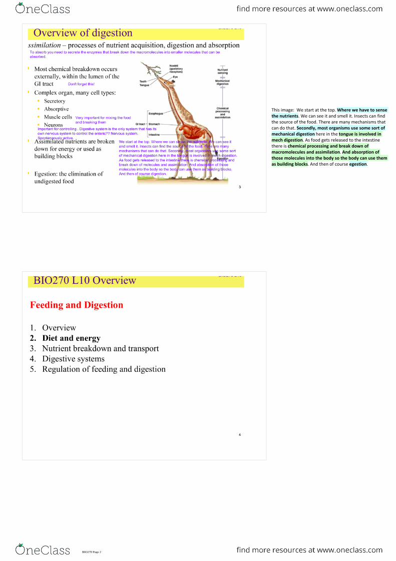
This image: We start at the top. Where we have to sense the nutrients. We can see it and smell it. Insects can find the source of the food. There are many mechanisms that can do that. Secondly, most organisms use some sort of mechanical digestion here in the tongue is involved in mech digestion. As food gets released to the intestine there is chemical processing and break down of macromolecules and assimilation. And absorption of those molecules into the body so the body can use them as building blocks. And then of course egestion.
BIO270 Page 2

Why are some calories not available for animals? This is why. We have our gross energy. The energy which is there. But much of it indigestible. They go straight to the digestive tract and gets released in the feces. The digestible energy which is left, much of that me even though they are digestible is unmetabolizable. That is lost in the urine. The left over is metabolic able energy but even that we don't get all because of specific dynamic action (SDA). As you eat it is energetically expensive. It produces a lot of heat. Specific dynamic action. We use a lot of energy as you eat, you digest, you absorb. You secrete all these enzymes. You use up energy. When you eat a full meal you want to lie down and get that warm fuzzy feeling, that is specific dynamic activity. The amount of heat produced by digestive system.So the net energy is nowhere near the gross energy of the food
There are times when we have lots of food in our body and times when we don’t have lots of food. There is an imbalance in our eating habits.So there should be a mechanism to store the contents of the food. Some animals can't eat always.
<-- to get proteins you have to eat two times as much of food to get the same energy as fat.
<-- you take the molecule, put in there, burn it. Oxidize it. The calorimeter measure the temperature change and converts it to calorie. We don’t burn food, we oxidize them.
We oxidize food carefully.
BIO270 Page 3

We don't have enough of them so we have lot of significant important deficiencies. Vitamin C - scurvy. A connective tissue problem. Can't make enough collagen. Vitamin k - required to blood clot. Without it you get haemophilia. Meaning When you get a cut your blood vessels cant clot you bleed out.
<00 D, and K can be synthesized in body.
James lind was a physician and British navy. He made a discovery. He identified a quality in a fruit which gave his sailors the ability to go and return. The naval ships would left with 1200 sailors and came after two months with 700 - 800 sailors. Because there was a vitamin deficiency. So he found this fruit. This was the first clinical trial ever. Gave everyone some limes, and vinegars, and other fruits and watched what would happen. The ones who got the limes were fine. The deficiency of vitamins C is called vimines? The Americans called the British limens, because they had lot of limes in their ship/
We don’t produce vitamin C. most mammals cant. Most of the research on vitamin c was done on guinea pigs because they can't synthesize their vitamin C as well
Have to get for our diets. Most of them are absorbed around the GI tract.
BIO270 Page 4

12/20 we can produce. The others are called essential AA.Dogs can only produce 10. so they have 10 essential AA.Cats have tauring? Which is not an AA, but essentialCat food doesn’t have taurins, and so cant mix cat and dog food.
We can male palmatate from acetyl CoA.
These must be gotten from the diet.
BIO270 Page 5
We do this by digestive enzymes.
<-- we absorb/ uptake the smaller components.
BIO270 Page 6

BIO270 Page 7

How does it happen? It starts very earl on. We release an enzyme from our salivary glands called salivary amylasewhich breaks down starch and glycogen into oligosaccharides. Occurs in the mouth. Goes to the stomach and breaks down carbohydrates. Very little very little carbs. In the Small intestine there's the release of pancreatic amylase and dischchardases( enzymes that breaks own disaccharides. Like lactase). That leads to the monosaccharide's in the small intestine (for absorption. Almost all absorption takes place here) Are absorbed in the small intestine by a special epithelium cells called enterocytes. Enterocytes are absorptive these cells in the GI tract. Cellulose, for most organisms, including us do not have any environment to break down cellulose. We don't have the enzymes to break it down.
Two situations to look at1) low glucose in the lumen of the GI tract.When there is low glucose, We don't have a strong gradient for glucose to move into the cell . Even if we had a transporter in these low glucose conditions Na/glucose transporter is key. It drags the glucose in along with its electrochemical gradient. Glut 5 transporters bring fructose into these enterocytes. Fructose can exit across the basolateral membrane via glut 5 and glut 2 transporter. Both are shown in the baso lateral membrane. This is how we can get glucose even if we have low glucose situations. Na/ glucose co transporter.
2) high glucose levels.
In high glucose levels why use Na/glucose transporter which requires ATP secondary active transporter. Doesn't use much of the Na/glucose transporter. What it does is It incorporates free form of glut two into the apical membrane. Now we have a high conc of glucose in the lumen lower in the cell. Now the glucose is going to freely pass. Facilitated diffusion. these glut 2 transporters are stored in vesicle within enterocytes. No energy required, especially when you cut these glut two transporters stored in vesicles. The Na glucose co transporter apparently signals these vesicles to move to the apical membrane. If that is the case what transporter is missing in this thing in the figure? It should be there. We saw the Na glucose co-transporter. How does it function? It makes no sense like this. Na K ATPase which will be in the basolateral membrane. If you don't this you don't have the electrochemical, gradient to pull glucose into the cell. It is a secondary active transporter, Na k ATP Ase.
Lmd n
BIO270 Page 8

More people have lactose intolerance these days. We reduce the amount of lactase as we move into adulthood.
Congenital:
• If we don’t have lactase we can't break down lactose.• Lactose goes to the large intestine, the gut flora acts on
it --> produces gas, produces muscle contraction.
○ Because lactose is a carbohydrate and it holds onto a lot of water and water cannot be reabsorbed and results in diarrhoea.
It also produces diarrhoea --> for lactose intolerant people
•
• Reading: lactose is not increasing in coagulation. What happens is that lots of people mix, and those with lactase has been brought up with milk. Lots of places in the world doesn’t use cow's milk.
<--no peptidases in the saliva so starts in the stomach. Enzyme called pepsin the active enzyme is released initially as a pepsinogen which is inactive. This is the pro enzyme. This is important because same as in the pancreatic enzymes, you don’t want this enzyme active in the cells. Because they will start breaking down proteins in the PM in the gut or in other organelles so they are as proenzymes.
Amino acid Na+ co transporters are in eh small intestine
Trypsin, chymotrypsin, Carboxypeptidase: they are initially as trypsionge, chymotrypsinogen, and procarboxypesinogen. The same as before. Don’t want to harm the lumen of the GI tract
BIO270 Page 9
GI tract has to secrete bile --> composed of amphipathic molecules. Positive and negative regions. This is very important. There are bile acids, and also phosphor lipids which are secreted with bile. These leads to emulsification. emulsify, mix up and break into smaller components fat molecules. This is really important. These are large fat globules, They have a really small surface area on the outside in which lipase can break down. If you emulsify, break them into fat globules, essentially then you are increasing the surface area to in which lipase can break down that fat. Or the fat is going to flow straight through and not going to be able break it down and take it in. so bile is important to break down and absorption of lipids.
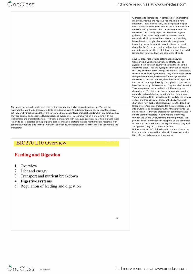
physical properties of lipids determines on how its transported. If you have short chains of fatty acids or glycerol It can be taken up, moved across the PM to the directly to blood. They are hydrophilic they can be moved that way. The most of these large triglycerides, cholesterols, they are much more hydrophobic. They are absorbed across the apical membrane, by simple diffusion, hydrophobic molecules so can cross the PM, then they are incorporated into the ER> thorough the Golgi. Through that transport you have the building of chylomicrons. They are label? Proteins. Tso many proteins are added to the lipids creating the chylomicrons. This is the mechanism in which triglyceride, monoglycerids and cholesterol get into the blood supply. They are released into the lactilc, which leads to the venous system and then circulates through the body. The small, short chain fatty acid of glycerol can get into the blood. But larger glycerol's such as triglycerides they get incorporated into chylomicrons, glycoproteins, they then move into the blood stream --> they are processed at peripheral tissues --> bind to specific receptors --> as those fats are moving through the ER and Golgi ,proteins are incorporated. The proteins binds into the specific receptors on the peripheral tissues. And can break down the triglyceride into fatty acids and glycerol. They can take up cholesterol Ultimately what's left of the chylomicrons are taken up by liver, and reincorporated into a bunch of molecules such a LDL , HDL. (not talking about it too much)
The image you see a chylomicron: in the central core you see triglycroles and cholesterols. You see the materials that want to be incorporated into cells. Can be used To build membranes. can be used for energy. but they are hydrophobic and they are surrounded by an outer layer of phospholipids which are amphipathic. They are positive and negative . Hydrophobic and hydrophilic. Hydrophobic region is interacting with the triglyceroled and cholesterol esters? Hydrophilic interacting with the aqueous extracellular fluid allowing those factors to be transported to the peripheral tissues. Then able proteins that are mentioned are receptors with peripheral protein to bind to them. Allowing the break down/incorporation into those cells of triglyceroils and cholesterol
BIO270 Page 10

<-- separating different function into different regions of the gut.
<-- food goes in , waste goes out exactly from the same opening. This is a huge advance in evolution so now you can secrete your digestive enzymes into the isolated environment, in this case the gastrovascular cavity. Before that they secreted the enzymes into the extracellular fluid or find a way to take them up, the large molecules, and process them inside a vacuole which was not the best way to do them.These organisms developed further and developed direticulum. They increases in the surface area. Secondly, they can allow the organism to specialize. So in one region of the gut you have an acidic region and in the other you may have basic region. This is not perfect, but this is better because there is one flow through.Hydra: is an cnidarian. They are constantly feeding organisms. Capture food with tentacles, have a mucus stream which delivers food into the mouth, and into the gastro vascular cavity if the mouth is open. In the epithelium the gastrodremis they have two primary types of cells. Enzymatic cells and neutral cell. enzymatic cell releases enzymes into the gastrovasular cavity breaking down macromoelcules. Nutritive cells phagocytose, takes up the somewhat broken down food molecules into the cell. In the food vacuoles there is more cleavage Of the macrmoelcules. In the end these vesicles move through the nutritive cell, released into the epidermis and providing energy to the epidermal cells, the muscle, neuronal cells. This is the two way gut
This is also called the GI tract. Very distinct functionally and anatomically separate regions. You have pharynx the esophagus, involved in the chemical breakdown of food, the stomach which is in the acidic regions which is important for the enzymes functioning in that regions.
<-- further digestion. Almost all the absorption via the enterocytes
<-- mostly absorption of water, some ions.
<--- release of indigestible material
Note that there are sphincters: they separate some of the anatomical regions, because enzyme in the stomach function at low pHs. Enzymes in the intestine will not function at low pH. So they separate these regions and create micro environments. The stomach of the horse for example create these regions. Sphincters can be controlled by the nervous system.
BIO270 Page 11

This is one of the evolutionary advances --> Increase in surface area of GI tract.
More surface area --> more transporters you can have --> the more time food spend in the gut --> the more nutrients you can absorb and break down. You don’t want the food to go too quickly through the gut or you won't be able to absorb nutrients. But on the other hand you don’t want this indigestible material hanging around. Most organisms increase the surface area or increase the length or both. So the tissue levels, the circular folds. At the tissue level there is villi. Increasing the surface area again. Blow the villi: on top of the enterocytes on the villi there are microvilli. These are formed by microfilaments. Important for the structure. This is called the brush order within the intestine,. Significantly increasing the surface area for the secretion of enzymes into the gut or absorption of small molecules. Specialize dcompaortnets
<-- increase the efficiency of digestion Because you can close off one region from another sequentially. You can't do this easily in a two way gut.
<-- alter pH: Also specific enzymes are released in one region, mucus is released in one region.
<-- and complexity : of gut morphology significantly is varies. Real Complexity at the ease of digestion (like upon what they eat): like plant eaters they have much longer stomachs with larger surface area because it is much harder to extract energy from them than in animals.
<-- gut morphology changes within taxa too: the insects have larva stage, which is an eating machine eating green leafy plant material, it pupates and become a nectar eater. So completely different guts. There is difference within and between guts. Female mosquitoes drink blood, males feed on nectar. So there is different in the gut morphology.
Talked about humans, start off with nutrients crossing the placenta, then you get only milk then you only get different types of diets. There is significant variation within taxa on what you eat developmentally. Great differences.
BIO270 Page 12

This is the guts of various animals. Lots of variation.Have a simple straight gut and is way shorter than the animalSharks: have a spiral valve, food has to go through the spiral surface and this increases the surface area, slows down the movement of food so you can enzymatically break down the foods or nutrients can be absorbed.
1. Increase surface area so there is more area for secretion of enzymes and absorption of molecules
2. Microbes: house microbes. Bacteria is not the only one that can be housed. Protozoans can also be housed. So should be microbes.
Many organisms have ceca: have two functions
Chickens/ birds : have a very interesting traits, They have a crop which is an extension of the esophagus. Which does stores food. The proventiculus is the active stomach of the birds.
Pigs: huge stretch which could go half way round this hall. Significant increasing the length and microvilli.
(and microbes)
The fact is that we can't break down cellulose. But Some organisms can. They can do that because they have specific microbes within certain regions of the gut/ ceca. Bacteria , fungi, photosynthetic organisms.
<-- most common well studied. Found within the lumen of the GI tract/ ceca. The first mage is a termite: they eat wood, but they can't eat wood on their own. But they store protozoans (second image) in the gut which can cleave the bond in cellulose (beta-1,4 bond on cellulose) we can only cleave the alpha 1,4 bond in starch.
<-- actively structure symbioses outside of the body. The leaf cutter ants. They bring that back to the fungal garden and they take care of the fungus that breaks down the leaves and then the leaves and fungus they have double?? They can break cellulose down and able to incorporate.
<-- much more less common, but they are out there. Deep sea vents, (rifts of large worms in the bottom of the ocean) they have symbiotic relationship between organisms that can oxidize H2S.
And symbiotic theory: mitochondria was thought to be phagocytosed into a cell, a bacteria, and incorporated into it. Now, symbiotic relationship in everyone.
BIO270 Page 13

<-- so ceca is a modification that improves the digestion. this is an accessory region in the gut. But ruminant is slightly different. They have a digastric stomach. Two regions in the stomach. The rumen in the reticulum, the first two regions in the stomach where initially the food goes. that’s where they found the fermentative bacteria in the stomach. You take the food in very quickly --> very little chemical breakdown is done --> goes into the rumen and the reticulum where they keep the fermented bacteria which breaks down the cellulose. They would reverge the food back into the mouth --> chew it and then after it is been partially broken down by the bacteria--> goes back down the esophagus but now it goes to the omasum and the abomasum. Abomasum is the glandular stomach, the acidic region of the stomach.
BIO270 Page 14

Salivary glands begins the breakdown of carbohydrates. They are collection of several exocrine glands: secreted by ducts outside the body into the GI tract. Ducts open to the mouth, and multicellular.Have mucus secreting serous cells which is a clear fluid with enzymes in it. Such as salivary amylase. This is important because it lubricates food and dissolves food and food can bind to receptors and stimulates the signal transdcution pathway. Cleanses the mouth with antimicrobial properties.
< salivation is controlled by parasympathetic nervous system. Stimulates the production of saliva. Sympathetic nervous system also stimulates the production of the saliva. Clear fluid with more salivary amylase.Sympathetic nervous system inhibits and also stimulates the salivation. 29. 00
Image: dogs have 4 sets of glands. We don’t have the orbital gland but we have the other three. All of these contribute to the production of saliva. Serving these functions.
Surface involves the columnar epithelium cells joined by tight junctions. This is important in the gut because it has a very acidic gastric juice in the stomach. And don’t want to leak.
4 cells are found within gastric pits or the walls of the stomach.
Mucous neck cells.Pariteal cellsChief cells - pepsin is secreted as an inactive precursor pepsinogen by this cell. This is activated by the HCL produced by the parietal cells.
G cells: called this because synthesize and release gastrin. And they are enterochromatin cells which release histidine.
Enterendocrine cells: endocrine is secreted into the blood. The first three secrete into the lumen of the gut. This secretes into the blood.
This guys ( I think showing an image) : they are bacteria found within the gut. The acidic compartment kills about everything and the pH there is 1-2. But this bacterium finds a way to survive. This is what produces gastritis, peptic ulcers and is associated with hylubacterim? This is found in the maze of this pit. It escapes the high acidity of the stomach.Also the gastric pruning frog? - (shows image): there were only two species found they both are extinct. They lived in Australia. The parents takes the young embryos into the stomach and the embryos secrete prostaglandin E2. which actually inhibits the production of HCL and allows this organisms to develop within the stomach of the gastric brooding frog. There is project called Lazarus project trying to bring them back again in university if new south wales.
BIO270 Page 15

Intestines: Structure - most nutrients are absorbed here. Forms the mucosa where you found the villi and the crypts. This is where the important cells are produced.
Sub mucosa is where you find the blood lymphatic vessels and the nerves which supply …?
Circular smooth muscle and longitudinal muscle: have two forms of smooth muscle allowing the gut to shorten and lengthen and allowing the circular diameter to change. length longitudinal.
Number of mucosal cells: Histologically: enterocytes are the primary absorptive cells in the GI. Have lots of microvilli increasing surface area.
Into the blood
<--- secrete antimicrobial molecules important in protecting stem cells produced at the bottom of the crypt of lieberkuhn. Cells in the GI tract, stomach and the intestine generally are there about 3 days. They are constantly been swapped off and reproduced by stem cells.
<--- goblet cells: secreting mucus
<-- secrete into the blood
BIO270 Page 16

Another secretion:
Into the duodenum, the upper portion of the small intestine.
And phospholipids which are also amphipathic <-- this shows what the amphipathic molecules can do.
Hydrophobic side shields from the aqueous extracellular fluid. Hydrophilic meets the aqueous side.
This is easy to get rid of hydrophilic waste products can send them straight to the urine. But hydrophobic ones are harder. One of the important ones we have to get rid of are breakdown of red blood cells. Break down of bilirubin. This is hydrophobic molecule and it is secreted with bile and released by desiccation?
Pancreases ducts is also released into the duodenum. The upper small intestine.Secretes as inactive pro enzyme protecting the cells which produce these enzymes. Activated upon release.
<--- these are released by the pancreatic juice.
Trypsiongen is released to the small intestine. This is acted upon by membrane bound enterokinase producing trypsin. Which then goes to activate chymotrypsin and Carboxypeptidase. Which were in the precursor form chymotripsinogen and procarboxypeptidase. What this doesn’t show is that trypsin can also cleave trypsiongen.one trypsin molecule and activate chrymptrypsiongen and and procarboxypeptidase and also other protrypsin molecule. This can now break down proteins in the small intestine.
BIO270 Page 17

Enzymes feed into wild animals: requires and energy and puts the animal at risk. This risk has to be waived. How much risk is there, and how badly I need the food. This is controlled by the CNS. The signal is sent to the hypothalamus. To feed. When you can't provide the fuel required for the circulation fuels we tend not to use the storage forms of the fuel unless we have to. We go back and eat so we have more circulation fuels.
There are three which are found to be particularly important in humans in controlling appetite and feeding. Leptin, peptide Yy and ghrelin. Leptin: secreted by
BIO270 Page 18

Leptin is considered to be as an white adipose stat? lots of lipids in adipose tissue and Leptin is released to inhibit appetite. Leptin is secreted by white adipose tissue when the lipid content of the white adipose tissue is high. Which is different from brown adipose tissue.
<-- peptide YY is secreted when the colon is full
<-- ghrelin stimulates appetite. Released when stomach is empty.
Leptin mouse: the one on the right is a normal mouse with normal Leptin.The left one has no Leptin. And can't stop eating. And gets enormous. Thought that when You pump in Leptin you can stop the appetite. But unfortunately this doesn’t work. Leptin doesn’t have the direct effect we want to have. So its long term adipose stat. Regulating the amount of stored fuel supply in terms of fat.
When lots of lipid is stored you don’t want to eat anymore
These hormones have an effect on the arcuate nucleus? On the hypothalamus where you find a number of cells which have receptors on for these hormones. These hypothalamic neurons release neurotransmitters in response. So there are synapses on the neurons, higher centers in the brain which influence the feeding behavior. Some of these neurotransmitters stimulate appetite. Pre neurotransmitters which are released from one type of neurons on the brain. from the same neurons you get all the peptides ( on slide)But we concentrate only on peptide Y.
There are neurotransmitters releases in response which inhibits appetite:POMC is not a neurotransmitter: it’s a pro hormone. There are three or four peptides which are released from proopiomelanocortin to differential cleavage in tissues.Important ones for us is alpha MHS: alpha melanocyte stimulating hormone. One of the cleavage products of precursor POMC.
BIO270 Page 19
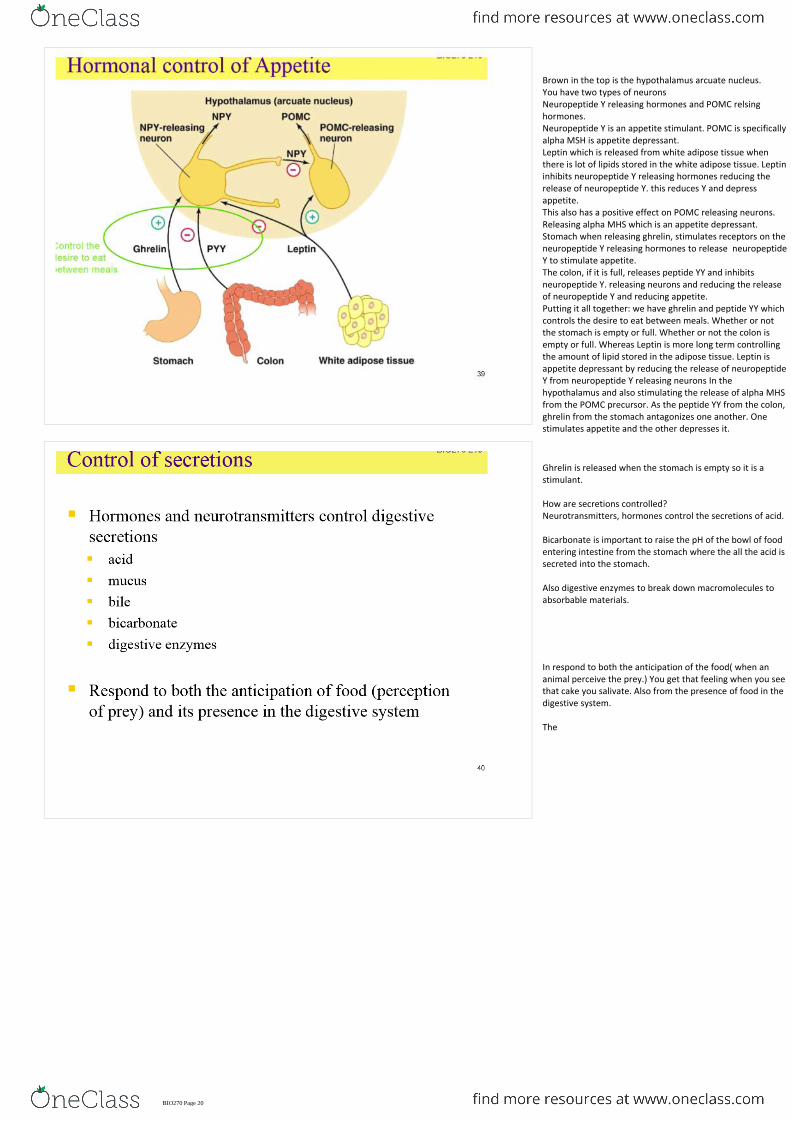
Brown in the top is the hypothalamus arcuate nucleus. You have two types of neuronsNeuropeptide Y releasing hormones and POMC relsing hormones.Neuropeptide Y is an appetite stimulant. POMC is specifically alpha MSH is appetite depressant. Leptin which is released from white adipose tissue when there is lot of lipids stored in the white adipose tissue. Leptin inhibits neuropeptide Y releasing hormones reducing the release of neuropeptide Y. this reduces Y and depress appetite.This also has a positive effect on POMC releasing neurons. Releasing alpha MHS which is an appetite depressant.Stomach when releasing ghrelin, stimulates receptors on the neuropeptide Y releasing hormones to release neuropeptide Y to stimulate appetite. The colon, if it is full, releases peptide YY and inhibits neuropeptide Y. releasing neurons and reducing the release of neuropeptide Y and reducing appetite. Putting it all together: we have ghrelin and peptide YY which controls the desire to eat between meals. Whether or not the stomach is empty or full. Whether or not the colon is empty or full. Whereas Leptin is more long term controlling the amount of lipid stored in the adipose tissue. Leptin is appetite depressant by reducing the release of neuropeptide Y from neuropeptide Y releasing neurons In the hypothalamus and also stimulating the release of alpha MHS from the POMC precursor. As the peptide YY from the colon, ghrelin from the stomach antagonizes one another. One stimulates appetite and the other depresses it.
Ghrelin is released when the stomach is empty so it is a stimulant.
How are secretions controlled?Neurotransmitters, hormones control the secretions of acid.
Bicarbonate is important to raise the pH of the bowl of food entering intestine from the stomach where the all the acid is secreted into the stomach.
Also digestive enzymes to break down macromolecules to absorbable materials.
In respond to both the anticipation of the food( when an animal perceive the prey.) You get that feeling when you see that cake you salivate. Also from the presence of food in the digestive system.
The
BIO270 Page 20

The anticipation of the food stimulates the parasympathetic neuron --> they have direct effect on the parietal cells which releases acid into the stomach. Parasympathetic neurons also activate G cells which release gastrin hormone --> stimulating enterochromofine cells which release histamine which also stimulate parietal cells to release acid.Gastrin also stimulates chief cells to release pepsinogen which gets activated in the stomach to pepsin by HCL. (pH 1-2)Also when you ingest food it activates chemoreceptors and mechanoreceptors, in the gut, esophagus, mouth etc.This has two effects:Can directly stimulate the G cells. ( gastrin)Can Also affect the enteric nerves which are inherent in the gut. The gut has its own nervous system, a physiological system which has its own nervous system.These enteric nerves can also effect G cells. G cells are the commanders . Everything goes through the G cells.
Second image: how does this work? Talked something like this in osmoregulation. We talked about proton ATPase. But in this the important pump is the K/H pump. This is an ATPase. Pumping protons into the lumen of the stomach in exchange for K. it also has Cl- channels. Cl- (negative)following the K+ (positive) into the stomach. That’s how we have the HCL secretions into the stomach. What's so important is the regulation of pH. Is the presence of the enzyme carbonic anhydrase. Hydrates Co2. producing carbonic acid which dissociates into protons and bicarbonate. Protons which supplies K/ H pump so that it doesn't run out of protons and bicarbonate which is exchanged for Cl- making sure you have enough Cl- to get into the stomach. Carbonic anhydrase is able to hydrate Co2 producing that proton, this is what supplies the proton and the bicarbonate is exchanged for Chloride. Of course we have the Na/K ATPase in the basolateral membrane.
Acid reflux: you may have taken papsidase C or anti? How do they work? They inhibit and binds to histamine receptors on parietal cells. The histamine released from the enetrochromaphin cells cannot stimulate the parietal cells to release acid to stomach. There are also other medicine which specifically targets the K/H ATPase. They fncion by inhibiting that pump which reduces the prodcutionof acid by parietal cells.
From the intestine we start to break down molecules. So we have acid and food entering the duodenum, the first section after the stomach.
1. Vasoactive intestinal peptide - works on the pancreas to release HCO3- to increase pH. Why? Important because the enzymes whicha re going to function in the intestine is not going to work in acdic level.
2. Secretin - stimualtes the prodcution and release of bile. Bile also has bicarbonate so contributes to the icnrease of pH. This stimulates the liver to produce bile.
Acids stimulate the enteroendocrine cells to stimulate two hormones:
Digestive products( AA, FA, small peptides) stimuatles the endocrine cells to release CCK (cholysictokinenie)
1. Stimulates the smooth muscle in the gallbladder to release the bile into the duodenum into the upper intestine.
2. Stimuatles the pancrease to release enzymes. Also the trypsinogen, chrymotrypsiogens, lipasess, nuclease
Has two effects:
Because all of the secretions are aquesou solutions and body is losing a lot of water as it secretes them in the digestive tract. Osmotically we have to recover most of those water.
BIO270 Page 21

Gut is controlled by muscles. Muscles which are controlled by neurons.Food moves along the GI tract in contractile waves. This is called peristalsis. This occurs because of intrinsic myogenic activity. Similar to pacemaker cells which spontaneously depolarize and don’t need signals. Receives signal from CNS but not directly to the muscle. Signals from the CNS go to the myentric plexus which is part of the enteric nervous system which controls the gut alone.CNS signals the gut VIA enteric nervous system. sturcturE: you have the intestinal vili, circular smooth muscle and longitudinal smooth muscle. In between that you see the myenteric plexus. Neurons which controls the actiivty of gut. They also control thesecretion of digestive enzymes.
What is important: controls the speed of contracions. If contracts too fast cant acsoprb nutrients. Too slow, then there is indegesible food so there shoulde a perfect balance. This happens because of the interstitial cells of cajal and also the effects of CNS on those cells.
How does it work: smooth and longitudinal muscles. So smooth muscles show rhythmic cycles that move along tract through peristalsis. ( From interstitial cells. If you look at step 1: you see contrction of circular mucsles. Circular muscles are invovled in controlling the diamtere. Logitudinal muscles : length. So contraction of circula smooth musclesinfront of the food.
Step 2: contraction of longitudinal muscles shortening the gut. At the same time you have relazation of the smooth muscles. (figure doesn’t show). Contraction of circulat infront of the food, relaxation behind the food, and contraction of the longitudinal muscles shortening the gut and reuslting in movement.This is peristalsis.
BIO270 Page 22
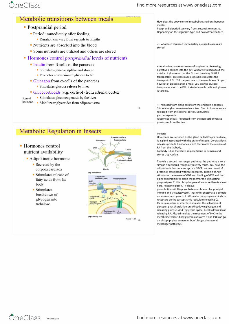
How does the body control metabolic transtiions between meals?Postprandial period can vary froms seconds to months. Depending on the orgnaism type and how often you feed.
<-- whatever you need immediately are used, excess are stored.
<--endocrine pancreas: iseltes of langhaerns. Releasing digestive enzymes into the gut. When we talked about the uptake of glucose across the GI tract involving GLUT 2 transporters, skeleton muscles insulin stimulates the transport of GLUT 4 tranpsorters to the membrane. So you have lot of glucose after a meal, you put the glucose tranpsoeters into the PM of skeltel muscle cells and glucose is take up.
Steroidhormone
<-- released from alpha cells from the endocrine pancres. Stimulates glucose release from liver. Steroid hormones are released from the adrenal cortex. Stimulates glucoenogenesis.Gluconeogenesis: Produced from the non carbohydrate presurosrs from the liver.
Insects:Homrones are secreted by the gland called Corpra cardiaca, Is a gland associated with the brain of insects. Corpra allata: releases juvenile hormones which Stimulates the release of FA from the fat body. Fat body is like the white adipose tissue in humans and stores triglyceride.
There is a second messenger pathway: the pathway is very similar. You should recognize this very much. You have the adipokinetic hormone receptor a GPCR. Heterotrimeric G protein is associated with this receptor. Binding of AdK stimulates the release of GDP and binding of GTP and the alpha subunit moves along the membrane stimulating phspholipase C. this phospholipase does more than is shown here. Phsopholipase C --> cleave phosphptilinositolbisphosphate membrane phsopholipid into IP3 and triecyloglycerol. Inositolbisphosphate is soluble on aqueous cytoplasm. It diffuses to the cytoplasm binds to receptors on the sarcoplasmic reticulum releasing Ca. Ca has a number of effects: stimulates the activation of glycogen phosphorylation breaking down glycogen and releasing glucose. And triglycerid lipase, breaks down lipase releasing FA. Also stimualtes the moement of PKC to the membrnae where diacylglycerola ctivates it and PKC can go an phsophprylate someone. Don’t forget the second messenger pathways.
BIO270 Page 23

<--if that postprandial? Happens too long it leads to starvation where you have the reorganization of metabolism.We have tissues that function only with glucose: e.g.: brain and red blood cells.Muscles shift to lipid metabolism. If there is lots of glucose muscles select glucose but in starvation response they switch to lipids saving glucose.Then when these stores are depleted body starts using proteins. We don’t have a store of protein in our body like glycogen and lipids. But it is stored in our skeleton muscles. So then the skeleton muscle starts to break down producing AA which can then feed into pathways to produce energy.
Early starvation:Adipose tissues releases FA and goes to liver.Liver sends glucose it produces to the brain.Skeleton muscles starts to use triglycerides to save that glucose which is been produced by the liver to the brain. As we lose that lipid and glycogen sotre, liver starts producing ketone bodies so brain can use them.You can see the muscles start to break down. We need the energy to supply the necessary tissues of the brain. AA are sent to the loiver wher they can feed into different pathway. They can feed into the trchloroacidic cycle. In the end generates energy.So we have this reorganization tissues stop using glucose and start using FA if they can.
In late starvation we have the breakdown of proteins from muscles which therefore allows those tissues to use AA to produce energy. And ketone bodies are produced by liver is released into circulation and can be used by the brain In late starvation response.
BIO270 Page 24


![Med · 270 342 [(+72) (270+72) 472 (270+72 [(+72) tztžU +130) (+102)] (+130) 520 (270+250) 1270+102 752 (270+102 (+480) 750 270+480) 852 (270+102 (270+102](https://static.fdocuments.net/doc/165x107/5fb23750d464052f95224679/med-270-342-72-27072-472-27072-72-tztu-130-102-130-520-270250.jpg)