Bio 211- Anatomy and Physiology I Today’s topics Tissues.
75
Bio 211- Anatomy and Physiology I Today’s topics •Tissues
-
Upload
emil-mcgee -
Category
Documents
-
view
230 -
download
2
Transcript of Bio 211- Anatomy and Physiology I Today’s topics Tissues.

Bio 211- Anatomy and Physiology I
Today’s topics •Tissues

Structural hierarchy in human biology
Human Body
Organ system (cardiovascular system)
Organ (Heart)
Tissue (cardiac muscle)
Cells (cardiomyocytes)
Organelles (nucleus, ribosome)
Macromolecule (Protein, DNA)
Molecule (amino acid)
Atom (nitrogen, carbon, oxygen)Smallest
Biggest, most complex
DONE!!

Histology
The study of cells and tissues and how they are arranged into organs• Allows us to see how cells are arranged in a particular
tissue type and how tissues help form an organ
•200+ types of cells in our bodies are broken down into 4 primary tissue classes or categories:1. Epithelial
2. Connective3. Nervous4. Muscular

Tissues
A group of similar cells derived from a common embryonic origin that are arranged in a way that allows them to carry out a particular STRUCTURAL or PHYSIOLOGICAL function• STRUCTURAL
Bone (type of connective tissue) supports and protects the body and allows muscles to produce movement
• PHYSIOLOGICAL Epithelial tissues lining the digestive system allows the body to absorb nutrients
from the foods we eat
• Many tissues have both a structural and physiological function (bone)
Tissues differ from one another in the types of cells which make up the tissue, as well as by the type and amount of EXTRACELLULAR MATRIX that surrounds the cells
• Matrix contains protein fibers, water, minerals, nutrients, waste products
Tissue = cells + extracellular matrix (or extracellular material)

Studying Histology
Biologists use microscopes to examine tissue sections (very thin slices) colored with various stains or antibodies (used to visualize proteins) to determine the location and arrangement of cells within a tissue
Depending on the stain used, you can visualize cytoplasm, nuclei, even specific proteins!
Purple nuclei
Pink cytoplasm
Brown stain shows location of a specific lung protein

Epithelial Tissues
EPITHELIAL TISSUES are characterized by a layer of tightly bound cells that are one or more layers thick (1-20), bound to a BASEMENT MEMBRANE and are usually exposed to the outside environment or to the inside of the body
• Examples : skin, lining of digestive system, lining of lungs, lining of blood vessels, etc…
Epithelial cells
Basement membrane Connective tissue
Basement membrane acts like a biological glue to hold cells to connective tissue underneath• composed of collagen, and a variety of glycoproteins and proteoglycans

Classification of epithelial tissues
Cell Shapes1. Squamous – thin, flat
• Good for exchange of gases, nutrients, waste products (lung and blood vessels)2. Cuboidal – typically square or round
• Found lining the ducts of many glands3. Columnar – tall and narrow
• Found lining the intestines• Often have small projections on exposed surface (MICROVILLI) that increase
surface area for absorption

Classification of epithelial tissues
1. Simple – have a single layer of cells
2. Stratified – Tissue contains a few to many layers of cells on top of each other• Only bottom layer are bound to basement membrane• Upper layers have cells bound to other cells
3. Transitional – Specialized type of epithelia found in organs that stretch
4. Pseudostratified columnar – all cells are bound to the basement membrane but not all cells reach the exposed surface.

Epithelial tissue histology
Simple Squamous:Single, layer of thin cells
Simple cuboidal:Single layer of cube or round cells

Simple columnar:Single layer of tall cells, all
cells reach the exposed surface, microvilli present
Pseudostratified columnar:Single layer of columnar cells,
nuclei are scattered, not all cells reach exposed surface
Microvilli

Keratinized, stratified squamous epithelia:Many layers of cells, lower cells are alive and dividing, upper cells are dead and filled with
keratin, CANNOT SEE NUCLEI
Non-keratinized stratified squamous epithelia:
Many layers of cells, all alive, CAN SEE NUCLEI

Transitional epithelium:Cells are alive and overlapping, may appear
single layer when tissue is stretched
-Found in tissues that need to stretch on a regular basis
-Have a cuboidal appearance in a relaxed tissue but “TRANSITION” to a squamous appearance when stretched

Tips to identifying and classifying epithelial tissues
1. Try to identify how many layers of cells are between the basement membrane (bottom) and the exposed surface. Living cells will have visible nuclei, but dead keratinized cells will not.
2. Try to identify the shape – thin, flat (squamous), cube or round (cuboidal), tall and thin (columnar)
3. Number of layers and shape of cell tells you the name of the tissue• Stratified sqaumous (SEVERAL LAYERS of THIN, FLAT cells)• Simple cuboidal (ONE LAYER of CUBE-SHAPED cells)• Etc….
Since epithelial tissues are often found lining something you will typically see epithelial tissues next to open spaces (white space on a microscope slide)

Connective Tissue
Tissue derived from the mesoderm that provides protection and support for the body and its organs, often composed of dense fibers, large amount of extracellular
material with relatively few cells
Major Functions:1. Connection – connective tissue binds bones to muscle (tendons), bones to other bones
(ligaments) and keeps organs in place2. Support – bones help support the weight of the body, cartilage supports structures like
the nose and ear3. Protection –
• Physical- delicate organs like the brain, kidneys, eyes, etc… are often surrounded by bone and adipose tissue for protection
• Immune – LEUKOCYTES (white blood cells) and LYMPHATIC organs are connective tissues that protects the body from invasion by bacteria and viruses
4. Movement – Bones help move body with help of muscle, muscles attached to bones compress some body cavities
5. Storage and heat production – Adipose tissue (Fat) stores energy for later use and preserves/generates heat
6. Transport – Blood transports gases, nutrients, waste, hormones to distant parts of the body

Types of connective tissue
1. Fibrous connective tissue – • What most people think of when talking about connective tissue• Ligaments and tendons contain fibrous C.T.
2. Cartilage – • Rubbery C.T. that supports nose and ears, found between bones
3. Bone – • Rigid C.T. that protects body organs, supports weight of body, helps
produce movement4. Blood –
• Fluid C.T. that functions to transport materials throughout body, also serves protective immune function through leukocytes

Fibrous Connective Tissue
A diverse type of C.T. composed of cells, fibers, and ground substance (appears as empty space)
1. Cells – • Fibroblasts – large cells that produce the fibers and ground substance
• Leukocytes (WBCs) – important part of immune system that monitors body for invasion and coordinates immune response
Lymphocytes produce ANTIBODIES when a bacteria or virus is detected.o Antibodies are proteins that recognize specific proteins on surface
of invaders and “flag” them so macrophages can destroy them Macrophages – phagocytic cells that look for, engulf, and destroy bacteria and
viruses• Adipocytes – cells designed to store large vacuoles of fat, cellular part of adipose
tissue

2. Fibers – • Collagenous – made of a protein called COLLAGEN
Very strong and flexible Major component of tendons and ligaments
• Reticular fibers – collagen fibers COATED WITH GLYCOPROTEINS Found inside several organs for structural support
• Elastic fibers – Thin fibers made of the protein ELASTIN Elastin fibers are coiled like a “slinky” and allow the fibers to stretch
and then return to normal shape Very important for elasticity of skin and lung tissue (makes things
stretchy)
3. Ground substance – • Fills the space not occupied by fibers and cells• Composed of large carbohydrates, proteoglycans, and glycoproteins• Cushion and protect the cells of the C.T.• Many molecules are negatively charged and attract Na+ ions and water
Chondroitin sulfate is part of the ground substance and is taken by many people with joint problems (Glucosamine and chondroitin)

Types of Fibrous Connective Tissue
1. Loose fibrous connective tissue
• Much more ground substance than fibers and cells 3 Types – Areolar : many randomly oriented fibers (COLLAGEN AND ELASTIN)
Reticular: many randomly oriented RETICULAR fibersAdipose: Few fibers and many adipose cells filled with lipid
2. Dense fibrous connective tissue• Many more collagen fibers than ground substance and cells
2 Types – Dense regular: collagen fibers are densely packed and parallel to each other.
Dense irregular: collagen fibers are densely packed but run in random directions.

Areolar tissue – relatively few fibers, lots of ground substance, randomly arranged fibers
Reticular – reticular fibers are loosely arranged and tissue possesses many lymphocytes
Note: we will not see this tissue in lab
Adipose – very few fibers, tissue contains MANY adipose cells that look empty (where lipids are stored)
Loose Fibrous Connective Tissue
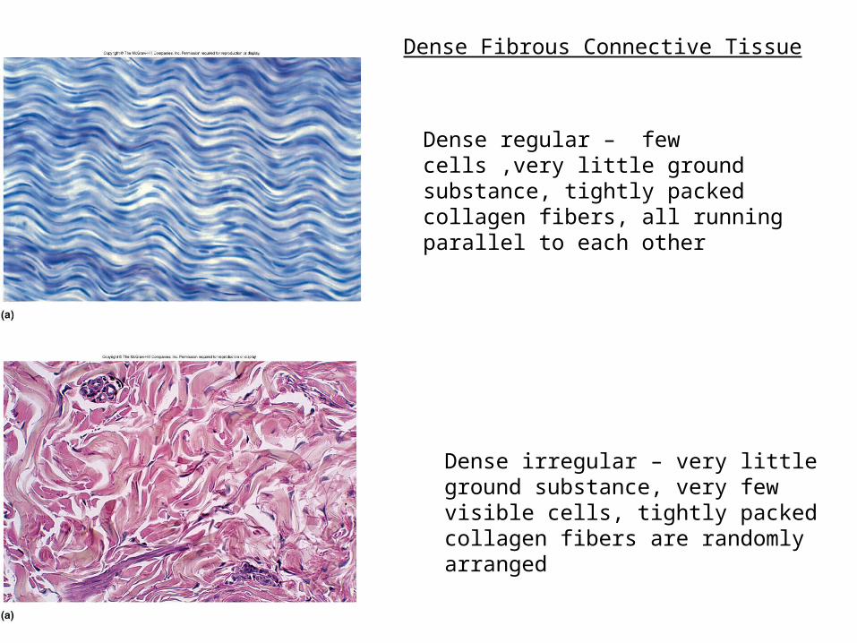
Dense regular – few cells ,very little ground substance, tightly packed collagen fibers, all running parallel to each other
Dense irregular – very little ground substance, very few visible cells, tightly packed collagen fibers are randomly arranged
Dense Fibrous Connective Tissue

Tips for identifying fibrous connective tissues
1. First look at relative amount of fibers • Many fibers=dense C.T.• Few fibers=loose C.T.
2. Next, look at the direction the fibers are running• Parallel = Dense regular• Random=Dense irregular or any of the loose C.T.
3. Finally, look at the relative amount of ground substance and cells• On histology slides, ground substance usually looks like open space• Lots of open space and a few cells = areolar• Few fibers and open space is filled with cells (lymphocytes, look for nuclei) = reticular• Few fibers and BIG OPEN SPACES where lipid vaculoles used to be = adipose
Sort of has a honeycomb structure

Bio 211- Anatomy and Physiology I
Today’s topics •Connective tissue•Bone•Blood

Cartilage
Flexible, rubbery connective tissue that plays an important supportive role• Ears, nose, between bones of some joints
•Cells of cartilage that secrete the matrix are known as CHONDROBLASTS, once surrounded by matrix (in small cavities called LACUNAE), they are known as CHONDROCYTES
Three types of cartilage:
1. Hyaline cartilage – has a clear, glassy appearance; contains many thin collagen fibers• Usually surrounded by PERICHONDRIUM made of dense, irregular CT (protective
covering)• Precursor to osseous tissue, • Articular cartilage, trachea, larynx, costal cartilage2. Elastic cartilage – contains many elastin fibers• Provides flexible support for the outer ear, part of larynx
3. Fibrocartilage – contains many course, collagen BUNDLES, rather than random collagen fibers, like in hyaline cartilage• Supportive CT that resists compression and absorbs shock• Found in pubic symphysis and between vertebrae (intervertebral discs)

Histology
Hyaline cartilage:Smooth, glassy appearance; individual fibers usually can’t be seen; often surrounded by
fibrous PERICHONDRIUM

Bone
•Technically, the word “bone” describes bone as an organ………. and not bone tissue
•OSSEOUS TISSUE is the connective tissue commonly known as “bone”
•All bones are covered by fibrous tissue known as PERIOSTEUM (similar to
perichondrium) that serves as an anchor point for attachment of tendons and ligaments
Two types of osseous tissue (bone):1. Spongy bone – osseous tissue found in the heads of long bones;
microscopically, the tissue looks like a sponge
2. Compact bone – dense osseous tissue that surrounds spongy bone
We’ll learn much more about bone anatomy and physiology shortly!!

Compact Bone
•Bone cells called OSTEOCYTES live in lacunae, like in cartilage
•Blood vessels and nerves run the length of the bone in canals called HAVERSIAN CANALS (sometimes called Central Canal)
•Osteocytes live in lacunae in concentric circles around the haversian canals (that’s where the blood supply and food is!)
•One OSTEON consists of a Central canal and rings of osteocytes that are laying down bone matrix
Blood
•Fluid connective tissue that plays an important role in transportation of many substances and also very important in immune protection via LEUKOCYTES (i.e., white blood cells)
•Non-cellular ground substance is known as PLASMA
•Cells and cell fragments are referred to as FORMED ELEMENTS of blood:
• Erythrocytes (red blood cells)The only cells in the body without a nucleusResponsible for carrying O2 and CO2 between lungs and body tissues
• Leukocytes (white blood cells)5 different types of cells all with specific functions that contribute to
immune protection• Platelets
Cell fragments that are important in the process of blood clotting and directing new blood vessel growth

Blood
•Consists of plasma, erythrocytes (RBCs) and leukocytes (WBCs)
•Erythrocytes are round, with a faint center, and NO NUCLEUS
•Leukocytes are larger and have a large, obvious nucleus
•Different types of leukocytes can be identified by the shape and size of its nucleus (later on in Bio 212)

Nervous tissue
Specialized, “excitable” tissue found in central nervous system and peripheral nervous system
• Tissues receive and transmit electrical signals (called an ACTION POTENTIAL) from one part of the body to another
Action potentials are generated when ions flow back and forth across the cell membrane
• Central nervous system = brain and spinal cord• Peripheral nervous system = all the other nerves in our body
• Cells are excitable, meaning they receive and respond to stimuli very rapidly and can transmit signals as well
• Cells of nervous tissue : NEURONS (nerve cells) – cells that transport signalsGLIAL CELLS – support cells in the nervous system that nourish and protect the
neurons

Neuron anatomy:
•SOMA = cell body, often round or oval shaped. The soma contains the nucleus and other organelles.
•DENDRITES = short processes that receive signals coming from other neurons
•AXON = large nerve fiber that sends signals to other neurons or eventually to final organ or tissue

Muscle Tissue
Specialized tissue with cells which are designed to contract when stimulated
Muscle contraction exerts force on other tissues and organs• Required for movement of bones, movement of food through digestive
system, breathing, movement of the eyeballs, constriction of blood vessels etc…
3 types of muscle tissue
1. Skeletal muscle – long cylindrical cells are called MUSCLE FIBERS• Overlapping ACTIN and MYOSIN protein fibers cause STRIATIONS (alternating light and
dark bands)• Voluntary muscle tissue- typically under conscious control• Long, cylindrical and usually multinucleated
2. Cardiac muscle – shorter, often branched cells are called CARDIOMYOCYTES• Also striated• Contain one centrally located nucleus often surrounded by a clear “halo” • INTERCALATED DISKS are found at the ends of each cell
These are connections that allow signals to travel from one myocyte to another allowing all the cells of the heart to beat at nearly the same time
• Cardiac muscle is INVOLUNTARY, meaning we can’t consciously control contraction

3. Smooth muscle – long cells that are tapered at the ends (thinner at the ends than in the middle)• Smooth muscle cells ARE NOT STRIATED• One nucleus per cell• Smooth muscle is involuntary• Body uses smooth muscle to control many important physiological functions
Dilation and constriction of the pupil in the eye Contraction of GI tract (esophagus, stomach, intestines) Dilation and constriction of blood vessels
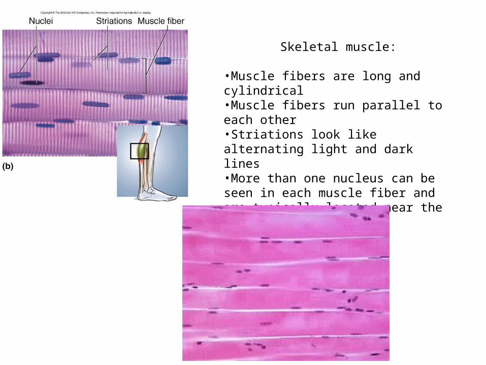
Skeletal muscle:
•Muscle fibers are long and cylindrical•Muscle fibers run parallel to each other•Striations look like alternating light and dark lines•More than one nucleus can be seen in each muscle fiber and are typically located near the plasma membrane

Cardiac muscle:
•Cardiomyocytes are shorter than muscle fibers and are often branched
•Visible striations
•Intercalated disks appear as thick dark bands on each end of the cell
•Single nucleus is centrally located

Smooth muscle:
•Smooth muscle cells overlap each other and are short and tapered•Single nucleus that is located in the center of the cell•NO STRIATIONS!!

Tips for identifying different types of muscle:
1. First, look for striations• YES – it is either skeletal or cardiac• NO – it must be smooth
2. Look at the nuclei and shape of the cell• Skeletal muscle fiber has more than one nucleus and cells looks like a long cylinder• Cardiac myocytes are shorter and are often branched and only contain one nucleus
surrounded by clear open space

Cell Junctions
With the exception of blood cells, all the cells in our bodies need to be in contact with other cells or tissues to function normally• allows nearby cells to communicate and perform coordinated functions• allows many cells to come together and form a tissue
Tight junctions:• Plasma membrane proteins form grooves and ridges that help adjacent cells attach to
one another like a zipper; like a Zip-loc bag• Tight junction completely circles the cell so there are no gaps between adjacent cells
Think of the cells as a six pack of soda and the tight junction as the plastic ring that holds them all together
• Ensures that most things can’t pass between the cells of an epithelial tissue Prevents bacteria and viruses from entering through skin, keeps fluids in proper
body cavities, etc…
3 Types of cell junctions

Desmosomes:• Proteins on the surface of adjacent cells form a “patch” with
intermediate filaments running through them • The desmosomes from 2 cells fit together like a button or snap• This provides a firm attachment between cells, but allows solutes to pass
between cells (PARACELLULAR ROUTE)
• Hemidesmosomes (half desmosome) are used to attach cells to the basement membrane of a tissue (cells of a simple epithelium or basal layer of cells in a stratified epithelium)
Gap junctions:• Proteins on the surface of a cell form a pore called a CONNEXON that
joins up with the connexon of an adjacent cell• This type of junction allows ions, nutrients, and cell signaling
molecules to pass freely between cells• Seen in several cell types : osteocytes, cardiomyocytes

Cell junctions:1. Tight junction – good for tissues that act as
barriers2. Desmosome- very strong and help tissues
resist stretching3. Gap junction – fairly weak, but great for
allowing cells to communicate quickly

Glands
Glands are organs or cells that produce a substance that is to be used by another nearby tissue, a tissue in another part of the body, or eliminated from the body (waste)
Examples:• Salivary glands, sweat glands, prostate, pituitary gland, etc…
2 Categories of glands:1. Endocrine glands – glands that produce substances that will be used by other
cells or tissues WITHIN the body• Typically glands that produce hormones that enter the bloodstream (i.e.,
endocrine system)
2. Exocrine glands – produce substances that will leave the body or be deposited into the cavity of another organ (i.e., digestive or reproductive tracts)• Sweat glands, salivary glands, liver, pancreas, prostate, etc….

Glands can also be classified by the type of secretions……..
• Serous – thin watery fluids (sweat, saliva)• Mucous – thick, stringy fluid (saliva, lung mucus)• Mixed – produce both serous and mucous secretions (some salivary glands)• Cytogenic – actually secrete whole cells!
Ovary and testes produce and secrete eggs and sperm
Or classified by their mode of secretion:• Merocrine – cells of the gland produce a
substance and then release it into the duct via exocytosis
• Holocrine – cells of the gland produce a substance and then rupture, thereby releasing the substance into the ducts

Membranes
Membranes describe the portion of an organ or tissue that is exposed to the lumen or external surface of that organ (3 types we’ll cover)
• Composed of epithelial cell layer, basement membrane, and layer of connective tissue• Skin is sort of a unique “dry” membrane• Mucous membranes secrete mucus and line organs exposed to the external environment
(lungs, digestive system, reproductive system)• Serous membranes produce serous fluid, a thin and slippery lubricating fluid
Pleural, pericardial, and peritoneal membranes are serous membranes• Synovial membranes secrete synovial fluid of joints

Bio 211- Anatomy and Physiology I
Today’s topics •Integumentary system I

Integumentary system
The integumentary system is the organ commonly referred to as “skin” or INTEGUMENT
• The integumentary system is composed of the skin itself PLUS all of the accessory organs associated with it:
Hair, nails, glands, etc….
•Largest organ of the body and makes up about 15% of our body weight

Skin is very important both medically and socially/psychologically
• The average person probably spends more time taking care of their skin than any other organ of the body
•People in the USA spend BILLIONS of dollars per year on skin care products and cosmeticsMany of which don’t actually help the HEALTH of our skin
•Whether we like it or not (or care to admit it) people often make judgments about a person based on their skin!
•Skin conditions can also affect people’s own self image and sociability Acne, dandruff, eczema, psoriasis, etc….

Skin is very important both medically and socially/psychologically
•Many systemic disorders have symptoms that are seen in the skin
You can learn a lot about a person’s health and well-being just by looking at their skin(important for those of you going into the healthcare field)
Pale skin – may be anemicExcessive or lack of hair – may be a hormonal disorderUlcers on foot – may be a complication of diabetesThickening or thinning of the skin – may be a connective tissue disorderEdema (Swelling) – may be an indication of heart disease or vascular damage
Overall skin tone:• Jaundice – a yellowing of the skin due to the buildup of bilirubin (indicates a
poorly functioning liver)

Zander at about two weeks old
• Jaundice is pretty common in babies since their livers may not be able to efficiently remove bilirubin from the bloodstream
Zander later on
•He’s OK now!!

Skin Functions
Skin has many functions including protection, vitamin synthesis, thermoregulation, sensation, and social functions
1. Protection – Keratinized epithelial cells make the skin resistant to many injuries and acts as a great protection barrier• Skin acts as a barrier to keep water, chemicals and foreign invaders out
Keratin in skin cells makes them waterproof Many dangerous chemicals can’t penetrate skin Tight junctions of skin epithelial cells prevent bacteria and viruses from
entering (skin surface is also slightly acidic)• Physical injuries are easily repaired since keratinized skin is very resistant to
abrasion and trauma.
Skin Functions, cont…
2. Synthesis – When exposed to sunlight, the skin plays a role in the synthesis of Vitamin D
3. Sensation – Skin is the most extensive and important sensory organ• Sensory organs in skin tell us when something is too hot, too cold, too sharp, etc…• Serves both a protective function and information processing• Distribution of sensory structures will vary in different parts of the body
4. Thermoregulation – Thermoreceptors in skin provide information on temperature and play a role in the feedback loops that cause shivering, sweating, vasoconstriction, and vasodilation
5. Social functions – facial expressions and blushing give non-verbal communication cues

Structure of the skin
Skin consists of two distinct layers: EPIDERMIS and the DERMIS
EPIDERMIS – the upper layer of skin that is composed of a layer of stratified, squamous, keratinized epithelial cells, stem cells, and sensory cells• This epithelial layer of the skin lacks blood vessels and contains cells packed
with tough keratin• There are 5 cell types found in the epidermis:
1. Keratinocytes: responsible for synthesizing keratin and make up the bulk of the cells in the epidermis
2. Stem cells: undifferentiated cells that actively divide and give rise to cell that become keratinocytes
3. Melanocytes: pigment cells found near the base of the epidermis. Melanocytes produce the pigment MELANIN that gives everyone’s skin a different shade of color
4. Merkel cells: specialized sensory cells located near the base of the epidermis. These cells are responsible for the sense of touch
5. Dendritic cells: specialized immune cells that monitor the skin for invasion by bacteria and viruses and help coordinate the immune defense. Sort of like security guards for the skin!

Layers of the epidermis
Histologists have divided the epidermis into 5 separate layers that overlap to a certain degree
1. Stratum corneum – layers of dead, keratinized skin cells
2. Stratum lucidum – thin layer of cells found only in thick skin. Filled with a precursor to keratin
3. Stratum granulosum – layer of cells that contain granules of a waterproofing substance and other material used to make keratin. Cells in this layer undergo APOPTOSIS (cell suicide)
4. Stratum spinosum – the thickest layer of the epidermis. Desmosomes that hold the cells together can be seen and make the cells look “spiny”
5. Stratum basale – lowest layer of cells attached to the basement membrane. Most cells are stem cells that divide and produce daughter cells that grow into keratinocytes. Melanocytes and Merkel cells are also found here

Thin Skin – Similar to what WE will see in lab

OLDEST
YOUNGEST
•Skin cells in stratum basale are cuboid or columnar in shape and become more flattened (squamous) as they move up towards the surface•As cells of stratum basale divide, cells above are pushed closer to the surface•Only the first 3-5 layers of cells divide (no blood supply in upper layers of epidermis)•EXFOLIATION is when dead keratinzed cells are shed or scraped off (DANDER)•Dandruff is a clumped mixture of dead keratinocytes and SEBUM (oil)
Skin Life Cycle

Dermis
The dermis is a layer of connective tissue directly beneath the epidermis that contains various types of connective tissue, blood vessels, nerves, sweat glands, and hair follicles
The dermis is subdivided into 2 regions: PAPILLARY LAYER and RETICULAR LAYER
•Papillary layer is a layer of AREOLAR connective tissue directly below the epidermisThe loose arrangement of thin collagen fibers allows leukocytes and other immune
cells to occupy the area and prevent invasionThis layer contains many small blood vessels (CAPILLARIES)Fingerlike projections of the papillary layer into the epidermis are called DERMAL
PAPILLAE and give skin ridges and grooves• Fingerprints and tiny wrinkles and grooves on your hands
•Reticular layer is thicker (about 80% of the dermis) and deep to the papillary layer Contains a great deal of DENSE IRREGULAR connective tissue and small
pockets of ADIPOSE tissue
Since the dermis contains the only blood supply to the skin, its health and function is critical to the survival of the epidermis!!

MEISSNER’S CORPUSCLE (Tactile Corpuscle) – sensory receptor in dermis that is responsible for detecting light or delicate touch, located closer to the surface of the skin
PACINIAN CORPUSCLE – sensory receptors that detect deep pressure, located deeper in the dermis

Hypodermis
The HYPODERMIS (sometimes called SUBCUTANEOUS TISSUE) is located directly beneath the dermis, but is not technically part of the skin• No distinct border between the dermis and hypodermis, although it is
identifiable since it contains a great deal of adipose tissue
• Hypodermis binds the skin to the underlying muscle and tissue and provides some physical protection (think of areas on your body where there is “extra padding”!!)
• HYPODERMIC needles are used to administer shots (the hypodermis is very vascular)

Bio 211- Anatomy and Physiology I
Today’s topics •Integumentary system II
Accessory organs of the integumentary system
Accessory organs of the integumentary system include: hair, nails, and a variety of glands
HAIRThin strands of DEAD keratinized cell that grow from HAIR FOLLICLES located in the dermis
•Hair is located on most surface of the skin, with a few exceptions (fingertips, palms, toes, lips, etc…)•The density of hair follicles varies greatly from one area to another
More follicles on scalp and face, fewer on arms and legsDifferences in individual appearance (i.e., hairiness) is due to thickness, texture,
growth rate, and color of hair!!
•As we grow older, our hair changesColor, texture, etc…Largely due to changes in hormone levels
Hair Anatomy
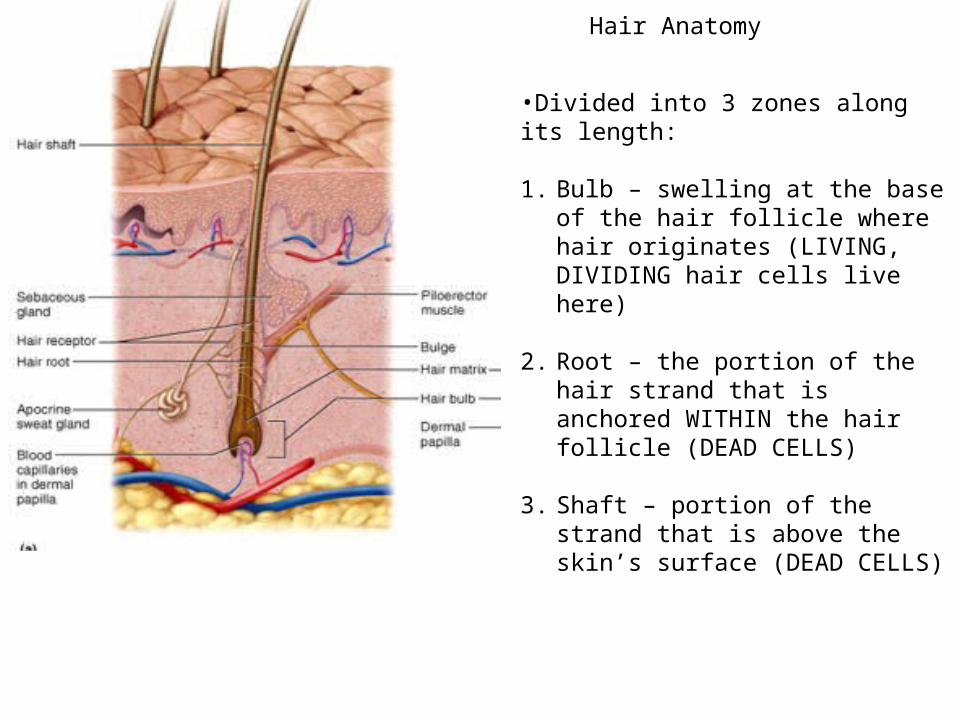
•Divided into 3 zones along its length:
1. Bulb – swelling at the base of the hair follicle where hair originates (LIVING, DIVIDING hair cells live here)
2. Root – the portion of the hair strand that is anchored WITHIN the hair follicle (DEAD CELLS)
3. Shaft – portion of the strand that is above the skin’s surface (DEAD CELLS)

•Hair follicle is attached to ARRECTOR PILI muscle that draws the root into a vertical position
•HAIR RECEPTORS are specialized nerve fibers that detect movement of hair shaft
•APOCRINE SWEAT GLANDS and SEBACEOUS GLANDS are not really part of the hair structure, but their ducts travel with the hair follicle
Hair anatomy cont….
Hair life cycle
•A hair strand goes cycles through 3 phases : ANAGEN, CATAGEN, and TELOGEN in which the hair transitions from rapid growth, to slow growth, to eventually falling out •ALOPECIA = loss or thinning of hair
Can be due to age, disease, nutrition, or genetics (male pattern baldness)Chemotherapeutic drugs cause alopecia since these drugs are designed to kill rapidly
dividing cells

Cutaneous Glands
5 types : merocrine sweat glands, apocrine sweat glands, sebaceous glands, ceruminous glands, and mammary glands (talk about later)
Merocrine sweat glands:
•“Regular” sweat glands that are found over the surface of most of the body•Secretion is mostly water and serves to cool the body•Simple, tubular gland with MYOEPITHELIAL cells that contract/ squeeze sweat to skin surface•Sweat is also slightly acidic (~pH 4-6) which helps inhibit bacteria growth•Most people lose over half a liter of sweat per day, even without exercise!
Apocrine sweat glands:
• Located in the axillary (armpit), groin, and anal region• Gland secretions flow into hair follicle, not directly onto the skin• Glands develop after puberty and are thought to produce scent molecules called
PHEROMONES• The metabolism of this type of sweat gland secretion by bacteria yields unpleasant
“body odor”

Sebaceous glands:
•Produce an oily secretion called SEBUM (mix of oil and cell fragments)•Glands are flask shaped and open into hair follicle •Sebum is a natural moisturizer meant to prevent skin from drying out
Yet we spend huge amounts of money on cleansers designed to remove it and then even more on creams to replace it!!! (i.e., lanolin)
Ceruminous glands:
•Ducts open directly to skin surface•Only found in outer ear canal•Secretion called CERUMEN mixes with sebum and dead epithelial cells to make earwax•Cerumen keeps eardrum pliable, waterprooofs the ear canal, and inhibits bacteria growth
Sebaceous glandApocrine gland
Merocrine gland

Cutaneous gland histology
1. Sebaceous and apocrine glands open into the hair follicle• Sebaceous glands are large and flask-shaped• Apocrine glands are simple coiled glands
2. Merocrine glands open directly onto the skin• Merocrine glands are simple coiled
When a coiled tubular structure is sectioned for histology, you will see a cluster of hollow circles and ovals where the gland was cut


Thin Skin – Similar to what WE will see in lab

Skin disorders-Burns
•In addition to exposure to extreme heat, fire, and boiling water; burns can also be caused by strong acids and bases, ionizing radiation, and electricity•Burns are classified (1st degree, 2nd degree, 3rd degree) by how deep the burn damages the tissue
1st degree burn:Damaged tissue is limited to the
epidermis(common sunburns, water
scalding, etc…)
2nd degree burn:Damage extends all the way through the epidermis and
enters the dermis (blistering sunburns, touching a hot
stove, etc…)
3rd degree burn: Damage extends through dermis and often involves damage to hypodermis.
Typically requires skin grafts since entire integument in
that area is destroyed
Extensive 3rd degree burns are often fatal if not treated immediately due to loss of body fluids and infection at burn site

Skin Cancer
There are three main forms of skin cancer : 1. Malignant Melanoma – cancer of the melanocytes in the stratum basale2. Non-melanoma skin cancer –
Basal cell carcinoma: cancer of cuboidal cells of the stratum basaleSquamous cell carcinoma: cancer of the keratinocytes of the stratum spinosum
Basal cell carcinoma: • Most common form of skin cancer• Extremely high survival rate since basal cell carcinoma rarely METASTACIZES (spreads
to other non-skin organs) •Like all skin cancers, fair skin, prior bad sunburns, and repeated sun exposure increase the risk of basal cell carcinoma•Treatment typically involves surgical removal of the cancerous tissue and/or radiation therapy in advanced cases•Like burns, cancers are graded on the depth of the damage (tumor), with deeper tumors being more serious•Often appears as a rounded, raised bump on sun-exposed skin

Squamous cell cariconoma:• Arises from squamous cells in the stratum spinosum• Often appears on sun-exposed skin as a red, scaly ulcer• Although the cosmetic damage can be extensive, this type of cancer is very curable
through surgery if caught early, radiation therapy for more advanced cases• Can be lethal if left unchecked due to metastasis
Stats• Each year there are 1,000,000 new cases of basal
cell and squamous cell carcinoma in the U.S.
• Of that number approximately 1,000 are lethal
• Basal cell and squamous cell carcinomas that appear on skin where “the sun don’t shine” are
usually much more dangerous since these cancers often involve genetic damage that did not involve sun exposure and often are very aggressive and
metastacize quickly

Malignant Melanoma: arises from melanocytes in the stratum basale• Least common, but definitely the most lethal all skin cancers
It spreads quickly and is often fatal if not treated immediately• Approximately 65,000 new cases each year
Of those, about 8,500 will be lethal (1 in 8 new cases!)• Melanoma typically originates in moles (NEVI), which are simply clusters of
melanocytes• Melanoma is more common in men than women, and is often linked to fair skin and a
history of bad sunburns
•Treatment options are pretty limited:VERY curable through radical surgery if the
melanoma is caught early Typically incurable if the melanoma has spread
since these type of cancer cells do not respond well to chemotherapy and radiation therapy
•Prognosis is related to location of melanoma:Locations close to lymph nodes have poor
prognosis (back, head, neck, groin, etc…) since metastasis is more common than in places like the hand or foot

Some of the (potentially) most important things you’ll learn in this class
Although very dangerous, malignant melanoma is EXTREMELY curable if it is caught early!!!
In a wide excision surgery the epidermis, dermis, and hypodermis are removed in a 2 cm radius around the melanoma. The dermis and hypodermis are then removed from an additional 2-3 cm and then sewn up.
ABCDs of melanoma detection
Bio 211- Anatomy and Physiology I
Today’s topics •Integumentary system
Damage and repair

Repairing damage to the integument
Since the skin plays such an important protective role in our bodies, any damage to it must be quickly repaired• Protection from bacteria, viruses, dirt, some chemicals• Keratinized epithelia prevents water loss
Type of wound healing depends on extent of damage1. Regeneration – involves replacement of damaged cells with the SAME type of cells
• Typically seen in VERY superficial scrapes and cuts• Damaged cells (i.e., keratinocytes) are replaced through proliferation of pre-
existing cells2. Fibrosis/Repair – involves formation of a clot, scab, and eventually replacing
damaged tissue with scar tissue• Typical in most deep cuts and scrapes• Need to repair not only epidermis, but also connective tissue, blood vessels of
dermis!

Repairing damage to the integument
•Immediately after a deep cut, blood from severed vessels enters into the wound Blood vessels become “leaky” allowing leukocytes, antibodies, clotting factors and
proteins to enter the wound Blood protein called FIBRIN polymerizes and forms meshwork that creates a clot A scab is just the dried part of the blood clot on the surface of the skin
•The formation of a blood clot is very important since it:
1. Helps stop bleeding2. Protects the open wound
from bacteria, dirt

Repairing damage to the integument
•Before the epidermis can be fixed, the damage to the dermis must be repaired Blood vessels grow and repair connections Macrophages enter and remove damaged tissue and blood clot Fibroblasts synthesize new collagen to repair connective tissue in dermis
•New collagen is laid down by fibroblasts quickly and is more dense and less flexible than original tissue (SCAR TISSUE)
•Once repair to the dermis is started, epithelial cells proliferate and help repair the epidermis
This phase is called REMODELING and can take many weeks