
Binding site density enables paralog-specific activity of SLM2 and ...
11
4120–4130 Nucleic Acids Research, 2017, Vol. 45, No. 7 Published online 19 December 2016 doi: 10.1093/nar/gkw1277 Binding site density enables paralog-specific activity of SLM2 and Sam68 proteins in Neurexin2 AS4 splicing control Marina Danilenko 1,† , Caroline Dalgliesh 1,† , Vittoria Pagliarini 2,3 , Chiara Naro 2,3 , Ingrid Ehrmann 1 , Mikael Feracci 4 , Mahsa Kheirollahi-Chadegani 1 , Alison Tyson-Capper 5 , Gavin J Clowry 6 , Philippe Fort 7 , Cyril Dominguez 4 , Claudio Sette 2,3 and David J. Elliott 1,* 1 Institute of Genetic Medicine, Newcastle University, Central Parkway, Newcastle NE1 3BZ, UK, 2 Department of Biomedicine and Prevention, University of Rome Tor Vergata, 00133 Rome, Italy, 3 Laboratory of Neuroembryology, Fondazione Santa Lucia, 00143 Rome, Italy, 4 Leicester Institute of Structural and Chemical Biology and Department of Molecular and Cell Biology, Henry Wellcome Building, University of Leicester, Lancaster Road, Leicester LE1 7RH, UK, 5 Institute forCellular Medicine, Newcastle University, Framlington Place, Newcastle NE2 4HH, UK, 6 Institute of Neuroscience, Newcastle University, Framlington Place, Newcastle, UK and 7 Universit´ e Montpellier, UMR 5237, Centre de Recherche de Biologie cellulaire de Montpellier, CNRS, 34293 Montpellier, France Received May 20, 2015; Revised December 02, 2016; Editorial Decision December 06, 2016; Accepted December 08, 2016 ABSTRACT SLM2 and Sam68 are splicing regulator paralogs that usually overlap in function, yet only SLM2 and not Sam68 controls the Neurexin2 AS4 exon impor- tant for brain function. Herein we find that SLM2 and Sam68 similarly bind to Neurexin2 pre-mRNA, both within the mouse cortex and in vitro. Pro- tein domain-swap experiments identify a region in- cluding the STAR domain that differentiates SLM2 and Sam68 activity in splicing target selection, and confirm that this is not established via the variant amino acids involved in RNA contact. However, far fewer SLM2 and Sam68 RNA binding sites flank the Neurexin2 AS4 exon, compared with those flanking the Neurexin1 and Neurexin3 AS4 exons under joint control by both Sam68 and SLM2. Doubling binding site numbers switched paralog sensitivity, by plac- ing the Neurexin2 AS4 exon under joint splicing con- trol by both Sam68 and SLM2. Our data support a model where the density of shared RNA binding sites around a target exon, rather than different paralog- specific protein–RNA binding sites, controls func- tional target specificity between SLM2 and Sam68 on the Neurexin2 AS4 exon. Similar models might explain differential control by other splicing regula- tors within families of paralogs with indistinguish- able RNA binding sites. INTRODUCTION Around 95% of human genes encode multiple mRNA iso- forms with different exon contents, thus greatly expanding the informational capacity of the genome. RNA binding proteins play a key role in creating different mRNA splice isoforms, by binding to target sites within pre-mRNAs and directing splice site choice by the spliceosome. These RNA– protein binding sites contribute to a splicing code through which the exon–intron structure of pre-mRNAs is deci- phered (1–3). This splicing code includes RNA binding sites for proteins that activate and repress exon inclusion, called splicing enhancer and repressor sequences respectively, as well as the splice sites that they regulate. RNA binding sites can be both exon-proximal and sometimes deep intronic (4). In vertebrates, many splicing regulator proteins belong to families of evolutionarily related, similar sister proteins that often, but not always, regulate the same target exons. These sister proteins include the STAR proteins SLM2, Sam68 and SLM1; the Transformer proteins Tra2 and Tra2; the Polypyrimidine Tract Binding Proteins PTBP1-3; the Mus- cleblind proteins MBNL1–3; the Epithelial Specific RNA Splicing regulator proteins ESRP1 and ESRP2; RNA bind- ing Fox1 homolog proteins RBFOX1–3; TIAL and TIA1; as well as others (5). Many of these paralogs were cre- ated by gene duplications very early in vertebrate evolution. However, to what extent very similar splicing regulator par- alogs might select overlapping versus distinct targets is often poorly understood, as are why such similar paralogs have been maintained often over considerable periods of evolu- tionary time. * To whom correspondence should be addressed. Tel: +44 191 241 8694; Fax: +44 191 241 8666; Email: [email protected] † These authors contributed equally to the work as first authors. C The Author(s) 2016. Published by Oxford University Press on behalf of Nucleic Acids Research. This is an Open Access article distributed under the terms of the Creative Commons Attribution License (http://creativecommons.org/licenses/by/4.0/), which permits unrestricted reuse, distribution, and reproduction in any medium, provided the original work is properly cited. Downloaded from https://academic.oup.com/nar/article-abstract/45/7/4120/2716528 by guest on 05 April 2018
Transcript of Binding site density enables paralog-specific activity of SLM2 and ...

4120–4130 Nucleic Acids Research, 2017, Vol. 45, No. 7 Published online 19 December 2016doi: 10.1093/nar/gkw1277
Binding site density enables paralog-specific activityof SLM2 and Sam68 proteins in Neurexin2 AS4splicing controlMarina Danilenko1,†, Caroline Dalgliesh1,†, Vittoria Pagliarini2,3, Chiara Naro2,3,Ingrid Ehrmann1, Mikael Feracci4, Mahsa Kheirollahi-Chadegani1, Alison Tyson-Capper5,Gavin J Clowry6, Philippe Fort7, Cyril Dominguez4, Claudio Sette2,3 and David J. Elliott1,*
1Institute of Genetic Medicine, Newcastle University, Central Parkway, Newcastle NE1 3BZ, UK, 2Department ofBiomedicine and Prevention, University of Rome Tor Vergata, 00133 Rome, Italy, 3Laboratory of Neuroembryology,Fondazione Santa Lucia, 00143 Rome, Italy, 4Leicester Institute of Structural and Chemical Biology and Departmentof Molecular and Cell Biology, Henry Wellcome Building, University of Leicester, Lancaster Road, Leicester LE1 7RH,UK, 5Institute for Cellular Medicine, Newcastle University, Framlington Place, Newcastle NE2 4HH, UK, 6Institute ofNeuroscience, Newcastle University, Framlington Place, Newcastle, UK and 7Universite Montpellier, UMR 5237,Centre de Recherche de Biologie cellulaire de Montpellier, CNRS, 34293 Montpellier, France
Received May 20, 2015; Revised December 02, 2016; Editorial Decision December 06, 2016; Accepted December 08, 2016
ABSTRACT
SLM2 and Sam68 are splicing regulator paralogsthat usually overlap in function, yet only SLM2 andnot Sam68 controls the Neurexin2 AS4 exon impor-tant for brain function. Herein we find that SLM2and Sam68 similarly bind to Neurexin2 pre-mRNA,both within the mouse cortex and in vitro. Pro-tein domain-swap experiments identify a region in-cluding the STAR domain that differentiates SLM2and Sam68 activity in splicing target selection, andconfirm that this is not established via the variantamino acids involved in RNA contact. However, farfewer SLM2 and Sam68 RNA binding sites flank theNeurexin2 AS4 exon, compared with those flankingthe Neurexin1 and Neurexin3 AS4 exons under jointcontrol by both Sam68 and SLM2. Doubling bindingsite numbers switched paralog sensitivity, by plac-ing the Neurexin2 AS4 exon under joint splicing con-trol by both Sam68 and SLM2. Our data support amodel where the density of shared RNA binding sitesaround a target exon, rather than different paralog-specific protein–RNA binding sites, controls func-tional target specificity between SLM2 and Sam68on the Neurexin2 AS4 exon. Similar models mightexplain differential control by other splicing regula-tors within families of paralogs with indistinguish-able RNA binding sites.
INTRODUCTION
Around 95% of human genes encode multiple mRNA iso-forms with different exon contents, thus greatly expandingthe informational capacity of the genome. RNA bindingproteins play a key role in creating different mRNA spliceisoforms, by binding to target sites within pre-mRNAs anddirecting splice site choice by the spliceosome. These RNA–protein binding sites contribute to a splicing code throughwhich the exon–intron structure of pre-mRNAs is deci-phered (1–3). This splicing code includes RNA binding sitesfor proteins that activate and repress exon inclusion, calledsplicing enhancer and repressor sequences respectively, aswell as the splice sites that they regulate. RNA binding sitescan be both exon-proximal and sometimes deep intronic (4).In vertebrates, many splicing regulator proteins belong tofamilies of evolutionarily related, similar sister proteins thatoften, but not always, regulate the same target exons. Thesesister proteins include the STAR proteins SLM2, Sam68and SLM1; the Transformer proteins Tra2� and Tra2�; thePolypyrimidine Tract Binding Proteins PTBP1-3; the Mus-cleblind proteins MBNL1–3; the Epithelial Specific RNASplicing regulator proteins ESRP1 and ESRP2; RNA bind-ing Fox1 homolog proteins RBFOX1–3; TIAL and TIA1;as well as others (5). Many of these paralogs were cre-ated by gene duplications very early in vertebrate evolution.However, to what extent very similar splicing regulator par-alogs might select overlapping versus distinct targets is oftenpoorly understood, as are why such similar paralogs havebeen maintained often over considerable periods of evolu-tionary time.
*To whom correspondence should be addressed. Tel: +44 191 241 8694; Fax: +44 191 241 8666; Email: [email protected]†These authors contributed equally to the work as first authors.
C© The Author(s) 2016. Published by Oxford University Press on behalf of Nucleic Acids Research.This is an Open Access article distributed under the terms of the Creative Commons Attribution License (http://creativecommons.org/licenses/by/4.0/), whichpermits unrestricted reuse, distribution, and reproduction in any medium, provided the original work is properly cited.
Downloaded from https://academic.oup.com/nar/article-abstract/45/7/4120/2716528by gueston 05 April 2018

Nucleic Acids Research, 2017, Vol. 45, No. 7 4121
An important example of differential splicing regula-tor paralog function is found within the nervous system.Here, the STAR family RNA binding proteins Sam68 (alsoknown as KHDRBS1), SLM1 (also known as KHDRBS2)and SLM2 (also known as KHDRBS3 and T-STAR) differ-entially regulate splicing patterns of the Neurexin1–3 genesinvolved in brain function (6–9). The Neurexin1–3 genesencode trans-membrane pre-synaptic proteins, mutationsin which are associated with various conditions includingschizophrenia and autism (10), indicating the crucial roleplayed by their regulation for proper brain functions. EachNeurexin gene has several alternative exons called alterna-tive segments 1–5 (abbreviated AS1–AS5), and through al-ternative splicing of these exons can produce many differ-ent mRNA isoforms (11). While SLM2 and Sam68 usu-ally function interchangeably in splicing regulation withintransfected cells and both repress inclusion of the Neurexin1and Neurexin3 AS4 exons, only SLM2 but not Sam68 reg-ulates the Neurexin2 AS4 exon (6). These AS4 splicingpatterns are physiologically important, as manipulation ofNeurexin1 and Neurexin3 AS4 alternative splicing affectsboth mouse behavior and synapse function (12,13).
SLM2 and Sam68 are 62% identical, and have similarmodular organisations, including a STAR domain com-prising a KH domain and flanking regions that medi-ate protein–protein and protein–RNA interactions, an RG(arginine/glycine)-rich region and C-termini enriched in ty-rosine residues (14). Based on atomic level resolution fromX-ray crystallography and nuclear magnetic resonance (15),SLM2 and Sam68 proteins bind RNA via their STAR do-mains, with largely overlapping but not identical RNA–protein contacts with the same U(A/U)AA target sequence(abbreviated UWAA) (15–17). There is a 51-nucleotide clus-ter of intronic Sam68/SLM2 binding sites immediatelydownstream of the Neurexin2 AS4 exon that mediates thesplicing response to SLM2 (6), but the features underlyingdifferential regulation of Neurexin2 AS4 splicing by SLM2and not by Sam68 remain unclear. Herein, we have investi-gated the features of the SLM2 and Sam68 proteins and ofthe target Neurexin2 pre-mRNA that enable this differentsplicing activity, and elucidate distinct physiological prop-erties of these closely related protein paralogs.
MATERIALS AND METHODS
Crosslinking-immunoprecipitation experiment
The CLIP assay was performed as previously described(18,19). In brief, dissociated cortex tissues were irradiatedon ice (100 mJ/cm2). The cell suspension was centrifugedat 4000 rpm for 3 min, and the pellet was incubated for 10min on ice in lysis buffer (50-mM Tris, pH 8.0, 100-mMNaCl, 1% NP-40, 1-mM MgCl2, 0.1-mM CaCl2, 0.5 mMNa3VO4, 1-mM DTT, protease inhibitor cocktail [Sigma-Aldrich] and RNase inhibitor [Promega]). Samples werebriefly sonicated and incubated with DNase (RNase-free;Ambion) for 10 min at 37◦C and then centrifuged at 15 000g for 10 min at 4◦C. One milligram of extract was immuno-precipitated using anti-STAR (Santa Cruz Biotechnology,Inc.) or IgG (negative control) in the presence of protein Gmagnetic (Life Technologies). 1 U RNase I (Ambion) wasadded to immunoprecipitates and incubated for 2 h at 4◦C
under rotation. After stringent washes, 10% was kept as acontrol to test efficiency of immunoprecipitation by west-ern blot, while the rest of the immunoprecipitated sampleswere treated with 50 �g Proteinase K and incubated for 1h at 55◦C. RNA was then isolated by standard proceduresand retrotranscribed with random primers, using M-MLVreverse transcriptase (Promega). qPCR was performed us-ing LightCycler 480 SYBR green I Master and the Light-Cycler 480 System (Roche) according to the manufacturer’sinstructions. RNA associated with STAR proteins is rep-resented as fold enrichment relative to IgG samples. Allprimers used are listed in Supplementary file 1.
Fluorescence polarization
Sam68 and SLM2 STAR domains were produced as previ-ously described (15). RNA oligonucleotides were purchasedfrom Dharmacon, GE Healthcare, deprotected accordingto the manufacturer’s instructions, lyophilized, and resus-pended in ddH2O. All RNAs used for fluorescence polar-ization contained a fluorescein tag and three cytosines atthe 5′ end of the sequence described in Table 1.
Fluorescence polarization experiments were carried outin black 96-well plates with a 50 �l sample volume perwell in 10 mM Tris pH 7, 100 mM NaCl, 0.1% �-mercaptoethanol. Sam68 and SLM2 domains were seriallydiluted across the plate from 200 to 0 �M. Fluorescein-labeled RNA was then added at 0.2 �M final concentration.Plates were analyzed using a Perkin Elmer Victor X5 platereader at excitation wavelength of 531 nm and emission at595 nm, and experiments were carried out in triplicate.
Genomic sequences
Neurexin1–3 sequences were retrieved from NCBI database(http://www.ncbi.nlm.nih.gov), using the annotationsearch tools available in the Geneious 7.1.5 package(Biomatters, http://www.geneious.com/). Accession num-bers: Homo sapiens (NG 011878.1; NC 000011.10;NC 000014.9), Mus musculus (18189; 18190; 18191),Sarcophilus harrisii (100090577; 100917908; 100928491),Macropus eugenii (ENSMEUG00000007055; ENS-MEUG00000016641; ENSMEUG00000001347), Or-nithorhynchus anatinus (100090577; 100073756), Pelodiscussiniensis (102449503; 102447445; 102449503), Alligatormississippiensis (102558540; 102566256; 102570703),Xenopus tropicalis (100127647), Danio rerio (BX255907;CR751231; BX571723; NW 001884422).
Minigenes and creation of hybrid proteins
Minigenes were cloned into pXJ41 using the primers in Sup-plementary File S1, and mutagenesis was carried out byoverlap PCR as previously described (20). Minigene splic-ing patterns were analysed in HEK293 cells 24 h after co-transfection either with GFP, SLM2-GFP or SAM68-GFPas previously described (6). Tissue culture, transfection ofHEK293 cells, RNA purification and RT-PCR was carriedout using primers and conditions as previously described(21) and the RT-PCR products were analysed by capillarygel electrophoresis (20). To enable analysis of splicing pat-terns as well as monitoring the expression of transfected
Downloaded from https://academic.oup.com/nar/article-abstract/45/7/4120/2716528by gueston 05 April 2018

4122 Nucleic Acids Research, 2017, Vol. 45, No. 7
Table 1. Affinity measurements of the STAR domains of T-STAR and SAM68 proteins to target RNAs from Neurexin2 and Stxbp5l carried out usingfluorescence polarization (FP)
SLM2 SAM68
Nrxn2 WT 5.9 ± 0.9 2.9 ± 0.3Nrxn2 mutant 7 3.9 ± 0.4 3.4 ± 0.3UAAAAx4 7.9 ± 1.2 6.9 ± 0.8Stxbp5l 1 24.0 ± 5.9 15.5 ± 4.5Stxbp5l 2 18.2 ± 3.3 13.8 ± 2.6Poly(C) >100 >100
Poly(C) was used as a negative control. Affinities are given in Kd (�M) with standard deviations derived from three independent experiments. The RNA molecules used for FPexperiments were Nrxn2 WT:5′-AAUUAAUUAAUUAAUUAACUAACUAACUAACUUUAAAAACACGAUCUUAAA-3′ ;Nrxn2mutant7:5′-AAUUAAUUAAUUAAUUAACUAACCCACCCACCCUAAAAACACGAUCUUAAA-3′ ;UAAAAx4: 5′-UAAAAUAAAAUAAAAUAAAA-3′;Stxbp5l 1: 5′-ACAGUUUAAAAUUUGAUAAAAUUU-3′;Stxbp5l 2: 5′-UUACAUUUAAAAGAUGAUUUAAAAA-3′.Poly(C): 5′-CCCCCCCCC-3′.
splicing factors, transfected cell cultures were split into twoportions and analysed by RT-PCR and western blotting aspreviously described (22). Splicing patterns were measuredas percentage splicing exclusion (6). All primers used forsplicing assays are provided in the supplementary informa-tion. Hybrid SLM2 and Sam68 proteins were made by Gblock synthesis, and cloned in frame with GFP.
Detection of gene expression in mouse brain tissue
RNA was prepared from brain tissue dissected from wildtype and Khdrbs3 (SLM2) and Khdrbs1 (SAM68) (23)knockout mice using TRIzol (Life Technologies). ThecDNA was generated using Superscript 3 (Invitrogen) andanalysed by RT-PCR using primer pairs described in theSupplementary Information.
Statistical analysis
Bar charts were plotted and statistical analyses performedusing Graphpad Prism (Graphpad software).
Western blots
Western blots were carried out as described previously (21).Proteins were detected by western blotting using antibod-ies specific for GFP (Clontech 632 381 Living Colors Av monoclonal antibody); SLM2 (6) SAM68 (Santa Cruzanti-Sam68 sc-333) and for actin �-Actin (Sigma-Aldrich,A5441,1:2000 dilution).
RESULTS
SLM2 and SAM68 proteins bind indistinguishably to theNeurexin2 pre-mRNA
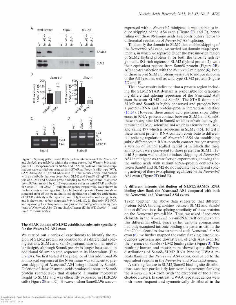
The mouse cortex is a brain structure that expresses bothSam68 and SLM2 but negligible levels of SLM1 (8). Tomonitor binding of endogenous SLM2 and Sam68 pro-teins to the Neurexin2 pre-mRNAs within the mouse cortexwe used CLIP (Cross Linking Immunoprecipitation), withan antibody that recognizes both Sam68 and SLM2 (anti-STAR antibody, Figure 1A). To properly discriminate be-tween the two proteins, CLIP experiments were performedto detect SLM2 in a Sam68 null mouse background, and todetect Sam68 in a Slm2 null mouse background.
Western blot analysis showed efficient immunoprecip-itation of both Sam68 and SLM2 from the mouse cor-tex (Figure 1A), whereas SLM1 was not detected in eitherbackground (data not shown). Quantitative analysis of theimmunoprecipitates showed efficient SLM2 protein cross-linking to the Neurexin2 pre-mRNA (Figure 1B; ∼100-foldenrichment of Neurexin2 precipitation with SLM2 com-pared with IgG), in line with the strong effect of SLM2 ex-pression on AS4 splicing in the cortex (Figure 1C). Surpris-ingly however since Sam68 does not control Neurexin2 AS4exon skipping within the mouse cortex (Figure 1C), Sam68protein was also efficiently cross-linked to the Neurexin2pre-mRNA (Figure 1B; ∼100-fold enrichment of Neurexin2precipitation with Sam68 compared with IgG). Precise mea-surements using fluorescence polarization (FP) also showedthat SLM2 and Sam68 have very similar in vitro affinityfor the 51 nucleotide UWAA-rich sequence downstream ofthe Neurexin2 AS4 exon (Table 1). Hence, these data indi-cate that the different splicing activity of SLM2 and Sam68on Neurexin2 AS4 does not correlate with detectable dif-ferences in Sam68 and SLM2 protein-binding levels to theendogenous pre-mRNA.
To test whether similar joint binding also occurred onanother functional SLM2 target in the mouse brain, weanalyzed a 171 nucleotide exon within the Stxbp5l gene,which is one of the few other known splicing targets ofSLM2 besides the Neurexin1–3 genes (6,13). CLIP also de-tected binding of both Sam68 and SLM2 to the Stxbp5l pre-mRNA within the mouse cortex, although with around 3-fold higher levels of Sam68 (Figure 1B). Both Sam68 andSLM2 proteins bound to Stxbp5l RNA probes in vitro inFP experiments (Table 1). Analysis of physiological splicingpatterns in the mouse cortex (Figure 1D) and other brainregions (quantitative data for Stxbp5l splicing patterns inthe cortex and other mouse brain regions are shown in Sup-plementary Figure S1) showed that splicing of this 171 nu-cleotide Stxbp5l exon responded to knockout of either Slm2and Sam68. Hence, Stxbp5l is a joint splicing target for bothSLM2 and Sam68, similar to the Neurexin1 and Neurexin3AS4 exons, and unlike Neurexin2. Note an additional up-stream 93 nucleotide exon is annotated in some mRNAsbut not in Refseq, and we could detect some inclusion ofthis additional exon in the brain (to give the +93 band) butsplicing of this did not respond to deletion of either Sam68or Slm2.
Downloaded from https://academic.oup.com/nar/article-abstract/45/7/4120/2716528by gueston 05 April 2018
Nucleic Acids Research, 2017, Vol. 45, No. 7 4123
Sam68 -/-
Stxbp5l
Nrxn2
RNA
enric
hmen
t α-
STAR
vs α
-IgG
**
**
0
50
100
150
200
0100200300400500
RNA
enric
hmen
t α-
STAR
vs α
-IgG
Stxbp5l
Nrxn2
Slm2 -/-**
**
B
A
Sam
68 -/
-
Slm
2 -/
-
WT
Sam
68 -/
-
Slm
2 -/
-
WT
Sam
68 -/
-
Slm
2 -/
-
WT
Total Extract α-IgG α-STAR
SAM68
SLM2
Sam
68 -/
-
Slm
2 -/
-
WT
Sam
68 -/
-
Slm
2 -/
-
WT
C
Nrxn2
AS4+AS4-
FL
D
Stxbp5l
+93
Δ171Δ93/171
Figure 1. Splicing patterns and RNA protein interactions of the Neurexin2and Stxbp5l pre-mRNAs within the mouse cortex. (A) Western blot anal-ysis of CLIP experiments for SLM2 and SAM68 proteins. Immunoprecip-itations were carried out using an anti-STAR antibody in wild-type (WT),SAM68 (Sam68−/−) or SLM2 (Slm2−/−) null mouse cortex, and probedwith an antibody that can detect both SLM2 and Sam68. (B) qPCR anal-ysis of SLM2 and SAM68 protein binding to the Stxbp5l and Neurexin2pre-mRNAs assayed by CLIP experiments using an anti-STAR antibodyin Sam68 −/− or Slm2−/− null mouse cortex, respectively. Data shown inthe bar charts are averages from four biological replicates. Error bars showstandard error of the mean. Statistical significance of mRNA enrichmentof STAR antibody with respect to control IgG was addressed using t tests,and is shown on the bar charts as: **P < 0.01. (C, D) Endpoint RT-PCRand agarose gel electrophoresis analysis of the endogenous splicing pat-terns of Neurexin2 AS4 (C) and Stxbp5l genes (D) in WT, Sam68−/− andSlm2−/- mouse cortex.
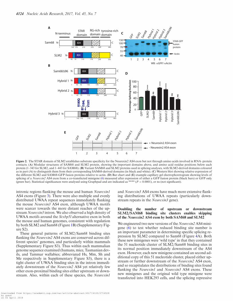
The STAR domain of SLM2 establishes substrate specificityfor the Neurexin2 AS4 exon
We carried out a series of experiments to identify the re-gion of SLM2 protein responsible for its differential splic-ing activity. SLM2 and Sam68 proteins have similar modu-lar designs, although Sam68 protein is longer because of anadditional 96 amino acid sequence at its N-terminus (Fig-ure 2A). We first tested if the presence of this additional 96amino acid sequence at the N-terminus was sufficient to pre-vent skipping of Neurexin2 AS4 being induced by Sam68.Deletion of these 96 amino acids produced a shorter Sam68protein (Sam68�96) that displayed a similar molecularweight to SLM2 and was efficiently expressed in HEK293cells (Figure 2B and C). However, when Sam68�96 was co-
expressed with a Neurexin2 minigene, it was unable to in-duce skipping of the AS4 exon (Figure 2D and E), henceruling out these 96 amino acids as a contributory factor todifferential regulation of Neurexin2 AS4 splicing.
To identify the domain in SLM2 that enables skipping ofthe Neurexin2 AS4 exon, we carried out domain swap exper-iments, in which we replaced either the tyrosine-rich regionof SLM2 (hybrid protein 1), or both the tyrosine rich re-gion and RG-rich regions of SLM2 (hybrid protein 2), withtheir equivalent regions from Sam68 protein (Figure 2B).After co-transfection with the Neurexin2 minigene (6), bothof these hybrid SLM2 proteins were able to induce skippingof the AS4 exon as well as wild type SLM2 protein (Figure2D and E).
The above results indicated that a protein region includ-ing the SLM2 STAR domain is responsible for establish-ing differential splicing repression of the Neurexin2 AS4exon between SLM2 and Sam68. The STAR domain ofSLM2 and Sam68 is highly conserved and provides botha protein–RNA and protein–protein interaction interface(15,24). However, three amino acid positions show differ-ences in RNA–protein contact between SLM2 and Sam68:these are arginine 180 in Sam68 which is substituted by glu-tamine in SLM2, isoleucine 184 which is a leucine in SLM2,and valine 197 which is isoleucine in SLM2 (15). To test ifthese variant protein–RNA contacts contribute to differen-tial splicing regulation of Neurexin2 AS4 via establishingsubtle differences in RNA–protein contact, we constructeda version of Sam68 (called hybrid 3) in which the threeamino acids were converted to those present in SLM2. Hy-brid 3 protein was unable to induce skipping of Neurexin2AS4 in minigene co-transfection experiments, showing thatthe amino acids with variant RNA–protein contacts be-tween Sam68 and SLM2 do not mediate the different splic-ing activity of these two splicing regulators on the Neurexin2AS4 exon (Figure 2D and E).
A different intronic distribution of SLM2/SAM68 RNAbinding sites flank the Neurexin2 AS4 compared with boththe Neurexin1 and Neurexin3 AS4 exons
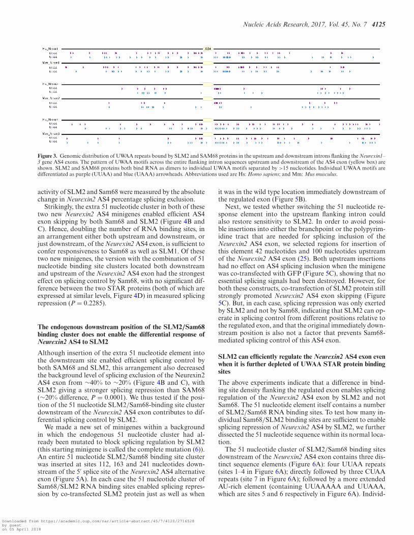
Taken together, the above data suggested that differentprotein–RNA binding abilities between SLM2 and Sam68do not differentiate the splicing specificity of these proteinson the Neurexin2 pre-mRNA. Thus, we asked if sequenceelements in the Neurexin2 pre-mRNA itself could explainthis differential effect. Since earlier genome comparisonshad only examined intronic binding site patterns within thefirst 200 nucleotides downstream of each Neurexin1–3 AS4exon (6), we further mapped the entire flanking intronic se-quences upstream and downstream of each AS4 exon forthe presence of Sam68/SLM2 binding sites (Figure 3). Theresulting human and mouse maps showed quite differentdistributions of Sam68/SLM2 RNA binding UWAA re-peats flanking the Neurexin2 AS4 exons, compared to theequivalent regions in the Neurexin1 and Neurexin3 genes.
A striking feature in SLM2/Sam68 binding site distribu-tions was their particularly low overall occurrence flankingthe Neurexin2 AS4 exon (with the exception of the 51 nu-cleotide cluster). In contrast, UWAA repeat sequences wereboth more frequent and symmetrically distributed in the
Downloaded from https://academic.oup.com/nar/article-abstract/45/7/4120/2716528by gueston 05 April 2018
4124 Nucleic Acids Research, 2017, Vol. 45, No. 7
KH1 443
N- -CSam68170 210143 276 349
KHN- -CSam68Δ96
KHN- -C
1 34370 11043 176 249
SLM2
KHN- -C
1 34370 11043 176 249
Hybrid 1
KH1 443
N- -C
R180
QR1
80Q
170 210143 276 349
I184
LI1
84L
V19
7IV
197I
130100
70
55
40
GFP
SLM
2Sa
m68
1 2 3 4 5 6 7
Actin
STAR-GFPfusionprotein
WB: αGFP/ αActin
Hyb
rid 1
Hyb
rid 2
Hyb
rid 3
KHN- -C
1 34370 11043 176 249
Hybrid 2
Hybrid 3
20
40
60
80
0
STAR domainN-terminus
RG-rich domain
tyrosine-richdomain
A
B
C
****
ns
ns
ns
Per
cen
tag
e Sp
licin
g E
xclu
sio
n 100
GFP
SLM
2
Sam
68
Hyb
rid
1
Hyb
rid
2
Hyb
rid
3
Sam
68Δ
96
Sam
68Δ9
6
D
1 2 3 4 5 6 7
GFP
SLM
2
Sam
68
Hyb
rid 1
Hyb
rid 2
Hyb
rid 3
Sam
68Δ9
6+ Neurexin2 AS4 exon
- Neurexin2 AS4 exon
E
****
****
Figure 2. The STAR domain of SLM2 establishes substrate specificity for the Neurexin2 AS4 exon but not through amino acids involved in RNA–proteincontacts. (A) Modular structures of SAM68 and SLM2 protein, showing the important domains above, and amino acid residue positions below eachprotein (1–343 for SLM2, and 1–443 for SAM68). (B) Variant SAM68 and SLM2 proteins used in splicing analyses, with SLM2-derived domains colouredas in part (A) to distinguish them from their corresponding SAM68-derived domains (in black and white). (C) Western blot showing relative expression ofthe different SLM2 and SAM68-GFP fusion proteins relative to actin. (D) Bar chart and (E) example capillary gel electrophoretogram showing levels ofsplicing of a Neurexin2 AS4 exon from a co-transfected minigene (6) measured after expression of either a GFP fusion protein (black bars) or GFP only(green bar). Statistical significances were analysed using Graphpad and are indicated as **** (P < 0.0001), or ns (not significant).
intronic regions flanking the mouse and human Neurexin1AS4 exons (Figure 3). There were also multiple and evenlydistributed UWAA repeat sequences immediately flankingthe mouse Neurexin3 AS4 exon, although UWAA motifswere scarcer towards the more distant reaches of the up-stream Neurexin3 intron. We also observed a high density ofUWAA motifs around the Stxbp5l alternative exon in boththe mouse and human genomes, consistent with regulationby both SLM2 and Sam68 (Figure 1B) (Supplementary Fig-ure S2).
These general patterns of SLM2/Sam68 binding sitesflanking the Neurexin2 AS4 exons are conserved across dif-ferent species’ genomes, and particularly within mammals(Supplementary Figure S3). Thus within each mammaliangenome sequence examined (humans, mice, Tasmanian dev-ils, and Tammar wallabies; abbreviated Hs, Mm, Sh andMe respectively in Supplementary Figure S3), there is atight cluster of UWAA binding sites in the intron immedi-ately downstream of the Neurexin2 AS4 yet relatively fewother exon-proximal binding sites either upstream or down-stream. Also, within each of these species, the Neurexin1
and Neurexin3 AS4 exons have much more extensive flank-ing distributions of UWAA repeats (particularly down-stream repeats in the Neurexin3 gene).
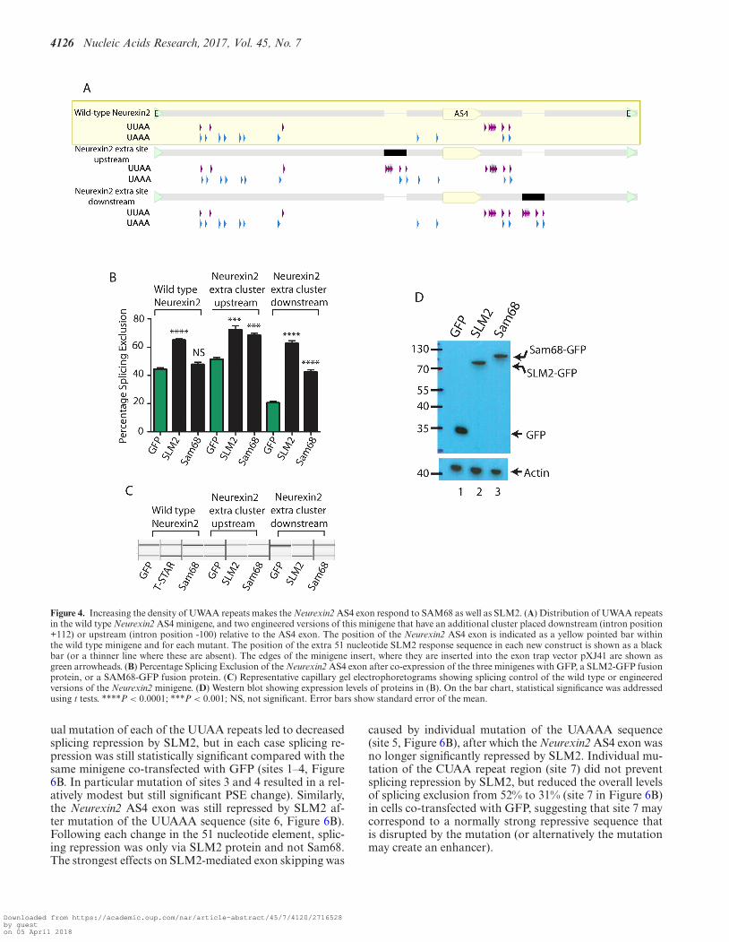
Doubling the number of upstream or downstreamSLM2/SAM68 binding site clusters enables skippingof the Neurexin2 AS4 exon by both SAM68 and SLM2
We engineered two new versions of the Neurexin2 AS4 mini-gene (6) to test whether reduced binding site number isan important parameter in determining specific splicing re-pression by SLM2 compared to Sam68 (Figure 4A). Boththese new minigenes were ‘wild type’ in that they containedthe 51 nucleotide cluster of SLM2/Sam68 binding sites inits normal position immediately downstream of the AS4exon. However, each new minigene contained an second ad-ditional copy of this 51 nucleotide cluster, placed either up-stream or further downstream of the Neurexin2 AS4 exon,and so recapitulates the distributions of binding sites foundflanking the Neurexin1 and Neurexin3 AS4 exons. Thesenew minigenes and the original wild type minigene weretransfected into HEK293 cells, and the splicing repressive
Downloaded from https://academic.oup.com/nar/article-abstract/45/7/4120/2716528by gueston 05 April 2018
Nucleic Acids Research, 2017, Vol. 45, No. 7 4125
Figure 3. Genomic distribution of UWAA repeats bound by SLM2 and SAM68 proteins in the upstream and downstream introns flanking the Neurexin1–3 gene AS4 exons. The pattern of UWAA motifs across the entire flanking intron sequences upstream and downstream of the AS4 exon (yellow box) areshown. SLM2 and SAM68 proteins both bind RNA as dimers to individual UWAA motifs separated by >15 nucleotides. Individual UWAA motifs aredifferentiated as purple (UUAA) and blue (UAAA) arrowheads. Abbreviations used are Hs: Homo sapiens; and Mm: Mus musculus.
activity of SLM2 and Sam68 were measured by the absolutechange in Neurexin2 AS4 percentage splicing exclusion.
Strikingly, the extra 51 nucleotide cluster in both of thesetwo new Neurexin2 AS4 minigenes enabled efficient AS4exon skipping by both Sam68 and SLM2 (Figure 4B andC). Hence, doubling the number of RNA binding sites, inan arrangement either both upstream and downstream, orjust downstream, of the Neurexin2 AS4 exon, is sufficient toconfer responsiveness to Sam68 as well as SLM1. Of thesetwo new minigenes, the version with the combination of 51nucleotide binding site clusters located both downstreamand upstream of the Neurexin2 AS4 exon had the strongesteffect on splicing control by Sam68, with no significant dif-ference between the two STAR proteins (both of which areexpressed at similar levels, Figure 4D) in measured splicingrepression (P = 0.2285).
The endogenous downstream position of the SLM2/Sam68binding cluster does not enable the differential response ofNeurexin2 AS4 to SLM2
Although insertion of the extra 51 nucleotide element intothe downstream site enabled efficient splicing control byboth SAM68 and SLM2, this arrangement also decreasedthe background level of splicing exclusion of the Neurexin2AS4 exon from ∼40% to ∼20% (Figure 4B and C), withSLM2 giving a stronger splicing repression than SAM68(∼20% difference, P = 0.0001). We thus tested if the posi-tion of the 51 nucleotide SLM2/Sam68-binding site clusterdownstream of the Neurexin2 AS4 exon contributes to dif-ferential splicing control by SLM2.
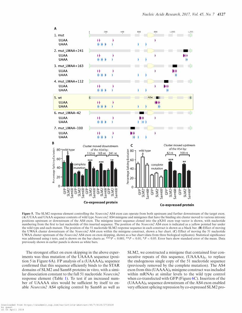
We made a new set of minigenes within a backgroundin which the endogenous 51 nucleotide cluster had al-ready been mutated to block splicing regulation by SLM2(this starting minigene is called the complete mutation (6)).An entire 51 nucleotide SLM2/Sam68 binding site clusterwas inserted at sites 112, 163 and 241 nucleotides down-stream of the 5′ splice site of the Neurexin2 AS4 alternativeexon (Figure 5A). In each case the 51 nucleotide cluster ofSam68/SLM2 RNA binding sites enabled splicing repres-sion by co-transfected SLM2 protein just as well as when
it was in the wild type location immediately downstream ofthe regulated exon (Figure 5B).
Next, we tested whether switching the 51 nucleotide re-sponse element into the upstream flanking intron couldalso restore sensitivity to SLM2. In order to avoid possi-ble insertions into either the branchpoint or the polypyrim-idine tract that are needed for splicing inclusion of theNeurexin2 AS4 exon, we selected regions for insertion ofthis element 42 nucleotides and 100 nucleotides upstreamof the Neurexin2 AS4 exon (25). Both upstream insertionshad no effect on AS4 splicing inclusion when the minigenewas co-transfected with GFP (Figure 5C), showing that noessential splicing signals had been destroyed. However, forboth these constructs, co-transfection of SLM2 protein stillstrongly promoted Neurexin2 AS4 exon skipping (Figure5C). But, in each case, splicing repression was only exertedby SLM2 and not by Sam68, indicating that SLM2 can op-erate in splicing control from different positions relative tothe regulated exon, and that the original immediately down-stream position is also not a factor that prevents Sam68-mediated splicing control of this AS4 exon.
SLM2 can efficiently regulate the Neurexin2 AS4 exon evenwhen it is further depleted of UWAA STAR protein bindingsites
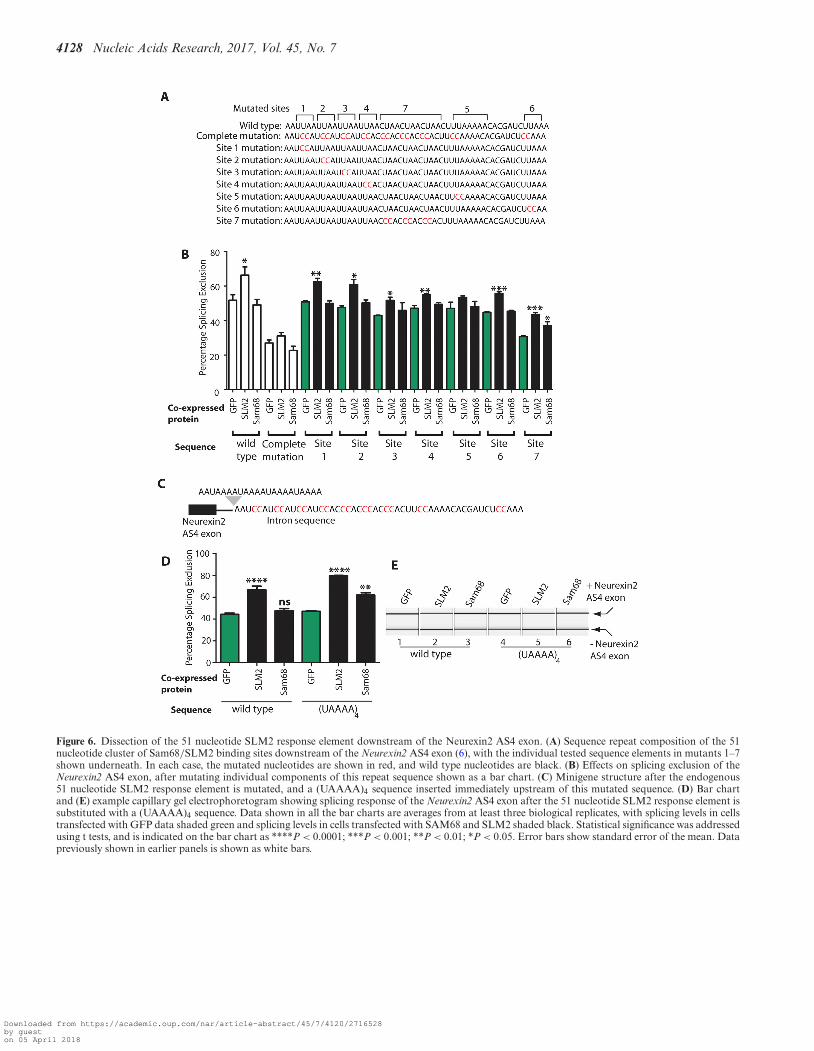
The above experiments indicate that a difference in bind-ing site density flanking the regulated exon enables splicingregulation of the Neurexin2 AS4 exon by SLM2 and notSam68. The 51 nucleotide element itself contains a numberof SLM2/Sam68 RNA binding sites. To test how many in-dividual Sam68/SLM2 binding sites are sufficient to enablesplicing repression of Neurexin2 AS4 by SLM2, we furtherdissected the 51 nucleotide sequence within its normal loca-tion.
The 51 nucleotide cluster of SLM2/Sam68 binding sitesdownstream of the Neurexin2 AS4 exon contains three dis-tinct sequence elements (Figure 6A): four UUAA repeats(sites 1–4 in Figure 6A); directly followed by three CUAArepeats (site 7 in Figure 6A); followed by a more extendedAU-rich element (containing UUAAAAA and UUAAA,which are sites 5 and 6 respectively in Figure 6A). Individ-
Downloaded from https://academic.oup.com/nar/article-abstract/45/7/4120/2716528by gueston 05 April 2018
4126 Nucleic Acids Research, 2017, Vol. 45, No. 7
Figure 4. Increasing the density of UWAA repeats makes the Neurexin2 AS4 exon respond to SAM68 as well as SLM2. (A) Distribution of UWAA repeatsin the wild type Neurexin2 AS4 minigene, and two engineered versions of this minigene that have an additional cluster placed downstream (intron position+112) or upstream (intron position -100) relative to the AS4 exon. The position of the Neurexin2 AS4 exon is indicated as a yellow pointed bar withinthe wild type minigene and for each mutant. The position of the extra 51 nucleotide SLM2 response sequence in each new construct is shown as a blackbar (or a thinner line where these are absent). The edges of the minigene insert, where they are inserted into the exon trap vector pXJ41 are shown asgreen arrowheads. (B) Percentage Splicing Exclusion of the Neurexin2 AS4 exon after co-expression of the three minigenes with GFP, a SLM2-GFP fusionprotein, or a SAM68-GFP fusion protein. (C) Representative capillary gel electrophoretograms showing splicing control of the wild type or engineeredversions of the Neurexin2 minigene. (D) Western blot showing expression levels of proteins in (B). On the bar chart, statistical significance was addressedusing t tests. ****P < 0.0001; ***P < 0.001; NS, not significant. Error bars show standard error of the mean.
ual mutation of each of the UUAA repeats led to decreasedsplicing repression by SLM2, but in each case splicing re-pression was still statistically significant compared with thesame minigene co-transfected with GFP (sites 1–4, Figure6B. In particular mutation of sites 3 and 4 resulted in a rel-atively modest but still significant PSE change). Similarly,the Neurexin2 AS4 exon was still repressed by SLM2 af-ter mutation of the UUAAA sequence (site 6, Figure 6B).Following each change in the 51 nucleotide element, splic-ing repression was only via SLM2 protein and not Sam68.The strongest effects on SLM2-mediated exon skipping was
caused by individual mutation of the UAAAA sequence(site 5, Figure 6B), after which the Neurexin2 AS4 exon wasno longer significantly repressed by SLM2. Individual mu-tation of the CUAA repeat region (site 7) did not preventsplicing repression by SLM2, but reduced the overall levelsof splicing exclusion from 52% to 31% (site 7 in Figure 6B)in cells co-transfected with GFP, suggesting that site 7 maycorrespond to a normally strong repressive sequence thatis disrupted by the mutation (or alternatively the mutationmay create an enhancer).
Downloaded from https://academic.oup.com/nar/article-abstract/45/7/4120/2716528by gueston 05 April 2018
Nucleic Acids Research, 2017, Vol. 45, No. 7 4127
Figure 5. The SLM2 response element controlling the Neurexin2 AS4 exon can operate from both upstream and further downstream of the target exon.(A) UUAA and UAAA sequence contents of wild type Neurexin2 AS4 minigene and minigenes that have the binding site cluster moved to various intronicpositions upstream or downstream of the AS4 exon. The minigene insert sequence cloned into the pXJ41 exon trap vector is shown, with nucleotidenumbering from the first to last nucleotide of this inserted sequence. The position of the Neurexin2 AS4 exon is indicated as a yellow pointed bar underthe wild type and each mutant. The position of the 51 nucleotide SLM2 response sequence in each construct is shown as a black bar. (B) Effect of movingthe UWAA cluster downstream of the Neurexin2 AS4 exon within the minigene construct, shown a bar chart. (C) Effect of moving the 51 nucleotideUWAA cluster upstream of the Neurexin2 AS4 exon on exon skipping, shown as a bar chart (data from three biological replicates). Statistical significancewas addressed using t tests, and is shown on the bar charts as: ***P < 0.001; **P < 0.01; *P < 0.05. Error bars show standard error of the mean. Datapreviously shown in earlier panels is shown as white bars.
The strongest effect on exon skipping in the above exper-iments was thus mutation of the UAAAA sequence (posi-tion 5 in Figure 6A). FP analysis of a (UAAAA)4 sequenceconfirmed that this sequence efficiently binds to the STARdomains of SLM2 and Sam68 proteins in vitro, with a simi-lar dissociation constant to the full 51 nucleotide Neurexin2response element (Table 1). To test if an increased num-ber of UAAAA sites would be sufficient by itself to en-able Neurexin2 AS4 splicing control by Sam68 as well as
SLM2, we constructed a minigene that contained four con-secutive repeats of this sequence, (UAAAA)4, to replacethe endogenous single copy of the 51 nucleotide sequence(previously removed by the complete mutation). The AS4exon from this (UAAAA)4 minigene construct was includedwithin mRNAs at similar levels to the wild type controlwhen co-transfected with GFP (Figure 6C). Insertion of the(UAAAA)4 sequence downstream of the AS4 exon enabledvery efficient splicing repression by co-expressed SLM2 pro-
Downloaded from https://academic.oup.com/nar/article-abstract/45/7/4120/2716528by gueston 05 April 2018
4128 Nucleic Acids Research, 2017, Vol. 45, No. 7
Figure 6. Dissection of the 51 nucleotide SLM2 response element downstream of the Neurexin2 AS4 exon. (A) Sequence repeat composition of the 51nucleotide cluster of Sam68/SLM2 binding sites downstream of the Neurexin2 AS4 exon (6), with the individual tested sequence elements in mutants 1–7shown underneath. In each case, the mutated nucleotides are shown in red, and wild type nucleotides are black. (B) Effects on splicing exclusion of theNeurexin2 AS4 exon, after mutating individual components of this repeat sequence shown as a bar chart. (C) Minigene structure after the endogenous51 nucleotide SLM2 response element is mutated, and a (UAAAA)4 sequence inserted immediately upstream of this mutated sequence. (D) Bar chartand (E) example capillary gel electrophoretogram showing splicing response of the Neurexin2 AS4 exon after the 51 nucleotide SLM2 response element issubstituted with a (UAAAA)4 sequence. Data shown in all the bar charts are averages from at least three biological replicates, with splicing levels in cellstransfected with GFP data shaded green and splicing levels in cells transfected with SAM68 and SLM2 shaded black. Statistical significance was addressedusing t tests, and is indicated on the bar chart as ****P < 0.0001; ***P < 0.001; **P < 0.01; *P < 0.05. Error bars show standard error of the mean. Datapreviously shown in earlier panels is shown as white bars.
Downloaded from https://academic.oup.com/nar/article-abstract/45/7/4120/2716528by gueston 05 April 2018
Nucleic Acids Research, 2017, Vol. 45, No. 7 4129
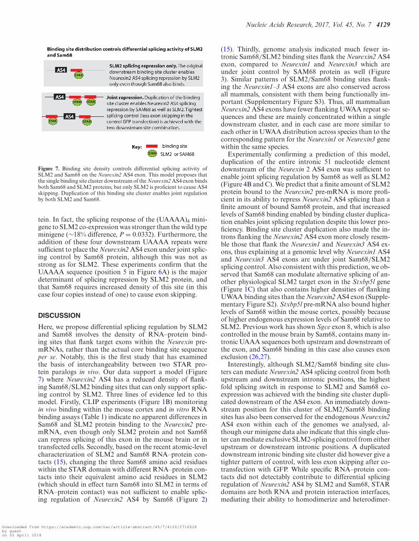
Figure 7. Binding site density controls differential splicing activity ofSLM2 and Sam68 on the Neurexin2 AS4 exon. This model proposes thatthe single binding site cluster downstream of the Neurexin2 AS4 exon bindsboth Sam68 and SLM2 proteins, but only SLM2 is proficient to cause AS4skipping. Duplication of this binding site cluster enables joint regulationby both SLM2 and Sam68.
tein. In fact, the splicing response of the (UAAAA)4 mini-gene to SLM2 co-expression was stronger than the wild typeminigene (∼18% difference, P = 0.0332). Furthermore, theaddition of these four downstream UAAAA repeats weresufficient to place the Neurexin2 AS4 exon under joint splic-ing control by Sam68 protein, although this was not asstrong as for SLM2. These experiments confirm that theUAAAA sequence (position 5 in Figure 6A) is the majordeterminant of splicing repression by SLM2 protein, andthat Sam68 requires increased density of this site (in thiscase four copies instead of one) to cause exon skipping.
DISCUSSION
Here, we propose differential splicing regulation by SLM2and Sam68 involves the density of RNA–protein bind-ing sites that flank target exons within the Neurexin pre-mRNAs, rather than the actual core binding site sequenceper se. Notably, this is the first study that has examinedthe basis of interchangeability between two STAR pro-tein paralogs in vivo. Our data support a model (Figure7) where Neurexin2 AS4 has a reduced density of flank-ing Sam68/SLM2 binding sites that can only support splic-ing control by SLM2. Three lines of evidence led to thismodel. Firstly, CLIP experiments (Figure 1B) monitoringin vivo binding within the mouse cortex and in vitro RNAbinding assays (Table 1) indicate no apparent differences inSam68 and SLM2 protein binding to the Neurexin2 pre-mRNA, even though only SLM2 protein and not Sam68can repress splicing of this exon in the mouse brain or intransfected cells. Secondly, based on the recent atomic-levelcharacterization of SLM2 and Sam68 RNA–protein con-tacts (15), changing the three Sam68 amino acid residueswithin the STAR domain with different RNA–protein con-tacts into their equivalent amino acid residues in SLM2(which should in effect turn Sam68 into SLM2 in terms ofRNA–protein contact) was not sufficient to enable splic-ing regulation of Neurexin2 AS4 by Sam68 (Figure 2)
(15). Thirdly, genome analysis indicated much fewer in-tronic Sam68/SLM2 binding sites flank the Neurexin2 AS4exon, compared to Neurexin1 and Neurexin3 which areunder joint control by SAM68 protein as well (Figure3). Similar patterns of SLM2/Sam68 binding sites flank-ing the Neurexin1–3 AS4 exons are also conserved acrossall mammals, consistent with them being functionally im-portant (Supplementary Figure S3). Thus, all mammalianNeurexin2 AS4 exons have fewer flanking UWAA repeat se-quences and these are mainly concentrated within a singledownstream cluster, and in each case are more similar toeach other in UWAA distribution across species than to thecorresponding pattern for the Neurexin1 or Neurexin3 genewithin the same species.
Experimentally confirming a prediction of this model,duplication of the entire intronic 51 nucleotide elementdownstream of the Neurexin 2 AS4 exon was sufficient toenable joint splicing regulation by Sam68 as well as SLM2(Figure 4B and C). We predict that a finite amount of SLM2protein bound to the Neurexin2 pre-mRNA is more profi-cient in its ability to repress Neurexin2 AS4 splicing than afinite amount of bound Sam68 protein, and that increasedlevels of Sam68 binding enabled by binding cluster duplica-tion enables joint splicing regulation despite this lower pro-ficiency. Binding site cluster duplication also made the in-trons flanking the Neurexin2 AS4 exon more closely resem-ble those that flank the Neurexin1 and Neurexin3 AS4 ex-ons, thus explaining at a genomic level why Neurexin1 AS4and Neurexin3 AS4 exons are under joint Sam68/SLM2splicing control. Also consistent with this prediction, we ob-served that Sam68 can modulate alternative splicing of an-other physiological SLM2 target exon in the Stxbp5l gene(Figure 1C) that also contains higher densities of flankingUWAA binding sites than the Neurexin2 AS4 exon (Supple-mentary Figure S2). Stxbp5l pre-mRNA also bound higherlevels of Sam68 within the mouse cortex, possibly becauseof higher endogenous expression levels of Sam68 relative toSLM2. Previous work has shown Sgce exon 8, which is alsocontrolled in the mouse brain by Sam68, contains many in-tronic UAAA sequences both upstream and downstream ofthe exon, and Sam68 binding in this case also causes exonexclusion (26,27).
Interestingly, although SLM2/Sam68 binding site clus-ters can mediate Neurexin2 AS4 splicing control from bothupstream and downstream intronic positions, the highestfold splicing switch in response to SLM2 and Sam68 co-expression was achieved with the binding site cluster dupli-cated downstream of the AS4 exon. An immediately down-stream position for this cluster of SLM2/Sam68 bindingsites has also been conserved for the endogenous Neurexin2AS4 exon within each of the genomes we analysed, al-though our minigene data also indicate that this single clus-ter can mediate exclusive SLM2-splicing control from eitherupstream or downstream intronic positions. A duplicateddownstream intronic binding site cluster did however give atighter pattern of control, with less exon skipping after co-transfection with GFP. While specific RNA–protein con-tacts did not detectably contribute to differential splicingregulation of Neurexin2 AS4 by SLM2 and Sam68, STARdomains are both RNA and protein interaction interfaces,mediating their ability to homodimerize and heterodimer-
Downloaded from https://academic.oup.com/nar/article-abstract/45/7/4120/2716528by gueston 05 April 2018

4130 Nucleic Acids Research, 2017, Vol. 45, No. 7
ize (15,28). As well as RNA binding, protein dimerisationis essential for the splicing repressive activity of both SLM2and Sam68 (15,16). SLM2 and Sam68 heterodimerise, andco-transfection of Sam68 with SLM2 protein inhibits theactivity of SLM2 in splicing control of Neurexin2 AS4, pre-sumably through inducing formation of a heterodimer thatis no longer sufficient to mediate skipping of this exon (6).
More generally, many splicing regulator proteins existin multi-protein families. Multi-protein families may en-able individual protein members to have evolved differ-ent patterns of expression, thus providing a mechanism tovary local concentrations of splicing regulator proteins likeSam68 (fairly ubiquitously expressed) and SLM2 (mainlyexpressed in the testis and brain). The existence of multi-protein families could also enable individual protein mem-bers of each family to specialise in splicing control of dif-ferent targets by evolving different RNA binding specifici-ties, although the STAR family members Sam68 and SLM2have been puzzling in sometimes selecting different func-tional targets while having indistinguishable RNA bindingsites (15). Our model presented here, that splice site den-sity within target pre-mRNAs influences splicing responsesto Sam68 and SLM2, might also apply to other families ofsplicing factors that have indistinguishable RNA target sitesbut that control different subsets of splicing targets.
SUPPLEMENTARY DATA
Supplementary Data are available at NAR Online.
FUNDING
Wellcome Trust [WT080368MA and WT089225/Z/09/Z];Biotechnological and Biological Sciences ResearchCouncil (BBSRC) [BB/K018957/1, BB/D013917/1 andBB/I006923/1]; Medical Research Council [CDA fel-lowship G1000526]; Telethon [GGP14095]; Associationfor International Cancer Research [14581]; NewcastleUniversity JW Luccock and E Jeffcock studentship (toM.D.). Funding for open access charge: BBSRC (RCUK).Conflict of interest statement. None declared.
REFERENCES1. Fu,X.D. and Ares,M. Jr (2014) Context-dependent control of
alternative splicing by RNA-binding proteins. Nat. Rev. Genet., 15,689–701.
2. Jangi,M. and Sharp,P.A. (2014) Building robust transcriptomes withmaster splicing factors. Cell, 159, 487–498.
3. Kelemen,O., Convertini,P., Zhang,Z., Wen,Y., Shen,M., Falaleeva,M.and Stamm,S. (2013) Function of alternative splicing. Gene, 514,1–30.
4. Lovci,M.T., Ghanem,D., Marr,H., Arnold,J., Gee,S., Parra,M.,Liang,T.Y., Stark,T.J., Gehman,L.T., Hoon,S. et al. (2013) Rbfoxproteins regulate alternative mRNA splicing through evolutionarilyconserved RNA bridges. Nat. Struct. Mol. Biol., 20, 1434–1442.
5. Rosenbloom,K.R., Armstrong,J., Barber,G.P., Casper,J., Clawson,H.,Diekhans,M., Dreszer,T.R., Fujita,P.A., Guruvadoo,L.,Haeussler,M. et al. (2015) The UCSC Genome Browser database:2015 update. Nucleic Acids Res., 43, D670–D681.
6. Ehrmann,I., Dalgliesh,C., Liu,Y., Danilenko,M., Crosier,M.,Overman,L., Arthur,H.M., Lindsay,S., Clowry,G.J., Venables,J.P.et al. (2013) The tissue-specific RNA binding protein T-STARcontrols regional splicing patterns of neurexin pre-mRNAs in thebrain. PLoS Genet., 9, e1003474.
7. Iijima,T., Wu,K., Witte,H., Hanno-Iijima,Y., Glatter,T., Richard,S.and Scheiffele,P. (2011) SAM68 regulates neuronal activity-dependentalternative splicing of neurexin-1. Cell, 147, 1601–1614.
8. Iijima,T., Iijima,Y., Witte,H. and Scheiffele,P. (2014) Neuronal celltype-specific alternative splicing is regulated by the KH domainprotein SLM1. J. Cell Biol., 204, 331–342.
9. Traunmuller,L., Bornmann,C. and Scheiffele,P. (2014) Alternativesplicing coupled nonsense-mediated decay generates neuronal celltype-specific expression of SLM proteins. J. Neurosci., 34,16755–16761.
10. Reissner,C., Runkel,F. and Missler,M. (2013) Neurexins. GenomeBiol., 14, 213.
11. Missler,M. and Sudhof,T.C. (1998) Neurexins: three genes and 1001products. Trends Genet., 14, 20–26.
12. Aoto,J., Martinelli,D.C., Malenka,R.C., Tabuchi,K. and Sudhof,T.C.(2013) Presynaptic neurexin-3 alternative splicing trans-synapticallycontrols postsynaptic AMPA receptor trafficking. Cell, 154, 75–88.
13. Traunmuller,L., Gomez,A.M., Nguyen,T.M. and Scheiffele,P. (2016)Control of neuronal synapse specification by a highly dedicatedalternative splicing program. Science, 352, 982–986.
14. Ehrmann,I., Fort,P. and Elliott,D.J. (2016) STARs in the CNS.Biochem. Soc. Trans., 44, 1066–1072.
15. Feracci,M., Foot,J.N., Grellscheid,S.N., Danilenko,M., Stehle,R.,Gonchar,O., Kang,H.S., Dalgliesh,C., Meyer,N.H., Liu,Y. et al.(2016) Structural basis of RNA recognition and dimerization by theSTAR proteins T-STAR and Sam68. Nat. Commun., 7, 10355.
16. Galarneau,A. and Richard,S. (2009) The STAR RNA bindingproteins GLD-1, QKI, SAM68 and SLM-2 bind bipartite RNAmotifs. BMC Mol. Biol., 10, 47.
17. Lin,Q., Taylor,S.J. and Shalloway,D. (1997) Specificity anddeterminants of Sam68 RNA binding. Implications for the biologicalfunction of K homology domains. J. Biol. Chem., 272, 27274–27280.
18. Pagliarini,V., Pelosi,L., Bustamante,M.B., Nobili,A.,Berardinelli,M.G., D’Amelio,M., Musaro,A. and Sette,C. (2015)SAM68 is a physiological regulator of SMN2 splicing in spinalmuscular atrophy. J. Cell Biol., 211, 77–90.
19. Bielli,P., Bordi,M., Di Biasio,V. and Sette,C. (2014) Regulation ofBCL-X splicing reveals a role for the polypyrimidine tract bindingprotein (PTBP1/hnRNP I) in alternative 5′ splice site selection.Nucleic Acids Res., 42, 12070–12081.
20. Grellscheid,S., Dalgliesh,C., Storbeck,M., Best,A., Liu,Y.,Jakubik,M., Mende,Y., Ehrmann,I., Curk,T., Rossbach,K. et al.(2011) Identification of evolutionarily conserved exons as regulatedtargets for the splicing activator tra2beta in development. PLoSGenet., 7, e1002390.
21. Venables,J.P., Bourgeois,C.F., Dalgliesh,C., Kister,L., Stevenin,J. andElliott,D.J. (2005) Up-regulation of the ubiquitous alternativesplicing factor Tra2beta causes inclusion of a germ cell-specific exon.Hum. Mol. Genet., 14, 2289–2303.
22. Liu,Y., Bourgeois,C.F., Pang,S., Kudla,M., Dreumont,N., Kister,L.,Sun,Y.H., Stevenin,J. and Elliott,D.J. (2009) The germ cell nuclearproteins hnRNP G-T and RBMY activate a testis-specific exon.PLoS Genet., 5, e1000707.
23. Richard,S., Torabi,N., Franco,G.V., Tremblay,G.A., Chen,T.,Vogel,G., Morel,M., Cleroux,P., Forget-Richard,A., Komarova,S.et al. (2005) Ablation of the Sam68 RNA binding protein protectsmice from age-related bone loss. PLoS Genet., 1, e74.
24. Chen,T., Damaj,B.B., Herrera,C., Lasko,P. and Richard,S. (1997)Self-association of the single-KH-domain family members Sam68,GRP33, GLD-1, and Qk1: role of the KH domain. Mol. Cell. Biol.,17, 5707–5718.
25. Schwartz,S., Hall,E. and Ast,G. (2009) SROOGLE: webserver forintegrative, user-friendly visualization of splicing signals. NucleicAcids Res., 37, W189–W192.
26. Chawla,G., Lin,C.H., Han,A., Shiue,L., Ares,M. Jr and Black,D.L.(2009) Sam68 regulates a set of alternatively spliced exons duringneurogenesis. Mol. Cell. Biol., 29, 201–213.
27. Paronetto,M.P., Messina,V., Barchi,M., Geremia,R., Richard,S. andSette,C. (2011) Sam68 marks the transcriptionally active stages ofspermatogenesis and modulates alternative splicing in male germcells. Nucleic Acids Res., 39, 4961–4974.
28. Lukong,K.E. and Richard,S. (2003) Sam68, the KHdomain-containing superSTAR. Biochim. Biophys. Acta, 1653, 73–86.
Downloaded from https://academic.oup.com/nar/article-abstract/45/7/4120/2716528by gueston 05 April 2018


![JavaFX.ppt [Uyumluluk Modu] - eskisehir.edu.trceng.eskisehir.edu.tr/aarslan2/BIM207/icerik/javafx.pdf · JavaFX introduces a new concept called binding property that enables a target](https://static.fdocuments.net/doc/165x107/5f9990ce435b11253e2d016f/uyumluluk-modu-eskisehiredutrcengeskisehiredutraarslan2bim207icerikjavafxpdf.jpg)


