
Binding Proteins from Animals with Possible Transport Function · Binding Proteins from Animals...
24
Binding Proteins from Animals with Possible Transport Function R. H. WASSERMAN, R. A. CORRADINO, and A. N. TAYLOR From the Department of Physical Biology,New York State Veterinary College, CorneAl University, Ithaca, New York 14850 A B S TR A CT Several proteins from various animal tissues with possible transport function have been briefly described, with emphasis given to a vitamin D- induced calcium-binding protein (CaBP) implicated in calcium translocation across epithelial membranes. The latter protein was shown to be present in the small intestine, colon, kidney, and the uterus (shell gland) of the chicken. CaBP was also found in the small intestine of the rat, dog, bovine, and monkey. This protein has been isolated in high purity from chick intestinal mucosa and some of its properties determined. Its molecular weight is about 28,000, its formation constant, about 2.6 X 10⁵ M -1, and its binding capacity, 1 calcium atom per protein molecule. Correlative studies have shown that CaBP concen- tration in intestinal mucosa varies with the calcium absorptive capacity of the gut, thereby suggesting that CaBP is intimately involved in the process of cal- cium absorption. CaBP has been localized in the brush border region of the intestinal absorptive cell and within goblet cells. Among other proteins men- tioned were the intrinsic factor required for vitamin B12 absorption and the protein(s) associated with iron translocation. Plasma membranes, to a large degree, determine the extent and rate of per- meation of substrates into and out of the cytoplasmic mass. Contributing to their passive permeability and selectivity are various physical and chemical properties of the membrane, such as its lipoid nature, the presence of aqueous channels, and the electrical charges associated with the cell surface and the core of the channel. These and other important aspects have been well docu- mented and have sometimes been amenable to detailed investigations by the use of artificially constructed membranes. Biological membranes, in addition, have the capacity to transport certain substrates against thermodynamic gra- dients (active transport) and to support reactions which accelerate the transfer of certain substrates down their electrochemical gradients (facilitated diffu- sion). Both of these latter two processes are hypothetically carrier-mediated and, in recent years, carrier-like molecules have been implicated in an increas- ing number of transport systems. These systems exhibit certain common characteristics including Michaelis-Menten kinetics, a high degree of struc- ix 4 s The Journal of General Physiology on April 4, 2017 Downloaded from Published July 1, 1969
-
Upload
truongkhue -
Category
Documents
-
view
231 -
download
0
Transcript of Binding Proteins from Animals with Possible Transport Function · Binding Proteins from Animals...
Binding Proteins from Animals with
Possible Transport Function
R. H. W A S S E R M A N , R. A. C O R R A D I N O , and A. N. T A Y L O R
From the Department of Physical Biology, New York State Veterinary College, CorneAl University, Ithaca, New York 14850
A B S TR A C T Several proteins from various animal tissues with possible transport function have been briefly described, with emphasis given to a vitamin D- induced calcium-binding protein (CaBP) implicated in calcium translocation across epithelial membranes. The latter protein was shown to be present in the small intestine, colon, kidney, and the uterus (shell gland) of the chicken. CaBP was also found in the small intestine of the rat, dog, bovine, and monkey. This protein has been isolated in high purity from chick intestinal mucosa and some of its properties determined. Its molecular weight is about 28,000, its formation constant, about 2.6 X 10⁵ M -1, and its binding capacity, 1 calcium atom per protein molecule. Correlative studies have shown that CaBP concen- tration in intestinal mucosa varies with the calcium absorptive capacity of the gut, thereby suggesting that CaBP is intimately involved in the process of cal- cium absorption. CaBP has been localized in the brush border region of the intestinal absorptive cell and within goblet cells. Among other proteins men- tioned were the intrinsic factor required for vitamin B12 absorption and the protein(s) associated with iron translocation.
Plasma membranes, to a large degree, determine the extent and rate of per- meation of substrates into and out of the cytoplasmic mass. Contributing to their passive permeabili ty and selectivity are various physical and chemical properties of the membrane, such as its lipoid nature, the presence of aqueous channels, and the electrical charges associated with the cell surface and the core of the channel. These and other important aspects have been well docu- mented and have sometimes been amenable to detailed investigations by the use of artificially constructed membranes. Biological membranes, in addition, have the capacity to transport certain substrates against thermodynamic gra- dients (active transport) and to support reactions which accelerate the transfer of certain substrates down their electrochemical gradients (facilitated diffu- sion). Both of these latter two processes are hypothetically carrier-mediated and, in recent years, carrier-like molecules have been implicated in an increas- ing number of transport systems. These systems exhibit certain common characteristics including Michaelis-Menten kinetics, a high degree of struc-
i x 4 s
The Journal of General Physiology
on April 4, 2017
Dow
nloaded from
Published July 1, 1969

WASSERMAN, CORRADINO, AND TAYLOR Binding Protdns and Transport Function x 15 s
tural specifcity, and relative stereospecificity. Because of the specificity de- manded, it is usually considered that the carriers are proteins, molecules having sufficient complexity to contain the required information to distinguish between chemically similar molecules. The de facto existence of membrane- associated proteins having a possible transport or carrier function is substan- tiated by the information given in the recent review article of Pardee (1) and the material presented at the present Symposium.
There appears to be at least two classes of proteins implicated in transport, those that are readily released from the cell or cell surface by the osmotic shock technique of Heppel (2) or by cell homogenization, and those that are firmly bound to membranous cell components and which can be solubilized by reagents such as Tri ton X-100 (3) and NaI (4). These may be designated as "soluble" and "insoluble" transport proteins, respectively, and the relation between these and solute movement, and between each other, is not clear at present. As an example of each, Anraku (5) has isolated a soluble protein from Escherichia coli K-12 with a high binding affinity for galactose; the insoluble " M " (membrane) protein of Kennedy and Fox (3), also from E. coli, ap- pears to interact with/~-galactosides during the transport reaction.
A few binding proteins with possible transport function have been detected in animal tissues. One of the first so designated is the membrane adenosine triphosphatase of the crab nerve and erythrocyte membrane; this enzyme is discussed in detail elsewhere in this volume. Others are the calcium-binding protein induced by vitamin D, the intrinsic factor related to vitamin B12 ab- sorption by the intestine, the galactose-binding component of intestinal brush border, a possible iron-binding protein of intestinal mucosa, the blood protein, transferrin, implicated in erythrocytic iron uptake, and a shockable protein from kidney that binds transportable organic acids. These will be subsequently discussed although emphasis will be given to the first mentioned.
C A L C I U M - B I N D I N G P R O T E I N S
The recognition of the existence of a calcium-binding protein (CaBP) in in- testinal mucosa was derived from studies on the mechanism of action of vita- min D. It was early shown that the protein was not present in tissue derived from rachitic animals but began to appear in such animals after vitamin D had been administered. From the older, classical studies of Mellanby (6) and Nicolaysen (7), and as reviewed recently by Wasserman and Taylor (8), such treatment also restores the capacity of the intestine to absorb calcium and eventually causes a remission of the rachitic syndrome. Because of the close interconnections between calcium movement, vitamin D, and a possible trans- port protein, it was considered worthwhile to review briefly certain aspects of the effect of vitamin D on calcium absorption and on cell behavior before describing the investigations on CaBP.
on April 4, 2017
Dow
nloaded from
Published July 1, 1969
i i6 s T R A N S P O R T P R O T E I N S
Physiological Site of Vitamin D Action
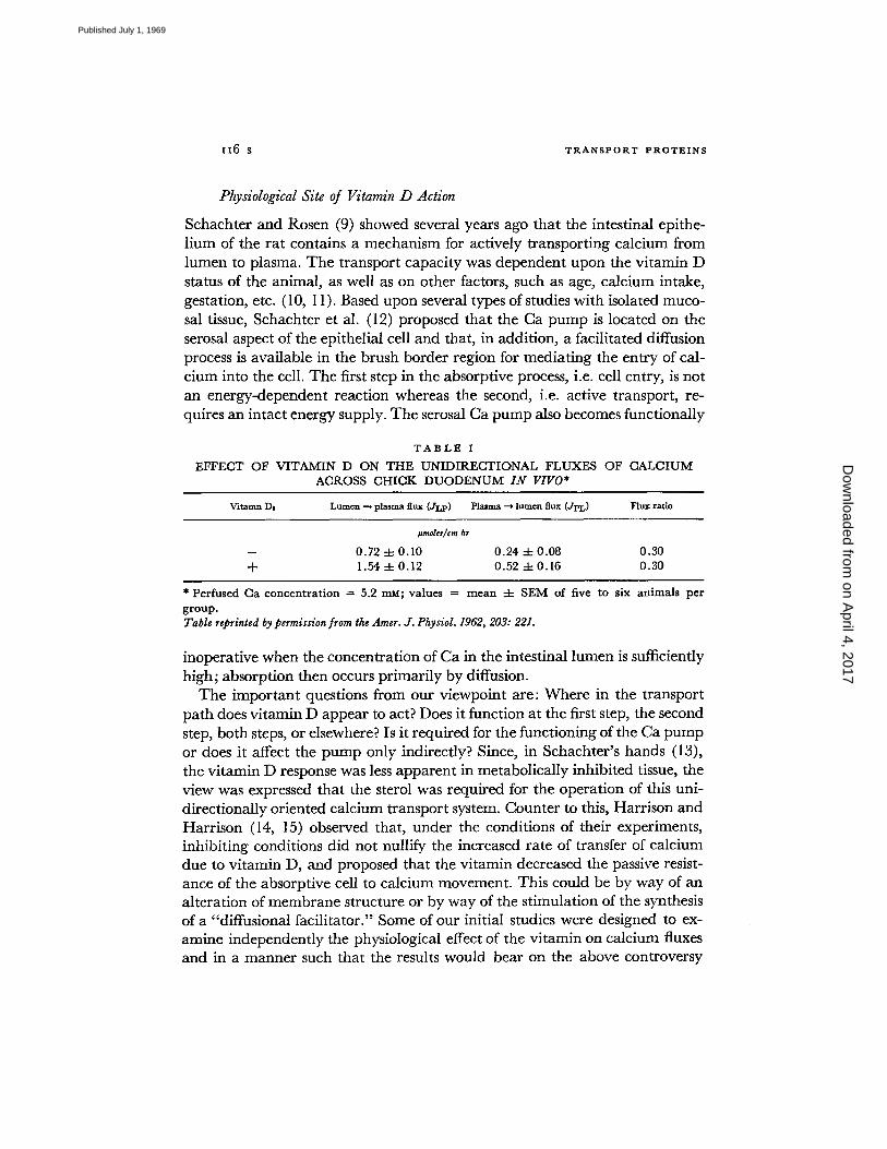
Schachter and Rosen (9) showed several years ago that the intestinal epithe- l ium of the rat contains a mechanism for actively transporting calcium from lumen to plasma. The transport capacity was dependent upon the vitamin D status of the animal, as well as on other factors, such as age, calcium intake, gestation, etc. (10, I 1). Based upon several types of studies with isolated muco- sal tissue, Schachter et al. (12) proposed that the Ca pump is located on the serosal aspect of the epithelial cell and that, in addition, a facilitated diffusion process is available in the brush border region for mediating the en t ry of cal- cium into the cell. The first step in the absorptive process, i.e. cell entry, is not an energy-dependent reaction whereas the second, i.e. active transport, re- quires an intact energy supply. The serosal Ca pump also becomes functionally
T A B L E I
E F F E C T O F V I T A M I N D O N T H E U N I D I R E C T I O N A L F L U X E S O F C A L C I U M
A C R O S S C H I C K D U O D E N U M I N VIVO*
Vitamn Ds Lumen --* plasma flux (JI,P) Plasma --, lumva flux (Jl~,) Flux ratio
~moles/¢m hr
- - 0 . 7 2 4- 0 . 1 0 0 . 2 4 4- 0 . 0 8 0 . 3 0
-t- 1 .54 4- 0 . 1 2 0 . 5 2 4- 0 . 1 6 0 . 3 0
* P e r f u s e d C a c o n c e n t r a t i o n = 5.2 m u ; v a l u e s = m e a n -4- S E M of f ive t o s ix a n i m a l s per g r o u p . Table reprinted by permission from the Amer. J. Physiol. 1962, 203: 221.
inoperative when the concentration of Ca in the intestinal lumen is sufficiently high; absorption then occurs primarily by diffusion.
The important questions from our viewpoint are: Where in the transport path does vitamin D appear to act? Does it function at the first step, the second step, both steps, or elsewhere? Is it required for the functioning of the Ca pump or does it affect the pump only indirectly? Since, in Schachter's hands (13), the vitamin D response was less apparent in metabolically inhibited tissue, the view was expressed that the sterol was required for the operation of this uni- directionally oriented calcium transport system. Counter to this, Harrison and Harrison (14, 15) observed that, under the conditions of their experiments, inhibiting conditions did not nullify the increased rate of transfer of calcium due to vitamin D, and proposed that the vitamin decreased the passive resist- ance of the absorptive cell to calcium movement. This could be by way of an alteration of membrane structure or by way of the stimulation of the synthesis of a "diffusional facilitator." Some of our initial studies were designed to ex- amine independently the physiological effect of the vitamin on calcium fluxes and in a manner such that the results would bear on the above controversy
on April 4, 2017
Dow
nloaded from
Published July 1, 1969

WASSEI~AN, COI~ADINO, AND TAYLOR Binding Proteins and Transport Function x l 7 s
(16, 17). I t was reasoned that, if the Ca pump were stimulated or activated, the unidirectional flux ratio of rachitic chicks should be less than that of vita- min D- t rea ted chicks. If the flux ratio remained the same, this would suggest that the permeabili ty theory was probably correct. The data, par t of which is presented in Table I, clearly showed that vitamin D treatment increased both the efflux (J,.p) and the influx (Jp,.) of Ca, and that the flux ratio remained essentially unchanged. The most obvious interpretation was that the sterol in
100 n I ABSORPTION J
75 (% 47Ca DOSE) ~ - ~
SO o . ~ e ~ e''s- ~VIT D3 ~
25 / je RACHITIC ~ ~ O
0 I I , - MUCOSAL UPTAKE
10 ~ (% 47Ca / g)
8
4 / ~
2 ~RACHITIC
I I 0 10 20 30
TIME (rain)
FIGURE I. Et~cct of vitamin 133 on the duodenal absorption and mucosal tissue uptake of radiocaldum. The dosing solution, contalnlng 47Ca and 4°Ca (I mg/ml), was placed in the lumen of a ligated segment of duodenum of anesthetized racldtic and vit~mln Ds-- treated chicks. At the time periods indicated, the duodenum was removed and then the residual radioactivity in the lumen and the mucosal content of the isotope were deter- mined by a previously described procedure (20). Each point represents the mean of five to six chicks.
some manner enhanced the permeabili ty of the epithelial barrier of the intes- tine, allowing a greater m o v e m e n t of Ca in both directions. Of the two hy- potheses previously offered, the one stating that the vitamin effect was by way of a nonenergy-requiring permeabili ty alteration appeared to be the more likely one.
In respect to the cellular site of the response, other studies were done in which the uptake of either intraluminal 47Ca or plasma 4~Ca by the mucosal tissue was determined. At the same time, the absorption of 47Ca or the "secre- t ion" of plasma 47Ca into the lumen was also measured. From the data de- picted in Fig. 1, it was apparent that vitamin D treatment enhanced *TCa absorption (per usual) and, at the earlier time periods, enhanced the quanti ty
on April 4, 2017
Dow
nloaded from
Published July 1, 1969

zz8 s T R A N S P O R T P R O T E I N s
of *7Ca associated with the mucosa. The rate of release of 47Ca by the mucosa was also stimulated by the treatment. In regard to movement in the opposite direction, the transfer of plasma 4rCa to the intestinal lumen was greater in those chicks treated with the vitamin but, on the other hand, the transfer from plasma to mucosal tissue was unaffected (Fig. 2). Tha t is, in both studies there was an apparent increase in Ca transfer across the mucosal border of the ab- sorbing cell no matter whether Ca was derived from the lumen or the plasma and, from this, one may conclude that this region is at least one of the promi- nent sites of expression of the vitamin D effect. These and other aspects of cal- cium transfer across the intestine were discussed recently (18).
( ~ I I 1 .I I I
z I ~ Plasma 4rCa -Lumen ooi-/\
Z 041- - x r Vit. "
I \ Rochitic i
o r I 1 I I I I
I Mucosa Uptake of Plasma 47Ca
06"-
- ,
I I / '°3 I I 5 IO 15
TIME (rain)
Fzotnu~ 2. Effect of vitamin D3 on the transfer of 47Ca from plasma to intestinal lumen and from plasma to mucosal tissue. The radiocalcium was injected intravenously into rachitic or vitamin Drtreated chicks. Prior to this, 1 ml of 0.15 t,i NaC1 was placed in a ligated loop of duodenum. At the times indi- cated, the duodenum was re- moved, and the radioactivity content of the duodenal fluid and the mucosa were meas- ured, as described previously (20). The values represent the mean 4- SEM of five to six chicks.
Molecular Basis of Vitamin D Action
The administration of vitamin D to a rachitic animal does not cause an imme- diate restoration of the absorptive process but requires several hours, the time depending upon the dose level of the vitamin. This lag effect was noted by Lindquist (19) in 1952 and a typical time-response curve from our studies is shown in Fig. 3 (20). I t is apparent that, at the dose level used (500 IU/chick) , between 8-16 hr must transpire before a significant response occurs. Consider- able attention has been given to the events which take place within this period. These have been tentatively identified as: (a) the time required for the sterol to localize in the target tissue and at the appropriate intracellular site; (b) the time for possible transformation of vitamin D to an active metabolite; and, finally, (c) time required for the induction of the synthesis of a necessary com- ponent of the calcium transport system (2 I, 22). The transformation of vitamin D to an active metaboli te was first shown by Norman et al. (23), using radio- actively labeled sterol. A metabolite was recently isolated and characterized
on April 4, 2017
Dow
nloaded from
Published July 1, 1969

WASSERMAN, CORRADINO, ANn TAYLOR Binding Proteins and Transport Function I 19 s
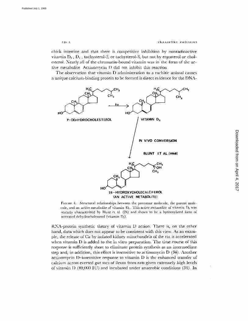
by Blunt e ta l . (24) and found to be 25-hydroxycholecalciferol (Fig. 4). The involvement of vitamin D in protein synthesis was initially derived from the experiments of Eisenstein and Passavoy (25) in which it was demonstrated that the hypercalcemia produced by massive doses of vitamin D was prevented by pretreatment with actinomycin D. Actinomycin D and other inhibitors of protein synthesis, such as puromycin and cycloheximide, were later shown by several groups to inhibit the intestinal response of various species to vitamin D (26-28) and, from this, the most reasonable hypothesis would be that the syn-
~: 1 0 0
80
60
< 40
l
2 0
Q
FIGURE 3.
1 I I I I
" ~CONTROL
I ,, I I I ~.~ I 2 4 8 16 4 0
TIME ( hr )
Delayed absorptive response to vitamin D3. At zero time, a group of rachitic chicks was given 500 IU vitamin D3 each. At the times indicated, the capacity of a ligated duodenal segment to absorb calcium was determined. The absorption period was 30 min. Each value represents the mean 4- SEM of five to six chicks. Figure reprinted by permission from the J. Nutr., 1962, 77:69.
thesis of a specific protein is induced by the vitamin. In this same vein, the incorporation of 3H-orotic acid and ~H-uridine into RNA of intestinal mucosa was shown to be stimulated by the prior injection of vitamin D; this response was also inhibited by actinomycin D (29, 30).
The subcellular localization of the vitamin is also consistent with the protein synthetic hypothesis since both Haussler and Norman (31) and Stohs and DeLuca (32) find this sterol in highest concentration in the nucleus of the target tissue. There is disagreement as to whether it accumulates cn the nu- clear membrane or is bound by specific structures within the nucleoplasm. However, Haussler et al. (33) recently presented substantial evidence indicat- ing that labeled vitamin D3 associates with the chromatin of mucosal cells of
on April 4, 2017
Dow
nloaded from
Published July 1, 1969
I 2 0 S T R A N S P O R T P R O T E I N S
chick intestine and that there is competitive inhibition by nonradioactive vitamin D2, D~, tachysterol-2, or tachysterol-3, but not by ergosterol or chol- esterol. Nearly all of the chromatin-bound vitamin was in the form of the ac- tive metabolite. Actinomycin D did not inhibit this reaction.
The observation that vitamin D administration to a rachitic animal causes a unique calcium-binding protein to be formed is direct evidence for the DNA-
HO / "L" ~J I'~~ J
hw
7-DEHYDROCHOLESTEROL
C,H,J,. I c" "V "l CH,
CH 21 I I
VITAMIN D~
IN VIVO CONVERSION
BLUNT ET AL.(1968)
H ~ C ~ C H 3 C.H:t,~ ~'OH
CH 2 I,~ ~J J
HO I ~
2 5- HYDROXYCHOLECALCIFE ROL (AN ACTIVE METABOLITE)
FIGURE 4. Structural relationships between the precursor molecule, the parent mole- cule, and an active metabolite of vitamin D3. This active metabolite of vitamin 1)~ was recently characterized by Blunt et al. (24) and shown to be a hydroxylated form of activated dehydrocholesterol (vitamin ])3).
RNA-protein synthetic theory of vitamin D action. There is, on the other hand, data which does not appear to be consistent with this view. As an exam- ple, the release of Ca by isolated kidney mitochondria of the rat is accelerated when vitamin D is added to the in vitro preparation. The time course of this response is sufficiently short to eliminate protein synthesis as an intermediate step and, in addition, this effect is insensitive to actinomycin D (34). Another actinomycin D-insensitive response to vitamin D is the enhanced transfer of calcium across everted gut sacs of ileum from rats given extremely high levels of vitamin D (40,000 IU) and incubated under anaerobic conditions (34). In
on April 4, 2017
Dow
nloaded from
Published July 1, 1969
WASSERMAN, CORRADINO, ANn TAYLOR Binding Proteins and Transport Function ~2x s
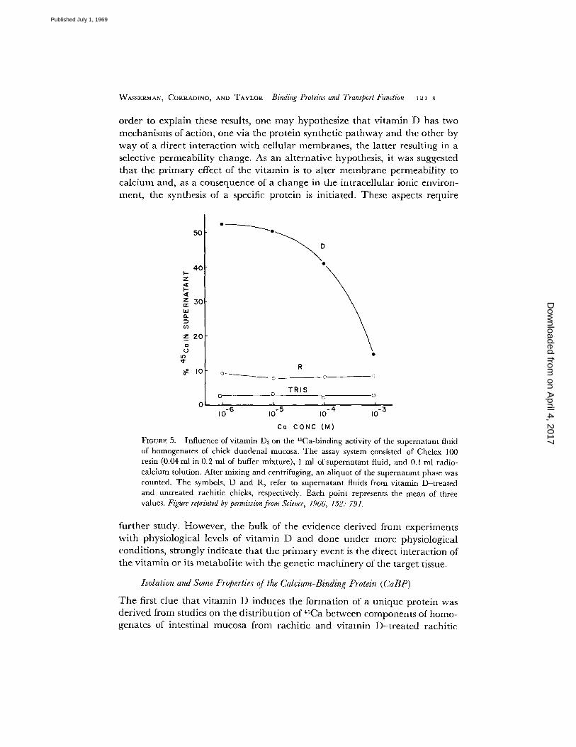
order to explain these results, one may hypothesize that vitamin D has two mechanisms of action, one via the protein synthetic pathway and the other by way of a direct interaction with cellular membranes, the latter resulting in a selective permeabili ty change. As an alternative hypothesis, it was suggested that the primary effect of the vitamin is to alter membrane permeability to calcium and, as a consequence of a change in the intracellular ionic environ- ment, the synthesis of a specific protein is initiated. These aspects require
50
4O z
so kd
Z 2 0
g, tg~ ¢
g Io -c.
z i
10 -3
R
° ~ c __ - o
TRIS ~. E3 .[],
0 I I I
10 - 6 10 - 5 10 . 4
Co CONC ( M )
FIGURE 5. Influence of v i tamin Da on the 4sCa-binding activity of the superna tan t fluid of homogenates of chick duodenal mucosa. The assay system consisted of Chelex 100 resin (0.04 ml in 0.2 ml of buffer mixture) , 1 ml of superna tan t fluid, and 0.1 ml radio- calcium solution. After mixing and centrifuging, an aliquot of the supernatant phase was counted. The symbols, D and R, refer to superna tan t fluids from vi tamin D- t rea ted and unt rea ted rachitic chicks, respectively. Each point represents the mean of three values. Figure reprinted by permission from Science, 1966, 152: 791.
further study. However, the bulk of the evidence derived from experiments with physiological levels of vitamin D and done under more physiological conditions, strongly indicate that the primary event is the direct interaction of the vitamin or its metabolite with the genetic machinery of the target tissue.
Isolation and Some Properties of tt~e Calcium-Binding Protein (CaBP)
The first clue that vitamin D induces the formation of a unique protein was derived from studies on the distribution of 45Ca between components of homo- genates of intestinal mucosa from rachitic and vitamin D-t rea ted rachitic
on April 4, 2017
Dow
nloaded from
Published July 1, 1969

I 2 2 S T R A N S P O R T P R O T E I N S
chicks (35). Signif icant ly more of the r ad ioca lc ium remained in the soluble phase of the mate-:ial f rom vi tamin D - t r e a t e d versus that f rom rachi t ic animals and most or all of this effect appea red to be due to the presence of a factor in the v i tamin D tissue with a capaci ty to bind Ca. T h e pro te inaceous na tu re of
FIGURE 6. Acrylamide gel electrophoretic pat- tern of proteins in the supernatant fluids of mucosal homogenates. The symbols, R and D, refer to ma- terial from rachitic and vitamin D3-treated chicks, respectively. The arrow designates the band with which the vitamin D-induced calcium-binding protein is associated. Figure reprinted by permission from Arch. Biochem. Biophys., 1967, 119: 536.
the factor was shown by the fact tha t it was nondia lyzable , hea t labile, t rypsin and Pronase digestible, and excluded by a Sephadex G-25 co lumn (36).
T h e assay system for CaBP was based upon the ion exchange p rocedu re of Briggs and Fle ishman (37) and depended upon the compet i t ion of the resin and the soluble b inding substance for added radiocalc ium. Wi th this assay procedure , the b inding activities of the v i tamin D and rachi t ic supe rna tan t fluids and the buffer alone were de t e rmined and the results, as a funct ion of ca lc ium concen t ra t ion in the system, are shown in Fig. 5. Clearly, the b inding
on April 4, 2017
Dow
nloaded from
Published July 1, 1969

WASSERMAN, CORRADINO, AND TAYLOR Binding Proteins and Transport Function I2 3 s
activity of the vitamin D supernatant was considerably greater than that of the rachitic supernatant or the buffer alone and that, as the calcium concentration was increased, the proportion of radiocalcium maintained in the soluble phase of the vitamin D supernatant decreased, suggesting that a binding component of the latter fluid was being saturated. A comparison of acrylamide gel electro- phoretic patterns also revealed that at least one protein was present in vitamin D supernatant which could not be visualized in the rachitic material (cf. arrow, Fig. 6). This band was later shown to be, or have associated with it, the vitamin D- induced calcium-binding protein.
The CaBP was obtained in high purity by the use of a three-step fractiona- tion procedure which included, in order: (a) ammonium sulfate treatment; (b) gel filtration on Sephadex G-100; and (c) preparative disc acrylamide gel electrophoresis (38). The product yielded a single band on an analytical elec- trophoretic gel and a single Schlieren peak with the analytical ultracentrifuge. The molecular weight was estimated to be in the range of 25,000-28,000, as determined by the use of a calibrated gel filtration column and by sedimenta- tion equilibrium. The association constant between Ca and the protein was determined by the procedure of Schubert et al. (39) and found to be about 2.6 × 10 a M -1. Those for Sr and Ba were observed to be about 3.0 × 10⁴ and 5.8 × 10³ M -1, respectively. Analysis of the binding data indicated that 1 molecule of protein bound 1 atom of Ca.
TISSUE LOCALIZATION OF CaBP The vitamin D- induced calcium-binding protein has been identified in all segments of the small intestine (40), the colon, 1 and the kidney of the chick (40), and the uterus (shell gland) of the laying hen (41). Each of these are epithelial tissues across which calcium is translocated. CaBP has not been found in liver, muscle, pancreas, or blood. Although considerable effort was given to uncovering the presence of CaBP in bone, the data have proven negative thus far.
SPECIES DISTRIBUTION OF CaBP A vitamin D- induced calcium-binding protein has been found in intestinal mucosa of the following species: chick (36), rat (42), dog (43), bovine, and monkey. There are some differences in the properties of these various proteins, such as electrophoretic mobility and sen- sitivity to proteolytic enzymes, but detailed studies in this direction have not yet been carried out.
CORRELATION BETWEEN CaBP AND CALCIUM ABSORPTION Knowing that vitamin D increases Ca absorption and observing that a product of vitamin D interaction with the target tissue is a calcium-binding protein, is by itself sug- gestive evidence that the product (i.e. CaBP) is involved in the translocation process. However, other evidence is required to build a substantial case in favor of this hypothesis. For example, it must be shown that the appearance of
1 Taylor , A. N., and R. H. Wasserman. 1969. Unpubl i shed data.
on April 4, 2017
Dow
nloaded from
Published July 1, 1969

O Q
.1-
O o
I_n,, n l - ~ Z O O ~O (,.) 00
. , JO
Z , ~ ixl Q O
¢1
i2 4 s T R A N S P O R T P R O T E I N S
C a B P in t h e r a c h i t i c c h i c k a f t e r v i t a m i n D a d m i n i s t r a t i o n p r e c e d e s o r a t l e a s t
a c c o m p a n i e s t he p h y s i o l o g i c a l c h a n g e . T h i s was s h o w n to be a p p r o x i m a t e l y
t h e s i t u a t i o n in a n e a r l i e r e x p e r i m e n t (36) a n d was i n v e s t i g a t e d in m o r e d e t a i l
r e c e n t l y (44). P a r t o f t he l a t t e r s t u d y was to e x a m i n e the t i m e r e q u i r e d for t h e
70
6 0
50
40
30
20
10
I I I I t
. / ¢;--ACR ,AM,OE GEL / 1 G~ ~ )Cv~/BPBy .~T = IMMUNODIFFUSION
, , ,
10 20 30 40 TIME (HR)
FIGURE 7. Correlation between the appearance of CaBP and the restoration of the calcium-absorptive mechanism by vitamin D3. At zero time, a group of rachitic chicks were given either 100 1U or 5000 IU vitamin 133. At the periods designated above, the absorption of radiocalcium from a ligated segment of duodenum was measured; the ab- sorptive period was 1~ hr. CaBP in the duodenal mucosa of other similarly treated chicks at these same time periods was also measured. Three procedures were used: G, detection of the characteristic band on an acrylamide gel electrophoretic pattern; I, immunodif- fusion against a CaBP-specific antibody; and R, Chelex 100 ion exchange assay. In the above figure, the periods at which CaBP was first detected have been designated by a G, I, or R. The symbol, A, refers to the time at which a significant increase in absorption was first observed. Each point represents the group mean of five to six chicks ± SEM. Figure reprinted by permij~ion from Amer. Jr. Clin. Nutr., 1969, 22: 431.
a p p e a r a n c e of C a B P a t two levels of v i t a m i n D, o n e a t a p h y s i o l o g i c a l l eve l
(100 I U ) a n d o n e a t a p h a r m a c o l o g i c leve l (5000 I U ) . By r e s o r t i n g to t h e m o s t
sens i t ive t e c h n i q u e a v a i l a b l e to us, i .e. d o u b l e i m m u n o d i f f u s i o n , i t was s h o w n
t h a t C a B P is p r e s e n t in m u c o s a l t issue a t t he s a m e t i m e t h a t C a a b s o r p t i o n was
50
on April 4, 2017
Dow
nloaded from
Published July 1, 1969
WASSERMAN, CORRADINO, AND TAYLOR Binding Proteins and Transport Function I2 5 s
enhanced, in spite of the fact that the 5000 I U dose decreased the lag time by about 10 hr (Fig. 7).
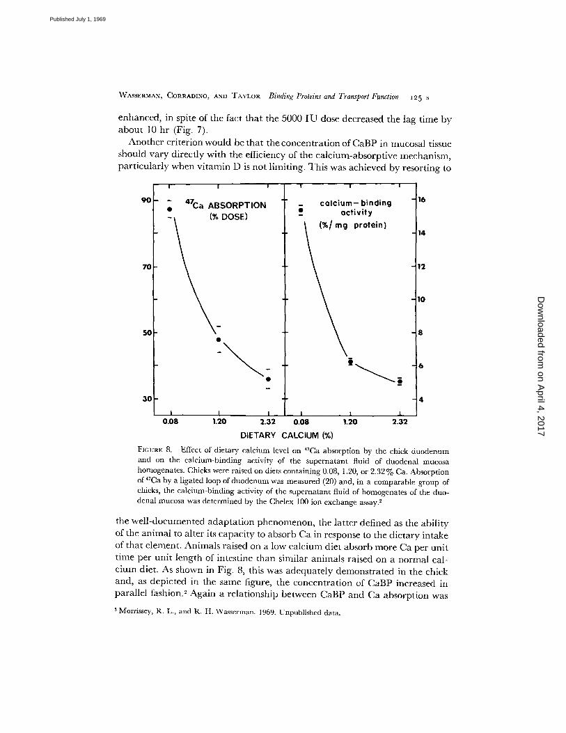
Another criterion would be that the concentration of CaBP in mucosal tissue should vary directly with the efficiency of the calcium-absorptive mechanism, particularly when vitamin D is not limiting. This was achieved by resorting to
I I i
9 o - - 4z . , a ~ " ABSORPTION
- D O S E )
70
I
5O
3 0
I I 0 .08 1.20
! I
c a l c i u m - b i n d i n g e~ a c t i v i t y
I
16
14
12
10
8
6
4
I I I I 2.32 0.08 1.20 2.32
DIETARY CALCIUM (%)
FIGURE 8. Effect of dietary calcium level on 47Ca absorption by the chick duodenum and on the calcium-binding activity of the supernatant fluid of duodenal mucosa homogenates. Chicks were raised on diets containing 0.08, 1.20, or 2.32% Ca. Absorption of 47Ca by a ligated loop of duodenum was measured (20) and, in a comparable group of chicks, the calcium-binding activity of the supernatant fluid of homogenates of the duo- denal mucosa was determined by the Chelex i00 ion exchange assay. 2
the well-documented adaptation phenomenon, the latter defined as the ability of the animal to alter its capacity to absorb Ca in response to the dietary intake of that element. Animals raised on a low calcium diet absorb more Ca per unit time per unit length of intestine than similar animals raised on a normal cal- cium diet. As shown in Fig. 8, this was adequately demonstrated in the chick and, as depicted in the same figure, the concentration of CaBP increased in parallel fashion. 2 Again a relationship between CaBP and Ca absorption was
2 Morrissey, R. L., and R. H. Wasserman. 1969. Unpublished data.
on April 4, 2017
Dow
nloaded from
Published July 1, 1969

In6 s T R A N S P O R T P R O T E I N S
a p p a r e n t . These and o ther corre la t ions s t rongly suggested tha t the hypothes is is cor rec t ; i.e., C a B P funct ions in the t rans loca t ion process.
F u r t h e r it was d e m o n s t r a t e d tha t C a B P synthesis is inhib i ted by ac t ino- myc in D (45), and tha t the concen t ra t ion of C a B P in intest inal m u c o s a is
FIGURE 9. Fluorescent antibody localization of CaBP in histological section of duodenal tissue from vitamin D~-treated chick. The sample was first treated with CaBP-specific antiserum from rabbits, then with fluoresceinisothiocyanate-conjugated anti-rabbit serum from sheep. Light areas represent specific fluorescence due to presence of CaB[. Rachitic intestine and immunologically blocked controls exhibited nonspecific fluores- cence only. × 275.
g rea t e r in y o u n g e r t han older an imals and g rea te r in lay ing t han in n o n - e g g lay ing hens (8).
T w o types of evidence, tha t would be unequivoca l , have not been ach ieved successfully. O n e would be to p lace CaBP in rachi t ic m u c o s a and restore its abso rp t ive capac i t y and, secondly, inhibi t the response wi th a specific r e agen t such as the a n t i b o d y to CaBP. Ne i the r of these have yet been accompl i shed .
LOCALIZATION OF CALCIUM-BINDING PROTEIN A n a n t i s e r u m shown to be
on April 4, 2017
Dow
nloaded from
Published July 1, 1969
WASSERMAN, CORRADINO, AND TAYLOR Binding Proteins and Transport Function I~ 7 s
specific for CaBP by Tay lo r (46) u a s used in the indirect fluorescent an t ibody me thod of Weller and Coons (47) for the purpose of localizing CaBP in intes- tinal tissue. Frozen sections of d u o d e n u m from rachitic and v i tamin D3- t reated rachit ic chicks were used, and Fig. 9 shows the distr ibution of fluores- cence obtained. This pat tern of fluorescence was shown to be immunologica l ly
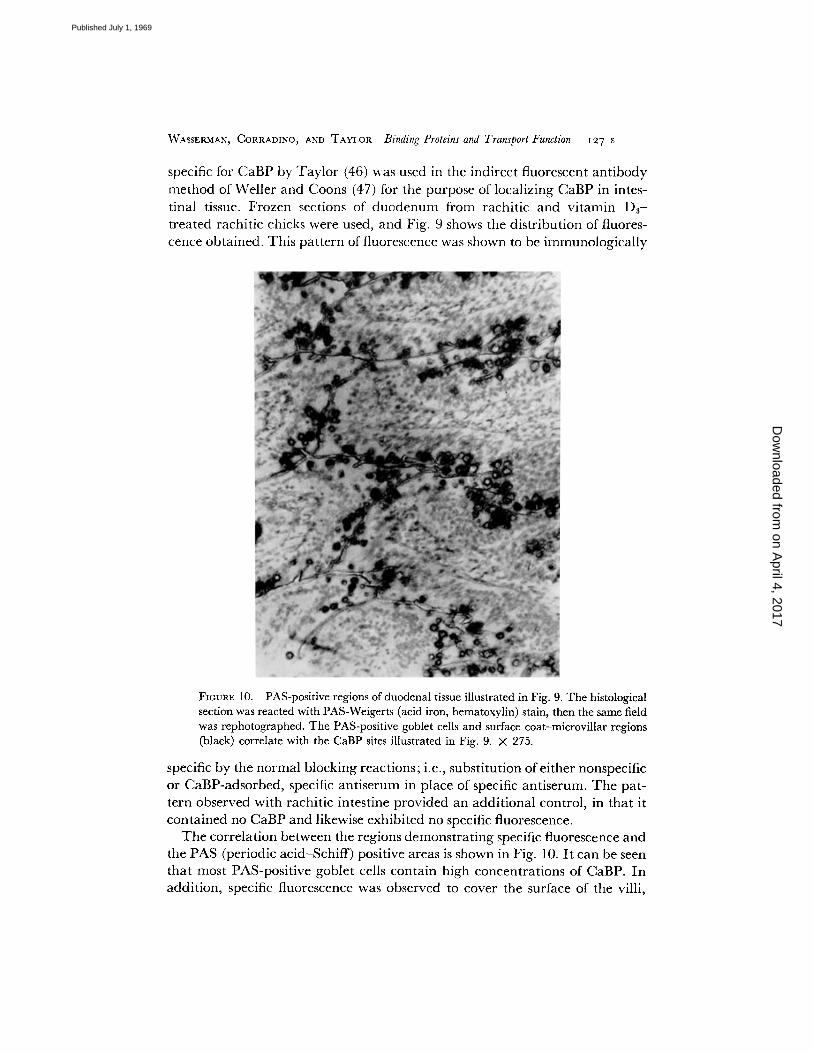
FIGURE 10. PAS-positive regions of duodenal tissue illustrated in Fig. 9. The histological section was reacted with PAS-Weigerts (acid iron, hematoxylin) stain, then the same field was rephotographed. The PAS-positive goblet cells and surface coat-microvillar regions (black) correlate with the CaBP sites illustrated in Fig. 9. × 275.
specific by the normal blocking reactions; i.e., substi tution of either nonspecific or CaBP-adsorbed, specific an t i serum in place of specific ant iserum. T h e pat- tern observed with rachit ic intestine provided an addi t ional control, in tha t it conta ined no CaBP and likewise exhibited no specific fluorescence.
The correlat ion between the regions demons t ra t ing specific fluorescence and the PAS (periodic acid-Schiff) positive areas is shown in Fig. 10. I t can be seen tha t most PAS-positive goblet cells conta in high concentrat ions of CaBP. In addi t ion, specific fluorescence was observed to cover the surface of the villi,
on April 4, 2017
Dow
nloaded from
Published July 1, 1969

i 2 8 s T R A N S P O R T P R O T E I N S
indicating that CaBP is present in the surface coat-microvilli region of the cells lining the villar surfaces. The resolution obtainable with 6-/z frozen sec- tions does not allow a definitive localization of CaBP in either the mucopoly- saccharide surface coat or the brush border (microvilli); however, when viewed at high magnification, a surface coat localization is the more probable. These results are analogous to those obtained by Nakane et al. (48) in which it was shown that a leucine-binding protein was localized in the cell envelope of E. coli. In both cases, the binding proteins are located in "logical" positions to function as transport proteins.
S P E C U L A T I O N O N T H E M E C H A N I S M O F A C T I O N O F C a B P The exact function of CaBP in the translocation process (if this is actually the case) is, of course, unknown. Some speculation perhaps is warranted if only to serve as a starting point for subsequent experimentation.
From physiological information, it seems that vitamin D-dependent cal- cium absorption is not necessarily dependent on an intact energy supply and, equating the vitamin D mechanism with CaBP function, one may tentatively conclude that CaBP does not function as part of an active transport system. Pertaining to this same point, the Ca pump appears to be associated with the basal membranes of the epithelial cell whereas CaBP is localized in highest concentration at the opposite pole of the cell, and most likely within the surface coat matrix. Further, the mucosal border has been identified as a region of the cell where vitamin D manifests an important part of its effect, and across which the translocation of calcium in both directions is enhanced.
Assuming that these features are the most pertinent, at least three models can be offered, these being depicted in Fig. 11. First, CaBP may serve as a "diffusional facilitator," enhancing-the transfer of calcium across the micro- villar membrane and /or through the sea of anionic charges that exists within the surface coat (cf. Borle (49) for a description of the relationship between the extraneous surface coat of isolated HeLa cells and calcium binding). Because of the large binding affinity of CaBP, this molecule could successfully compete for luminal Ca with respect to the anionic groups in the surface coat. However, two difficulties arise with this first suggestion. The protein must be able to penetrate through the core of the membrane in order to act as a membrane facilitator and, secondly, some mechanism must be available to release Ca from the protein (unless a Ca sink of sufficient magnitude is maintained).
A second possibility is that CaBP serves as an "intracellular carrier," facili- tating the movement of calcium through the cytoplasmic milieu. At the basal aspect of the cell, the transported Ca could be come attached to a component of the Ca pump and then be actively extruded from the cell or, in conjunction with a dumping system, leave the cell by diffusion. This type of mechanism appears unlikely because it was estimated that insufficient CaBP is available to
on April 4, 2017
Dow
nloaded from
Published July 1, 1969

WASSERMAN, CORRAD1NO, AND TAYLOR Binding Proteins and Transport Function 129 s
complex all of the ca lc ium in transi t when a relat ively high concen t ra t ion of
ca lc ium is p laced in the intestinal lumen; thus, CaBP appears to funct ion catalytical ly.
T h e third possibility is tha t CaBP m a y act p r imar i ly as a "microv i l l a r con- c e n t r a t o r " of calcium, being analogous to an ion exchange resin and accumu- lat ing Ca in the region ad jacent to the plasma membrane . T h e bound Ca is then avai lable for the next step in the chain, the transfer of the e lement across the m e m b r a n e into the cell cytoplasm. T h e la t ter step might involve ano the r car r ier of calcium.
FIGURE 11. Diagrammatic representation of speculative ways by which CaBP may func- tion. The solid circle represents CaBP; X represents an intra- cellular, soluble complex of calcium, such as citrate; the open circle with squiggle, the Ca pump.
O T H E R P O S S I B L E T R A N S P O R T P R O T E I N S
T h e r e are several o ther an imal proteins tha t appea r to be involved in the t ranspor t of specific substrates into the cell. O n e of these, " int r ins ic f ac to r" (IF) has been studied for over 35 yr and its existence was suggested by Castle in 1930 (50). H e observed a hematopo ie t i c response in pernicious anemia pa- tients after no rmal h u m a n gastric juice, previously incuba ted with beef muscle, was in t roduced into the pat ient ' s s tomach. Castle was able to conc lude on the basis of this and o ther exper iments tha t no rma l gastric ju ice con ta ined an " in t r ins ic f ac to r" lacking in pernicious anemia pat ients tha t was essential for the absorp t ion of some substance genera ted by the in terac t ion be tween it and an "extr ins ic f ac to r" con ta ined in cer ta in foods, such as beef. I t is now known
on April 4, 2017
Dow
nloaded from
Published July 1, 1969
~3 o s T R A N S P O R T P R O T E I N S
that the "extrinsic factor" is vitamin B12 which was first isolated in pure form in 1948 (51, 52) and fully characterized in 1955 (53). Complete characteriza- tion of IF has not yet been realized, but it appears to be a glycoprotein of molecular weight 50,000-60,000 (54) and formed in the stomach, perhaps, in the chief cells (55).
The exact mechanism of IF-mediated B12 transport is unknown but certain features have been elucidated. It is known that IF, though produced in the stomach, promotes B12 absorption in the small intestine (56). It is generally accepted that B~2 must combine with IF prior to absorption (57, 58) and that calcium is necessary for optimal absorption (58). Very recent evidence has indicated that there is a specific receptor for the IF-B~2 complex in the intes- tinal surface coat. In one study, fluorescein-labeled rabbit antibodies to ham- ster microvillous membranes showed specific reaction with hamster intestine brush borders (59). In a related study, IF-mediated at tachment of B12 to brush borders isolated from hamster small intestine was shown to be prevented by incubation in vitro with rabbit antiserum to hamster microvillous membranes (60). Once attached to the luminal surface of the intestinal cell, the IF-B12 complex may penetrate the cell as a unit but may be altered in transit since, in one study, the vitamin entering intestinal lymph, though not dialyzable, was devoid of IF activity (61). Serious doubt has been cast on this concept by a very recent study, however, which showed that IF was not absorbed during the process of IF-mediated vitamin B,2 absorption (62).
There is a Bi2-binding factor in the blood, appearing both in normal humans and in pernicious anemia patients having no gastrointestinal IF. This factor facilitates uptake of B~2 by reticulocyte-rich erythrocyte preparations by an adsorption mechanism quite similar to the IF-mediated B,2 uptake by the intestine (63). The binding factor involved appears to be a ~-globulin and has been called transcobalamin II (64). It is possible to speculate that both the "intrinsic factor" and transcobalamin I I -media ted B12 uptake are components of a sequential mechanism operating to assure optimal availability of B~2 for erythrocyte production and /o r maturation.
Specific proteins have also been implicated in the intestinal absorption and erythrocytic uptake of iron. In the former case, apoferritin was considered to function in the transepithelial movement of iron (65). Little support for this concept has developed and, in fact, the evidence on which it was based was highly circumstantial. Ferritin was qualitatively estimated in guinea pig intes- tine by histological means and shown to increase with iron feeding (65). It was speculated that this stored iron could be called upon as needed to maintain blood levels (66). This proposal has been discounted and apoferritin is not even considered per se in a recent model of intestinal iron transport although the data presented are consistent with the presence of some carrier within the intestinal cell (67).
on April 4, 2017
Dow
nloaded from
Published July 1, 1969
WASSERMAN, CORRADINO, AND TAYLOR Binding Proteins and Transport Function I3I s
A second iron-binding protein, gastroferrin, appears to be involved in in- testinal iron transport (68) but in a negative sense. This protein is found in gastric juice and has the capacity to complex iron, perhaps rendering it un- available for absorption by the intestinal mucosa. Supporting this concept are two observations. In hemochromatosis, in which there is excessive iron absorp- tion, gastroferrin is absent (69). Iron-deficiency anemia caused by severe blood loss was associated with low gastroferrin levels which returned to normal after restoration of normal blood levels by transfusion (70).
With regard to erythrocytic iron translocation, the case for the participation of a specific protein, transferrin, is quite good. Transferrin is the major non- heme, iron-binding protein of serum. In studies of human red cell suspensions, the uptake of iron-laden transferrin was much greater than the uptake of apo- transferrin (71). Once attached to the red cell membrane, the iron is released into the red cell while the apotransferrin molecule becomes detached. The transferrin-mediated uptake of iron was prevented by metabolic inhibitors with the iron accumulation process being far more sensitive to inhibition than the transferrin at tachment step (71).
Proteins have been suspected of being involved in other transport systems in animals but little is known of their nature. Transport of glucose by intestine (72, 73) and by the erythrocyte (4, 74, 75), for example, have been related to binding by proteins or protein-like substances. Similarly, work is proceeding on the purification of a carrier-like protein for organic bases 8 that was released by osmotic shock from the kidney (76).
The investigations from our laboratory were supported by National Institute of Health Grants AM-04652 and AM-06271-NTN, and Contract AT(30-1)-2147 from the United States Atomic Energy Commission.
R E F E R E N C E S
1. PARDE~, A. B. 1968. Membrane transport proteins. Science. 162:632. 2. HSP~EL, L. A. 1967. Selective release of enzymes from bacteria. Science. 156:1451. 3. Fox, C. F., and E. P. I~NNEDY. 1965. Specific labeling and partial purification of the M
protein, a component of the 3-galactoslde transport system of Escherichia coli. Proc. Nat. Acad. &i. U.S.A. 54:891.
4. Bo~INSm, H., and W. D. STEm. 1966. Isolation of a glucose-binding component from human erythrocyte membranes. Nature (London). 211:1366.
5. A ~ K u , Y. 1968. Transport of sugars and amino acids in bacteria. I. Purification and specificity of the galactose- and leucine-binding proteins. J. Biol. Chem. 243:3116.
6. MELLAr~Y, E. 1919. An experimental investigation on rickets. Lancet. 196:409. 7. NlcoI.AYSmq R. 1937. The influence of vitamin D on the absorption of calcium and phos-
phorus in the rat. Biochem. J. 31:122. 8. WAssE~an, R. H., and A. N. TAx, OR. 1969. Some aspects of the intestinal absorption of
calcium, with special reference to vitamin D. In Mineral Metabolism, An Advanced Treatise. C. L. Comar and F. Brormer, editors. Vol. I I I , p. 321.
8 Magour, S., A. Farah, and A. Sroka. The partial purification of a carrier-like protein for organic bases from the kidney. Private communication.
on April 4, 2017
Dow
nloaded from
Published July 1, 1969
I32 S T R A N S P O R T P R O T E I N S
9. SCHACHTER, ~)., and S. M. ROS,~N. 1959. Active transport of Ca 45 by the small intestine and its dependence on vitamin D. Amer. J. Physiol. 196:357.
10. SCHACHTER, D., E. B. DOWDLE, and H. SCHEN~ER. 1960. Active transport of calcium by the small intestine of the rat. Amer. J. Physiol. 198:263.
11. KIMnERa, D. V., D. SCHACHTER, and H. SCHENm~R. 1961. Active transport of calcium by intestine: effects of dietary calcium Amer. J. Physiol. 200:1256.
12. SCHACHTER, D., S. KOWARSr, t, J. D. FIN:~ELSTEIN, and R. W. MA. 1966. Tissue concentra- tion differences during active transport of calcium by intestine. Amer. J. Physiol. 211:1131.
13. ScaAcrrmR, D., D. V. KIMBERG, and H. SCrmNg.~R. 1961. Active transport of calcium by intestine: action and bio-assay of vitamin D. Amer. J. Physiol. 200:1263.
14. HARtUSON, H. E., and H. C. HAmUSON. 1960. Transfer of Ca 4~ across the intestinal wall in vitro in relation to action of vitamin D and cortisol. Amer. J. Physiol. 199:265.
15. HARI~ISON, H. E., and H. C. HARRISOn. 1965. Vitamin D and permeability of intestinal mucosa to calcium. Amer. J. Physiol. 208:370.
16. WASSEm~AN, R. H., and F. A. KALLWLZ. 1962. Vitamin D3 and unidirectional calcium fluxes across the rachitic chick duodenum. Amer. J. Physiol. 203:221.
17. WASS]~P.~AN, R. H., A. N. TAYLOR, and F. A. KALLFELZ. 1966. Vitamin D and the transfer of plasma calcium to intestinal lumen in chicks and rats. Amer. J. Physiol. 211:419.
18. WASSEm~AN, R. H. 1968. Calcium transport by the intestine: a model and comment on vitamin D action. Calc. Tiss. Res. 2:301.
19. LmoQutsT, B. 1952. Effect of vitamin D on the metabolism of radiocalcium in rachitic rats. Acta Paediat. 41 (Suppl.):86.
20. WASSSm~AN, R. H. 1962. Studies on vitamin ]:)8 and the intestinal absorption of calcium and other ions in the rachitic chick. J. Nutr. 77:69.
21. DELucA, H. F. 1967. Mechanism of action and metabolic fate of vitamin D. Vitamins Hormones. 25:315.
22. NOlU~AN, A. W. 1968. The mode of action of vitamin D. Biol. Rev. (Cambridge) 43".97. 23. NOm~AN, A. W., J . LUND, and H. F. DELucA. 1964. Biologically active forms of vitamin D8
in kidney and intestine. Arch. Biochem. Biophys. 108:12. 24. BLUNT, J. W., H. F. DELucA, and H. K. SCHNOES. 1968. 25-Hydroxycholecaleiferol: a
biologically active metabolite of cholecalciferol. Chem. Commun. 14:801. 25. EISENSTEm, R., and M. PASSAVOY. 1964. Actinomycin D inhibits parathyroid hormone
and vitamin D activity. Proc. Soc. Exp. Biol. Med. 117:77. 26. ZULL, J. E., E. CZAP, NOWSKA-MJszTAL, and H. F. DELucA. 1965. Actinomycin D inhibition
of vitamin D action. Science. 149:182. 27. NO,AN, A. W. 1965. Actinomycin D and the response to vitamin D. Science. 149:184. 28. BOSMANN, H. B., and P. S. CHE~, JR. 1966. Actinomycin D inhibition of vitamin D- and
dihydrotachysterol-induced responses in the chick. J. Nutr. 90:405. 29. STOHS, S. J., J . E. ZULL, and H. F. DELucA. 1967. Vitamin D stimulation of [3H]-orotic
acid incorporation into ribonucleic acid of rat intestinal mucosa. Biochemistry 6:1304. 30. NORMAN, A. W. 1966. Vitamin D mediated synthesis of rapidly labeled RNA from intestinal
mucosa. Biochem. Biophys. Res. Commun. 23:335. 31. HAUSSLZR, M. R., and A. W. NORMAN. 1967. The subcellular distribution of physiological
doses of vitamin D~. Arch. Biochem. Biophys. 118:145. 32. STOHS, S. J., and H. F. DELucA. 1967. SubceUular location of vitamin D and its metabo-
lites in intestinal mucosa after a 10-IU dose. Biochemistry. 6:3338. 33. HAUSSLER, M. R., J. F. M~mTLE, and A. W. NORMAN. 1968. The association of a metabo-
lite of vitamin D~ with intestinal mucosa chromatin "in vivo". J. Biol. Chem. 243:4055. 34. ZULL, J. E., E. CZARNOWSKA-M~szTAL, and H. F. DELucA. 1966. On the relationship be-
tween vitamin D action and actinomycin-sensitive processes. Proc. Nat. Acad. Sci. U.S.A. 55:177.
35. WASS~m~A~, R. H., and A. N. TA~_~OR. 1963. Vitamin Dn inhibition of radiocalcium bind- ing by chick intestinal homogenates. Nature (London). 198:30.
36. WASS~R~A~, R. H., and A. N. TnYgo~. 1966. Vitamin Ds-induced calcium-binding pro- rein in chick intestinal mucosa. Science. 152:791.
on April 4, 2017
Dow
nloaded from
Published July 1, 1969

WAss~.~a~, C o g R ~ m o , ~-~ TA~-~OR Binding Proteins and Transport Function I33 s
37. BgIaos, F. N., and M. FLmsx~a~. 1965. Calcium binding by particle-free supernatants of homogenates of skeletal muscle. J. Gen. Physiol. 49:131.
38. WASSEm~AN, R. H., R. A. C o - - t o o , and A. N. TAYLOR. 1968. Vitamin D-dependent calcium-blnding protein. J. BioL Chem. 243:3978.
39. SCmmZRT, J., E. R. RUSSELL, and L. S. M ~ g s , JR. 1950. DiSSociation constants of radium- organic acid complexes measured by ion exchange. J. Biol. Chem. 185:387.
40. TA~rnOR, A. N., and R. H. WASS~.~AN. 1967. Vitamin D3-induced calcium-binding pro- tein: partial purification, electrophoretic visualization, and tissue distribution. Arch. Biochem. Biophys. 119:536.
41. CORg~a~INO, R. A., R. H. WASSE~.~, M. H. PtrBoLs, and S. I. CI-n~o. 1968. Vitamin Ds induction of a calcium-binding protein in the uterus of the laying hen. Arch. Biochem. Biophys. 125:378.
42. KALLF~LZ, F. A., A. N. TAYLOR, and R. H. WASS~AN. 1967. Vitamin D-induced calcium binding factor in rat intestinal mucosa. Proc. Soc. Exp. Biol. Med. 125:54.
43. TAYLOR, A. N., R. H. WASSERMAa~, andJ . JowsEY. 1968. A vitamin D-dependent calcium- binding protein in canine intestinal mucosa. Fed. Proc. 27:675.
44. EBEL, J. G., A. N. TAYnOR, and R. H. WASSE~AN. 1969. The vitamin D-induced calcium- binding protein of intestinal mucosa: relation to vitamin D dose level and the lag period. Amer. J. Clin. Nutr. 22:431.
45. CORg~a~INO, R. A., and R. H. WASSERM.~. 1968. Actinomycin D inhibition of vitamin D3- induced calcium-bindlng protein (CaBP) formation in chick duodenal mucosa. Arch. Biochem. Biophys. 126:957.
46. TAYLOR, A. N. 1969. Vitamin Ds-induced calcium-binding protein: immunochemical and fluorescent antibody localization studies. Ph.D. Thesis. Cornell University, Ithaca.
47. WELL, R, T. H., and A. H. CooNs. 1954. Fluorescent antibody studies with agents of Varicella and Herpes Zoster propagated in vitro. Proc. Soc. Exp. Biol. Med. 86:789.
48. N~a~ANE, P. K., G. E. NICHOALDS, and D. L. OXENDER. 1968. Cellular localization of leucine-binding protein from Escherichia coli. Science. 161:182.
49. BoRnE, A. B. 1968. Calcium metabolism in HeLa cells and the effects of parathyroid hor- mone. J. Cell Biol. 36:567.
50. CASTLe, W. B., W. C. TOWNSEND, and C. W. H~ATH. 1930. Observations on the etiologic relationship of achylia gastrica to pernicious anemia. I I I . The nature of the reaction between normal gastric juice and beef muscle leading to clinical improvement and in- creased blood formation similar to the effect of liver feeding. Amer. J. Med. Sci. 180:305.
51. RICKES, E. L., N. G. BRinK, F. R. KONIUSZKY, T. R. WOOD, and K. Fon~mRs. 1948. Crystal- line vitamin B12. Science. 107:396.
52. SMITH, E. L., and L. F. J. PARKER. 1948. Purification of anti-pernicious anemia factor. Biochem. J. 43:viii.
53. HODO~aN, D. C., J . I~CKWORTH, J. H. ROBERTSON, K. N. TRUEBLOOD, R. J. PgOSEN, and J. G. WI-nTE, 1955. The crystal structure of the hexacarboxylic acid derived from BI~ and the molecular structure of the vitamin. Nature (London). 176:325.
54. GR~SBZCK, R. 1967. Intrinsic factor and the transcobalamins with reflections on the general function and evolution of soluble transport proteins. Scand. J. Clin. Lab. Invest. Suppl. 95:7.
55. KEUNmG, F. J. , A. Ams~s, E. M~NDEMA, and H. O. Nn~wlo. 1959. Observations on the site of production of Castle's intrinsic factor in the rat. J. Lab. Clin. Med. 53:127.
56. I ~ L L , P. C., G. H. SPRAY, and K. B. TAYLOR. 1957. The site of absorption of vitamin Ba2 in the rat. Clin. Sci. (London). 16:663.
57. Nn~wiG, H. O., S. C. Sn~N, and W. B. CASTLE. 1957. Mechanism of intrlnsic factor action in the gastrectomized rat. Proc. Soc. Exp. Biol. Med. 94:223.
58. I ~ R T , V. 1959. Mechanism of intrinsic factor action in everted sacs of rat small intes- tine. J. Clin. Invest. 38:102.
59. Kom,, W. L., J . S. T ~ R , I. L. MACI~Nzr~, andR. M. DONALDSON, JR. 1968. Antibodies to intestinal microvillous membranes. I. Characterization and morphologic localization. .7. Exp. Med. 128:357.
on April 4, 2017
Dow
nloaded from
Published July 1, 1969

x34 s T R A N S P O R T P R O T E I N S
60. MAcKENzIE, I. L., R. M. DONALDSON, JR., W. L. KOPP, andJ. S. TRIER. 1968. Antibodies to intestinal microvillous membranes. II. Inhibition of intrinsic factor-mediated attach- ment of vitamin B12 to hamster brush borders. J. Exp. Med. 128:375.
61. BoAss, A., and T. H. WILSON. 1964. Intestinal absorption of intrinsic factor and BI~ intrinsic factor complex. Amer. J. Physiol. 207:27.
62. HinEs, J. D., A. ROS~NBERO, and J. w. HAmuS. 1968. Intrinsic factor-mediated radio-B12 uptake in sequential incubation studies using everted sacs of guinea pig small intestine: evidence that IF is not absorbed into the intestinal cell. Pro¢. So¢. Exp. Biol. Med. 129:653.
63. I~TmF, F. P., C. W. GOTTLmB, and V. I~P.B~.RT. 1966. Mechanism of vitamin Bx~ uptake by erythrocytes. J'. Clin. Invest. 45:1907.
64. HALL, C. A., and A. E. FmKL~m 1965. The dynamics of transcobalamln II. A vitamin B12 binding substance in plasma. J. Lab. Clin. Med. 65:459.
65. GRAmfiK, S. 1946. Ferritin. IX. Increase of the protein apoferritin in the gastrointestinal mucosa as a direct response to iron feeding. The function of ferritin in the regulation of iron absorption. J. Biol. Chem. 164:737.
66. GRANICK, S. 1954. Iron Metabolism. Bull. N.Y. Acad. Med. 30:81. 67. HELBOCK, H. J., and P. SALTMAN. 1967. The transport of iron by rat intestine. Biochim.
Biophys. Acta. 135:979. 68. DAws, P. S., C. G. Lur~, and D. J. I~LI~R. 1967. Gastric iron binding protein in iron
chelation by gastric juice. Nature (London). 214:1126. 69. DAms, P. S., C. G. LuI~, and D. J. DELLER. 1966. Reduction of gastric iron-binding pro-
tein in haemochromatosis. Lancet. II:1431. 70. L u g , C. G., P. S. DAms, and D. J. DELLER. 1967. Change in gastric iron-binding protein
(gastroferrin) during iron-deficiency anaemia. Lancet. I:926. 71. JANDL, J. J., and J. H. KOTZ. 1964. The plasma-to-cell cycle of transferrin. J. Clin. Invest.
42:314. 72. FAUST, R. G., S-M. L. Wu, and M. L. FAGGARD. 1967. D-glucose: preferential binding to
brush borders disrupted with Tris (hydroxymethyl) ~minomethane. Science. 155:1261. 73. F~.m~m, S. M., S. L. HART, and J. A. NIsslM. 1967. Glucose binding by homogenates of
intestinal mucosa. Nature (London). 213:985. 74. BONSALL, R. W., and S. HUNT. 1966. Solubilization of a glucose-binding component of the
red cell membrane. Nature (London). 211:1368. 75. LEVmE, M., and W. D. STEm. 1967. Techniques for analysis of glucose binding by human
erythrocyte membranes. Biochim. Biophys. Acta. 135:710. 76. Ross, C. R., N. I. PESSAH, and A. E. FARAH. 1969. Attempts to label the renal carrier for
organic bases with dibenamine. J. Pharma¢ol. Exp. Ther. In press.
Discussion from the Floor
Felix Bronner (University of Louisville, Louisville, Ky.): Dr. Maddaiah and I, stimulated by Dr. Wasserman's studies, have investigated the occurrence of this calcium-binding protein in rat mucosa and have studied the amount of binding ac- tivity as a function of calcium intake. We find, in agreement with what Dr. Wasser- man has shown, that there is an inverse relationship between the amount absorbed and the amount bound; i.e., the amount of binding activity.
When animals that have been on a high calcium diet are placed on a low calcium diet, within 24 hr the amount of binding activity increases as shown on the slide (Fig. 1). As can be seen, there was a marked increase in calcium-binding activity, expressed as moles Ca X 10-u/rag protein, assayed by the Wasserman method. This increase was marked, then decreased in intensity, and stabilized at about the level
on April 4, 2017
Dow
nloaded from
Published July 1, 1969

WASSERMAN, CORRADINO, AND TAYIA3R Binding Proteins and Transport Function I35 s
you see on the right-hand side of the slide. The animals on diet I I I were absorbing roughly 100 mg calcium per day, the animals on diet I approximately 5 mg calcium per day.
When you treat animals with an antibiotic such as puromycin you can inhibit the increase of binding activity shown on the left-hand side of the slide. We have not done the reverse. However, when you place animals that are on the low calcium diet and shift them to a high calcium diet you see there is a decrease in activity. The increase
DIET I l l and shift to DIET 1"
0 ~ 0
0
->' ~. 2 0
~ . E
0 . 0
o X ("~w 0
v
30
DI ET I and shift to DIET I I [
s TI I ) ...,~li ' f! Time {doys) f! Time (doys)
Fioum~ l.
in activity, which can be inhibited by puromycin, is not parathyroid dependent, as animals that have been parathyroidectomized show behavior similar to that of intact animals.
Studies are now in progress to further characterize the nature of this calcium-bind- ing substance which, in agreement with Dr. Wasserman, we believe is a protein. We have preliminary evidence to this effect as 4~Ca activity coincides with the first pro- tein peak when the material is passed through Sephadex G-25 gel. Moreover, the activity disappears when the extract is digested with trypsin. The protein does not oc- cur in the brush borders of the mucosal cells (prepared by the method of D. Miller and R. K. Crane. 1961. Anal. Biochem. 2:284). Rather, most of the activity is localized in the microsomal fraction of the mucosal cells (as determined by ultracentrifugal fractionation studies).
on April 4, 2017
Dow
nloaded from
Published July 1, 1969
t36 s T R A N S P O R T P R O T E I N S
Dr. Wasserman: I t is not surprising that CaBP was not observed to be associated with isolated brush borders since the protein is easily disengaged from the mucosal tissue by simple homogenization. The fluorescent antibody study, however, does tend to show that the protein is localized in the brush border region in vivo, but again the bonding between CaBP and the brush border material must be quite labile.
Dr. Geoffrey W. G. Sharp (Massachusetts General Hospital): In connection with your suggestion that the calcium-binding protein is situated in the mucus layer and may be synthesized in the goblet cells, do you have evidence of specific nuclear locali- zation of vitamin D in the goblet ceils, or is it localized in most of the ceils?
Dr. Wasserman: We have no evidence on this point. Dr. Pardee: Dr. Roseman asks whether it has carbohydrate in it. Dr. Wasserman: We concluded from our previous analysis that CaBP would contain
1% or less of a glucose equivalent on a weight basis. This was done when limited amounts of purified protein were available.
Mr. Arthur B. Chausmer (State University of New York, Downstate): The other mediator of calcium absorption which springs to mind is parathyroid hormone. Have you looked for this binding protein under the influence of parathyroid hormone? The second question I have goes back to whether your aetinomycin itself will lower serum calcium in a parathyroidectomized rat. I didn' t notice whether you had given aetinomycin to an animal which was essentially normal, and whether you had noted any changes in serum calcium levels.
Dr. Wasserman: No, we do not have any specific information on the influence of parathyroid hormone on the calcium-binding protein. The hormone, I 'm sure, would not induce the synthesis of the protein. However, if vitamin D is not limiting, it may possibly alter the concentration of CaBP in the target tissue.
Regarding the actinomycin D response, several groups (Norman, A. W. 1965. Science. 149:134; Zull, J. E., et al. 1965. Science. 149:182; and Bosmann, H. B., and P. S. Chen, Jr. 1966. J. Nutr. 90:405) showed that the increment in Ca absorption in rachitic animals due to vitamin D treatment was prevented when they were given aetinomycin D before or simultaneously with the vitamin. If the antibiotic were administered several hours after the vitamin was given, little or no inhibition was noted, indicating that there was a direct effect on the vitamin D-mediated process. However, H. E. Harrison and H. C. Harrison (Proc. Soc. Exp. Biol. Med. 1966. 121:312) observed that aetinomycin D decreased Ca absorption by raehitie rats and this suggested that the antibiotic acted on the "transport system itself rather than on the enhancing effect of vitamin D on this process."
Dr. Joseph F. Gennaro (New York University): Is the anticalcium-binding protein species-specific?
Dr. Wasserman: The antibody formed against chick CaBP does not interact with the dog protein.
Dr. Gennaro: Will the anti-mammalian antibodies react with the chick CaBP? Dr. Wasserman: The chick CaBP antibody is the only antibody we have in hand. Dr. Gennaro: If you treated this tissue on the mueosal surface with neuraminidase,
is it possible that you could eliminate this calcium-binding effect? Secondly, in your first electrophoresis slide you mentioned the increase in the staining intensity of the
on April 4, 2017
Dow
nloaded from
Published July 1, 1969
WAK'$ERMAN, CORRADINO, AND TAYLOR Binding Proteins and Transport Function I37 s
furthermost band, but the two above it also showed variations. Do you have any com- ments on that?
Dr. Wasserman: In regard to your first question, those treatments have not been attempted as yet.
You are quite correct in noting that there are other alterations in the gel electro- phoretic pattern of proteins due to vitamin D treatment or to a low calcium intake. In the past, however, we have given attention only to CaBP and its characteristic band, but eventually will attempt to characterize or at least gain some information on the other proteins.
Dr. George Nichols, Jr. (Harvard Medical School): Dr. Wasserman, I have two questions. The first was somewhat similar to the comments of the last gentleman, namely what are the effects of proteolytic enzymes on the presence of the protein? The second is: Have you yet had a chance to see how the presence or absence of this protein on the surface may effect the actual transfer of calcium 45 across the mucosal membrane? Can you put the two together yet?
Dr. Wasserman: You mean restore the system? Dr. Nichols: Yes. Dr. Wasserrnan: We have made some attempts in this direction but thus far we
have not been successful.
on April 4, 2017
Dow
nloaded from
Published July 1, 1969


![Zipcode RNA-Binding Proteins and Membrane Trafficking ... · Zipcode RNA-Binding Proteins and Membrane Trafficking Proteins Cooperate to Transport Glutelin mRNAs in Rice Endosperm[OPEN]](https://static.fdocuments.net/doc/165x107/5fedaa08e6ee6243c45b24a5/zipcode-rna-binding-proteins-and-membrane-trafficking-zipcode-rna-binding-proteins.jpg)
