
BINDING PROTEIN Is a Master Regulator of the Endoplasmic Reticulum Stress Sensor/Transducer
15
BINDING PROTEIN Is a Master Regulator of the Endoplasmic Reticulum Stress Sensor/Transducer bZIP28 in Arabidopsis C W OA Renu Srivastava, a Yan Deng, a Shweta Shah, b Aragula Gururaj Rao, b and Stephen H. Howell a,c,1 a Plant Sciences Institute, Iowa State University, Ames, Iowa 50011 b Roy J. Carver Department of Biochemistry, Biophysics, and Molecular Biology, Iowa State University, Ames, Iowa 50011 c Department of Genetics, Development, and Cell Biology, Iowa State University, Ames, Iowa 50011 BINDING PROTEIN (BiP) is a major chaperone in the endoplasmic reticulum (ER) lumen, and this study shows that BiP binds to the C-terminal tail of the stress sensor/transducer bZIP28, a membrane-associated transcription factor, retaining it in the ER under unstressed conditions. In response to ER stress, BiP dissociates from bZIP28, allowing it to be mobilized from the ER to the Golgi where it is proteolytically processed and released to enter the nucleus. Under unstressed conditions, BiP binds to bZIP28 as it binds to other client proteins, through its substrate binding domain. BiP dissociates from bZIP28 even when bZIP28’s exit from the ER or its release from the Golgi is blocked. Both BiP1 and BiP3 bind bZIP28, and overexpression of either BiP detains bZIP28 in the ER under stress conditions. A C-terminally truncated mutant of bZIP28 eliminating most of the lumenal domain does not bind BiP and is not retained in the ER under unstressed conditions. BiP binding sites in the C-terminal tail of bZIP28 were identified in a phage display system. BiP was found to bind to intrinsically disordered regions on bZIP28’s lumen-facing tail. Thus, the dissociation of BiP from the C-terminal tail of bZIP28 is a major switch that activates one arm of the unfolded protein response signaling pathway in plants. INTRODUCTION BINDING IMMUNOGLOBULIN PROTEIN, or simply Binding Pro- tein (BiP), known in animal systems as 78-kD Glc-regulated pro- tein or heat shock 70-kD protein 5, and calnexin/calreticulin are the two major chaperone systems in the endoplasmic reticulum (ER) lumen (Otero et al., 2010). BiP is primarily involved in the maturation and folding of nonglycosylated proteins (Hendershot, 2004). BiPs form complexes with the HSP40-like cochaperones containing J domains (ERdj3) and stromal-derived factor-2 (Jin et al., 2008; Nekrasov et al., 2009; Schott et al., 2010). These BiP complexes maintain nascent proteins in a competent state for subsequent folding and oligomerization (Anelli and Sitia, 2008). BiP also plays an important role in the unfolded protein response (UPR) by regulating stress transducers, such as ACTIVATING TRANSCRIPTION FACTOR6 (ATF6), Protein kinase RNA-like ER kinase, and Inositol Requiring Enzyme1 (IRE1) in animal cells (Bertolotti et al., 2000; Shen et al., 2002). In response to stress, BiP and ATF6 rapidly dissociate, and ATF6 becomes a cargo in the ER-to-Golgi trafficking system (Shen et al., 2002). Shen et al. (2002) observed that BiP binds to three regions in the lumenal domain of ATF6. They reasoned that binding to these sites might mask Golgi localization signals (GLSs) on ATF6. They located the GLSs in two regions of the lumenal domain of ATF6 by showing that when these regions were deleted, transport of ATF6 to the Golgi and its processing by the Golgi-resident proteases were blocked. To demonstrate that BiP masked GLSs under unstressed conditions, they developed constructs in which the BiP binding sites were deleted, but putative GLSs were retained. These con- structs were constitutively translocated to the Golgi, supporting the idea that BiP retains ATF6 in the ER by blocking its GLSs (Shen et al., 2002). In plants, BiP is reported to play a role in the defense against various stresses. In particular, the overexpression of BiP in Nicotiana tabacum (tobacco) has been shown to protect against tunicamycin (TM) inhibition of seed germination and against water stress during plant growth (Alvim et al., 2001). When BiP was overexpressed in Glycine max (soybean), leaves of transgenic plants were more resistant to wilt and more tolerant of the loss of water potential than control plants when subjected to drought or osmotic stress (Valente et al., 2009). The greater stress re- sistance has been attributed to an attenuation of a cell death signal produced by ER and osmotic stress through N-RICH PROTEIN (NRP) and NAC6-mediated pathways (Reis et al., 2011). This implies that BiP is a negative regulator of stress-induced NRP- mediated cell death. In plants, BiP has also been shown to be a limiting factor in the folding of certain secreted proteins under ER stress conditions. Leborgne-Castel et al. (1999) reported that the production of a-amylase was reduced in a tobacco transient expression system subjected to ER stress but could be restored by cotransfection with BiP. BiP2 loss-of-function mutants are defective PATHOGENESIS RELATED1 protein secretion in response 1 Address correspondence to [email protected]. The author responsible for distribution of materials integral to the findings presented in this article in accordance with the policy described in the Instructions for Authors (www.plantcell.org) is: Stephen H. Howell (shh@ iastate.edu). C Some figures in this article are displayed in color online but in black and white in the print edition. W Online version contains Web-only data. OA Open Access articles can be viewed online without a subscription. www.plantcell.org/cgi/doi/10.1105/tpc.113.110684 The Plant Cell, Vol. 25: 1416–1429, April 2013, www.plantcell.org ã 2013 American Society of Plant Biologists. All rights reserved.
Transcript of BINDING PROTEIN Is a Master Regulator of the Endoplasmic Reticulum Stress Sensor/Transducer

BINDING PROTEIN Is a Master Regulator of theEndoplasmic Reticulum Stress Sensor/TransducerbZIP28 in ArabidopsisC W OA
Renu Srivastava,a Yan Deng,a Shweta Shah,b Aragula Gururaj Rao,b and Stephen H. Howella,c,1
a Plant Sciences Institute, Iowa State University, Ames, Iowa 50011bRoy J. Carver Department of Biochemistry, Biophysics, and Molecular Biology, Iowa State University, Ames, Iowa 50011cDepartment of Genetics, Development, and Cell Biology, Iowa State University, Ames, Iowa 50011
BINDING PROTEIN (BiP) is a major chaperone in the endoplasmic reticulum (ER) lumen, and this study shows that BiP bindsto the C-terminal tail of the stress sensor/transducer bZIP28, a membrane-associated transcription factor, retaining it in theER under unstressed conditions. In response to ER stress, BiP dissociates from bZIP28, allowing it to be mobilized from theER to the Golgi where it is proteolytically processed and released to enter the nucleus. Under unstressed conditions, BiPbinds to bZIP28 as it binds to other client proteins, through its substrate binding domain. BiP dissociates from bZIP28 evenwhen bZIP28’s exit from the ER or its release from the Golgi is blocked. Both BiP1 and BiP3 bind bZIP28, and overexpressionof either BiP detains bZIP28 in the ER under stress conditions. A C-terminally truncated mutant of bZIP28 eliminating mostof the lumenal domain does not bind BiP and is not retained in the ER under unstressed conditions. BiP binding sites in theC-terminal tail of bZIP28 were identified in a phage display system. BiP was found to bind to intrinsically disordered regionson bZIP28’s lumen-facing tail. Thus, the dissociation of BiP from the C-terminal tail of bZIP28 is a major switch that activatesone arm of the unfolded protein response signaling pathway in plants.
INTRODUCTION
BINDING IMMUNOGLOBULIN PROTEIN, or simply Binding Pro-tein (BiP), known in animal systems as 78-kD Glc-regulated pro-tein or heat shock 70-kD protein 5, and calnexin/calreticulin arethe two major chaperone systems in the endoplasmic reticulum(ER) lumen (Otero et al., 2010). BiP is primarily involved in thematuration and folding of nonglycosylated proteins (Hendershot,2004). BiPs form complexes with the HSP40-like cochaperonescontaining J domains (ERdj3) and stromal-derived factor-2 (Jinet al., 2008; Nekrasov et al., 2009; Schott et al., 2010). These BiPcomplexes maintain nascent proteins in a competent state forsubsequent folding and oligomerization (Anelli and Sitia, 2008).
BiP also plays an important role in the unfolded protein response(UPR) by regulating stress transducers, such as ACTIVATINGTRANSCRIPTION FACTOR6 (ATF6), Protein kinase RNA-likeER kinase, and Inositol Requiring Enzyme1 (IRE1) in animal cells(Bertolotti et al., 2000; Shen et al., 2002). In response to stress,BiP and ATF6 rapidly dissociate, and ATF6 becomes a cargo inthe ER-to-Golgi trafficking system (Shen et al., 2002). Shen et al.(2002) observed that BiP binds to three regions in the lumenal
domain of ATF6. They reasoned that binding to these sites mightmask Golgi localization signals (GLSs) on ATF6. They locatedthe GLSs in two regions of the lumenal domain of ATF6 by showingthat when these regions were deleted, transport of ATF6 to theGolgi and its processing by the Golgi-resident proteases wereblocked. To demonstrate that BiP masked GLSs under unstressedconditions, they developed constructs in which the BiP bindingsites were deleted, but putative GLSs were retained. These con-structs were constitutively translocated to the Golgi, supportingthe idea that BiP retains ATF6 in the ER by blocking its GLSs(Shen et al., 2002).In plants, BiP is reported to play a role in the defense against
various stresses. In particular, the overexpression of BiP inNicotiana tabacum (tobacco) has been shown to protect againsttunicamycin (TM) inhibition of seed germination and against waterstress during plant growth (Alvim et al., 2001). When BiP wasoverexpressed in Glycine max (soybean), leaves of transgenicplants were more resistant to wilt and more tolerant of the lossof water potential than control plants when subjected to droughtor osmotic stress (Valente et al., 2009). The greater stress re-sistance has been attributed to an attenuation of a cell death signalproduced by ER and osmotic stress through N-RICH PROTEIN(NRP) and NAC6-mediated pathways (Reis et al., 2011). Thisimplies that BiP is a negative regulator of stress-induced NRP-mediated cell death. In plants, BiP has also been shown to bea limiting factor in the folding of certain secreted proteins underER stress conditions. Leborgne-Castel et al. (1999) reported thatthe production of a-amylase was reduced in a tobacco transientexpression system subjected to ER stress but could be restoredby cotransfection with BiP. BiP2 loss-of-function mutants aredefective PATHOGENESIS RELATED1 protein secretion in response
1Address correspondence to [email protected] author responsible for distribution of materials integral to the findingspresented in this article in accordance with the policy described in theInstructions for Authors (www.plantcell.org) is: Stephen H. Howell ([email protected]).C Some figures in this article are displayed in color online but in black andwhite in the print edition.W Online version contains Web-only data.OAOpen Access articles can be viewed online without a subscription.www.plantcell.org/cgi/doi/10.1105/tpc.113.110684
The Plant Cell, Vol. 25: 1416–1429, April 2013, www.plantcell.org ã 2013 American Society of Plant Biologists. All rights reserved.

to salicylic acid elicitation and are more susceptible to Pseudo-monas syringae infection (Wang et al., 2005).
BiP plays a dual role in plant UPR. First, the genes encodingBiP are upregulated by the UPR. There are three BiP codinggenes in Arabidopsis thaliana, and BiP3, in particular, is mosthighly upregulated by abiotic stress or by ER stress agents(Koizumi, 1996; Martínez and Chrispeels, 2003; Iwata andKoizumi, 2005; Liu et al., 2007b; Iwata et al., 2008, 2010; Tajimaet al., 2008; Liu and Howell, 2010). Since BiP is a chaperone, it isthought to mitigate stress by binding to misfolded proteins in theER, preventing their aggregation during refolding processes.Second, BiP is thought to regulate the activity of the ER stresssensor/transducers, bZIP17 and bZIP28, plant homologs ofmammalian ATF6 and related factors. bZIP17 and bZIP28 likeATF6 are membrane-associated transcription factors activatedby various stresses in a process that involves their mobilizationfrom the ER to the Golgi where they are processed and releasedby site 1 and site 2 proteases (S1P and S2P) (Liu et al., 2007a,2007b).
In this study, we examined the role of BiP in retaining bZIP28in the ER under unstressed conditions and allowing it to mobilizeto the nucleus in response to stress to upregulate target genes.BiP binds to the C-terminal, lumen-facing tail of bZIP28, and inresponse to ER stress, BiP dissociates from bZIP28, releasing itfrom the ER. The deletion of the bZIP28 C-terminal tail preventsbZIP28’s retention in the ER under unstressed conditions andallows it to relocate to the nucleus to constitutively upregulatestress response genes.
RESULTS
BiP Binds to bZIP28 under Unstressed Conditions
BiP binds to bZIP28 in unstressed Arabidopsis seedlings asdemonstrated by the coimmunoprecipitation of BiP with myc-bZIP28 in transgenic lines expressing myc-bZIP28 (Figure 1A).Arabidopsis encodes three BiP isoforms, BiP1 to BiP3, that arepredicted to be ER lumenal proteins (see Supplemental Figure 1online). The anti-BiP antibody used in these studies does notdiscriminate between the BiPs. Therefore, to determine whichisoforms of BiP bind to bZIP28, we FLAG-tagged BiP1 and BiP3and used the epitope-tagged forms in immunoprecipitation ex-periments with myc-bZIP28 transiently expressed in Nicotianabenthamiana leaves. BiP2 was not used in these experimentsbecause BiP1 and BiP2 are nearly identical in sequence (seeSupplemental Figure 1 online). We observed that both BiP1-flgand BiP3-flg coimmunoprecipitate with myc-bZIP28 (Figure 1B).We were also able to pull down myc-bZIP28 using BiP anti-bodies from unstressed Arabidopsis plants (see SupplementalFigure 2 online). These results confirmed that bZIP28 does in-deed interact with BiP.
Characteristics of BiP Binding to bZIP28
We performed further experiments to determine the character-istics of BiP binding to bZIP28. BiP appears to bind myc-bZIP28as it does to other client proteins. BiP coimmunoprecipitated
Figure 1. BiP Binds to bZIP28.
(A) myc-bZIP28 was detected in extracts and immunoprecipitates (iP) from of roots of unstressed 7-d-old transgenic (T) and nontransgenic control (NT)Arabidopsis seedlings. Immunoblots were probed with anti-BiP and anti-myc antibodies. The anti-BiP antibody that was used for immunoprecipitationsdid not bind to agarose beads alone.(B) Both BiP1-flg and BiP3-flg bind to myc-bZIP28. myc-bZIP28 was immunoprecipitated from extracts of N. benthamiana leaves transiently expressingBiP1-flg and BiP3-flg. Immunoblots were probed with anti-flg and anti-myc antibodies.(C) BiP bound to myc-bZIP28 extracted from myc-bZIP28 expression lines is released by ATP. myc-bZIP28 was immunoprecipitated with anti-mycantibodies and incubated for 30 min with or without 2 mM ATP and 2 mM MgCl2. Immunoblot was probed with anti-BiP and anti-myc antibodies.(D) Mutant BiP with a defect in substrate binding does not bind bZIP28. myc-bZIP28 coexpressed with BiP1-flg or BiP1P503L-flg in the transientexpression system was immunoprecipitated with anti-myc antibodies. Immunoblot was probed with anti-flg and anti-myc antibodies.
BiP Regulates bZIP28 in Endoplasmic Reticulum Stress 1417

with carboxypeptidase Y star–green fluorescent protein (CPY*-GFP), a known BiP substrate (Izawa et al., 2012), when it wasexpressed from a transgene in Arabidopsis (see SupplementalFigure 3 online). Client proteins can be released from BiP byADP/ATP exchange in BiP’s N-terminal nucleotide binding do-main (Wei et al., 1995). BiP was released from myc-bZIP28 uponincubation of the immunoprecipitate with ATP and MgCl2 andwas not released in the absence of ATP (Figure 1C). In addition,BiP did not bind to myc-bZIP28 when BiP’s substrate bindingdomain was disabled by substituting Pro at position 503 for Leu.In mammalian systems, the equivalent substitution of a Pro forLeu at position 495 (BiPP495L) gives rise to a form that is de-fective in substrate binding (Shen et al., 2005). When Arabi-dopsis BiP1P503L-flg was expressed along with myc-bZIP28,
-BiP1P503L-flg did not coimmunoprecipitate with myc-bZIP28(Figure 1D). Thus, we concluded that under unstressed con-ditions, BiP binds to bZIP28 similarly to the manner in which BiPbinds to other client proteins.
BiP Dissociates from bZIP28 under Stress
When seedlings are treated with ER stress agents, such as TM,bZIP28 is transported from the ER to the nucleus via the Golgiapparatus (Liu et al., 2007b; Srivastava et al., 2012). We wereinterested in determining whether BiP dissociates from bZIP28in response to stress and whether the dissociation of BiP is cor-related with other events involved in the mobilization of bZIP28.Therefore, we performed coimmunoprecipitations of myc-bZIP28
Figure 2. BiP Dissociates from bZIP28 in Response to ER Stress.
(A) Seven-day-old Arabidopsis seedlings expressing myc-bZIP28 were treated with 2 mg/mL TM, and proteins were extracted (from whole seedlings) attimes indicated following TM treatment. Extracts were immunoprecipitated with anti-myc antibodies, and immunoblot was probed with anti-BiP andanti-myc antibodies. The fast migrating band visualized by the anti-myc antibody is the proteolytically processed form of myc-bZIP28.(B) Confocal images of roots of Arabidopsis seedlings expressing YFP-bZIP28. Seedlings were treated for 1 h with 2 mg/mL TM. Roots were stainedwith propidium iodide to show cell outlines. Bars = 50µm.(C) BiP dissociates in response to stress from mutant forms of bZIP28 that are prevented from exiting the ER or from being released from Golgi bodies.Seven-day-old Arabidopsis expressing various forms of myc-bZIP28 were treated with TM for 1 h, and proteins were extracted, immunoprecipitatedwith anti-myc, subjected to immunoblot analysis, and probed with anti-BiP and anti-myc antibodies. bZIP28KK320AA is blocked in exiting the ER, andbZIP28G329A is not proteolytically cleaved by S2P and therefore is not released from Golgi bodies.
1418 The Plant Cell

and BiP during a time course of movement of bZIP28 from theER to the nucleus as described by Srivastava et al. (2012). BiPprogressively dissociated from myc-bZIP28 during an hour aftertreating seedlings with TM (Figure 2A; see Supplemental Figure2 online). The loss of BiP from myc-bZIP28 immunoprecipitateswas due to dissociation and not to degradation of BiP becauseBiP levels did not decline during the time course of these experi-ments (see Supplemental Figure 4 online). At ;30 min after thestart of stress treatment, bZIP28 begins to exit the ER (Srivastavaet al., 2012), and by 1 h, myc-bZIP28 was being proteolyticallyprocessed (Figure 2A) and yellow fluorescent protein (YFP)-bZIP28 appeared in the nucleus (Figure 2B; see Supplemental
Figure 5 online). Since BiP dissociation and YFP-bZIP28 movementoccurred at about the same time, we asked whether the dissocia-tion of BiP depends on exit of bZIP28 from the ER. To do so, welooked for the dissociation of BiP from myc-bZIP28KK320AA,a mutant that is incapable of exiting the ER. bZIP28KK320AAhas an altered pair of Lys residues on the cytoplasmic side of themembrane, which impedes its interaction with COPII vesicle com-ponents and its exit from the ER (Srivastava et al., 2012). BiP boundto myc-bZIP28KK320AA under unstressed conditions and dis-sociated from it following TM treatment (Figure 2C). Since it takesabout an hour of TM treatment for most of BiP to dissociate fromwild-type myc-bZIP28, the question can be asked whether the
Figure 3. The Effect of Truncations on bZIP28 Mobilization.
(A) Map shows truncations in myc-bZIP28 and the region of the protein (blue line) that was used in the phage display analysis.(B) Coimmunoprecipitation experiments of BiP with truncated forms of bZIP28. Proteins were extracted from 7-d-old Arabidopsis seedlings expressingmyc-tagged forms of the truncated constructs myc-bZIP28D591 and myc-bZIP28D355 and subjected to immunoblot analysis probed with anti-BiP andanti-myc antibodies. The expression levels of truncated forms of the myc-bZIP28 constructs in the lines used are shown in the crude protein extracts[labeled anti-myc (extract)].(C) Subcellular localization of YFP-bZIP28D591 in root cells under unstressed conditions.(D) Relocation of YFP-bZIP28D591 to nuclei in root cells of seedlings treated with 2 mg/mL TM for 1 h. Confocal images of Arabidopsis rootscounterstained with propidium iodide to show cell outlines. Bars = 50 µm.
BiP Regulates bZIP28 in Endoplasmic Reticulum Stress 1419

dissociation is only apparent because the myc-tagged N terminusof bZIP28 is cleaved off in the Golgi. To test this, we determinedwhether BiP dissociated frommyc-bZIP28G329A, a form of bZIP28that has a mutation in its transmembrane domain, making it re-sistant to proteolytic cleavage by S2P and incapable of beingreleased from the Golgi (Srivastava et al., 2012). BiP dissociatedfrom myc-bZIP28G329A following TM treatment (Figure 2C).Therefore, BiP dissociates from bZIP28 in response to ER stresswhether or not its exit is blocked from either the ER or Golgi.
BiP Binding Retains bZIP28 in the ER
To test whether BiP retains bZIP28 in the ER by binding to bZIP28’sC-terminal tail, two major truncation constructs were developed:YFP-bZIP28D591, which eliminated most of the C terminusdownstream of the S1P site; and YFP-bZIP28D355, which re-moved most of bZIP28’s C-terminal tail residing in the ER lumen(Figure 3A). We tested these truncations of bZIP28 for their abilityto bind BiP in stable transgenic overexpression lines. BiP boundnormally to myc-bZIP28D591 but did not coimmunoprecipitatewith myc-bZIP28D355, the construct lacking most of the C-terminaltail (Figure 3B). We also examined the localization and movementof YFP-tagged versions of the two truncation constructs. YFP-bZIP28D591 was retained in the ER under unstressed conditionsand migrated to the nucleus only when seedlings were subjectedto TM stress (Figures 3C and 3D). However, YFP-bZIP28D355was located in the nucleus even under unstressed conditions(Figure 4A) and also under TM stressed conditions (Figure 4B).To demonstrate that YFP-bZIP28D355 is transcriptionally activein unstressed cells, we looked for the expression of a bZIP28 targetgene, BiP3 (Liu et al., 2007b). We found that unstressed seedlingsexpressing the full-length form of YFP-bZIP28 did not upregulateBiP3, but seedlings expressing YFP-bZIP28D355 upregulated Bip3expression even under unstressed conditions (see SupplementalFigure 6 online). Thus, bZIP28D355 does not bind to BiP and is notretained in the ER, but instead migrates to the nucleus in unstressedseedlings where it upregulates stress response genes.
It is possible that the YFP-bZIP28D355 truncation constructfails to be retained in the ER by shortcutting its normal route,which involves translocation through the secretion pathway to theGolgi where it would be released by S1P and S2P proteolysis. Totest whether YFP-bZIP28D355 follows the conventional route tothe nucleus, we introduced this construct into an s2p mutantbackground (Che et al., 2010). Under unstressed conditions,YFP-bZIP28D355 was prevented from moving constitutively intothe nucleus (Figures 4C and 4D). In a wild-type background andunder unstressed conditions, YFP-bZIP28D355 was observed notonly in the nucleus, but also in small punctate structures thatcolocalize with a Golgi body marker (see Supplemental Figure7 online). Thus, YFP-bZIP28D355 moved via the Golgi to thenucleus, and this was prevented by blocking proteolysis of thebZIP28 protein in an s2p mutant.
Identifying BiP Binding Regions in the C-TerminalTail of bZIP28
Based on the results described above in which bZIP28D591 boundBiP and was retained in the ER under unstressed conditions, but
bZIP28D355 did not bind BiP and was not retained in the ER, wefocused on a region (positions 376 to 555) between these sites toidentify the BiP binding and ER retention domains (Figure 3A). Weinitially attempted to use a yeast two-hybrid system to study theinteraction between BiP and bZIP28. We had to abandon thisapproach due to high backgrounds in controls with the lumenaldomain proteins. Blond-Elguindi et al. (1993) developed a meansfor identifying binding sites for mammalian BiP based on a phage-panning assay for octapeptides that bind to BiP. From their find-ings, they generated a scoring algorithm to identify potential BiPbinding sites. However, their scoring algorithm did not allow us tounambiguously identify potential BiP binding sites in the lumenaltail of bZIP28. To better identify potential BiP binding sites inbZIP28, we conducted phage panning experiments using puri-fied, immobilized Arabidopsis BiP1-His (see Supplemental Figure8 online) and a phage library of 12 overlapping peptides (seeSupplemental Table 1 online) from the lumenal domain of bZIP28displayed in a M13 phage display system (Figure 5A).We subjected the overlapping peptide phage library to four
rounds of panning. Among the 25 phages sequenced from thesecond round of panning, we recovered eight of the 12 input
Figure 4. Translocation of bZIP28D355 to the Nucleus Requires S2P.
YFP-bZIP28D355 was imaged by confocal microscopy in a wild-typebackground at zero time (A) and after 1 h treatment (B) with 2 mg/mL TM.Arrows point out the nuclear localization of some of the YFP-bZIP28D355. YFP-bZIP28D355 was imaged in an s2p background atzero time (C) and after 1 h treatment (D) with TM. In each case the rootcells were counterstained with propidium iodide. Bars = 50 µm.
1420 The Plant Cell

Figure 5. bZIP28 Peptides Used in Phage Display Library.
(A) Overlapping peptides from residues 376 to 555 in the lumenal domain of bZIP28 were displayed in M13 phage. Tendency for intrinsic disorder in thelumenal domain of bZIP28 is plotted against the map of the lumenal domain for bZIP28. The tendency for disorder was determined by IUPred (http://iupred.enzim.hu/pred.php).
BiP Regulates bZIP28 in Endoplasmic Reticulum Stress 1421

sequences, and the phage displaying peptide 376 led all othersin frequency of recovery (Figure 5B). In the third and fourth rounds,we recovered fewer sequences, but the recovered phage wereenriched further for peptides 376 and 471. Thus, in the panningassay, peptides 376 and 471 clearly outcompeted the others inbinding to immobilized BiP1. The results of the panning wereconfirmed by scoring the binding of phage separately to immo-bilized BiP1 in an ELISA assay (Figure 5C). When the peptideswere not challenged competitively, the difference in the bindingof phage displaying peptides 376 and 471 compared with phage-displaying peptides 441 and 501 was more modest. Nonetheless,the binding of these peptides to immobilized BiP1 was compa-rable to the binding of phage displaying the entire region coveredby the peptide library. Also, to demonstrate that the binding of thepeptides to immobilized BiP is not a contextual artifact, we per-formed protein overlay assays. The four peptides, 376, 441, 471,and 501, were fused to a glutathione S-transferase (GST) tag ina pET42a vector and expressed in BL-21 cells. The GST-taggedpeptides were pulled down using glutathione beads and loadedon SDS gels. The blots were probed with purified BiP1-His fol-lowed by primary and secondary antibodies to detect the bindingof BiP1 to the individual peptides. The four GST-tagged peptideswere capable of binding to BiP1 (Figure 5D). The GST control andpeptides that failed to bind to immobilized BiP in the panningprocedures did not show any binding in the overlay assays (seeSupplemental Figure 9 online).
In surveying the region covered by the phage panning anal-ysis, it was observed that the peptides that showed the greatestbinding to BiP were derived from regions of the bZIP28 lumenaltail that had the highest tendency for disorder (Figure 5A). Ingeneral, the bZIP28 C-terminal tail has three broad regions, la-beled R1 to R3, that showed greater tendency for disorder. Thetendency for disorder was determined by IUPred (http://iupred.enzim.hu/pred.php), a prediction program for disorder tendencybased on the estimation of stabilizing contacts in the pairwiseinteractions between residues. Regions with fewer inter-residueinteractions are predicted to be intrinsically unfolded regions(Dosztányi et al., 2005). The bZIP28 tail peptides that showedthe greatest binding affinity for BiP1 were located in R1 and R2.These regions of bZIP28 showed little correlation with hydro-phobicity index (see Supplemental Figure 10 online). It has beenreported that BiP tends to bind to solvent-accessible hydrophobic
patches on proteins (Caramelo et al., 2003). That does not appearto be case in the binding of BiP to the lumenal tail of bZIP28.To determine if bZIP28 bearing the R1 region alone at its
C-terminal tail could bind BiP, we developed another bZIP28truncated construct, myc-bZIP28D450 (Figure 3A). This construct,along with BiP1-flg, was coexpressed in a tobacco leaf transientexpression assay, and it was found that BiP1-flg coimmunopre-cipitated with myc-bZIP28D450 in the leaf extracts (Figure 5E). Weconcluded that myc-bZIP28 bearing only the R1 region, whichshows tendencies for disorder, can interact with BiP in planta.These results also demonstrated that BiP interacts independentlywith the different regions of the bZIP28 lumen-facing tail.
Effect of Overexpression of BiP on theMobilization of bZIP28
We manipulated the levels of BiP expression to explore its im-pact on bZIP28 mobilization. Arabidopsis has three BiP genes,but since BiP1 and BiP2 are nearly identical, we overexpressedBiP1 and BiP3 with the 35S promoter in lines containing YFP-bZIP28 (see Supplemental Figure 11 online). To demonstrate thatthe overexpressed forms are appropriately localized at the sub-cellular level, we showed that BiP1-YFP-HDEL colocalized withan ER marker, sPMcherry (see Supplemental Figure 12 online).In the wild-type lines (not containing the BiP1 or BiP3 transgenes),YFP-bZIP28 was located in the ER in unstressed seedlings (Figure6A) and translocated to nuclei after 2 h of TM treatment (Figure6B). However, in BiP1 and BiP3 overexpressors, most YFP-bZIP28was retained in the ER even after 2 h of TM treatment (Figures 6Cand 6D). Although BiP1 and BiP3were overexpressed, TM treatmentstill induced ER stress as evidenced by the splicing of bZIP60 (seeSupplemental Figure 13 online and Deng et al., 2011). Nonethe-less, BiP1 or BiP3 overexpression interfered with the release ofYFP-bZIP28 from the ER. As a control, we showed that over-expression of BiP1P503L-flg, the construct that is defective insubstrate binding, did not prevent bZIP28’s movement to nucleifollowing stress treatment (Figure 6E).To determine if we could further restrain the stress mobiliza-
tion of YFP-bZIP28 by preventing its dissociation from BiP, weintroduced into Arabidopsis mutated forms of BiP (BiPG235D)that are known in other systems to interfere with ATP bindingand the release of client proteins (Wei and Hendershot, 1995;
Figure 5. (continued).
(B) Recombinant phages were pooled and panned against immobilized BiP1-His in four rounds of panning. At each round, bound phages were releasedand the inserts encoding the bZIP28 peptides were sequenced. The frequency in recovering phage expressing the various peptides in progressiverounds of screening is shown. Red asterisks indicate the peptides in phage recovered with the highest frequency in the fourth round of panning.(C) Separate recombinant phage lines were incubated with immobilized BiP1-His, and bound phage were quantified in an ELISA assay. Error barsindicate SE.(D) Overlay immunoblot demonstrating that soluble BiP1-His binds to GST-tagged bZIP28 peptides. The four peptides (441, 471, 376, and 501)enriched in panning were tagged with GST, purified by binding to glutathione beads, eluted, subjected to SDS-PAGE, and transferred to a nitrocellulosefilter. The filter was incubated with purified BiP1-His, washed, and incubated with a primary anti-BiP antibody and then a secondary antibody was usedto detect BiP binding. The GST-tagged peptides pulled down with glutathione beads and stained with Coomassie blue were used as a loading control.(E) bZIP28 construct containing only the R1 region of lumenal domain (as shown in [A]) binds BiP1-flg in vivo. bZIP28 truncation constructs myc-bZIP28D450 (containing region R1) and myc-bZIP28D355 (lacking region R1) were each coexpressed with BiP1-flg in a tobacco leaf transient ex-pression assay. Leaf extracts were subjected to immunoblotting and probed with anti-flg and anti-myc antibodies.[See online article for color version of this figure.]
1422 The Plant Cell
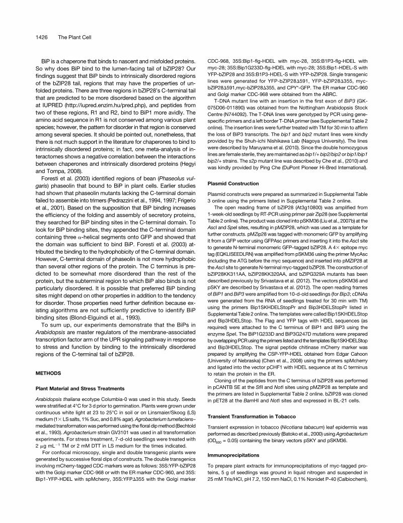
Snowden et al., 2007). In response to TM-induced stress, theexpression of BiP1G233D or BiP3G247D transgenes appearedto prevent the dissociation from myc-bZIP28 in pull-down ex-periments (Figure 7A). The overexpression of BiP1G233D withreduced capacity for client protein dissociation prevented theprocessing of myc-bZIP28 in response to DTT treatment (Figure7B) and also effectively blocked the nuclear relocation of YFP-bZIP28 in response to TM-induced stress (Figures 7C and 7D).DTT and not TM was used as a stress agent in this experimentbecause TM treatment produces additional nonglycosylatedforms of myc-bZIP28 that make the gel patterns difficult to in-terpret. In our hands, DTT has been just as effective as TM ineliciting ER stress. The overexpression BiP3G247D similarly
prevented the relocation of YFP-bZIP28 to nuclei in response toTM-induced stress (Figure 7E). Thus, we conclude that over-expression of BiP, particularly forms of BiP that interfere with therelease of client proteins, prevents the normal mobilization ofbZIP28 from the ER to the nucleus by binding to bZIP28 and notallowing its release.
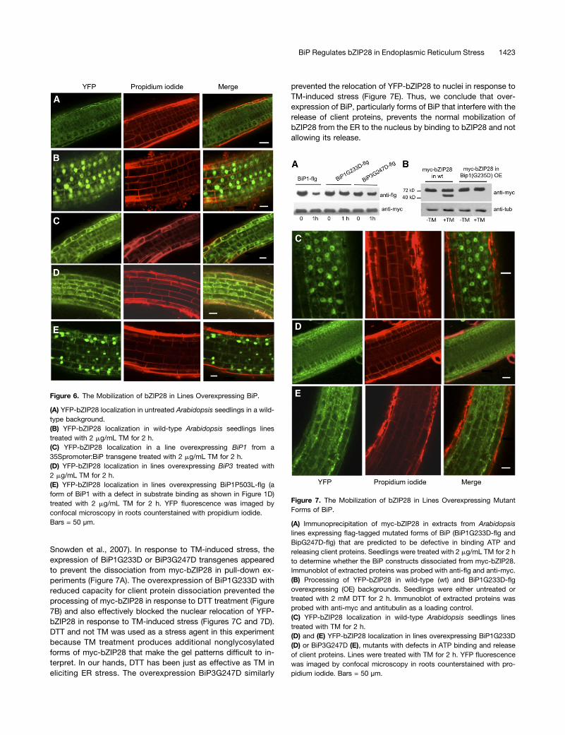
Figure 6. The Mobilization of bZIP28 in Lines Overexpressing BiP.
(A) YFP-bZIP28 localization in untreated Arabidopsis seedlings in a wild-type background.(B) YFP-bZIP28 localization in wild-type Arabidopsis seedlings linestreated with 2 mg/mL TM for 2 h.(C) YFP-bZIP28 localization in a line overexpressing BiP1 from a35Spromoter:BiP transgene treated with 2 mg/mL TM for 2 h.(D) YFP-bZIP28 localization in lines overexpressing BiP3 treated with2 mg/mL TM for 2 h.(E) YFP-bZIP28 localization in lines overexpressing BiP1P503L-flg (aform of BiP1 with a defect in substrate binding as shown in Figure 1D)treated with 2 mg/mL TM for 2 h. YFP fluorescence was imaged byconfocal microscopy in roots counterstained with propidium iodide.Bars = 50 µm.
Figure 7. The Mobilization of bZIP28 in Lines Overexpressing MutantForms of BiP.
(A) Immunoprecipitation of myc-bZIP28 in extracts from Arabidopsislines expressing flag-tagged mutated forms of BiP (BiP1G233D-flg andBipG247D-flg) that are predicted to be defective in binding ATP andreleasing client proteins. Seedlings were treated with 2 mg/mL TM for 2 hto determine whether the BiP constructs dissociated from myc-bZIP28.Immunoblot of extracted proteins was probed with anti-flg and anti-myc.(B) Processing of YFP-bZIP28 in wild-type (wt) and BiP1G233D-flgoverexpressing (OE) backgrounds. Seedlings were either untreated ortreated with 2 mM DTT for 2 h. Immunoblot of extracted proteins wasprobed with anti-myc and antitubulin as a loading control.(C) YFP-bZIP28 localization in wild-type Arabidopsis seedlings linestreated with TM for 2 h.(D) and (E) YFP-bZIP28 localization in lines overexpressing BiP1G233D(D) or BiP3G247D (E), mutants with defects in ATP binding and releaseof client proteins. Lines were treated with TM for 2 h. YFP fluorescencewas imaged by confocal microscopy in roots counterstained with pro-pidium iodide. Bars = 50 µm.
BiP Regulates bZIP28 in Endoplasmic Reticulum Stress 1423

Effect of BiP Knockouts on bZIP28 Mobilization
Similar experiments were conducted in BiP knockout lines con-taining YFP-bZIP28. Our interest here was whether the knockoutof BiP genes would prevent the retention of bZIP28 in the ER underunstressed conditions. Homozygous T-DNA insertion lines areavailable for all three BiP genes; however, multiple mutants, suchas the bip1 bip2 double mutant, are not viable. In an effort to re-duce BiP levels as much as possible, we obtained lines homozy-gous for either bip1 or bip2 but heterozygous for the other locus asdescribed by Maruyama et al. (2010). The BiP3 T-DNA homozy-gous insertion line (GK-075D06-011890) was obtained from theNottingham Arabidopsis Stock Centre and found not to produceBiP3 transcripts under stress conditions (see Supplemental Figure14 online). Compared with the wild type, detectable amounts ofYFP-bZIP28 escaped from the ER to nuclei under unstressedconditions in the bip1/bip1 bip2/+ and in the bip1/+ bip2/bip2and bip3 mutant lines, indicating a reduced ability of the BiPknockout mutants to retain bZIP28 in the ER (Figures 8A to 8D).We observed processed myc-bZIP28 in the various knockoutmutant lines under unstressed conditions, confirming these resultsand indicating that bZIP28 is constitutively activated in these lines(Figure 8E).
DISCUSSION
Our results show that BiP binds to bZIP28 under unstressed con-ditions and appears to bind to it in the same manner as it does toother client proteins, such as CPY*. We have also shown that BiPdissociates from bZIP28 in response to ER stress, and in doing sobZIP28 is activated, allowing it to make its way to the nucleuswhere it upregulates stress response genes (Figures 9A and 9B).The release of bZIP28 from BiP corresponds closely with its exitfrom the ER, but its release is not dependent on the trafficking ofbZIP28 from organelle to organelle. In animal systems, it is thoughtthat BiP binding retains ATF6 in the ER under nonstress conditions,putatively by blocking ATF6’s GLSs, preventing it from beingtransported through the secretory pathway (Shen et al., 2002).There are different ideas as to how BiP relinquishes its hold onATF6 under stress conditions (Parmar and Schröder, 2012). Oneidea, called the dynamic competition model, is that BiP is com-peted away from ATF6 by misfolded proteins in the ER (Hardinget al., 2002; Kaufman et al., 2002; Kimata et al., 2003). In thismodel, BiP bound to ATF6 is thought to be in equilibrium with freeBiP and BiP associated with misfolded proteins, and when un-folded proteins accumulate in the ER as a result of stress, thebinding of BiP to ATF6 would be competed away. Our results inArabidopsis support such a dynamic competition model.
We observed that the overexpression of BiP1 or BiP3 delaysor blocks the translocation of YFP-bZIP28 to the nucleus. WhenBiP is overexpressed, bZIP28 appears not to be fully deployed inresponse to stress but seems to be largely retained in the ER.The overexpression effect is enhanced using mutant versions ofBiP, which interfere with the release of substrates, as has also beenshown by Snowden et al. (2007). On the other hand, overexpressionof Bip1P503L defective in substrate binding does not block themovement of bZIP28. In animal systems, similar results were alsoobserved by Shen et al. (2002), who overexpressed BiP in HeLa
cells and found that ATF6 translocated to the Golgi, but pro-cessing by S1P and S2P, the Golgi resident proteases, was de-layed following stress treatment. Also, overexpression of BiPT37G,an ATPase mutant compromised in the release of bound proteins,prevented the translocation of ATF6 to the Golgi and its processingby the Golgi-resident proteases (Wei et al., 1995). The BiP knockoutmutants (bip1/bip1 bip2/+, bip1/+ bip2/bip2, and bip3/bip3) allowsome migration of bZIP28 to nuclei under unstressed conditions.
Figure 8. Mobilization of bZIP28 in BiP Knockout Lines.
(A) YFP-bZIP28 in wild-type unstressed Arabidopsis seedlings.(B) to (D) YFP-bZIP28 in the bip1/bip1 bip2/+ (B), bip1/+ bip2/bip2 (C),and bip3/bip3 (D) mutant lines. YFP fluorescence was imaged by con-focal microscopy in roots stained with propidium iodide. Arrows pointout nuclear localization. Bars = 50 µm.(E) Proteolytic processing of mycbZIP28 in wild-type seedlings in re-sponse to 2 mg/mL TM treatment for 2 h. Processing of myc-bZIP28 inuntreated BiP knockout lines as indicated.
1424 The Plant Cell

All three BiP genes cannot be knocked out because even homo-zygous bip1 bip2 mutants are not viable (Maruyama et al., 2010).We observed that these partial knockdown BiP mutants appear tobe defective in retaining bZIP28 in the ER under unstressed con-ditions. In all, our results involving the overexpression and under-expression of BiP have demonstrated the critical role of BiP in theretention and activation of bZIP28 in Arabidopsis.
Another model for the release of ATF6 from BiP in animal sys-tems does not invoke dynamic competition, instead positing thatthe association is stable, but can be disrupted by a signal frommisfolded proteins. Several arguments favor a stability model (Shenet al., 2005), with one being that association between BiP and ATF6is stable enough to survive immunoprecipitations without the needfor cross-linking. Second, if BiP is continually binding and disso-ciating from ATF6, then the T37G BiP mutant with a defect inATPase activity (protein releasing activity) should build up onATF6 when introduced into monkey kidney fibroblast cells. Ap-parently, it does not (Shen et al., 2005). Shen et al. (2005) comparedthe dissociation of BiP from ATF6 and from unassembled Ig heavy
chains, which are also retained in the ER by their association withBiP. They found that in response to stress, BiP dissociated fromATF6, but not from unassembled Ig heavy chains. From this theyhypothesized that ATF6 contains BiP binding and release ele-ments, the latter referred to as stress-responsive domains.Our experiments demonstrate that BiP binds to the C-terminal,
lumen-facing tail of bZIP28, and when the tail is eliminated as inbZIP28D355, the protein is not retained in the ER and behaveslike an activated form of bZIP28. Truncated bZIP28 constitutivelymoves to the nucleus where it upregulates stress genes, includingBiP3. The movement takes place via the Golgi and requires S2Pprocessing. As an aside, it is interesting that YFP-bZIP28D355appears to require S2P to relocate to the nucleus and is ap-parently a substrate for S2P. YFP-bZIP28D355 lacks most of itsC-terminal tail and its S1P site. Cleavage at the S1P site is usuallyconsidered to be a prerequisite for S2P cleavage (Espenshadeet al., 1999; Shen and Prywes, 2004). The implication from this isthat S1P cleavage is not required for S2P proteolysis as long asthe C-terminal tail on bZIP28 has been removed.
Figure 9. The Effect of BiP Expression on the Mobilization of bZIP28.
(A) BiP normally associates with bZIP28 under unstressed conditions and detains bZIP28 in the ER.(B) In response to stress, BiP is competed away by the accumulation of misfolded proteins, releasing bZIP28 to relocate to the nucleus via the Golgi.(C) When BiP is overexpressed, the accumulation of misfolded proteins fails to compete BiP away from bZIP28 under stress. As a result bZIP28 isdetained in the ER even under stress conditions.(D) When BiP is underexpressed, bZIP28 escapes to the nucleus under unstressed conditions.[See online article for color version of this figure.]
BiP Regulates bZIP28 in Endoplasmic Reticulum Stress 1425
BiP is a chaperone that binds to nascent and misfolded proteins.So why does BiP bind to the lumen-facing tail of bZIP28? Ourfindings suggest that BiP binds to intrinsically disordered regionsof the bZIP28 tail, regions that may have the properties of un-folded proteins. There are three regions in bZIP28’s C-terminal tailthat are predicted to be more disordered based on the algorithmat IUPRED (http://iupred.enzim.hu/pred.php), and peptides fromtwo of these regions, R1 and R2, bind to BiP1 more avidly. Theamino acid sequence in R1 is not conserved among various plantspecies; however, the pattern for disorder in that region is conservedamong several species. It should be pointed out, nonetheless, thatthere is not much support in the literature for chaperones to bind tointrinsically disordered proteins; in fact, one meta-analysis of in-teractomes shows a negative correlation between the interactionsbetween chaperones and intrinsically disordered proteins (Hegyiand Tompa, 2008).
Foresti et al. (2003) identified regions of bean (Phaseolus vul-garis) phaseolin that bound to BiP in plant cells. Earlier studieshad shown that phaseolin mutants lacking the C-terminal domainfailed to assemble into trimers (Pedrazzini et al., 1994, 1997; Frigerioet al., 2001). Based on the supposition that BiP binding increasesthe efficiency of the folding and assembly of secretory proteins,they searched for BiP binding sites in the C-terminal domain. Tolook for BiP binding sites, they appended the C-terminal domaincontaining three a-helical segments onto GFP and showed thatthe domain was sufficient to bind BiP. Foresti et al. (2003) at-tributed the binding to the hydrophobicity of the C-terminal domain.However, C-terminal domain of phaseolin is not more hydrophobicthan several other regions of the protein. The C terminus is pre-dicted to be somewhat more disordered than the rest of theprotein, but the subterminal region to which BiP also binds is notparticularly disordered. It is possible that preferred BiP bindingsites might depend on other properties in addition to the tendencyfor disorder. Those properties need further definition because ex-isting algorithms are not sufficiently predictive to identify BiPbinding sites (Blond-Elguindi et al., 1993).
To sum up, our experiments demonstrate that the BiPs inArabidopsis are master regulators of the membrane-associatedtranscription factor arm of the UPR signaling pathway in responseto stress and function by binding to the intrinsically disorderedregions of the C-terminal tail of bZIP28.
METHODS
Plant Material and Stress Treatments
Arabidopsis thaliana ecotype Columbia-0 was used in this study. Seedswere stratified at 4°C for 3 d prior to germination. Plants were grown undercontinuous white light at 23 to 25°C in soil or on Linsmaier/Skoog (LS)medium (13 LSsalts, 1%Suc, and 0.8%agar).Agrobacterium tumefaciens–mediated transformationwasperformedusing the floral dipmethod (Bechtoldet al., 1993). Agrobacterium strain GV3101 was used in all transformationexperiments. For stress treatment, 7-d-old seedlings were treated with2 mg mL21 TM or 2 mM DTT in LS medium for the times indicated.
For confocal microscopy, single and double transgenic plants weregenerated by successive floral dips of constructs. The double transgenicsinvolving mCherry-tagged CDC markers were as follows: 35S:YFP-bZIP28with the Golgi marker CDC-968 or with the ER marker CDC-960, and 35S:Bip1-YFP-HDEL with spMcherry, 35S:YFPD355 with the Golgi marker
CDC-968, 35S:Bip1-flg-HDEL with myc-28, 35S:B1P3-flg-HDEL withmyc-28; 35S:Bip1G233D-flg-HDEL with myc-28; 35S:Bip1-HDEL-S withYFP-bZIP28 and 35S:B1P3-HDEL-S with YFP-bZIP28. Single transgeniclines were generated for YFP-bZIP28D591, YFP-bZIP28D355, myc-bZIP28D591,myc-bZIP28D355, and CPY*-GFP. The ER marker CDC-960and Golgi marker CDC-968 were obtained from the ABRC.
T-DNA mutant line with an insertion in the first exon of BiP3 (GK-075D06-011890) was obtained from the Nottingham Arabidopsis StockCentre (N744092). The T-DNA lines were genotyped by PCR using gene-specific primers and a left border T-DNA primer (see Supplemental Table 2online). The insertion lines were further treated with TM for 30 min to affirmthe loss of BiP3 transcripts. The bip1 and bip2 mutant lines were kindlyprovided by the Shuh-ichi Nishikawa Lab (Nagoya University). The lineswere described by Maruyama et al. (2010). Since the double homozygouslines are female sterile, they aremaintained asbip1/+ bip2/bip2orbip1/bip1bip2/+ strains. The s2pmutant line was described by Che et al., (2010) andwas kindly provided by Ping Che (DuPont Pioneer Hi-Bred International).
Plasmid Construction
Plasmid constructs were prepared as summarized in Supplemental Table3 online using the primers listed in Supplemental Table 2 online.
The open reading frame of bZIP28 (At3g10800) was amplified from1-week-old seedlings by RT-PCR using primer pair Zip28 (see SupplementalTable 2 online). The productwas cloned into pSKM36 (Liu et al., 2007b) at theAscI and SpeI sites, resulting in pMZIP28, which was used as a template forfurther constructs. pMZip28 was tagged with monomeric GFP by amplifyingit from a GFP vector using GFPAsc primers and inserting it into the AscI siteto generate N-terminal monomeric GFP–tagged bZIP28. A 43 epitope myctag (EQKLISEEDLRN) was amplified from pSKM36 using the primer MycAsc(including the ATG before the myc sequence) and inserted into pMZIP28 attheAscI site to generate N-terminal myc-tagged bZIP28. The construction ofbZIP28KK311AA, bZIP28KK320AA, and bZIPG329A mutants has beendescribed previously by Srivastava et al. (2012). The vectors pSKM36 andpSKY are described by Srivastava et al. (2012). The open reading framesofBiP1 andBiP3were amplified from 10-d-old seedlings (for Bip3; cDNAswere generated from the RNA of seedlings treated for 30 min with TM)using the primers Bip1SKHDELStopPr and Bip3HDELStopPr listed inSupplemental Table 2 online. The templates were called Bip1SKHDELStopand Bip3HDELStop. The Flag and YFP tags with HDEL sequences (asrequired) were attached to the C terminus of BiP1 and BiP3 using theenzyme SpeI. The BiP1G233D and BiP3G247D mutations were preparedbyoverlappingPCRusing theprimers listedand the templatesBip1SKHDELStopand Bip3HDELStop. The signal peptide chitinase mCherry marker wasprepared by amplifying the CSP-YFP-HDEL obtained from Edgar Cahoon(University of Nebraska) (Chen et al., 2008) using the primers spMcherryand ligated into the vector pCHF1 with HDEL sequence at its C terminusto retain the protein in the ER.
Cloning of the peptides from the C terminus of bZIP28 was performedin pCANTB SE at the SfiI and NotI sites using pMZIP28 as template andthe primers are listed in Supplemental Table 2 online. bZIP28 was clonedin pET28 at the BamHI and NotI sites and expressed in BL-21 cells.
Transient Transformation in Tobacco
Transient expression in tobacco (Nicotiana tabacum) leaf epidermis wasperformed as described previously (Batoko et al., 2000) usingAgrobacterium(OD600 = 0.05) containing the binary vectors pSKY and pSKM36.
Immunoprecipitations
To prepare plant extracts for immunoprecipitations of myc-tagged pro-teins, 5 g of seedlings was ground in liquid nitrogen and suspended in25 mM Tris/HCl, pH 7.2, 150 mm NaCl, 0.1% Nonidet P-40 (Calbiochem),
1426 The Plant Cell
and 10% glycerol. Anti-c-myc agarose conjugate (Sigma-Aldrich) wasadded to the filtered lysate and incubated for 2 h at 4°C. The mixture wasrotated at 4°C for 2 h, and beads were washed four times for 5 min eachwith the buffer described above. The recovered beads were resuspendedand boiled in 23 SDS buffer for 5 min, and the eluted material wassubjected to immunoblotting. Plant crude extracts (input material for theimmunoprecipitation reactions) were analyzed for the presence of thetagged protein using c-myc antibody (9E10; Santa Cruz Biotechnology)as probe. BiP-bound proteins were immunoprecipitated from 2 g of plantmaterial using a BiP antibody, ADI-SPA-818 D from Enzo Life Sciences.The mixture was rotated at 4°C for 2 h. This was followed by binding toprotein agarose 916-157 (Millipore), and bound beads were washed fourtimes for 5 min each with the buffer described above followed by loadingon SDS gel. Flag antibodies (F1804; Sigma-Aldrich) and GFP antibodies(11814460001; Roche) were used in immunoprecipitations and in probingimmunoblots.
Immunoblot Analysis
Immunoblots were performed as described by Liu et al. (2007a, 2007b). Toexamine the processing of bZIP28, plants were grown vertically on Petriplates containing agar medium. Ten-day-old seedlings were treated with2 mm DTT in LS medium, and 300 mg of root material was harvested fromthe treated plants. Roots were homogenized in liquid nitrogen, and 30 mgof protein was loaded per lane on gels. T8203 monoclonal anti-atubulinantibody from Sigma-Aldrich was used to detect tubulin as a loadingcontrol.
Confocal Microscopy
Subcellular localization andproteinmovement experiments for fluorescent-tagged proteins were performed using a NikonC1si confocal scanningsystem attached to a 90i microscope (Nikon Instruments). Roots wereused for microscopy from plants pretreated with TM, and untreated plantswere used as controls. The roots were observed under320 and360 waterlenses. Some rootswere counterstainedwith 50mgmL21 propidium iodideand Syto Red. The emission signals for YFP, propidium iodide, and SYTORed 59 were acquired using sequential scanning mode to eliminatecrosstalk and emission signal bleed-through. Fluorescence emission wasobtained by laser excitation of YFP at 488 nm, and for mCherry, propidiumiodide, or Syto Red, excitation was at 591 nm. Emission was in the range500 to 575 and 590 to 700 nm, respectively. Syto Red 59 (S-11341) wasobtained from Molecular Probes.
Gene Expression Analysis
Total RNA was isolated from ground plant tissues using an RNeasy kit,treatedwithRNase-freeDNase I, according to themanufacturer’s instructions(Qiagen), and was quantified by 260/280-nm UV light absorption. A 1-mgportion of total RNA was reverse transcribed using the Supertranscript IIIRT kit (Invitrogen). A 2-mL volumeof cDNAwas used for RT-PCR.All primersare listed in Supplemental Table 2 online.
Construction of Phage-Displayed Overlapping Peptide Library
Twelve overlapping regions of the lumenal domain of bZIP28 were gen-erated by PCR using bZIP28 cDNA as a template and forward and reverseprimers to incorporate SfiI and NotI sites. The PCR fragments were thendigested with SfiI and NotI, ligated into a similarly digested pCANTAB 5Ephagemid vector, and subsequently transformed into Escherichia coli XL1-Blue cells (Stratagene). Single colonies fromeach platewere inoculated into5 mL 2YT/carbenicillin/tetracycline media, grown to OD600 of;0.2 to 0.3 at37°C and then infected with helper phage-VCSM13 (Stratagene). After 1 h,the cell culture was transferred to 25 mL 2YT/Kan media and further
incubated overnight at 37°C. After removing cell debris by centrifugation,phage particles were precipitated from the supernatant using 7.5 mL 20%polyethylene glycol solution containing 2.5 M NaCl. The precipitate wasresuspended in 1mLPBS, and thephage concentrationwas determinedbymeasuring absorbance at 268l (OD268 = 1.0 for a solution containing 531012
phage per mL). A peptide library was prepared by mixing equal concen-trations of individual phage peptide.
Production and Purification of BiP for Phage Panning Experiment
BiP1 was His tagged at its N terminus in the pET28a vector. The constructwas introduced into E. coli strain BL21, and cells were induced with 300 mMisopropyl b-D-1 thiogalacopyranodise overnight at 16°C. His-tagged BiP1was purified using nickel-nitrilotriacetic acid agarose beads (Qiagen). Theincubation buffer contained 50 mM sodium phosphate buffer, pH 8.0,300mMNaCl, 20mM imidazole, and 0.05%Tween. Themixturewas rotatedat 4°C for 2 h and washed four times with incubation buffer. The protein waseluted by increasing the imidazole concentration in the incubation buffer to250mM.Thebeadswere resuspended and boiled in 23SDSbuffer for 5minand subjected to SDS-PAGE to check for purity. The purified BiP-His wasused for panning experiments, ELISA, and overlay immunoblotting.
Phage Panning
His-tagged BiP1 protein was immobilized in the wells of a Nunc MaxisorpELISA plate by aliquoting 100mL of protein at a concentration of 10 µg/mLin 50 mM NaHCO3, pH 9.6, at room temperature with gentle rotation for2 h. Wells were then blocked with PBS containing 0.2% BSA for 1 hfollowed by three washings with PBS containing 0.05% Tween 20 (PBST)and then incubated with 100 mL of the phage peptide library from thelumenal domain of bZIP28 for 3 h at room temperature with gentle ro-tation. After removing unbound phage by washing with PBST five times,bound phage was eluted by incubating with 500 mL of 0.1 M HCl for 5 minat room temperature with shaking. The eluted phage were immediatelyneutralized by the addition of one-third phage volume of 1 M Tris-HClbuffer, pH 8.0, followed by infection of XL1-Blue cells (grown to <0.6 OD)with the phage. Infected XL1-Blue cells were incubated at 37°C for 20 minfollowed by addition of the helper phage (VCSM13) and again incubatingat 37°C for 30 min. The phage-infected XL1-Blue cells were transferredinto a conical flask containing 50 mL of 2YT medium containing 10 µg/mLof tetracycline and 100 µg/mL of ampicillin that was further incubated at37°C overnight with shaking at 210 rpm. Phage was prepared as describedearlier, yielding the first round of enriched phage. The entire process wasrepeated for four rounds, and after two, three, and four rounds, phage-infectedXL1-Blue cells were grown on 2YT ampicillin plates. Plasmidswereprepared from 25 randomly selected colonies and their DNA sequenced.
Phage ELISA for Binding Specificity
For phage ELISAs, 100 mL of His-tagged BiP1 protein and BSA (control)(10 µg/mL in 50 mM NaHCO3, pH 9.6) were immobilized in the wells of anELISA plate at room temperature with gentle rotation for 2 h. The plate wasthen washed two times with PBS followed by blocking with PBST con-taining 0.2% BSA for 1 h. Subsequently, after three washings with PBST,wells were incubated with the 100 mL of phage displaying lumenal domainpeptides diluted in PBST containing 0.2% BSA for 2 h at room tem-perature with gentle shaking. The plate was again washed three timeswithPBST followed by incubation with anti-M13 HRP conjugated antibody for1 h. After washing four more times with PBST, bound phage in each wellwas detected by incubating for ;10 min with 50 mL of a solution con-taining 0.01% hydrogen peroxide and 0.8 mg/mL o-phenylenediaminedihydrochoride. Reactions were terminated by the addition of 50 mL 3MHCl, and absorbance of the developed yellow color was measured at490 nm.
BiP Regulates bZIP28 in Endoplasmic Reticulum Stress 1427

Overlay Immunoblotting
The four peptides (441, 471, 376, and 501) were tagged on their N terminuswith a GST tag in pET42a vector. For in vitro GST pull-down assays, GST-441, GST-471, GST-376, GST-501, and GST-H6 (control) were bound toglutathione agarose beads (Sigma-Aldrich). The pull-down mixture con-tained 25 mM Tris-HCl, pH 7.4, 75 mM NaCl, 0.5 mM EDTA, 0.5 mM DTT,0.05% Nonidet P-40, and 1 mg/mL BSA (NEB). The mixture was rotated at4°C for 2 h and washed four times with pull-down buffer. The beads wereresuspended and boiled in 23 SDS buffer for 5 min, and the eluted materialwas subjected to SDS-PAGE. GST-tagged peptides were transferred ontoa nitrocellulose membrane after separation on SDS-PAGE, and the mem-brane was blocked with 1% BSA in TBS buffer. Ten micrograms of BiPproteinwas allowed to bind to the respective peptides by incubation in T-TBSbuffer containing 0.1% BSA. This was followed by incubation of the blot withthe BiP antibody, which was detected by a secondary antibody, as describedin the immunoblotting procedure.
Accession Numbers
Sequence data from this article can be found in the Arabidopsis GenomeInitiative or GenBank/EMBL databases under the following accessionnumbers: BiP1 (At5g28540), BiP2 (At5g42020), BiP3 (At1g09080), bZIP17(At2g40950), bZIP28 (At3g10800), Erdj3a (At3g08970), Erdj3b (At3g62600),and S2P (At4g20310).
Supplemental Data
The following materials are available in the online version of this article.
Supplemental Figure 1. Sequence Alignment of the Three BiPProteins from Arabidopsis.
Supplemental Figure 2. Immunoprecipitation of myc-bZIP28 byBead-Bound BiP Antibody.
Supplemental Figure 3. BiP Binds CPY*-GFP Expressed as a Trans-gene in Arabidopsis Seedlings.
Supplemental Figure 4. BiP Levels in Stressed Arabidopsis Seedlings.
Supplemental Figure 5. Subcellular Localization of YFP-bZIP28 inStressed Seedlings.
Supplemental Figure 6. Constitutive Expression of BiP3 in a Line ThatFails Retain bZIP28 in the ER.
Supplemental Figure 7. Subcellular Localization of YFP-bZIP28D355in Unstressed Seedlings.
Supplemental Figure 8. BiP1-His Extracted from E. coli.
Supplemental Figure 9. BiP Binding to Peptides Enriched in PhageDisplay Panning.
Supplemental Figure 10. Hydrophobicity Plot of the Lumenal Domainof bZIP28.
Supplemental Figure 11. Expression Levels of BiP in Untreated BiP-Overepressing Lines.
Supplemental Figure 12. Subcellular Localization of BiP1-YFP-HDEL.
Supplemental Figure 13. bZIP60 Splicing in BiP Overexpression Lines.
Supplemental Figure 14. Noninduction of BiP3 in bip3 HomozygousMutant.
Supplemental Table 1. List of Peptides Used in the Phage DisplayExperiments.
Supplemental Table 2. Primers Used in This Study.
Supplemental Table 3. Cloning Information.
ACKNOWLEDGMENTS
This work was supported by the Iowa State University Plant SciencesInstitute and by a National Science Foundation grant (IOS90917) to S.H.H.The bip1/bip1 bip2/+ and bip1/+ bip2/bip2 mutant lines were obtainedfrom the Shuh-ichi Nishikawa Lab (Nagoya University, Nagoya, Japan). Thes2p mutant line was provided generously by Ping Che (DuPont Pioneer Hi-Bred International, Johnston, IA).
AUTHOR CONTRIBUTIONS
R.S., Y.D., S.S., and S.H.H. conceived and designed the experiments.R.S. and S.S. performed the experiments. R.S., S.S., and A.G.R. analyzedthe data. R.S. and S.H.H. wrote the article.
Received February 13, 2013; revised March 28, 2013; accepted April 10,2013; published April 24, 2013.
REFERENCES
Alvim, F.C., Carolino, S.M., Cascardo, J.C., Nunes, C.C., Martinez,C.A., Otoni, W.C., and Fontes, E.P. (2001). Enhanced accumulation ofBiP in transgenic plants confers tolerance to water stress. Plant Physiol.126: 1042–1054.
Anelli, T., and Sitia, R. (2008). Protein quality control in the earlysecretory pathway. EMBO J. 27: 315–327.
Batoko, H., Zheng, H.Q., Hawes, C., and Moore, I. (2000). A rab1GTPase is required for transport between the endoplasmic reticulumand golgi apparatus and for normal Golgi movement in plants. Plant Cell12: 2201–2218.
Bechtold, N., Ellis, J., and Pelletier, G. (1993). In planta Agrobacterium-mediated gene transfer by infiltration of adult Arabidopsis thaliana plants.C. R. Acad. Sci. Paris 316: 1194–1199.
Bertolotti, A., Zhang, Y., Hendershot, L.M., Harding, H.P., and Ron,D. (2000). Dynamic interaction of BiP and ER stress transducers inthe unfolded-protein response. Nat. Cell Biol. 2: 326–332.
Blond-Elguindi, S., Cwirla, S.E., Dower, W.J., Lipshutz, R.J., Sprang,S.R., Sambrook, J.F., and Gething, M.J. (1993). Affinity panning ofa library of peptides displayed on bacteriophages reveals the bindingspecificity of BiP. Cell 75: 717–728.
Caramelo, J.J., Castro, O.A., Alonso, L.G., De Prat-Gay, G., andParodi, A.J. (2003). UDP-Glc:glycoprotein glucosyltransferase recognizesstructured and solvent accessible hydrophobic patches in molten globule-like folding intermediates. Proc. Natl. Acad. Sci. USA 100: 86–91.
Che, P., Bussell, J.D., Zhou, W., Estavillo, G.M., Pogson, B.J., andSmith, S.M. (2010). Signaling from the endoplasmic reticulum activatesbrassinosteroid signaling and promotes acclimation to stress in Arabidopsis.Sci. Signal. 3: ra69.
Chen, M., Markham, J.E., Dietrich, C.R., Jaworski, J.G., and Cahoon,E.B. (2008). Sphingolipid long-chain base hydroxylation is important forgrowth and regulation of sphingolipid content and composition inArabidopsis. Plant Cell 20: 1862–1878.
Deng, Y., Humbert, S., Liu, J.X., Srivastava, R., Rothstein, S.J., andHowell, S.H. (2011). Heat induces the splicing by IRE1 of a mRNAencoding a transcription factor involved in the unfolded protein responsein Arabidopsis. Proc. Natl. Acad. Sci. USA 108: 7247–7252.
Dosztányi, Z., Csizmok, V., Tompa, P., and Simon, I. (2005). IUPred:Web server for the prediction of intrinsically unstructured regions ofproteins based on estimated energy content. Bioinformatics 21:3433–3434.
1428 The Plant Cell

Espenshade, P.J., Cheng, D., Goldstein, J.L., and Brown, M.S.(1999). Autocatalytic processing of site-1 protease removes propeptideand permits cleavage of sterol regulatory element-binding proteins. J. Biol.Chem. 274: 22795–22804.
Foresti, O., Frigerio, L., Holkeri, H., de Virgilio, M., Vavassori, S.,and Vitale, A. (2003). A phaseolin domain involved directly in trimerassembly is a determinant for binding by the chaperone BiP. PlantCell 15: 2464–2475.
Frigerio, L., Pastres, A., Prada, A., and Vitale, A. (2001). Influence ofKDEL on the fate of trimeric or assembly-defective phaseolin: Selectiveuse of an alternative route to vacuoles. Plant Cell 13: 1109–1126.
Harding, H.P., Calfon, M., Urano, F., Novoa, I., and Ron, D. (2002).Transcriptional and translational control in the mammalian unfoldedprotein response. Annu. Rev. Cell Dev. Biol. 18: 575–599.
Hegyi, H., and Tompa, P. (2008). Intrinsically disordered proteins displayno preference for chaperone binding in vivo. PLoS Comput. Biol. 4:e1000017.
Hendershot, L.M. (2004). The ER function BiP is a master regulator ofER function. Mt. Sinai J. Med. 71: 289–297.
Iwata, Y., Fedoroff, N.V., and Koizumi, N. (2008). Arabidopsis bZIP60 isa proteolysis-activated transcription factor involved in the endoplasmicreticulum stress response. Plant Cell 20: 3107–3121.
Iwata, Y., and Koizumi, N. (2005). An Arabidopsis transcription factor,AtbZIP60, regulates the endoplasmic reticulum stress response ina manner unique to plants. Proc. Natl. Acad. Sci. USA 102: 5280–5285.
Iwata, Y., Sakiyama, M., Lee, M.-H., and Koizumi, N. (2010).Transcriptomic response of Arabidopsis thaliana to tunicamycin inducedendoplasmic reticulum stress. Plant Biotechnol. 27: 161–171.
Izawa, T., Nagai, H., Endo, T., and Nishikawa, S. (2012). Yos9p andHrd1p mediate ER retention of misfolded proteins for ER-associateddegradation. Mol. Biol. Cell 23: 1283–1293.
Jin, Y., Awad, W., Petrova, K., and Hendershot, L.M. (2008).Regulated release of ERdj3 from unfolded proteins by BiP. EMBO J.27: 2873–2882.
Kaufman, R.J., Scheuner, D., Schröder, M., Shen, X., Lee, K., Liu,C.Y., and Arnold, S.M. (2002). The unfolded protein response innutrient sensing and differentiation. Nat. Rev. Mol. Cell Biol. 3: 411–421.
Kimata, Y., Kimata, Y.I., Shimizu, Y., Abe, H., Farcasanu, I.C.,Takeuchi, M., Rose, M.D., and Kohno, K. (2003). Genetic evidencefor a role of BiP/Kar2 that regulates Ire1 in response to accumulation ofunfolded proteins. Mol. Biol. Cell 14: 2559–2569.
Koizumi, N. (1996). Isolation and responses to stress of a gene thatencodes a luminal binding protein in Arabidopsis thaliana. Plant CellPhysiol. 37: 862–865.
Leborgne-Castel, N., Jelitto-Van Dooren, E.P., Crofts, A.J., andDenecke, J. (1999). Overexpression of BiP in tobacco alleviatesendoplasmic reticulum stress. Plant Cell 11: 459–470.
Liu, J.X., and Howell, S.H. (2010). bZIP28 and NF-Y transcriptionfactors are activated by ER stress and assemble into a transcriptionalcomplex to regulate stress response genes in Arabidopsis. Plant Cell22: 782–796.
Liu, J.X., Srivastava, R., Che, P., and Howell, S.H. (2007a). Saltstress responses in Arabidopsis utilize a signal transduction pathwayrelated to endoplasmic reticulum stress signaling. Plant J. 51: 897–909.
Liu, J.X., Srivastava, R., Che, P., and Howell, S.H. (2007b). Anendoplasmic reticulum stress response in Arabidopsis is mediatedby proteolytic processing and nuclear relocation of a membrane-associated transcription factor, bZIP28. Plant Cell 19: 4111–4119.
Martínez, I.M., and Chrispeels, M.J. (2003). Genomic analysis of theunfolded protein response in Arabidopsis shows its connection toimportant cellular processes. Plant Cell 15: 561–576.
Maruyama, D., Endo, T., and Nishikawa, S. (2010). BiP-mediatedpolar nuclei fusion is essential for the regulation of endospermnuclei proliferation in Arabidopsis thaliana. Proc. Natl. Acad. Sci.USA 107: 1684–1689.
Nekrasov, V., et al. (2009). Control of the pattern-recognition receptorEFR by an ER protein complex in plant immunity. EMBO J. 28:3428–3438.
Otero, J.H., Lizák, B., and Hendershot, L.M. (2010). Life and death ofa BiP substrate. Semin. Cell Dev. Biol. 21: 472–478.
Parmar, V.M., and Schröder, M. (2012). Sensing endoplasmicreticulum stress. Adv. Exp. Med. Biol. 738: 153–168.
Pedrazzini, E., Giovinazzo, G., Bollini, R., Ceriotti, A., and Vitale, A.(1994). Binding of BiP to an assembly-defective protein in plantcells. Plant J. 5: 103–110.
Pedrazzini, E., Giovinazzo, G., Bielli, A., de Virgilio, M., Frigerio, L.,Pesca, M., Faoro, F., Bollini, R., Ceriotti, A., and Vitale, A. (1997).Protein quality control along the route to the plant vacuole. PlantCell 9: 1869–1880.
Reis, P.A., Rosado, G.L., Silva, L.A., Oliveira, L.C., Oliveira, L.B.,Costa, M.D., Alvim, F.C., and Fontes, E.P. (2011). The binding proteinBiP attenuates stress-induced cell death in soybean via modulation ofthe N-rich protein-mediated signaling pathway. Plant Physiol. 157:1853–1865.
Schott, A., Ravaud, S., Keller, S., Radzimanowski, J., Viotti, C.,Hillmer, S., Sinning, I., and Strahl, S. (2010). Arabidopsis stromal-derivedFactor2 (SDF2) is a crucial target of the unfolded protein response in theendoplasmic reticulum. J. Biol. Chem. 285: 18113–18121.
Shen, J., Chen, X., Hendershot, L., and Prywes, R. (2002). ER stressregulation of ATF6 localization by dissociation of BiP/GRP78 bindingand unmasking of Golgi localization signals. Dev. Cell 3: 99–111.
Shen, J., and Prywes, R. (2004). Dependence of site-2 proteasecleavage of ATF6 on prior site-1 protease digestion is determined by thesize of the luminal domain of ATF6. J. Biol. Chem. 279: 43046–43051.
Shen, J., Snapp, E.L., Lippincott-Schwartz, J., and Prywes, R.(2005). Stable binding of ATF6 to BiP in the endoplasmic reticulumstress response. Mol. Cell. Biol. 25: 921–932.
Snowden, C.J., Leborgne-Castel, N., Wootton, L.J., Hadlington,J.L., and Denecke, J. (2007). In vivo analysis of the lumenal bindingprotein (BiP) reveals multiple functions of its ATPase domain. Plant J.52: 987–1000.
Srivastava, R., Chen, Y., Deng, Y., Brandizzi, F., and Howell, S.H.(2012). Elements proximal to and within the transmembrane domainmediate the organelle-to-organelle movement of bZIP28 under ER stressconditions. Plant J. 70: 1033–1042.
Tajima, H., Iwata, Y., Iwano, M., Takayama, S., and Koizumi, N.(2008). Identification of an Arabidopsis transmembrane bZIP transcriptionfactor involved in the endoplasmic reticulum stress response. Biochem.Biophys. Res. Commun. 374: 242–247.
Valente, M.A., et al. (2009). The ER luminal binding protein (BiP) mediatesan increase in drought tolerance in soybean and delays drought-inducedleaf senescence in soybean and tobacco. J. Exp. Bot. 60: 533–546.
Wang, D., Weaver, N.D., Kesarwani, M., and Dong, X. (2005). Inductionof protein secretory pathway is required for systemic acquired resistance.Science 308: 1036–1040.
Wei, J., Gaut, J.R., and Hendershot, L.M. (1995). In vitro dissociationof BiP-peptide complexes requires a conformational change in BiPafter ATP binding but does not require ATP hydrolysis. J. Biol. Chem.270: 26677–26682.
Wei, J., and Hendershot, L.M. (1995). Characterization of the nucleotidebinding properties and ATPase activity of recombinant hamster BiPpurified from bacteria. J. Biol. Chem. 270: 26670–26676.
BiP Regulates bZIP28 in Endoplasmic Reticulum Stress 1429

DOI 10.1105/tpc.113.110684; originally published online April 26, 2013; 2013;25;1416-1429Plant Cell
Renu Srivastava, Yan Deng, Shweta Shah, Aragula Gururaj Rao and Stephen H. HowellArabidopsisSensor/Transducer bZIP28 in
BINDING PROTEIN Is a Master Regulator of the Endoplasmic Reticulum Stress
This information is current as of November 21, 2018
Supplemental Data /content/suppl/2013/04/11/tpc.113.110684.DC1.html
References /content/25/4/1416.full.html#ref-list-1
This article cites 48 articles, 26 of which can be accessed free at:
Permissions https://www.copyright.com/ccc/openurl.do?sid=pd_hw1532298X&issn=1532298X&WT.mc_id=pd_hw1532298X
eTOCs http://www.plantcell.org/cgi/alerts/ctmain
Sign up for eTOCs at:
CiteTrack Alerts http://www.plantcell.org/cgi/alerts/ctmain
Sign up for CiteTrack Alerts at:
Subscription Information http://www.aspb.org/publications/subscriptions.cfm
is available at:Plant Physiology and The Plant CellSubscription Information for
ADVANCING THE SCIENCE OF PLANT BIOLOGY © American Society of Plant Biologists