Biliary tract interventions
73
biliary tract interventions THORSANG CHAYOVAN/AJ.KITTIPITCH BANNANGKUN INTERVENTIONAL RADIOLOGY, PRINCE OF SONGKLA UNIVERSITY 18.11.2015 Topic in Interventional Radiology
-
Upload
thorsang-chayovan -
Category
Health & Medicine
-
view
1.109 -
download
0
Transcript of Biliary tract interventions
biliary tract interventionsTHORSANG CHAYOVAN/AJ.K IT TIPITCH BANNANGKUN
INTERVENTIONAL RADIOLOGY, PRINCE OF SONGKLA UNIVERSITY
18.11.2015
Topic in Interventional Radiology
Percutaneous transhepatic biliary drainage
Biliary tract obstructionBenign causes of biliary obstruction are often iatrogenic in nature◦ Biliary tree injury during surgical procedures
◦ Others: trauma, inflammatory processes (stone disease or pancreatitis), sphincter of Oddi dysfunction, and a late complication of intraabdominalprocesses
Malignant biliary strictures are much more common ◦ Cholangiocarcinoma, gallbladder and pancreas cancer
◦ Lymphoma and metastasis◦ Hepatocellular carcinoma
Percutaneous TranshepaticCholangiography (PTC)Introduced clinically in the 1970s
Popular in the 1980s-early 1990s
ERCP has largely replaced PTC in the diagnosis and treatment of biliary tract obstruction
However, PTC still has an important role in the management of patients with biliary tract disease
Current role of PTCPTC often when endoscopic techniques are not possible◦ Hilar obstruction
◦ Surgically altered anatomy◦ When ERCP is technically unsuccessful
◦ When additional information about the intrahepatic extent of disease is desired
Careful patient selection
Review of preprocedural diagnostic imaging studies
Appropriate planning
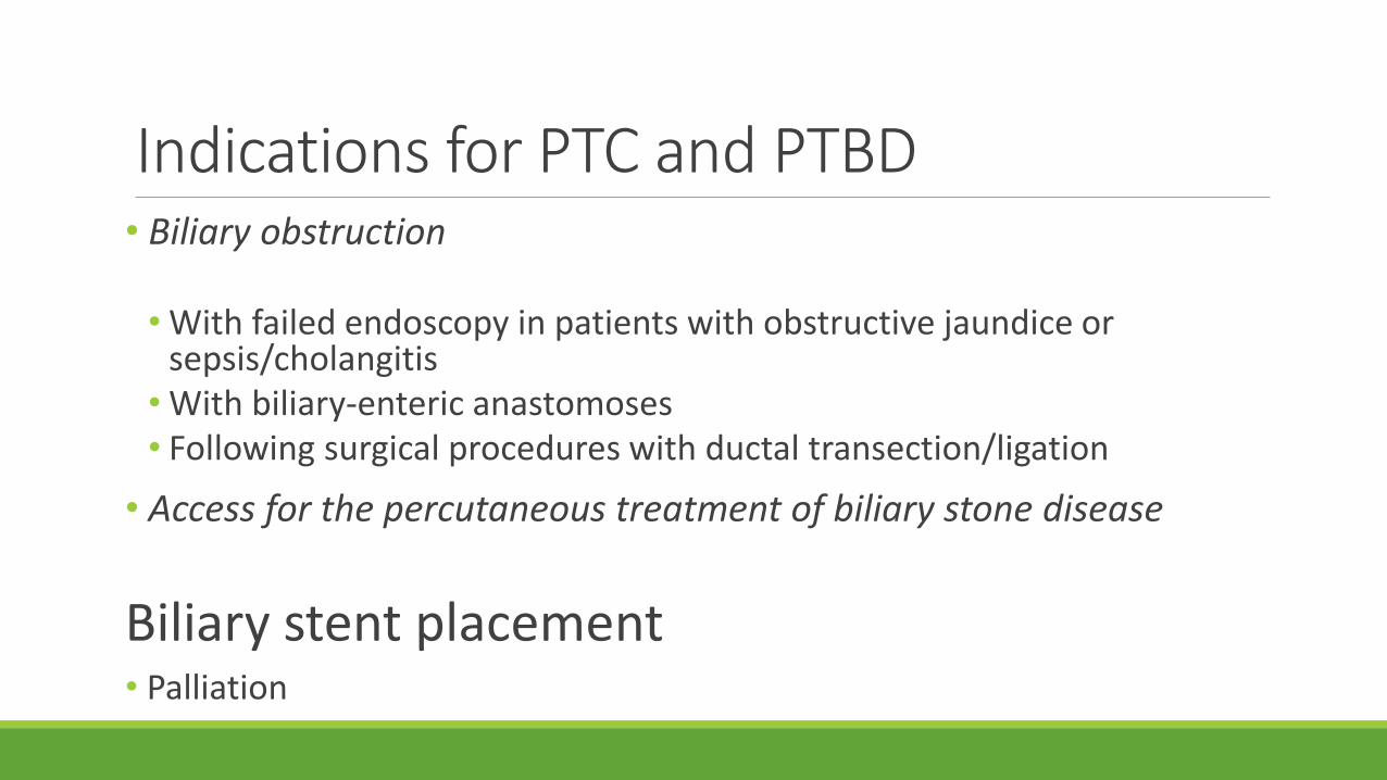
Indications for PTC and PTBD• Biliary obstruction
• With failed endoscopy in patients with obstructive jaundice or sepsis/cholangitis
• With biliary-enteric anastomoses • Following surgical procedures with ductal transection/ligation
• Access for the percutaneous treatment of biliary stone disease
Biliary stent placement• Palliation
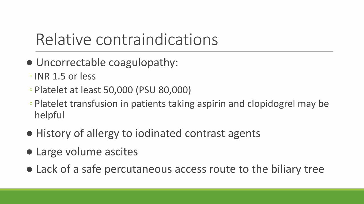
Relative contraindications● Uncorrectable coagulopathy: ◦ INR 1.5 or less
◦ Platelet at least 50,000 (PSU 80,000)
◦ Platelet transfusion in patients taking aspirin and clopidogrel may be helpful
● History of allergy to iodinated contrast agents
● Large volume ascites
● Lack of a safe percutaneous access route to the biliary tree
Contraindications for PTBD
● Segmental high bile duct obstruction
● Multiple isolations
Contraindications for biliary stent placement
● Candidate for surgery
● Benign disease
● Sepsis
Patient Selectionand Preparation
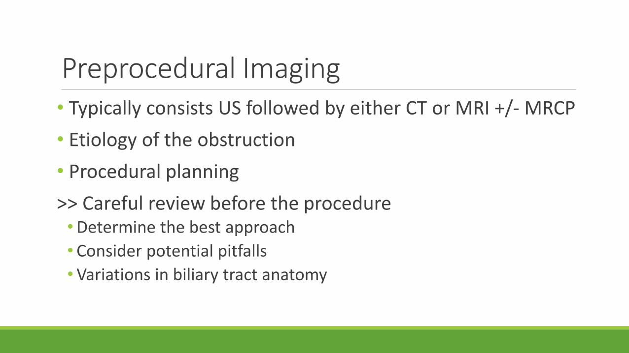
Preprocedural Imaging • Typically consists US followed by either CT or MRI +/- MRCP
• Etiology of the obstruction
• Procedural planning
>> Careful review before the procedure •Determine the best approach
• Consider potential pitfalls
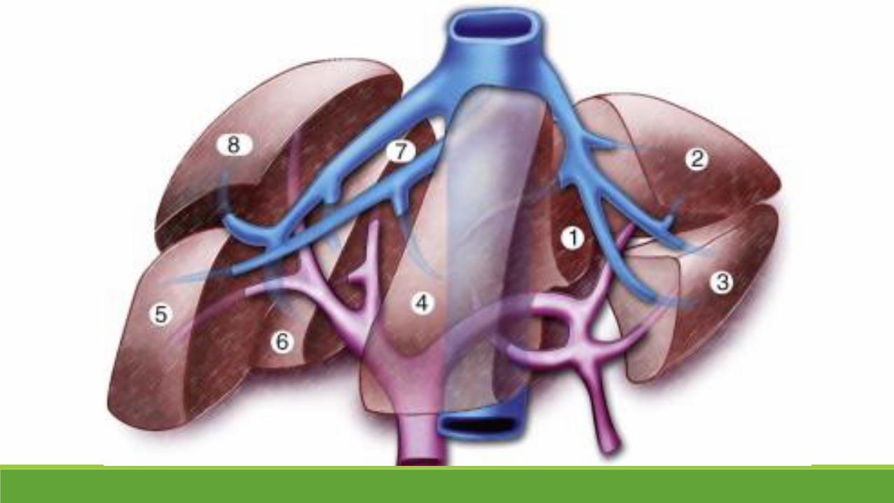
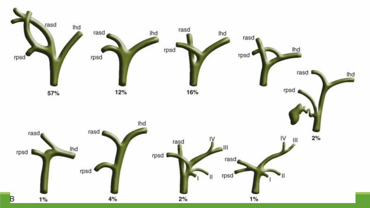
•Variations in biliary tract anatomy

Initial Access
Right-sided Left-sided
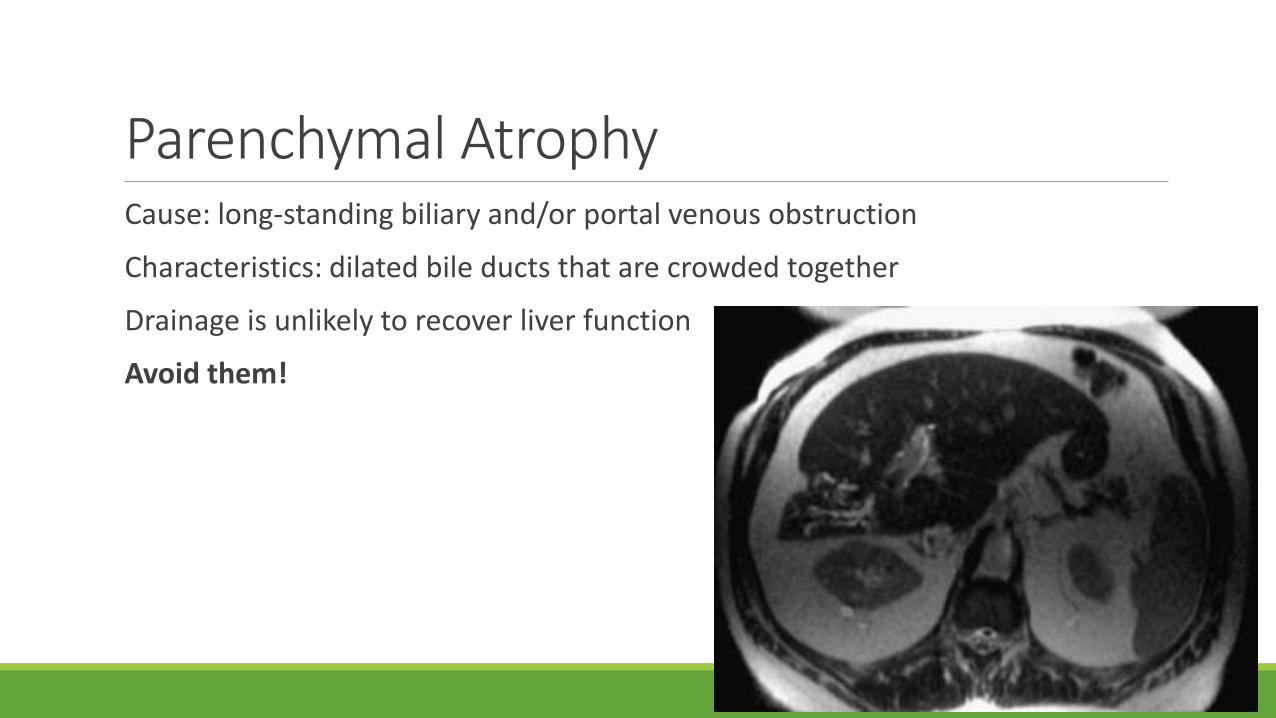
Parenchymal AtrophyCause: long-standing biliary and/or portal venous obstruction
Characteristics: dilated bile ducts that are crowded together
Drainage is unlikely to recover liver function
Avoid them!
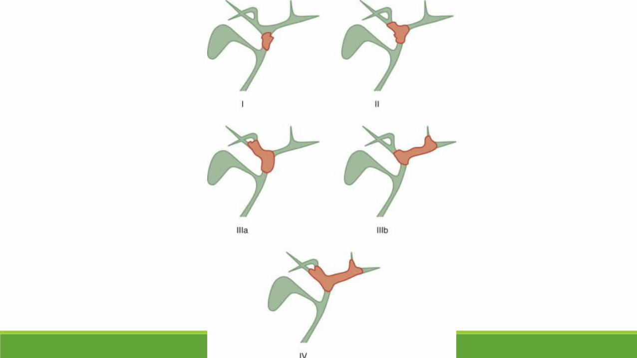
Portal Vein Status
Only liver segments with patent portal venous supply should be targeted during biliary drainage
Preprocedural Preparation: LabsINR 1.5 or less
Platelet count of at least 50,000/dL (PSU 80,000/dL)
Normal PTT
◦ Elevated INR◦ Vitamin K--elective procedure that can be delayed until the coagulations normalize
◦ Fresh frozen plasma--urgent or emergent evaluation
◦ Low platelet count◦ Platelet transfusions
Preprocedural PreparationHydration
Prophylactic antibiotics◦ Transient bacteremia--to minimize the risk of sepsis or abscess formation
◦ Patients with biliary-enteric anastomoses/dysfunctional sphincter of Oddi(sphincterotomy or endoscopic stent placement)
◦ More susceptible to infectious complications secondary to colonization of the biliary tree and infection of the bile
◦ Ampicillin-gentamicin, piperacillin/tazobactam, ampicillin/sulbactum, or a fluoroquinolone (ciprofloxacin or Levaquin)
Sedation and analgesia Elective: moderate sedation◦ Narcotics (fentanyl citrate) and benzodiazepines (midazolam or
diazepam) +/- meperidine◦ Clear liquid diet for a minimum of 2 hours before the procedure
Urgent or emergent intervention ◦ Hemodynamic instability making procedural management more
complicated◦ Monitoring or sedation by an anesthesiologist may be necessary
Patient PreparationA supine or a slight RAO position, preferably with the arm elevated above the head or extended to the side◦ allow wide access to the right hepatic lobe
US◦ Help to determine the optimal route for access of the biliary tree
Skin sterilized and draped
Sedation agents should be given before attempting to access the biliary system◦ Patient comfort ◦ Cephalad migration of the liver may occur because of decreased tidal volumes
Technique
Choose the access route
Lidocaine to the skin, subcutaneous tissues, and the hepatic capsule
Right-sided Access
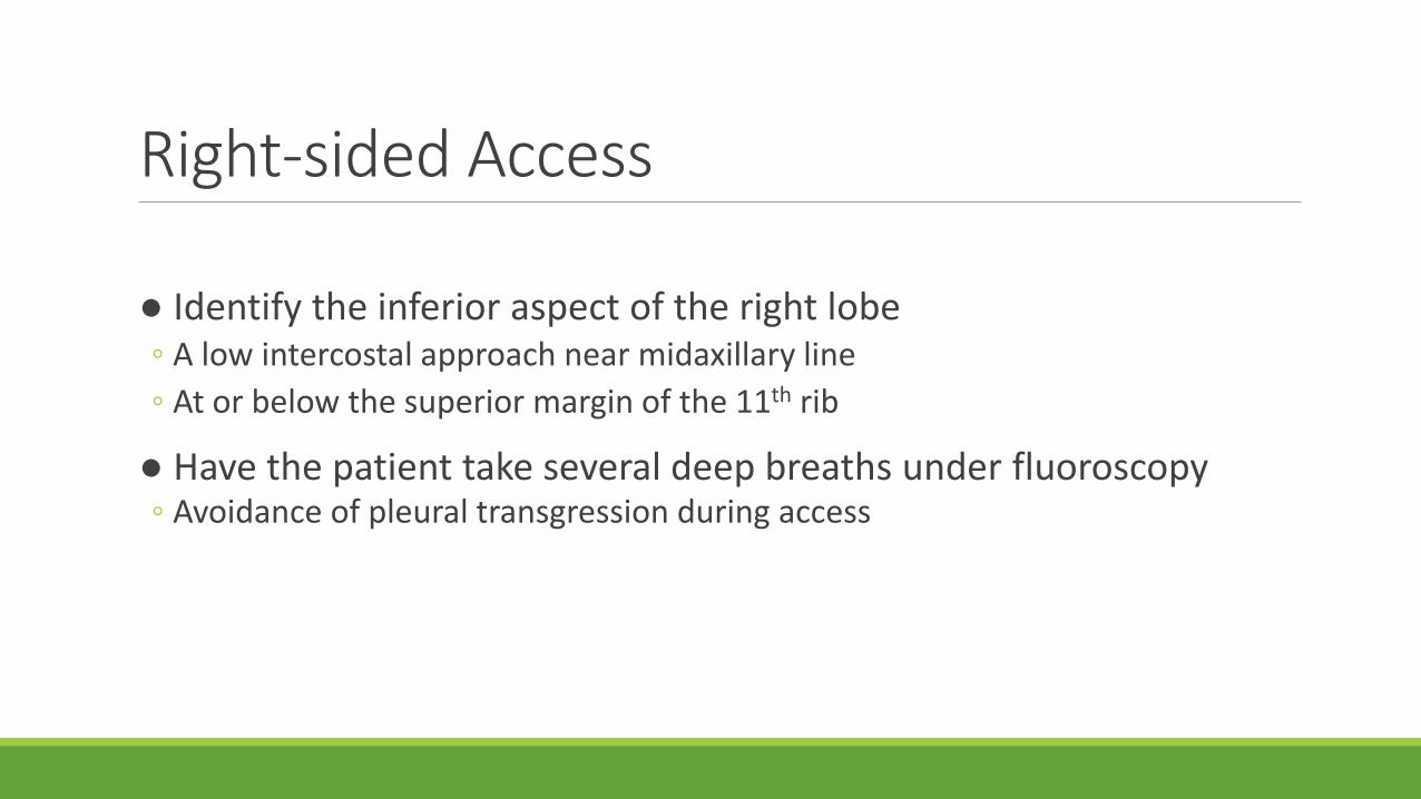
● Identify the inferior aspect of the right lobe ◦ A low intercostal approach near midaxillary line
◦ At or below the superior margin of the 11th rib
● Have the patient take several deep breaths under fluoroscopy◦ Avoidance of pleural transgression during access
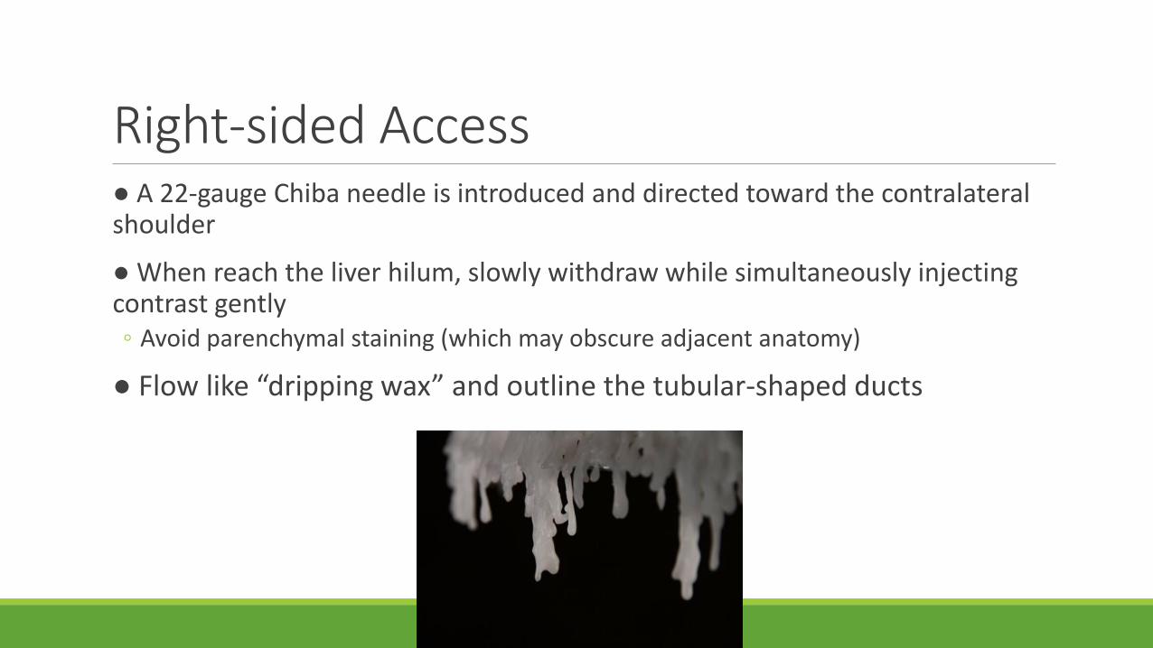
Right-sided Access● A 22-gauge Chiba needle is introduced and directed toward the contralateral shoulder
● When reach the liver hilum, slowly withdraw while simultaneously injecting contrast gently ◦ Avoid parenchymal staining (which may obscure adjacent anatomy)
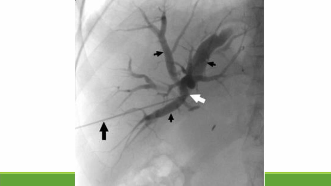
● Flow like “dripping wax” and outline the tubular-shaped ducts
Left-sided AccessA short needle ◦ Left lobe biliary ducts are anterior and close to the anterior abdominal wall
US may be used to identify an enlarged duct and provide guidance for initial needle placement
The needle is inserted just lateral to the xiphoid at the costal margin
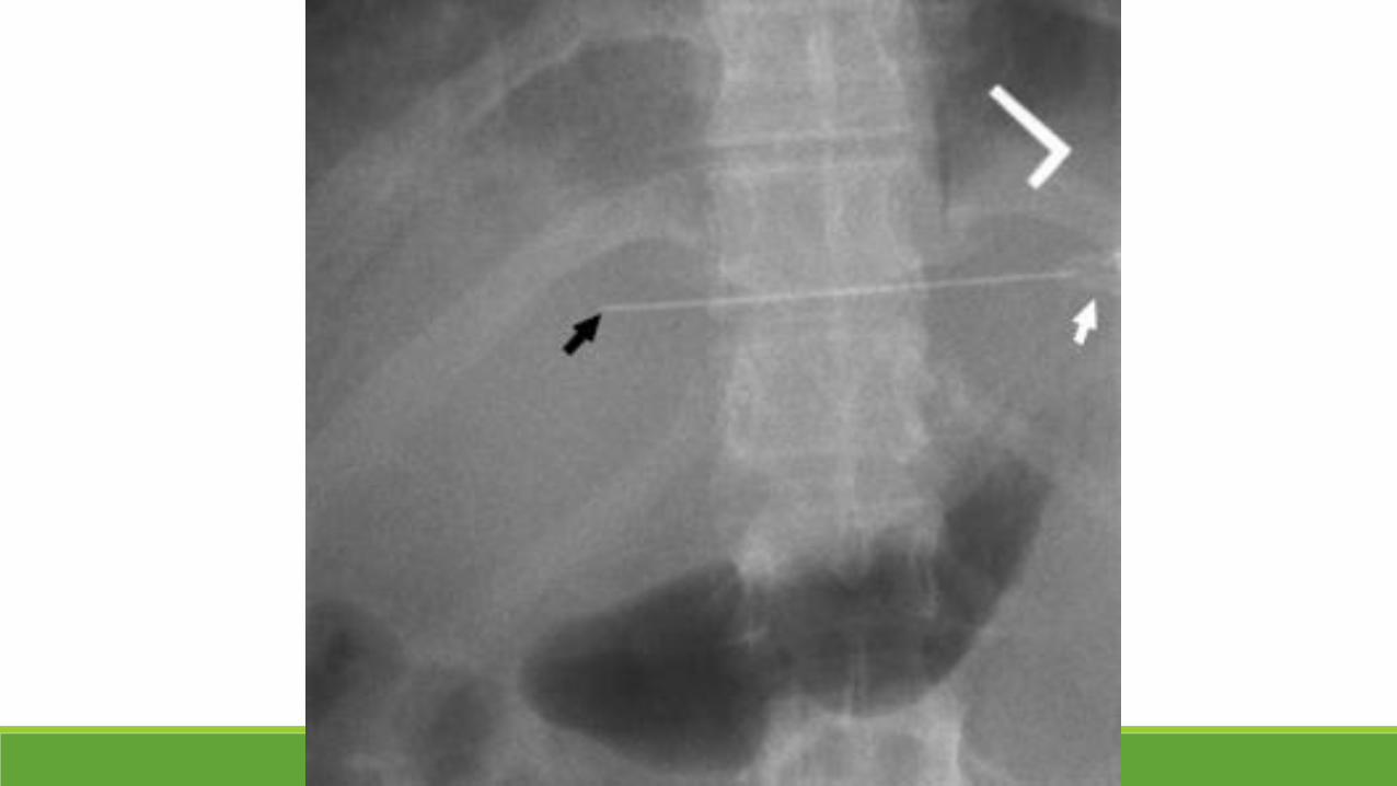
Initial Access
Aspirate a small amount of bile for microscopy and culture
Do not overinject the biliary system, particularly in the presence of obstruction◦ Possibility of inducing sepsis
After needle access• Adequate opacification of the ductal system
• Select a suitable duct ◦ Inferior ducts are preferred
◦ For avoidance of pleural transgression, especially on the right
◦ Relatively straight course toward the hilum◦ Facilitate the passage of a guidewire and catheter
◦ Access the duct peripherally ◦ Decrease the chance of injury to the larger vascular structures near the
hilum
1-stick technique
• A 0.018- inch wire can be introduced
• Wire advanced needle removed dilator replaced
• Extreme care passing dilators over a guidewire• One should advance the dilator under fluoroscopy
1-stick technique
0.018-inch stiffer 0.038-inch wire◦ Most are a triaxial design
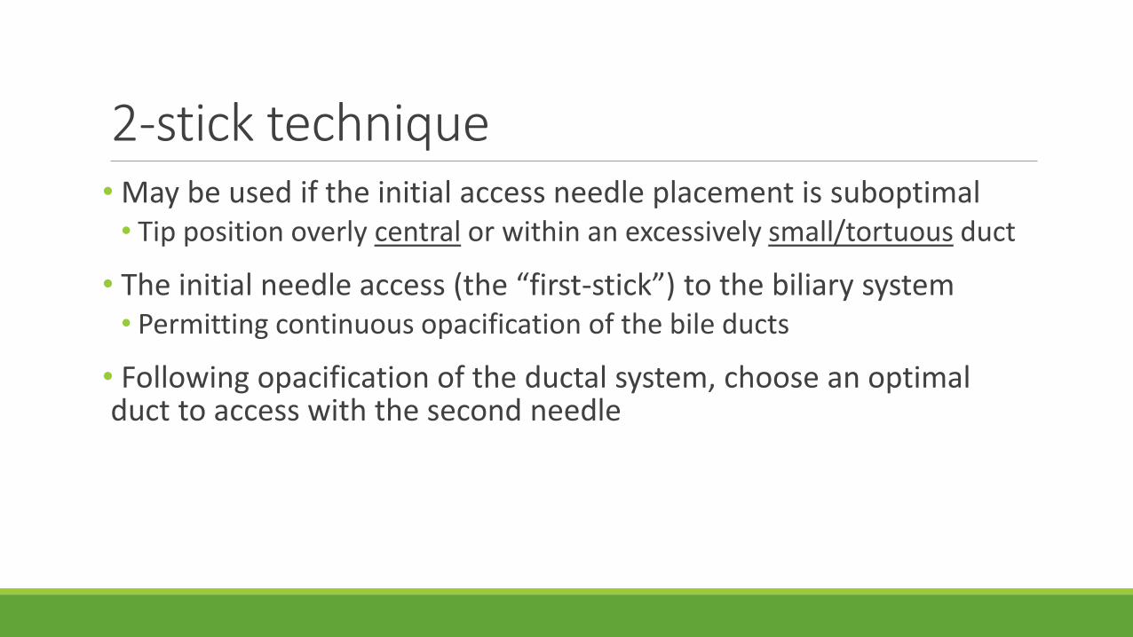
2-stick technique• May be used if the initial access needle placement is suboptimal • Tip position overly central or within an excessively small/tortuous duct
• The initial needle access (the “first-stick”) to the biliary system• Permitting continuous opacification of the bile ducts
• Following opacification of the ductal system, choose an optimal duct to access with the second needle
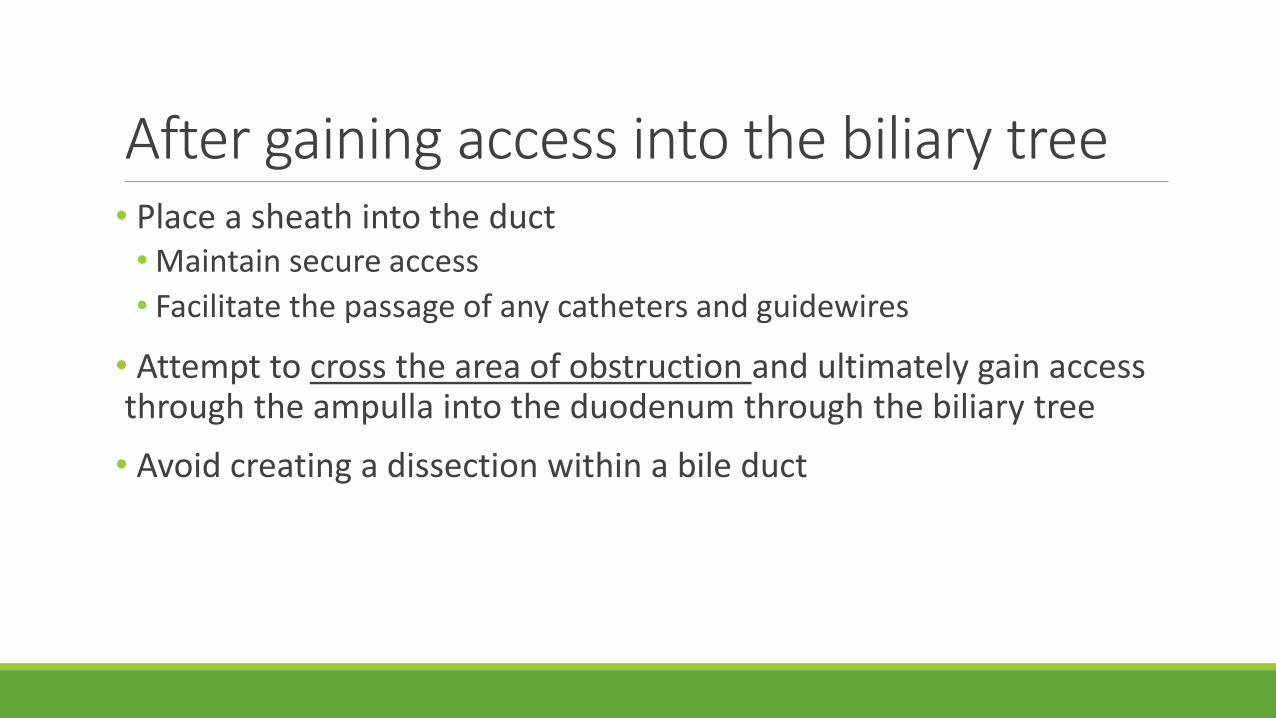
After gaining access into the biliary tree• Place a sheath into the duct• Maintain secure access
• Facilitate the passage of any catheters and guidewires
• Attempt to cross the area of obstruction and ultimately gain access through the ampulla into the duodenum through the biliary tree
• Avoid creating a dissection within a bile duct
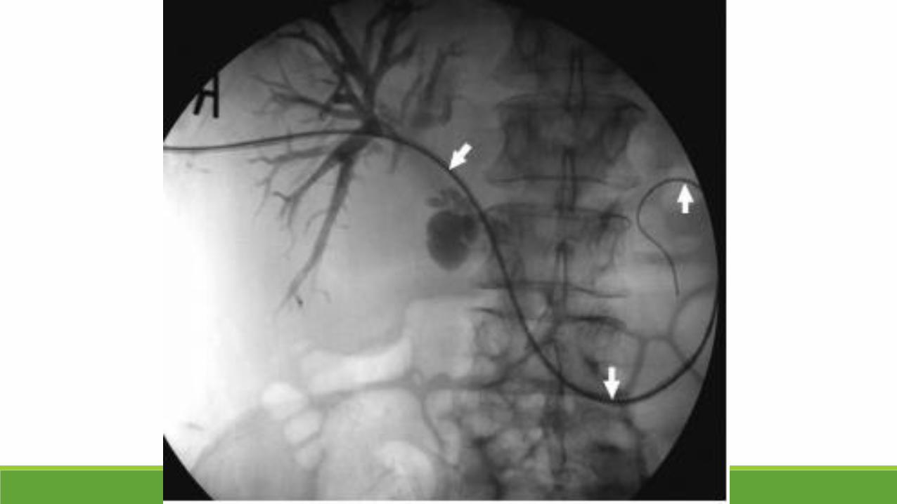

Drainage catheter
INTERNAL-EXTERNAL
Allows bile to drain ◦ externally into a bag
◦ internally into the small bowel
Preserving the normal enterohepatic circulation of bile
EXTERNAL
Unable to advance a catheter through an obstruction and into the duodenum
Drainage catheter• 8 to 14 Fr with multiple side holes
• A radiopaque marker proximal extent of the side holes
• A “pigtail” distal/internal end of the catheter with a locking mechanism to maintain the position in bowel/ducts
• In position which the side holes will drain all the intrahepatic ducts
• Confirmed with contrast injection
• If there are any ducts that are not adequately drained, placement of a second catheter may be required
Postprocedural Management• Routine postsedation monitoring for 2-3 hours
• Hospital admission in intensive care unit
• Beware of sepsis and/or hemorrhage
• Antibiotics should be continued and adjusted according to results of the Gram stain and cultures• Escherichia coli, enterococci, Klebsiella species, and Streptococcus viridans
Postprocedural Management• Postprocedural pain control • Discomfort, particularly if the route of access was via an intercostal space
• Pain will gradually decrease over 24-48 hours
• Use soft drainage catheter
• Catheters should be routinely flushed with 5-10 mL NSS every 8 hours to maintain patency
• Exchange q 3 months (2 months at PSU)
• Prior to removing any biliary drainage catheter, clamped for 24-48 hours• Assessment of the adequacy of internal drainage
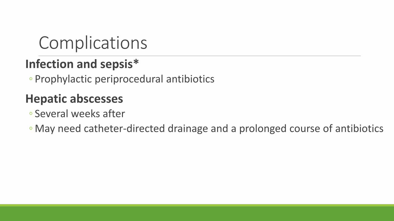
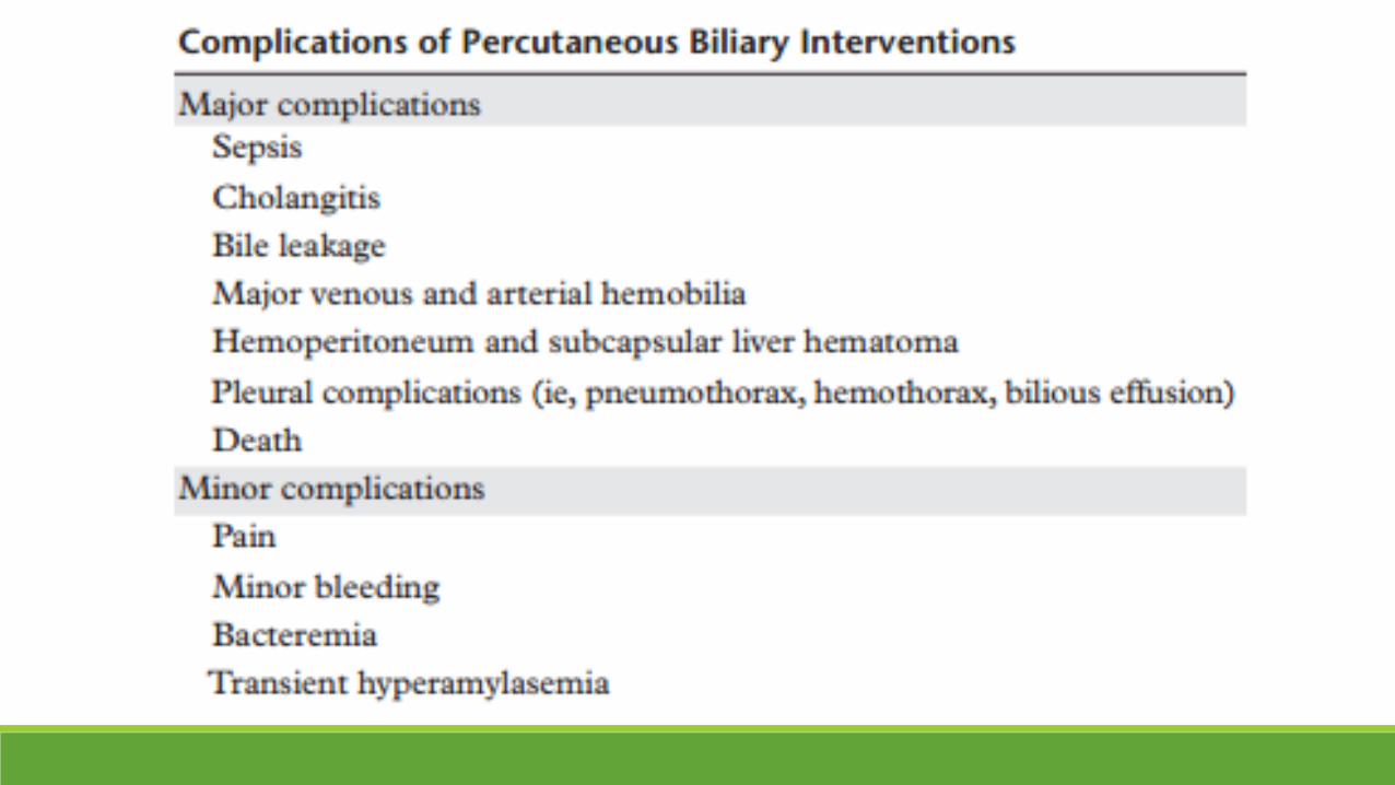
ComplicationsInfection and sepsis* ◦ Prophylactic periprocedural antibiotics
Hepatic abscesses ◦ Several weeks after
◦ May need catheter-directed drainage and a prolonged course of antibiotics
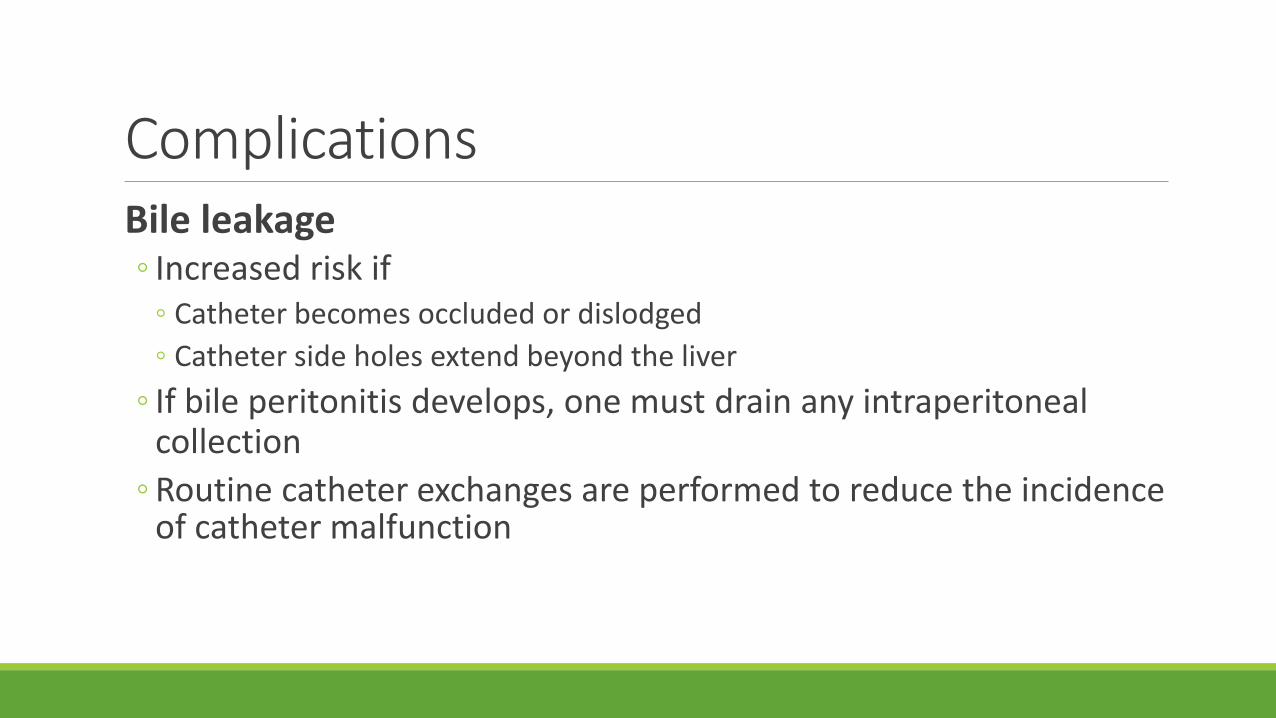
ComplicationsBile leakage ◦ Increased risk if
◦ Catheter becomes occluded or dislodged
◦ Catheter side holes extend beyond the liver
◦ If bile peritonitis develops, one must drain any intraperitoneal collection
◦ Routine catheter exchanges are performed to reduce the incidence of catheter malfunction
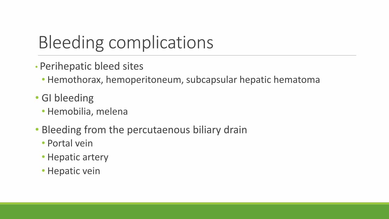
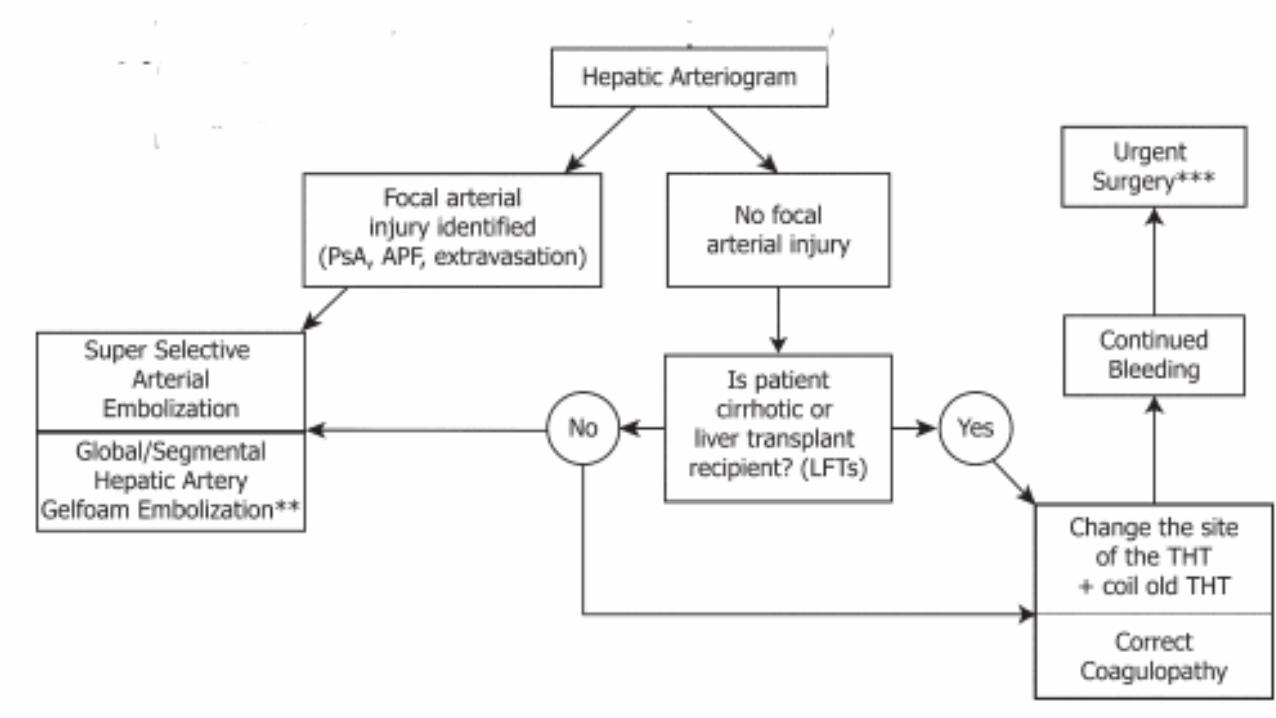
Bleeding complications• Perihepatic bleed sites• Hemothorax, hemoperitoneum, subcapsular hepatic hematoma
• GI bleeding• Hemobilia, melena
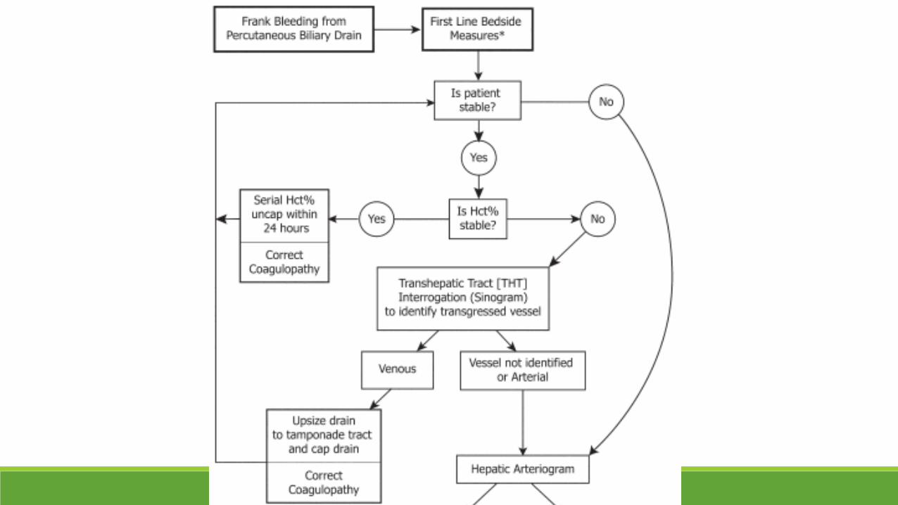
• Bleeding from the percutaenous biliary drain• Portal vein
• Hepatic artery
• Hepatic vein

Cholecystostomy Tube Placement
Percutaneous cholecystostomy
An alternative treatment method in patients with
acute cholecystitis
who are at high risk for surgery due to comorbid diseases
Indications for Percutaneous cholecystostomy
Critically ill patients with
calculous or acalculous cholecystitis
who are poor or nonoperative candidates
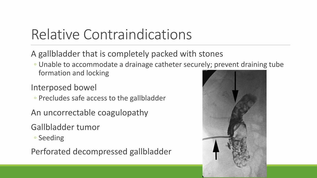
Relative ContraindicationsA gallbladder that is completely packed with stones◦ Unable to accommodate a drainage catheter securely; prevent draining tube
formation and locking
Interposed bowel◦ Precludes safe access to the gallbladder
An uncorrectable coagulopathy
Gallbladder tumor◦ Seeding
Perforated decompressed gallbladder
Preprocedure PreparationLabs and antibiotics
review of preprocedural imaging is mandatory
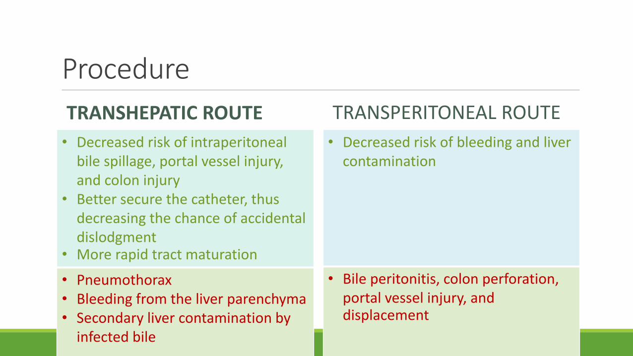
Procedure
TRANSHEPATIC ROUTE
• Decreased risk of intraperitoneal bile spillage, portal vessel injury, and colon injury
• Better secure the catheter, thus decreasing the chance of accidental dislodgment
• More rapid tract maturation
• Pneumothorax• Bleeding from the liver parenchyma• Secondary liver contamination by
infected bile
TRANSPERITONEAL ROUTE
• Decreased risk of bleeding and liver contamination
• Bile peritonitis, colon perforation, portal vessel injury, and displacement
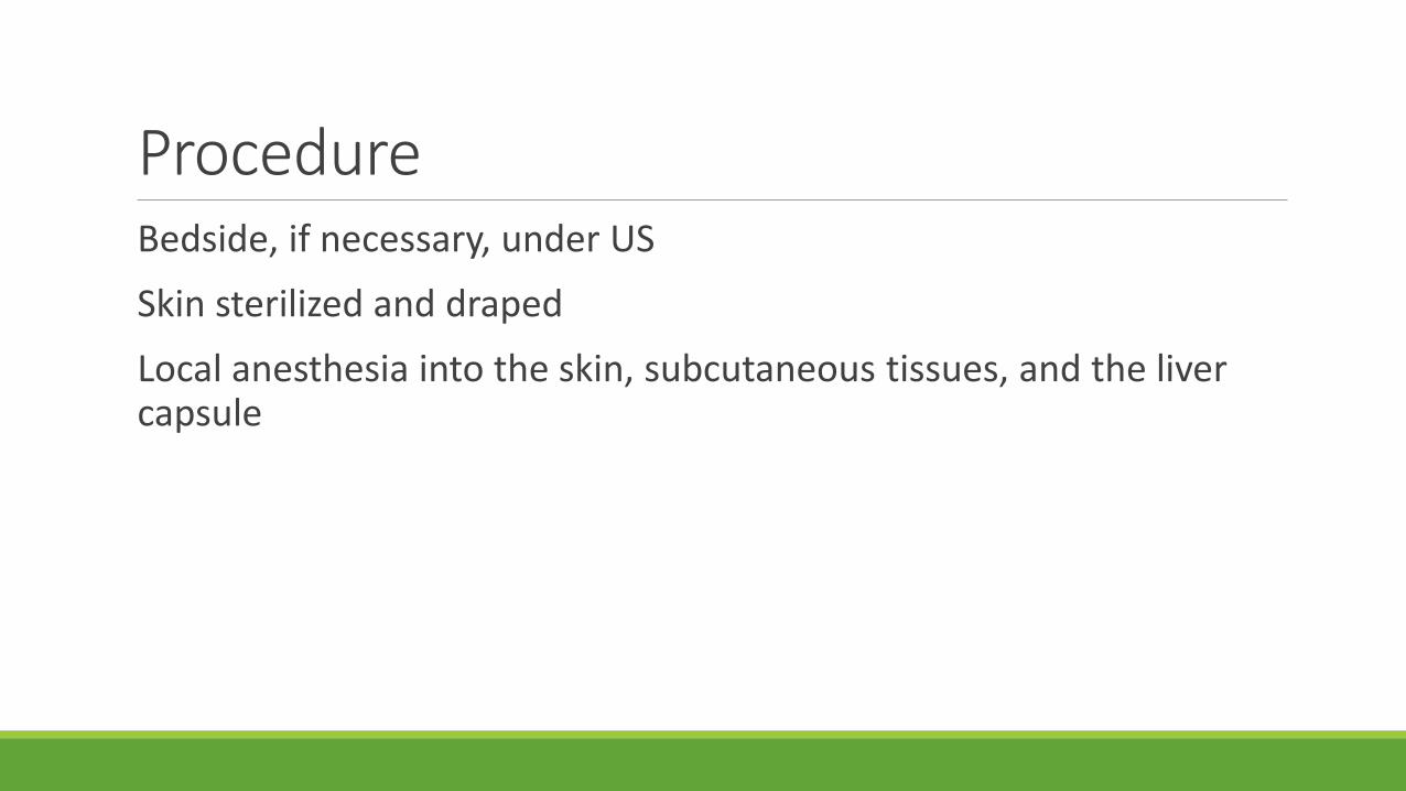
ProcedureBedside, if necessary, under US
Skin sterilized and draped
Local anesthesia into the skin, subcutaneous tissues, and the liver capsule
Placing a cholecystostomy tubeOften useful to mark the estimated depth that the catheter should be advanced into the gallbladder
Either a Trocar or a Seldinger’s technique
Trocar techniqueSuited to those with a large distended gallbladder that can be easily visualized with a clear percutaneous window
An 8F to 10F self-retaining/locking all-purpose drain
Remove the stylet and sample fluid confirming the position◦ If bile is aspirated, the catheter is advanced off the trocar and fixed in the
gallbladder and deployed
◦ If no bile is aspirated, the system is retracted while continuously exerting suction
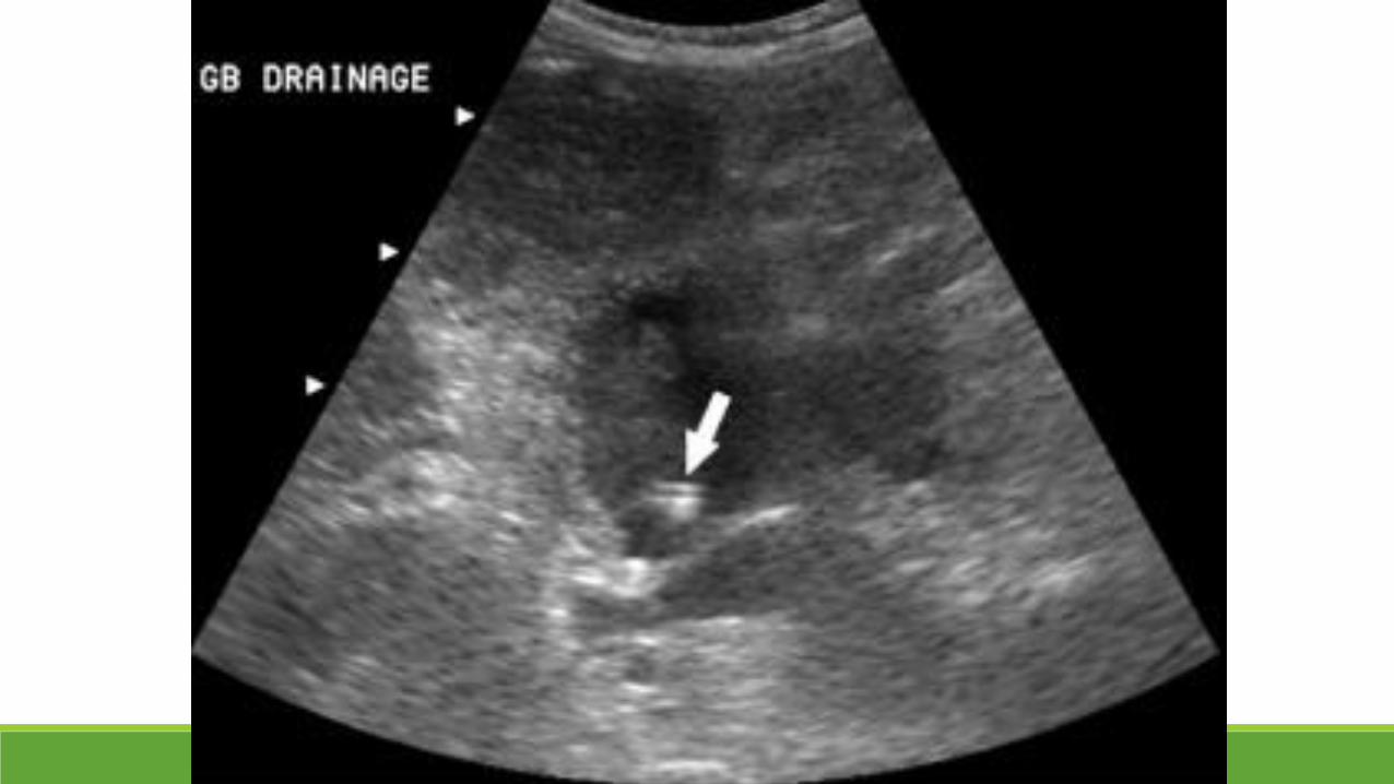
Seldinger’s techniqueBetter suited for difficult access or a small gallbladder
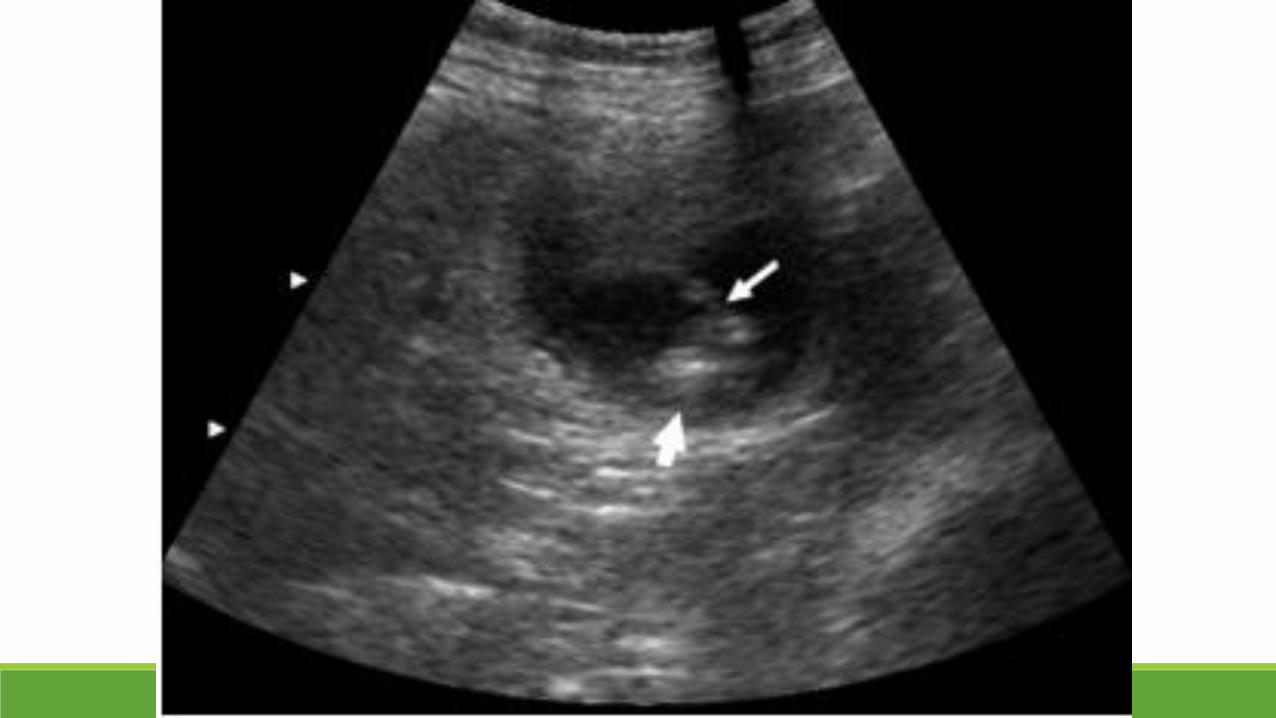
A small needle, such as a 18G to 22G Chiba or a Ring needle, is advanced under ultrasound into the gallbladder
A 0.018-inch guidewire is introduced followed by a triaxial set
A stiff working guidewire must be introduced into the gallbladder
Serial dilation
All these wire placement and dilation should be performed using fluoroscopy to avoid inadvertent perforation of the gallbladder
Following dilation, the catheter can be deployed over the wire, using extreme care not to advance the catheter into the gallbladder with the metal inner stiffener still in place
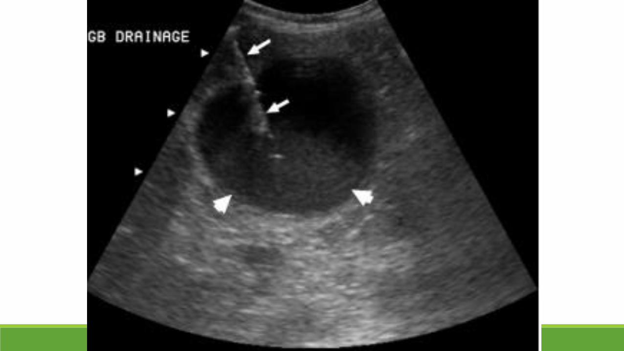
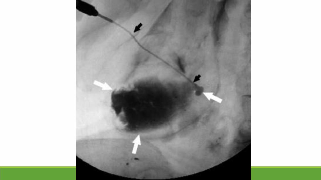
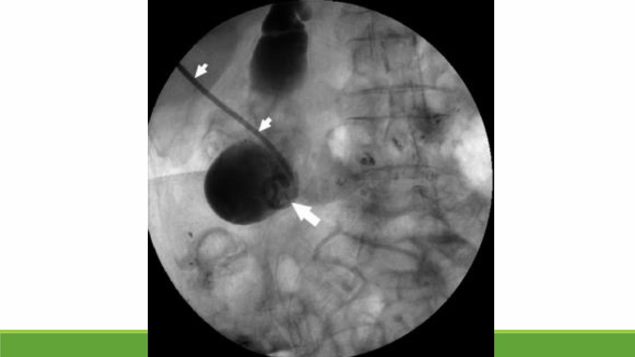
Seldinger’s techniqueSample of bile sent for laboratory analysis◦ Gram stain and both aerobic and anaerobic cultures
Bile aspiration◦ Gallbladder decompression
Contrast injection via the catheter◦ Demonstrate the final catheter position and complete decompression
Do not overinject◦ Bacteremia ◦ Possible gallbladder perforation
Postprocedural Managementroutine monitoring for signs of hemorrhage, worsening sepsis, or other clinical deterioration
Antibiotics according to Gram stain and culture
The catheter◦ Placed to gravity drainage◦ periodic catheter flushing using 5-10 mL NSS
Removal of the catheterDepends on the indication for placement, the clinical condition of the patient, and the time interval since placement
◦ Calculous cholecystitisuntil cholecystectomy◦ In rare circumstances, demonstration of patent cystic and common ducts, and a small or
absent stone burden, may allow for catheter removal without cholecystectomy
◦ Persistent cholelithiasis in non-surgical candidatespercutaneous stone removal/lithotripsy
◦ Acalculous cholecystitis = definitive treatment
Removal of the catheter
• 3 to 6 weeks before removal• Mature tract forms
• A tube cholangiogram•Demonstrate cystic and common duct patency
• Tube should be clamped for 24-48 hours before tube removal•To demonstrate patient tolerance
SuccessTechnical success rates for percutaneous cholecystostomy◦ 95% to 100%
Technical failures tend to occur in ◦ Decompressed gallbladders
◦ Impacted stones and porcelain gallbladders◦ Significant gallbladder wall thickening
ComplicationsBile leakage◦ Transhepatic route to prevent bile
leakage and painful bile peritonitis
Pneumothorax and sympathetic pleural effusion◦ Pleura traversing and any bile
leakage into the pleural space
Accidental gallbladder perforation ◦ Acutely inflamed and friable
Colonic transgression◦ Review of preprocedural imaging
Large hepatic vessel injury◦ Color- flow Doppler ultrasound
Bleeding◦ Usually self-limited
Conclusions: Percutaneous biliary tract interventionsDiagnostic and therapeutic option
Alternative for critically ill patients in whom endoscopic intervention has failed or in whom surgery is a poor option
Proper patient assessment and preparation
Indications and contraindications
Techniques
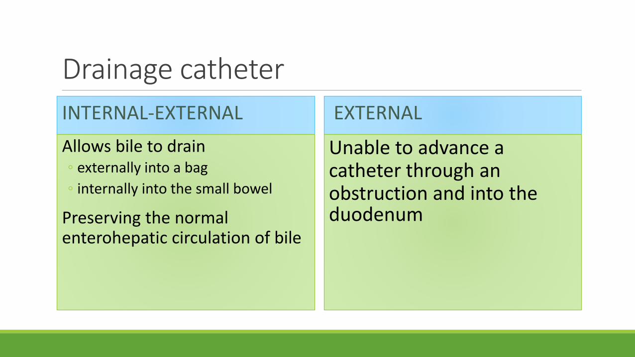
Drainage catheter
INTERNAL-EXTERNAL
Allows bile to drain ◦ externally into a bag
◦ internally into the small bowel
Preserving the normal enterohepatic circulation of bile
EXTERNAL
Unable to advance a catheter through an obstruction and into the duodenum
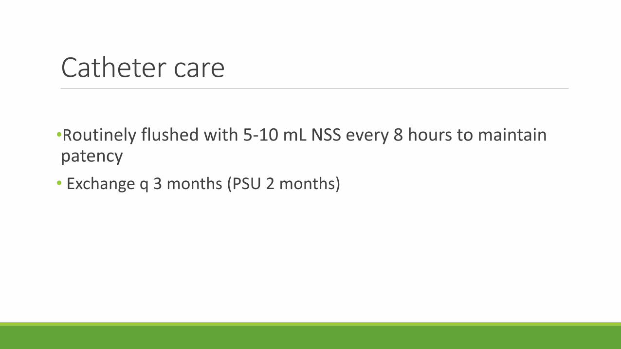
Catheter care
•Routinely flushed with 5-10 mL NSS every 8 hours to maintain patency
• Exchange q 3 months (PSU 2 months)
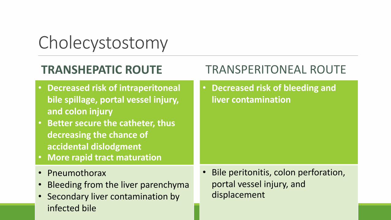
Cholecystostomy
TRANSHEPATIC ROUTE
• Decreased risk of intraperitoneal bile spillage, portal vessel injury, and colon injury
• Better secure the catheter, thus decreasing the chance of accidental dislodgment
• More rapid tract maturation
• Pneumothorax• Bleeding from the liver parenchyma• Secondary liver contamination by
infected bile
TRANSPERITONEAL ROUTE
• Decreased risk of bleeding and liver contamination
• Bile peritonitis, colon perforation, portal vessel injury, and displacement