Upper GI Histopathology Update Dr David Cundell ST4 Histopathology, BRI.
Upload
elwin-bennettCategory
view
217download
0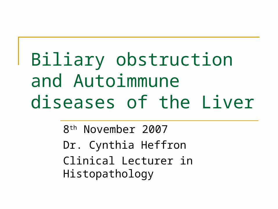
Biliary obstruction and Autoimmune diseases of the Liver
8th November 2007
Dr. Cynthia Heffron
Clinical Lecturer in Histopathology
Biliary drainage system
Canaliculi (between abutting hepatocytes) 1-2um in diameter
Canals of Hering & cholangioles Canaliculi join to form these larger structures
Intra-hepatic bile ducts Extra-hepatic bile ducts
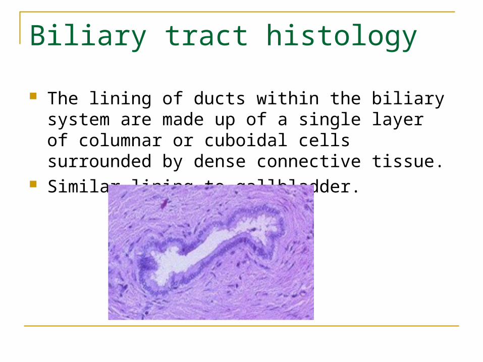
Biliary tract histology
The lining of ducts within the biliary system are made up of a single layer of columnar or cuboidal cells surrounded by dense connective tissue.
Similar lining to gallbladder.
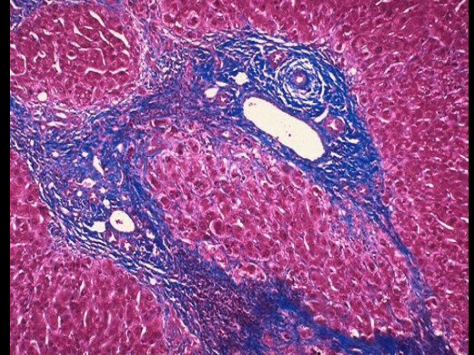
Biliary obstruction
Intrahepatic Primary biliary cirrhosis Primary sclerosing cholangitis
Extrahepatic Biliary atresia Gallstones Strictures Carcinoma of pancreatic head
PRIMARY BILIARY CIRRHOSIS
Definition: A chronic cholestatic disease due to a non-suppurative destructive
cholangitis of intrahepatic bile ducts, immune mediated. Epidemiology:
F:M as 10:1. Age range 20-80years, peak at 40-50. Clinical features:
May be associated with other autoimmune diseases. Very insidious onset, may be asymptomatic for decades, pruritis,
fatigue, xanthelasmas, leading to frank cholestatic jaundice, cirrhosis. Major cause of death is liver failure. Liver transplantation is the definitive treatment.
90-95% positive antimitochondrial antibodies (AMA) against E2 subunit of pyruvate dehydrogenase complex inner mitochondrial membrane.
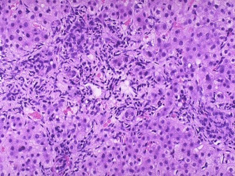
Histology: Characteristically a granulomatous destruction of bile
ducts. Leads to ductopenia. Copper accumulates in periportal hepatocytes due to
chronic cholestasis. Chronic inflammation in portal tracts with interface
hepatitis. Portal fibrosis progresses to cirrhosis.
Liver biopsy used to confirm diagnosis and to stage the disease.

PRIMARY SCLEROSING CHOLANGITIS
Definition: A chronic cholestatic disease due to a non-specific inflammatory obliterative
fibrosis of bile ducts, intrahepatic and extrahepatic.
Epidemiology: M:F as 2:1. Affects mostly young men. Third to fifth decades.
Clinical features: 70% have ulcerative colitis. Linkage with HLAB8, DR2, DR3. pANCA+ May be associated with other rare fibrosing conditions. May be asymptomatic, pruritis and cholestatic jaundice and then cirrhosis
develop over many years. Characteristic beading on barium radiology studies. 10% at risk of developing cholangiocarcinoma.
Aetiology: Unknown.
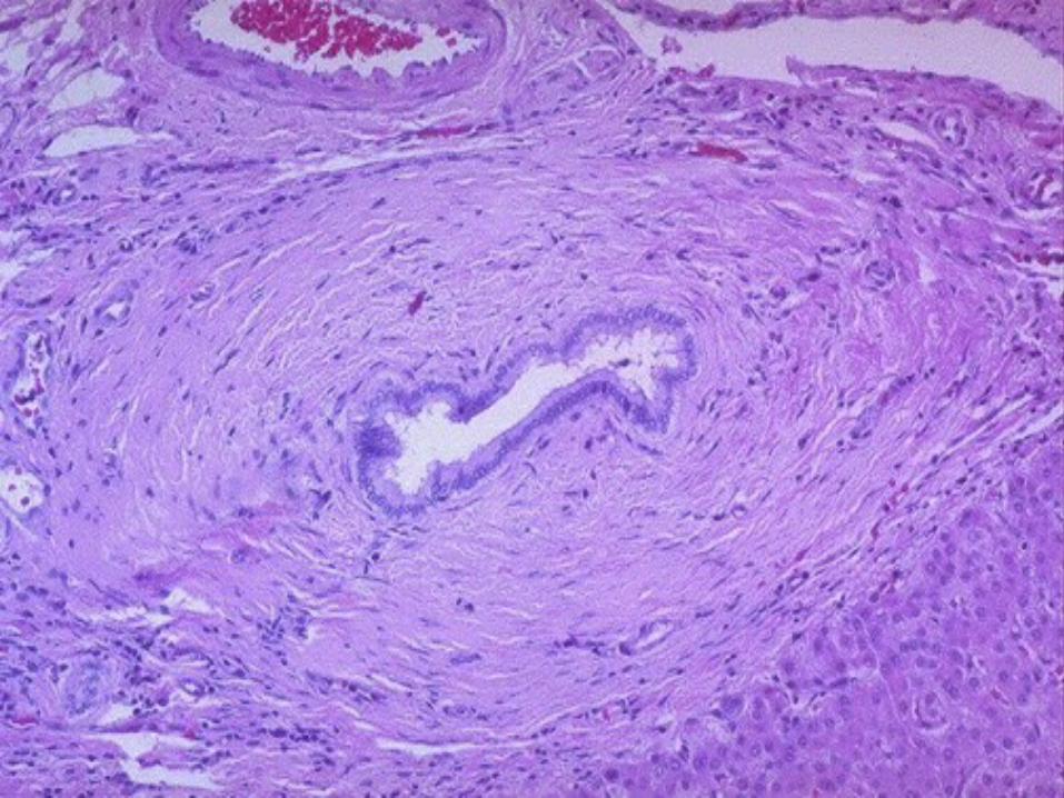
Histology: Concentric fibrosis of bile ducts; may result in a
scar at site of duct, “onion skin” fibrosis. Leads to ductopenia. Copper accumulates in periportal hepatocytes. Usually scanty lymphocytic infiltrate. Portal fibrosis progresses to cirrhosis.
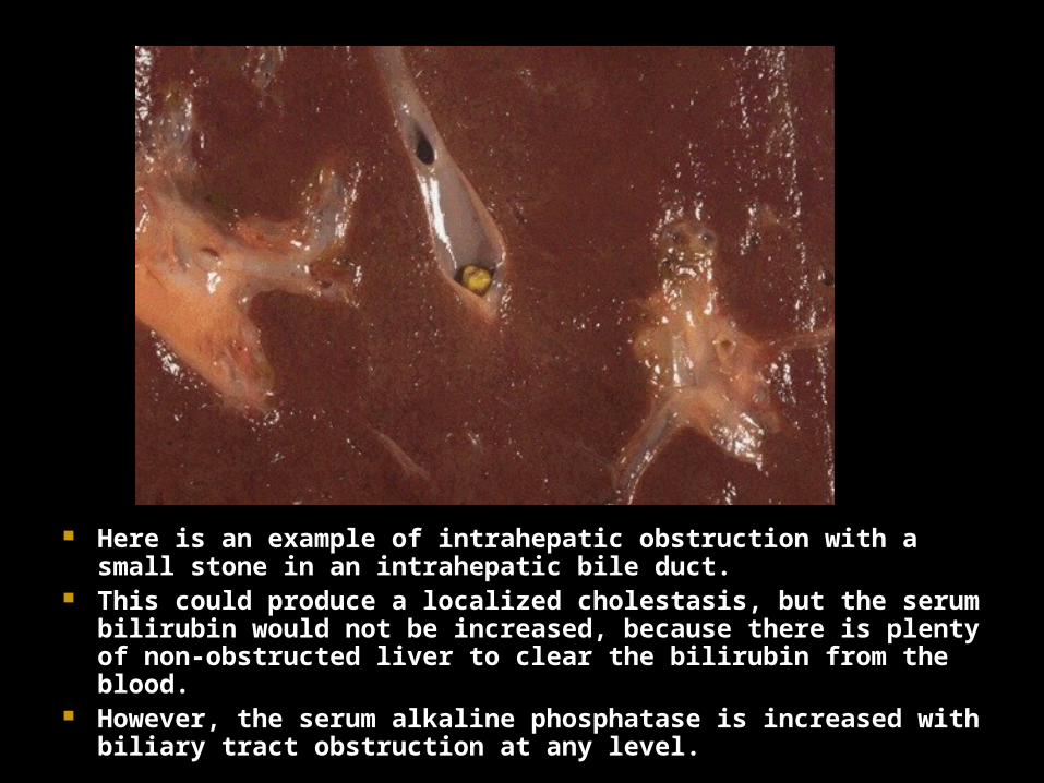
Here is an example of intrahepatic obstruction with a small stone in an intrahepatic bile duct.
This could produce a localized cholestasis, but the serum bilirubin would not be increased, because there is plenty of non-obstructed liver to clear the bilirubin from the blood.
However, the serum alkaline phosphatase is increased with biliary tract obstruction at any level.
Extrahepatic biliary obstruction
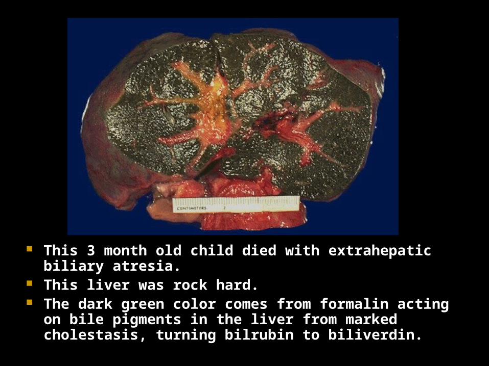
This 3 month old child died with extrahepatic biliary atresia.
This liver was rock hard. The dark green color comes from formalin acting on bile
pigments in the liver from marked cholestasis, turning bilrubin to biliverdin.
Extrahepatic biliary atresia
Defined as a complete obstruction of bile flow due to destruction or absence of all or part of the extrahepatic bile ducts.
Undergo progressive inflammation with stricture of hepatic or common bile ducts within weeks of birth.
Accounts for one third of neonatal cholestasis. Diagnosed on biopsy. Treated surgically
Kasai procedure Hepatic portoenterostomy performed early in life, before 10
weeks of age. Transplantation also considered as a treatment option.
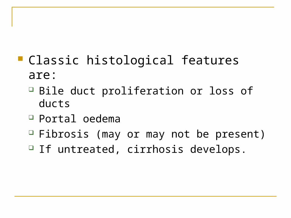
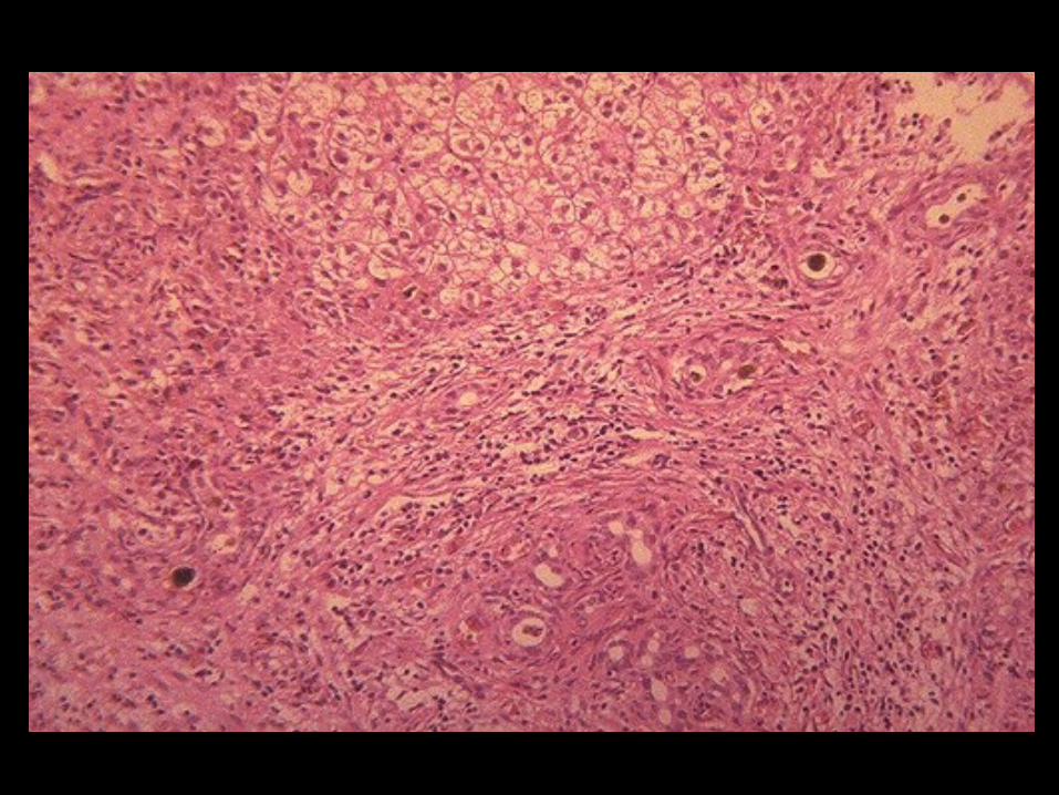
Classic histological features are: Bile duct proliferation or loss of ducts Portal oedema Fibrosis (may or may not be present) If untreated, cirrhosis develops.
Cause unknown. Viral Exposure to enviromental toxins Disordered iummunity Defective morphogenesis of bile ducts
Surgical intervention required at an early stage.
If unrecognised or uncorrected, cirrhosis develops within 3-6 months.
MISCELLANEOUS BILE DUCT DISEASES
Acquired sclerosing cholangitis can occur in a number of conditions including AIDS.
Bile duct injury can also occur with liver allografts, graft-versus-host-disease (GVHD), viral hepatitis, drugs, toxins, pyogenic infections, parasitic infestations and other rare conditions.
Definition: Syndrome of chronic hepatitis in patients with a heterogeneous set of
immunologic abnormalities. Epidemiology:
More common in females (78%) than in males. Any age but especially young women and perimenopausal women.
Clinical features: Up to 60% have other forms of autoimmune disease eg rheumatoid
arthritis, thyroiditis, Sjogren’s syndrome, ulcerative colitis. HLA B8 and HLA DR3 frequent. High titres of IgG and antibodies to various bacterial and viral antigens. High titres of autoantibodies - antinuclear (ANA), antismooth muscle
(SMA), and/or antiliver/kidney microsomes (anti-LKM1) antibodies. Present similar to other forms of chronic hepatitis.
Autoimmune Hepatitis
Autoimmune Hepatitis
Pathogenesis: Reduced number and function of suppressor T lymphocytes. Is a cytotoxic T-cell attack on hepatocytes.
Histology: Severe chronic hepatitis with marked interface hepatitis and
numerous plasma cells in the inflammatory infiltrate. Appearances not specific to autoimmune hepatitis so careful
clinical correlation required. Treatment/Prognosis:
Responds well to steroids. Liver transplantation may be necessary. Untreated 40% die of liver failure in 6months. 40% of survivors develop cirrhosis.
DRUG,TOXIC AND METABOLIC DISORDERS
DRUGS AND THE LIVER
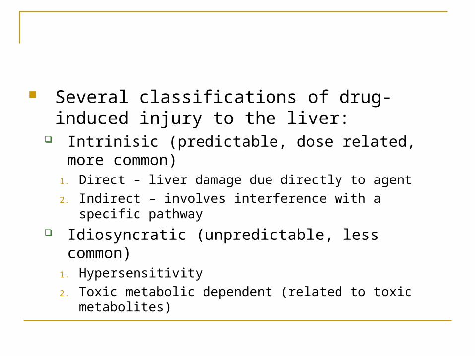
Several classifications of drug-induced injury to the liver:
Intrinisic (predictable, dose related, more common)
1. Direct – liver damage due directly to agent
2. Indirect – involves interference with a specific pathway Idiosyncratic (unpredictable, less common)
1. Hypersensitivity
2. Toxic metabolic dependent (related to toxic metabolites)
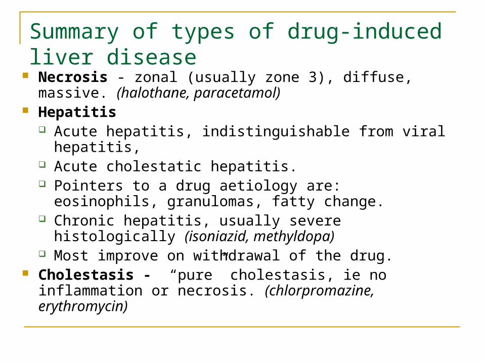
Summary of types of drug-induced liver disease
Necrosis - zonal (usually zone 3), diffuse, massive. (halothane, paracetamol)
Hepatitis Acute hepatitis, indistinguishable from viral hepatitis, Acute cholestatic hepatitis. Pointers to a drug aetiology are: eosinophils,
granulomas, fatty change. Chronic hepatitis, usually severe histologically
(isoniazid, methyldopa) Most improve on withdrawal of the drug.
Cholestasis - “pure” cholestasis, ie no inflammation or necrosis. (chlorpromazine, erythromycin)
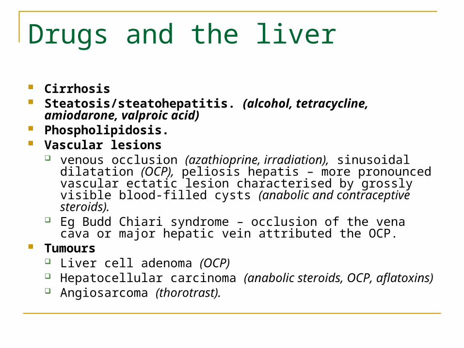
Drugs and the liver
Cirrhosis Steatosis/steatohepatitis. (alcohol, tetracycline, amiodarone,
valproic acid) Phospholipidosis. Vascular lesions
venous occlusion (azathioprine, irradiation), sinusoidal dilatation (OCP), peliosis hepatis – more pronounced vascular ectatic lesion characterised by grossly visible blood-filled cysts (anabolic and contraceptive steroids).
Eg Budd Chiari syndrome – occlusion of the vena cava or major hepatic vein attributed the OCP.
Tumours Liver cell adenoma (OCP) Hepatocellular carcinoma (anabolic steroids, OCP, aflatoxins) Angiosarcoma (thorotrast).
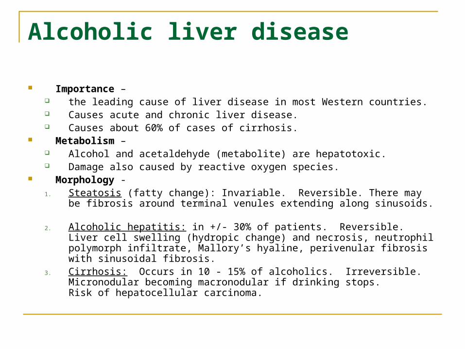
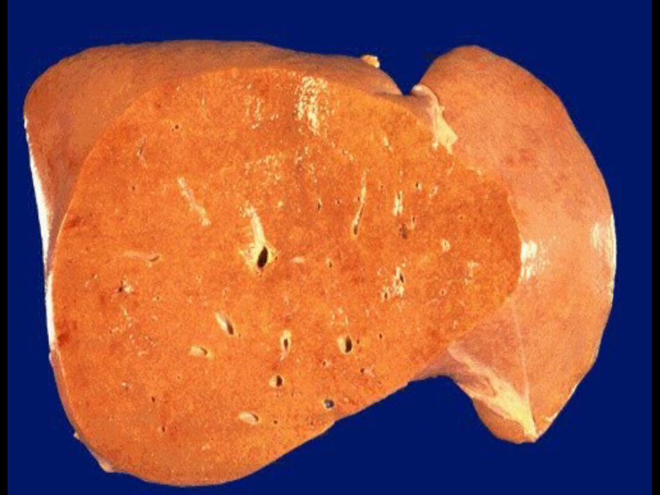
Alcoholic liver disease
Importance – the leading cause of liver disease in most Western countries. Causes acute and chronic liver disease. Causes about 60% of cases of cirrhosis.
Metabolism – Alcohol and acetaldehyde (metabolite) are hepatotoxic. Damage also caused by reactive oxygen species.
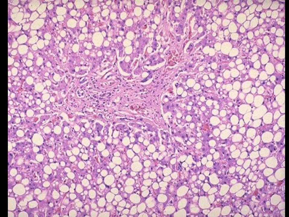
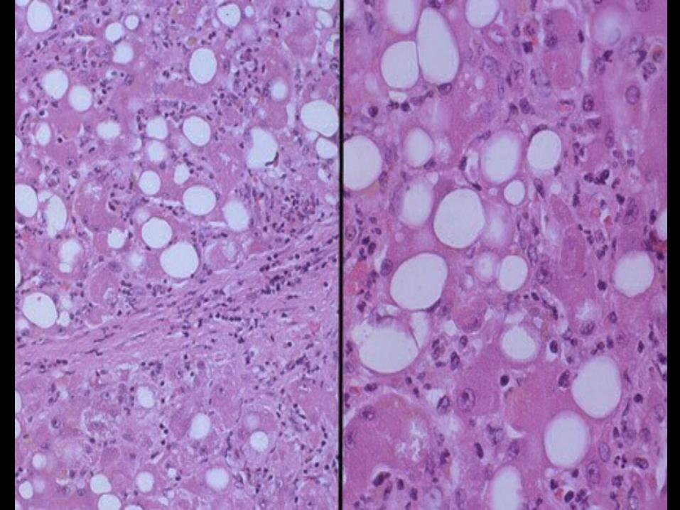
Morphology - 1. Steatosis (fatty change): Invariable. Reversible. There may be fibrosis around
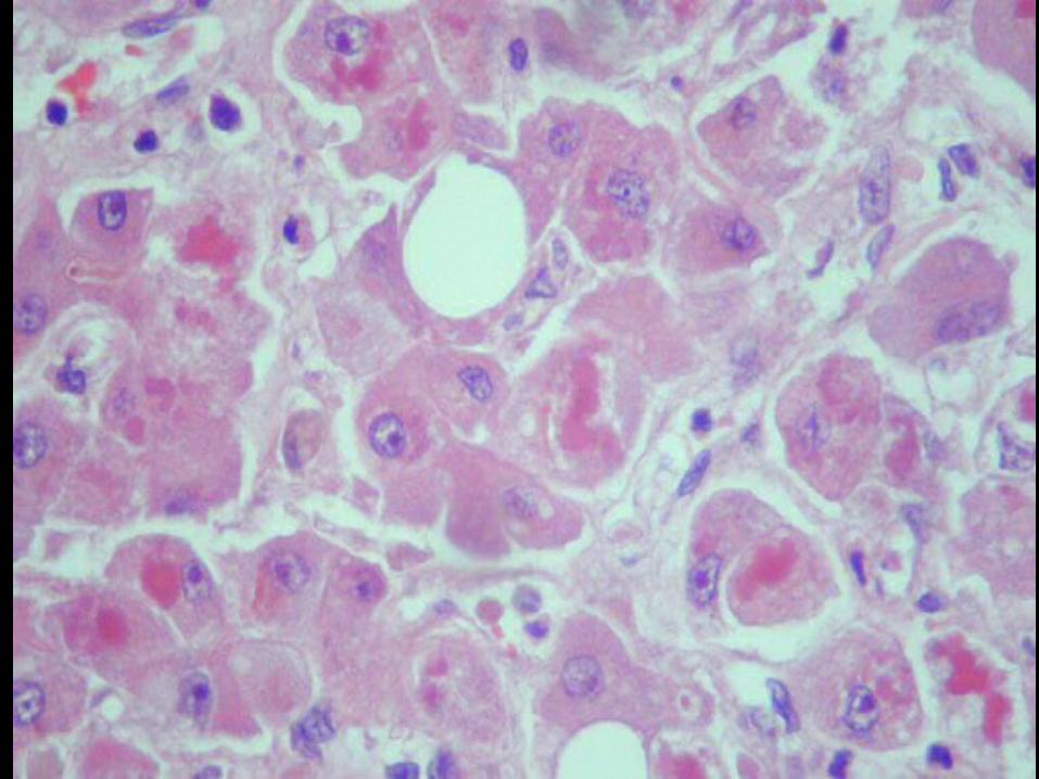
terminal venules extending along sinusoids.2. Alcoholic hepatitis: in +/- 30% of patients. Reversible. Liver cell swelling
(hydropic change) and necrosis, neutrophil polymorph infiltrate, Mallory’s hyaline, perivenular fibrosis with sinusoidal fibrosis.
3. Cirrhosis: Occurs in 10 - 15% of alcoholics. Irreversible. Micronodular becoming macronodular if drinking stops. Risk of hepatocellular carcinoma.

Alcoholic liver disease
Liver biopsy is required to distinguish the three morphological types of alcoholic liver disease. All three may have hepatomegaly. Steatosis tends to be mild clinically & biochemically; alcoholic hepatitis can be mild or have life-threatening liver failure. It may be superimposed on cirrhosis.
Factors influencing alcoholic liver disease. Dose and duration. Continuous worse than binge drinking. Immune reaction to neo-antigens from hepatocellular proteins. Female gender. Genetic polymorphisms. Nutritional deficiencies. Chronic viral infections. Drugs and toxins.
Pathogenesis
The relationship of steatosis, alcoholic hepatitis to cirrhosis is not clear.
Perivenular sinusoidal fibrosis due to hepatic stellate cell activation is important.
Alcohol is fibrogenic. Its perivenular (zone 3) location (and that of
necroinflammation) can be explained by the localisation here of drug metabolising enzymes.
Alcohol releases endothelins from sinusoidal endothelial cells which induce myofibroblasts to contract, constricting sinusoids and leading to localised hypoxia, a further damaging factor
Toxins
A few examples of other toxins encountered through occupation, in the home or elsewhere in the environment:-
Carbon tetrachloride Phosphorus Paraquat Mushroom poisoning (Amanita phalloides). Plants containing pyrrolizidine alkaloids. Herbal teas, herbal remedies.
Metabolic liver disease NONALCOHOLIC FATTY LIVER DISEASE AND STEATOHEPATITIS
Known as NAFLD and NASH respectively. Mimics alcoholic liver disease both histologically and clinically. A diagnosis of exclusion especially of heavy alcohol intake. Equally common in males and females (alcoholic liver disease M>F). Strong association with obesity, hyperlipidemia, hyperinsulinemia and
insulin resistance and diabetes mellitus type 2. Patients often asymptomatic and discovered on incidental finding of
abnormal liver blood tests. Diagnosed with increasing frequency with the epidemic of obesity.
Morphologically steatosis (NAFL) and steatohepatitis (NASH) are indistinguishable from their counterparts due to alcohol.
10% to 30% of NASH progress to cirrhosis. (May be the most common cause of “cryptogenic” cirrhosis).
Contributes to progression of other liver diseases such as HCV hepatitis.
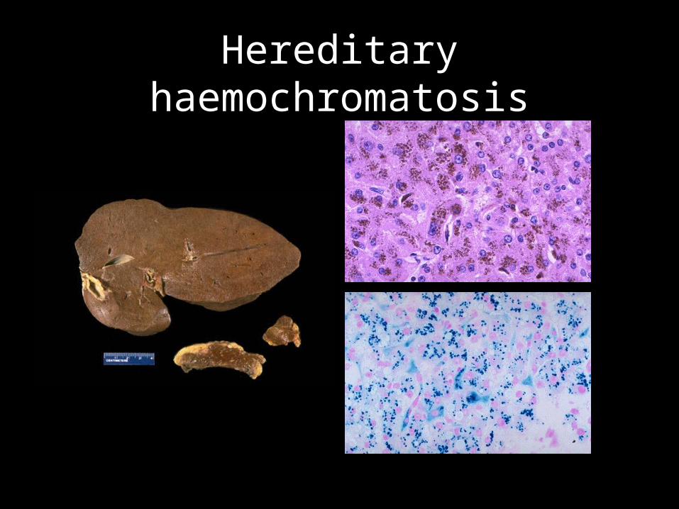
Hereditary haemochromatosis
Wilson’s Disease Inherited abnormality of copper metabolism. Autosomal recessive inheritance. Gene ATP7B on chromosome 13 encodes for transmembrane copper-transporting
ATPase on hepatocyte canalicular membrane. Mutations lead to decreased biliary copper excretion into bile. Excess copper accumulates, toxic via Cu-catalysed reactive oxygen species. Onset during 2nd to 4th decades of life. Clinical manifestations non specific. Disease prevalence 1:30,000. Low plasma ceruloplasmin; high urinary copper. Copper deposited in liver, brain, and cornea (resulting in characteristic Kayser-
Fleischer rings) = liver disease and neuropsychiatric features. Histology:
Pathological changes in liver very variable. Fatty change, acute hepatitis, massive necrosis, chronic hepatitis, steatonecrosis, cirrhosis. Risk of hepatocellular carcinoma low. Distinctive mitochondrial changes on electron microscopy. Copper or its binding protein can be demonstrated in the liver.
Early diagnosis and treatment have markedly improved prognosis.

Diagnosis
serum iron % saturation Serum ferritin estimation of hepatic iron content
DDx in liver haemosiderosis - alcoholic liver disease lipofuscin
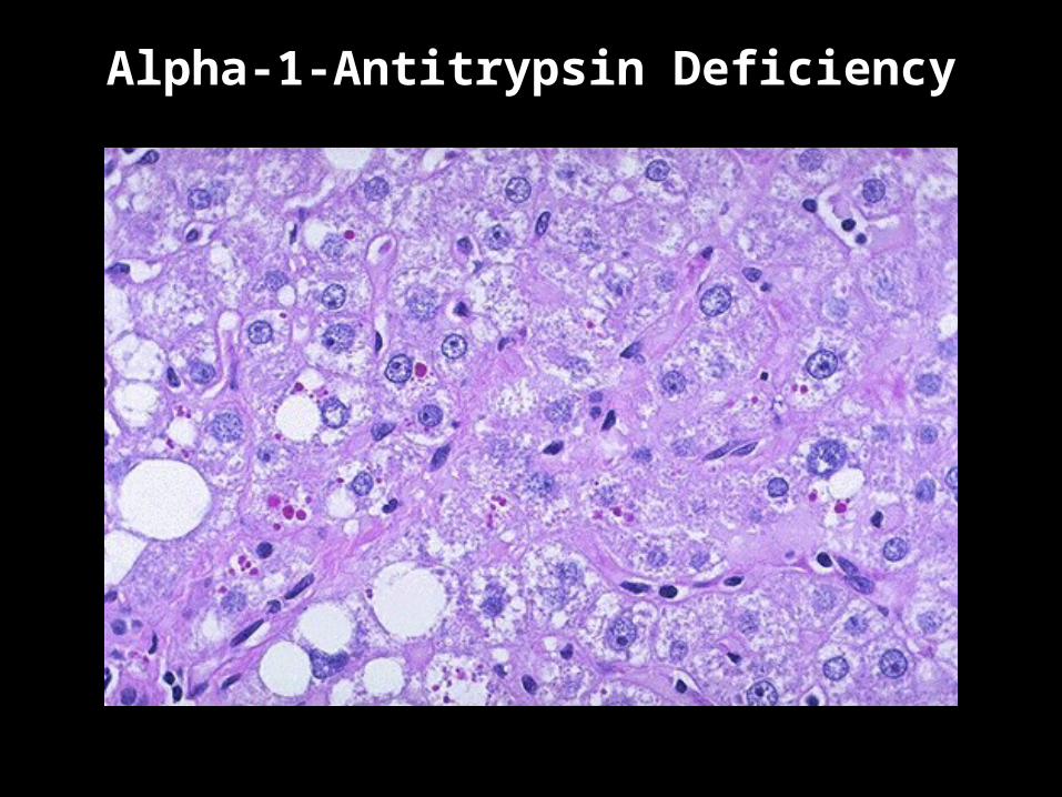
Alpha-1-Antitrypsin Deficiency
Other metabolic diseases
Numerous metabolic disease affect the liver including those of: carbohydrate metabolism (eg glycogen storage
diseases) protein & amino acid metabolism lipid metabolism (eg Gaucher’s disease) porphyrin metabolism and others.
Vascular Disorders
Portal veins Agenesis or hypoplasia. Thrombosis of large and/or small veins. Obliterative portal venopathy (various causes). Possible sequelae:- Zahn “infarcts”; non-cirrhotic portal hypertension.
Hepatic veins Thrombosis of large veins, the Budd-Chiari syndrome. Occlusion of small veins, veno-occlusive disease.
Sinusoids Dilatation. Peliosis hepatis.
Arteries atheroma, embolism, arteritis.
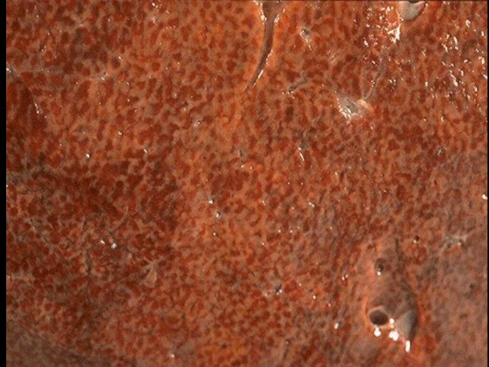
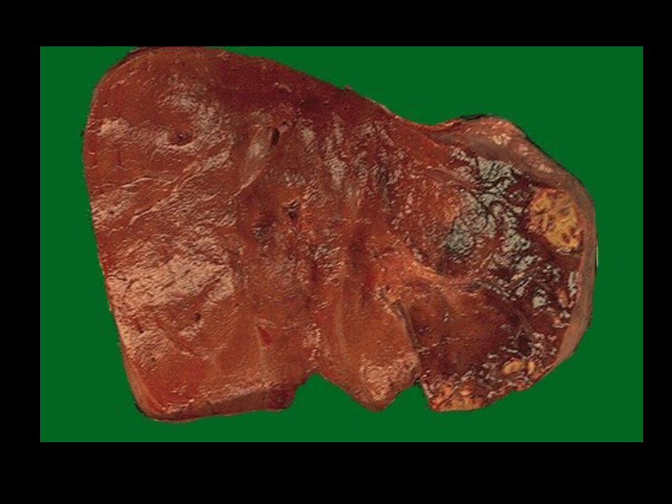
Hepatic congestion and oedema
Hepatic congestion Chronic passive venous congestion due to right heart failure. Liver enlarged, tense, cyanotic with rounded edges. Congestion in perivenular (zone 3) with atrophy of liver cell plates and fatty change in
other zones result in a characteristic appearance called “nutmeg liver.” In long-standing cases, perivenular fibrosis with venular to venular bridging fibrosis
leads to “cardiac sclerosis” (referred to as cardiac cirrhosis in the distant past) Acute passive congestion with hypoperfusion due to co-existent left heart failure leads
to a haemorrhagic centrilobular necrosis. Hepatic ischaemia
Shock causes confluent necrosis in zone 3 (“shock liver”); there may be a neutrophil polymorph response.
Left sided heart failure may lead to hepatic hypoperfusion and hypoxia also. Infarcts are rare due to the dual blood supply. Infarcts of cirrhotic nodules may occur.
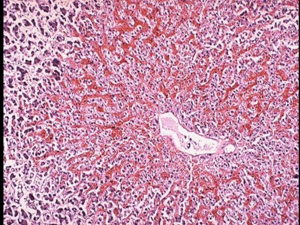
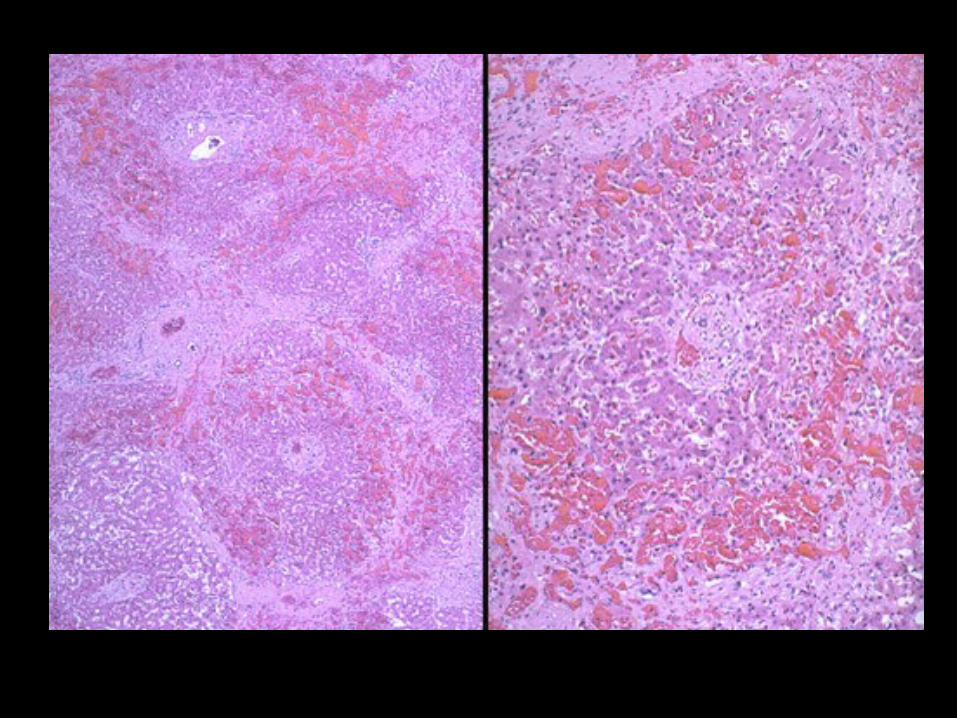
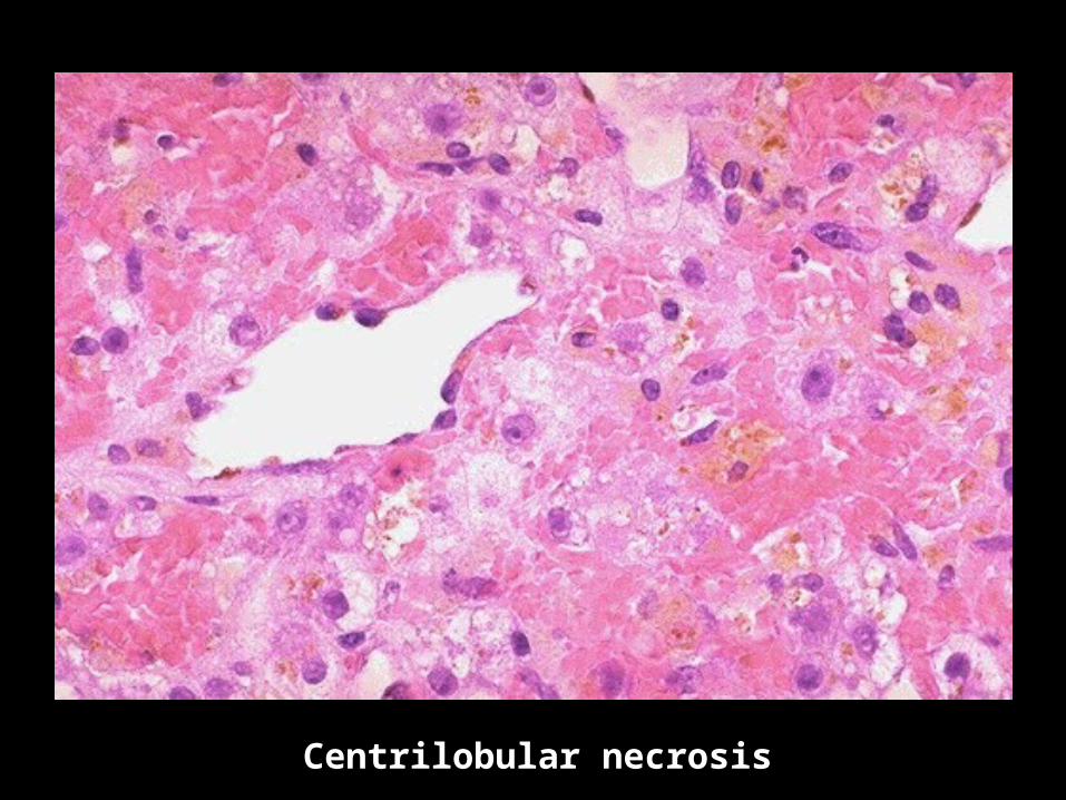
Centrilobular necrosis
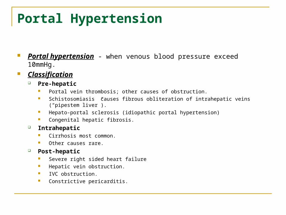
Portal Hypertension
Portal hypertension - when venous blood pressure exceed 10mmHg.
Classification Pre-hepatic
Portal vein thrombosis; other causes of obstruction. Schistosomiasis causes fibrous obliteration of intrahepatic veins (“pipestem liver”). Hepato-portal sclerosis (idiopathic portal hypertension) Congenital hepatic fibrosis.
Intrahepatic Cirrhosis most common. Other causes rare.
Post-hepatic Severe right sided heart failure Hepatic vein obstruction. IVC obstruction. Constrictive pericarditis.