Bilateral Shoulder Edge Adduction Contracture: Anatomy and ... · Grishkevich VM, Grishkevich M...
7
Central Bringing Excellence in Open Access JSM Burns and Trauma Cite this article: Grishkevich VM, Grishkevich M (2017) Bilateral Shoulder Edge Adduction Contracture: Anatomy and Treatment with Axillary Adipose- Cutaneous Trapezoid and Quadrangular Flaps, a New Approach. JSM Burns Trauma 2(1): 1009. *Corresponding author Viktor M. Grishkevich M.D., Department of Reconstructive and Plastic Surgery, Vishnevsky Institute of Surgery, Russia, Email: Submitted: 10 November 2016 Accepted: 21 December 2016 Published: 02 January 2017 Copyright © 2017 Grishkevich et al. OPEN ACCESS Keywords • Quadrangular Flaps • Anatomy • Bilateral • Tissues Research Article Bilateral Shoulder Edge Adduction Contracture: Anatomy and Treatment with Axillary Adipose-Cutaneous Trapezoid and Quadrangular Flaps, a New Approach Viktor M. Grishkevich M.D.* and Max Grishkevich M.D. Department of Reconstructive and Plastic Surgery, Vishnevsky Institute of Surgery, Russia Abstract Background: Bilateral shoulder edge adduction contractures are result of vast burns and scars, restricting the upper limb motion and its development in children. There is no published research pertaining specifically to the anatomy of bilateral axillary contractures. Therefore, the used techniques based on triangular and other flaps’ forms, local and regional, were not anatomically substantiated. The cause of scar contracture (scar surface deficit) was not explored. Therefore, the suggested flaps did not match the scar surface deficit, and the contracture was often released incompletely. For this reason, we researched bilateral contractures’ anatomy and proposed an adequate scar surface deficit compensation local flap method. Methods: Anatomy and scars’ surface deficit (contracture cause) of bilateral contractures were studied in 45 patients (children and adults), before operation, during, and after surgery. Results: Using the new evidence, a new approach to treatment of shoulder bilateral contracture was developed. The treatment lies in surface deficit compensation using the surface surplus of sheets of the fold and entire tissue of the axillary fossa in the form of trapezoid adipose-cutaneous and neighboring flaps and tissues displacement on the donor wound. The surface surplus of sheets of the fold and undamaged adipose-cutaneous layer of axillary fossa allows scar surface deficit compensation and contracture elimination with one or two-stage technique without skin transplants, triangular local flaps and regional complex traumatic regular and expanded flaps that are typically used in the original technique. The procedure significantly improves the outcomes of the bilateral shoulder contracture treatment. Conclusion: The understanding of the edge contracture formation, anatomy and contracture cause open a new approach to forming of a diagnosis, planning of operation, and successful reconstruction without skin transplants and complex flaps. INTRODUCTION Shoulder contractures are formed most frequently among big joints (38%) [1]. The bilateral shoulder adduction contracture is a result of vast burns and scars, covering the shoulder joint anterior and posterior surfaces, trunk and upper extremity, restricting all upper limb function and limiting donor sites. Literature is scarce on the treatment of bilateral shoulder adduction contracture and provides minimum research regarding other aspects of the contracture: anatomy, cause, and criteria for technique selection. Most often an III-Type axillary scar contractures classification is used [2]. It is an accepted notion that edge contractures are caused by the scar’s band or web located along the edge of axillary fossa. The authors did not find publications regarding anatomy description of the band, web, and fold, and their meaning in type formation and its role in edge contracture reconstruction. These edge contractures have many names (long, wide, lineal, cord-like and more) which are edge type (I and II). Regarding bilateral contractures treatment, Yang [3] wrote that the use of skin grafts, especially full-thickness, local Z-plasties could suffice for the release of the contracture; sometimes, multiple Z-plasties would be needed to correct a longer lineal contracture band. If contractures were severe, a local fascio- cutaneous flap was a better choice; for correction of extensive scar contractures of the axilla, the fascio-cutaneous and musculo- cutaneous flaps were an alternative choice for correction of extensive scar contractures. Chan and Donelan [4] presented a patient with shoulder bilateral contractures who was treated with the Z-plasty. Duncan and Smith [5] indicated that bilateral adduction contracture was caused by scar bands/folds located along both axillary fossa edges. From the undamaged axilla, a subcutaneous pedicle propeller flap was elevated longitudinally
Transcript of Bilateral Shoulder Edge Adduction Contracture: Anatomy and ... · Grishkevich VM, Grishkevich M...

CentralBringing Excellence in Open Access
JSM Burns and Trauma
Cite this article: Grishkevich VM, Grishkevich M (2017) Bilateral Shoulder Edge Adduction Contracture: Anatomy and Treatment with Axillary Adipose-Cutaneous Trapezoid and Quadrangular Flaps, a New Approach. JSM Burns Trauma 2(1): 1009.
*Corresponding authorViktor M. Grishkevich M.D., Department of Reconstructive and Plastic Surgery, Vishnevsky Institute of Surgery, Russia, Email:
Submitted: 10 November 2016
Accepted: 21 December 2016
Published: 02 January 2017
Copyright© 2017 Grishkevich et al.
OPEN ACCESS
Keywords•Quadrangular Flaps•Anatomy•Bilateral•Tissues
Research Article
Bilateral Shoulder Edge Adduction Contracture: Anatomy and Treatment with Axillary Adipose-Cutaneous Trapezoid and Quadrangular Flaps, a New ApproachViktor M. Grishkevich M.D.* and Max Grishkevich M.D.Department of Reconstructive and Plastic Surgery, Vishnevsky Institute of Surgery, Russia
Abstract
Background: Bilateral shoulder edge adduction contractures are result of vast burns and scars, restricting the upper limb motion and its development in children. There is no published research pertaining specifically to the anatomy of bilateral axillary contractures. Therefore, the used techniques based on triangular and other flaps’ forms, local and regional, were not anatomically substantiated. The cause of scar contracture (scar surface deficit) was not explored. Therefore, the suggested flaps did not match the scar surface deficit, and the contracture was often released incompletely. For this reason, we researched bilateral contractures’ anatomy and proposed an adequate scar surface deficit compensation local flap method.
Methods: Anatomy and scars’ surface deficit (contracture cause) of bilateral contractures were studied in 45 patients (children and adults), before operation, during, and after surgery.
Results: Using the new evidence, a new approach to treatment of shoulder bilateral contracture was developed. The treatment lies in surface deficit compensation using the surface surplus of sheets of the fold and entire tissue of the axillary fossa in the form of trapezoid adipose-cutaneous and neighboring flaps and tissues displacement on the donor wound. The surface surplus of sheets of the fold and undamaged adipose-cutaneous layer of axillary fossa allows scar surface deficit compensation and contracture elimination with one or two-stage technique without skin transplants, triangular local flaps and regional complex traumatic regular and expanded flaps that are typically used in the original technique. The procedure significantly improves the outcomes of the bilateral shoulder contracture treatment.
Conclusion: The understanding of the edge contracture formation, anatomy and contracture cause open a new approach to forming of a diagnosis, planning of operation, and successful reconstruction without skin transplants and complex flaps.
INTRODUCTIONShoulder contractures are formed most frequently among big
joints (38%) [1]. The bilateral shoulder adduction contracture is a result of vast burns and scars, covering the shoulder joint anterior and posterior surfaces, trunk and upper extremity, restricting all upper limb function and limiting donor sites. Literature is scarce on the treatment of bilateral shoulder adduction contracture and provides minimum research regarding other aspects of the contracture: anatomy, cause, and criteria for technique selection. Most often an III-Type axillary scar contractures classification is used [2]. It is an accepted notion that edge contractures are caused by the scar’s band or web located along the edge of axillary fossa. The authors did not find publications regarding anatomy description of the band, web, and fold, and their meaning in type formation and its role in edge contracture
reconstruction. These edge contractures have many names (long, wide, lineal, cord-like and more) which are edge type (I and II). Regarding bilateral contractures treatment, Yang [3] wrote that the use of skin grafts, especially full-thickness, local Z-plasties could suffice for the release of the contracture; sometimes, multiple Z-plasties would be needed to correct a longer lineal contracture band. If contractures were severe, a local fascio-cutaneous flap was a better choice; for correction of extensive scar contractures of the axilla, the fascio-cutaneous and musculo-cutaneous flaps were an alternative choice for correction of extensive scar contractures. Chan and Donelan [4] presented a patient with shoulder bilateral contractures who was treated with the Z-plasty. Duncan and Smith [5] indicated that bilateral adduction contracture was caused by scar bands/folds located along both axillary fossa edges. From the undamaged axilla, a subcutaneous pedicle propeller flap was elevated longitudinally

CentralBringing Excellence in Open Access
Grishkevich et al. (2016)Email:
JSM Burns Trauma 2(1): 1009 (2017) 2/7
and rotated on 90 degrees release. Severe or moderate (bilateral) contractures were treated by Chen et al. [6], treated severe or moderate axillary burn scar contracture with large transverse island scapular flap and expanded transverse island scapular flap in 15 adult and pediatric patients and transposed to axilla. Walash et al. [7], write that for Type II (bilateral contractures) and III (total) contractures, the regional flaps were a preferred choice whenever available. Depending of contracture severity, Devi and Baishya [8] use Z-plasty, propeller flap regional flaps. The tendency to the large flaps use when tissue of axillary fossa is undamaged could not be anatomically justified.
The different opinions about treatment, indications and surgical techniques, outcomes of treatment of relatively simple contracture are caused by insufficiently researches anatomy, contracture cause, role of the folds, tissue displacement.
Most known flaps and techniques were tested in our institute, and we did not reach the desired results. Surgical research proved that many aspects of bilateral shoulder adduction contractures were insufficiently researched. The need for research concerning the following aspects became obvious: contractures’ formation, contracture anatomy, scar surface deficit (contracture cause) form and location, tissue surface surplus formation in folds (indication for reconstruction), and the condition of the axilla as an excellent donor site. All these aspects of bilateral contractures anatomy and treatment were researched; the new approach, flaps and technique significantly improved the outcomes and made the operation easier, safer, preventing the donor site injury. The research, results, and discussion are presented in this paper.
MATERIAL AND METHODSPatients
Forty-five patients with bilateral shoulder adduction contractures were operated personally; of them, 6 patients had edge posterior contractures of both shoulder joints; 25 were men and 20 women; ages 5 to 65 years; 9 were children. TBSA was 12 to 56% (mean 31%).Twenty-two patients had combinations of shoulder bilateral contractures with elbow or/and wrist and truncal lateral scar contractures which were released simultaneously.
Methods
Anatomy of bilateral shoulder edge contractures were explored before surgery, during operation, and 6 months to 7 years after reconstruction. Anatomic criteria were: scar location and spreading in relation to joint’s flexion surfaces; the fold presence (scar surface surplus) and its anatomy; scar surface deficit determination (contracture cause); needed form of the flap for scars surface deficit compensation and edge contracture elimination. Follow- up results observed from 6 months to 8 years. Full shoulder abduction (180 degrees) and rotation were considered as good results. Received data laid an anatomical basis for technique choice, development of the new approach, and a new method with a justified rationale for local tissue use.
RESULTSAnatomy of bilateral shoulder adduction contracture
Functional zones/surfaces of the shoulder joint in case of bilateral edge contracture (Figure 1): Before surgery, it is
necessary to divide the shoulder joint’s surface into flexion (F) and extension (E); the boundary between them passes along the joint rotation axis level (“+”symbol). The flexion surface (F) has a curvature of nearly 90 degrees, caused by latissimus dorsi and pectoralis major muscles edges, which divided it into two parts: flexion lateral (FL) surface and flexion medial (FM) surface (Figure 1A and B). The divided line passes along the edge of the axillary fossa. The scars of flexion lateral (FL) surfaces spread from the joint rotation axis distally and form folds (Fd) the sheets of which is divided by fold crest (Cr). The flexion medial (FM) surface lies between fossa edges. Contracture type formation depends on the contracted scars location: shoulder edge posterior, anterior, and bilateral contractures are caused by scars covering corresponding the flexion lateral (FL) and fold one or both zones; the flexion medial and extension surfaces stay undamaged; scars, covering the flexion medial surface, cause medial contracture; scars, located on both flexion lateral and medial surfaces or joint circularly create total contracture.
Edge shoulder bilateral contracture formation (Figure 1): Edge shoulder bilateral adduction contracture is caused by burn and scars, covering both joint flexion lateral surfaces (FL), from the crests of the fold to the joint rotation axis. During wound healing, scars grow distally, between the shoulder and chest wall anteriorly and posteriorly. Moving distally, scars involve the healthy skin of axillary fossa. As a result, the crescent-shaped folds are formed, located along the anterior and posterior edges of axillary fossa. The scars lateral fold sheets are a part of flexion lateral surfaces which becomes wider (distance from the fold’s crest to the joint rotation axis). At the same time, scars are contracted in length and shorten distance between shoulder and chest wall, causing adduction contracture. Axillary fossa stays undamaged.
Anatomy of shoulder edge bilateral adduction contracture (Figure 1): Shoulder edge bilateral contracture has specific anatomic and clinical features: (a) contracture is caused by scar which cover the joint flexion lateral surfaces. (b) Scars of the flexion lateral surfaces grow distally, involving the healthy
A) B) C)
Figure 1 Functional zones of shoulder joint surface and anatomy of bilateral edge shoulder adduction contracture. (A and B) Anterior and posterior views: E- joint extension surface: “:+” – joint rotation axis; FL- flexion lateral surfaces (from the fold crest (Fd) to the joint rotation axis) is scars causing contracture; Fd- the fold: the lateral fold’s sheet is scars and part and continuation of FL surface; the medial, is healthy skin and continuation of FM surface; Cr- crest of the fold is edge of scars; FM- flexion medial surface (between fossa edges or fossa) is healthy skin. Scars of flexion lateral surfaces (FL) and lateral scars sheets of the fold contracture cause. Medial sheets are part of the flexion medial surface (FM) and are healthy skin and donor site. The folds are newly tissue formation and presents surface surplus of scars and healthy skin; (C) contracture released in one -stage with two adipose-scar trapezoid flaps.
CentralBringing Excellence in Open Access
Grishkevich et al. (2016)Email:
JSM Burns Trauma 2(1): 1009 (2017) 3/7
fossa skin and forming the folds (Fd), that pass along the edges of axillary fossa, between flexion lateral (FL) and flexion medial (FM) surfaces. (c) The fold consists of two different qualities sheets: the lateral fold sheets are scars and are the continuation of the flexion lateral surfaces; the medial fold sheets and axillary fossa (flexion medial surface, FM) are healthy skin. (d) The crest of the fold (Cr) is the edge of scars. These four anatomic features and clinical signs determine the scar contracture name, edge, and are constant independent of contracture location and severity and are basis of diagnose. In case of edge shoulder bilateral contracture, the distance from the fold crest to the joint rotation axis (the width of contracted scars covering the joint flexion lateral surface), is 6-8 cm (in children) to 10 -12 cm and more (in adults). The fold becomes a new anatomic formation that creates the surface surplus useful for contracture elimination.
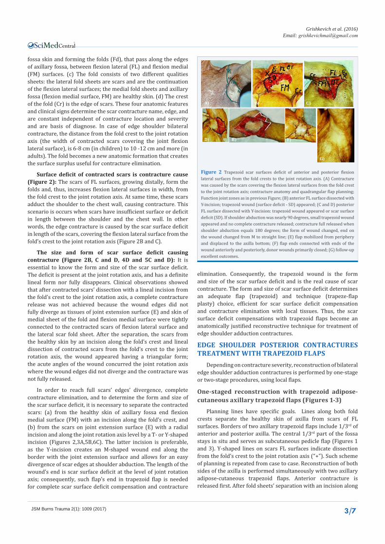
Surface deficit of contracted scars is contracture cause (Figure 2): The scars of FL surfaces, growing distally, form the folds and, thus, increases flexion lateral surfaces in width, from the fold crest to the joint rotation axis. At same time, these scars adduct the shoulder to the chest wall, causing contracture. This scenario is occurs when scars have insufficient surface or deficit in length between the shoulder and the chest wall. In other words, the edge contracture is caused by the scar surface deficit in length of the scars, covering the flexion lateral surface from the fold’s crest to the joint rotation axis (Figure 2B and C).
The size and form of scar surface deficit causing contracture (Figure 2B, C and D, 4D and 5C and D): It is essential to know the form and size of the scar surface deficit. The deficit is present at the joint rotation axis, and has a definite lineal form nor fully disappears. Clinical observations showed that after contracted scars’ dissection with a lineal incision from the fold’s crest to the joint rotation axis, a complete contracture release was not achieved because the wound edges did not fully diverge as tissues of joint extension surface (E) and skin of medial sheet of the fold and flexion medial surface were tightly connected to the contracted scars of flexion lateral surface and the lateral scar fold sheet. After the separation, the scars from the healthy skin by an incision along the fold’s crest and lineal dissection of contracted scars from the fold’s crest to the joint rotation axis, the wound appeared having a triangular form; the acute angles of the wound concurred the joint rotation axis where the wound edges did not diverge and the contracture was not fully released.
In order to reach full scars’ edges’ divergence, complete contracture elimination, and to determine the form and size of the scar surface deficit, it is necessary to separate the contracted scars: (a) from the healthy skin of axillary fossa end flexion medial surface (FM) with an incision along the fold’s crest, and (b) from the scars on joint extension surface (E) with a radial incision and along the joint rotation axis level by a T- or Y-shaped incision (Figures 2,3A,5B,6C). The latter incision is preferable, as the Y-incision creates an M-shaped wound end along the border with the joint extension surface and allows for an easy divergence of scar edges at shoulder abduction. The length of the wound’s end is scar surface deficit at the level of joint rotation axis; consequently, such flap’s end in trapezoid flap is needed for complete scar surface deficit compensation and contracture
elimination. Consequently, the trapezoid wound is the form and size of the scar surface deficit and is the real cause of scar contracture. The form and size of scar surface deficit determines an adequate flap (trapezoid) and technique (trapeze-flap plasty) choice, efficient for scar surface deficit compensation and contracture elimination with local tissues. Thus, the scar surface deficit compensations with trapezoid flaps become an anatomically justified reconstructive technique for treatment of edge shoulder adduction contractures.
EDGE SHOULDER POSTERIOR CONTRACTURES TREATMENT WITH TRAPEZOID FLAPS
Depending on contracture severity, reconstruction of bilateral edge shoulder adduction contractures is performed by one-stage or two-stage procedures, using local flaps.
One-staged reconstruction with trapezoid adipose-cutaneous axillary trapezoid flaps (Figures 1-3)
Planning lines have specific goals. Lines along both fold crests separate the healthy skin of axilla from scars of FL surfaces. Borders of two axillary trapezoid flaps include 1/3rd of anterior and posterior axilla. The central 1/3rd part of the fossa stays in situ and serves as subcutaneous pedicle flap (Figures 1 and 3). Y-shaped lines on scars FL surfaces indicate dissection from the fold’s crest to the joint rotation axis (“+”). Such scheme of planning is repeated from case to case. Reconstruction of both sides of the axilla is performed simultaneously with two axillary adipose-cutaneous trapezoid flaps. Anterior contracture is released first. After fold sheets’ separation with an incision along
A)
D) E) F)
G)
B) C)
Figure 2 Trapezoid scar surfaces deficit of anterior and posterior flexion lateral surfaces from the fold crests to the joint rotation axis. (A) Contracture was caused by the scars covering the flexion lateral surfaces from the fold crest to the joint rotation axis; contracture anatomy and quadrangular flap planning; Function joint zones as in previous Figure; (B) anterior FL surface dissected with Y-incision; trapezoid wound (surface deficit - SD) appeared; (C and D) posterior FL surface dissected with Y-incision: trapezoid wound appeared or scar surface deficit (SD). If shoulder abduction was nearly 90 degrees, small trapezoid wound appeared and no complete contracture released; contracture full released when shoulder abduction equals 180 degrees; the form of wound changed, end on the wound changed from M to straight line; (E) flap mobilized from periphery and displaced to the axilla bottom; (F) flap ends connected with ends of the wound anteriorly and posteriorly, donor wounds primarily closed; (G) follow-up excellent outcomes.

CentralBringing Excellence in Open Access
Grishkevich et al. (2016)Email:
JSM Burns Trauma 2(1): 1009 (2017) 4/7
the fold’s crest, the contracted scars are dissected from the fold’s crest to the joint rotation axis and separated from the tissues of the extension surface (E) with a Y-shaped incision. The split end of a Y-incision separates scars from the extension surface tissues, As a result, the wound edges freely diverge and the contracture is fully released and a trapezoid wounds appears on the joint’s anterior flexion lateral surfaces. Considering the wound’s size and form, but by about 30% wider, a trapezoid flap is mobilized that consists of the fold’s medial sheet and adipose-cutaneous layer of 1/3rd of fossa (in anterio- posterior direction). The flap is transposed on the wound with tension and sutured to the wound’s borders. The angles of the fold’s sheet approach the flap’s base, covering the donor wounds aside the flap. Then, the posterior contracture is released. The central 1/3rd part of the fossa stays in situ and serves as subcutaneous pedicle (Figures1 and 3). Blood circulation in flaps is sufficient and no complications occurred. 6.2. Two-staged bilateral shoulder adduction contracture treatment (Figure 4)
For elimination of more severe contractures and scar surface deficit compensation, the two-staged technique is preferable. First, the more severe contracture is released: scars are separated from healthy skin with an incision along the fold’s crest; then, the contracted scars are dissected with a Y- incision from the fold’s crest to the joint rotation axis, separated contracted scars from the joint extension tissues (E). As a result, a trapezoid wound appears (Figure 4D); matching the wound, a trapezoid adipose cutaneous flap was prepared from all axilla and medial fold sheet. The flap was transposed on the wound and donor wounds aside the flap was covered with scar fold sheet. Six months later, when skin tension disappeared and healthy skin surface increased due to growth, the contracture of the other axilla side was removed using the same procedure (Figure 4A-C, left joint; D-I, right joint).
Bilateral edge shoulder contracture in children elimination with quadrangular subcutaneous pedicle flap, edge elbow, and medial lateral truncal contractures with trapezoid flaps (Figure 5)
If scars cover а wide surface, form axillary, elbow and truncal
contractures, axillary and elbow fossa can have healthy skin. The scars displace the axillary tissue downward which increases the scar surface deficit. The anatomy and planning of the operation is shown in Figure (5A). First, the edge elbow contracture is eliminated with a trapezoid adipose-cutaneous flap. With incisions along both folds’ crests, the sheets are separated; all axillary healthy skin is included in the quadrangular flap. Using perpendicular Y-shaped incisions, scars are dissected from the folds’ crests to the joint rotation axis anteriorly and posteriorly. The axillary flap (skin and subcutaneous fat layer) is separated from the scars and mobilized from periphery (Figure 5B). The flap has steady blood circulation, and stretching it in anterio-posterior direction is safe, what allows the flap ends connect with the wounds’ edges anteriorly and posteriorly by displacing also the skin of the extension surfaces on the flexion lateral surfaces (Figures 5C and D). The wounds aside the flap are primarily closed, starting from the flap’s ends (Figure 5E). Truncal contracture is eliminated at last using trapezoid adipose-scar flaps. Two weeks after surgery is shown in Figure (5F-K).
Bilateral edge severe contracture treatment with quadrangular axillary subcutaneous pedicle flap (Figure 6)
Severe bilateral shoulder adduction contracture in adults is also treated with the island quadrangular subcutaneous pedicle
A) B) C)
Figure 3 Bilateral shoulder edge adduction contracture treatment using one-stage trapeze-flap technique. (A) Pre surgery view: joint zones: E- extension surface; FL- flexion lateral surface; “+”- joint rotation axis; Fd- fold; FM- flexion medial surface; planning anterior and posterior edge contracture release: solid lines- incisions, two trapezoid flap with common pedicle, lines of the fold’s sheets division, Y-shaped lines for scars dissection; (B) end of operation: trapezoid adipose-cutaneous flaps compensated scar surface deficit; (C) follow- up results: contracture released, and contours of axilla restored (one year and 8 months after surgery).
A) B) C)
D) E) F)
Figure 4 (A) Two-staged bilateral axillary edge contractures treatment with trapezoid adipose- cutaneous flaps. (A) Left shoulder joint: anterior contracture removed three months before, planned trapezoid flap for posterior contracture release; (B) contracture eliminated, axillary contours restored. Two-staged bilateral edge axillary contracture treatment with trapezoid adipose-cutaneous flaps. (c) Anterior right edge contracture released before reconstruction posterior contracture; posterior FL surface and scar fold sheets; cars sheet dissected with Y-shaped incision, trapezoid wound appeared (scar surface deficit): SD-surface deficit, DW-donor wound; FP- flap; FL surface; Cr- crest of the fold; E- extension surface. Trapezoid adipose-cutaneous flap mobilized; (D) mobilized flap covered wound; (E) donor wounds aside the flap covered with adipose-scar trapezoid flaps prepared from the scar sheet; surface deficit compensated and fully contracture released;(F) follow-up result (two years after surgery): excellent functional and cosmetic outcomes.

CentralBringing Excellence in Open Access
Grishkevich et al. (2016)Email:
JSM Burns Trauma 2(1): 1009 (2017) 5/7
flap and primarily donor wound closure (Figures 5 A-D).
Edge shoulder bilateral contracture treatment using island axillary healthy tissue (Figure 7).
The island axillary flap is mobilized on periphery, displaced on the axilla’s bottom and transposed in anterio-posterior direction with tension. Due to flap tension: (a) the edges of pectoralis major and latissimus dorsi muscles approach each other and axillary fossa is obliterated; (b) all healthy tissue is used for surface deficit compensation and wound covering on anterior and posterior joint lateral flexion surfaces; (c) extended flap squeezes the soft tissue around the joint and displaces the skin of the joint extension surfaces downward, covering part of the joint’s flexion lateral surfaces. As a result, the flap’s ends reach the wound edges and suspend the axilla at the normal level, preventing contracture recurrence. The presented flaps and techniques allow to eliminate the bilateral shoulder adduction contracture with local tissues and to avoid skin grafting and the use of complex regional pedicle and free flaps in most cases.
RESULTS OF OPERATIONSThe edge shoulder bilateral contractures were eliminated
with trapezoid flaps in all 45 patients and fully shoulder abduction was achieved (Figures 1-7). The folds’ surface surplus in conjunction with healthy axillary tissue allowed contractures elimination with local flaps. Adipose-cutaneous trapezoid flaps were alive, no flap loss occurred. After reconstruction, extended
flaps and surrounding tissues grow and tension gradually diminished; axillary fossa accepted normal shape. Hair-bearing skin was situated in axillary cupola. Flaps stable suspend the axilla and thus prevent contracture recurrence. Follow-up results improve with time. Appearance of the shoulder joint region was significantly improved.
DISCUSSIONDifferent surgical techniques are used for edge shoulder
A) B) C)
D) E) F) G)
H) I) J) K)
Figure 5 One-stage reconstruction contractures in children: bilateral axillary edge adduction with quadrangular subcutaneous pedicle flap, edge elbow flexion contracture and medial lateral truncal contracture with trapeze-flap plasty. (A and B) Pre surgery: axilla anatomy and joint functional zones; only fossa’s skin is healthy; planning shoulder treatment with quadrangular flap, edge elbow contracture with trapezoid elbow flap, and last truncal contracture with adipose-scar trapezoid flaps; (C) edge elbow contracture eliminated first with one adipose-scar trapezoid elbow flap; (D - H) steps of shoulder contracture treatment with quadrangular flap; truncal contracture with adipose-scar trapezoid flaps; (I -K) results (one and two weeks after surgery).
C)
A) B)
D)
Figure 6 Results of bilateral edge shoulder adduction contracture treatment with quadrangular subcutaneous pedicle flap. (A and B) Pre surgery: functional joint zones and planning; (C) two weeks after reconstruction; (D) follow-up results: contracture eliminated and axillary region restored.
C)
A) B)
D)
Figure 7 Shoulder adduction contracture treatment using island healthy skin in axillary cupola. (A and B) Pre surgery, functional joint zones, anatomy and planning; (C) contracture released and island flap mobilized on periphery; (D) ends of the flap sutured to the wound’s edges anteriorly and posteriorly, donor wounds aside the flap primarily closed, starting from the flap ends.

CentralBringing Excellence in Open Access
Grishkevich et al. (2016)Email:
JSM Burns Trauma 2(1): 1009 (2017) 6/7
adduction contractures treatment:
Skin grafting
Yang [3] indicated that the skin grafts’ use was a basic technique for axillary contracture correction. Some authors [9,10] suggested skin transplants use if other techniques were not viable or suggested as an addition to other methods. Many local-flap techniques have been suggested for edge contractures release.
Z-plasty stays most often used procedure
Chan and Donelan [4] wrote that Z-plasty had found many uses in plastic surgery and was definitely a part of every plastic surgeon’s armamentarium; authors presented pictures of treatment of bilateral shoulder edge contracture using Z-plasty. Suzuki [11] proposed a comprehensive classification of V-Y flaps and their analogs; flaps could be easily designed according to the degree of contracture and the shape of the scars and that was useful to combine V-Y flaps with plan metric Z-plasties.
Buis et al. [12], pointed out to the problem of necrosis of the triangular flaps’ ends; for release of flap tension, the authors proposed two technical modifications preventing flaps necrosis. Huang [13] determined that Z-plasty was not possible in cases of limited availability of uninjured skin adjacent to the wound; interposition flap technique was presented.
V-Y plasty
This is rare used for edge axillary contracture treatment.van Niekerk et al. [14] noticed that the success of V-Y plasty depended on the depth of the flap’s advancement only which was restricted as the flaps were not mobilized. Gumus [15] used a deep incision through the fascia, making advancements of the Y- flap easy by sliding. Some authors [5,16] used propeller flaps for bilateral edge shoulder contracture treatment. Axillary longitudinal subcutaneous pedicle flap was rotated at 90 degrees for release of the edge contractures. Hyakusoku and Akimoto [17] proposed the square flap method; Ertas and Borman [18], double opposing rectangular advancement flap. Authors who performed analysis outcomes of scar contractures treatment by the triangular flaps use did not receive satisfaction [12-15]. Balumuka et al. [19], reported that out of 58 patients with shoulder and elbow contractures, 30 (52 %) had a recurrence with 67% being shoulder contracture. The most commonly employed operative technique was the local triangular flaps. Complete release of the shoulder contracture was achieved in 56 % of right joint contracture and in 33 % of left joint contractures. Incomplete contracture release resulted in contracture recurrence. Klein [20] concludes that is apparent that one should expect an evolution in surgical techniques and technologies that can improve the function and appearance of person with burn injuries. No real evolution is observed; first, it concerns scar contractures treatment.
Many regional flaps were proposed for axillary scar contractures treatment: transverse island scapular flap [6]; thoracodorsal perforator based cutaneous island flap [21]; scapula skin island flap [22], scapular island flap in pediatric axillary contracture [23], extended lower trapezius island myo-cutaneous flap [24]; latissimus dorsi flap with soft tissue
expansion [25]. The problem is that many authors do not classify their patients; classification into anatomy-based three types: edge, medial, and total [26] and the trapeze-flap plasty use will open a new page in burned patient rehabilitation. Stekelenburg et al. [27], explored efficacy of scar contractures treatment with different techniques and concluded that at present, no consensus exists on the technique to be used; no definite conclusions could be reached about the effectiveness of different techniques; therefore, no direct implication for daily practice could be made.
The literature analysis proves how complicated, intricate, and perplex the issue concerning the edge shoulder contractures elimination is. New anatomical data explain why all techniques based on triangular flap can’t complete edge bilateral contracture elimination.
a. Contracture is caused by scars, covering the joint anterior and posterior flexion lateral surface which spreads from the fold’s crests to the joint rotation axis, 8-12 cm in length. Such length of triangular flap is not mobilized and not displaced
b. Scar surface deficit or contracture cause has a trapezoid shape, maximal at the fold’s crest and at the joint rotation axis is 5-6 cm in length. Triangular flaps do not match the shape of the scar surface deficit which is trapeze-shaped.
c. Contracture scars of flexion lateral surface (from anterior and posterior edge of fossa to the joint rotation axis), and the fold, which is a new anatomic structure, has scar surface surplus 3-6 cm in width (from the edge of fossa to the crest of the fold). The triangular rotation and advancement techniques are working in borders the fold’s width and do not exceed 6 cm in depth from the fold’s crest.
d. The flap has different quality sheets and only lateral scar sheet participates in contracture formation; medial fold sheet is healthy skin and a part of FM surface; flap presents a surface surplus; the tissues of the fold sheets are used for scar surface deficit compensation and contracture elimination. Pointed triangular rotation and advancement triangular flaps are prepared from the fold sheets, and different quality flaps are counter transposed; scar flaps are penetrated inside the healthy skin where absent the scar surface deficit.
Presented anatomical data confirm that all techniques based on triangular pointed flaps can’t significantly release the edge shoulder bilateral contracture; research results explain why so many different composed flaps and traumatic techniques are used for a treatment of a relatively simple contracture, which can be easily eliminated with an axillary adipose-cutaneous trapezoid flap.
CONCLUSIONBilateral shoulder adduction contracture is caused by scars
covering the joint FL surface, and having insufficient surface trapezoid form among shoulder and chest wall. The surface deficit spreads from the fold crest to the joint rotation axis, 8-12 cm in length and can’t be covered with triangular flaps. Contractures are successfully eliminated by scar surface deficit

CentralBringing Excellence in Open Access
Grishkevich et al. (2016)Email:
JSM Burns Trauma 2(1): 1009 (2017) 7/7
compensation with axillary trapezoid or quadrangular adipose-cutaneous subcutaneous pedicle flap and trapezoid counter transposed flaps prepared from the fold’s sheets. Donor wounds are primarily closed. The excellent outcomes allow flaps and method belong to the preferable.
REFERENCES1. Schneider JC, Holavanahalli R, Helm Ph, Golstein R, Kowalske K.
Contractures in burn injury: defining the problem. J Burn Care Research. 2006; 27: 508-514.
2. Kurzman LC, Stern PJ. Upper extremity burn contracture. Hand Clin. 1990; 67: 261-279.
3. Yang JY. Reconstruction of axillary contracture. Functional and aesthetic reconstruction of burned patients. Taylor and Francis: Boca Raton. 2005; 116: 367-378.
4. Chan RK, Donelan MB. Use of Z-plasty in Burn Reconstruction. Color atlas of burn reconstructive surgery. Springer. Verlag Berlin Heidelberg 2010; 172-177.
5. Duncan SFM, Smith AA. Treatment of the burned axilla. Achauer and Sood’s burn surgery. Reconstruction and rehabilitation. 2006; 282-298.
6. Chen B, Xu M, Chai J, Song H, Gao Q. Surgical treatment of severe or moderate axillary burn scar contracture with transverse island scapular flap in adult and pediatric patients – A clinical experience of 15 cases. Burns. 2015; 41: 872-880.
7. Walash A, Kishk T, Ghareeb FM. Treatment of postburn axillary contracture. Menoufia Medical J. 2014; 267: 278-283.
8. Devi SR, Baishya J. Management of post burn axillary contracture along with breast contracture: Our experience. Indian J Burns. 2012; 20: 23-29.
9. Asuku ME, Iobrahim A, Ijekeye FO. Post burn axillary contractures in pediatric patients: A retrospective survey of management and outcome. Burns. 2008; 34: 1190-1195.
10. Karki D, Mehta N, Narayan RP. Post-burn axillary contracture: A therapeutic challenge! Indian J Plast Surg. 2014; 47: 375-380.
11. Suzuki SH, Kawai K, Morimoto N. Z- plasties and V-Y flaps. Color atlas of burn reconstructive surgery. Springer-Verlag Berlin Heidelberg. 2010; 20; 160- 171.
12. Buis J, Soupre V, Picard A, Le Louam C, Servant JM, Vazquez MP. The plasty low tension. Ann Chir Plast Esthet. 2009; 54: 370-373.
13. Huang TM, Lee SS, Lai ChSh, Lin SD. Treatment of axillary burn scar
contracture using opposite running Y-V-plasty. Burns. 2005; 31: 894-900.
14. Van Niekerk WJC, Taggart I. The size of the Y: The multiple Y-V plasty revisited. Burns. 2008; 34: 257-261.
15. Gumus N. Difficulties with running V-Y plasty in releasing burn scar contracture. Turkish Trauma Emergency Surg. 2010; 16: 407-412.
16. Karki D, Mehta N, Narayan RP. Subcutaneous pedicle propeller flap: An old technique revisited and modified! Indian J Plast Surg. 2016; 49: 220-224.
17. Hyakusoku H, Akimoto M. The square flap method. Color atlas of burn reconstructive surgery. Springer –Verlag Berlin Heidelberg. 2010; 23: 186-197.
18. Ertaş N, Borman H. Double opposing rectangular advancement flap is an alternative technique in the treatment of wide linear post burn scar contractures. Burns. 2011; 37: 1449-1457.
19. Balumuka DD, Galiwango GW, Alenyo R. Recurrence of post burn contractures of the elbow and shoulder joints: experience from a ugandian hospital. BMC Surgery. 2015; 15: 103-111.
20. Klein MB. Physical Medicine and Rehabilitation Clinics of North America. Burn Reconstruction. 2011; 22: 311-326.
21. Er E, Ucar C. Reconstruction of axillary contractures with thoracodorsal perforator island flap. Burns. 2005; 31: 726-730.
22. Nisanci M, Er E, Isic S, Sengezer M. Treatment modalities for post burn axillary contractures and the versatility of scapular flap. Burns. 2002; 28: 177-180.
23. Turkaslan T, Turan A, Dayicioglu D, Ozsoy Z. Uses of scapular island flap in pediatric axillary burn contracture (correction of contractures). Burns. 2006; 32: 885-890.
24. Elshaer WM. Extended lower trapezius island myocutaneous flap in the repair of post burn axillary contracture. Plast Reconstr Surg. 2004; 113: 2076-2081.
25. Lykoudis EG, Seretis K, Ziogas D. Tissue expansion and latissimus dorsi transfer for arm-thorax synechia reconstruction. J Burn Care Research. 2011; 31: e15-e20.
26. Grishkevich VM. The basic types of scar contractures after burns and methods eliminating them with trapezeplasty flaps. Plast Reconstr Surg. 1991; 88: 1044-1054.
27. Stekelenburg CM, Marck RE, Tuinebreijer WE, de Vet HC, Ogawa R, van Zuijlen PP. A systematic review on burn scar contracture treatment: searching for evidence. J Burn Care Res. 2015; 36: e153-161.
Grishkevich VM, Grishkevich M (2017) Bilateral Shoulder Edge Adduction Contracture: Anatomy and Treatment with Axillary Adipose-Cutaneous Trapezoid and Quadrangular Flaps, a New Approach. JSM Burns Trauma 2(1): 1009.
Cite this article

![Systèmes à Adduction d’Air - delamet.com · Systèmes à Adduction d’Air [Respirez en toute sécurité] Les systèmes à adduction d’air sont particulièrement requis pour](https://static.fdocuments.net/doc/165x107/5bd6ac4709d3f2e17c8bc19a/systemes-a-adduction-dair-systemes-a-adduction-dair-respirez-en.jpg)