Bibliography on Neuroinformatics - The Human Brain Project
112
Bibliography on Neuroinformatics Finn ˚ Arup Nielsen CIMBI at DTU Informatics and NRU Rigshospitalet Lyngby and Copenhagen, Denmark June 24, 2008 $Revision: 1.368 $ $Date: 2008/06/13 13:17:13 $ Abstract Neuroinformatics references are collected, especially in the area of functional neuroimaging of the human brain. The emphasis is on tools for analysis of neuroscience data, particularly programs for functional neuroimaging. The original URL to this document is PDF: http://www.imm.dtu.dk/˜fn/bib/Nielsen2001BibNeuroinformatics.pdf PostScript: http://www.imm.dtu.dk/˜fn/bib/Nielsen2001BibNeuroinformatics.ps HTML: http://www.imm.dtu.dk/˜fn/bib/Nielsen2001BibNeuroinformatics/. This bibliography is part of a larger collection of bibliographies that was begun in 2001 see http://www.imm.dtu.dk/˜fn/bib/Nielsen2001Bib/. The bibliography is written in L A T E X and BIB- TeX and should be available both as HTML and PostScript. The bibliography is probably far from complete, but new references are added whenever the author finds new material and has the time to add them. You can email the author if corrections are required or you have found some references that you feel ought to be included: [email protected]. Acknowledged are Lael Gatewood, Fr´ ed´ eric Schoenahl, Andrew Straw, Thomas E. Nichols and Yaroslav Halchenko. Much of the information in this bibliography is from the SPM mailing list posted by numerous researchers. Funding was provided through Lundbeck Foundation, the Amer- ican Human Brain Project in the International Neuroimaging Consortium (INC), the European Union project MAPAWAMO, the Danish THOR Center for Neuroinformatics and the Villum Kann Rasmussen Foundation. Contents 1 Neuroinformatics — definitions 4 2 General reference for neuroinformatics 4 3 Levels of the nervous system 4 4 Data sources 5 4.1 Brain images ........................................... 8 5 Experimental design 11 5.1 Statistical approach to optimize experimental design ..................... 12 5.1.1 Unclassified ........................................ 12 5.2 Neuropsychological tests ..................................... 12 5.3 Psychological test software tools ................................ 13 5.4 MRI compatible hardware .................................... 15 1
Transcript of Bibliography on Neuroinformatics - The Human Brain Project

Bibliography on Neuroinformatics
Finn Arup Nielsen
CIMBI at DTU Informatics and NRU Rigshospitalet
Lyngby and Copenhagen, Denmark
June 24, 2008
$Revision: 1.368 $
$Date: 2008/06/13 13:17:13 $
Abstract
Neuroinformatics references are collected, especially in the area of functional neuroimaging of thehuman brain. The emphasis is on tools for analysis of neuroscience data, particularly programs forfunctional neuroimaging. The original URL to this document is� PDF: http://www.imm.dtu.dk/˜fn/bib/Nielsen2001BibNeuroinformatics.pdf� PostScript: http://www.imm.dtu.dk/˜fn/bib/Nielsen2001BibNeuroinformatics.ps� HTML: http://www.imm.dtu.dk/˜fn/bib/Nielsen2001BibNeuroinformatics/.
This bibliography is part of a larger collection of bibliographies that was begun in 2001 seehttp://www.imm.dtu.dk/˜fn/bib/Nielsen2001Bib/. The bibliography is written in LATEX and BIB-TeX and should be available both as HTML and PostScript.
The bibliography is probably far from complete, but new references are added whenever theauthor finds new material and has the time to add them. You can email the author if correctionsare required or you have found some references that you feel ought to be included: [email protected].
Acknowledged are Lael Gatewood, Frederic Schoenahl, Andrew Straw, Thomas E. Nichols andYaroslav Halchenko. Much of the information in this bibliography is from the SPM mailing listposted by numerous researchers. Funding was provided through Lundbeck Foundation, the Amer-ican Human Brain Project in the International Neuroimaging Consortium (INC), the EuropeanUnion project MAPAWAMO, the Danish THOR Center for Neuroinformatics and the Villum KannRasmussen Foundation.
Contents
1 Neuroinformatics — definitions 4
2 General reference for neuroinformatics 4
3 Levels of the nervous system 4
4 Data sources 54.1 Brain images . . . . . . . . . . . . . . . . . . . . . . . . . . . . . . . . . . . . . . . . . . . 8
5 Experimental design 115.1 Statistical approach to optimize experimental design . . . . . . . . . . . . . . . . . . . . . 12
5.1.1 Unclassified . . . . . . . . . . . . . . . . . . . . . . . . . . . . . . . . . . . . . . . . 125.2 Neuropsychological tests . . . . . . . . . . . . . . . . . . . . . . . . . . . . . . . . . . . . . 125.3 Psychological test software tools . . . . . . . . . . . . . . . . . . . . . . . . . . . . . . . . 135.4 MRI compatible hardware . . . . . . . . . . . . . . . . . . . . . . . . . . . . . . . . . . . . 15
1

6 Analysis and processing 176.1 Methods in neuroimaging . . . . . . . . . . . . . . . . . . . . . . . . . . . . . . . . . . . . 176.2 Tools . . . . . . . . . . . . . . . . . . . . . . . . . . . . . . . . . . . . . . . . . . . . . . . . 19
6.2.1 Neuroimaging . . . . . . . . . . . . . . . . . . . . . . . . . . . . . . . . . . . . . . . 196.2.2 Computational neuroscience and simulators . . . . . . . . . . . . . . . . . . . . . . 296.2.3 Other areas . . . . . . . . . . . . . . . . . . . . . . . . . . . . . . . . . . . . . . . . 29
6.3 Unclassified . . . . . . . . . . . . . . . . . . . . . . . . . . . . . . . . . . . . . . . . . . . . 31
7 Computer, computational data modeling issues 327.1 Image Compression . . . . . . . . . . . . . . . . . . . . . . . . . . . . . . . . . . . . . . . . 327.2 Databases . . . . . . . . . . . . . . . . . . . . . . . . . . . . . . . . . . . . . . . . . . . . . 32
7.2.1 Image databases . . . . . . . . . . . . . . . . . . . . . . . . . . . . . . . . . . . . . 327.2.2 Medical image databases . . . . . . . . . . . . . . . . . . . . . . . . . . . . . . . . . 337.2.3 Neuroimaging image databases . . . . . . . . . . . . . . . . . . . . . . . . . . . . . 337.2.4 Neuroinformatics database . . . . . . . . . . . . . . . . . . . . . . . . . . . . . . . 34
7.3 Computational neuroscience . . . . . . . . . . . . . . . . . . . . . . . . . . . . . . . . . . . 347.4 Unclassified . . . . . . . . . . . . . . . . . . . . . . . . . . . . . . . . . . . . . . . . . . . . 34
8 Meta-analyses 368.1 Function/location meta-analysis . . . . . . . . . . . . . . . . . . . . . . . . . . . . . . . . . 368.2 Connectivity analyses . . . . . . . . . . . . . . . . . . . . . . . . . . . . . . . . . . . . . . 388.3 Unclassified . . . . . . . . . . . . . . . . . . . . . . . . . . . . . . . . . . . . . . . . . . . . 38
9 Neuroscience terminology 39
10 PET and SPECT 4910.1 Molecular imaging . . . . . . . . . . . . . . . . . . . . . . . . . . . . . . . . . . . . . . . . 4910.2 Molecular neuroimaging and personality . . . . . . . . . . . . . . . . . . . . . . . . . . . . 54
11 Magnetic resonance imaging (MRI) 5511.1 Contrast agents . . . . . . . . . . . . . . . . . . . . . . . . . . . . . . . . . . . . . . . . . . 56
12 Anatomical tracing 57
13 File formats 5813.1 Neuroimaging volume data file formats . . . . . . . . . . . . . . . . . . . . . . . . . . . . . 5813.2 Other file formats . . . . . . . . . . . . . . . . . . . . . . . . . . . . . . . . . . . . . . . . . 61
References 62
Index 108
List of Tables
1 Levels of the nervous system . . . . . . . . . . . . . . . . . . . . . . . . . . . . . . . . . . 52 Specific data sources . . . . . . . . . . . . . . . . . . . . . . . . . . . . . . . . . . . . . . . 53 Size of data . . . . . . . . . . . . . . . . . . . . . . . . . . . . . . . . . . . . . . . . . . . . 84 Brain images . . . . . . . . . . . . . . . . . . . . . . . . . . . . . . . . . . . . . . . . . . . 95 Experimental designs in functional neuroimaging. . . . . . . . . . . . . . . . . . . . . . . . 116 Neuropsychological tests. . . . . . . . . . . . . . . . . . . . . . . . . . . . . . . . . . . . . 127 Psychological software and hardware. . . . . . . . . . . . . . . . . . . . . . . . . . . . . . . 148 MRI compatible hardware . . . . . . . . . . . . . . . . . . . . . . . . . . . . . . . . . . . . 169 Analysis methods . . . . . . . . . . . . . . . . . . . . . . . . . . . . . . . . . . . . . . . . . 1710 Neuroinformatics tools . . . . . . . . . . . . . . . . . . . . . . . . . . . . . . . . . . . . . . 1911 “Meta”-software . . . . . . . . . . . . . . . . . . . . . . . . . . . . . . . . . . . . . . . . . 2512 SPM plugins . . . . . . . . . . . . . . . . . . . . . . . . . . . . . . . . . . . . . . . . . . . 26
2

13 Computational neuronal models . . . . . . . . . . . . . . . . . . . . . . . . . . . . . . . . . 2914 Neuroinformatics tools — other . . . . . . . . . . . . . . . . . . . . . . . . . . . . . . . . . 3015 Computer/database issues . . . . . . . . . . . . . . . . . . . . . . . . . . . . . . . . . . . . 3216 Medical image retrieval systems . . . . . . . . . . . . . . . . . . . . . . . . . . . . . . . . . 3317 Mathematical meta-analyses in functional neuroimaging. . . . . . . . . . . . . . . . . . . . 3618 Meta-analysis of function . . . . . . . . . . . . . . . . . . . . . . . . . . . . . . . . . . . . 3719 Connectivity analyses . . . . . . . . . . . . . . . . . . . . . . . . . . . . . . . . . . . . . . 3821 Functional and other areas . . . . . . . . . . . . . . . . . . . . . . . . . . . . . . . . . . . . 3920 Neuroscience terminology, taxonomies and ontologies. . . . . . . . . . . . . . . . . . . . . 4422 Brodmann areas . . . . . . . . . . . . . . . . . . . . . . . . . . . . . . . . . . . . . . . . . 4523 Tracers . . . . . . . . . . . . . . . . . . . . . . . . . . . . . . . . . . . . . . . . . . . . . . 4924 MRI sequences and techniques . . . . . . . . . . . . . . . . . . . . . . . . . . . . . . . . . 5525 Tracing techniques . . . . . . . . . . . . . . . . . . . . . . . . . . . . . . . . . . . . . . . . 5726 Neuroimaging volume file formats . . . . . . . . . . . . . . . . . . . . . . . . . . . . . . . . 58
3

1 Neuroinformatics — definitions
Neuroinformatics is:
neuroinformatics = neuroscience + informatics.
A more detailed definition appears in (Beltrame and Koslow, 1999):
. . . combining neuroscience and informatics research to develop and apply advanced toolsand approaches essential for a major advancement in understanding the structure and functionof the brain. Neuroinformatics research is uniquely placed at the intersections of medicaland behavioral sciences, biology, physical and mathematical sciences, computer science, andengineering. The synergy from combining these approaches will accelerate scientific andtechnological progress, resulting in major medical, social, and economic benefits.
In (Luscombe et al., 2001, page 347) a definition of bioinformatics was proposed. A parallel definitionof neuroinformatics is:
Neuro-informatics: neuroinformatics is conceptualizing neuroscientific data and ap-plying “informatics techniques” (derived from disciplines such as applied mathematics,computer science and statistics) to understand and organise the information associatedwith the data on a large scale.
2 General reference for neuroinformatics
Dedicated books about neuroinformatics are (Arbib and Grethe, 2001; Koslow and Huerta, 1997).(Rashidi and Buehler, 2000, section 5.1) contains a small section on neuroinformatics.
General review and discussion articles are (Shepherd et al., 1998; Fox and Lancaster, 1994; Cohenet al., 2001; Chicurel, 2000; Beaulieu, 2001; Brinkley and Rosse, 2002; Nielsen et al., 2006) and the mostrecent (French and Pavlidis, 2007).
Philosophical Transactions of the Royal Society of London, Series B, Biological Sciences, 2001 Au-gust is a theme issue on neuroscience databases and contains articles describing some of the existing andplanned databases (Kotter, 2001): LGICdb (Le Novere and Changeux, 2001), L-Neuron (Ascoli et al.,2001), XANAT (Press et al., 2001b), CoCoMac (Stephan et al., 2001), NeuroScholar (Burns, 2001), Neu-roML (Goddard et al., 2001), SuMS (Dickson et al., 2001), fMRIDC (Van Horn et al., 2001a). Abstractsfrom “Neuroinformatics Workshop: Computing the Chromaffin Cell”, September 7, 2001, San Diego,California are available on http://www.nimh.nih.gov/neuroinformatics/compchroabstracts.cfm. Journalof the American Medical Informatics Association, volume 8, issue 1; Jan/Feb, 2001 has a short focuson neuroinformatics: (Wong and Koslow, 2001; Miller et al., 2001; Gardner et al., 2001b). Neuroin-formatics. Proceedings of a workshop. Arlington, Virginia, USA, September 19-20, 1995 is publishedas NeuroImage, 1996 Dec, 4(3 Pt 2):S1-61. More recent is Nature Neuroscience, 2004 May, volume 7,number 5, with a focus on “scaling up neuroscience”.
A group under OECD has issued a report on neuroinformatics (Global Science Forum, 2002) listingformal neuroinformatics projects throughout the world, see also (Working Group on Biological Informat-ics, 1999). This work has resulted in the formation of the “International Neuroinformatics CoordinatingFacility” (INCF) (Eckersley et al., 2003; Working Group on Neuroinformatics, 2003; Butler, 2004). Thehomepage of this organization is http://incf.org/.
Journals with neuroinformatics content are Neuroinformatics, NeuroImage, Human Brain Mappingand many more.
A web-site with links are available from Berlin http://www.neuroinf.de/.
3 Levels of the nervous system
Table 1 shows a way to subdivide neuroscience that focuses on the scale: Macroscopic neurosciencefocuses on a system level description with, e.g., brain areas. Microscopic neuroscience focus on, e.g.,molecular biology.
4

A Behavior Psychological descriptions, . . .
B Distributed systems Brain mapping on a macroscopic level as obtainable with PET,SPECT, fMRI, EEG
C Local Circuits Cortex layers, . . .
D Neuron
E Microcircuit The pattern of synaptic connections
F Synapse
G Macromolecules Membranes, molecules, ions
H Genes
Table 1: Levels of the nervous system. Based on (Shepherd, 1994, figure 1.1).
4 Data sources
Table 2: Specific data sources. A star “*” indicates that the datasource has public data easily available, e.g., through the Inter-net. The second column refer to Shepherd’s levels listed in ta-ble 1. See also the The SfN Neuroscience Database Gateway athttp://big.sfn.org/ndg/site/, Neuroguide web-page on databases:http://www.neuroguide.com/neuroresac 4.html.
Name Description Reference
Allen BrainAtlas *
AH Mouse brain atlas of gene transcrip-tions. In situ hybridization stained 3Dimages
http://www.brain-map.org/,http://www.brainatlas.org/, (Dong,2006; Lein et al., 2007; Markram, 2007)
(Old) Brain-Map� *
AB Neuroimaging location data. Con-tains Bibliographic information, exper-imental descriptors and 3D Talairachcoordinates. This “old” and closeddatabase is continued as “BrainMapDBJ” (which now is referred to as“BrainMap”)
(Fox and Lancaster, 1994; Fox et al.,1994; Fox et al., 1995; Lancaster et al.,1994; Lancaster et al., 1997a; Fox andLancaster, 2002)
(New) Brain-Map�/ Brain-Map DBJ®*
AB Neuroimaging location data. Continua-tion of (original) BrainMap. As of 2005referred to as “BrainMap”
(Fox et al., 2002; Fox and Lancaster,2002; Fox et al., 2005a; Laird et al.,2005b), http://www.brainmap.org
BredeDatabase*
AB Neuroimaging location data, brain re-gion taxonomy, brain function taxon-omy.
(Nielsen, 2003),http://hendrix.imm.dtu.dk-/services/jerne/brede/,http://hendrix.imm.dtu.dk/software-/brede/
fMRIDC * (A)B fMRI scanning data, summary images (Van Horn et al., 2001a; Van Horn andGazzaniga, 2002; Grethe et al., 2001;Van Horn et al., 2003; Van Horn, 2003)http://www.fmridc.org
Neuro-Generator
AB Database for human brain imaging. (Roland et al., 2001; Svensson andForsberg, 2002; Halldorsson andFredriksson, 2002; Svensson et al.,2003; Forsberg, 2003; Forsberg, 2004)http://www.neurogenerator.org
5

Name Description Reference
OMIM * AH “Online Mendelian Inheritance in Man”links gene and genetic disorders
(McKusick, 1998),http://www.ncbi.nlm.nih.gov-/entrez/query.fcgi?db=OMIM
BrainWeb * B Simulated MRIs of the human brain (Cocosco et al., 1997; Kwan et al., 1996;Kwan et al., 1999; Collins et al., 1998)http://www.bic.mni.mcgill.ca/brainweb/
IBSR * B MGH CMA Internet Brain Seg-mentation Repository (IBSR).Repository with raw and segmented(GM/WM/CSF) MRI volumes
http://neuro-www.mgh.harvard.edu-/cma/ibsr/
IBVD * B MGH CMA Internet Brain VolumeDatabase (IBVD). “Neuroanatomicvolumetric observations”, e.g., fromMRI morphological studies.
(Haselgrove and Kennedy, 2003),http://www.cma.mgh.harvard.edu/ibvd/
CoCoMac * B Neural connectivity database for theMacaque brain
(Stephan et al., 2001; Stephanet al., 2000b; Stephan and Kotter,1999; Kotter and Wanke, 2005)http://cocomac.org/
“Cat connec-tivity” *
B Cortico-cortical, thalamo-cortical, andcortico-thalamic connectivity of the cat.Distributed as ASCII and Excel files.
(Scannell et al., 1995)http://www.ncl.ac.uk/biol/research-/psychology/nsg/neuroinformatics.htm
MC-ET * B Simulation data sets for emission to-mography
http://www.ibfm.cnr.it/mcet-/index.html
NeSys * B Database of pontine projections to SIcortex in rat
(Bjaalie, 2002),http://www.nesys.uio.no/Database/
OASIS B Open Access Series of Imaging Studies.Structural MR from 416 subjects.
http://www.oasis-brains.org
ThalamicConnectivityAtlas *
B Probabilistic mapping of thalamo-cortical connections based on proba-bilistic diffusion tractography
(Behrens et al., 2003a; Behrens et al.,2003b), http://www.fmrib.ox.ac.uk-/connect/
XANAT B Neural connectivity (Press et al., 2001b; Ol-shausen and Press, 1994),http://redwood.ucdavis.edu/xanat/
SumsDB B “Surface Management SystemsDataBase”
(Van Essen et al., 2001; VanEssen, 2002; Van Essen et al.,2005; Dickson et al., 2001),http://sumsdb.wustl.edu:8081/sums-/index.jsp
CoCoDat C Database “Collation of Cortical Data”— for “organization of single cell andmicrocircuitry data”
(Dyhrfjeld-Johnsen et al., 2001)
WormAtlas CD Complete neuronal connectivity for thesmall worm C. elegans
(Chen et al., 2006),http://www.wormatlas.org
L-NEURON CD Database with dendritic trees. L-Neuron and ArborVitae are programs.
(Ascoli et al., 2001),http://www.krasnow.gmu.edu/L-Neuro/
Duke-Southamptonarchive ofneuronalmorphology
D Database with rat hippocampal neu-rons.
(Cannon et al., 1998)http://www.cns.soton.ac.uk-/˜jchad/cellArchive/index-/topindex.html
6

Name Description Reference
Neurodata-base.org *
D Neurophysiology database with time-series and histogram data. Java pro-gram as entrance and with the datastructure used termed “common datamodel” (CDM). Previously called “Cor-tical Neuron Net Database”. Associ-ated with BrainML
(Gardner et al., 2001b; Gardner et al.,2001a), http://neurodatabase.org/,http://brainml.org/
Neocorticalmicrocircuitdatabase *
DE(H) Neuron type (anatomical, electrophysi-ological), neuron connections
http://microcircuit.epfl.ch/
WebQTL * H (Wang et al., 2003; Chesler et al., 2004),http://www.webqtl.org/
SenseLab * DG Organization of neurons, neurotrans-mitters, receptors, cellular ionic cur-rents, computation neuroscientific mod-els (GENESIS, NEURON). Separatedatabase for olfactory receptors.
(Mirsky et al., 1998; Marencoet al., 1999; Skoufos et al.,1990; Skoufos et al., 2000;Crasto et al., 2003; Marencoet al., 2003; Crasto et al., 2007),http://senselab.med.yale.edu/senselab/
GPCRDB * G A database of G protein-coupled recep-tors. Ligand dissociation constants, 3Dmodels
(Horn et al., 1998; Horn et al., 2001),http://www.gpcr.org/
LGICdb * G “The Ligand Gated Ion ChannelDatabase”.
(Le Novere and Changeux, 1999;Le Novere and Changeux, 2001;Le Novere and Changeux, 2001),http://www.pasteur.fr/recherche-/banques/LGIC/
HPMR * G “Human Plasma Membrane Recep-tome”. A database of plasma mem-brane receptors
(Ben-Shlomo et al., 2003),http://receptome.stanford.edu-/HPMR/
WormBase GH Gene expression in Caenorhabditis ele-gans and other worms
(Harris et al., 2004; Bieri et al., 2007),http://www.wormbase.org/
PubMed * — Bibliographic data within biomedicalsciences
http://www.ncbi.nlm.nih.gov/entrez/(Wilbur and Yang, 1996)
CiteSeer * — Also called ResearchIndex. Index of re-search articles in PostScript on the In-ternet
(Lawrence et al., 1999)http://citeseer.ist.psu.edu/
Publisher’swebsite *
— E.g., http://www.sciencedirect.com/,http://www.interscience.wiley.com/
Bibliographicdatabases *
— Special subject databases, e.g., Mu-SICA (previously “Music & BrainInformation Database” — MBI)and “Copenhagen NeuropsychologyDatabase”
E.g., http://www.musica.uci.edu/,http://www.open-rehab.com/ris/risweb.isa
Researchers in functional neuroimaging typically both acquired (from original experiments) and an-alyze the data. However, a number of Internet services store data (raw or analyzed), enabling otherresearchers to reanalyze, meta-analyze: Table 2 lists Internet databases with available neuroscientificdata.
Biochemical pathways databases are aMAZE http://www.amaze.ulb.ac.be/, KEGGhttp://www.genome.ad.jp/kegg/pathway.html and “Signal Transduction Knowledge Environment”
7

(STCK, not public) http://stke.sciencemag.org/.APLYSIA (“APLYSIA Proficienty Lets You Scan Indentifying Attributes”) once available from
http://mollusc.med.cornell.edu/ seems to have been superseded http://neurodatabase.org/.Neuroscientific data can be voluminous, e.g., A fMRI study can have a size of a gigabyte and a few
minutes of a single EEG multi-electrode recordings can generate hundreds of megabyte. Table 3 is anoverview of the different types and sizes of data from experimental measurements and associated sourcesthat are collected in neuroscientific databases.
Table 3: Size of data. Table parallel to (Luscombe et al., 2001,table 1).
Name Description Reference
Data source Data size Neuroinformatics topic
Neuroimaging scanning data Gigabytes / study Reanalyses
Meta-analyses
Neuroimaging summary images Megabyte / study Meta-analyses
Volume searches
Neuroimaging location data Kilobytes / study Meta-analyses
volumes searches
Neural connectivity Kilobytes / study Neural network analysis
Literature 11 million Bibliographic searches
4.1 Brain images
Table 4 shows some to the web-sites with brain images and programs. These are usually not in anyreference (stereotaxic) space. The images are often more applicable for educational purposes rather thanresearch purposes.
A dead link is “David” (Online Atlas of Human Anatomy for Clinical Imaging Diagnosis developedby J.-C. Oberson. CT and MRI (T1, T2, PD) labeled sections) http://www.cid.ch/DAVID/head.html
Some of the many books with brain images are (Mai et al., 1997; Talairach and Tournoux, 1988;Heimer, 1994; Moos and Møller, 2001; Damasio, 1995).
8

Table 4: Brain images — either on the web, as program or digi-tized. More brain images listed in (Toga, 2002a, table 1) and athttp://www.msu.edu/˜brains/atlases/.
Name Description Reference
BrainInfo ‘The Template Atlas’ 62 draw-ings from the longtailed macaque(Macaca fascicularis), nissl, myelin,photographs (unstained), MRI ofMacaca fascicularis. Integrated withNeuroNames
http://www.elsevier.com-/homepage/sah/pbm/,http://rprcsgi.rprc.washington.edu-/˜atlas/,http://braininfo.rprc.washington.edu/;(Martin and Bowden, 1996;Rauschning, 1983)
GENSAT Gene expression atlas of the mousebrain
(Heintz, 2004),http://www.gensat.org
Brain stem (MSU) “Atlas of the Human Brain Stem”with axial images
http://www.msu.edu/˜brains-/brains/human/brainstem/
Brain stem (Swen-son)
“Atlas of the Brain Stem” with axialimages
http://www.dartmouth.edu-/˜rswenson/Atlas/
The Whole BrainAtlas
Extensive site with human braintransversal sections images (andmovies) of normal as well as patho-logical brains in multiple modalities(T1, T2, PD, PET, CT). Navigationand often labeled images
http://www.med.harvard.edu-/AANLIB/, (Johnson and Becker,1999).
SPL/NSL AnatomyBrowser
3D views of segmented human brain (Golland et al., 1998; Andersonet al., 1998; Umans et al., 1997; Kiki-nis et al., 1996; Shenton et al., 1995)http://splweb.bwh.harvard.edu:8000-/pages/papers/AnatomyBrowser-/current/index.html
Atlas of the SheepBrain
Cell and fiber stained sections withoptional labeling. Labeled externalsurface images.
http://www.msu.edu-/user/brains/sheepatlas/
Columbia Brain At-las
MRI and In vivo (PET) and invitro (autoradiography) molecularneuroimages
http://cba.cpmc.columbia.edu/
Flybrain Images and illustration of Drosophila (Armstrong et al., 1995),http://www.flybrain.org/
Digital Anatomist Human brain image in sections, sur-face and 3D views with interactivelabels from Seattle
(Brinkley and Rosse,1997), http://www9.biostr.-washington.edu/da.html
Neur@nat Human brain images with interactivelabels
http://www.chups.jussieu.fr/ext-/neuranat/
MBL C57BL/6J At-las
Mouse brain pictures in coronaland horizontal sections with/outgrid. C57BL/6J is a specific strain.DBA/2J atlas is also available.
http://www.mbl.org/atlas232-/atlas232 frame.html,http://www.mbl.org/atlas170-/atlas170 frame.html
PET Brain Atlas(PBA)
PET images of the normal and dis-eased human brain. Labeled and un-label illustrations.
http://oracle.crump.ucla.edu:8001-/pet/pba/.http://laxmi.nuc.ucla.edu:8000-/PBA/HTML/
9

Name Description Reference
Neuroanatomy Neuroanatomy, A Photografic Studyof the Central Nervous System
http://www.neuroanatomy.hpg.-ig.com.br/
The NPAC VisibleHuman Viewer
Java applet for viewing the VisibleHuman data set in coronal, transver-sal and sagittal sections
(Chang et al., 1998)http://www.npac.syr.edu/projects-/vishuman/VisibleHuman.html
Comparative Mam-malian Brain Collec-tions
Photographs of external surfacesof 175 mammalian species, some(stained?) section images
http://brainmuseum.org/
The Human Brain.Dissection of theReal Brain
Photographs and labeled drawings ofthe human brain from the VirtualHospital
http://www.vh.org/Providers-/Textbooks/BrainAnatomy-/BrainAnatomy.html
Brain stem of Rhesus Label sections in stereotaxic coordi-nates with links to BrainInfo
(Smith et al., 1972)http://braininfo.rprc.-washington.edu-/otheratlas/Brainstem/index.html
The HCIL VisibleHuman Explorer
Not a web application but a SUNcomputer program to display imagesfrom the visible human data set
(North and Korn, 1996; North et al.,1996) http://www.cs.umd.edu/hcil-/visible-human/
(Items below do not seem (or has notseemed) to work on a Linux system)
LONI Human atlas Cryosection images from two nor-mal female cadavers. Possible thetermed “Human cryotome data” and“LADY” before.
(Toga et al., 1994) (?),http://www.loni.ucla.edu/Research-/Atlases/HumanAtlas.html
LONI Monkey Atlas Sections and images of 3D models (Cannestra et al., 1997) (?),http://www.loni.ucla.edu/Research-/Atlases/MonkeyAtlas.html
LONI Rat Atlas Labeled Coronal, sagittal andtransversal cryosections with coor-dinates
(Toga et al., 1995) (?)http://www.loni.ucla.edu/Research-/Atlases/RatAtlas.html
DTI Atlas Labeled images of analyzed diffusiontensor images
http://www.DTIatlas.org
10

5 Experimental design
Type Subtype Description Reference
Subtraction (Lassen et al., 1978)
Double sub-traction
fMRI Subtraction of the form (A−B)−(C − B)
(Poldrack et al., 1998; Poldracket al., 1999)
Parametric PET (Grafton et al., 1992)
Parametric Presentation rate of heard wordsis varied
(Price et al., 1992)
Parametric Nonlinear (Buchel et al., 1996)
Parametric (Buchel et al., 1998)
Block Block-design in fMRI presentsthe paradigm
Event-related
fMRI design where the paradigmis presented as short stimuli.
Mixed fMRI design Mixed betweenblock design and event-related.
e.g., (Visscher et al., 2003; Don-aldson, 2004)
Stochasticevent-related
Event-related design where theperiods between the stimuli arevaried.
2×2 factorial PET Motor activation and time (Friston et al., 1992a)
2×2 factorial PET Memory and drug (apomorphineand buspirone) interaction
(Friston et al., 1992b)
2×2 factorial efMRI Maintenance and manipulation (Honey et al., 2001)
PPI Pycho-physiological interactionwhere the change in interactionbetween one area and another isanalyzed
(Friston et al., 1997),Example analysis:http://www.fil.ion.ucl.ac.uk-/˜wpenny/datasets/attention-/README GLM PPI.txt
Conjunction (A − B) ∧ (C − D) (Price and Friston, 1997; Fristonet al., 1999a; Nichols et al., 2005;Brett et al., 2004; Friston et al.,2005)
Conjunction Multimodal (Hayasaka et al., 2006)
Table 5: Experimental designs in functional neuroimaging.
Table 5 shows some of the experimental designs in (cognitive) functional neuroimaging.In pycho-physiological interaction (PPI) experiments (Friston et al., 1997) the change in interaction
strength between one area and another are investigated. The change appears when the experimentalparadigm is changed.
Cognitive conjunction (Price and Friston, 1997) includes a series of subtraction designs that differ inthe cognitive component they elicit. The resulting statistical maps are “and’ed” to form a statistical mapfor (usually) a single cognitive component. The exact interpretation of the “and” can vary dependingon the statistics used (Brett et al., 2004).
(Hemodynamic) response modeling in fMRI can model shape and amplitude of hemodynamic responsefunction (HRF), e.g., (Boynton et al., 1996) where the function between the contrast of a visual stimuli(flickering checkerboard pattern) and the BOLD response is described.
In fMRI and especially in event-related fMRI (efMRI) the paradigm is either a “Dirac” impulse ora short block of stimulus/response. Common for efMRI is that the measured signal is not regarded as
11

being in a steady state. In these kind of experiments the paradigm pattern can be varied in many ways,e.g., the interstimulus interval (ISI) can be varied. Jittering is when the presentation of the paradigmis deliberately varied so that it is not correlated in phase with the acquisition rate of the scanner. Instochastic designs the paradigm is presented aperiodically (Heid et al., 1997).
A program for optimizing experiment design with genetic algorithms has been constructed byTor Wagner and Tom E. Nichols (Wager and Nichols, 2002): http://www.lsa.umich.edu/psych-/research&labs/jjonides/download.html.
5.1 Statistical approach to optimize experimental design
(Liu et al., 2001a) distinguishes between estimation efficiency and detection power, where the formeris the ability to estimate the shape of the hemodynamic response function (HRF) and the latter is theability to estimated the brain activation. In connection with the general linear model the measure forthe efficiency is (Dale, 1999, equation 12), (Friston et al., 1999b, page 608 and equation A.3), (Liu et al.,2001a, equation 4), (Birn et al., 2002, equation 12)
E =1
trace[
(XTX)−1] (1)
where X is the design matrix and σ2 is the covariance of the Gaussian distributed noise. This is calledthe inverse A-optimality criterion (Montgomery, 2001, page 468).
The direct optimization of experimental design for BOLD fMRI should take into account the nonlineareffect occurring when a stimulus is presented rapidly and the 1/f-noise which prevents the stimulus to bepresented to long. (Aguirre et al., 2002) find that BOLD fMRI are contaminated by autocorrelated noisecontrary to perfusion arterial spin labeling (ASL) fMRI in which the autocorrelated noise is absent.
Other discussions of functional neuroimaging experimental design are found in (Birn et al., 2002).
5.1.1 Unclassified
J. E. Desmond, G. H. Glover, Estimating sample size in functional MRI (fMRI) neuroimaging studies:statistical power analyses. J Neurosci Methods 2002 Aug 30;118(2):115-28 PMID: 12204303
Mapping Voxel-Based Statistical Power on Parametric Images” by JD Van Horn et al. Neuroimage7, 97-107 (1998)
Jason Steffener SPM-mailing list
5.2 Neuropsychological tests
Table 6 shows a number of tests that are particularly used in clinical neuropsychology. Description ofmany neuropsychological tests are available in (Crawford et al., 1992; Lezak, 1995).
Table 6: Neuropsychological tests.
Abbrev. Name Description Reference
ADIS Adult Diagnostic InterviewSchedule
BPRS Brief Psychiatric Rating Scale Southwick et al., 1993
CRAS Clinician Rated Anxiety Scale
EPQ Eysenck Personality Question-naire
HAMA Hamilton Anxiety Rating Scale “Hamilton Anxiety scale” (Hamilton, 1959)
HAMD Hamilton depression scale
MAS Manifest Anxiety Scale (Taylor, 1953)
12

Abbrev. Name Description Reference
MMPI Minnesota Multiphasic Personal-ity Inventory
Wikipedia
MMSE Mini-Mental State Examination A brief test for dementia (Folstein et al., 1975), (Mor-ris and Kopelman, 1992, page304)
NEO PI-R Revised NEO Personality Inven-tory
(Costa and McCrae, 1992)
PANAS Positive Affect Negatve AffectSchedule
(Watson et al., 1988)
PASS Panic Attack Symptom Scale A panic attack is defined inDSMIV as a PASS score ofhigher than 8
POMS Profile of Mood States. Orig-inally “Psychiatric OutpatientMood Scale”
(McNair et al., 1981; McNairand Lorr, 1964)
SAM Self-Assessment Manikin A quick non-verbal pictorialassessment of emotional reac-tion split in pleasure, arousal,and dominance components
(Bradley and Lang, 1994)
SCID Structured Clinical Interview forDSM-IV
(Spitzer et al., 1987)
SCL-90-R Symptom Check List 90 Revised Hopkins(?)
SHSS Stanford Hypnotic SusceptibilityScale
“Form A” “Form C”, “FormI” and Form II” seem to exist
(Hilgard et al., 1963)
SDS Self-rating Depression Scale.Also “Zung’s depression scale”.
20 questions answered by thepatient. < 50 is normal, > 70is severe depression
(Zung et al., 1965)
STAIS-s Spielberger’s State Anxiety In-ventory
SUB Subjective Units of Distress
UPDRS Unified Parkinson’s Disease Rat-ing Scale
(Fahn et al., 1987)
WAIS Wechsler Adult IntellingenceScale
(Wechsler, 1955; Crawford,1992)
WAIS-R Wechsler Adult IntellingenceScale — Revised
(Wechsler, 1981; Crawford,1992)
Y-BOCS Yale-Brown obsessive-compulsive scale
(Goodman et al., 1989)
YGTSS Yale Global Tic Severity Scale Used for Tourette Syndrome
5.3 Psychological test software tools
Table 7 displays a list of psychological experiment generator software. Some of these only incorporatepresentation of visual stimulus, while others also allow for collection of responses from a number ofdifferent devices as well as synchronization with an fMRI scanner. Psychology Software Distribution,http://www.psychologysoftwaredistribution.com/, maintains a short list of experiments written for anumber of these software packages. It is based on data collected by CTI from University of York,http://www.york.ac.uk/inst/ctipsych/expgen/entry.html. (Hammond and Trapp, 1996) is a 1996 review
13
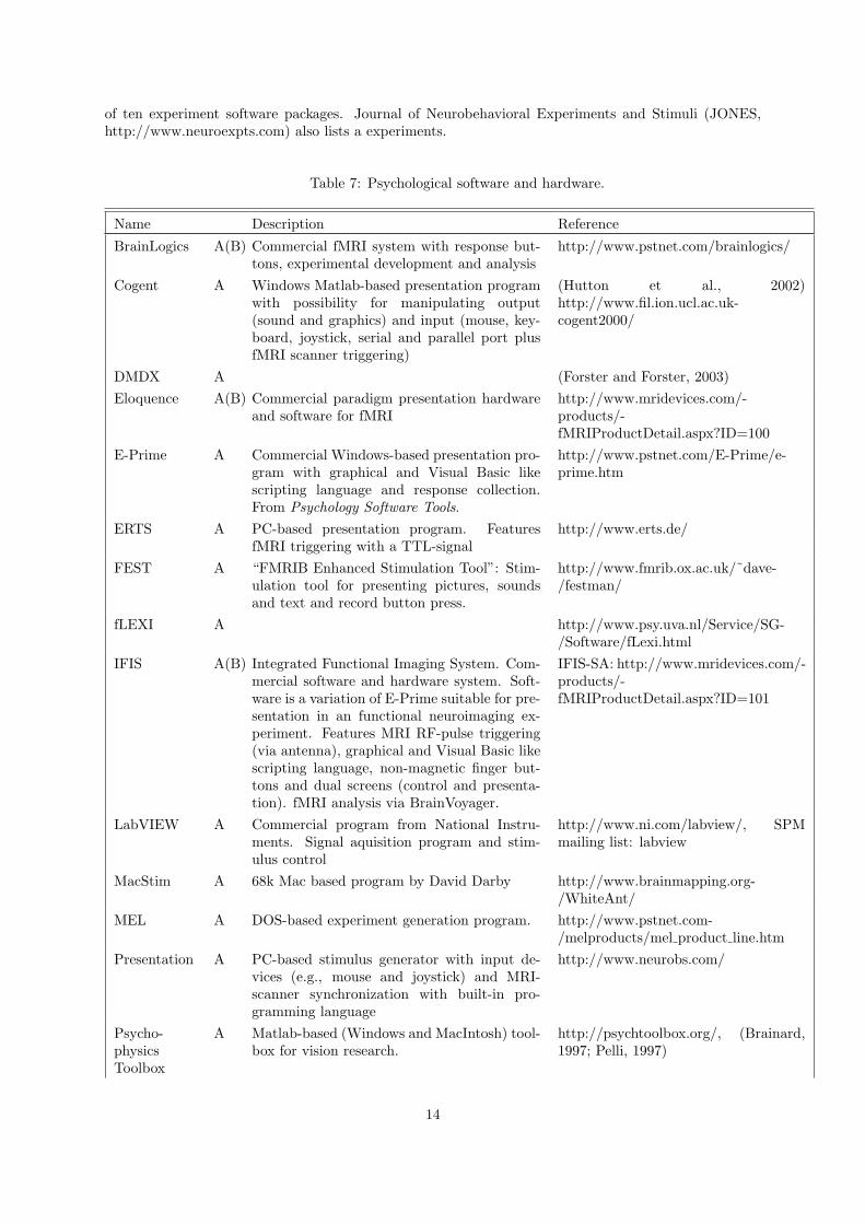
of ten experiment software packages. Journal of Neurobehavioral Experiments and Stimuli (JONES,http://www.neuroexpts.com) also lists a experiments.
Table 7: Psychological software and hardware.
Name Description Reference
BrainLogics A(B) Commercial fMRI system with response but-tons, experimental development and analysis
http://www.pstnet.com/brainlogics/
Cogent A Windows Matlab-based presentation programwith possibility for manipulating output(sound and graphics) and input (mouse, key-board, joystick, serial and parallel port plusfMRI scanner triggering)
(Hutton et al., 2002)http://www.fil.ion.ucl.ac.uk-cogent2000/
DMDX A (Forster and Forster, 2003)
Eloquence A(B) Commercial paradigm presentation hardwareand software for fMRI
http://www.mridevices.com/-products/-fMRIProductDetail.aspx?ID=100
E-Prime A Commercial Windows-based presentation pro-gram with graphical and Visual Basic likescripting language and response collection.From Psychology Software Tools.
http://www.pstnet.com/E-Prime/e-prime.htm
ERTS A PC-based presentation program. FeaturesfMRI triggering with a TTL-signal
http://www.erts.de/
FEST A “FMRIB Enhanced Stimulation Tool”: Stim-ulation tool for presenting pictures, soundsand text and record button press.
http://www.fmrib.ox.ac.uk/˜dave-/festman/
fLEXI A http://www.psy.uva.nl/Service/SG-/Software/fLexi.html
IFIS A(B) Integrated Functional Imaging System. Com-mercial software and hardware system. Soft-ware is a variation of E-Prime suitable for pre-sentation in an functional neuroimaging ex-periment. Features MRI RF-pulse triggering(via antenna), graphical and Visual Basic likescripting language, non-magnetic finger but-tons and dual screens (control and presenta-tion). fMRI analysis via BrainVoyager.
IFIS-SA: http://www.mridevices.com/-products/-fMRIProductDetail.aspx?ID=101
LabVIEW A Commercial program from National Instru-ments. Signal aquisition program and stim-ulus control
http://www.ni.com/labview/, SPMmailing list: labview
MacStim A 68k Mac based program by David Darby http://www.brainmapping.org-/WhiteAnt/
MEL A DOS-based experiment generation program. http://www.pstnet.com-/melproducts/mel product line.htm
Presentation A PC-based stimulus generator with input de-vices (e.g., mouse and joystick) and MRI-scanner synchronization with built-in pro-gramming language
http://www.neurobs.com/
Psycho-physicsToolbox
A Matlab-based (Windows and MacIntosh) tool-box for vision research.
http://psychtoolbox.org/, (Brainard,1997; Pelli, 1997)
14

Name Description Reference
PsyScope A Macintosh-based (OS7-OS9) “interactivegraphic system for experimental design andcontrol”. Development has ceased.
(Cohen et al., 1993; MacWhinney et al.,1997), http://psyscope.psy.cmu.edu/(original site), http://cnts.uia.ac.be-/psyscope/index.html (Belgian mirror)
PyEPL A “The Python Experiment-Programming Li-brary”: Python-based with playback andrecording of sound, displaying text, imagesand 3 dimensional environments with inputfrom keyboard and synchronization with ex-ternal recorded events
(Geller, 2006; Geller et al., 2006),http://pyepl.sourceforge.net/
Stim2 A Commerical Windows-based stimulus presen-tation from Neuroscan.
http://www.neuro.com-/product.sstg?id=58
Rings A Mac-based program for generation of simplevisual stimuli
http://porkpie.loni.ucla.edu-/BMD HTML/SharedCode-/SharedSoftware.html#Stimuli
SuperLab A Commercial Macintosh/Windows-based ex-periment generator supporting a number ofinput devices (keyboard, mouse, microphone,Cedrus and PST response boxes, I/O cards).From Cedrus Corporation.
http://www.superlab.com/
Vision Egg A 2D and 3D visual stimulus creation and con-trol open source software based on python andOpenGL for Windows, Mac OS X, Linux, SGI
http://www.visionegg.org
5.4 MRI compatible hardware
Table 8 shows publicly available MRI compatible hardware. A fiber optic joystick is used in (Van Hornet al., 2001b). The Altra Felix pointing device is not made for use in MRI, but it has been used in a1.5T scanner (Balslev et al., 2004).
15

Name Items Reference
ADInstruments Finger electrodes, galvanic skin re-sponse, respiratory transducer, laserDoppler probe, termocouple probe
http://www.adinstruments.com-/products/MR list/research/
ASL 504LRO fMRI compatible eyetracker http://www.a-s-l.com-/504lro.htm
Avotec LCD projection system with eye track-ing, e.g., “Silent Vision” system.
http://www.avotec.org-/silentvision.htm
Cambridge Research Sys-tems
“Lumina” response pads and “MR-Eyetracker”
E.g., http://www.crsltd.com-/catalog/lumina/
Coldswitch LUMItouch� response pad and LUMI-touch Joystick
http://www.coldswitch.com-/medical.asp
Current Designs Fiber Optic Response Pad system(fORP): Buttons, joystick, trackball
http://www.curdes.com/
Imagelys fMRI response pads and fMRI synchro-nization interface
http://www.imagilys.com/fmri-dti-neurosurgery-products/
Invivo Research Patient monitoring, Pulse Oximetry,and e.g., “monitors ECG, Respira-tion, HR, SpO2, NIBP, with optionalEtCO2”
http://invivoresearch.com-/prod mpm.html
Mag Design and Engineer-ing
Response box, head constraints, tactilestimuli, eye tracking, joystick. Com-mercial products by Ben Krasnow
http://www.magconcept.com-/mri.html
Measurand “ShapeHand MRI” data glove http://www.measurand.com-/products/ShapeHand.html
MRI Devices Corporation IFIS, response pad, earphones, visualdisplay, control room console
http://www.mridevices.com-/products/ifis/
NeuroScan STIM stereoscopic visual (Silent Vi-sion�) and stereo auditory (SilentScan�) stimulation, MagLink EEGrecording
http://www.neuro.com-/neuroscan/stimfmri.htm
Nonin Portable Pulse Oximeter http://www.nonin.com
Resonance Technology Commander XG earphones, MRIVision2000 and VisuaStim XGA stereoscopichead-mounted displays
http://www.mrivideo.com-/temp/functionalmri/
Rowland Institute Response box by Chris Stokes(?) http://hill-server.rowland.org-/rurb/
Sven Haller Pneumatic MR compatible responsebox available “at a reasonable price”
SPM Mailing list, 2002-9-11
Table 8: MRI compatible hardware: Response pads, joysticks, displays, earphones and EEG recording.
16

6 Analysis and processing
6.1 Methods in neuroimaging
The literature presents (exceedingly) many methods for analysis of neuroscience data. Table 9 lists onlyfew for use within functional and molecular neuroimaging.
Table 9: Analysis methods.
Domain Name/topic Description Reference
DynamicPET
“Logan plot”, theclassic Logan plot
(Logan et al., 1996)
DynamicPET
Logan’s ReferenceTissue Model
Logan’s plot with a reference re-gion used with k′
2 Implementedin PMOD.
http://pmod.com-/technologies/doc/pkin-/2322.htm
DynamicPET
MRTM0 Ichise’s (original) multi-linearreference tissue model
(Ichise et al., 1996),http://www.pmod.com-/technologies/doc/pxmod-/3250.htm
DynamicPET
MRTM Ichise’s (modified) multi-linearreference tissue model, a three-parameter model
(Ichise et al., 2003;Ichise et al., 2002),http://www.pmod.com-/technologies/doc/pkin-/2316.htm
DynamicPET
MRTM2 Ichise’s multi-linear reference tis-sue model, a two-parametermodel with fixed k′
2
(Ichise et al., 2003),http://www.pmod.com-/technologies/doc/pxmod-/3251.htm
fMRI BOLDresponsemodeling
Convolution withFIR-filter
Linear modeling of the responsefrom paradigm to fMRI signalwith a finite impulse response(FIR) filter.
(Nielsen et al., 1997; Goutteet al., 2000; Lange et al.,1999)
fMRI re-sponsemodeling
Convolution withBayesian estimatedFIR
Modeling the fMRI responsewith a finite impulse response(FIR) filter, Bayesian estima-tion, a Gaussian process prior,and Markov chain Monte Carlo(MCMC)
(Goutte et al., 2000; Ander-sen et al., 2002),
fMRI BOLDresponsemodeling
Convolution,Gamma
A gamma probability densityfunction modeling the hemody-namic response, estimation inthe frequency domain, and mod-eling of the noise in the frequencydomain. Partly implemented inLyngby
(Lange and Zeger, 1997)
fMRI BOLDresponsemodeling
Bayesian balloons Balloon models, extended bal-loon models with BayesianMarkov chain Monte Carlo andsplit-half resampling.
(Jacobsen et al., 2008)
17

Domain Name/topic Description Reference
fMRI un-supervisedmultivariate
K-means clustering K-means clustering of voxelswith use of cross-correlationfunction between paradigm andfMRI signal. Estimation of thenumber of clusters.
(Goutte et al., 1999b)
fMRI un-supervizedmultivariate
K-means clustering Clustering of voxel based on fea-tures from, e.g., and FIR-filtermodeling of the hemodynamicresponse
(Goutte et al., 2001; Goutteet al., 1999a)
fMRI un-supervisedmultivariate
Independent com-ponent analysis(ICA)
There are many studies in thisdomain
See Bibliography on Indepen-dent Component Analysis inFunctional Neuroimaging
fMRI semi-supervized
Univariate correla-tion across subjects
Showing the same stimulus toseveral subjects and analysis theintersubject correlation
(Hasson et al., 2004)
Analysis Image-based co-variates
(Biological parametric mapping) (Casanova et al., 2007; Oakeset al., 2007; Mehta et al.,2006)
Prediction PCA and neuralnetwork classifier
Principal component analysisand an artificial neural network
(Lautrup et al., 1995; Hansenet al., 1994)
Prediction Neural networkclassifier
SPECT predicting Alzheimerand controls
(Halkjær et al., 1997a;Halkjær et al., 1997b)
Prediction PET and fMRI,neural network,principal com-ponent analysis
(Mørch et al., 1997; Mørchet al., 1995; Mørch et al.,1996)
Prediction fMRI, neural net-work and principalcomponent analysiswith saliency maps
(Nielsen, 1996)
Predictionand repro-ducibility
Learning curves Demonstrated on four differentPET data sets. Prediction er-ror expressed in terms of mutualinformation and sensitivity mapshown.
(Kjems et al., 2002)
Prediction fMRI, evolutionaryalgorithm featureselection, linearclassifier, neuralnetwork, supportvector machinewith relevancemaps
Prediction (Aberg et al., 2006)
Connectivitymodeling
Structural equationmodeling, etc.
See the Bibliography on PathAnalysis
Imaging Location identifica-tion
(Reimold et al., 2005)
18

Domain Name/topic Description Reference
Multiplecomparisons
Permutation teston the maximumin the statisticalsummery image
(Holmes et al., 1996; Nicholsand Holmes, 2001)
Multiplecomparisons
Two-step FDR (Jiang and Doerge, 2006)
6.2 Tools
There exists a large number of programs for processing and analysis of brain signals, particularly withinthe functional neuroimaging field.
6.2.1 Neuroimaging
A review of some of the programs for fMRI analysis is available in (Gold et al., 1998): AFNI 2.01,SPM96, Stimulate 5.0, MEDIMAX 2.01, and FIT were tested; FIASCO, Yale, and MEDx 2.0. FIT(functional imaging toolkit) does not seem to be available any longer. More recent comments on toolsfor fMRI appear in (Nielsen et al., 2006).
For lists of image registration software see the Image Registration bibliogra-phy: http://www.imm.dtu.dk/˜fn/bib/Nielsen2001BibImage/. And for lists of brainsegmentation software see the “Bibliography of Segmentation in Neuroimaging”http://www.imm.dtu.dk/˜fn/bib/Nielsen2001BibSegmentation/.
Most programs work with volumes. The programs that allow for analysis or visualization of the 2Dcortical surface are BrainVoyager, CirclePack, FreeSurfer, SureFit/Caret, SUMA, SurfRelax and McGray
Apart from the listed programs there is also, e.g., CliniViewer, a program for display of multipleMRIs (Uttecht and Thulborn, 2002).
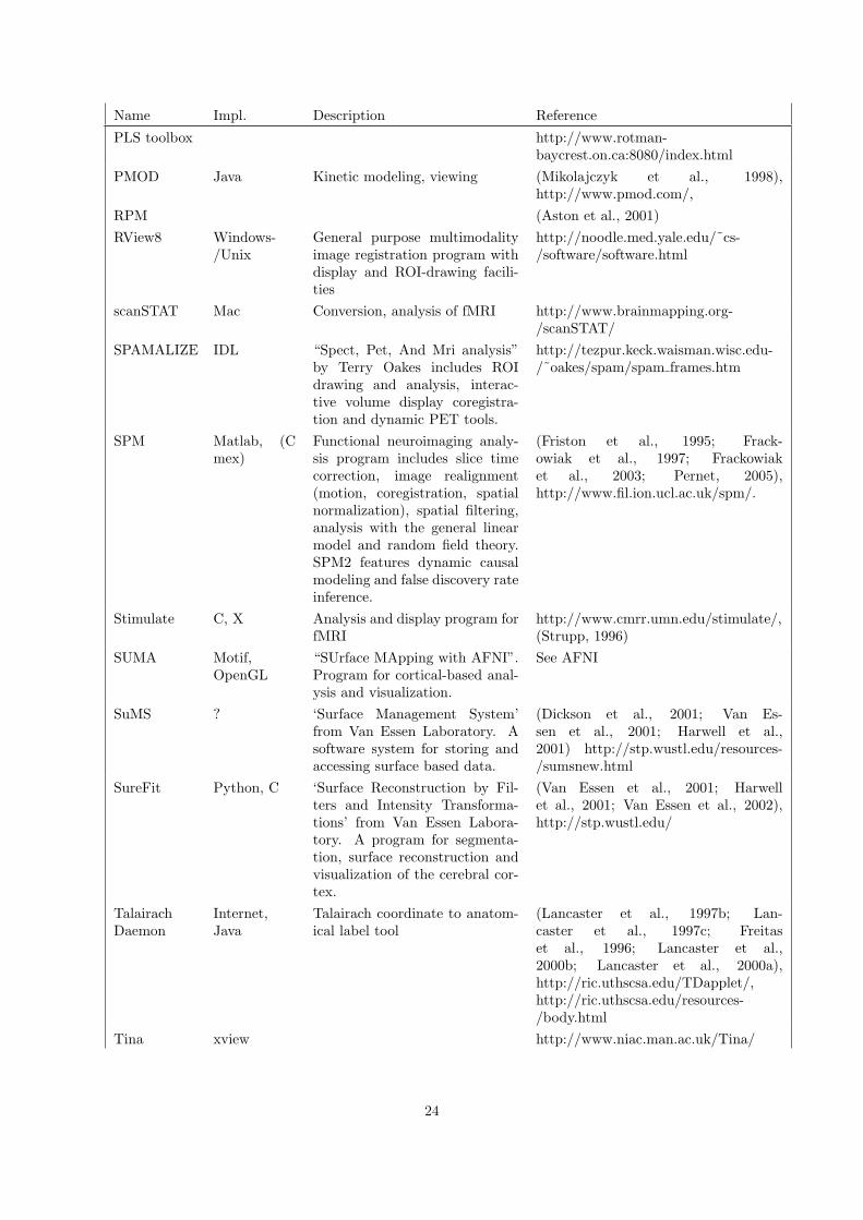
Table 10: Neuroinformatics tools for functional neuroimag-ing (PET and fMRI). The entries are alphabetically or-dered. See also The SfN Neuroscience Database Gatewayat http://big.sfn.org/ndg/site/, Internet Analysis Tools Registryat http://www.cma.mgh.harvard.edu/tools/, Andrew Crabb’sI do Imaging at http://idoimaging.com/ and NIH’s list ofvisualization software http://www.cc.nih.gov/cip/visualization-/vis packages.html.
Name Impl. Description Reference
3D slicer Visualization, segmentation, reg-istration
(Gering et al., 1999; Gering, 1999)http://www.slicer.org/
3DVIEWNIX X Commercial program for prepro-cessing, visualization and analy-sis of 3D imaging data. From theMedical Image Processing Groupand University of Pennsylvania.
http://www.mipg.upenn.edu/˜Vnews/
ABLe Solaris,Linux, SGIIRIX
Commercial program part ofMEDx for “Analysis of Brain Le-sions”
http://medx.sensor.com/products-/medx/able.html
Activ2000 Delphi5 Windows-based program forfMRI processing and analysis.
http://www.multimania.com-/dducreux/activ2000.htm
19

Name Impl. Description Reference
AFNI C Functional neuroimaging analy-sis program
(Cox, 1996; Cox and Hyde, 1997; Coxand Ward, 1997; Ward and Cox, 1997),http://afni.nimh.nih.gov/afni/
AIR C Image registration program (Woods et al., 1998a;Woods et al., 1998b)http://bishopw.loni.ucla.edu/AIR3/
AMIDE Unix, Mac,Windows
Volume viewing (Loening and Gambhir, 2003),http://amide.sourceforge.net/
Anatomist Unix Structural morphometry analy-sis, e.g., sulci identification. Partof BrainVISA
(Riviere et al., 2003),http://anatomist.info
Atamai Linux, MacOS X, win-dows
“Atamai Viewer” with 3D visual-ization. Image registration (com-mericial)
http://www.atamai.com/
BAMM “[S]oftware library for statisticalanalysis of structural and func-tional magnetic resonance (MR)images”
http://www-bmu.psychiatry.cam.ac.uk/BAMM/
BET Brain Extraction Tool byStephen Smith et al. Included inMRIcro
http://www.fmrib.ox.ac.uk/fsl/bet/
BPM Matlab “Biological Parametric MappingToolbox” matlab toolbox withimage-based regressors
(Casanova et al., 2007),http://www.fmri.wfubmc.edu
BrainMap� Internet,Java
Database with functional neu-roimaging results
(Fox and Lancaster, 1994; Fox et al.,1994; Fox et al., 1995; Lancaster et al.,1994; Lancaster et al., 1997a; Fox andLancaster, 2002; Fox et al., 2005a)http://www.brainmap.org
BRAINS2 X11,OpenGL,TCL/TK
“Brain Research: Analysis ofImages Networks and Systems”.Manual and automated tools forstructural analysis, tissue classi-fication and cortical surface ex-traction.
(Magnotta et al., 2002; Mag-notta and Andreasen, 2001),http://moniz.psychiatry.uiowa.edu-/local/brains2/brains2.html
Brain Tools Software by Krish Singh for theAnalysis of Structural and Func-tional Brain images. Consists ofmri3dX and mriWorkshopX.
http://www-users.aston.ac.uk-/˜singhkd/software.html
BrainVoyager C++/-OpenGL/Qt
Commercial program for fMRIanalysis and visualization
http://www.brainvoyager.de/
BrainWeb Internet ser-vice
Database with simulated MRI ofthe human brain
(Cocosco et al., 1997; Kwan et al., 1996;Kwan et al., 1999; Collins et al., 1998)http://www.bic.mni.mcgill.ca/brainweb/
BRIAN C, C++,Unix
Perhaps continued as LIPSIA? (Kruggel and Lohmann, 1996)
BSE Automated Brain Surface Ex-traction
http://www.loni.ucla.edu/NCRR-/Software/BSE.html
20

Name Impl. Description Reference
CamBA Windows,POSIX
Cambridge Brain Activation.fMRI analysis.
(Suckling et al., 2006b; Bullmoreet al., 1999; Bullmore et al., 2001;Suckling and Bullmore, 2004; Suck-ling et al., 2006a), http://www-bmu.psychiatry.cam.ac.uk/software/
Caret SGI/Linux-/Sun/Mac
‘Computerized AnatomicalReconstruction and EditingToolkit’ from Van Essen Labo-ratory. for ‘visualizing, editing,analyzing, and flattening corticalsurface reconstructions’.
(Drury et al., 1996; Van Essenet al., 2001; Harwell et al., 2001),http://brainmap.wustl.edu/caret/
CCHIPS© IDL “Cincinnati Children’s HospitalImage Processing Software” Pre-processing and analysis softwarefor fMRI, dMRI, spectroscopy,and pMRI.
http://www.irc.chmcc.org/chips.htm
CirclePack X-windows Software for flatmapping http://www.math.utk.edu/˜kens/,http://neurovia.umn.edu/incweb-/flatmap info.html
CLEAVE C “C Language Exploratory Anal-ysis of Variance with Enhance-ments” that is “designed for theanalysis of very large data sets ofthe sort obtained in experimentsusing EEG and fMRI”
http://www.ebire.org/hcnlab-/software/cleave.html
Corner Cube IDL or Web Visualization environment (Rehm et al., 1998; Schaperet al., 1996; Rehm et al., 1997),http://www.neurovia.umn.edu/-incweb/ccinfo.html Web demo:http://neurovia.umn.edu/incweb/-ccweb/
DP Tools Delphi5 Windows-based diffusion andperfusion MRI analysis programby Denis Ducreux
(Smith et al., 2000; Ducreux et al.,2001) http://www.multimania.com-/dducreux/DPTools.htm
Display Unix,OpenGL
A program to view MINC files ftp://ftp.bic.mni.mcgill.ca-/pub/register+Display/,http://www.bic.mni.mcgill.ca-/software/Display/Display.html,http://sourceforge.net/projects/mni-minc-win32/
EMMA Matlab “Extensible MATLAB Medicalimage Analysis” from BIC, MNI,McGill Reading and writing ofMINC files. Visualization, ROIand dynamic PET analyses
http://www.bic.mni.mcgill.ca-/software/emma/
EvIdent (Fuzzy) cluster analysis for neu-roimaging
(Pizzi et al., 2001),http://www.ibd.nrc-cnrc.gc.ca/english/info e evident.htm
FIASCO Shell, C Collection of programs (C andshell scripts) for processing offMRI.
http://lib.stat.cmu.edu/˜fiasco/ (Eddyet al., 1996)
21

Name Impl. Description Reference
fMRI Anal-ysis Package(Yale)
Matlab Analysis http://mri.med.yale.edu/individual/-pawel/fMRIpackage.html
FMRISTAT Matlab fMRI analysis program. Alsocalled BICstat and multistat
(Worsley et al., 2002;Worsley et al., 2000)http://www.bic.mni.mcgill.ca/˜keith/
FMRLAB Matlab Independent component analysisfor fMRI
http://sccn.ucsd.edu/fmrlab/
FreeSurfer Linux, Dar-win, Solaris,IRIX
Program for reconstruction ofthe cortical surface.
(Dale et al., 1999; Fischl et al., 1999a;Fischl et al., 1999b; Fischl and Dale,2000; Fischl et al., 2001; Busa, 2002)http://surfer.nmr.mgh.harvard.edu/
FSL C Preprocessing and analysis instructural and functional neu-roimaging by Stephen Smith etal.
(Smith et al., 2001; Smith et al.,2004), http://www.fmrib.ox.ac.uk-/fsl/index.html
GIFT Matlab Group ICA of fMRI Toolbox http://icatb.sourceforge.net/
GpetView C, Gtk+ Simple neuroimaging visualiza-tion of ANALYZE files
http://homepage2.nifty.com-/peco/gpetview/gpetview.html, Hi-roshi Watabe
HAMMER(*)
“Hierarchical Attribute Match-ing Mechanism for Elastic Reg-istration”. Skull-stripping andelastic warping
(Shen and Davatzikos, 2002;Shen and Davatzikos, 2003),http://www.rad.upenn.edu/sbia-/rsoftware.html
iBrain (Abbott and Jackson, 2001)
LIMA C++/-OpenGL
Software package for segmenta-tion and visualization of MRIs.
(Busch et al., 2001)
Lipsia C fMRI analysis program (Lohmann et al., 2001b; Lohmannet al., 2001a; Lohmann et al., 2000)
LOFA (Gokcay et al., 1999)
Lyngby Matlab Neuroimaging analysis (fMRIand PET) with a number of mul-tivariate (and univariate) analy-sis tools
(Hansen et al., 1999; Hansenet al., 2000; Hansen et al., 2001)http://hendrix.imm.dtu.dk/software-/lyngby/
MARINA Windows,Linux withWine
“MAsks for Region of INter-est Analysis” enables to “create,smooth, threshold, edit, and savemasks in an SPM-ANALYZEformat”
(Walter et al., 2003),http://www.bion.de/Marina.htm
MartinezFlat-Mapper
Web Flatmap and orthogonal planevisualizations of, e.g., specifiedTalairach coordinates
(Kang et al., 2004),http://www.ebire.org/hcnlab/cortical-mapping/
MEDx Solaris,Linux, IRIX,Tru64
Commercial multimodality brainimaging processing and anal-ysis software by Sensor Sys-tems. MEDx 3.4 contains, e.g.,SPM99, FSL, ABLe and “Func-tional Data Simulator”.
http://medx.sensor.com/
22

Name Impl. Description Reference
MIDAS(Tsui)
Solaris “Multimodal Image Data Anal-ysis System”. Region-of interestanalysis for multimodal 3D data
(Tsui et al., 2001)
MINC Unix, Win-dows
Tools for processing MINC files http://sourceforge.net-/projects/mni-minc-win32/,http://www.bic.mni.mcgill.ca/˜rotor-/distro/deb/
MPITool Unix “Multi Purpose Imaging Tool”from Advanced Tomo Vision.Reading and displaying of ECATfiles. Reslicing, region of inter-est, filter.
http://www.atv-gmbh.de/mpitoolh/,(Advanced Tomo Vision, 1999)
MRIcro Windows,Linux, So-laris
A program for conversion andviewing of 3D medical images byChris Rorden. Features also re-gion of interest (ROI) drawingand volume rendering
(Rorden and Brett, 2000),http://www.psychology.nottingham.-ac.uk/staff/cr1/mricro.html
MRIcron Linux, OSXand Win-dows (viaGTK)
A program somewhat similar toMRIcro
http://www.sph.sc.edu/comd/rorden-/mricron/
mri3dX Unix/OpenGL Program by Krish Singh “for vi-sualization and analysis of struc-tural and functional magneticresonance images”. Part of BrainTools
http://www-users.aston.ac.uk-/˜singhkd/mri3dX/
MRIVIEW IDL Visualization and segmentationof 2D and 3D brain images
(Ranken and George, 1997),http://www.biophysics.lanl.gov/brain-imaging/mriview/mriview.html
MRIWarp C Image registration program forMRI
(Kjems et al., 1999b; Kjems et al.,1999a), http://hendrix.imm.dtu.dk-/software/mriwarp/
MRVision Unix/Linux Commercial program for visual-ization and simple fMRI analysis
http://www.mrvision.com/
N3 Correction of intensity non-uniformity in MRI
(Sled et al., 1998; Sled et al., 1997; Sled,1997) http://www.bic.mni.mcgill.ca-/software/N3/
Neuro-Modeller
Windows 3D Visualization, 3D model gen-eration from contours
http://users.infohouse.com/amiller/
NPAIRS Unix, IDL “Nonparametric, Prediction,Activation, Influence, Re-producibility, re-Sampling”.Package with resampling forcomparing preprocessing andanalysis choices and returningreproducibility and predictionindices.
(Strother et al., 2002),http://www.neurovia.umn.edu-/incweb/npairs info.html
PI-WAVE Matlab Wavelet modeling for positronemission tomography
(Turkheimer et al., 1999; Turkheimeret al., 2000a; Turkheimer et al.,2000b), http://www.irsl.org/˜fet-/piwave/piwave.html
23
Name Impl. Description Reference
PLS toolbox http://www.rotman-baycrest.on.ca:8080/index.html
PMOD Java Kinetic modeling, viewing (Mikolajczyk et al., 1998),http://www.pmod.com/,
RPM (Aston et al., 2001)
RView8 Windows-/Unix
General purpose multimodalityimage registration program withdisplay and ROI-drawing facili-ties
http://noodle.med.yale.edu/˜cs-/software/software.html
scanSTAT Mac Conversion, analysis of fMRI http://www.brainmapping.org-/scanSTAT/
SPAMALIZE IDL “Spect, Pet, And Mri analysis”by Terry Oakes includes ROIdrawing and analysis, interac-tive volume display coregistra-tion and dynamic PET tools.
http://tezpur.keck.waisman.wisc.edu-/˜oakes/spam/spam frames.htm
SPM Matlab, (Cmex)
Functional neuroimaging analy-sis program includes slice timecorrection, image realignment(motion, coregistration, spatialnormalization), spatial filtering,analysis with the general linearmodel and random field theory.SPM2 features dynamic causalmodeling and false discovery rateinference.
(Friston et al., 1995; Frack-owiak et al., 1997; Frackowiaket al., 2003; Pernet, 2005),http://www.fil.ion.ucl.ac.uk/spm/.
Stimulate C, X Analysis and display program forfMRI
http://www.cmrr.umn.edu/stimulate/,(Strupp, 1996)
SUMA Motif,OpenGL
“SUrface MApping with AFNI”.Program for cortical-based anal-ysis and visualization.
See AFNI
SuMS ? ‘Surface Management System’from Van Essen Laboratory. Asoftware system for storing andaccessing surface based data.
(Dickson et al., 2001; Van Es-sen et al., 2001; Harwell et al.,2001) http://stp.wustl.edu/resources-/sumsnew.html
SureFit Python, C ‘Surface Reconstruction by Fil-ters and Intensity Transforma-tions’ from Van Essen Labora-tory. A program for segmenta-tion, surface reconstruction andvisualization of the cerebral cor-tex.
(Van Essen et al., 2001; Harwellet al., 2001; Van Essen et al., 2002),http://stp.wustl.edu/
TalairachDaemon
Internet,Java
Talairach coordinate to anatom-ical label tool
(Lancaster et al., 1997b; Lan-caster et al., 1997c; Freitaset al., 1996; Lancaster et al.,2000b; Lancaster et al., 2000a),http://ric.uthscsa.edu/TDapplet/,http://ric.uthscsa.edu/resources-/body.html
Tina xview http://www.niac.man.ac.uk/Tina/
24

Name Impl. Description Reference
TSU ‘TalairachSpace util-ity’. MatlabSPM plugin
Neuroimaging visualization http://www.ihb.spb.ru-/˜pet lab/TSU/TSUMain.html
UCLA BMD fMRI analysis programs contain-ing CC gr, T gr, Cproto, Over-lay
http://porkpie.loni.ucla.edu-/BMD HTML/SharedCode-/SharedSoftware.html
VoxBo UNIX ‘Software package for process-ing voxel-based functional brainimaging data’ made at the Uni-versity of Pennsylvania
http://www.voxbo.org
WFU Pick-Atlas
ROI-based analysis based on theTalairach Daemon and SPM
(Maldjian et al., 2003; Maldjian et al.,2004), http://www.fmri.wfubmc.edu-/download.htm
(X)MedCon C, Gtk+ Simple neuroimaging visualiza-tion and conversion
http://xmedcon.sourceforge.net, ErikNolf
Name Description Reference
BrainFX+GenericFX Execution environment http://www-staff.psychiatry.cam.ac.uk/˜co224-/projects/brainfx/
BrainVISA Execution environment in Python.Includes Anatomist
(Cointepas et al., 2001),http://brainvisa.free.fr/
FisWidgets Execution environment in Java (withSwing)
(Fissell et al., 2003),http://neurocog.lrdc.pitt.edu-/fiswidgets/
RUMBA Execution environment with visual-ization and visual data flow writtenin python
(Bly et al., 2004; Bly et al., 2001a),http://www.rumba.rutgers.edu/-projects.php
LONI Pipeline Pro-cessing Environment
Execution environment implementedwith visualization and visual dataflow in XML and Java
(Rex et al., 2003; Toga et al.,2001), http://www.loni.ucla.edu/-Software/Installing Detail.jsp?-software id=2
PVElab (PVEOut) Execution environmentand data processing environment inMatlab for, e.g., partial volume ef-fect correction
(Quarantelli et al., 2004),http://nru.dk/software/pveout/
SPM5 (spm jobman) Execution environ-ment for SPM5 functions with XMLderived from work of Philippe Ciuciuand Guillaume Flandin
http://www.fil.ion.ucl.ac.uk/spm/-software/spm5/
Table 11: “Meta”-software.
Table 11 lists a number of execution environments (or wrappers or pipelines) which can encapsulateother programs. An execution environment typically offers a graphical user interface where programvariables can be setup and the programs can be executed in arbitrary order. They will typically alsoallow for the storage of the parameters or the script so the setup and execution can be separated.
25

A few programs, e.g., SPM and AFNI, offer the possibility of plugins. SPM plugins are listed intable 12. These plugins are usually written by researchers that are not (directly) associated with theSPM developers. Note that some of the plugins are now outdated since their functionality is directlyimplemented in the standard distribution of later version of SPM, i.e., SPM2 and SPM5..
Table 12: SPM plugins. See also Thomas Nichols’ collectionof SPM extensions http://www.fil.ion.ucl.ac.uk/spm/ext/(where some of this information is taken from) as wellas his collection of John Ashburner’s code snippetshttp://www.sph.umich.edu/˜nichols/JohnsGems.html: ‘JohnGems’. A multivariate toolbox has been announced onhttp://www.fil.ion.ucl.ac.uk/spm/spm99.html but is presently(2002-10-24) not available.
Name Description Reference
ArtRepair fMRI artifact detection and repairingby Paul Mazaika
http://cibsr.stanford.edu/tools-/ArtRepair.htm
autospm2 Batch scripts for SPM2 http://www.md.ucl.ac.be/rdgn-/autospm2.html
batch general Front end to SPM batch analysis byRuss Poldrack
http://sourceforge.net/projects/spm-toolbox
batch subject Toolbox by Russ Poldrack http://sourceforge.net/projects/spm-toolbox
CBMG-Tools A few function for visualization and ex-traction of fMRI time series
http://www.brain.northwestern.edu-/cbmg/cbmg-tools/
CCA-fMRI Canonical correlation analysis for fMRI (Borga, 1998; Friman et al., 2003),http://cca-fmri.sourceforge.net
Conjunction SPM2 (1spm2 conj) and SPM99(spm99 conj) modifications for “con-junction null” conjunction inference
(Brett et al., 2004),http://www.sph.umich.edu/˜nichols-/Conj/
Deformations Toolbox by John Ashburner with spa-tial normalization utilities, e.g., ex-traction of information from SPM99spatial normalization file ( sn3d.mat)for deformation-based morphometry(DBM) or tensor-based morphometry(TBM)
ftp://ftp.fil.ion.ucl.ac.uk/spm-/toolbox/Deformations/
diffusion Toolbox by Russ Poldrack http://sourceforge.net/projects/spm-toolbox
Diffusion Toolbox by Volkmar Glauche for diffu-sion weighted MRI
http://www.uke.uni-hamburg.de-/kliniken/neurologie/spm/downloads/
DISTANCE Specialized permutation test. (Meriaux et al., 2006a;Meriaux et al., 2006b),http://www.madic.org/download-/DISTTBx/DISTTBx main.html
Dynamic PET Toolbox by Florian Wilke and RalphBuchert from Hamburg includes re-alignment, spatial normalization, mod-eling of dynamic PET and SPECT andVOI analysis.
Available via email (SPM mailing list,2000-04-04)
26

Name Description Reference
ezSPM2 GUI for batch construction in SPM2 http://www.izkf.rwth-aachen.de/downloads/ezspm2.htm
FDR False discovery rate extension andpatch for SPM by Thomas Nichols.This plugin has been included in SPM2.
http://www.sph.umich.edu-/˜nichols/FDR/
IBASPM “Individual Brain Atlases using Sta-tistical Parametric Mapping software”.SPM2 plugin that combines the AALatlas and tissue segmentation for con-struction of segmentation of individu-als.
http://www.thomaskoenig.ch/Lester/-ibaspm.htm
KUL Also called KUL SPM, kulSPM andKULeuven. Batch script for SPM99
http://www.kuleuven.ac.be/radiology-/Research/fMRI/kulSPM/
LI-Tool SPM2/5 toolbox for computation of lat-eralization indices
Marko Wilke, Tubingen
LOR2SPM Converts output from LORETA intoSPM compatible format. Program bySergey Pakhomov.
http://www.ihb.spb.ru/˜pet lab/L2S-/L2SMain.htm
MarsBar Region of interest toolbox by MatthewBrett
(Brett et al., 2002), http://www.mrc-cbu.cam.ac.uk/Imaging/marsbar.html
mascoi “MASked COntrast Images”. A tool-box/function to combine a contrast im-age with z-score image
(Reimold et al., 2005),http://homepages.uni-tuebingen.de-/matthias.reimold/mci/mascoi.m
mfBox Model-free toolbox http://mips.gsf.de/proj/cmb-/researchmfbox.html
MM Toolbox Multivariate analysis (principal compo-nent analysis and multivariate linearmodels, partial least squares) by CEA-SHFJ
http://www.madic.org/download-/MMTBx/
MNI Space utility Anatomical label tool http://www.ihb.spb.ru/˜pet lab-/MSU/MSUMain.html
PCT ‘Percent Change Threshold’ toolbox byThomas Nichols
http://www.sph.umich.edu/fni-stat-/PCT/ (Nichols, 2002)
PSPM Parallel implementation of SPM imageprocessing and analysis procedures us-ing LAM/MPI and MPICH
http://sourceforge.net/projects-/parallelspm/
RobustWLS A toolbox that performs weighted leastsquares estimation to account for arti-facts, e.g., head motion.
(Diedrichsen and Shadmehr, 2005),http://www.bangor.ac.uk/˜pss412-/imaging/robustWLS.html
roi Definition of ROI and extraction of sig-nal by Russ Poldrack
http://sourceforge.net/projects/spm-toolbox
SEM Structural equation modeling by Chris-tian Buchel
In beta test.
slice overlay 2D visualization by Matthew Brett ca-pable of merging an anatomical andfunctional image. Also called “Slice dis-play”
http://www.mrc-cbu.cam.ac.uk-/Imaging/display slices.html
27

Name Description Reference
SLT Region of interest analysis, batchscripting, semiautomated ROI iden-tification, Contains RAT2, ASAP,SurfTools, FSTools.
(Nieto-Castanon et al.,2003; SpeechLab, 2003),http://speechlab.bu.edu/SLT.php
SnPM Nonparametric permutation tests.SnPM99 is for SPM99 and SnPM2 isfor SPM2
(Nichols and Holmes, 2001; Holmeset al., 1996; Holmes, 1998),http://www.fil.ion.ucl.ac.uk/spm-/snpm
spm2 batch (Ash-burner)
Batch program for SPM2 programmedby John Ashburner
ftp://ftp.fil.ion.ucl.ac.uk/spm/toolbox-/spm2 batch 031210.tar.gz (previewversion)
spm2 batch Batch program from SPM programmedin Cambridge
ftp://ftp.mrc-cbu.cam.ac.uk/pub-/imaging/SPM2 batch
SPM Anatomytoolbox
Region of interest tools with probabilis-tic Brodmann areas atlas
(Eickhoff et al., 2005; Eick-hoff, 2005), http://www.fz-juelich.de/ime/spm anatomy toolbox
spm batch Batch preprocessing by SebastianThees
http://www.charite.de/ch/neuro-/forschung/teams/klinisch/people-/thees/index.html
SPMd ‘Statistical Parametric Mapping Diag-nosis’ by Wen-Lin Luo and ThomasNichols for residual analysis
(Luo and Nichols, 2003),http://www.sph.umich.edu/˜nichols-/SPMd/
spmjob Batch processing toolbox for SPM2with GUI.
http://spmjob.ffii.org/
spm loop Batch processing for display multipleresult images (SPMs).
http://www.mrc-cbu.cam.ac.uk-/Imaging/Common/spm loop.shtml
spm orthoviews ROI and movie tools by VolkmarGlauche.
http://www.uke.uni-hamburg.de-/kliniken/neurologie/spm/downloads/
SPM Script Front-end tool for automatic fMRIanalysis
http://www.nfil.rwth-aachen.de
spm xbrain Visualization extension by Tom Sieger http://www.neuro.lf1.cuni.cz/spm-/spm xbrain 3d.html
SPM XML Toolbox A toolbox for storing point activationfrom SPM in the XML file format.
(Keator et al., 2005; Keator,2005), http://www.nbirn.net-/Resources/Users/Applications-/xcede/SPM XMLTools.htm
Talairach Spaceutility
Neuroimaging visualization http://www.ihb.spb.ru/˜pet lab/TSU-/TSUMain.html
Volumes Toolbox by Volkmar Glauche for vol-ume operations, e.g., extraction of sub-volumes.
http://www.uke.uni-hamburg.de-/kliniken/neurologie/spm/downloads/
Unwarp ‘Toolbox for estimation and removalof movement-by-susceptibility inducedvariance in fMRI time series’
(Andersson, 2001),http://www.fil.ion.ucl.ac.uk/spm-/toolbox/unwarp.html
xjView SPM2 visualization http://people.hnl.bcm.tmc.edu/cuixu-/xjView/
Zephyr SPM2 batch for preprocessing http://diddy.dartmouth.edu/zephyr/
28

6.2.2 Computational neuroscience and simulators
Table 13 shows software in connection with neuronal modeling. (De Schutter, 1992) is an early review ofseven neuronal modeling software: AXONTREE, GENESIS, NEURON, NEMOSYS, NODUS, SABERand SPICE. GENESIS and NEURON are probably the most widely used. See also the sort review of(Jeong, 2005).
Table 13: Computational neuronal models. See alsolist at http://www.emsl.pnl.gov:2080/proj/neuron/neuro/systems-/shareware.html
Name Description Reference
GENESIS http://www.bbb.caltech.edu-/GENESIS/
NEST The “NEural Simulation TechnologyInitiative”. A program suitable formany simple neurons.
(Diesmann and Gewaltig, 2003),http://www.nest-initiative.org/
NeuGen “Generation of dendritic and axonalmorphology”
(Eberhard et al., 2006),http://neugen.uni-hd.de/
neuroConstruct “Biophysical neural network modelingsoftware”
(Gleeson et al., 2007),http://www.neuroconstruct.org/
Neurolator http://www.brainvoyager.com-/Neurolator.htm
NEURON http://www.neuron.yale.edu/
NEUSIM http://www.neosim.org/
NODUS http://bbf-www.uia.ac.be/SOFT/
NSL “Neural Simulation Language”. Neuralnetwork simulator language
(Weitzenfeld et al., 1999; Weitzenfeldet al., 2002), http://www-hbp.usc.edu-/Projects/nsl.htm
Surf-Hippo http://www.cnrs-gif.fr/iaf/iaf9/surf-hippo.html
XNBC (Vibert et al., 1997)http://www.u444.jussieu.fr/xnbc/
BMW “Brain Models on the Web” is adatabase of neuronal models
http://www-hbp.usc.edu/Projects-/bmw.htm
SenseLab ModelDB Database with “compartmental neuronmodels”. Contained 15 models 2001-10-25
(Peterson et al., 1996),http://senselab.med.yale.edu-/senselab/ModelDB/
6.2.3 Other areas
29

Table 14: Neuroinformatics tools — other tools. E.g., MEG, EEGand electrophysiology.
Name Impl. Description Reference
4D Toolbox Matlab Analysis and visualization of(Neuromag) MEG data
http://boojum.hut.fi/˜ojensen-/4Dtools/
ASA Windows(?) “Advanced source analysis”.EEG or MEG analysis withsource analysis from ANT.
http://www.ant-software.nl/asa/
BESA Windows “Brain Electrical Source Anal-ysis”. Commercial EEG andMEG source localization.
http://www.besa.de/
Brain project(bp)
Windows,Linux
“a Delphi Library for EEG dataAnalysis and Display”
http://www.irisa.fr/siames/GENS-/mcongedo/MC Projects.html
BrainStorm Matlab MEG and EEG data processing,visualization and source localiza-tion
http://neuroimage.usc.edu/brainstorm/
CogniTrace Linux “EEG/ERP data acquisition for32 to 128 channels” from ANT.
http://www.ant-software.nl/erp/
Curry PC, HP,Sun, SGI
Commercial program fromNeuroscan with electromag-netic source localization andvisualization
http://www.neuro.com-/product.sstg?id=39
cvapp Java Neuronal morphology editor http://www.cns.soton.ac.uk-/˜jchad/cellArchive/cellArchive.html
EEGLAB Matlab Analysis (independent compo-nent analysis) and visualizationfor EEG and MEG by Ar-naud Delorme and Scott Makeigfrom Swartz Center for Compu-tational Neuroscience et al.
http://sccn.ucsd.edu/eeglab/
EEG toolbox Matlab EEG toolbox for visualization http://eeg.sourceforge.net/
ERPWAVELAB Matlab Analysis of event related EEGand MEG data in the time-frequency domain.
(Mørup et al., 2007),http://www.erpwavelab.org/
G-clamp LabView http://hornlab.neurobio.pitt.edu/
ILAB© Matlab Eye movement analysis soft-ware for data generated by IS-CAN©and ASL© (Gitelman et al., 2001),
http://www.brain.northwestern.edu-/ilab/
Klusters Linux “A graphical interface for spikesorting of extracellular neuronalrecordings”
http://klusters.sourceforge.net
LOFA (Gokcay et al., 1999)
LORETA “Low Resolution Brain Electro-magnetic Tomography”. Pro-gram for inversion reconstructionof the 3D electromagnetic dis-tribution from surface EEG andMEG recordings.
(Pascual-Marqui, 1999),http://www.unizh.ch/keyinst-/NewLORETA/LORETA01.htm
30

Name Impl. Description Reference
NeuroBench Java Program to analyze and viewelectrophysiological recordings
http://www-hbp.usc.edu/Projects-/neuronBench.htm
Neurolucida� Commercial software from Mi-croBrightField for neuroanatom-ical analysis, e.g., 3D neuron re-construction.
(Glaser and Glaser, 1990)http://www.microbrightfield.com-/prod-nl.htm
Neuro-Modeller
Windows 3D Visualization, 3D model gen-eration from contours
http://users.infohouse.com/amiller/
NeuroScholar Java MySQL Database frontend withmanagement of bibliography, his-tological and tracing data.
(Burns, 2001; Burns, 1998; Sha-habi et al., 1999; Burns et al.,2002; Burns et al., 2003),http://www.neuroscholar.org,http://chasseur.usc.edu/ns/
NeuroScope C++,Linux/KDE
“Viewer for electrophysiologicaland behavioral data with limitedediting capabilities”
http://neuroscope.sourceforge.net
NeuroSim Matlab “Neurosim is a MATLABprogram that implements aconductance-based model sym-pathetic neuron and can also cancreate synaptic template files fordriving G-clamp”
http://hornlab.neurobio.pitt.edu/
SOURCE Commercial EEG dipole sourcelocalization from Neuroscan
http://www.neuro.com/-product.sstg?id=40
Spike2 Windows Commercial software for datacapture and analysis of, e.g.,electroneurophysiological datamade by Cambridge ElectronicDesign.
http://www.ced.co.uk/spk4wglu.htm
TEMPO OpenGL 3D EEG scalp data plotting http://tempo.sourceforge.net/
XANAT X Windows Analysis and storage of neuralconnectivity data
(Press et al., 2001b; Ol-shausen and Press, 1994),http://redwood.ucdavis.edu/bruno-/xanat/xanat.html
6.3 Unclassified
(Stiber et al., 1997)http://www.compneuro.org/ includes CD-ROM that is included in connect ion with the book Com-
putational Neuroscience: Realistic Modeling for Experimentalists (Schutter, 2000).
31

7 Computer, computational data modeling issues
Name Description Reference
Computer Cluster fMRI preprocessing with a computer cluster (Erberich et al., 2000)
Peer-to-peer database Napster like service for brain imaging data (Bly et al., 2001b)
QBISM Database for 3D spatial data especially brain mappingdata. Built around Starburst DBMS.
(Arya et al., 1996a; Arya et al., 1994;Arya et al., 1993; Arya et al., 1996b)
BRAID Object-oriented database augmented with image pro-cessing and statistical operations based on Illustra� (Herskovits, 2000a; Letovsky et al.,
1998) http://braid.uphs.upenn.edu-/websbia/braid/
B-SPID Object-relational database for neuroimaging databased on Illustra� (Diallo et al., 1999a; Diallo et al.,
1997a)
BIRN “Biomedical Informatics Resource Network”: Ameri-can project to closely link universities for collaborationin neuroimaging by application of SDSC’s Storage Re-
source Broker (SRB).
http://birn.ncrr.nih.gov/http://www.npaci.edu/DICE/SRB/
NeuroCore Database framework used in, e.g., fMRIDC http://www-hbp.usc.edu/Projects-/neurocore.htm
NeuroML XML for neuroscience, more specifically neuronalmodeling
(Goddard et al., 2001; Crook et al.,2005) http://www.neuroml.org/
XNAT Software platform to store MR neuroimaging and re-lated data. The technologies used are Java, DICOMand XML.
(Marcus et al., 2007),http://www.xnat.org/
— 3-dimensional database of deep brain functionalanatomy for image-guided neurosurgery (IGNS). Cod-ing structure according to Tasker
(Finnis et al., 2000; Tasker et al., 1978)
Table 15: Computer/database issues.
7.1 Image Compression
There exist a number of general purpose lossless compression programs, e.g., gzip and compress. Theseprograms seldom make a very efficient compression of neuroimaging data. SmallTime (Cohen, 2000),(http://www.brainmapping.org/SmallTime/) is a program specifically designed for MRI sequences andworks with differences of 16 bit images. Other algorithms have been described (Thiran et al., 1996;Adamson, 2002; Wu and Forchhammer, 2004), some based on wavelets.
The DICOM standard specifies lossless compression via the JPEG-LS (ISO 14495-1) and JPEG 2000(15444-1) standards.
7.2 Databases
Most databases management systems (DBMS) are relational. These consist of tables and links betweentables. A wide-spread query language is SQL. Early relation database management systems (RDBMS)had very simple types: integer, floats, string, etc. and would not represent 3D neuroimaging data well.Extensible DBMS such as so-called object-relation DBMS (ORDBMS) augment the ordinary relationDBMS with (complex) user-defined types (objects). One of the first ORDBMS was POSTGRES, see,e.g., (Stonebraker and Kemnitz, 1991). This database was commercialized by Illustra, which in December1995 was bought by Informix. Informix was in turn bought by IBM in April 2001. Another extensibleDBMS is/was the Starburst DBMS, see, e.g., (Lohman et al., 1991). It’s query language “hydrogen” isbased on SQL. Open source ORDBMS version exists with PostgreSQL (http://www.postgresql.org/).EXODUS is yet another extensible database system (Carey et al, 1991).
7.2.1 Image databases
Content-based image retrieval (CBRI) uses image feature, e.g., for finding similar images in a database.
32

One finds descriptions of general systems (not necessarily for neuroscience images) with Photobook(Pentland et al., 1993), http://vismod.media.mit.edu/vismod/demos/photobook/, QBIC (Flickner et al.,1995) and Virage (Bach et al., 1996).
7.2.2 Medical image databases
(Tagare et al., 1997) discusses some of the issues in content-based retrieval approaches in medicaldatabase.
Clinical medical image databases are often refered to as “picture archiving and communication sys-tems” PACS (Huang and Taira, 1992). There are probably many commercial systems in this area. Onesuch is EasyViz by Medical Insight.
7.2.3 Neuroimaging image databases
(Bjaalie, 2002) discusses neuroscientific databases, particularly the NeSys database, and (Kotter, 2002)is an edited book about neuroscience databases. Other methods of image retrieval are presented in (Liuand Dellaert, 1998a; Liu and Dellaert, 1998b; Liu et al., 1998; Liu et al., 2001b; Liu et al., 2002).
A review of databases for neuroimaging appears in (Diallo et al., 1999b; Diallo et al., 1997b).Image archives and data sharing in research oriented brain mapping have been discussed in a number
of articles (Chicurel, 2000; Aldhous, 2000; Marshall, 2000; Nature editorial, 2000; Cohen et al., 2001;Barinaga, 2003; Toga, 2002b). This discussion was initiated after suggestion for required submission tothe fMRI Data center (Van Horn et al., 2001a; Van Horn and Gazzaniga, 2002). A review of neuroscienceatlas/databases is (Van Essen, 2002).
(Wang et al., 2006) implemented content-based image retrieval on fMRI contrast maps with a combi-nation of wavelets and image feature extraction. (Cho, 2005) discusses a retrieval system with fMRI andsingle unit recordings. The CCVT tool also enables query-by-example (Cornea, 2005). NeuroServ andNIRV are software tools mentioned by (Carley-Spencer et al., 2006). Table 16 lists other content-basedimage retrieval systems in the neuroimaging context.
Table 16: Medical image retrieval systems
Name Area Features and description Reference
— Lympho-proliferativedisorders
Shape (Fourier descriptors), texture(MRSAR), color. Image-based queries,trained classifier
(Comaniciu et al., 1999;Comaniciu et al., 1998).
— (Synthetic)MRI
Topographical relations, size, round-ness, orientation. Images representedas “attributed relational graphs”
(Petrakis and Faloutsos,1997; Petrakis, 2002)
BredeDatabase
Functionalneuroimag-ing
Finding nearest Talairach coordinate toa given location. “Grey-level” volumefrom voxelized locations. Determina-tion of related volumes in the BredeDatabase.
(Nielsen and Hansen,2004b; Nielsen,2003; Nielsen andHansen, 2002b),http://hendrix.imm.dtu.dk-/services/jerne/brede/
Dermatlas Clinical der-matologicalimages (pho-tographs,histological,biopsy)
Text-based (keywords, diagnosis, bodysite, pigmentation, ...). Internet-basedwith collaboration (users are able tosubmit new images).
http://www.dermatlas.org
33

Name Area Features and description Reference
I2Cnet MRI, ... ROI (location, shape, size: round-ness, compactness, area, orientation),texture (maximum probability, angularsecond moment, contrast, inverse dif-ference moment, entropy, correlation,variance, cluster shade, diagonal mo-ment, k statistics, fractal signature).Internet-based.
(Orphanoudakis et al.,1996)
(Jerne,“Relatedvolumes”)
Functionalneuroimag-ing
“Grey-level” volume: Voxelized loca-tions. Determination of related vol-umes in the BrainMap� database byvoxelization
(Nielsen and Hansen,2004b; Nielsen andHansen, 2002b),http://hendrix.imm.dtu.dk-/services/jerne/ninf-/relvol.html
(KMeD) MRI, x-ray,...
Shape, size, texture, topological re-lations. Spatial temporal query lan-guage: (KSTL). Features in a hierar-chical structure
(Chu et al., 1998)
MIMS Image descriptors: file type (JPEG,GIF), device (MRI, radio), domain (ra-diology), ... Topographical relations.Java Internet-based. Stores imagesvoice reports and general (text) data.Thesaurus on descriptors
(Chbeir et al., 1999;Chbeir et al., 2000)
WebMIRS X-rays http://archive.nlm.nih.gov-/proj/webmirs/
xBrain Functionalneuroimag-ing
Search for nearest Talairach coordi-nates.
http://www.xbrain.org/
7.2.4 Neuroinformatics database
In neuroimaging contexts the neuroimages themselves do not only have to be databased but all theassociated data as well. These associated data might be subject information such as demographic,clinical and genetic. Complications for the construction of databases for such data might be extensibilityand the secure transportation of data across multiple sites. Thoughts on the problems for such systemsappear, e.g., in (Bockholt et al., 2006) (“The MIND Institute”) and (Keator et al., 2006) (“XCEDE”,“BIRN” ).
Unique identifiers for authors are partially implemented in the Brede Database (Nielsen, 2003). Thishas also been suggested by (Falagas, 2006) for more broader application.
7.3 Computational neuroscience
The Blue Brain Project aims at modeling the neocortical column of the somatosensory cortex of youngrats based on the NEURON program and using supercomputers, and is introduced in (Markram, 2006).
7.4 Unclassified
OSIRIS (Ligier et al., 1994): This seems to be an image analysis/processing program rather than adatabase. http://www.expasy.ch/www/UIN/html1/projects/osiris/osiris.html
34

M. Bota and M. A. Arbib. The NeuroHomology Database. Neurocomputing, 38-40, pp. 1627-1631,2001.
(Hirsch and Koslow, 1999)(Miller et al., 2001; Gardner et al., 2001b).(Guimond et al., 1997): retrieval of corresponding brain structures from a database of medical images.Database Challenges and Solutions in Neuroscientific Applications (1997). Ali E. Dashti, Shahram
Ghandeharizadeh, James Stone, Larry W. Swanson, Richard H. Thompson
35

8 Meta-analyses
8.1 Function/location meta-analysis
Data source Purpose Method Reference
BrainMap� Assessment of variation inactivation focus
‘Functional volumes modeling’:Probability density modelingincorporating sample size
(Fox et al., 1997b; Fox et al.,1999; Fox et al., 2001; Foxet al., 1997a)
Lesion database (BRAID) Determine association be-tween 14 functional vari-ables and 90 brain struc-tural variables in elderlypeople
Manual delineation of infarct-likelesions. Chi-square contingencytable test between pairs of func-tional and structural variables.
(Letovsky et al., 1998; Her-skovits, 2000a)
Lesion database (BRAID) Determine associationbetween lesion sites anddevelopment of secondaryattention-deficit hyperac-tivity disorder (S-ADHD)in children
Manual delineation of lesion fromMRI, Mann-Whitney and Fisherexact test statistics in a brain im-age database (BRAID)
(Herskovits et al., 1999; Her-skovits, 2000a)
Lesion database (BRAID) (Herskovits, 2000b)
Literature Determination of areas in-volved in language produc-tion
(Indefrey and Levelt, 2000; In-defrey and Levelt, 1999)
Literature (25 studies) Distinction between dorso-lateral and frontopolar cor-tex
Hotelling’s T 2 statistics used tocompare two groups of activationsin Talairach space. χ2 used for re-gion test.
(Christoff and Grabrieli, 2000,page 176)
Literature Determining of differentactivation between 5 sets ofactivation foci each set in-volving different cognitivedemand
Multidimensional Kolmogorov-Smirnov applied multiple times.
(Duncan and Owen, 2000)
Literature (28 studies) Determination of areas in-volved in syntactic parsing
Region-based analysis (102 areas)with P -value threshold determinedfrom a binomial distribution.
(Indefrey, 2001)
BrainMap� Novelty detection innomenclature
Probability density modelingthrough adaptive kernel den-sity modeling conditioned onanatomical labels.
(Nielsen and Hansen, 2002c;Nielsen et al., 2001)
Literature (9 studies) Prediction of areas in-volved in single word read-ing
Probability density modelingthrough kernel density modelingof 154 Talairach coordinates
(Turkeltaub et al., 2002;Turkeltaub et al., 2001)
Table 17: Mathematical meta-analyses in functional neuroimaging.
Table 17 displays some of the meta-analyses that use mathematics/statistics. For an early review see(Fox et al., 1998). For a more recent overview see (Wager et al., 2007).
Meta-analysis of Talairach coordinates was pioneered by Peter T. Fox et al. under the name “func-tional volumes modeling” (FVM) (Fox et al., 1997b; Fox et al., 1999; Fox et al., 1997a). These originalstudies used parametric Gaussian models. Non-parametric modeling of the distribution of brain fociwas first described in two unpublished studies with Gaussian mixture modeling (Nielsen and Hansen,1999) and adaptive Gaussian kernel density modeling (Nielsen and Hansen, 2000). Later studies includeadaptive Gaussian kernel density modeling for database outlier detection (Nielsen and Hansen, 2002c;Nielsen et al., 2001; Nielsen et al., 2000), Gaussian kernel density modeling in connection with singleword processing (Turkeltaub et al., 2002; Turkeltaub et al., 2001), kernel density estimation in connectionwith Broca’s area and verbal working memory (Chein et al., 2002; Chein et al., 2001), kernel densitymodeling with a spheric uniform kernel in emotion (Wager et al., 2003), and a model combining kerndeldensity modeling with a Gaussian mixture model (Neumann et al., 2008). Another study is (Wager et al.,2004). A large number of similar studies appears in a special issue of the Human Brain Mapping, volume
36

25, issue 1 (Fox et al., 2005b). Functional volumes modeling is sometimes referred to as “voxelization”or “activation likelihood estimation” (ALE). Some form of meta-analysis with the use the BrainMap�database has been briefly described (Mahurin et al., 1995).
Determining statistical thresholds in one set of voxelized Talairach coordinates is described in(Turkeltaub et al., 2002; Turkeltaub et al., 2001) and (Nielsen, 2005). Statistical methods for deter-mining whether two sets are different are described in (Christoff and Grabrieli, 2000; Duncan and Owen,2000) and (Nielsen and Hansen, 2004a; Nielsen et al., 2004b; Nielsen et al., 2005; Nielsen et al., 2004a)and (Laird et al., 2005a). The multidimensional Kolmogorov-Smirnov used in (Duncan and Owen, 2000)is originally from (Peacock, 1983) and is also described and implemented in (Press et al., 1992, section14.7). Hotelling’s T 2 test was also used in (Berman et al., 1999, page 212) but not in connection with ameta-analysis. Statistical tests on warped coordinates are described in (Steel and Lawrie, 2004).
The Brede Toolbox automatically performs multivariate analyses such as singular value decomposition(principal component analyses), independent component analyses, non-negative matrix factorization andK-means on voxelized Talairach coordinates on the entire Brede Database (Nielsen, 2003; Nielsen et al.,2004c). Another multivariate analysis method, “replicator dynamics”, is suggested in (Neumann et al.,2005).
A number of meta-analytic studies have grown out of the BRAID database: (Herskovits et al., 1999;Megalooikonomou et al., 1999; Megalooikonomou et al., 2000; Megalooikonomou and Herskovits, 2001;Lazarevic et al., 2001; Herskovits et al., 2002).
Descriptive statistics of activation foci appears in (Markowitsch and Tulving, 1994), where the fractionof fundus activations over 30 PET studies is found.
Function/location meta-analysis without spatial information other than the text can also extractrelevation associations. Name entities has been extracted from well over 100,000 PubMed abstractswith the use of Unified Medical Language System (UMLS) and manually developed rules for rule-basedname entity extraction (Hsiao et al., 2007). This system extracted phrases/terms for brain function andneuroanatomy and built an interactive visualization system to display the function-anatomy graph.
Table 18: Meta-analysis in Talairach space of brain function. KDEis kernel density estimation (in Talairach space).
.
Area Function Method Description Reference
Left inferiorfrontal cortex
Semantic andphonologicalprocessing
Tables, plots Phonological pro-cessing dorsallywhile semanticventrally
(Poldrack et al.,1999)
Anterior cingu-late
Cognition, emo-tion
(Bush et al.,2000)
Many Cognition Tables, plots (Cabeza and Ny-berg, 2000)
Inferior frontal Phonological pro-cessing
Plots (Burton, 2001)
Prefrontal Cognition, emo-tion
Plots, warptransformation,MANOVA, KDE
Resampling wasused for signifi-cance test
(Steel andLawrie, 2004)
Orbitofrontal Plots (Kringelbach andRolls, 2004)
Medial frontal Self/Other Clustering(?) (Seitz et al.,2005)
37

Area Function Method Description Reference
Posterior cingu-late
As many as possi-ble
Text clustering,Hotelling’s test
Text mining onPubMed abstractfor clustering ar-ticles. There-after determina-tion of segrega-tion between co-ordinates in clus-tered articles
(Nielsen et al.,2005)
8.2 Connectivity analyses
Species System Reports Areas Conn. Levels Method ReferencesRat Hippocampus > 900 (14000) 23 0–3, c, x 2D and 5D MDS (from SAS
MDS), Cluster analysis (SAS6.09 MODECLUS), Venn dia-gram
(Burns andYoung, 2000)
Macaque Cortical sensory (Felleman andVan Essen,1991)
30/14 <,≤, ∅,≥, > Hierarchical analysis (Hilgetag et al.,2000b)
Cat Cortical sensory (Scannell et al.,1995)
22 <,≤, ∅,≥, > Hierarchical analysis as above
Cat Cortical Part of (Scan-nell et al., 1999)
55 892 0–3 Optimal set analysis, MDS (SASMDS), Cluster analysis (SASMODECLUS), small-world coef-ficient
(Hilgetag et al.,2000a)
Macaque Visual (Felleman andVan Essen,1991)
32 319 as above as above
‘Primate’ (Young, 1993) 73 834 as above as above
Macaque Somatosensorymotor
(Felleman andVan Essen,1991)
15 66 as above as above
Primate Cortical 19 (CoCoMac-Stry)
39 “3897tests”
0–3 Optimal set analysis, MDS (SASMDS), small-world coefficient
(Stephan et al.,2000a)
Table 19: Connectivity analyses
An early program for connectivity analysis is “Connection” (Nicolelis et al., 1990).(Kaiser and Hilgetag, 2004) used data from CoCoMac together with spatial positions from Caret to
get an approximation for the wiring length in cortex.(Toro and Paus, 2006) described a co-activation analysis of functional activation recorded in the
BrainMap� database and constructed a program for interactive visualization of the 6-D probabilisticmap. A smaller co-activation study analysing data from 126 papers focused on connections from thebasal ganglia (Postuma and Dagher, 2006).
A database with volumes for anatomo-functional connectivity might become available (Poupon et al.,2006).
There is a number of studies using the connectivity of the small worm Caenorhabditis elegans (Wor-mAtlas), e.g., for examining the small-world phenomenon (Watts and Strogatz, 1998), or explaining theneuronal placement (Chen et al., 2006; Ahn et al., 2006) and (Kaiser and Hilgetag, 2005, Society forNeuroscience).
8.3 Unclassified
Meta-analysis of ERP in schizophrenia (Bramon et al., 2001).(Young and Scannell, 2000)
38

9 Neuroscience terminology
Nomenclature for receptors and ion channels is found in (Alexander et al., 1999). The Mai atlas (Maiet al., 1997) has a hierarchy for selected brain structures. A text mining system for neuroanatomicalterms has been applied for thalamic nuclei (Srinivas et al., 2005).
Table 20 lists some fo the frameworks for description of neuroscience terms, particularly neu-roanatomy. Other frameworks: GRAFIP (Hudelot et al., 2006) and the system of (Dameron et al.,2004; Golbreich et al., 2005) where they are able to link the terms to 3D data.
Jorg Niggemann: Representation of Neuroanatomical Knowledge: The Description Language ADL.Terminology and Knowledge Engineering (Vol. 1) 1990: 200-209
Table 21: Functional and cytoarchitectonic areas. Seealso http://defiant.ssc.uwo.ca/Jody web/fMRI4Dummies-/functional brain areas.htm and (Heimer, 1994, pages 285–286).
Name Description Reference
cmc Caudal cingulate motor area. Posterior cin-gulate motor areas. Human area corre-sponding to macaque 24d
(Zilles et al., 1995)
cmr Rostral cingulate motor area. Human areacorresponding to macaque 24c. Anteriorcingulate motor areas
(Zilles et al., 1995)
DP Dorsal prelunate area. An area in themacaque bordering V3A, V4, 7a, LIP,VIP according to Felleman and Van Essen.Seems to be mostly used by Van Essen.
(Andersen, 1985; Van Essen, 2003;Felleman and Van Essen, 1991)
F1 Designation used by Matelli for macaqueprimary motor cortex.
(Matelli et al., 1985; Matelli et al.,1991)
F2 Macaque area in Brodmann area 6 (Matelli et al., 1985; Matelli et al.,1991)
F3 Macaque area in Brodmann area 6 (Matelli et al., 1985; Matelli et al.,1991)
F4 Macaque area in Brodmann area 6 (Matelli et al., 1985; Matelli et al.,1991)
F5 Macaque area in Brodmann area 6. Re-ported to contain “canonical” and “mirror”neurons
(Matelli et al., 1985; Matelli et al.,1991; Picard and Strick, 2001)
F6 Macaque area in Brodmann area 6. (Matelli et al., 1985; Matelli et al.,1991)
F7 Macaque area in Brodmann area 6 (Matelli et al., 1985; Matelli et al.,1991)
FA Precentral area (von Economo and Koskinas, 1925, pp.218, 260+)
FB Agranular frontal area (von Economo and Koskinas, 1925, pp.260+)
FBA
FC Intermediate frontal area (von Economo and Koskinas, 1925, pp.260+, 315+)
FCBm
PCop
39

Name Description Reference
FD Granular frontal area (von Economo and Koskinas, 1925, pp.260+, 336–364)
FD∆
FDΓ
FDC
FDE
FDL
FE Frontopolar area (von Economo and Koskinas, 1925, pp.260+, 364–373)
FEF Frontal eye field. Precentral cortex
FEL Limbic frontopolar area (von Economo and Koskinas, 1925, p.371)
FF Orbital area (von Economo and Koskinas, 1925, pp.260+, 373+)
FG Rectus gyrus area (von Economo and Koskinas, 1925, pp.260+, 384+)
FH Prefrontal area (von Economo and Koskinas, 1925, pp.391+)
FI Area frontoinsularis (von Economo and Koskinas, 1925, p.219)
FK Area piriformis frontalis (von Economo and Koskinas, 1925, p.219)
FL Area parolfactoria (von Economo and Koskinas, 1925,p. 219)
FM Area geniculata (von Economo and Koskinas, 1925, p.219)
FN Area praecommissuralis (von Economo and Koskinas, 1925, p.219)
FST Fundus superior temporal. Floor of superiortemporal area
hMT Human correspondent to MT
IA Insula (Bonin and Bailey, 1947, p. 78)
IB Insula (Bonin and Bailey, 1947, p. 78)
KO Kinetic occipital. A shape/contour (frommotion?) selective area
LA Anterior limbic area in macaque correspond-ing to Brodmann area 24. Heterotypic andagranular cortex
(Bonin and Bailey, 1947, pp. 26 and78)
LC “Posterior part of the cingulate gyrus andextends into the cingulate sulcus”. Corre-sponding to Brodmann area 23.
(Bonin and Bailey, 1947, p. 78)
LIP Lateral intraparietal area
LO “Lateral occipital”.
M1 Primary motor cortex.
MDP Medial dorsal parietal area
MST Medial superior temporal area
40
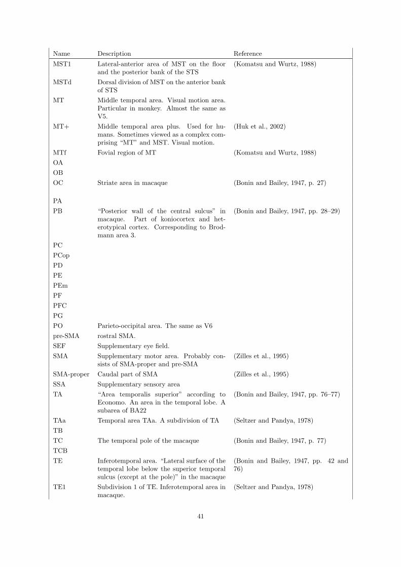
Name Description Reference
MST1 Lateral-anterior area of MST on the floorand the posterior bank of the STS
(Komatsu and Wurtz, 1988)
MSTd Dorsal division of MST on the anterior bankof STS
MT Middle temporal area. Visual motion area.Particular in monkey. Almost the same asV5.
MT+ Middle temporal area plus. Used for hu-mans. Sometimes viewed as a complex com-prising “MT” and MST. Visual motion.
(Huk et al., 2002)
MTf Fovial region of MT (Komatsu and Wurtz, 1988)
OA
OB
OC Striate area in macaque (Bonin and Bailey, 1947, p. 27)
PA
PB “Posterior wall of the central sulcus” inmacaque. Part of koniocortex and het-erotypical cortex. Corresponding to Brod-mann area 3.
(Bonin and Bailey, 1947, pp. 28–29)
PC
PCop
PD
PE
PEm
PF
PFC
PG
PO Parieto-occipital area. The same as V6
pre-SMA rostral SMA.
SEF Supplementary eye field.
SMA Supplementary motor area. Probably con-sists of SMA-proper and pre-SMA
(Zilles et al., 1995)
SMA-proper Caudal part of SMA (Zilles et al., 1995)
SSA Supplementary sensory area
TA “Area temporalis superior” according toEconomo. An area in the temporal lobe. Asubarea of BA22
(Bonin and Bailey, 1947, pp. 76–77)
TAa Temporal area TAa. A subdivision of TA (Seltzer and Pandya, 1978)
TB
TC The temporal pole of the macaque (Bonin and Bailey, 1947, p. 77)
TCB
TE Inferotemporal area. “Lateral surface of thetemporal lobe below the superior temporalsulcus (except at the pole)” in the macaque
(Bonin and Bailey, 1947, pp. 42 and76)
TE1 Subdivision 1 of TE. Inferotemporal area inmacaque.
(Seltzer and Pandya, 1978)
41

Name Description Reference
TE2 Subdivision 2 of TE. Inferotemporal area inmacaque.
(Seltzer and Pandya, 1978)
TE3 Subdivision 3 of TE. Inferotemporal area inmacaque.
(Seltzer and Pandya, 1978)
TEa Subdivision a of TE. Inferotemporal area inmacaque.
(Seltzer and Pandya, 1978)
TEc
TEm Subdivision m of TE. Inferotemporal area inmacaque.
(Seltzer and Pandya, 1978)
TEO
TEr
TF (Bonin and Bailey, 1947, pp. 42–43)
TG (Bonin and Bailey, 1947, pp. 38–39)
TH (Bonin and Bailey, 1947, p. 78)
TS1 Area in the supratemporal plane (inmacaque)
TS2 Area in the supratemporal plane (inmacaque)
TS3 Area in the supratemporal plane (inmacaque)
TSA Transitional sensory area. Upper bank ofcingulate sulcus (Macaca)
(Morecraft et al., 2004)
V1 Visual area one. Primary visual cortex.Striate cortex. Brodmann area 17.
V2 Visual area two. Extrastriate. Approxi-mately(?) Brodmann area 18 (with V3).
(Roland and Zilles, 1998)
V2d Visual area two, dorsal. Cuneus.
V2v Visual area two, ventral. Lingual gyrus
V3 complex Group of areas consisting of V3 and V3A(and V3B? ...). Orientation and form sensi-tive. Color insensitive.
V3 A visual area between V2 and V4. Some-times V3 is used to designate only the dorsalpart of V3, aka V3d. V3 is not a superareaof V3A
(Zeki, 1969; Van Essen, 2003)
V3A A dorsal area next to V3
V3B Visual area three, B. Possible the same areaas KO. V3A are possibly two area: V3A andV3B according to human studies by Smithand Press, see Van Essen 2003
(Smith et al., 1998; Press et al., 2001a;Van Essen, 2003; Zeki et al., 2003)
V3d Visual area three, dorsal. Lower visual field (Van Essen, 2003)
V3v Visual area three, ventral. Upper visualfield. Situated between V2 (V2v) and V4(V4v)
(Van Essen, 2003)
V4 Visual area four. Brodmann area 19. Colorsensitive. The prelunate gyrus in macaque
(Zeki, 1973), (Zeki, 1977, figure 16)
V4d Visual area four, dorsal.
42
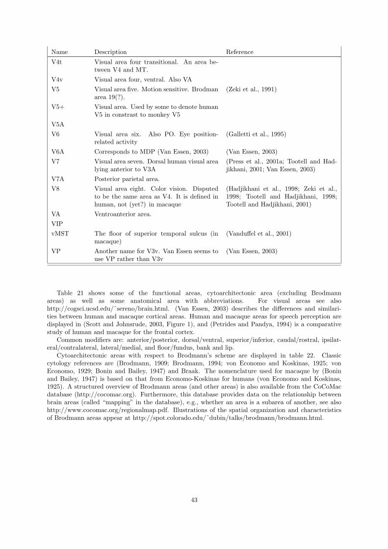
Name Description Reference
V4t Visual area four transitional. An area be-tween V4 and MT.
V4v Visual area four, ventral. Also VA
V5 Visual area five. Motion sensitive. Brodmanarea 19(?).
(Zeki et al., 1991)
V5+ Visual area. Used by some to denote humanV5 in constrast to monkey V5
V5A
V6 Visual area six. Also PO. Eye position-related activity
(Galletti et al., 1995)
V6A Corresponds to MDP (Van Essen, 2003) (Van Essen, 2003)
V7 Visual area seven. Dorsal human visual arealying anterior to V3A
(Press et al., 2001a; Tootell and Had-jikhani, 2001; Van Essen, 2003)
V7A Posterior parietal area.
V8 Visual area eight. Color vision. Disputedto be the same area as V4. It is defined inhuman, not (yet?) in macaque
(Hadjikhani et al., 1998; Zeki et al.,1998; Tootell and Hadjikhani, 1998;Tootell and Hadjikhani, 2001)
VA Ventroanterior area.
VIP
vMST The floor of superior temporal sulcus (inmacaque)
(Vanduffel et al., 2001)
VP Another name for V3v. Van Essen seems touse VP rather than V3v
(Van Essen, 2003)
Table 21 shows some of the functional areas, cytoarchitectonic area (excluding Brodmannareas) as well as some anatomical area with abbreviations. For visual areas see alsohttp://cogsci.ucsd.edu/˜sereno/brain.html. (Van Essen, 2003) describes the differences and similari-ties between human and macaque cortical areas. Human and macaque areas for speech perception aredisplayed in (Scott and Johnsrude, 2003, Figure 1), and (Petrides and Pandya, 1994) is a comparativestudy of human and macaque for the frontal cortex.
Common modifiers are: anterior/posterior, dorsal/ventral, superior/inferior, caudal/rostral, ipsilat-eral/contralateral, lateral/medial, and floor/fundus, bank and lip.
Cytoarchitectonic areas with respect to Brodmann’s scheme are displayed in table 22. Classiccytology references are (Brodmann, 1909; Brodmann, 1994; von Economo and Koskinas, 1925; vonEconomo, 1929; Bonin and Bailey, 1947) and Braak. The nomenclature used for macaque by (Boninand Bailey, 1947) is based on that from Economo-Koskinas for humans (von Economo and Koskinas,1925). A structured overview of Brodmann areas (and other areas) is also available from the CoCoMacdatabase (http://cocomac.org). Furthermore, this database provides data on the relationship betweenbrain areas (called “mapping” in the database), e.g., whether an area is a subarea of another, see alsohttp://www.cocomac.org/regionalmap.pdf. Illustrations of the spatial organization and characteristicsof Brodmann areas appear at http://spot.colorado.edu/˜dubin/talks/brodmann/brodmann.html.
43

Name Description Reference
BAMS “The Brain Architecture ManagementSystem”. Brain structure hierarchy,connections and chemicals
(Bota et al., 2003; Bota and Ar-bib, 2004; Bota and Swanson,2006; Bota and Swanson, 2007),http://brancusi.usc.edu/bkms/
BIRNLex Ontology for, e.g., neuroanatomy, be-havioral processes, diseases.
http://fireball.drexelmed.edu/birnlex-/OWLdocs/
BrainInfo Merging of NeuroNames and TemplateAtlas
(Bowden and Dubach, 2003),http://braininfo.rprc.washington.edu/
BrainMap� The BrainMap� database includes,e.g., a taxonomy for “behavioral do-mains”
http://brainmap.org
BredeDatabase
Small taxonomy for neuroanatomy(WOROI) and, e.g., brain func-tions, neuroreceptors, brain diseases(WOEXT). Used for classifying ex-periments and locations in the BredeDatabase.
(Nielsen, 2003; Nielsen, 2005),http://hendrix.imm.dtu.dk-/services/jerne/brede-/index roi alphabetic.html,http://hendrix.imm.dtu.dk-/services/jerne/brede-/index ext alphabetic.html
CoCoMac Brain areas such as Brodmann areas.Mapping between them called “objec-tive relational transformation” (ORT).
(Stephan et al., 2001; Stephan et al.,2000b; Stephan and Kotter, 1999),http://cocomac.org/
FMA Foundational Model of Anatomy. AWeb-based ontology for anatomy, in-cluding the brain. An item such as“straight gyrus” is defined with syn-onyms, latin name, part-of information,arterial supply and whether it has di-mension, mass and boundary.
(Rosse and Mejino Jr., 2003),http://sig.biostr.washington.edu-/projects/fm/.
(Jerne, “Vol-umes of In-terest”)
List of words and phrases used in the“lobar anatomy” field of BrainMap�.Generation of volumes and determina-tion of related volumes.
(Nielsen and Hansen, 2002a),http://hendrix.imm.dtu.dk/services-/jerne/ninf/voi.html
MeSH A hierarchy of medical terms used inconjunction with the PubMed web-service
http://www.nlm.nih.gov/mesh/
NeuroNames A hierarchy and thesaurus of neu-roanatomical names with an Internetservice. See also BrainInfo
(Bowden and Martin, 1995; Bow-den and Martin, 1997; Bowden andDubach, 2003; Martin et al., 1990)http://rprcsgi.rprc.washington.edu-/neuronames/
OpenGALEN Ontology for clinical data which in-clude, e.g., neuroanatomy
http://www.opengalen.org/, (Rectoret al., 1998)
Petilla 2005 GABAergic interneurons (Alonso-Nanclares et al., 2005)http://www.columbia.edu/cu/-biology/faculty/yuste/petilla/, Back-ground: (Yuste, 2005)
SBML Systems Biology Markup Language (Hucka et al., 2003), http://sbml.org/
Table 20: Neuroscience terminology, taxonomies and ontologies.
44

Table 22: Brodmann areas. BA and the Brodmann area num-bers. EK refers to Economo-Koskinas identifiers (von Economo andKoskinas, 1925; Zilles and Palomero-Gallagher, 2001) and (Roland,1992, figure 5.20).
BA EK Area Description Reference
1 PC Intermediatepostcentral area
Postcentral region (Brodmann, 1994, p. 112), (Zillesand Palomero-Gallagher, 2001, ta-ble 1)
2 PD, PDE (Caudal) post-central gyrus
(Brodmann, 1994, p. 112), (Zillesand Palomero-Gallagher, 2001, ta-ble 1)
3 (Rostral) post-central gyrus
(Brodmann, 1994, p. 112)
3a PA1 (Zilles and Palomero-Gallagher,2001)
3b PB1,PB2
Primary somatosensory area (Zilles and Palomero-Gallagher,2001)
4 Giant pyramidalarea
Precentral region, primary motorcortex
(Brodmann, 1994, p. 113)
4a 4 anterior “slightly larger, more denselypacked pyramidal cells in layerIII than [. . . ] 4p”
(Geyer et al., 1996), Julich atlas
4p 4 posterior Between 3a and 4a towards thebottom of the precentral gyrus.
(Geyer et al., 1996)
5 Preparietal area Parietal region
6 Agranular frontalarea
(Brodmann, 1994, p. 115)
7 Superior parietalarea
8 Intermediatefrontal area
(Brodmann, 1994, p. 116)
9 Granular frontalarea
10 Frontopolar area
11
12
13 Does not appear in humans
14 Does not appear in humans
15 Does not appear in humans
16 Does not appear in humans
17 Striate area (Brodmann, 1994, p. 119)
18 Occipital area (Brodmann, 1994, p. 120)
19 Preoccipital area (Brodmann, 1994, p. 120)
20 Inferior temporalarea
(Brodmann, 1994, p. 124)
21 Middle temporalarea
(Brodmann, 1994, p. 121)
45
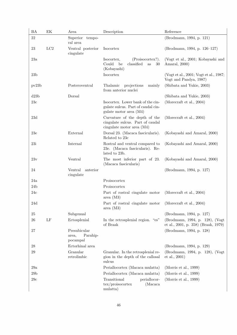
BA EK Area Description Reference
22 Superior tempo-ral area
(Brodmann, 1994, p. 121)
23 LC2 Ventral posteriorcingulate
Isocortex (Brodmann, 1994, p. 126–127)
23a Isocortex, (Proisocortex?).Could be classified as 30(Kobayashi)
(Vogt et al., 2001; Kobayashi andAmaral, 2000)
23b Isocortex (Vogt et al., 2001; Vogt et al., 1987;Vogt and Pandya, 1987)
pv23b Posteroventral Thalamic projections mainlyfrom anterior nuclei
(Shibata and Yukie, 2003)
d23b Dorsal (Shibata and Yukie, 2003)
23c Isocortex. Lower bank of the cin-gulate sulcus. Part of caudal cin-gulate motor area (M4)
(Morecraft et al., 2004)
23d Curvature of the depth of thecingulate sulcus. Part of caudalcingulate motor area (M4)
(Morecraft et al., 2004)
23e External Dorsal 23. (Macaca fascicularis).Related to 23c
(Kobayashi and Amaral, 2000)
23i Internal Rostral and ventral compared to23e. (Macaca fascicularis). Re-lated to 23b.
(Kobayashi and Amaral, 2000)
23v Ventral The most inferior part of 23.(Macaca fascicularis)
(Kobayashi and Amaral, 2000)
24 Ventral anteriorcingulate
(Brodmann, 1994, p. 127)
24a Proisocortex
24b Proisocortex
24c Part of rostral cingulate motorarea (M3)
(Morecraft et al., 2004)
24d Part of rostral cingulate motorarea (M3)
(Morecraft et al., 2004)
25 Subgenual (Brodmann, 1994, p. 127)
26 LF Ectosplenial In the retrosplenial region. “es”of Braak
(Brodmann, 1994, p. 128), (Vogtet al., 2001, p. 358) (Braak, 1979)
27 Presubiculararea, Parahip-pocampal
(Brodmann, 1994, p. 128)
28 Ectorhinal area (Brodmann, 1994, p. 129)
29 Granularretrolimbic
Granular. In the retrosplenial re-gion in the depth of the callosalsulcus
(Brodmann, 1994, p. 128), (Vogtet al., 2001)
29a Periallocortex (Macaca mulatta) (Morris et al., 1999)
29b Periallocortex (Macaca mulatta) (Morris et al., 1999)
29c Transitional periallocor-tex/proisocortex (Macacamulatta)
(Morris et al., 1999)
46

BA EK Area Description Reference
29d Proisocortex. Most medial partof 29. (Macaca mulatta)
(Morris et al., 1999)
29l Lateral 29 (Kobayashi and Amaral, 2000;Vogt et al., 2001)
29m Medial 29 (Kobayashi and Amaral, 2000;Vogt et al., 2001)
30 LD Agranularretrolimbic
Dysgranular. Proisocortex. Inthe retrosplenial region in the libof the callosul sulcus. “Variablethickness layer IV that is inter-rupted by large NFP-ir neuronsin layers IIIc and Va”
(Brodmann, 1994, p. 128), (Vogtet al., 2001; Morris et al., 1999;Morecraft et al., 2004)
30v Transition between 29 and visualassociation cortex
(Kobayashi and Amaral, 2000)
31 LC1 Dorsal posteriorcingulate
32 Dorsal anteriorcingulate
(Brodmann, 1994, p. 127)
33 Pregenual
34
35
36 Ectorhinal area,(Parahippocam-pal?)
(Brodmann, 1994, p. 122)
36’d (Vogt et al., 2001)
36’v (Vogt et al., 2001)
37
38 Temporopolar (Brodmann, 1994, p. 124)
39 PG Angular “Corresponds broadly to the an-gular gyrus”. Myeloarchitec-tonic PG and PEG (Eidelberg-Galaburda)
(Brodmann, 1994, p. 119), (Eidel-berg and Galaburda, 1984), (Zillesand Palomero-Gallagher, 2001, ta-ble 1)
40 Supramarginal
41 Medial (ante-rior transversetemporal area
(Brodmann, 1994, p. 125), Julichatlas
42 Lateral (poste-rior) transversetemporal area
(Brodmann, 1994, p. 125)
43 PDF (Zilles and Palomero-Gallagher,2001, table 1)
44 Opercular area Subfrontal region
45 Triangular area Subfrontal region
46 Middle frontalarea
46d Dorsal area 46
46v Ventral area 46(also v46)
47

BA EK Area Description Reference
47 Orbital area Subfrontal region
48
49
50
51
52 Parainsular (Brodmann, 1994, p. 125)
48

10 PET and SPECT
10.1 Molecular imaging
Table 23: Tracers. Partially based on (Gjedde, 2001; Smith et al.,2003; Knudsen and Svarer, 2002) and list from Turku PET Centrehttp://www.turkupetcentre.fi/pet tracers.html. Compound ini-tial keys: SB (SmithKline Beecham), GR (Amersham PhamarciaBiotech?).
Transmitter Enzyme, receptoror metabolism
Radiosubstrate orligand
Reference for PET, SPECT or autoradiogra-phy
Several D2+5HT2 +α1 [C-11]NMSP (N-methyl-spiperone)
(Wong et al., 1984; Morris et al., 1993), Nys-berg S 1993.
Dopamine DOPA-decarboxylase
[F-18]FDOPA
D1 [C-11]NNC 112 (Halldin et al., 1998; Abi-Dargham et al.,2000), Slifstein and Laruelle 2000(?)
D1 [C-11]NNC 756 (Laihinen et al., 1994)
D1 [H-3]SCH 23390 (De Keyser et al., 1988; Hall et al., 1988)
D1 [C-11]SCH 23390 (Farde et al., 1987; Wang et al., 1998; Chanet al., 1998; Karlsson et al., 2002)
D1 [C-11]SCH 39166 (Laihinen et al., 1994)
D2/D3 [C-11]-epidepride (Langer et al., 1999), Almeida P 1999, Hall H1997
D2/D3 [I-123]-epidepride (Leslie et al., 1996; Kuikka et al., 1997; Var-rone et al., 2000; Fujita et al., 2004), Kornhu-ber J 1995, Kessler RM 1991(?), Almeida P1999, Goldsmith and Joyce 1996.
D2/D3 [I-125]-epidepride (Hall et al., 1996; Hall et al., 1997)
D2/D3 [F-18]-fallypride (Mukherjee et al., 2002)
D2/D3 [C-11]FLB 457 (Farde et al., 1997b; Olsson et al., 1999;Okubo et al., 1999; Vilkman et al., 2000;Kaasinen et al., 2001; Suhara et al., 2001;Cselenyi et al., 2002), Sudo Y, 2001, Talvik2003.
D2/D3 [I-125]NCQ 298 (Hall et al., 1997)
D2 [H-3]-raclopride (Hall et al., 1988)
D2/D3 [C-11]-raclopride (Breier et al., 1998; Kestler et al., 2000), Farde1987, Volkov N 1993
D2 [H-3]-spiroperidol (De Keyser et al., 1988)
D2 [I-123]IBZM(iodobenzamide)
(Leslie et al., 1996), Kung HF 1990, LaulumaaV 1993, Seibyl 1996
D2/5HT-2A [F-18]FESP See under 5HT-2A
[I-123]IBZP
D2 [I-123]IDF
D2 R-[C-11]SKF 82957
D4 [C-11]SCH 66712
49

Transmitter Enzyme or Re-ceptor
Radiosubstrate orligand
Reference for PET, SPECT or autoradiogra-phy
DAT [C-11]CFT, [C-11]WIN 35,428
Wong 1993, Rinne 2004
DAT [F-18]CFT, [F-18]WIN 35,428
Laakso 1998, Nurmi 2000a, 2000b, Laakso2000
DAT [C-11]beta-CFT Rinne 1998
DAT [C-11]-cocaine (Telang et al., 1999)
DAT [N-C-11-methyl](-)-cocaine
Logan J 1990
DAT [F-18]-cocaine
DAT [C-11]FPCIT
DAT [C-11]PE2I (Halldin et al., 2003)
DAT [C-11]RTI 32
Norepine-phrine(nora-drenaline)
NET [C-11]MeNER (Wilson et al., 2003), Schou M 2003 (postmortem)
Histamine H1 [C-11]-doxepin (Yanai et al., 1992; Tagawa et al., 2001)
H1 [C-11]-pyrilamine (Yanie et al., 1992)
Serotonin(5-HT)
(Metabolism) α-[C-11]MTrp([C-11]AMT orα-[C-11]methyl-L-tryptophan)
(Okazawa et al., 2000).
[C-11]-HTP, [C-11]-5-hydroxy-L-tryptophan
Hagberg 2002
Tryptophan hy-droxylase
5-hydroxy-tryptophan-decarboxylase
5HT-1(?) [H-3]serotonin (Pazos et al., 1987a)
5HT-1A [H-3]8-hydroxy-2-[N,N-di-N-propyl-amino]tetralin
(Pazos et al., 1987a)
5HT-1A [H-3]8-OHDPAT
5HT-1A [C-11] CPC-222 Houle 1997
5HT-1A [O-methyl-C-11]WAY-100635
Pike 1995
5HT-1A [Carbonyl-C-11]WAY-100635
(Farde et al., 1998; Ito et al., 1999; Tauscheret al., 2001; Andree et al., 2002; Rabineret al., 2002; Parsey et al., 2002), Parsey 2000Pike 1996, Gunn 1998, Slifstein and Laruelle2000(?)
5HT-1A [Carbonyl-C-11]Desmethyl-WAY-100635
(Andree et al., 2002)
50
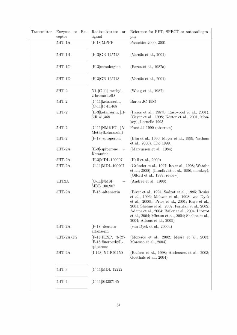
Transmitter Enzyme or Re-ceptor
Radiosubstrate orligand
Reference for PET, SPECT or autoradiogra-phy
5HT-1A [F-18]MPPF Passchier 2000, 2001
5HT-1B [H-3]GR 125743 (Varnas et al., 2001)
5HT-1C [H-3]mesulergine (Pazos et al., 1987a)
5HT-1D [H-3]GR 125743 (Varnas et al., 2001)
5HT-2 N1-[C-11]-methyl-2-bromo-LSD
(Wong et al., 1987)
5HT-2 [C-11]ketanserin,[C-11]R 41,468
Baron JC 1985
5HT-2 [H-3]ketanserin, [H-3]R 41,468
(Pazos et al., 1987b; Eastwood et al., 2001),(Geyer et al., 1998; Kotter et al., 2001, Mon-key), Laruelle 1993
5HT-2 [C-11]NMKET (N-Methylketanserin)
Frost JJ 1990 (abstract)
5HT-2 [F-18]-setoperone (Blin et al., 1990; Meyer et al., 1999; Yathamet al., 2000), Cho 1999.
5HT-2A [H-3]-spiperone +Ketamine
(Marcusson et al., 1984)
5HT-2A [H-3]MDL-100907 (Hall et al., 2000)
5HT-2A [C-11]MDL-100907 (Grunder et al., 1997; Ito et al., 1998; Watabeet al., 2000), (Lundkvist et al., 1996, monkey),(Offord et al., 1999, review)
5HT2A [C-11]NMSP +MDL 100,907
(Andree et al., 1998)
5HT-2A [F-18]-altanserin (Biver et al., 1994; Sadzot et al., 1995; Rosieret al., 1996; Meltzer et al., 1998; van Dycket al., 2000b; Price et al., 2001; Kaye et al.,2001; Sheline et al., 2002; Forutan et al., 2002;Adams et al., 2004; Bailer et al., 2004; Liptrotet al., 2004; Mintun et al., 2004; Sheline et al.,2004; Adams et al., 2005)
5HT-2A [F-18]-deutero-altanserin
(van Dyck et al., 2000a)
5HT-2A/D2 [F-18]FESP, 3-(2’-[F-18]fluoroethyl)-spiperone
(Moresco et al., 2002; Messa et al., 2003;Moresco et al., 2004)
5HT-2A [I-123]-5-I-R91150 (Baeken et al., 1998; Audenaert et al., 2003;Goethals et al., 2004)
5HT-3 [C-11]MDL 72222
5HT-4 [C-11]SB207145
51

Transmitter Enzyme or Re-ceptor
Radiosubstrate orligand
Reference for PET, SPECT or autoradiogra-phy
SERT [C-11]ADAM Shiue GG 2003 (in vitro, rat and baboon)
SERT [C-11]AFA
SERT [C-11]AFM
SERT [C-11]-cyano-imipramine
(Takano et al., 2002)
SERT [C-11]DAPA
SERT [C-11]DAPP (Houle et al., 2000)
SERT [C-11]DASB (Houle et al., 2000; Frankle et al., 2004)
SERT [C-11]MADAM Chalon 2003
SERT [C-11](+)McN-5652
(Szabo et al., 1995; Szabo et al., 1996; Bucket al., 2000; Takano et al., 2002; Frankleet al., 2004), McCann 1998, Parsey 2000, Sz-abo 1999, Simpson 2003, Szabo 2004, Ikoma2002
SERT [C-11]-NS 4194
SERT [I-123]ADAM
SERT [I-123]β-CIT Brucke 1993, Semple 1999, (Neumeister et al.,2000)
SERT [C-11]β-CIT Farde 1994
SERT [I-123]ZIENT Chen 2000
5HT-1A mRNA (Burnet et al., 1995)
5HT-2A mRNA (Burnet et al., 1995)
GABA GABA-A (BZ) [C-11]-flumazenil,[C-11]FMZ
(Holthoff et al., 1991; Grunder et al., 2001),Koeppe 1991
Microglia GABA-A (BZ)
[C-11](R)-PK11195 Banati
GABA-A (BZ) [F-18]-fluoroethyl-flumazenil (FEF)
(Grunder et al., 2001)
GABA-A (BZ) [I-123]Iomazenil Laruelle 1993, Onishi Y 1996
GABA-A (BZ) NNC 13-8241 (Kuikka et al., 1996)
GABA-A (BZ) Ro 15-4513
GABA-A (BZ) [C-11] Ro 15-1788 (Persson et al., 1985)
GABA-A (BZ) Diazepam
GABA-A [H-3]Muscimol (Geyer et al., 1998; Kotter et al., 2001, Mon-key)
Glutamate AMPA [H-3]AMPA (Geyer et al., 1998; Kotter et al., 2001, Mon-key)
Kainate [H-3]kainate (Geyer et al., 1998; Kotter et al., 2001, Mon-key)
NMDA [H-3]MK-801 (Geyer et al., 1998; Kotter et al., 2001, Mon-key)
Opioid µ, κ [F-18]-cyclofoxy Carson RE 1993
nonselective [C-11]-diprenor-phine
Jones 1988, Jones 1994, Frost 1990, Sadzot1991, Prevett 1994
52

Transmitter Enzyme or Re-ceptor
Radiosubstrate orligand
Reference for PET, SPECT or autoradiogra-phy
µ [C-11]-carfentanil (Frost et al., 1989; Zubieta et al., 1999), Frost1985, Frost 1990
δ [C-11]MeNTI (Smith et al., 1999)
Sigma σ1 [C-11]SA4503 Kawamura
Acetylcholine Nicotine [C-11]-nicotine
nAChR α4β2 [F-18] A85380
α1 [H-3]Prazosin (Geyer et al., 1998; Kotter et al., 2001, Mon-key)
α2 [H-3]Idazoxan(RX781094)
α2 [H-3]UK-14304 (Geyer et al., 1998; Kotter et al., 2001, Mon-key)
α 2A/2B H. De Vos 1992.
M1 [H-3]pirenzepine (Geyer et al., 1998; Kotter et al., 2001, Mon-key)
M2 [C-11]-tropanyl-benzilat (TRB)
Koeppe 1994, Lee 1996
M2 [F-18] FP-TZTP (Podruchny et al., 2003)
Muscarinic [C-11]NMPB(N-[C-11]methyl-4-piperidyl benzilate)
(Zubieta et al., 1998; Yoshida et al., 2000), Zu-bieta 2001, Sudo 1998, Kakiuchi 2001 (mon-key)
M2/M4 [H-3]oxotremorine-M
(Geyer et al., 1998; Kotter et al., 2001, Mon-key)
[C-11]scopolamine Frey 1992
[I-123]QNB(quinuclidinyl-iodo-benzilate)
Acetylcholinesteraseactivity
N-[C-11]methyl-piperidin-4-ylpropionate, [C-11]PMP
(Koeppe et al., 1999)
Acetylcholinesterasactivity
N -[C-11]methylpiperidin-4-yl acetate, [C-11]MP4A
(Nagatsuka Si et al., 2001)
— Monoamine oxi-dase A
[C-11]Deprenyl
— Monoamine oxi-dase B
[C-11]Clorgyline
— Vesicularmonoaminetransporter 2
[C-11]DTBZ
— — [C-11]-methyl-tetrahydroamino-acridine
(Traykov et al., 1999)
— — [C-11]-vinpocetine (Gulyas et al., 2002)
53

Table 23 lists a number of the radiotracers for PET and SPECT human brain imaging. Another list isthe North American PET directory of radiotracers available from http://www.snidd.org/petsearch.cfm.
Binding characterstics are available from http://www.gpcr.org/7tm/ligand/Seeman/S1A.html
10.2 Molecular neuroimaging and personality
(Farde et al., 1997a; Breier et al., 1998; Kestler et al., 2000; Suhara et al., 2001; Moresco et al., 2002).With CBF: (Johnson et al., 1999; Youn et al., 2002)
54

11 Magnetic resonance imaging (MRI)
Table 24: MRI sequences and techniques. See also General MRIAcronym Directory
.
Name Type Description Reference
ASL Perfusion Arterial spin labeling (Detre et al., 1994)
DW Diffusion weighted
EPI Echo planar imaging
EPISTAR Perfusion Echo planar imaging and signal targeting withalternating radiofrequency
(Edelman et al., 1994)
FAIR Perfusion Flow-sensitive alterating inversion recovery (Kim, 1995; Kwong et al.,1995)
FISP GRE Fast Imaging with Steady-State Precession
FLAIR IR Fluid Attenuation Inversion Recovery
FLASH GRE Fast Low Angle Shot
GE-MBEST Gradient Echo Modulus Blipped Echo planarShot Technique
GEMS Gradient Echo Multi Slice
GRASS GRE Gradient Recalled Acquisition in the SteadyState
GRE Gradient echo
HASTE Half-Fourier single shot turbo spin-echo
IR T1 Inversion recovery. Initial 180-pulse (inver-sion) followed by a 90-pulse
MPRAGE Magnetization Prepared RApid GradientEcho
PICORE ASL, tagging Wong et al 1997
PRESTO
QUIPPS ASL Quantitative imaging of perfusion using a sin-gle subtraction
(Wong et al., 1998)
RR-EPI Robarts Research Echo Planar Imaging
SE T1, T1, PD,diffusion
Spin echo. Slice selective 90 pulse and one ormore 180-pulse
SE-MBEST Spin Echo Modulus Blipped Echo planar Sin-gle pulse Technique
SEMS Spin Echo Multi Slice
SENSE Sensitivity Encoding (Puessmann et al., 1999),http://www.mr.ethz.ch-/sense/
SPGR GRE Spoiled Grass
Spiral Readout Readout in a spiral pattern in the k-space
SR T1 Saturation recovery
SS-FSE Single shot fast spin echo
STAR Signal Targeting with Alternating Radiofre-quency
STIR
55

Name Type Description Reference
Turbo FLASH FLASH
Turbo-PICORE ASL, tagging Wong et al 2000
UNFAIR ASL Uninverted FAIR
Object Oriented Development Interface for NMR (ODIN, http://od1n.sourceforge.net/) includingPulsar is a collection of for generation and simulation of MR sequences.
11.1 Contrast agents� Gadolinium diethylenetriaminepenta-acetic acid (Gd-DTPA) in fMRI: (Belliveau et al., 1991).� Iron: Superparamagnetic iron oxide (SPIO), ultra small paramagnetic iron oxide particles (USPIO).CASRN: 1317-61-9 (ferrosoferric oxide) (?).
– Monocryalline iron oxide MION: fMRI In rat (Palmer et al., 1999), fMRI in monkey (Vanduffelet al., 2001), Leite NeuroImage 16, 2002.
– Combidex� (Advanced Magnetics) in fMRI in rat: (van Bruggen et al., 1998), Combidex®– SPBPA (?)
– AMI-227 (?) (Sinerem or Combidex)
– NC 100150. (Clariscan, PEG-Ferron) (?)� Mn2+. Pautler 2002
56

12 Anatomical tracing
A list of tracer substances is available in table 25. A much more extensive list is available fromhttp://cocomac.org in the “tracer substance” field: http://cocomac.org/Help/Tracer.asp
“Afferent” (“sensory”) means a pathway from the peripheral system towards the central system, while“efferent” (“Motor”) is from the central to the peripheral system.
Table 25: Tracing techniques (Far from complete).
Name Transport Description Reference
Calbindin-D28K
A
FB R Fast blue. Fluorescent neuronal tracer.
HRP HR Horseradish peroxide.
TAA Tritiated amino acid
WGA-HRP Wheat germ agglutinin-conjugatedhorseradish peroxidase
MRI DWI
MRI restingstate
Strycnine
57

13 File formats
Alois Schlogl maintains a list of signal data formats mostly for EEG and other biomedical signals:http://www-dpmi.tu-graz.ac.at/˜schloegl/matlab/eeg/.
13.1 Neuroimaging volume data file formats
Table 26: Neuroimaging volume file formats. ECAT and Interfileinformation kindly provided by Frederic Schoenahl. Not all infor-mation might be accurate.
Name Ext. Description Conversion software Reference
AFNI BRIK,HEAD
Format with two files: bi-nary BRIK data file andACII HEAD header file.(Presumably) only used inAFNI.
AFNI http://afni.nimh.nih.gov-/afni/afni faq.shtml-#BRIKformathttp://afni.nimh.nih.gov-/afni/docREADME-/README.attributes
ANALYZE img,hdr
Format with two files: Bi-nary img data file andbinary hdr header file.There are different ver-sions of ANALYZE files,see Mayo Clinic’s informa-tion.
Mayo Clinic’s ANALYZE,NIH’s ImageJ, AndrewJankes’ software,
ANALYZE(SPM)
img,hdr,(mat)
Variation of an ANA-LYZE file format (version7) with offset and scalefields added. Widespreadfile format and used inSPM. SPM also uses anextra Matlab file to de-scribe a 3D transforma-tion.
Andrew Jankes’ soft-ware, Matthew Brett’sana4dto3d http://www.mrc-cbu.cam.ac.uk/Imaging-/ana4dto3d.html
http://www.fil.ion.ucl.ac.uk-/spm/distrib.html#AzeFmt
Bruker .5X1(?)
pvconvhttp://pvconv.sourceforge.net/
http://www.mrc-cbu.cam.ac.uk-/Imaging/Common-/brukerformat.shtml
COR (noneand“.info”)
MGH-NMR COR formatused in FreeSurfer. Onefile for each coronal slice.Prefix as converted byFreeSurfer is “COR”.
mri convert program inFreeSurfer.
?
Curry(CDR)
iso(???) Current density resultfrom the Curry Program
CdrSPM 1.0 from Seoul Na-tional University converts toSPM ANALYZE.
Descriptor des,dat
File format used by RIC,San Antonio
HIPG’s Alice� http://ric.uthscsa.edu-/projects/chsn/des.html
DICOM Extensive image data for-mat. Different vari-ants: DICOM3, DICOM2(ACR-NEMA)
NIH’s ImageJ, The commer-cial dicomread.m and dicom-info.m from Matlab Image Pro-cessing toolbox (works bothon Siemens and GE DICOM),MedX. SPM2, IDICON, Di-comWorks. See also next row.
http://medical.nema.org-/dicom.html, Newsgroup:comp.protocols.dicom, DavidClunie’s Medical ImageFormat Site
58

Name Ext. Description Conversion software Reference
DICOMSuper-Mosaic(?)
IMA(?)
Format used in SiemensSyngo (It is not clear —for the author — whichsubset of DICOM is usedby Siemens and GE and issupported, more informa-tion is needed)
volumestack fromxmedcon (see, e.g.,http://xmedcon.sourceforge.net-/faq/stack.html, kulCONV,MRIcro, Pieter Vandemaele’sdcmsmosaic.c, MySplitMo-saic.m
Newsgroup:comp.protocols.dicom.
ECAT .s .S .v.V .a.A .N
Format by CTI/Siemensprimarily for PET andSPECT able to store2D/3D sinograms andvolumes as well as bull-eyes. Filename extensionsare lowercase for data, up-percase for database entrycontaining sinogram,volume, attenuationdata, normalization data,respectively. ‘One file(datablock) per dataset,which contains a binaryheader with commonfields in nuclear medicine(dose.. patient name..)+ an image-type specificsubheader, and the imagedata coded in function ofthe image-type’.
cti2analyze(ftp://dormeur.topo.ucl.ac.be-/pub/ecat/); (x)medcon(http://xmedcon.sourceforge.-net/, version 6.4 of ECAT);
GE General Electric scannerformat
Souheil Inati’s GE2SPM(http://dbic.dartmouth.edu-/˜inati/tools/ge2spm.php).Krish Singh’s ge2spm(http://www.aston.ac.uk-/lhs/staff/singhkd/mri3dX-/mri3dX download.html) inBrainTools
Interfile HDR,IMG(CRV,ROI,H01,I01,C01,R01)
Format for nuclearmedicine (SPECT) witha binary image file andan ASCII header (notthe same header as ANA-LYZE though they havethe same file extension)
(x)medcon (http://xmedcon.-sourceforge.net/); IDI-CON Interfile/DICOMConversion Programhttp://www.inf.u-szeged.hu-/˜idicon/shortdoc.html
http://www.keston.com-/Interfile/, (Todd-Pokropeket al., 1992), Extension forPET http://www.irsl.org-/˜kris/petinterfile.html
MINC mnc File format from MNI inMontreal. Related toNetCDF. Used in MNI’sand Keith Worsley’s soft-ware. 3D and 4D versions.
EMMA, Andrew Jankes’ana2mnc—mnc2anahttp://www.cmr.uq.edu.au-/ rotor/software/, SatrajitGhosh’s ‘MINC to Analyze’http://www.cns.bu.edu-/˜satra/software.php
http://www.bic.mni.mcgill.ca-/software/minc/minc.html,http://poldracklab.psych.-ucla.edu/spm/minc spm.html
NetCDF General array data format http://www.unidata.ucar.edu-/packages/netcdf/index.html
59

Name Ext. Description Conversion software Reference
NIfTI-1 nii,hdr,img
A standard by “Neu-roimaging InformaticsTechnology Initiative”.Extension of ANALYZE.Planned support in AFNI,FSL and SPM.
http://nifti.nimh.nih.gov-/dfwg/src/
(Cox et al., 2004),http://nifti.nimh.nih.gov/dfwg/
PFF “The Pittsburgh File For-mat”. A file format usedin Pittsburgh fMRI com-munity
http://lib.stat.cmu.edu-/˜fiasco/overview/pff intro.html
SDT sdt,spr
Format used in Stimulate http://www.cmrr.umn.edu-/stimulate/stimUsersGuide-/node57.html
Siemens IMA Internal format used bySiemens. Not DICOM for-mat although it has thesame file extension, see(SPM mailing 2002-04-18)(???)
(x)medcon, visionToSPM
SimpleFileFormat
sfd,sfh
Used at Hvidovre Hospi-tal, Denmark
sf utils.tar.gz from HvidovreHospital.
http://www.drcmr.dk-/software/index.html
VAPET (none) File format used in VAMedical center, PETimaging service, Min-neapolis. Exists in a 3Dand 4D version. Headerinformation is in thebeginning of the file inASCII representation
MRIWarp from the TechnicalUniversity of Denmark
UNC s??? Multi-dimensional dataformat from University OfNorth Carolina. Used byBAMM
http://www-bmu.psychiatry.cam.ac.uk-/BAMM/FBAMM-/FILEFORMAT-/unc.html, unc2mnc,http://www.cmr.uq.edu.au-/˜rotor/software/
VISTA .v ? ? ? http://www.cs.ubc.ca/nest-/lci/vista/vista.html
XPrime ? (File format known fromHvidovre Hospital)
MRIWarp from the TechnicalUniversity of Denmark
Table 13.1 displays some of the file formats for functional neuroimaging (volumes). See also AndrewJanke’s list at http://www.cmr.uq.edu.au/˜rotor/minc/ as well as the list in connection with (x)medcon(http://xmedcon.sourceforge.net/docs/content.html). Chris Rorden has also a list of conversion software:http://www.psychology.nottingham.ac.uk/staff/cr1/mricro.html#otherconv.
The “Medical Image Format FAQ” is available, e.g., at http://www.cs.uu.nl/wais/html/na-dir/medical-image-faq/.html.
SPM uses a specific prefixing scheme for processed files: A character is added in front of the filename:masked files (m), realign/reslice (r), spatial smoothing (s), spatial normalization (n), slice timing (a) andwarped (w). MRIcro uses (l) for exported region of interests volume files.
60

NIfTI-1 is a new standard that is planned to be supported by Brain Voyager, AFNI, FSL and SPM(Cox et al., 2004). It is based on ANALYZE.
13.2 Other file formats
The Protein Data Bank format is for atomic coordinate files with the extension .mod, seehttp://www.umass.edu/microbio/rasmol/pdb.htm. These can be read by molecular visualization pro-grams, such as RasMol
61

References
Abbott, D. and Jackson, G. (2001). iBrain — software for analysis and visualisation of function MR im-ages. NeuroImage, 13(6, part 2):S59. http://www.apnet.com/www/journal/hbm2001/10839.html.ISSN 1053-8119.
Aberg, M. C. B., Wessberg, J., and Loken, L. S. (2006). A multivariate pattern recognition approach tofMRI analysis. In Neuroscience. Society for Neuroscience. #492.11/PP86.
Abi-Dargham, A., Martinez, D., Mawlawi, O., Simpson, N., Hwang, D.-R., Slifstein, M., Anjilvel, S.,Pidcock, J., Guo, N.-N., Lombardo, I., Mann, J. J., Heertum, R. V., Foged, C., Halldin, C., andLaruelle, M. (2000). Measurement of striatal and extrastriatal dopamine D1 receptor binding po-tential with [11C]NNC 112 in humans: validation and reproducibility. Journal of Cerebral BloodFlow and Metabolism, 20(2):225–43. PMID: 10698059. ISSN 0271-678X.
Adams, K. H., Hansen, E., Pinborg, L. H., Hasselbalch, S. G., Svarer, C., Holm, S., Bolvig, T. G., andKnudsen, G. M. (2005). Patients with obsessive-compulsive disorder have increased 5-HT2A receptorbinding in the caudate nuclei. International Journal of Neuropsychopharmacology. PMID: 15801987.
Adams, K. H., Pinborg, L. H., Svarer, C., Hasselbalch, S. G., Holm, S., Haugbol, S., Madsen,K., Frokjaer, V., Martiny, L., Paulson, O. B., and Knudsen, G. M. (2004). A database of[18F]-altanserin binding to 5-HT2A receptors in normal volunteers: normative data and relation-ship to physiological and demographic variables. NeuroImage, 21(3):1105–1113. PMID: 15006678.DOI: 10.1016/j.neuroimage.2003.10.046. ISSN 1053-8119.
Adamson, C. (2002). Lossless compression of magnetic resonance imaging data. Bachelor thesis, Schoolof Computer Science and Software Engineering, Monash University, Melbourne, Australia.
Advanced Tomo Vision (1999). MPITool. Multi Purpose Imaging Tool. Manual. AdvancedTomo Vision GmbH, Erftstadt, Germany, version 2.58, revision a6e edition. ftp://www.atv-gmbh.de/mpitool/mpitool e.pdf.
Aguirre, G. K., Detre, J. A., Zarahn, E., and Alsop, D. C. (2002). Experimental design and the relativesensitivity of BOLD and perfusion fMRI. NeuroImage. IDEALfirst.
Ahn, Y.-Y., Jeong, H., and Kim, B. J. (2006). Wiring cost in the organization of a biological neuronalnetwork. Physica A, 367:531+. DOI: 10.1016/j.physa.2005.12.013. http://www.nd.edu/˜hjeong-/physicaCelegans.pdf.
Aldhous, P. (2000). Prospect of data sharing gives brain mappers a headache. Nature, 406(6795):445–446.ISSN 0028-0836.
Alexander, S., Peters, J., Mead, A., and Lewis, S. (1999). TiPS receptor and ion chan-nel nomenclature supplement 1999. Trends in Pharmacological Sciences, 19(Sup-plement 1):1–85. http://www.sciencedirect.com/science/article/B6T1K-44P89DY-1/2-/dfed9e9f8e4cd9a0419808d5e4fdbc03.
Alonso-Nanclares, L., Anderson, S., Ascoli, G., Benavides-Piccione, R., Burkhalter, A., Buzsaki, G.,Cauli, B., DeFelipe, J., Fairen, A., Feldmeyer, D., Fishel, G., Fregnac, Y., Freund, T. F., Fukuyi,K., Glarreta, M., Goldberg, J., Helmstaedter, M., Hensch, T., Hestrin, S., Kisvarday, Z., Lam-bolez, B., Lewis, D., McBain, C., Marin, O., Markham, H., Monyer, H., Munoz, A., Petersen, C.,Rockland, K., Rossier, H., Ruby, B., Somogyi, P., Staiger, J. F., Tamas, G., Thomason, A., Toledo-Rodriguez, M., Wang, X.-J., Wang, Y., West, D., and Yuste, R. (2005). Petilla 2005: Nomencla-ture of features of GABAergic interneurons of the cerebral cortex. http://www.columbia.edu/cu-/biology/faculty/yuste/petilla/petilla-webpages/Nomenclature/PetillaNomenclaturefinal.pdf.
Andersen, I. K., Szymkowiak, A., Rasmussen, C. E., Hanson, L. G., Marstrand, J. R., Larsson, H. B.,and Hansen, L. K. (2002). Perfusion quantification using Gaussian process deconvolution. MagneticResonance in Medicine, 48(2):351–361. PMID: 12210944.
62

Andersen, R. A. (1985). Callosal and prefrontal associational projecting cell populations in area 7A of themacaque monkey: a study using retrogradely transported fluorescent dyes. Journal of ComparativeNeurology, 232(4):443–455. PMID: 3980763.
Anderson, J. E., Umans, C., Halle, M., Golland, P., Jakab, M., McCarley, R. W., Jolesz, F. A., Shenton,M. E., and Kikinis, R. (1998). Anatomy browser: Java-based interactive teaching tool for learninghuman neuroanatomy. RSNA EJ, 2. http://ej.rsna.org/ej2/0050-97.fin/index.html. ISSN 1090-7629.
Andersson, J. L. R. (2001). Modeling geometric deformations in EPI time series. NeuroImage, 13(5):903–919. PMID: 11304086. http://www.fil.ion.ucl.ac.uk/spm/papers/Unwarp/Unwarp.pdf.
Andree, B., Halldin, C., Pike, V. W., Gunn, R. N., Olsson, H., and Farde, L. (2002). The PET radioligand[carbonyl-11C]Desmethyl-WAY-100635 binds to 5-HT1A receptors and provides a higher radioactivesignal than [carbonyl-11C]WAY-100635 in the human brain. Journal Nuclear Medicine, 43(3):292–303. PMID: 11884487. ISSN 0161-5505.
Andree, B., Nyberg, S., Ito, H., Ginovart, N., Brunner, F., Jaquet, F., Halldin, C., and Farde, L.(1998). Positron emission tomographic analysis of dose-dependent MDL 100,907 binding to 5-hydroxytryptamine-2A receptors in the human brain. Journal of Clinical Psychopharmacology,18(4):317–323. PMID: 9690698. ISSN 0271-0749. Positron emission tomography study with the NMSPtracer after administration of MDL100907. Time-activity curves are shown for the frontal cortex withreference to cerebellum. No values for other regions are quantitatively reported.
Arbib, M. A. and Grethe, J. S., editors (2001). Computing the Brain: A Guide to Neuroinformatics.Academic Press. ISBN 0120597810.
Armstrong, J. D., Kaiser, K., Muller, A., Fischbach, K.-F., Merchant, N., and Strausfeld, N. J. (1995).Flybrain, an on-line atlas and database for the drosophila nervous system. Neuron, 15(1):17–20.
Arya, M., Cody, W., Faloutsos, C., Richardson, J., and Toga, A. (1993). Qbism:a prototype 3-d medical image database system. IEEE Data Engineering Bul-letin, 16(1):38–42. ftp://olympos.cs.umd.edu/pub/TechReports/qbism.ps.gz. CiteSeer:http://citeseer.ist.psu.edu/178468.html.
Arya, M., Cody, W., Faloutsos, C., Richardson, J., and Toga, A. (1994). Qbism: Extending a dbms tosupport 3d medical images. In Proceedings of the Tenth IEEE International Conference on DataEngineering, pages 314–325.
Arya, M., Cody, W., Faloutsos, C., Richardson, J., and Toga, A. W. (1996a). A 3D medical im-age database management system. Computerized Medical Imaging and Graphics, 20(4):269–284.PMID: 8954234. http://linkinghub.elsevier.com/retrieve/pii/S0895611196000195.
Arya, M., Cody, W., Faloutsos, C., Richardson, J., and Toga, A. W. (1996b). Design and implementationof QBISM, a 3D medical image database system. In V, S, S. and Jajodia, S., editors, MultimediaDatabase Systems: Issues and Research Directions, Artificial Intelligence, pages 79–100. Springer,Heidelberg, Germany. ISBN 3540587101.
Ascoli, G. A., Krichmar, J. L., Nasuto, S. J., and Senft, S. L. (2001). Generation, description and storageof dendritic morphology data. Philosophical Transactions of the Royal Society of London. Series B,Biological Sciences, 356(1412):1131–1145. PMID: 11545695.
Aston, J. A. D., Worsley, K. J., and Gunn, R. N. (2001). RPM STATISTICS —a statistical tool for receptor parametric mapping. NeuroImage, 13(6, part 2):S65.http://www.apnet.com/www/journal/hbm2001/9786.html.
Audenaert, K., Van Laere, K., Dumont, F., Vervaet, M., andd Guido Slegers, I. G., Mertens, J., vanHeeringen, C., and Dierckx, R. A. (2003). Decreased 5-HT2a receptor binding in patients withanorexia nervosa. Journal of Nuclear Medicine, 44(2):163–169. PMID: 12571204.
63

Bach, J., Fuler, C., Gupta, A., Hampapur, A., Horowitz, B., Humphrey, R., Jain, R., and Shu, C. (1996).The virage image search engine: An open framework for image management. In SPIE Conferenceon Storage and Retrieval for Image and Video Databases IV, volume 2670, pages 76–87.
Baeken, C., D’haenen, H., Flamen, P., Mertens, J., Terriere, D., Chavatte, K., Boumon, R., and Bossuyt,A. (1998). 123I-5-I-R91150, a new single-photon emission tomography ligand for 5-HT2A receptors:influence of age and gender in healthy subjects. European Journal of Nuclear Medicine, 25(12):1617–22. PMID: 9871092. ISSN 0340-6997. A study of human sertonin-2A receptors with the 5-I-R91150SPECT radioligand. Values are presented over cortical regions that do not follow standard neuroanatom-ical nomenclature, e.g., left frontal cortex in “high transversal slices”. The intersubject average regionalvalues vary very little: All values are either 1.7 or 1.8. It is shown that the 5-I-R91150 binding declineswith age.
Bailer, U. F., Price, J. C., Meltzer, C. C., Mathis, C. A., Frank, G. K., Weissfeld, L., McConaha,C. W., Henry, S. E., Brooks-Achenbach, S., Barbarich, N. C., and Kaye, W. H. (2004). Altered5-HT2A receptor binding after recovery from bulimia-type anorexia nervosa: Relationships to harmavoidance and drive for thinness. Neuropsychopharmacology, 29:1143–1155. http://www.medical-tribune.at/mm/Bailer NPP20046.pdf.
Balslev, D., Nielsen, F. A., Paulson, O. B., and Law, I. (2004). Right temporoparietal cortex activationduring visuo-proprioceptive conflict. Cerebral Cortex. PMID: 152384438. WOBIB: 128. Accepted.
Barinaga, M. (2003). Still debated, brain image archives are catching on. Science, 300(5616):43–45.PMID: 12677038.
Beaulieu, A. (2001). Voxels in the brain: Neuroscience, informatics and changing the notions of objec-tivity. Social Studies in Science, 31(5):635–680. http://sss.sagepub.com/cgi/reprint/31/5/635.pdf.ISSN 0306-3127.
Behrens, T. E. J., Johansen-Berg, H., Woolrich, M. W., Smith, S. M., Wheeler-Kingshott, C. A. M.,Boulby, P., Barker, G. J., Sillery, E. L., Sheehan, K., Ciccarelli, O., Thompson, A. J., Brady, J. M.,and Matthews, P. M. (2003a). Non-invasive mapping of connections between human thalamus andcortex using diffusion imaging. Nature Neuroscience, 6(7):750–757. DOI: 10.1038/nn1075.
Behrens, T. E. J., Woolrich, M. W., Jenkinson, M., Johansen-Berg, H., Nunes, R. G., Clare, S.,Matthews, P. M., Brady, J., and Smith, S. (2003b). Characterisation and propagation of un-certainty in diffusion weighted mr imaging. Magnetic Resonance in Medicine, 50:1077–1088.http://www3.interscience.wiley.com/cgi-bin/abstract/106559616/ABSTRACT.
Belliveau, J., Fox, P., Kennedy, D., Rosen, B., and Ungeleider, L., editors (1996). Second InternationalConference on Functional Mapping of the Human Brain, volume 3. Academic Press.
Belliveau, J. W., Kennedy Jr, D. N., McKinstry, R. C., Buchbinder, B. R., Weisskoff, R. M., Cohen,M. S., Vevea, J. M., Brady, T. J., and Rosen, B. R. (1991). Functional mapping of the human visualcortex by magnetic resonance imaging. Science, 254(5032):716–719.
Beltrame, F. and Koslow, S. H. (1999). Neuroinformatics as a megascience issue. IEEE Transactions onInformation Technology in Biomedicine, 3(3):239–240. PMID: 10719488.
Ben-Shlomo, I., Yu Hsu, S., Rauch, R., Kowalski, H. W., and Hsueh, A. J. (2003). Signaling receptome: agenomic and evolutionary perspective of plasma membrane receptors involved in signal transduction.Science STKE, (187):RE9. PMID: 12815191.
Berman, R. A., Colby, C. L., Genovese, C. R., Voyvodic, J. T., Luna, B., Thulborn, K. R., and Sweeney,J. A. (1999). Cortical networks subserving pursuit and saccadic eye movements in humans: anFMRI study. Human Brain Mapping, 8(4):209–225. PMID: 10619415. WOBIB: 46. ISSN 1065-9471.
Bieri, T., Blasiar, D., Ozersky, P., Antoshechkin, I., Bastiani, C., Canaran, P., Chan, J., Chen, N., Chen,W. J., Davis, P., Fiedler, T. J., Girard, L., Han, M., Harris, T. W., Kishore, R., Lee, R., McKay, S.,Muller, H.-M., Nakamura, C., Petcherski, A., Rangarajan, A., Rogers, A., Schindelman, G., Schwarz,
64

E. M., Spooner, W., Tuli, M. A., Auken, K. V., Wang, D., Wang, X., Williams, G., Durbin, R.,Stein, L. D., Sternberg, P. W., and Spieth, J. (2007). WormBase: new content and better access.Nucleic Acids Research, 35:D506–D510. DOI: 10.1093/nar/gkl818. http://nar.oxfordjournals.org-/cgi/content/abstract/35/suppl 1/D506.
Birn, R. E., Cox, R. W., and Bandettini, P. A. (2002). Detection versus estimation in event-relatedfMRI: Choosing the optimal stimulus timing. NeuroImage, 15(1):252–264. PMID: 11771993.
Biver, F., Goldman, S., Luxen, A., Monclus, M., Forestini, M., Mendlewicz, J., and Fran c. L.(1994). Multicompartmental study of fluorine-18 altanserin binding to brain 5HT2 receptors inhumans using positron emission tomograpy. European Journal of Nuclear Medicine, 21(9):937–946.PMID: 7995287.
Bjaalie, J. G. (2002). Localization in the brain: new solutions emerging. Nature reviews neuroscience,3:322–325. http://www.nesys.uio.no/Database/Bjaalie NRN2002.pdf.
Blin, J., Sette, G., Fiorelli, M., Bletry, O., Elghozi, J. L., Crouzel, C., and Baron, J. C. (1990). A methodfor the in vivo investigation of the serotonergic 5HT2 receptors in the human cerebral cortex usingpositron emission tomography and 18F-labelled setoperone. Journal of Neurochemistry, 54:1744–1754.
Bly, B. M., Rebbechi, D., Grasso, G., and Hanson, S. J. (2001a). A framework for software interoperabilityin brain imaging data analysis. NeuroImage, 13(6):S83. Briefly describes rumbaGUI and rumba C++ forneuroimaging image processing and analysis. RumbaGUI is an environment for inclusion og other programwith builtin tools for loading, preprocessing, analyzing and displaying while rumba C++ contains classesand methods for development of new programs to analyze brain imaging data.
Bly, B. M., Rebbechi, D., Grasso, G., and Hanson, S. J. (2001b). A peer-to-peer database for brainimaging data. NeuroImage, 13(6):S82. http://www.apnet.com/www/journal/hbm2001/11785.html.Brief description of an idea for a peer-to-peer (Napster-like) database for brain imaging data.
Bly, B. M., Rebbechi, D., Hanson, S. J., and Grasso, G. (2004). The RUMBA software: tools forneuroimaging data analysi. Neuroinformatics, 2(1):71–100. PMID: 15067169.
Bockholt, H. J., Ling, J., Scully, M., Magnotta, V., Gollub, R., White, T., Schultz, C., and An-dreasen, N. C. (2006). The MIND clinical imaging consortium as a case study of novel multi-center neuroinformatics software for integrating socio-demographic, behavioral, clinical, genetic,and neuroimaging data sources. In Corbetta, M., Nichols, T., and Pietrini, P., editors, Neu-roImage special issue: Twelfth Annual Meeting of the Organization for Human Brain Map-ping, volume 31, supplement 1. Elsevier. http://www.meetingassistant.com/ohbm2006/referee-/abstract popup.php?abstractno=1281.
Bonin, G. and Bailey, P. (1947). The neocortex of Macaca mulatta, volume 5 of Illinois monographs inthe medical sciences. University of Illinois Press, Urbana, Illinois.
Borga, M. (1998). Learning Multidimensional Signal Processing. PhD thesis, Linkoping University,Linkoping, Sweden. ftp://ftp.imt.liu.se/pub/bildb/Theses/PhDTheses/M Borga thesis.ps.gz.
Bota, M. and Arbib, M. A. (2004). Integrating databases and expert systems for the analysis of brainstructures. Neuroinformatics, 2(1):19–58. ISSN 1539-2791.
Bota, M., Dong, H.-W., and Swanson, L. W. (2003). From gene networks to brain networks. NatureNeuroscience, 6(8):795–799. PMID: 12886225. DOI: 10.1038/nn1096. ISSN 1097-6256.
Bota, M. and Swanson, L. W. (2006). A new module for on-line manipulation and display of molec-ular information in the Brain Architecture management System. Neuroinformatics, 4(4):275–298.http://brancusi.usc.edu/bkms/pub/bota&swanson-2006.pdf.
Bota, M. and Swanson, L. W. (2007). Online workbenches for neural network connections. The Journal ofComparative Neurology, 500(5):807–814. DOI: 10.1002/cne.21209. http://brancusi.usc.edu/bkms-/pub/bota&swanson-jcn.pdf. Review of some of the neuroinformatics databases particular the authorsown Brain Architecture Management System (BAMS).
65

Bowden, D. M. and Dubach, M. F. (2003). NeuroNames 2002. Neuroinformatics, 1(1):43–59. ISSN 1539-2791.
Bowden, D. M. and Martin, R. F. (1995). NeuroNames brain hierarchy. NeuroImage, 2(1):63–84.PMID: 9410576. ISSN 1053-8119.
Bowden, D. M. and Martin, R. F. (1997). A digital Rosetta stone for primate brain terminology. In Bloom,F. E., Bjorklund, A., and Hokfelt, T., editors, The Primate Nervous System, Part 1, volume 13 ofHandbook of Chemical Neuroanatomy, chapter 1. Elsevier, Amsterdam. ISBN 0444825584.
Boynton, G. M., Engel, S. A., Glover, G. H., and Heeger, D. J. (1996). Linear systems analysis offunctional magnetic resonance imaging in human V1. The Journal of Neuroscience, 16(13):4207–4221.
Braak, H. (1979). Pigment architecture of the human telencephalic cortex. IV. regio retrosplenialis. CellTissue Research, 204(3):431–440. PMID: 527027. ISSN 0302-766X.
Bradley, M. M. and Lang, P. J. (1994). Measuring emotion: The self-assessment manikin (SAM) andthe semantic differential. Journal of Experimental Psychiatry & Behavior Therapy, 25(1):49–59.PMID: 7962581.
Brainard, D. H. (1997). The psychophysics toolbox. Spatial Vision, 10(4):433–436. PMID: 9176952.http://color.psych.ucsb.edu/brainard/PsychToolbox.pdf.
Bramon, E., Rabe-Hesketh, S., Murray, R., and Frangou, S. (2001). Meta-analysis of evoked potential data in schizophrenia. NeuroImage, 13(6):S1031.http://www.apnet.com/www/journal/hbm2001/11391.html.
Breier, A., Kestler, L., Adler, C., Elman, I., Wiesenfeld, N., Malhotra, A., and Pickar, D. (1998).Dopamine d2 receptor density and personal detachment in healthy subjects. American Journal ofPsychiatry, 155(10):1440–1442. http://ajp.psychiatryonline.org/cgi/reprint/155/10/1440. Study ofcorrelation between striatal raclopride binding to the dopamine D2 receptor and personality where asignificant correlation is found with the detachment score fo the Karolinska Scales of Personality.
Brett, M., Anton, J.-L., Valabregue, R., and Poline, J.-B. (2002). Region of in-terest analysis using an SPM toolbox. NeuroImage, 16(2). http://www.mrc-cbu.cam.ac.uk/˜matthew.brett/abstracts/Marsbar/marsbar abs.html. Presented at the 8thInternational Conference on Functional Mapping of the Human Brain, June 2–6, 2002, Sendai,Japan. Available on CD-Rom.
Brett, M., Nichols, T., Andersson, J., Wagner, T., and Poline, J.-B. (2004). When is a conjunction nota conjunction? NeuroImage, 22. Presented at the 10th Annual Meeting of the Organization forHuman Brain Mapping, June 14–17, 2004, Budapest, Hungary. Available on CD-ROM.
Brinkley, J. F. and Rosse, C. (1997). The digital anatomist distributed frame-work and its applications to knowledge-based medical imaging. Journal ofthe American Medical Informatics Association, 4(3):165–183. PMID: 9147337.http://www.pubmedcentral.gov/articlerender.fcgi?pubmedid=9147337.
Brinkley, J. F. and Rosse, C. (2002). Imaging and the human brain project: Areview. Methods of Information in Medicine, 41(4):245–260. PMID: 12425235.http://www.schattauer.de/index.php?id=738&artikel=388&cHash=64f2835c4e.
Brodmann, K. (1909). Vergleichende Lokalisationslehre der Grosshirnrinde in ihren Prinzipien dargestelltauf Grund des Zellenbaues. Johann Ambrosius Barth Verlag, Leipzig.
Brodmann, K. (1909/1994). Brodmann’s ‘Localisation in the Cerebral Cortex’. Smith-Gordon, London,UK. ISBN 1854630288.
Buchel, C., Holmes, A. P., Rees, G., and Friston, K. J. (1998). Characterizing stimulus-responsefunctions using nonlinear regressors in parametric fMRI experiments. NeuroImage, 8(2):140–148.PMID: 9740757.
66

Buchel, C., Wise, R. J. S., Mummery, C. J., Poline, J.-B., and Friston, K. J. (1996). Nonlinear regressionin parametric activation studies. NeuroImage, 4(1):60–66. PMID: 9345497.
Buck, A., Gucker, P. M., Schonbachler, R. D., Arigoni, M., Kneifel, S., Vollenweider, F. X., Ametamey,S. M., and Burger, C. (2000). Evaluation of serotonergic transporters using PET and [11C](+)McN-5652: Assessment of methods. Journal of Cerebral Blood Flow and Metabolism, 20(2):253–262.PMID: 10698061.
Bullmore, E. T., Long, C., Suckling, J., Fadili, J., Calvert, G., Zelaya, F., Carpenter, T., and Brammer,M. J. (2001). Colored noise and computational inference in neurophysiological time series analysis:Resampling methods in time and wavelet domains. Human Brain Mapping, 12:61–78.
Bullmore, E. T., Suckling, J., Overmeyer, S., Rabe-Hesketh, S., Taylor, E., and Brammer, M. J. (1999).Global, voxel and cluster tests, by theory and permutation for a difference between two groups ofstructural MR images of the brain. IEEE Transactions in Medical Imaging, 18:32–42.
Burnet, P. W. J., Eastwood, S. L., Kasey, K., and Harrison, P. J. (1995). The distribution of 5-HT1A
and 5-HT2A receptor mRNA in human brain. Brain Research, 676(1):157–168. PMID: 7796165.DOI: 10.1016/0006-8993(95)00104-X. http://www.sciencedirect.com/science/article/B6SYR-4002M45-27/2/a36cf74b66c0d0e344ed5959ae12cc64.
Burns, G. A. P. C. (1998). Neuroscholar 1.00, a neuroinformatics databasing website. In Bower,J. M., editor, Neurocomputing: Proceedings of the Computational Neuroscience Conference. Elsevier.http://neuroscholar.usc.edu/.
Burns, G. A. P. C. (2001). Knowledge management of the neuroscientific literature: the data model andunderlying strategy of the NeuroScholar system. Philosophical Transactions of the Royal Societyof London. Series B, Biological Sciences, 356(1412):1187–1208. http://chasseur.usc.edu/website-/Download files/Burns 2001 PTRS.pdf.
Burns, G. A. P. C., Bian, F., Cheng, W.-C., Shahabi, S. K. C., and Ghandeharizadeh, S. (2002).Software engineering tools and approaches for neuroinformatics: the design and implementa-tion of the view-primitive data model framework, VPDMf. Neurocomputing, 44–46:1049–1056.http://chasseur.usc.edu/website/Download files/cns2001.pdf.
Burns, G. A. P. C., Khan, A. M., Ghandeharizadeh, S., O’Neill, M. A., and Chen, Y.-S. (2003). Toolsand approaches for the construction of knowledge models from the neuroscintific literature. Neu-roinformatics, 1(1):81–109. PMID: 15055395. ISSN 1539-2791.
Burns, G. A. P. C. and Young, M. P. (2000). Analysis of the connectional organization of neural systemsassociated with the hippocampus in rats. Philosophical Transactions of the Royal Society of London.Series B, Biological Sciences, 355(1393):55–70.
Burton, M. W. (2001). The role of inferior frontal cortex in phonological processing. Cognitive Science,25:695–709. http://www.museunacional.ufrj.br/labcoglin/Poeppel Burton.pdf.
Busa, E. (2002). FreeSurfer Manual. Massachusetts General Hospital, Boston, Massachusetts.ftp://surfer.nmr.mgh.harvard.edu/pub/docs/FreeSurferGuide.pdf.
Busch, N. H., Lohmann, G., and von Cramon, D. Y. (2001). LIMA — an object-oriented segmentationand visualization framework for medical data sets. NeuroImage, 13(6):S87. Briefly describes a softwarepackage for segmentation and visualization of MRI data sets.
Bush, G., Luu, P., and Posner, M. I. (2000). Cognitive and emotional influences in an-terior cingulate cortex. Trends in Cognitive Sciences, 4(6):215–222. PMID: 10827444.http://www.nmr.mgh.harvard.edu/BushLab/Bush 2000 TICS CingReview.pdf.
Butler, D. (2004). Ministers prepare to back neuroscience network. Nature, 427:275.DOI: 10.1038/427275b.
67

Cabeza, R. and Nyberg, L. (2000). Imaging cognition II: An empirical review of 275 PET and fMRIstudies. Journal of Cognitive Neuroscience, 12(1):1–47. PMID: 10769304. http://jocn.mitpress.org-/cgi/content/abstract/12/1/1.
Cannestra, A. F., Santori, E. M., Holmes, C. J., and Toga, A. W. (1997). A three-dimensional multi-modality brain map of the nemestrina monkey. Brain Research Bulletin, 43(2):141–148.
Cannon, R. C., Turner, D. A., Pyapali, G. K., and Wheal, H. V. (1998). An on-line archive of recon-structed hippocampal neurons. Journal of Neuroscience Methods, 84(1–2):49–54. PMID: 9821633.
Carley-Spencer, M. P., Hamilton, G., and Woodard, J. P. (2006). NIRV: Neuroimagery retrieval & visu-alization. In Corbetta, M., Nichols, T., and Pietrini, P., editors, NeuroImage special issue: TwelfthAnnual Meeting of the Organization for Human Brain Mapping, volume 31, supplement 1. Elsevier.http://www.meetingassistant.com/ohbm2006/referee/abstract popup.php?abstractno=3012.
Casanova, R., Srikanth, R., Baer, A., Laurienti, P. J., Burdette, J. H., Hayasaka, S., Flowers, L., Wood,F., and Maldjian, J. A. (2007). Biological parametric mapping: A statistical toolbox for multi-modality brain image analysis. NeuroImage, 34(1):137–143. DOI: 10.1016/j.neuroimage.2006.09.011.http://www.pubmedcentral.nih.gov/articlerender.fcgi?artid=1994117. Description of a toolbox withimage-based regressors.
Chan, G. L.-Y., Holden, J. E., Stoessl, A. J., Doudet, D. J., Wang, Y., Dobko, T., Morrison, K. S., Huser,J. M., English, C., Leg, B., Schulzer, M., Calne, D. B., and Ruth, T. J. (1998). Reproducibilityof the distribution of carbon-11-SCH 23390, a dopamine D1 receptor tracer, in normal subjects.Journal of Nuclear Medicine, 39(5):792–797. PMID: 9591577.
Chang, Y.-J., Coddington, P., and Hutchens, K. (1998). Viewing the Visible Human using Java and theweb. In Yang, Y., editor, Web Technologies and Applications, Proceedings of Asia Pacific Web(APWeb’98). International Academic Publishers. http://dhpc.adelaide.edu.au/reports/044/abs-044.html.
Chbeir, R., Amghar, Y., and Florey, A. (2000). MIMS: A prototype for medical im-age retrieval. In Recherche d’Informations Assistee par Ordinateur (RIAO), Paris,France. Centre de Hautes Etudes Internationales d’Informatique Documentaires.http://133.23.229.11/˜ysuzuki/Proceedingsall/RIAO2000/Thursday/71FGHP1.pdf. CiteSeer:http://citeseer.ist.psu.edu/412075.html. Description of the MIMS system: An Internet-based medicalimage retrieval system able to store text, images and voice reports with semi-automatic indexing and athesaurus for medical terms.
Chbeir, R., Amghar, Y., and Flory, A. (1999). System for medical image retrieval: The MIMS model.In Huijsmans, D. P. and Smeulders, A. W. M., editors, Visual Information and InformationSystems, Third International Conference, VISUAL ’99, Amsterdam, The Netherlands, June 2-4,1999, Proceedings, volume 1614 of Lecture Notes in Computer Science, pages 37–42, Heidelberg,Germany. Springer. http://link.springer.de/link/service/series/0558/bibs/1614/16140037.htm.ISBN 3540660798.
Chein, J. M., Fissel, C., Jacobs, S., and Fiez, J. A. (2001). Functional distinct subdivisions withinBroca’s area during verbal working memory: results from a novel method for meta-analysis. InSociety for Neuroscience, number 27, 81.10.
Chein, J. M., Fissell, K., Jacobs, S., and Fiez, J. A. (2002). Functional heterogeneity within Broca’sarea during verbal working memory. Physiology & Behavior, 77(4-5):635–639. PMID: 12527011.http://www.lrdc.pitt.edu/fiez/publications/FunctionalHeterogeneity2002.pdf.
Chen, B. L., Hall, D. H., and Chklovskii, D. B. (2006). Wiring optimization can relate neuronal structureand function. Proceedings of the National Academy of Sciences of the United States of America,103(12):4723–4728.
Chesler, E. J., Lu, L., Wang, J., Williams, R. W., and Manly, K. F. (2004). WebQTL: Rapid exploratoryanalysis of gene expression and genetic networks for brain and behavior. Nature Neuroscience,7:485–486.
68

Chicurel, M. (2000). Databasing the brain. Nature, 406:822–825. http://www.hirn.uni-duesseldorf.de/rk/Pdf/Nature BrainDatabasing.pdf. ISSN 0028-0836. News feature.
Cho, H. (2005). Classification of Functional Brain Data for Multimedia Retrieval. PhD thesis, Universityof Washington. http://sigpubs.biostr.washington.edu/archive/00000186/01/Hansang.pdf.
Christoff, K. and Grabrieli, J. D. E. (2000). The frontopolar cortex and human cognition: Evidencefor a rostrocaudal hierarchical organization within the human prefrontal cortex. Psychobiology,28(2):168–186. http://www-psych.stanford.edu:80/˜kalina/papers/psbi2000.pdf. ISSN 0889-6313.
Chu, W. W., Hsu, C.-C., Cardenas, A. F., and Taira, R. K. (1998). Knowledge-based im-age retrieval with spatial and temporal constructs. IEEE Transactions on Knowledge andData Engineering, 10(6):872–888. http://www.cobase.cs.ucla.edu/tech-docs/tkde98.ps. CiteSeer:http://citeseer.ist.psu.edu/chu98knowledgebased.html. Description of a medical image retrieval sys-tem.
Cocosco, C. A., Kollokian, V., Kwan, R. K.-S., and Evans, A. C. (1997). BrainWeb:Online interface to a 3D MRI simulated brain database. In (Friberg et al., 1997).http://www.bic.mni.mcgill.ca/users/crisco/HBM97 abs/HBM97 abs.ps.gz. ISSN 1053–8119.
Cohen, J., Dale, A., Evans, A., Mazziota, J., and Roland, P. (2001). Neu-roimaging databases. Science, 292(5522):1673–1676. PMID: 11387466.http://www.sciencemag.org/cgi/content/full/292/5522/1673. The Governing Council of theOrganization for Human Brain Mapping.
Cohen, J. D., MacWhinney, B., Flatt, M., and Provost, J. (1993). PsyScope: A new graphic interactiveenvironment for designing psychology experiments. Behavioral Research Methods, Instruments, andComputers, 25(2):257–271. ISSN 0743-3808.
Cohen, M. S. (2000). A fast and efficient method for compression of digital image time series. In The 8thScientific Meeting & Exhibition of the International Society for Magnetic Resonance in Medicine,page 1557. http://www.brainmapping.org/SmallTime/Poster1557.html.
Cointepas, Y., Mangin, J.-F., Garnero, L., Poline, J.-B., and Benali, H. (2001). BrainVISA: Soft-ware platform for visualization and analysis of multi-modality brain data. NeuroImage, 13(6):98.http://www.academicpress.com/www/journal/hbm2001/11140.html. Modular software environmentin Python for processing and visualizing multi-modality (MRI, fMRI, EEG, MEG) brain data.
Collins, D. L., Ziljenbos, A. P., Kollokian, V., Sled, J. G., Kabani, N. J., and Holmes, C. J. (1998).Design and construction of a realistic digital brain phantom. IEEE Transactions on Medical Imaging,17(3):463–468. http://www.bic.mni.mcgill.ca/users/louis/papers/phantom/.
Comaniciu, D., Meer, P., Foran, D., and Medl, A. (1998). Bimodal system for interac-tive indexing and retrieval of pathology images. In Proceedings of the 4th IEEE Work-shop on Applications of Computer Vision (WACV’98), Princeton, NJ, pages 76–81. CiteSeer:http://citeseer.ist.psu.edu/comaniciu98bimodal.html. .
Comaniciu, D., Meer, P., and Foran, D. J. (1999). Image-guided decision sup-port system for pathology. Machine Vision and Applications, 11(4):213–224.http://www.caip.rutgers.edu/˜comanici/Papers/Pathosys.pdf. Describes a medical image retrievalsystem for decision support in pathology in connection with lymphoma.
Cornea, N. D. (2005). A visualization tool for fMRI data mining. Master’s thesis, Rutgers, TheState University of New Jersey, New Brunswick, New Jersey. http://www.caip.rutgers.edu/˜cornea-/Publications/master-full.pdf.
Costa, P. and McCrae, R. R. (1992). Revised NEO Personality Inventory (NEO-PI-R) and NEOFive-Factor Inventory (NEO-FFI). Professional Manual. Psychological Assessment Resources, Inc,Odessa, Florida.
69

Cox, R. W. (1996). AFNI: Software for analysis and visualization of functional magnetic res-onance neuroimages. Computers and Biomedical Research, 29(3):162–173. PMID: 8812068.http://afni.nimh.nih.gov/ssc/afni paper1.ps. Description of the AFNI program for fMRI data analy-sis.
Cox, R. W., Ashburner, J., Breman, H., Fissell, K., Haselgrove, C., Holmes, C. J., Lancaster, J. L., Rex,D. E., Smith, S. M., Woodward, J. B., and Strother, S. C. (2004). A (sort of) new image data for-mat standard: NIfTI-1. NeuroImage, 22. http://nifti.nimh.nih.gov/dfwg/NIFTI-1-Abstract.html.Presented at the 10th Annual Meeting of the Organization for Human Brain Mapping, June 14–17,2004, Budapest, Hungary. Available on CD-ROM.
Cox, R. W. and Hyde, J. S. (1997). Software tools for analysis and visualization of FMRI data. NMRin Biomedicine, 10(4–5):171–178. PMID: 9430344. http://afni.nimh.nih.gov/ssc/afni paper2.ps.
Cox, R. W. and Ward, B. D. (1997). Software tools for functional MRI. NeuroImage, 5(4):S387.
Crasto, C. J., Marenco, L. N., Liu, N., Morse, T. M., Cheung, K.-H., Lai, P. C., Bahl, G., Masiar, P.,Lam, H. Y., Lim, E., Chen, H., Nadkarni, P., Migliore, M., Miller, P. L., and Shepherd, G. M.(2007). SenseLab: new developments in disseminating neuroscience information. Briefings in Bioin-formatics, 8(3):150–162. DOI: 10.1093/bib/bbm018.
Crasto, C. J., Marenco, L. N., Migliore, M., Mao, B., Madkarni, P. M., Miller, P., and Shepherd, G. M.(2003). Text mining neuroscience journal articles to populate neuroscience databases. Neuroinfor-matics, 1:215–237. ISSN 1539-2791. Description of a natural language text processing called NeuroTextfor information extraction. Part of the SenseLab system.
Crawford, J. R. (1992). Current and premorbib intellingence measures in neuropsychological assessment.In Crawford, J. R., Parker, D. M., and McKinlay, W. W., editors, A Handbook of NeuropsychologicalAssessment, chapter 3, pages 21–49. Lawrence Erlbaum Associates, Hove, United Kingdom.
Crawford, J. R., Parker, D. M., and McKinlay, W. W., editors (1992). A Handbook of NeuropsychologicalAssessment. Lawrence Erlbaum Associates, Hove, United Kingdom. ISBN 0863772749.
Crook, S., Beeman, D., Gleeson, P., and Howell, F. (2005). XML for model specification in neuroscience— an introduction and workshop summary. Brains, Minds & Media, 1. http://www.brains-minds-media.org/archive/228. ISSN 1861-1680.
Cselenyi, Z., Olsson, H., Farde, L., and Gulyas, B. (2002). Wavelet-aided parametric mapping of cerebraldopamine D2 receptors using the high affinity PET radioligand [11C]FLB 457. NeuroImage, pages47–60. DOI: 10.1006/nimg.2002.1152.
Dale, A. M. (1999). Optimal experimental design for event-related fMRI. Human Brain Mapping, 8(2–3):109–114. PMID: 10524601. http://www3.interscience.wiley.com/cgi-bin/abstract/66000615/.ISSN 1065-9471.
Dale, A. M., Fischl, B., and Sereno, M. I. (1999). Cortical surface-based anal-ysis i: Segmentation and surface reconstruction. NeuroImage, 9(2):179–194.ftp://ftp.nmr.mgh.harvard.edu/pub/flat/freesurfer/docs/recon1.ps.
Damasio, H. (1995). Human Brain Anatomy in Computerized Images. Oxfrd University Press, Oxford.ISBN 0195082044. A book with magnetic resonance images of the human brain both as volume render-ing of the cortical surface and slices in several different orientations using the Brainvox program. Thestructures of the brain (mostly major gyri and sulci visible) are labeled on almost all images. Exampleson lesions are included.
Dameron, O., Gibaud, B., and Morandi, X. (2004). Numeric and symbolic representation of the cerebralcortex anatomy: Methods and preliminary results. Surgical and Radiologic Anatomy, 26(3):191–197.http://smi-web.stanford.edu/people/dameron/publis/2004sra-dameron.pdf.
70

De Keyser, J., Claeys, A., De Backer, J.-P., Ebinger, G., Roels, F., and Vauquelin, G. (1988). Autora-diographic localization of D1 and D2 dopamine receptors in the human brain. Neuroscience Letters,91:142–147.
De Schutter, E. (1992). A consumer guide to neuronal modeling software. Trend in Neurosciences,15:462–464. http://bbf-www.uia.ac.be/publications/pub002/TNB pub2.shtml.
Detre, J. A., Zhang, W., Roberts, D. A., Silva, A. C., Williams, D. S., Grandis, D. J., Koretsky, A. P.,and Leigh, J. S. (1994). Tissue specific perfusion imaging using arterial spin labeling. NMR Biomed,7(1-2):75–82. PMID: 8068529. ISSN 0952-3480.
Diallo, B., Dolidon, F., Travere, J.-M., and Mazoyer, B. (1999a). B-SPID: an object-relational databasearchitecture to store, retrieve, and manipulate neuroimaging data. Human Brain Mapping, 7(2):136–150. PMID: 9950070. http://www3.interscience.wiley.com/cgi-bin/abstract/35000107/. Descriptionof an object-relation database for neuroimaging data based on Illustra, that can contain, e.g., PET andMRI volumes and set of point for volume of interests. The AMASS storage system is also described.
Diallo, B., Dolidon, F., Tzourio, N., Travere, J.-M., and Mazoyer, B. (1997a). Implementation of B-SPID,a multi-media neuroimaging database. NeuroImage, 5(4, part 2):S426.
Diallo, B., Travere, J.-M., and Mazoyer, B. (1999b). A review of database management systems suitablefor neuroimaging. Methods of Information in Medicine, 38(2):132–139. PMID: 10431518.
Diallo, B., Travere, J.-M., and Mazoyer, B. M. (1997b). Neuroimaging database softwares comparison.NeuroImage, 5(4, part 2):S427.
Dickson, J., Drury, H., and Van Essen, D. C. (2001). ‘the surface management system’ (SuMS) database:a surface-based database to aid cortical surface reconstruction, visualization and analysis. Philosoph-ical Transactions of the Royal Society of London. Series B, Biological Sciences, 356(1412):1277–1292.PMID: 11545703.
Diedrichsen, J. and Shadmehr, R. (2005). Detecting and adjusting for artifacts in fMRI time se-ries data. NeuroImage, 27(3):624–634. PMID: 15975828. DOI: 10.1016/j.neuroimage.2005.04.039.http://www.pubmedcentral.nih.gov/articlerender.fcgi?artid=1479857.
Diesmann, M. and Gewaltig, M.-O. (2003). NEST: An environment for neural systems simulations.In Plesser, T., editor, Forschung und wissenschaftliches Rechnen, Beitrage zum Heinz-Billing-Preis, number 58 in GWDG Bericht. Ges. fur Wiss. Datenverarbeitung, Gottingen, Germany.http://www.billingpreis.mpg.de/hbp01/diesmann.pdf.
Donaldson, D. I. (2004). Parsing brain activity with fMRI and mixed designs : what kind of a state isneuroimaging in? Trends in Neuroscience, 27(8):442–444.
Dong, H.-W. (2006). The Allen atlas: a digital brain atlas of the C57BL/6J male mouse. John Wiley &Sons, New York.
Drury, H. A., ad C. H. Anderson, D. C. V., Lee, C. W., Coogan, T. A., and Lewis, J. W. (1996).Computerized mappings of the cerebral cortex: A multiresolution flattening method and a surface-based coordinate system. Journal of Cognitive Neuroscience, 8(1):1–28. PMID: 11539144.
Ducreux, D., Smith, A. M., Oppenheim, C., Cosnard, G., Bittoun, J., and Lasjaunnias, P. (2001).DPTOOLS - diffusion, perfusion sous Windows. In INFORMAG 2001.
Duncan, J. and Owen, A. M. (2000). Common regions of the human frontal lobe re-cruited by diverse cognitive demands. Trends in Neurosciences, 23(10):475–483.DOI: 10.1016/S0166-2236(00)01633-7. http://www.sciencedirect.com/science/article/B6T0V-417MKDS-F/1/48e8aad434785265d476e00c6d76ecbf. ISSN 0378-5912.
Dyhrfjeld-Johnsen, J., Stephan, K. E., Kamper, L., Luhmann, H. J., and Kotter, R. (2001). CoCoDat:A database of microcircuitry data for large-scale modelling of cortical networks. NeuroImage, 13(6,part 2):S112. http://www.apnet.com/www/journal/hbm2001/10412.html.
71

Eastwood, S. L., Burnet, P. W. J., Gittins, R., Baker, K., and Harrison, P. J. (2001). Expression ofserotonin 5-HT2A receptors in the human cerebellum and alterations in schizophrenia. Synapse,42:104–114. Immunocytochemical and immunoautoradiographic, H-3-ketanserin autoradiography andhomogenate binding study in 16 normals and 16 schizophrenics. The autoradiography study shows aquite significant increase in ketanserin binding with age in the human cerebellum.
Eberhard, J. P., Wanner, A., and Wittum, G. (2006). NeuGen: A tool for the generation of realis-tic morphology of cortical neurons and neural networks in 3D. Neurocomputing, 70(1–3):327–342.DOI: 10.1016/j.neucom.2006.01.028.
Eckersley, P., Egan, G. F., ichi Amari, S., Beltrame, F., Bennett, R., Bjaalie, J. G., Dalkara, T.,Schutter, E. D., Gonzalez, C., Grillner, S., Herz, A., Hoffmann, K. P., Jaaskelainen, L. P., Koslow,S. H., Lee, S.-Y., Matthiessen, L., Miller, P. L., da Silva, F. M., Novak, M., Ravindranath, V.,Ritz, R., Ruotsalainen, U., Subramaniam, S., Toga, A. W., Usui, S., van Pelt, J., Verschure, P.,Willshaw, D., Wrobel, A., and Yiyuan, T. (2003). Neuroscience data and tool sharing. a legal andpolicy framework for neuroinformatics. Neuroinformatics, 1(2):149–166.
Eddy, W. F., Fitzgerald, M., Genovese, C. R., Mockus, A., and Noll, D. C. (1996). Functional imageanalysis software — computational olio. In Prat, A., editor, Proceedings in Computational Statistics,pages 39–49. Physica-Verlag.
Edelman, R. R., Siewert, B., Darby, D. G., Thangaraj, V., Nobre, A. C., Mesulam, M. M., and Warach,S. (1994). Qualitative mapping of cerebral blood flow and functional localization with echo-planarMR imaging and signal targeting with alternative radio frequency. Radiology, 192(2):513–520.PMID: 8029425.
Eickhoff, S. (2005). SPM Anatomy toolbox. Institute for Medicine, Research Center Julich, Julich,Germany, 1.0 edition. http://www.fz-juelich.de/ime/datapool/SPM/Manual v10.pdf.
Eickhoff, S. B., Stephan, K. E., Mohlberg, H., Grefkes, C., Fink, G. R., Amunts, K., and Zilles, K. (2005).A new SPM toolbox for combining probabilistic cytoarchitectonic maps and functional imaging data.NeuroImage, 25(4):1325–1335. PMID: 15850749. DOI: 10.1016/j.neuroimage.2004.12.034.
Eidelberg, D. and Galaburda, A. M. (1984). Inferior parietal lobule. divergent architectonic asymmetriesin the human brain. Archives of Neurology, 41(8):843–852. PMID: 6466160. ISSN 0003-9942.
Erberich, S. G., Hoppe, M., Liebert, M., Schmidt, T., Jansen, C., and Thron, A. (2000). A clini-cal research framework for fast parallel analysis of functional magnetic resonance imaging (fMRI).NeuroImage, 11(5):S560. Describes the use of a computer cluster for preprocessing (realignment, nor-malization and spatially smoothing) and analysis of fMRI.
Fahn, S., Elton, R., and Members of the UPDRS Development Committee (1987). Unified Parkinson’sdisease rating scale. In Fahn, S., Marsden, C. D., Calne, D. B., and Goldstein, M., editors, RecentDevelopments in Parkinson’s Disease, volume 2, pages 153–163, 293–304. Macmillan Health CareInformation, Florham Park, New Jersey.
Falagas, M. E. (2006). Unique author identification number in scientific databases: A suggestion. PLoSMedicine, 3(5):e249. DOI: 10.1371/journal.pmed.0030249.
Farde, L., Gustavsson, J. P., and Jonsson, E. (1997a). D2 dopamine receptors and personality traits.Nature, 385(6617):590. PMID: 9024656. A positron emission study with the raclopride radiotracer fordetermination D2 dopamine receptors in the putamen. Among 24 subjects it was found that D2-receptordensity correlated with the ‘irritability’ and especially ‘detachment’ variables of the Karolinska scales ofpersonality.
Farde, L., Halldin, C., Stone-Elander, S., and Sedvall, G. (1987). PET analysis of human dopaminereceptor subtypes using 11C-SCH 23390 and 11-raclopride. Psychopahrmacology, 92:278–284.
Farde, L., Ito, H., Swahn, C.-G., Pike, V. W., and Halldin, C. (1998). Quantitative analyses of carbonyl-carbon-11 WAY-100635 binding to central 5-hydroxytryptamine-1A receptors in man. The Journalof Nuclear Medicine, 39(11):1965–1971.
72

Farde, L., Suhara, T., Nyberg, S., Karlsson, P., Nakashima, Y., Hietala, J., and Halldin, C. (1997b).A PET-study of [11C]FLB 457 binding to extrastriatal D2-dopamine receptors in healthy subjectsand antiphychotic drug-treated patients. Psychopharmacology, 133(4):396–404. PMID: 9372541.DOI: 10.1007/s002130050420. ISSN 0033-3158.
Felleman, D. J. and Van Essen, D. C. (1991). Distributed hierarchical processing in the primate cerebralcortex. Cerebral Cortex, 1(1):1–47. PMID: 1822724.
Finnis, K. W., Starreveld, Y. P., Parrent, A. G., and Peters, T. M. (2000). A 3-dimensional databaseof deep brain functional anatomy, and its application to image-guided neurosurgery. In Delp, S. L.,Digioia, A. M., and Jaramaz, B., editors, Medical Image Computing and Computer-Assisted Inter-vention — MICCAI 2000, volume 1935 of Lecture Notes in Computer Science, pages 1–8, Berlin,Germany. Springer. http://www.irus.rri.on.ca/igns/documents/Miccai 2000.pdf. ISSN 0302-9743.ISBN 3540411895.
Fischl, B. and Dale, A. (2000). Measuring the thickness of the human cerebral cortex from mag-netic resonance images. Proceedings of the National Academy of Sciences, 97:11044–11049.ftp://surfer.nmr.mgh.harvard.edu/pub/docs/cortical thickness.pdf.
Fischl, B., Liu, A., and Dale, A. M. (2001). Automated manifold surgery: Constructing geometrically ac-curate and topologically correct models of the human cerebral cortex. IEEE Transactions on MedicalImaging, 20(1):70–80. ftp://surfer.nmr.mgh.harvard.edu/pub/docs/topology fixing reprint.pdf.gz.
Fischl, B., Sereno, M. I., and Dale, A. M. (1999a). Cortical surface-based analysis. ii: Inflation,flattening, and a surface-based coordinate system. NeuroImage, 9(2):195–207. PMID: 9931269.ftp://ftp.nmr.mgh.harvard.edu/pub/flat/freesurfer/docs/recon2.ps.
Fischl, B., Sereno, M. I., Tootell, R. B., and Dale, A. M. (1999b). High-resolution intersubject averaging and a coordinate system for the cor-tical surface. Human Brain Mapping, 8(4):272–284. PMID: 10619420.ftp://ftp.nmr.mgh.harvard.edu/pub/flat/freesurfer/docs/morphing resubmitted.doc.ps.
Fissell, K., Tseytlin, E., Cunningham, D., Iyer, K., Carter, C. S., Schneider, W., and Cohen, J. D.(2003). Fiswidgets: A graphical computing environment for neuroimaging analysis. Neuroinformat-ics, 1(1):111–126. ISSN 1539-2791.
Flickner, M., Sawhney, H., Niblack, W., Ashley, J., Huang, Q., Dom, B., Gorkani, M., Hafner, J., Lee,D., Petkovic, D., Steele, D., and Yanker, P. (1995). Query by image and video content: The QBICsystem. IEEE Computer, 28(9):23–32. http://www.computer.org/computer/co1995/r9023abs.htm.
Folstein, M. F., Folstein, S. E., and McHugh, P. R. (1975). Mini-mental state. a practival mehtod forgradign the cognitiva state of patients for the clinician. Journal of Psychiatric Research, 12:189–198.
Forsberg, L. (2003). NeuroViz - a visualizer for the NeuroGenerator database. NeuroImage, 19(2).http://208.164.121.55/hbm2003/abstract/abstract826.htm. Presented at the 9th International Con-ference on Functional Mapping of the Human Brain, June 19–22, 2003, New York, NY. Availableon CD-Rom.
Forsberg, L. (2004). Visualization and query tool for a human brain database. Master’s the-sis, Department of Numerical Analysis and Computer Science, Royal Institute of Technol-ogy, Stockholm, Sweden. http://www.nada.kth.se/utbildning/grukth/exjobb/rapportlistor/2004-/rapporter04/forsberg lars 04049.pdf.
Forster, K. I. and Forster, J. C. (2003). DMDX: A windows display program with millisecond accuracy.Behavior Research Methods and Instrumentation, 35:116–124.
Forutan, F., Estalji, S., Beu, M., Nikolaus, S., Hamacher, K., Coenen, H. H., Vosberg, H., Muller-Gartner,H.-W., and Larisch, R. (2002). Distribution of 5HT2A receptors in the human brain: comparisonof data in vivo and post mortem. Nuklearmedizin, 4:197–201. http://www.schattauer.de/zs/nukl/-2002/4/pdf/02040197.pdf.
73

Fox, P., Crank, M., Uecker, A., Kochunov, P., Petersen, S., Xiong, J., and Lancaster, J.(2002). BrainMap DBJ: Implementing an e-journal model. In Kawashima, R., Sakata, H.,Nakamura, K., and Taira, M., editors, Eighth International Conference on Functional Map-ping of the Human Brain, June 2–6, 2002, Sendai, Japan, San Diego, CA. Academic Press.http://www.academicpress.com/www/journal/hbm2002/15154.html.
Fox, P. T., Huang, A., Parsons, L. M., Xiong, J.-H., Zamarippa, F., Rainey, L., and Lancaster, J. L.(2001). Location-probability profiles for the mouth region of human primary motor-sensory cortex:Model validation. NeuroImage, 13(1):196–209. PMID: 11133322. DOI: 10.1006/nimg.2000.0659.ISSN 1053-8119.
Fox, P. T., Huang, A. Y., Parsons, L. M., Xiong, J.-H., Rainey, L., and Lancaster, J. L. (1999). Functionalvolumes modeling: Scaling for group size in averaged images. Human Brain Mapping, 8(2–3):143–150. PMID: 10524606. Special Issue: Proceedings of the BrainMap ’98 Workshop.
Fox, P. T., Laird, A. R., Fox, S. P., Fox, P. M., Uecker, A. M., Crank, M., Koenig, S. F., and Lancaster,J. L. (2005a). BrainMap taxonomy of experimental design: Description and evaluation. HumanBrain Mapping, 25:185–198. DOI: 10.1002/hbm.20141.
Fox, P. T., Laird, A. R., and Lancaster, J. L. (2005b). Coordinate-based voxel-wise meta-analysis:dividends of spatial normalization. report of a virtual workshop. Human Brain Mapping, 25(1):1–5.PMID: 15846826.
Fox, P. T. and Lancaster, J. L. (1994). Neuroscience on the net. Science, 266(5187):994–996.PMID: 7973682.
Fox, P. T. and Lancaster, J. L., editors (2000). Sixth International Conference on Functional Mappingof the Human Brain. NeuroImage, volume 11. Academic Press.
Fox, P. T. and Lancaster, J. L. (2002). Mapping context and content: the BrainMap model. NatureReviews Neuroscience, 3(4):319–321. http://www.brainmapdbj.org/Fox01context.pdf. Describes thephilosophy behind the (new) BrainMap functional brain imaging database with “BrainMap ExperimentCoding Scheme” and tables of activation foci. Furthermore discusses financial issues and quality controlof data.
Fox, P. T., Lancaster, J. L., Davis, G., and Mikiten, S. A. (1995). BrainMap : A community databaseof human functional neuroanatomy. In First International conference on Functional Mapping of theHuman Brain.
Fox, P. T., Lancaster, J. L., Parsons, L. M., and Xiong, J.-H. (1997a). Functional volumes model-ing: Metanalytic models for statistical parametric imaging. In (Friberg et al., 1997), page S397.ISSN 1053–8119.
Fox, P. T., Lancaster, J. L., Parsons, L. M., Xiong, J.-H., and Zamarripa, F. (1997b). Functionalvolumes modeling: Theory and preliminary assessment. Human Brain Mapping, 5(4):306–311.http://www3.interscience.wiley.com/cgi-bin/abstract/56435/START.
Fox, P. T., Mikiten, S., Davis, G., and Lancaster, J. L. (1994). BrainMap: A database of human functionbrain mapping. In Thatcher, R. W., Hallett, M., Zeffiro, T., John, E. R., and Huerta, M., editors,Functional Neuroimaging: Technical Foundations, chapter 9, pages 95–105. Academic Press, SanDiego, California. ISBN 0126858454.
Fox, P. T., Parsons, L. M., and Lancaster, J. L. (1998). Beyond the single study: functional/locationmetanalysis in cognitive neuroimaging. Current Opinion in Neurobiology, 8(2):178–187. ISSN 0959-4388.
Frackowiak, R. S. J., Friston, K. J., Dolan, R., and Price, C. (2003). Human Brain Function. AcademicPress, second edition. ISBN 0122648412.
Frackowiak, R. S. J., Friston, K. J., Frith, C. D., Dolan, R., and Mazziotta, J. C. (1997). Human BrainFunction. Academic Press, San Diego, California. ISBN 0122648404.
74

Frankle, W. G., Huang, Y., Hwang, D.-R., Talbot, P. S., Slifstein, M., Heertum, R. V., Abi-Dargham,A., and Laruelle, M. (2004). Comparative evaluation of serotonin transporter radioligands 11C-DASB and 11C-McN 5652 in healthy humans. Journal of Nuclear Medicine, 45(4):682–694.PMID: 15073266. http://jnm.snmjournals.org/cgi/reprint/45/4/682.
Freitas, C. S., Summerlin, J. L., Lancaster, J. L., and Fox, P. T. (1996). Talairach Daemon: Automatedmapping of surface anatomy onto standard coordinates. In Society for Neuroscience, volume 22,page 675.
French, L. and Pavlidis, P. (2007). Informatics in neuroscience. Briefings in Bioinformatics, 8(6):446–456.DOI: 10.1093/bib/bbm047.
Friberg, L., Gjedde, A., Holm, S., Lassen, N. A., and Nowak, M., editors (1997). Third InternationalConference on Functional Mapping of the Human Brain, NeuroImage, volume 5. Academic Press.
Friman, O., Borga, M., Lundberg, P., and Knutsson, H. (2003). Adap-tive analysis of fmri data. NeuroImage, 19(3):837–845. PMID: 12880812.http://www.imt.liu.se/mi/Publications/Papers/AdaptiveAnalysisoffMRIData.pdf.
Friston, K. J., Buchel, C., Fink, G. R., Morris, J., Rolls, E., and Dolan, R. J. (1997). Psychophysio-logical and modulatory interactions in neuroimaging. NeuroImage, 6(3):218–229. PMID: 9344826.http://www.fil.ion.ucl.ac.uk/spm/doc/papers/karl ppi.pdf.
Friston, K. J., Frith, C. D., Passingham, R. E., Liddle, P. F., and Frackowiak, R. S. J. (1992a). Mo-tor practice and neurophysiological adaptation in the cerebellum: a positron tomography study.Proceedings of the Royal Society of London, Series B, Biological Sciences, 248(1323):223–228.PMID: 1354360.
Friston, K. J., Grasby, P. M., Bench, C. J., Frith, C. D., Cowen, P. J., Liddle, P. F., Frackowiak, R.S. J., and Dolan, R. (1992b). Measuring the neuromodulatory effects of drugs in man with positronemission tomography. Neuroscience Letters, 141(1):106–110. PMID: 1354845.
Friston, K. J., Holmes, A. P., Price, C. J., B:chel, C., and Worsley, K. J. (1999a). Multisubject fMRIstudies and conjunction analyses. NeuroImage, 10:385–396.
Friston, K. J., Holmes, A. P., Worsley, K. J., Poline, J.-B., Frith, C. D., and Frackowiak, R. S. J.(1995). Statistical parametric maps in functional imaging: A general linear approach. HumanBrain Mapping, 2(4):189–210. http://www.fil.ion.ucl.ac.uk/spm/papers/SPM 3/. A classic paperdescribing the statistical model implemented in the SPM program.
Friston, K. J., Penny, W. D., and Glaser, D. E. (2005). Conjunction revisited. NeuroImage, 25(3):661–667. DOI: 10.1016/j.neuroimage.2005.01.013.
Friston, K. J., Zarahn, E., Joseph, O., Henson, R. N. A., and Dale, A. (1999b). Stochastic designs inevent-related fMRI. NeuroImage, 10(5):609–619. PMID: 10547338. DOI: 10.1006/nimg.1999.0498.http://www.fil.ion.ucl.ac.uk/spm/doc/papers/karl stochastic.pdf.
Frost, J. J., Douglass, K. H., Mayberg, H., Dannals, R. F., Links, J. M., Wilson, A. A., Ravert, H. T.,Crozier, W. C., and Wagner, H. N. (1989). Multicompartmental analysis of [c]-carfentanil bindingto opiate receptors in humans measured by positron emission tomography. Journal of Cerebral BloodFlow and Metabolism, 9:398–409.
Fujita, M., Varrone, A., Kim, K. M., Watabe, H., Zoghbi, S. S., Seneca, N., Tipre, D., Seibyl, J. P., Innis,R. B., and Iida, H. (2004). Effect of scatter correction on the compartmental measurement of striataland extrastriatal dopamine D2 receptors using [123I]epidepride SPET. European Journal of NuclearMedicine and Molecular Imaging, 31(5):644–654. PMID: 14730406. DOI: 10.1007/s00259-003-1431-7.ISSN 1619-7070.
Galletti, C., Battaglini, P. P., and Fattori, P. (1995). Eye position influence on the parieto-occipital areaPO (V6) of the macaque monkey. European Journal of Neuroscience, 7(12):2486–2501. ISSN 0953-816X.
75

Gardner, D., Abato, M., Knuth, K. H., DeBellis, R., and Erde, S. M. (2001a). Dynamic publicationmodel for neurophysiology databases. Philosophical Transactions for the Royal Society of London.Series B, Biological Sciences, 356:1229–1247.
Gardner, D., Knuth, K. H., Abato, M., Erde, S. M., White, T., DeBellis, R., and Gardner, E. P. (2001b).Common data model for neuroscience data and data model exchange. Journal of the AmericanMedical Informatics Association, 8(1):17–33. http://www.jamia.org/cgi/content/abstract/8/1/17.
Geller, A. (2006). PyEPL Users’ Manual. Department of Psychology, University of Pennsylvania,Philadelphia, Pennsylvania.
Geller, A. S., Schleifer, I. K., Sederberg, P. B., Jacobs, J., and Kahana, M. J. (2006). PyEPL:A cross-platform experiment-programming library. Submitted. http://memory.psych.upenn.edu-/publications/files/GellEtal06.pdf.
Gering, D. T. (1999). A system for surgical planning and guidance using image fusion and interventionalMR. Master’s thesis, MIT.
Gering, D. T., Nabavi, A., Kikinis, R., Grimson, W. E. L., Hata, N., Everett, P., Jolesz, F., and Wells,W. M. (1999). An integrated visualization system for surgical planning and guidance using imagefusion and interventional imaging. In Medical Image Computing and Computer-Assisted Intervention(MICCAI), Cambridge, England. http://www.slicer.org.
Geyer, S., Ledberg, A., Schleicher, A., Kinomura, S., Schormann, T., Burgel, U., Klingberg, T., Larsson,J., Zilles, K., and Roland, P. E. (1996). Two different areas within the primary motor cortexof man. Nature, 382(6594):805–807. PMID: 8752272. WOBIB: 163. http://www.nature.com/cgi-taf/DynaPage.taf?file=/nature/journal/v382/% -n6594/full/382805a0.html. ISSN 0028-0836.
Geyer, S., Matelli, M., Luppino, G., Schleicher, A., Jansen, Y., Palomero-Gallagher, N., andZilles, K. (1998). Receptor autoradiographic mapping of the mesial motor and premo-tor cortex of the macaque monkey. The Journal of Comparative Neurology, 397(2):231–250. PMID: 9658286. DOI: 10.1002/(SICI)1096-9861(19980727)397:2¡231::AID-CNE6¿3.0.CO;2-1.http://www3.interscience.wiley.com/cgi-bin/fulltext/31559/.
Gitelman, D. R., Bernero, J., and Ray, R. (2001). ILAB: a program for analyzing eye move-ments. Cognitive Neurology and Alzheimer’s Disease Center, Chicago. version 3.1b4.http://www.brain.northwestern.edu/ilab/.
Gjedde, A. (2001). Positron-emissionstomografisk kortlægning af den levende menneskehjernes recep-torer. Ugeskrift for Læger, 163:5199–5205.
Glaser, J. R. and Glaser, E. M. (1990). Neuron imaging with Neurolucida — a PC-based system forimage combining microscopy. Comput. Med. Imaging Graph., 14(5):307–317. PMID: 2224829.
Gleeson, P., Steuber, V., and Silver, R. A. (2007). neuroconstruct: A tool for modeling net-works of neurons in 3D space. Neuron, 54(2):219–235. http://www.neuron.org/content/article-/abstract?uid=PIIS0896627307002486.
Global Science Forum (2002). Report on Neuroinformatics. The Global Science Forum Neuroin-formatics Working Group of the Organisation for Economic Co-operation and Development.http://www.oecd.org/pdf/M00033000/M00033112.pdf.
Goddard, N. H., Hucha, M., Howell, F., Cornelis, H., Shankar, K., and Beeman, D. (2001). TowardsNeuroML: Model description methods for collaborative modelling in neuroscience. PhilosophicalTransactions of the Royal Society of London. Series B, Biological Sciences, 356(1412):1209–1228.PMID: 11545699. DOI: 10.1098/rstb.2001.0910.
Goethals, I., Vervaet, M., Audenaert, K., Van de Wiele, C., Ham, H., Vandecapelle, M., Slegers, G.,Dierckx, R., and van Heeringen, C. (2004). Comparison of cortical 5-HT2A receptor binding inbulimia nervosa patients and healthy volunteers. American Journal of Psychiatry, 161(10):1916–1918. PMID: 15465993. DOI: 10.1176/appi.ajp.161.10.1916. ISSN 0002-953X. Analysis of 5 large brain
76

areas where there is found no difference between patients and healthy controls in serotonin 2A receptorbinding with a SPECT ligand. Independent values for patients and controls are given. One region is“frontal” while another is “prefrontal” and the delineation is not explained further.
Gokcay, D., Mohr, C. M., Crosson, B., Leonard, C. M., and Bobholz, J. A. (1999). LOFA: software forindividualized localization of functional mri activity. NeuroImage, 10(6):749–755. PMID: 10600420.
Golbreich, C., Bierlaire, O., Dameron, O., and Gibaud, B. (2005). Use case: Ontology with rules foridentifying brain anatomical structures. In W3C Workshop on Rule Languages for Interoperability,Washington, D.C., USA, April 2005. http://www.ea3888.univ-rennes1.fr/lim/doc 111.pdf.
Gold, S., Christian, B., Arndt, S., Zeien, G., Cizadlo, T., Johnson, D. L., Flaum, M., and An-dreasen, N. C. (1998). Functional MRI statistical software packages: A comparative analy-sis. Human Brain Mapping, 6(2):73–84. PMID: 9673664. http://www3.interscience.wiley.com/cgi-bin/fulltext?ID=38759&PLACEBO=IE.pdf.
Golland, P., Kikinis, R., Umans, C., Halle, M., Shenton, M. E., and Richolt, J. A. (1998). Anatomy-Browser: A framework for integration of medical information. In Proceedings of First InternationalConference on Medical Image Computing and Computer-Assisted Intervention (MICCAI’98), pages720–731.
Goodman, W. K., Price, L. H., Rasmussen, S. A., Mazure, C., Fleischmann, R. L., Hill, C. L., Heninger,C. R., and Charney, D. S. (1989). The Yale-Brown obsessive-compusive scale. i. development, use,and reliability. Archives of General Psychiatry, 46:1006–1011.
Goutte, C., Hansen, L. K., Liptrot, M. G., and Rostrup, E. (1999a). Feature space clustering forfMRI meta-analysis. Technical Report IMM-REP-1999-13, Department of Mathematical Modelling,Technical University of Denmark, Lyngby, Denmark.
Goutte, C., Hansen, L. K., Liptrot, M. G., and Rostrup, E. (2001). Feature-space clus-tering for fMRI meta-analysis. Human Brain Mapping, 13(3):165–183. PMID: 11376501.http://www3.interscience.wiley.com/cgi-bin/abstract/82002382/START.
Goutte, C., Nielsen, F. A., and Hansen, L. K. (2000). Modeling the haemodynamic response in fMRIusing smooth FIR filters. IEEE Transactions on Medical Imaging, 19(12):1188–1201.
Goutte, C., Toft, P., Rostrup, E., Nielsen, F. A., and Hansen, L. K. (1999b). On clustering fMRI timeseries. NeuroImage, 9(3):298–310.
Grafton, S., Mazziotta, J., Presty, S., Friston, K. J., Frackwiak, R. S. J., and Phelps, M. (1992). Func-tional anatomy of human procedural learning determined with regional cerebral blood flow. Journalor Neuroscience, 12:2542–2548.
Grethe, J. S., Van Horn, J. D., Woodward, J. B., Inati, S., Kostelec, P. J., Aslam, J. A., Rockmore,D., Rus, D., and Gazzaniga, M. S. (2001). The fMRI data center: An introduction. NeuroImage,13(6):S135. http://www.apnet.com/www/journal/hbm2001/11329.html.
Grunder, G., Siessmeier, T., Lange-Asschenfeldt, C., Vernaleken, I., Buchholz, H.-G., Stoeter, P.,Drzezga, A., Luddens, H., Rosch, F., and Bartenstein, P. (2001). [18F]fluoroethylflumazenil: anovel tracer for PET imaging of human benzodiazepine receptors. European Journal of NuclearMedicine, 28(10):1463–1470. PMID: 11685488. DOI: 10.1007/s002590100594. ISSN 0340-6997.
Grunder, G., Yokoi, F., Offord, S. J., Ravert, H. T., Dannals, R. F., Salzmann, J. K., Szymanski, S.,Wilson, P. D., Howard, D. R., and Wong, D. F. (1997). Time course of 5-HT2A receptor occupancyin the human brain after a single oral dose of the putative antipsychotic drug MDL 100,907 mea-sured by positron emission tomography. Neuropsychopharmacology, 17(3):175–185. PMID: 9272484.DOI: 10.1016/S0893-133X(97)00044-4. ISSN 0893-133X. Time courses displayed for receptor occu-pancy. The are no multiregional values for binding potentials.
77

Guimond, A., Subsol, G., and Thirion, J.-P. (1997). Automatic MRI database exploration and appli-cations. International Journal of Pattern Recognition and Articial Intelligence, 11(8):1345–1365.http://www.spl.harvard.edu:8000/pages/ppl/guimond/publications/guimond97.3.ps.gz. CiteSeer:http://citeseer.ist.psu.edu/guimond97automatic.html. ISSN 0218-0014.
Gulyas, B., Halldin, C., Sandell, J., Karlsson, P., Sovago, J., Karpati, E., Kiss, B., Vas, A., Cselenyi, Z.,and Farde, L. (2002). PET studies on the brain uptake and regional distribution of [11C]vinpocetinein human subjects. Acta Neurologica Scandinavica, 106(6):325–332. PMID: 12460136. ISSN 0001-6314.
Hadjikhani, N., Liu, A. K., Dale, A. M., Cavanagh, P., and Tootell, R. B. (1998). Retinotopy and colorsensitivity in human visual cortical area V8. Nature Neuroscience, 1(3):235–241. PMID: 10195149.
Halkjær, S., Waldemar, G., Lautrup, B., and Paulson, O. B. (1997a). Correlation between cognitivefunction scores and the response of a neural network classifier for SPECT data in patients withalzheimer’s disease. Manuscript. http://www.nbi.dk/˜lautrup/papers/alzheimer.ps.
Halkjær, S., Waldemar, G., Lautrup, B., and Paulson, O. B. (1997b). A neural network classifier forSPECT in Alzheimer’s disease: Correlation with cognitive function. NeuroImage, 5(4):S48. ThirdInternational Conference on Functional Mapping of the Human Brain.
Hall, H., Farde, L., Halldin, C., Hurd, Y. L., Pauli, S., and Sedvall, G. (1996). Autoradiographic local-ization of extrastriatal D2-dopamine receptors in the human brain using [125I]epidepride. Synapse,23(2):115–123. PMID: 8723716. Regional values for autoradiographic-measured binding of epideprideover a large number of brain regions.
Hall, H., Farde, L., Halldin, C., Lundkvist, C., and Sedvall, G. (2000). Autoradiographic localization of5-HT2A receptors in the human brain using [3H]M1000907 and [11C]M100907. Synapse, 38(4):421–431. PMID: 11044889.
Hall, H., Farde, L., and Sedvall, G. (1988). Human dopamine receptor subtypes — in vitro bindinganalysis using [3H]SCH 23390 and [3H]raclopride. Journal of Neural Transmission, 73:7–21.
Hall, H., Halldin, C., Jerning, E., Osterlund, M., Farde, L., and Sedvall, G. (1997). Autoradiographiccomparison of [125I]epidepride and [125I]NCQ 298 binding to human brain extrastriated dopaminereceptors. Nuclear Medicine & Biology, 24(5):389–393. PMID: 9290072. DOI: 10.1016/S0969-8051(97)80004-2. Autoradiographic study of epidepride and NCQ298. There are no quantitative valuesgiven and the images in the electronic version of the paper are very dark. They write that the resultsmatch the results of the Hall et al. 1996 epidepride study.
Halldin, C., Erixon-Lindroth, N., Pauli, S., Chou, Y. H., amd P. Karlsson, Y. O., Lundkvist, C., Olsson,H., Guilloteau, D., Emond, P., and Farde, L. (2003). [11C]PE2I: a highly selective radioligand forPET examination of the dopamine transporter in monkey and human brain. European Journal ofNuclear Medicine and Molecular Imaging, 30(9):1220–1230. Positron emission tomography study of aradioligand for the dopamine transporter. Region of interest analysis is performed and results are shownin figures but not in tables.
Halldin, C., Foged, C., Chou, Y. H., Karlsson, P., Swahn, C. G., Sandell, J., Sedvall, G., and Farde,L. (1998). Carbon-11-NNC 112: a radioligand for PET examination of striatal and neocortical D1-dopamine receptors. Journal of Nuclear Medicine, 39(12):2061–2068. PMID: 9867142. ISSN 0161-5505. PET measurements with human and monkey. No quantitative values are given only ratios.
Halldorsson, H. and Fredriksson, J. (2002). Automatic processing of imaging data in the neuro-generator database system. In Eighth International Conference on Functional Mapping of theHuman Brain, June 2–6, 2002, Sendai, Japan, page 489, San Diego, CA. Academic Press.http://www.academicpress.com/journals/hbm2002/15413.html.
Hamilton, M. (1959). The assessment of anxiety states by rating. British Journal of Medical Psychology,32:50.
78

Hammond, N. V. and Trapp, A. L. (1996). Experiment generator software in psychology teaching andresearch. Psychology Software News, 7:15–17. http://cti-psy.york.ac.uk/cti/publications/hamm1-96.html.
Hansen, L. K., Lautrup, B., Law, I., Mørch, N. J. S., and Thomsen, J. (1994). Extremely ill-posedlearning. Technical University of Denmark. http://www.nbi.dk/˜lautrup/papers/ill-posed.ps.
Hansen, L. K., Nielsen, F. A., Liptrot, M. G., Goutte, C., Strother, S. C., Lange, N., Gade, A.,Rottenberg, D. A., and Paulson, O. B. (2000). lyngby 2.0 – a modeler’s matlab toolbox forspatio-temporal analysis of functional neuroimages. In (Fox and Lancaster, 2000), page S917.http://www.apnet.com/www/journal/hbm2000/6990.html. ISSN 1053-8119.
Hansen, L. K., Nielsen, F. A., Liptrot, M. G., Strother, S. C., Lange, N., Gade, A., Rottenberg, D. A., andPaulson, O. B. (2001). “lyngby” a matlab toolbox for fMRI analysis: Results of a user questionnaire.NeuroImage, 13(6):S145. http://www.apnet.com/www/journal/hbm2001/11156.html.
Hansen, L. K., Nielsen, F. A., Toft, P., Liptrot, M. G., Goutte, C., Strother, S. C., Lange, N.,Gade, A., Rottenberg, D. A., and Paulson, O. B. (1999). “lyngby” — a modeler’s Matlab tool-box for spatio-temporal analysis of functional neuroimages. In (Rosen et al., 1999), page S241.http://isp.imm.dtu.dk/publications/1999/hansen.hbm99.ps.gz. ISSN 1053–8119.
Harris, T. W., Chen, N., Cunningham, F., Tello-Ruiz, M., Antoshechkin, I., Bastiani, C., Bieri, T.,Blasiar, D., Bradnam, K., Chan, J., Chen, C.-K., Chen, W. J., Davis, P., Kenny, E., Kishore, R.,Lawson, D., Lee, R., Muller, H.-M., Nakamura, C., Ozersky, P., Petcherski, A., Rogers, A., Sabo, A.,Schwarz, E. M., Auken, K. V., Wang, Q., Durbin, R., Spieth, J., Sternberg, P. W., and Stein, L. D.(2004). WormBase: a multi-species resource for nematode biology and genomics. Nucleic AcidsResearch, 32:D411–D417. http://nar.oxfordjournals.org/cgi/content/abstract/32/suppl 1/D411.
Harwell, J., Van Essen, D., Hanlon, D., and Dickson, J. (2001). Integrated software for surface-basedanalyses of cerebral cortex. NeuroImage, 13(6):S148. Brief description of the SureFit, Caret and SuMSprograms.
Haselgrove, C. and Kennedy, D. (2003). Internet brain volume database. NeuroImage. Presented at theNinth Annual Meeting of the Organization for Human Brain Mapping.
Hasson, U., Nir, Y., Levy, I., Fuhrmann, G., and Malach, R. (2004). Intersubject synchonization ofcortical activity during natural vision. Science, 303:1634–1640. fMRI study where subjects free-viewedthe film ‘The Good, The Bad, and The Ugly’. An analysis seeking common activation found widespreadcorrelation across subjects in visual and auditory areas. A substantial component of the correlation camefrom ‘emotionally charged and surprising moments in the original movie’ or ‘surprising shifts in the movieplot’. The authors further reported that fMRI signal in the fusiform gyrus correlated with face closeups,signal in the collateral sulcus with indoor or outdoor scenes, and the post-central sulcus signal showedpreference for (actor) hand motor task.
Hayasaka, S., Du, A.-T., Duarte, A., Kornak, J., Jahng, G.-H., Weiner, M. W., and Schuff, N. (2006).A non-parametric approach for co-analysis of multi-model brain imaging data: Application toAlzheimer’s disease. NeuroImage, 30:768–779.
Heid, O., Gronner, F., and Scroth, G. (1997). Stochastic functional MRI. In (Friberg et al., 1997), pageS476. ISSN 1053–8119.
Heimer, L. (1994). The human brain and spinal cord: functional neuroanatomy and dissection guide.Springer-Verlag, New York, second edition. ISBN 0387942270.
Heintz, N. (2004). Gene expression nervous system atlas (GENSAT). Nature Neuroscience, 7:483.
Herskovits, E. H. (2000a). An architecture for a brain-image database. Methods of Information inMedicine, 39(4–5):291–297. PMID: 11191696. Description of the BRAID object-relational brain imagedatabase and related analysis methods (contingency tables, logistic regression) with examplification ontwo data sets.
79

Herskovits, E. H. (2000b). Bayesian mining of spatial lesion-deficit databases. NeuroImage, 11(5):S490.
Herskovits, E. H., JP, J. P. G., Davatzikos, C., and Bryan, R. N. (2002). Is the spatial distributionof brain lesions associated with closed-head injury in children predictive of subsequent develop-ment of posttraumatic stress disorder? Radiology, 224(2):345–351. DOI: 10.1148/radiol.2242011439.http://radiology.rsnajnls.org/cgi/content/abstract/224/2/345.
Herskovits, E. H., Megalooikonomou, V., Davatzikos, C., Chen, A., Bryan, R. N.,and Gerring, J. P. (1999). Is the spatial distribution of brain lesions associ-ated with closed-head injury predictive of subsequent development of attention-deficit/hyperactivity disorder? analysis with brain-image database. Radiology, 213(2):389–394. PMID: 10551217. http://ditzel.rad.jhu.edu/papers/radiology1999.ps. CiteSeer:http://citeseer.ist.psu.edu/319461.html.
Hilgard, E. R., Lauer, L. W., and Morgan, A. H. (1963). Manual for Standard Profile Scales of HypnoticSusceptibility, Forms I and II. Consulting Psychologists Press, Palo Alto.
Hilgetag, C.-C., Burns, G. A. P. C., O’Neill, M. A., Scannel, J. W., and Young, M. P. (2000a). Anatom-ical connectivity defines the organization of clusters of cortical areas in the macaque monkey andthe cat. Philosophical Transactions of the Royal Society of London. Series B, Biological Sciences,355(1393):91–110. PMID: 10703046.
Hilgetag, C.-C., O’Neill, M. A., and Young, M. P. (2000b). Hierarchical organization of macaque andcat cortical sensory systems explored with a novel network processor. Philosophical Transactions ofthe Royal Society of London. Series B, Biological Sciences, 355(1393):71–89. PMID: 10703045.
Hirsch, M. and Koslow, S. (1999). Interoperable neuroinformatics management systems for understandingfunctional neuroanatomy. In (Rosen et al., 1999), page S70. ISSN 1053–8119.
Holmes, A. P. (1998). Statistical Issues in functional Brain Mapping. PhD thesis, Depart-ment of Statistics, University of Glasgow, Glasgow, United Kingdom. Revised version.http://www.fil.ion.ucl.ac.uk/spm/course/notes02/theses/andrew/welcome.htm.
Holmes, A. P., Blair, R. C., Watson, J. D. G., and Ford, I. (1996). Nonparametric analysis of statisticimages from functional mapping experiments. Journal of Cerebral Blood Flow and Metabolism,16(1):7–22. PMID: 8530558.
Holthoff, V., Koeppe, R. A., Frey, K. A., Paradise, A. H., and Kuhl, D. E. (1991). Differentiationof radioligand delivery and binding in the brain: Validation of a tow-compartment model for[11C]flumazenil. Journal of Cerebral Bllod Flow and Metabolism, 11(5):745–752. PMID: 1874806.ISSN 0271-678X.
Honey, R. A. E., Anderson, J. M., Michie, P. T., Papadakis, N., Bullmore, E. T., Donovan, T., Liversidge,V. C., Carpenter, T. A., and Fletcher, P. C. (2001). An fMRI study of manipulation and maintenanceprocesses in verbal working memory. NeuroImage, 13(6):684.
Horn, F., Vriend, G., and Cohen, F. E. (2001). Collecting and harvesting biological data:the GPCRDB and NucleaRDB information systems. Nucleic Acids Research, 29(1):346–349.http://www.cmpharm.ucsf.edu/cohen/research/images/horn NAR01.pdf.
Horn, F., Weare, J., Beukers, M. W., Horsch, S., Bairoch, A., Chen, W., Edvardsen, Ø., Cam-pagne, F., and Vriend, G. (1998). GPCRDB: an information system for G protein-coupled recep-tors. Nucleic Acids Research, 26(1):277–281. PMID: 9399852. http://www.pubmedcentral.nih.gov-/articlerender.fcgi?artid=165550.
Houle, S., Ginovart, D., Hussey, J. H., and Wilson, A. A. (2000). Imaging the serotonin transported withpositron emission tomography: initial human studies with [11C]DAPP and [11C]DASB. EuropeanJournal of Nuclear Medicine, 27(11):1719–1722. A human PET study of two tracers for SERT. Nonumeric results are given.
80

Hsiao, M.-Y., Chen, D.-Y., and Chen, J.-H. (2007). Constructing human brain-function associationmodels for fMRI literature. In Proceedings of the 29th Annual Internal Conference of the IEEEEMBS, pages 1188–1191. Engineering in Medicine and Biology Society, IEEE. ISBN 9781424407873.
Huang, H. K. and Taira, R. K. (1992). Infrastructure design of a picture archiving and communicationsystem. American Journal of Roentgenology, 158(4):743–749. PMID: 1546584.
Hucka, M., Finney, A., Sauro, H., and Bolouri, H. (2003). Systems Biology Markup Language (SBML)Level 1: Structures and Facilities for Basic Model Definitions. Systems Biology Workbench Devel-opment Group, California Institute of Technology, Pasadena, USA. http://sbml.org/specifications-/sbml-level-1/version-2/html/sbml-level-1.html.
Hudelot, C., Atif, J., Batrancourt, B., Nempont, O., Angelini, E., and Bloch, I. (2006). GRAFIP: aframework for the representation of healthy and pathological anatomical and functional cerebral in-formation. In Corbetta, M., Nichols, T., and Pietrini, P., editors, NeuroImage special issue: TwelfthAnnual Meeting of the Organization for Human Brain Mapping, volume 31, supplement 1. Elsevier.http://www.meetingassistant.com/ohbm2006/referee/abstract popup.php?abstractno=1618.
Huk, A. C., Dougherty, R. F., and Heeger, D. J. (2002). Retinotopy and functional subdivisionof human areas MT and MST. Journal of Neuroscience, 22(16):7195–7205. DOI: 20026661.http://sirl.stanford.edu/˜bob/pubs/pdfs/Huk mtRet JNeurosci02.pdf. ISSN 1529-2401.
Hutton, C., Featherstone, E., and Josephs, O. (2002). Cogent 2000 UserManual. Functional ImagingLaboratory, Laboratory of Neurobiology, Institute of Cognitive Neuroscience, University CollegeLondon, 1.19 edition. http://www.fil.ion.ucl.ac.uk/cogent2000/cogmanv1.19.pdf.
Ichise, M., Ballinger, J. R., Golan, H., Vines, D., Luong, A., Tsai, S., and Kung, H. F. (1996). Noninvasivequatification of dopamine d2 receptros with iodine-123-IBF SPECT. Journal of Nuclear Medicine,37:513–520.
Ichise, M., Liow, J.-S., Lu, J.-Q., Takano, A., Model, K., Toyama, H., Suhara, T., Suzuki,K., Innis, R. B., and Carson, R. E. (2003). Linearized reference tissue parametric imag-ing methods: Application to [11C]DASB positron emission tomography studies of the sero-tonin transporter in human brain. Journal of Cerebral Blood Flow & Metabolism, 23:1096–1112.DOI: 10.1097/01.WCB.0000085441.37552.CA. Describes the MRTM and MRTM2 models in a casewith DASB PET imaging.
Ichise, M., Toyama, H., Innis, R. B., and Carson, R. E. (2002). Strategies to improve neuroreceptorparameter estimation by linear regression analysis. Journal of Cerebral Blood Flow & Metabolism,22(10):1271–1281. PMID: 12368666. DOI: 10.1097/00004647-200210000-00015.
Indefrey, P. (2001). A meta-analysis of PET and fMRI experiments on syntactic parsing. NeuroImage,13(6):S545. http://www.apnet.com/www/journal/hbm2001/10136.html.
Indefrey, P. and Levelt, W. J. M. (1999). A meta-analysis of neuroimaging experiments on word produc-tion. In (Rosen et al., 1999), page S1028. ISSN 1053–8119.
Indefrey, P. and Levelt, W. J. M. (2000). The neural correlates of language production. In Gazzaniga,M. S., editor, The New Cognitive Neurosciences, chapter 59, pages 845–865. MIT Press, Cambridge,MA, 2nd edition. Meta-analysis of language production where the function is split in core processes, and(brain region times experiment)-matrix is built and quantitatively analyzed.
Ito, H., Halldin, C., and Farde, L. (1999). Localization of 5-HT1A receptors in theliving human brain using [Carbonyl-11C]WAY-100635: PET with anatomic stan-dardization technique. Journal of Nuclear Medicine, 40(1):102–109. PMID: 993506.http://jnm.snmjournals.org/cgi/content/abstract/40/1/102.
Ito, H., Nyberg, S., Halldin, C., Lundkvist, C., and Farde, L. (1998). PET imaging of central 5-HT2A re-ceptors with carbon-11-MDL 100,907. Journal of Nuclear Medicine, 39(1):208–214. PMID: 9443763.
81

Jacobsen, D., Hansen, L. K., and Madsen, K. (2008). Bayesian model compari-son in non-linear BOLD fMRI hemodynamics. Neural Computation, 20(3):738–755.http://neco.mitpress.org/cgi/content/abstract/20/3/738.
Jeong, J. (2005). Computational neuroscience: Toward an integrative understanding of brain func-tions. BioSystems Review, 1(1):18–25. http://www.cmsmedia.org:8080/bsr/papers/vol01no01-/V01N01 Jeong.pdf.
Jiang, H. and Doerge, R. W. (2006). A two-step multiple comparison procedure for a large numberof tests and multiple treatments. Statistical Applications in Genetisc and Molecular Biology, 1(5).http://www.bepress.com/sagmb/vol5/iss1/art28. Article 28.
Johnson, D. L., Wiebe, J. S., Gold, S. M., Andreasen, N. C., Hichwa, R. D., Watkins, G. L.,and Boles Ponto, L. L. (1999). Cerebral blood flow and personality: a positron emis-sion tomography study. American Journal og Psychiatry, 156(2):252–257. PMID: 9989562.http://ajp.psychiatryonline.org/cgi/content/full/156/2/252.
Johnson, K. A. and Becker, J. A. (1999). The Whole Brain Atlas on CD-ROM. Lippincott Williams &Wilkins. ISBN 0781718414.
Kaasinen, V., Nagren, K., Hietala, J., Farde, L., and Rinne, J. O. (2001). Sex differences in extrastriataldopamine D2-like receptors in the human brain. American Journal of Psychiatry, 158:308–311.PMID: 11156817.
Kaiser, M. and Hilgetag, C. C. (2004). Modelling the development of cortical systems networks.Neurocomputing, 58–60:297–302. DOI: 10.1016/j.neucom.2004.01.059. http://www.biological-networks.org/pubs/Kaiser2004c.pdf.
Kang, X., Bertrand, O., Alho, K., Yund, E. W., Herron, T. J., and Woods, D. L. (2004).Local landmark-based mapping of human auditory cortex. NeuroImage, 22(4):1657–1670.DOI: 10.1016/j.neuroimage.2004.04.013.
Karlsson, P., Farde, L., and Halldin, C. (2002). PET study of D1 dopamine receptor binding inneuroleptic-naive patients with schizophrenia. American Journal of Psychiatry, 159(5):761–767.PMID: 11986129. http://ajp.psychiatryonline.org/cgi/content/abstract/159/5/761.
Kaye, W. H., Frank, G. K., Meltzer, C. C., Price, J. C., McConaha, C. W., Crossan, P. J., Klump,K. L., and Rhodes, L. (2001). Altered serotonin 2A receptor activity in women who have recov-ered from bulimia nervosa. American Journal of Psychiatry, 158(7):1152–1155. PMID: 11431241.http://www.wpic.pitt.edu/research/anbnbrainimaging/altser.pdf.
Keator, D. (2005). SPM XML Toolbox. Biomedical Informatics Research Network, 1.0 edition.http://www.nbirn.net/Resources/Users/Applications/xcede/XML Toolbox Manual.pdf.
Keator, D., Gadde, S., Grethe, J., Taylor, D., and Potkin, S. (2005). A general XML schema andassociated SPM toolbox for storage and retrieval of neuro-imaging results and anatomical labels.Submitted.
Keator, D. B., Ozyurt, B., Wei, D., Gadde, S., Function BIRN, and Grethe, J. (2006). A generaland extensible multi-site database and XML based informatics system for the storage, retrieval,transport and maintenance of human brain imaging and clinical data. In Corbetta, M., Nichols,T., and Pietrini, P., editors, NeuroImage speciel issue: Twelfth Annual Meeting of the Organizationfor Human Brain Mapping, volume 31, supplement 1. Elsevier. http://www.meetingassistant.com-/ohbm2006/referee/abstract popup.php?abstractno=2968.
Kestler, L. P., Malhotra, A. K., Finch, C., Adler, C., and Breier, A. (2000). The relation be-tween dopamine D2 receptor density and personality: Preliminary evidence from NEO person-ality inventory-revised. Neuropsychiatry Neuropsychology and Behavioral Neurology, 13(1):48–52.PMID: 10645736. ISSN 0894-878X. Study on the correlation between raclopride D2 binding and per-sonality. Significant correlation is found with the NEO PI-R depression personality trait.
82

Kikinis, R., Shenton, M. E., Iosifescu, D. V., McCarley, R. W., Saiviroonporn, P., Hokama,H. H., Robatino, A., Metcalf, D., Wible, C. G., Portas, C. M., Donnino, R. M., andJolesz, F. A. (1996). A digital brain atlas for surgical planning, model-driven segmenta-tion, and teaching. IEEE Transactions on Visualization and Computer Graphics, 2(3):232–241.http://www.spl.harvard.edu:8000/pages/papers/atlas/text.html.
Kim, S.-G. (1995). Quantification of relative blood flow change by flow-sensitive alternating inversionrecovery (FAIR) technique: application to functional mapping. Magnetic Resonance in Medicine,34:293–301.
Kjems, U., Hansen, L. K., Anderson, J., Frutiger, S., Muley, S., Sidtis, J., Rottenberg, D., andStrother, S. C. (2002). The quantitative evaluation of functional neuroimaging experiments:mutual information learning curves. NeuroImage, 15(4):772–786. DOI: 10.1006/nimg.2001.1033.http://www.idealibrary.com/links/doi/10.1006/nimg.2001.1033. Express classification/prediction er-ror as mutual information and access the reproducibility in line with the NPAIRS framework. Describesthe evolution of these measures (the “learning curve”) as the number of subjects going into the modelestimation are increased.
Kjems, U., Strother, S. C., Anderson, J. R., and Hansen, L. K. (1999a). Enhancing the mul-tivariate signal of [15O] water PET studies with a new nonlinear neuroanatomical regis-tration algorithm. IEEE Transaction on Medical Imaging, 18(4):306–319. PMID: 10385288.http://www.imm.dtu.dk/pubdb/views/publication details.php?id=2837.
Kjems, U., Strother, S. C., Anderson, J. R., and Hansen, L. K. (1999b). A new unix toolbox for non-linear warping of MR brain images applied to a [15O] water PET functional experiment. In (Rosenet al., 1999), page S18. ISSN 1053–8119.
Knudsen, G. M. and Svarer, C. (2002). Neurorecceptor imaging: Considerations for design, data analysis,and interpretation. In Senda, M., Kimura, Y., and Herscovitch, P., editors, Brain Imaging usingPET, chapter 4. Academic Press, San Diego, California, USA.
Kobayashi, Y. and Amaral, D. G. (2000). Macaque monkey retrosplenial cortex: I. three-dimensionaland cytoarchitectonic organization. The Journal of Comparative Neurology, 426(3):339–365.PMID: 10992242.
Koeppe, R. A., Frey, K. A., Snyder, S. E., Meyer, P., Kilbourn, M. R., and Kuhl, D. E. (1999). Ki-netic modeling of N-[11C]Methylpiperidin-4-yl propionate: Alternatives for analysis of an irreversiblepositron emission tomography tracer for measurement of acetylcholinesterase activity in humanbrain. Journal of Cerebral Blood Flow and Metabolism, 19:1150–1163. DOI: 10.1097/00004647-199910000-00012. http://www.nature.com/jcbfm/journal/v19/n10/abs/9590613a.html.
Komatsu, H. and Wurtz, R. H. (1988). Relation of cortical areas MT and MST to pursuit eye move-ments. I. localization and visual properties of neurons. Journal of Neurophysiology, 60(2):580–603.ISSN 0022-3077.
Koslow, S. H. and Huerta, M. F., editors (1997). Neuroinformatics, An Overview of the Human BrainProject. A Volume in the Progress in Neuroinformatics Research Series. Lawrence Erlbaum Asso-ciates. ISBN 080582099X.
Kotter, R. (2001). Neuroscience databases: tools for exploring brain structure-function relation-ships. Philosophical Transaction of the Royal Society of London. Series B, Biological Sciences,256(1412):1111–1120. PMID: 11545693.
Kotter, R., editor (2002). Neuroscience databases. A practical guide. Kluwer academic publishers, Boston.
Kotter, R., Stephan, K. E., Palomero-Gallager, N., Geyer, S., Schleicher, A., and Zilles, K. (2001). Mul-timodal characterisation of cortical areas by multivariate analyses of receptor binding and connec-tivity. Anatomy and Embryology, 204(4):333–349. PMID: 11720237. DOI: 10.1007/s004290100199.ISSN 0340-2061. A study on macaque brain regions using binding characteristics from 9 different ligandsas well as using anatomical connectivity information. Multidimensional scaling and hierarchical clusteringare used on the two receptor-times-brain-regions data matrices.
83

Kotter, R. and Wanke, E. (2005). Mapping brains without coordinates. Philosophical Transactions ofthe Royal Society, Series B, Biological Sciences, 360(1456):751–766. DOI: 10.1098.rstb.2005.1625.
Kringelbach, M. L. and Rolls, E. T. (2004). The functional neuroanatomy of the human orbitofrontalcortex: evidence from neuroimaging and neuropsychology. Progress in Neurobiology, 72:341–372.http://www.kringelbach.dk/papers/PN Kringelbach2004.pdf.
Kruggel, F. and Lohmann, G. (1996). BRIAN (brain image analysis) - ein programmsystem zuranalyse multimodaler datensatze des gehirns. In Lehmann, T., Scholl, I., and Spitzer, K., ed-itors, Bildverarbeitung fur die Medizin: Algorithmen, Systeme, Anwendungen, Proceedings desAachener Workshops am 8. und 9. November 1996, volume 6 of CEUR Workshop Proceedings.http://www.billingpreis.mpg.de/hbp97/kruggel.pdf.
Kuikka, J. T., Akerman, K. K., Hiltunen, J., Bergstrom, K. A., Rasanen, P., Vanninen, E., Halldin, C.,and Tiihonen, J. (1997). Strital and extrastriatal imaging of dopamine D2 receptors in the livinghuman brain with [123I]epidepride single-phton emission tomography. European Journal of NuclearMedicine, 24(5):483–487.
Kuikka, J. T., Hiltunen, J., Foged, C., Bergstrom, K. A., Halldin, C., Akerman, K., Tiihonen, J., andFarde, L. (1996). Initial human studies with single-photon emission tomography using iodine-123labelled 3-(5-cyclopropyl-1,2,4-oxadiazo-3-yl)- 7-iodo-5, 6-dihydro-5-methyl-6-oxo-4h-imidazo[1,5-a][1,4]-benzodiazepine (NNC 13-8241). European Journal of Nuclear Medicine, 23(7):798–803.PMID: 8662119. ISSN 0340-6997.
Kwan, R. K.-S., Evans, A. C., and Pike, G. B. (1996). An extensible MRI simulator for post-processingevaluation. In Hohne, K. H. and Kikinis, R., editors, Visualization in Biomedical Computing,4th International Conference, VBC ’96, Hamburg, Germany, September 22–25, 1996, Proceedings,volume 1131 of Lecture Notes in Computer Science, pages 135–140, Heidelberg, Germany. SpringerVerlag. http://www.bic.mni.mcgill.ca/users/rkwan/vbc96/paper/vbc96.ps.gz. ISBN 3540616497.
Kwan, R. K.-S., Evans, A. C., and Pike, G. B. (1999). MRI simulation-based evaluation of image-processing and classification methods. IEEE Transactions on Medical Imaging, 18(11):1085–1097.PMID: 10661326.
Kwong, K. K., Chesler, D. A., Weisskoff, R. M., Donahue, K. M., Davis, T. L., Ostergaard, L., Campbell,T. A., and Rosen, B. R. (1995). MR perfusion studies with t1-weighted echo planar imaging.Magnetic Resonance in Medicine, 34:878–887.
Laihinen, A. O., Rinne, J. O., Ruottinen, H. M., Nagren, K. A., Lehikoinen, P. K., Oikonen, V. J.,Ruotsalainen, U. H., and Rinne, U. K. (1994). PET studies on dopamine D1 receptors in thehuman brain with carbon-11-SCH 39166 and carbon-11-NNC 756. Journal of Nuclear Medicine,35(12):1916–1920. PMID: 7989969. http://jnm.snmjournals.org/cgi/content/abstract/35/12/1916.Measurement of binding to dopamine receptors in caudate, putamen, frontal and cerebellar cortex butonly few quantitative values are given.
Laird, A. R., Fox, P. M., Price, C. J., Glahn, D. C., Uecker, A. M., Lancaster, J. L., Turkeltaub, P. E.,Kochunov, P., and Fox, P. T. (2005a). ALE meta-analysis: Controlling the false discovery rate andperforming statistical contrasts. Human Brain Mapping, 25:155–164. DOI: 10.1002/hbm.20316.
Laird, A. R., Lancaster, J. L., and Fox, P. T. (2005b). BrainMap: the social evolution of a human brainmapping database. Neuroinformatics, 3(1):65–78. PMID: 15897617.
Lancaster, J. L., Chan, E., Mikiten, S. A., Nguyen, S., and Fox, P. T. (1997a). BrainMapTM search andview. In (Friberg et al., 1997), page S634. ISSN 1053–8119.
Lancaster, J. L., Fox, P. T., Davis, G., and Mikiten, S. (1994). BrainMap: A database of human func-tional brain mapping. In The Fifth International Conference: Peace through Mind/Brain Science,Hammamatsu, Japan. http://ric.uthscsa.edu.
Lancaster, J. L., Kochunov, P., Nickerson, D., and Fox, P. T. (2000a). Stand-alone java-base version ofthe Talairach Daemon database system. In (Fox and Lancaster, 2000), page S923. . ISSN 1053-8119.
84

Lancaster, J. L., Rainey, L. H., Summerlin, J. L., Freitas, C. S., Fox, P. T., Evans, A. C.,Toga, A. W., and Mazziotta, J. C. (1997b). Automated labeling of the human brain: A pre-liminary report on the development and evaluation of a forward-transform method. HumanBrain Mapping, 5(4):238–242. DOI: 10.1002/(SICI)1097-0193(1997)5:4¡238::AID-HBM6¿3.0.CO;2-4.ttp://www3.interscience.wiley.com/cgi-bin/abstract/56443/START. ISSN 1065-9471. Description ofthe Talairach Daemon for neuroanatomical labeling from Talairach coordinates.
Lancaster, J. L., Summerlin, J. L., Rainey, L., Freitas, C. S., and Fox, P. T. (1997c). The Talairachdaemon, a database server for Talairach atlas labels. In (Friberg et al., 1997), page S633. ISSN 1053–8119.
Lancaster, J. L., Woldorff, M. G., Liotti, M., Freitas, C. S., Rainey, L., Kochunov, P. V., Nickerson,D., Mikiten, S. A., and Fox, P. T. (2000b). Automated Talairach atlas labels for functional brainmapping. Human Brain Mapping, 10(3):120–131. PMID: 10912591. Describes the Talairach Daemonand validation of the scheme through BrainMap labels, expert annotations and an activation study.
Lange, N., Strother, S. C., Anderson, J. R., Nielsen, F. A., Holmes, A. P., Kolenda, T., Savoy, R., andHansen, L. K. (1999). Plurality and resemblance in fMRI data analysis. NeuroImage, 10(3):282–303.PMID: 10458943. DOI: 10.1006/nimg.1999.0472. http://www.sciencedirect.com/science/article-/B6WNP-45FCP48-13/2/bd7e7f72099b83540609e24c627a2fc4.
Lange, N. and Zeger, S. L. (1997). Non-linear fourier time series analysis for human brain mapping byfunctional magnetic resonance imaging (with discussion). Applied Statistics, Journal of the RoyalStatistical Society, Series C, 46(1):1–29.
Langer, O., Halldin, C., Dolle, F., Swahn, C.-G., Olsson, H., Karlsson, P., Hall, H., Sandell, J., Lundkvist,C., Vaufrey, F., Loc’h, C., Crouzel, C., Maziere, B., and Farde, L. (1999). Carbon-11 epidepride:a suitable radioligand for PET investigation of striatal and extrastriatal dopamine D2 receptors.Nuclear Medicine and Biology, 26(5):509–518. PMID: 10473189. ISSN 0969-8051.
Lassen, N. A., Ingvar, D. H., and Skinhøj, E. (1978). Brain function and blood flow. Scientific American,239(4):62–71. PMID: 705327.
Lautrup, B., Hansen, L. K., Law, I., Mørch, N. J. S., Svarer, C., and Strother, S. C. (1995).Massive weight sharing: A cure for extremely ill-posed problems. In Herman, H. J., Wolf,D. E., and Poppel, E. P., editors, Proceedings of the Workshop on Supercomputing in BrainResearch: From Tomography to Neural Networks, pages 137–148, Singapore. World Scientific.http://isp.imm.dtu.dk/publications/1994/lautrup.massive.ps.Z.
Lawrence, S., Giles, C. L., and Bollacker, K. (1999). Digital libraries and autonomous citation indexing.IEEE Computer, 32(6):67–71. http://www.neci.nec.com/˜lawrence/papers/aci-computer99/.
Lazarevic, A., Pokrajac, D., Megalooikonomou, V., and Obradovic, Z. (2001). Distinguishing among 3-Ddistributions for brain image data classification. In Papadouraki, G. M., editor, Neural Networksand Expert Systems in Medicine and Healthcare Conference, Proceedings of the Fourth InternationalConference. http://www-users.cs.umn.edu/˜aleks/papers/nnesmed01.pdf.
Le Novere, N. and Changeux, J.-P. (1999). The ligand-gated ion channel database. Nucleic Acid Research,27:340–342. http://www.pasteur.fr/recherche/unites/neubiomol/ARTICLES/Lenov1999a.pdf.
Le Novere, N. and Changeux, J.-P. (2001). LGICdb: the ligand-gated ion channel database. Nucleic Acid Research, 29:294–295.http://www.pasteur.fr/recherche/unites/neubiomol/ARTICLES/Lenov2001a.pdf.
Le Novere, N. and Changeux, J.-P. (2001). The Ligand Gated Ion Channel database: an example of asequence database in neuroscience. Philosohilcal Transaction of the Royal Society, London. SeriesB, Biological Sciences, 356(1412):1121–1130. PMID: 11545694.
85

Lein, E. S., Hawrylycz, M. J., Ao, N., Ayres, M., Bensinger, A., Bernard, A., Boe, A. F., Boguski,M. S., Brockway, K. S., Byrnes, E. J., Chen, L., Chen, L., Chen, T.-M., Chin, M. C., Chong, J.,Crook, B. E., Czaplinska, A., Dang, C. N., Datta, S., Dee, N. R., Desaki, A. L., Desta, T., Diep,E., Dolbeare, T. A., Donelan, M. J., Dong, H.-W., Dougherty, J. G., Duncan, B. J., Ebbert, A. J.,Eichele, G., Estin, L. K., Faber, C., Facer, B. A., Fields, R., Fischer, S. R., Fliss, T. P., Frensley,C., Gates, S. N., Glattfelder, K. J., Halverson, K. R., Hart, M. R., Hohmann, J. G., Howell, M. P.,Jeung, D. P., Johnson, R. A., Karr, P. T., Kawal, R., Kidney, J. M., Knapik, R. H., Kuan, C. L.,Lake, J. H., Laramee, A. R., Larsen, K. D., Lau, C., Lemon, T. A., Liang, A. J., Liu, Y., Luong,L. T., Michaels, J., Morgan, J. J., Morgan, R. J., Mortrud, M. T., Mosqueda, N. F., Ng, L. L., Ng,R., Orta, G. J., Overly, C. C., Pak, T. H., Parry, S. E., Pathak, S. D., Pearson, O. C., Puchalski,R. B., Riley, Z. L., Rockett, H. R., Rowland, S. A., Royall, J. J., Ruiz, M. J., Sarno, N. R., Schaffnit,K., Shapovalova, N. V., Sivisay, T., Slaughterbeck, C. R., Smith, S. C., Smith, K. A., Smith, B. I.,Sodt, A. J., Stewart, N. N., Stumpf, K.-R., Sunkin, S. M., Sutram, M., Tam, A., Teemer, C. D.,Thaller, C., Thompson, C. L., Varnam, L. R., Visel, A., Whitlock, R. M., Wohnoutka, P. E., Wolkey,C. K., Wong, V. Y., Wood, M., Yaylaoglu, M. B., Young, R. C., Youngstrom, B. L., Yuan, X. F.,Zhang, B., Zwingman, T. A., and Jones, A. R. (2007). Genome-wide atlas of gene expression inthe adult mouse brain. Nature, 445:168–176. DOI: 10.1038/nature05453. http://www.nature.com-/nature/journal/v445/n7124/full/nature05453.html.
Leslie, W. D., Abrams, D. N., Greenberg, C. R., and Hobson, D. (1996). Comparison of iodine-123-epidepride and iodine-123-IBZM for dopamine D2 receptor imaging. Journal of Nuclear NuclearMedicine, 37(10):1589–1591. PMID: 8862288. ISSN 0161-5505.
Letovsky, S. I., Whitehead, S. H., Paik, C. H., Miller, G. A., Gerber, J., Herskovits, E. H.,Fulton, T. K., and Bryan, R. N. (1998). A brain image database for structure/functionanalysis. AJNR American Journal of Neuroradiology, 19(10):1869–1877. PMID: 9874539.http://www.ajnr.org/cgi/reprint/19/10/1869.pdf. Description of the BRAID brain image database.
Lezak, M. D. (1995). Neuropsychological Assessment. Oxford University Press, New York, third edition.ISBN 0195090314.
Ligier, Y., Ratib, O., and Girard (1994). OSIRIS: A medial manipulation system. M.D. computing:Computers in medical practice, 11(4):212–218. PMID: 8072405. ISSN 0724-6811.
Liptrot, M. G., Adams, K. H., Martiny, L., Pinborg, L. H., Lonsdale, M. N., Olsen, N. V., Holm,S., Svarer, C., and Knudsen, G. M. (2004). Cluster analysis in kinetic modelling of the brain:a noninvasive alternative to arterial sampling. NeuroImage, 21(2):483–493. PMID: 14980551. Analtanserin PET study where K-means cluster analysis is used as an image-based estimator for the inputcurve. Distribution volume and binding potentials are given for 8 brain regions.
Liu, T. T., Frank, L. R., Wong, E. C., and Buxton, R. B. (2001a). Detection power, es-timation efficiency, and predictability in event-related fMRI. NeuroImage, 13(4):759–773.http://www.idealibrary.com/links/doi/10.1006/nimg.2000.0728. ISSN 1053-8119.
Liu, Y. and Dellaert, F. (1998a). A classification based similarity metric for 3D image retrieval.In 1998 Conference on Computer Vision and Pattern Recognition (CVPR’98), June 23-25,1998 — Santa Barbara, CA, pages 800–805, Los Alamitos, California. IEEE Computer Society.http://www.cs.cmu.edu/afs/cs.cmu.edu/project/nist/ftp/cvpr98.ps.Z. ISBN 0818684976.
Liu, Y. and Dellaert, F. (1998b). Classification-driven medical image retrieval. In DAPAR Image Under-standing Workshop, IUW’98. http://www.cs.cmu.edu/afs/cs.cmu.edu/project/nist/ftp/iuw.ps.Z.
Liu, Y., Dellaert, F., Rothfus, W. E., Moore, A., Scheider, J., and Kanade, T. (2001b). Classification-driven pathological neuroimage retrieval using statistical asymmetry measures. In Proceedings ofthe Medical Imaging Computing and Computer Assisted Intervention Conference (MICCAI 2001).
Liu, Y., Lazar, N., and Rothfus, W. (2002). Semantic-based biomedical image indexing andretrieval. In International Conference on Diagnostic Imaging and Analysis (ICDIA 2002).http://www.ri.cmu.edu/pubs/pub 4035.html.
86

Liu, Y., Rothfus, W. E., and Kanada, T. (1998). Content-based 3d neuroradi-ologic image retrieval: Preliminary results. In IEEE International Workshopon Content-based Access of Image and Video Databases, pages 91–100. IEEE.http://www.ri.cmu.edu/pub files/pub2/liu yanxi 1998 3/liu yanxi 1998 3.pdf.
Loening, A. M. and Gambhir, S. S. (2003). AMIDE: a free software tool for multimodality medical imageanalysis. Molecular Imaging, 2(3):131–137. PMID: 14649056.
Logan, J., Fowler, J. S., Volkow, N. D., jack Wang, G., Ding, Y.-S., and Alexoff, D. L. (1996). Distributionvolume ratios without blood sampling from graphical analysis of PET data. Journal of CerebralBlood Flow and Metabolism, 16:834–840.
Lohman, G. M., Lindsay, B., Pirahesh, H., and Schiefer, K. B. (1991). Extensions to starburst: Objects,types, functions, and rules. Communications of the ACM, 34(10):94–109.
Lohmann, G., Muller, K., Bosch, V., Mentzel, H., Busch, N., and von Cramon, D. Y.(2001a). Lipsia - a software package for the analysis of fMRI data. NeuroImage, 13(6):S190.http://www.apnet.com/www/journal/hbm2001/10174.html.
Lohmann, G., Muller, K., Bosch, V., Mentzel, H., Hessler, S., Chen, L., and von Cra-mon, D. Y. (2000). LIPSIA: Leipzig image processing and statistical inference algo-rithms. Technical report, Max-Planck-Institute of Cognitive Neuroscience, Leipzig, Germany.http://www.cns.mpg.de/Research/Groups/Group lipsia/summary.ps.gz.
Lohmann, G., Muller, K., Bosch, V., Mentzel, H., Hessler, S., Chen, L., Zysset, S., and von Cramon, D. Y.(2001b). LIPSIA — a new software system for the evaluation of functional magnetic resonance imagesof the human brain. Computerized Medical Imaging and Graphics, 26(6):449–457. http://personal-homepages.mis.mpg.de/lchen/Ps/l.ps.gz.
Lundkvist, C., Halldin, C., Ginovart, N., Nyberg, S., Swahn, C. G., Carr, A. A., Brunner, F., and Farde,L. (1996). [11C]MDL 100907, a radioligland for selective imaging of 5-ht(2a) receptors with positronemission tomography. Life Science, 58(10):187–192. PMID: 8602111. ISSN 0024-3205. A PET studywith Cynomolgus monkeys for the M100907 targeting serotonin 2A receptor.
Luo, W.-L. and Nichols, T. E. (2003). Diagnosis and exploration of massively univariate neuroimagingmodels. NeuroImage, 19(3):1014–1032. PMID: 12880829.
Luscombe, N. M., Greenbaum, D., and Gerstein, M. (2001). What is bioinformatics? a proposeddefinition and overview of the field. Method. Inform. Med., 40(4):346–258. PMID: 11552348.
MacWhinney, B., Cohen, J. D., and Provost, J. (1997). The PsyScope experiment-building system. Spat.Vis., 11(1):99–101. PMID: 9304758.
Magnotta, V. and Andreasen, M. (2001). BRAINS2 User’s Guide. Iowa Mental Health Clinical ResearchCenter, Image Processing Lab, The University of Iowa Hospitals and Clinics, Iowa City, IA, USA.http://moniz.psychiatry.uiowa.exdu/local/brains2/acrobat/BRAINS2 Users Guide.pdf.
Magnotta, V. A., Harris, G., Andreasen, N. C., O’Leary, D. S., Yuh, W. T. C., andHeckel, D. (2002). Structural MR image processing using the view image BRAINS2toolbox. Computerized Medical Imaging and Graphics, 26(4):251–264. PMID: 12074920.DOI: 10.1016/S0895-6111(02)00011-3. http://www.sciencedirect.com/science/article/B6T5K-45CDCB4-1/1/9f2fa5c959b71c5d238d87096fbb067c.
Mahurin, R., Miller, A., Velligan, D., Ryder, K., Lancaster, J., and Fox, P. (1995). BrainMap modelingof prefrontal cognitive dysfunction in negative symptom schizophrenia. Schizophrenia Research,15(1–2):90–91.
Mai, J. K., Assheuer, J., and Paxinos, G. (1997). Atlas of the Human Brain. Academic Press, San Diego,California. ISBN 0124653618.
87

Maldjian, J. A., Laurienti, P. J., Kraft, R. A., and Burdette, J. B. (2003). An automated methodfor neuroanatomic and cytoarchitectonic atlas-based interrogation of fMRI data sets. NeuroImage,19(3):1233–1239. PMID: 12880848.
Maldjian, J. A., Paurienti, P. J., and Burdette, J. H. (2004). Precentral gyrus dis-crepancy in electronic versions of the Talairach atlas. NeuroImage, 21(1):450–455.PMID: 14741682. http://www.sciencedirect.com/science/article/B6WNP-4B5JN94-5/2/fed6977f235358409b93d63a4bd7e2e8.
Marcus, D. S., Olsen, T. R., Ramaratnam, M., and Buckner, R. L. (2007). The ex-tensible neuroimaging archive toolkit: An informatics platform for managing, exploring,and sharing neuroimaging data. Neuroinformatics, 5(1):11–33. DOI: 10.1385/NI:5:1:11.http://www.xnat.org/xnat neuroinformatics.pdf. Description of the XNAT system, a software plat-form for storage of MR neuroimages and using Java, DICOM, XML and XML Schema technologies. Thesystem incorporates some logging and security facilities, e.g., virtual quarantine, and means for directingand monitoring a preprocessing pipeline, as well as viewing and searching functionality. XNAT generatesa relational database from XML.
Marcusson, J. O., Morgan, D. G., Winblad, B., and Finch, C. E. (1984). Serotonin-2 binding sitesin human frontal cortex and hippocampus. selective loss of S-2A sites with age. Brain Research,311(1):51–516. PMID: 6488044. ISSN 0006-8993.
Marenco, L., Nadkarni, P., Skoufos, E., Shepherd, G., and Miller, P. (1999). Neu-ronal database integration: the senselab EAV data model. In Proceedings of theAmerican Medical Informatics Association Symposium, pages 102–106. PMID: 10566329.http://www.amia.org/pubs/symposia/D005622.PDF.
Marenco, L., Tosches, N., Crasto, C., Shepherd, G., Miller, P. L., and Nadkarni, P. M. (2003). Achievingevolvable web-database bioscience applications using the EAV/CR framework: recent advances.Journal of the American Medical Informatics Association, 10:444–453. DOI: 10.1197/jamia.M1303.
Markowitsch, H. J. and Tulving, E. (1994). Cognitive processes and cerebral cortical fundi: Findingsfrom positron-emission tomography studies. Proceedings of the National Academy of Sciences of theUnited States of America, 91(22):10507–10511. PMID: 7937984. The “Fundus Story” with a meta-analysis of functional positron emission tomography studies showing a disproportional high fraction offunctional activations in the fundus region of cortical sulci compared with the non-fundi regions.
Markram, H. (2006). The blue brain project. Nature Neuroscience Review, 7(2):153–160.PMID: 16429124.
Markram, H. (2007). Bioinformatics: Industrializing neuroscience. Nature, 445:160–161.http://www.nature.com/nature/journal/v445/n7124/full/445160a.html.
Marshall, E. (2000). A ruckus over releasing images of the human brain. Science, 289(5484):1458–1459.PMID: 10991728.
Martin, R. F. and Bowden, D. M. (1996). A stereotaxic template atlas of the macaque brainfor digital imaging and quantitative neuroanatomy. NeuroImage, 4(2):119–150. PMID: 9345504.http://www.idealibrary.com/retrieve/doi/10.1006/nimg.1996.0036.
Martin, R. F., Dubach, J., and Bowden, D. M. (1990). Nauronames: human/macaque neuroanatomicalnomenclature. In Fourteen Annual Symposium on Computer Applications In Medical Care, pages1018–1019, Los Alamitos, California. IEEE Computer Society Press.
Matelli, M., Luppino, G., and Rizzolatti, G. (1985). Patterns of cytochrome oxidase activity in the frontalagranular cortex of macaque monkey. Behavioral Brain Research, 18(2):125–137. PMID: 3006721.DOI: 10.1016/0166-4328(85)90068-3.
Matelli, M., Luppino, G., and Rizzolatti, G. (1991). Architecture of superior and mesial area 6 andthe adjacent cingulate cortex in the macaque monkey. The Journal of Comparative Neurology,311(4):445–465. PMID: 1757597. DOI: 10.1002/cne.903110402.
88

McKusick, V. A. (1998). Mendelian Inheritance in Man. A Catalog of Human Genes and GeneticDisorders. Johns Hopkins University Press, Baltimore, 12 edition.
McNair, D. M. and Lorr, M. (1964). An analysis of mood in neurotics. J. Anbormal Soc. Psychology,69:620–627.
McNair, D. M., Lorr, M., and Dropplema, L. F. (1981). Profile of Mood States. Educational andIndustrial Testing Service, San Diego, California.
Megalooikonomou, V., Davatzikos, C., and Herskovits, E. H. (1999). Mining lesion-deficit associationsin a brain image database. In Proceedings of the fifth ACM SIGKDD international conference onKnowledge discovery and data mining, August 15–18, 1999, San Diego, CA USA, pages 347–351.CiteSeer: http://citeseer.ist.psu.edu/333930.html.
Megalooikonomou, V., Davatzikos, C., and Herskovits, E. H. (2000). A simulator for evaluating methodsfor the detection of lesion-deficit associations. Human Brain Mapping, 10(2):61–73. PMID: 10864230.ISSN 1065-9471.
Megalooikonomou, V. and Herskovits, E. H. (2001). Mining structure-function associations in a brainimage database. In Cios, K. J., editor, Medical Data Mining and Knowledge Discovery, volume 60 ofStudies in Fuzziness and Soft Computing. Springer-Verlag, Heidelberg, Germany. ISBN 3790813400.
Mehta, S., Rudrauf, D., Graves, W. W., and Grabowski, T. J. (2006). Voxel-based correlation of MRsignal with performance: observations in normal subjects. NeuroImage, 31, suppl. 1. Twelfth AnnualMeeting of the Organization for Human Brain Mapping.
Meltzer, C. C., Smith, G., Price, J. C., 3rd, C. F. R., Mathis, C. A., Greer, P., Lopresti, B., Mintun,M. A., Pollock, B. G., Ben-Eliezer, D., Cantwell, M. N., Kaye, W., and DeKosky, S. T. (1998).Reduced binding of [18F]altanserin to serotonin type 2A receptors in aging: persistence of effectafter partial volume correction. Brain Research, 813(1):167–171. PMID: 9824691. ISSN 0006-8993.
Meriaux, S., Roche, A., Dehaene-Lambertz, G., Thirion, B., and Poline, J.-B. (2006a). Combinedpermutation test and mixed-effect model for group average analysis in fMRI. Human Brain Mapping,27(5):402–410. PMID: 16596617.
Meriaux, S., Roche, A., Thirion, B., and Dehaene-Lambertz, G. (2006b). Robust statistics for nonpara-metric group analysis in fMRI. In Macro to Nano, 2006. 3rd IEEE International Symposium onBiomedical Imaging, pages 936–939. IEEE. ISBN 078039576X.
Messa, C., Colombo, C., Moresco, R. M., Gobbo, C., Galli, L., Lucignani, G., Gilardi, M. C., Rizzo, G.,Smeraldi, E., Zanardi, R., Artigas, F., and Fazio, F. (2003). 5-HT2A receptor binding is reducedin drug-naive and unchanged in SSRI-responder depressed patients compared to healthy controls: aPET study. Psychopharmacology, 167(1):72–78. DOI: 10.1007/s00213-002-1379-5.
Meyer, J. H., Kapur, S., Houle, S., DaSilva, J., Owczarek, B., Brown, G. M., Wilson, A. A., andKennedy, S. H. (1999). Prefrontal cortex 5-HT2 receptors in depression: an [18F]setoperone PETimaging study. American Journal of Psychiatry, 156:1029–1034.
Mikolajczyk, K., Szabatin, M., Rudnicki, P., Grodzki, M., and Burger, C. (1998).A JAVA environment for medical image data analysis: initial application forbrain PET quantitation. Med. Inform. (London), 23(3):207–214. PMID: 9785322.http://www.pmod.com/technologies/support/doc/papers/Mikolajczyk PMOD.pdf.
Miller, P. L., Nadkarni, P., Singer, M., Marenco, L., Hines, M., and Shepherd, G.(2001). Integration of multidisciplinary sensory data: A pilot model of the human brainproject approach. Journal of the American Medical Informatics Association, 8(1):34–48.http://www.jamia.org/cgi/content/abstract/8/1/34. ISSN 1067-5027.
89

Mintun, M. A., Sheline, Y. I., Moerlein, S. M., Vlassenko, A. G., Huang, Y., and Snyder, A. Z.(2004). Decreased hippocampal 5-ht2a receptor binding in major depressive disorder: in vivo mea-surement with [18F]altanserin positron emission tomography. Biological Psychiatry, 55(3):Febru-ary. DOI: 10.1016/j.biopsych.2003.08.015. http://www.sciencedirect.com/science/article/B6T4S-49W2R30-8/2/57b08341b1f9b8ffe9bf77edd2ec34e9.
Mirsky, J. S., Nadkarni, P. M., Healy, M. D., Miller, P. L., and Shepherd, G. M. (1998). Databasetools for integrating and searching membrane property data correlated with neuronal morphology.J. Neurosci Methods, 82(1):105–121. PMID: 10223520.
Montgomery, D. C. (2001). Design and analysis of experiments. John Wiley & Sons, New York, NewYork, fifth edition. ISBN 0471316490.
Moos, T. and Møller, M. (2001). Basal neuroanatomi. Foreningen af Danske Lægestuderendes Forlag,Copenhagen, Denmark, 1st edition. ISBN 8777492668. In Danish.
Mørch, N. J. S., Hansen, L. K., Strother, S. C., Law, I., Svarer, C., Lautrup, B., Anderson, J. R.,Lange, N., and Paulson, O. B. (1996). Generalization performance of nonlinear vs. linear models for[ 15O]water PET functional activation studies. In (Belliveau et al., 1996), page S258. ISSN 1053–8119.
Mørch, N. J. S., Hansen, L. K., Strother, S. C., Svarer, C., Rottenberg, D. A., Lautrup, B., Savoy, R.,and Paulson, O. B. (1997). Nonlinear versus linear models in functional neuroimaging: Learningcurves and generalization crossover. In Duncan, J. and Gindi, G., editors, Information Processingin Medical Imaging, 15th International Conference, IPMI’97, Poultney, Vermont, USA, June 1997,Proceedings, volume 1230 of Lecture Notes in Computer Science, pages 259–270, Berlin. Springer-Verlag. http://www.nbi.dk/˜lautrup/papers/morchn1997a.ps.
Mørch, N. J. S., Kjems, U., Hansen, L. K., Svarer, C., Law, I., Lautrup, B., Strother, S. C.,and Rehm, K. (1995). Visualization of neural networks using saliency maps. In Proceed-ings of 1995 IEEE International Conference on Neural Networks, volume 4, pages 2085–2090.http://isp.imm.dtu.dk/publications/1995/nmorch.icnn95.ps.Z.
Morecraft, R. J., Cipolloni, P. B., Stilwell-Morecraft, K. S., Gedney, M. T., and Pandya, D. N. (2004).Cytoarchitecture and cortical connections of the posterior cingulate and adjacent somatosensoryfields in the rhesus monkey. Journal of Comparative Neurology, 469(1):37–69. PMID: 14689472.DOI: 10.1002/cne.10980. ISSN 0021-9967.
Moresco, R. M., Cavallaro, R., Messa, C., Bravi, D., Gobbo, C., Galli, L., Lucig-nani, G., Rizzo, G., Velona, I., Smeraldi, E., and Fazio, F. (2004). Cerebral D2
and 5-HT2 receptor occupancy in schizophrenic patients treated with olanzapine andclozapine. Journal of Psychopharmacology, 18(3):355–365. DOI: 10.1177/0269881104044567.http://jop.sagepub.com/cgi/reprint/18/3/355.pdf.
Moresco, R. M., Dieci, M., Vita, A., Messa, C., Gobbo, C., Galli, L., Rizzo, G., Panzacchi, A., De Peri,L., Invernizzi, G., and Fazio, F. (2002). In Vivo serotonin 5HT2A receptor binding and personalitytraits in healthy subjects: A positron emission tomography study. NeuroImage, 17(3):1470–1478.PMID: 12414286. WOBIB: 161.
Morris, P. L., Mayberg, H. S., Bolla, K., Wong, D. F., Dannals, R. F., Starkstein, S. E., and Robinson,R. G. (1993). A preliminary study of cortical S2 serotonin receptors and cognitive performancefollowing stroke. Journal of Neuropsychiatry, 5(4):395–400. PMID: 8286937.
Morris, R., Petrides, M., and Pandya, D. N. (1999). Architecture and connections of retrosplenial area30 in the rhesus monkey (Macaca mulatta). European Journal of Neuroscience, 11(7):2506–2518.PMID: 10383640. ISSN 0953-816X.
Morris, R. G. and Kopelman, M. D. (1992). The neuropsychologial assessment of dementia. In Crawford,J. R., Parker, D. M., and McKinlay, W. W., editors, A Handbook of Neuropsychological Assessment,chapter 14, pages 295–321. Lawrence Erlbaum Associates, Hove, United Kingdom.
90

Mørup, M., Hansen, L. K., and Arnfred, S. M. (2007). ERPWAVELAB a toolbox for multi-channelanalysis of time-frequency transformed event related potentials. Journal of Neuroscience Methods.PMID: 17204335. DOI: 10.1016/j.jneumeth.2006.11.008. In press.
Mukherjee, J., Christian, B. T., Dunigan, K. A., Shi, B., Narayanan, T. K., Satter, M., and Man-til, J. (2002). Brain imaging of 18F-fallypride in normal volunteers: Blood analysis, distribution,test-retest studies, and preliminary assessment of sensitivity to aging effects on dopamine D-2/D-3 receptors. Synapse, 46(3):170–188. PMID: 12325044. http://www3.interscience.wiley.com/cgi-bin/abstract/98515963/ABSTRACT.
Nagatsuka Si, S., Fukushi, K., Shinotoh, H., Namba, H., Iyo, M., Tanaka, N., Aotsuka, A., Ota, T.,Tanada, S., and Irie, T. (2001). Kinetic analysis of [11C]MP4A using a high-radioactivity brain regionthat represents an integrated input function for measurement of cerebral acetylcholinesterase activitywithout arterial blood sampling. Journal of Cerebral Blood Flow and Metabolism, 21(11):1354–1166.PMID: 11702050.
Nature editorial (2000). Whose scans are they, anyway? Nature, 406(6795):443.
Neumann, J., Lohmann, G., Derrfuss, J., and von Cramon, D. Y. (2005). Meta-analysis offunctional imaging data using replicator dynamics. Human Brain Mapping, 25(1):165–173.http://www3.interscience.wiley.com/cgi-bin/abstract/110474181/. ISSN 1065-9471.
Neumann, J., von Cramon, D. Y., and Lohmann, G. (2008). Model-based clustering of meta-analyticfunctional imaging data. Human Brain Mapping, 29(2):177–192. DOI: 10.1002/hbm.20380. A de-scription of meta-analytic modeling of Talairach coordinates from functional neuroimaging experimentscombining kernel density estimation (”Activation Likelihood” Estimation - ALE) and Gaussian mixturemodels.
Neumeister, A., Pirker, W., Willeit, M., Praschak-Rieder, N., Asenbaum, S., Bruckeb, T., and Kasper, S.(2000). Seasonal variation of availability of serotonin transporter binding sites in healthy female sub-jects as measured by [123I]-2β-carbomethoxy-3β- (4-iodophenyl)tropane and single photon emissioncomputed tomography. Biological Psychiatry, 47(2):158–160. DOI: 10.1016/S0006-3223(99)00241-3.A I-123 beta-CIT SPECT study scanning 5 womens in summer and 6 in winter and examining whetherthere is a difference in a hypothalamus/thalamus region or a midbrain/pons region. There was probablya difference in both regions with the lowest binding in the winter scans. The hypothalamus/thalamusdifference were quite significant even though there were only 11 subjects, while the midbrain/pons regiondid not reached standard levels of significance.
Nichols, T., Brett, M., Andersson, J., Wager, T., and Poline, J.-B. (2005). Valid conjunction infer-ence with the minimum statistic. NeuroImage, 25(3):653–660. http://www.sph.umich.edu/˜nichols-/Conj/ValidConj.pdf.
Nichols, T. E. (2002). Visualizing variance with percent change threshold. Technical note, Department ofBiostatistics, University of Michigan, Ann Arbor, Michigan, USA. http://www.sph.umich.edu/fni-stat/PCT/PCT.pdf.
Nichols, T. E. and Holmes, A. P. (2001). Nonparametric permutation tests for functional neuroimag-ing experiments: A primer with examples. Human Brain Mapping, 15(1):1–25. PMID: 11747097.http://www3.interscience.wiley.com/cgi-bin/abstract/86010644/. ISSN 1065-9471. A reiteration ofan older permutation test article by Andrew Holmes. Lengthier and more accessible with three illustrativeexamples. An example shows that the permutation test with a pseudo-t statistics detects more “active”voxels than a parametric approach.
Nicolelis, M. A., Tinone, G., Sameshima, K., Timo-Iaria, C., Yu, C. H., and de Bilt, M. T. V. (1990).Connection, a microcomputer program for storing and analyzing structural properties of neuralcircuits. Comput Biomed Res, 23(1):64–81. PMID: 2306935. ISSN 0010-4809.
Nielsen, F. A. (1996). Visualization and analysis of 3D functional brain images. Master’s thesis,Department of Mathematical Modelling, Technical University of Denmark, Lyngby, Denmark.http://www2.imm.dtu.dk/pubdb/views/edoc download.php/4678/pdf/imm4678.pdf.
91

Nielsen, F. A. (2003). The Brede database: a small database for functional neuroimaging. NeuroImage,19(2). http://208.164.121.55/hbm2003/abstract/abstract906.htm. Presented at the 9th Interna-tional Conference on Functional Mapping of the Human Brain, June 19–22, 2003, New York, NY.Available on CD-Rom.
Nielsen, F. A. (2005). Mass meta-analysis in Talairach space. In Saul, L. K., Weiss, Y., and Bottou, L.,editors, Advances in Neural Information Processing Systems 17, pages 985–992, Cambridge, MA.MIT Press. http://books.nips.cc/papers/files/nips17/NIPS2004 0511.pdf.
Nielsen, F. A., Balslev, D., and Hansen, L. K. (2004a). Mining posterior cingulate. NeuroImage, 22.Presented at the 10th Annual Meeting of the Organization for Human Brain Mapping, June 14–17,2004, Budapest, Hungary. Available on CD-ROM.
Nielsen, F. A., Balslev, D., and Hansen, L. K. (2005). Mining the posterior cingulate: Segregation betweenmemory and pain component. NeuroImage, 27(3):520–532. DOI: 10.1016/j.neuroimage.2005.04.034.Text mining of PubMed abstracts for detection of topics in neuroimaging studies mentioning posteriorcingulate. Subsequent analysis of the spatial distribution of the Talairach coordinates in the clusteredpapers.
Nielsen, F. A., Chen, A. C. N., and Hansen, L. K. (2004b). Testing for difference between two groups offunctional neuroimaging experiments. In Olsen, S. I., editor, Proceedings fra den 13. Danske Kon-ference i Mønstergenkendelse og Billedanalyse, number 2004/10 in DIKU Technical Reports, pages121–129, Copenhagen, Denmark. Dansk Selskab for Automatisk Genkendelse af Mønstre, Datalo-gisk Institut, University of Copenhagen. http://www.diku.dk/dsagm04/proceedings.dsagm04.pdf.ISSN 0107-8283.
Nielsen, F. A., Christensen, M. S., Madsen, K. H., Lund, T. E., and Hansen, L. K. (2006). fMRI neuroin-formatics. IEEE Engineering in Medicine and Biology Magazine, 25(2):112–119. PMID: 16568943.http://www2.imm.dtu.dk/pubdb/views/publication details.php?id=3516. An overview of some ofthe tools for and issues in fMRI neuroinformatics with description of, e.g., the SPM, AFNI and FSLprograms and the BrainMap, fMRIDC and Brede databases.
Nielsen, F. A. and Hansen, L. K. (1999). Modeling of BrainMap data. Sub-mitted to but not accepted for Neural Information Processing Systems 12.http://isp.imm.dtu.dk/publications/1999/nielsen.nips99.ps.gz. Nonparametric modeling of thedistribution of Talairach coordinates. The BrainMap database provides Talairach coordinates and labelsfor an application.
Nielsen, F. A. and Hansen, L. K. (2000). Functional volumes modeling using ker-nel density estimation. Submitted to Sixth International Conference on Func-tional Mapping of the Human Brain but lost in the submission database.http://hendrix.imm.dtu.dk/staff/fnielsen/ps/Nielsen2000Functional.ps.gz. Presumably the firstdescription of kernel density modeling of Talairach coordinates. Examples are shown with modeling thespatial distribution of Talairach coordinates from the BrainMap database based on the database labels.
Nielsen, F. A. and Hansen, L. K. (2002a). Automatic anatomical labeling of Talairach coordi-nates and generation of volumes of interest via the BrainMap database. NeuroImage, 16(2).http://www.imm.dtu.dk/˜fn/ps/Nielsen2002Translating abstract.ps.gz. Presented at the 8th In-ternational Conference on Functional Mapping of the Human Brain, June 2–6, 2002, Sendai, Japan.Available on CD-Rom.
Nielsen, F. A. and Hansen, L. K. (2002b). Finding related functional neuroimaging volumes. NeuroImage,16(2). http://www.imm.dtu.dk/˜fn/ps/Nielsen2002Finding abstract.ps.gz. Presented at the 8thInternational Conference on Functional Mapping of the Human Brain, June 2–6, 2002, Sendai,Japan. Available on CD-Rom.
Nielsen, F. A. and Hansen, L. K. (2002c). Modeling of activation data in theBrainMapTM database: Detection of outliers. Human Brain Mapping, 15(3):146–156.DOI: 10.1002/hbm.10012. http://www3.interscience.wiley.com/cgi-bin/abstract/89013001/.CiteSeer: http://citeseer.ist.psu.edu/nielsen02modeling.html.
92

Nielsen, F. A. and Hansen, L. K. (2004a). Assessing the reproducibility in sets of Talairach coordinates.NeuroImage, 22. Presented at the 10th Annual Meeting of the Organization for Human BrainMapping, June 14–17, 2004, Budapest, Hungary. Available on CD-ROM.
Nielsen, F. A. and Hansen, L. K. (2004b). Finding related functional neuroimag-ing volumes. Artificial Intelligence in Medicine, 30(2):141–151. PMID: 14992762.http://www.imm.dtu.dk/˜fn/Nielsen2002Finding/.
Nielsen, F. A., Hansen, L. K., and Balslev, D. (2004c). Mining for associations between textand brain activation in a functional neuroimaging database. Neuroinformatics, 2(4):369–380.http://www2.imm.dtu.dk/˜fn/ps/Nielsen2004Mining submitted.pdf.
Nielsen, F. A., Hansen, L. K., and Kjems, U. (2000). Mining the BrainMapTM
database: Detection of outliers. Submitted to NIPS*2000 and not accepted.http://hendrix.imm.dtu.dk/staff/fnielsen/ps/Nielsen2000Data.ps.gz. Modeling of text and Ta-lairach coordinates in the BrainMap database with kernel density modeling. The results are described inmore detail in a paper published in 2002 in Human Brain Mapping.
Nielsen, F. A., Hansen, L. K., and Kjems, U. (2001). Modeling of locationsin the BrainMap database: Detection of outliers. NeuroImage, 13(6, part2):S211. http://www.imm.dtu.dk/˜fn/ps/Nielsen2001Modeling abstract.ps.gz. CiteSeer:http://citeseer.ist.psu.edu/406116.html.
Nielsen, F. A., Hansen, L. K., Toft, P., Goutte, C., Mørch, N. J. S., Svarer, C.,Savoy, R., Rosen, B. R., Rostrup, E., and Born, P. (1997). Comparison of twoconvolution models for fMRI time series. In (Friberg et al., 1997), page S473.http://isp.imm.dtu.dk/publications/1997/HBM97.compare.poster473.ps.gz. ISSN 1053–8119.
Nieto-Castanon, A., Ghosh, S. S., Tourville, J. A., and Guenther, F. H. (2003). Re-gion of interest based analysis of functional imaging data. NeuroImage, 19(4):1303–1316.DOI: 10.1016/S1053-8119(03)00188-5. http://www.sciencedirect.com/science/article/B6WNP-48YVVMC-7/2/6c1e63fdb70887ffd372aa50f4bb31d8.
North, C. and Korn, F. (1996). Browsing anatomical image databases: A case study of the Visible Human.In Conference Companion and Video Proceedings of ACM CHI’96 Human Factors in ComputingSystems Conference. ACM. http://www.cs.umd.edu/hcil/visible-human/vhp/chi96/cn txt.htm.
North, C., Shneiderman, B., and Plaisant, C. (1996). User controlled overviews of an image library:A case study of the Visible Human. In Proc. ACM Digital Libraries ’96 Conf. ACM, ACM Press.http://www.cs.umd.edu/hcil/visible-human/vhp/dl96/vhe dl96.html.
Oakes, T. R., Fox, A. S., Johnstone, T., Chung, M. K., Kalin, N., and Davidson, R. J. (2007). IntegratingVBM into the general linear model with voxelwise anatomical covariates. NeuroImage, 34:500–508.http://brainimaging.waisman.wisc.edu/publications/2007/OakesVBMNeuroImage.pdf.
Offord, S. J., Wong, D. F., and Nyberg, S. (1999). The role of positron emission tomography in the drugdevelopment of M1000907, a putative antipsychotic with a novel mechanism of action. The Journalof Clinical Pharmacology, 5:17S–24S. PMID: 10434243. A short review of the M100907. There are noregional values given.
Okazawa, H., Leyton, M., Benkelfat, C., Mzengeza, S., and Diksic, M. (2000). Statistical mapping anal-ysis of serotonin synthesis images generated in healthy volunteers using positron-emission tomog-raphy and alpha-[11C]methyl-L-tryptophan. Journal of Psychiatry & Neuroscience, 25(4):359–370.PMID: 11022401. WOBIB: 89. ISSN 1180-4882.
Okubo, Y., Olsson, H., Ito, H., Lofti, M., Suhara, T., Halldin, C., and Farde, L. (1999). PET mappingof extrastriatal D2-like dopamine receptors in the human brain using an anatomic standizationtechnique and [11C]FLB 457. NeuroImage, 10(6):666–674. DOI: 10.1006/nimg.1999.0502. ISSN 1053-8119.
93

Olshausen, B. A. and Press, W. A. (1994). XANAT: A Graphical Anatomical Database for the X WindowsSystem. Department of Anatomy and Neurobiology, Washington University School of Medicine, St.Louis, USA. ftp://redwood.ucdavis.edu/pub/xanat/guide.ps.
Olsson, H., Halldin, C., Carl-Gunnar-Swahn, and Farde, L. (1999). Quatification of [11C]FLB 457binding to extrastriatal dopamine receptors in the human brain. Journal of Cerebral Blood Flowand Metabolism, 19(10):1164–1173. PMID: 10532641.
Orphanoudakis, S. C., Chronaki, C. E., and Vamvaka, D. (1996). I2Cnet: Content-based similarity search in geographically distributed repositories of medical im-ages. Computerized Medical Imaging and Graphics, 20(4):193–207. PMID: 8954228.http://www.ics.forth.gr/ICS/acti/cmi hta/publications/papers/1996/i2cnet96/i2cnet.pdf.
Palmer, J. T., de Crespigny, A., Williams, S., Busch, E., and van Bruggen, N. (1999).High-resolution mapping of discrete representational areas in rat somatosensory cortex us-ing blood volume-dependent functional MRI. NeuroImage, 9(4):383–392. PMID: 10191167.http://linkinghub.elsevier.com/retrieve/pii/S1053811998904153.
Parsey, R. V., Oquendo, M. A., Simpson, N. R., Ogden, R. T., Heertum, R. V., Arango, V., and Mann,J. J. (2002). Effects of sex, age, and aggressive traits in man on brain serotonin 5-HT1A receptorbinding potential measured by PET using [C-11]WAY-100635. Brain Research, 954(2):173–182.PMID: 12414100. ISSN 0006-8993.
Pascual-Marqui, R. D. (1999). Review of methods for solving the EEG in-verse problem. International Journal of Bioelectromagnetism, 1(1):75–86.http://www.ee.tut.fi/rgi/ijbem/volume1/number1/pdf/ijbem a75-86.pdf. ISSN 1457-7857.
Pazos, A., Probst, A., and Palacios, J. M. (1987a). Serotonin receptors in the human brain — III.autoradiographic mapping of serotonin-1 receptors. Neuroscience, 21(1):97–122. PMID: 2955249.
Pazos, A., Probst, A., and Palacios, J. M. (1987b). Serotonin receptors in the human brain — IV.autoradiographic mapping of serotonin-2 receptors. Neuroscience, 21(1):123–139. PMID: 3601071.
Peacock, J. A. (1983). Two-dimensional goodness-of-fit testing in astron-omy. Monthly Notices of the Royal Astronomical Society, 202(2):615–627. http://www.sciencedirect.com/science/article/B6WS6-403X4BY-38V/1/c554b4dea0f198febddba59e5dc9e777. ISSN 0035-8711.
Pelli, D. G. (1997). The VideoToolbox software for visual psychophysics: Trans-forming numbers into movies. Spatial Vision, 10:437–442. PMID: 9176953.http://vision.nyu.edu/VideoToolbox/VideoToolbox.html.
Pentland, A., Picard, R. W., and Sclaroff, S. (1993). Photobook: Content-based manipulation of imagedatabases. Perceptual Computing Technical Report 255, M.I.T. Media Laboratory, Massachusetts.http://vismod.media.mit.edu/pub/tech-reports/TR-255.ps.Z.
Pernet, C. (2005). SPM2 User guide for fMRI. INSERM Unit 455, Universite Sabatier, Toulouse, France.http://membres.lycos.fr/wamcyril/Documents/SPM2 eng.pdf.
Persson, A., Ehrin, E., Eriksson, L., Farde, L., Hedstrom, C. G., Litton, J. E., Mindus, P., and Sedvall, G.(1985). Imaging of [11C]-labelled Ro 15-1788 binding to benzodiazepine receptors in the human brainby positron emission tomography. J Psychiatr Res, 19(4):609–622. PMID: 3001301. ISSN 0022-3956.
Peterson, B. E., Healy, M. D., Nadkarni, P. M., Miller, P. L., and Shepherd, G. M. (1996). ModelDB: anenvironment for running and storing computational models and their results applied to neuroscience.Journal of the American Medical Informatics Association, 3(6):389–398. PMID: 8930855. ISSN 1067-5027.
Petrakis, E. G. (2002). Content-based retrieval of medical images. International Journal of ComputerResearch, 11(2):171–182. http://www.ced.tuc.gr/˜petrakis/publications/petr01.pdf.
94

Petrakis, E. G. and Faloutsos, C. (1997). Similarity searching in medical im-age databases. IEEE Transactions on Knowledge and Data Engineering,9(3):435–447. http://www.ced.tuc.gr/˜petrakis/publications/pefa97.pdf. CiteSeer:http://citeseer.ist.psu.edu/petrakis97similarity.html.
Petrides, M. and Pandya, D. N. (1994). Comparative architectonic analysis of the human and macaquefrontal cortex. In Boller, F. and Grafman, J., editors, Handbook of Neuropsychology, volume 9,chapter 2. Elsevier. ISBN 0444899669.
Picard, N. and Strick, P. L. (2001). Imaging the premotor areas. Current Opinion in Neurobiology,11(6):663–672. PMID: 11741015. DOI: 10.1016/S0959-4388(01)00266-5.
Pizzi, N. J., Vivanco, R. A., and Somorjai, R. L. (2001). Evident(tm): a functional mag-netic resonance image analysis system. Artificial Intelligence in Medicine, 21(1–3):263–269.http://www.ee.umanitoba.ca/˜pizzi/Papers/p008.pdf.
Podruchny, T. A., Connolly, C., Bokde, A., Herscovitch, P., Eckelman, W. C., Kiesewetter, D. O.,Sunderland, T., Carson, R. E., and Cohen, R. M. (2003). In vivo muscarinic 2 receptor imaging incognitively normal young and older volunteers. Synapse, 48(1):39–44. PMID: 12557271.
Poldrack, R. A., Desmond, J. E., Glover, G. H., and Gabrieli, J. D. E. (1998). The neural basis ofvisual skill learning: An fMRI study of mirror reading. Cerebral Cortex, 8(1):1–10. PMID: 9510380.WOBIB: 15.
Poldrack, R. A., Wagner, A. D., Prull, M. W., Desmond, J. E., Glover, G. H., and Gabrieli, J. D. E.(1999). Functional specialization for semantic and phonological processing in the left inferiorprefrontal cortex. NeuroImage, 10(1):15–35. PMID: 10385578. DOI: 10.1006/nimg.1999.0441.WOBIB: 178. http://www.sciencedirect.com/science/article/B6WNP-45FCP4S-19/2/22ce9903da63d3db7f801d23303f08d6. An fMRI study and a review of studies with Talairachcoordinates of the activation distribution in the left inferior prefrontal cortex. Abstract/concretejudgement, syllable counting and case judgment are used as tasks.
Postuma, R. B. and Dagher, A. (2006). Basal ganglia functional connectivity based on a meta-analysis of126 positron emission tomography and functional magnetic resonance imaging publications. Cere-bral Cortex, 16(10):1508–1521. DOI: 10.1093/cercor/bhj088. http://cercor.oxfordjournals.org/cgi-/content/abstract/16/10/1508.
Poupon, C., Poupon, F., Allirol, L., and Mangin, J.-F. (2006). A database dedicated to anatomo-functional study of human brain connectivity. In Corbetta, M., Nichols, T., and Pietrini, P.,editors, NeuroImage speciel issue: Twelfth Annual Meeting of the Organization for Human BrainMapping, volume 31, supplement 1. Elsevier. http://www.meetingassistant.com/ohbm2006/referee-/abstract popup.php?abstractno=646.
Press, W. A., Brewer, A. A., Dougherty, R. F., Wade, A. R., and Wandell, B. A. (2001a). Visual areaand spatial summation in human visual cortex. Visual Research, 41(10–11):395–404.
Press, W. A., Olshausen, B. A., and Van Essen, D. C. (2001b). A graphical anatomical database ofneural connectivity. Philosophical Transactions of the Royal Society, London, Series B, BiologicalSciences, 356(1412):1147. PMID: 11545696.
Press, W. H., Teukolsky, S. A., Vetterling, W. T., and Flannery, B. P. (1992). Numerical Recipes in C:The Art of Scientific Computing. Cambridge University Press, Cambridge, England, second edition.ISBN 0521341085.
Price, C., Wise, R., Ramsey, S., Fristona, K. J., Howard, D., Patterson, K., and Frackowiak, R. S. J.(1992). Regional response differences within the human auditory cortex when listening to words.Neuroscience Letters, 146(2):179–182. PMID: 1491785.
Price, C. J. and Friston, K. J. (1997). Cognitive conjunction: A new approachto brain activation experiments. NeuroImage, 4(5):261–270. PMID: 9345555.http://www.idealibrary.com/links/doi/10.1006/nimg.1997.0269.
95

Price, J. C., Lopresti, B. J., Meltzer, C. C., Smith, G. S., Mason, N. S., Huang, Y., Holt, D. P.,Gunn, R. N., and Mathis, C. A. (2001). Analyses of [18F]altanserin bolus injection PET data.II: consideration of radiolabeled metabolites in humans. Synapse, 41(1):11–21. PMID: 11354009.ISSN 0887-4476.
Puessmann, K. P., Weiger, M., Scheidegger, M. B., and Boesiger, P. (1999). SENSE:Sensitivity encoding for fast fMRI. Magnetic Resonance in Medicine, 42:952–962.http://www.its.caltech.edu/˜cedric/Papers/sense.pdf.
Quarantelli, M., Berkouk, K., Prinster, A., Landeau, B., Svarer, C., Balkay, L., Alfano, B., Brunetti,A., Baron, J.-C., and Salvatore, M. (2004). Integrated software for the analysis of brainPET/SPECT studies with partial-volume-effect correction. Journal of Nuclear Medicine, 45(2):192–201. http://jnm.snmjournals.org/cgi/content/abstract/45/2/192.
Rabiner, E. A., Messa, C., Sargent, P. A., Husted-Kjaer, K., Montgomery, A., Lawrence, A. D., Bench,C. J., Gunn, R. N., Cowen, P., and Grasby, P. M. (2002). A database of [11C]WAY-100635 bindingto 5-HT1A receptors in normal male volunteers: normative data and relationship to methodological,demographic, physiological, and behavioral variables. NeuroImage, 15(3):620–632. PMID: 11848705.
Ranken, D. and George, J. (1997). MRIVIEW: A software tool for integrating brain structural andfunctional information. NeuroImage, 5(4):S388.
Rashidi, H. H. and Buehler, L. K. (2000). Bioinformatics. Applications in Biological Science andMedicine. CRC Press, Boca Raton. ISBN 0849323754.
Rauschning, W. (1983). Computed tomography and cryomicrotomy of lumbar spine specimens. a newtechnique for multiplanar anatomic correlation. Spine, 8(2):170–180. PMID: 6857388.
Rector, A. L., Zanstra, P. E., Solomon, W. D., Rogers, J. E., Baud, R., Ceusters, W., Claasen, W.,Kirby, J., Rodrigues, J. M., Mori, A. R., van der Haring, E. J., and Wagner, J. (1998). Reconcil-ing users’ needs and formal requirements: issues in developing a reusable ontology for medicine.IEEE Transactions on Information Technology in Biomedicine, 2(4):229–242. PMID: 10719533.DOI: 10.1109/4233.737578. ISSN 1089-7771.
Rehm, K., Lakshminarayan, K., Frutiger, S. A., Schaper, K. A., Sumners, D. L., Strother,S. C., Anderson, J. R., and Rottenberg, D. A. (1998). A symbolic environment forvisualizing activated foci in functional neuroimaging datasets. Medical Image Analysis,2(3):215–226. PMID: 9873900. http://www.sciencedirect.com/science/article/B6W6Y-45PJY0D-7/1/48196224354fdd62ea8c5a0d85379b07.
Rehm, K., Schaper, K. A., Strother, S. C., Anderson, J. R., and Rottenberg, D. A. (1997). Visualizationtools for comparing functional neuroimaging datasets. NeuroImage, 5(4):S390.
Reimold, M., Slifstein, M., Heinz, A., Mueller-Schauenburg, W., and Bares, R. (2005). Effect of spatialsmoothing on t-maps: arguments for going back from t-maps to masked contrast images. Journal ofCerebral Blood Flow & Metabolism. PMID: 16208316. DOI: 10.1038/sj.jcbfm.9600231. Considerationsaround the use of spatial smoothing in functional neuroimaging with simulation showing that t-maps canhave local maxima in regions with no signal and noise, e.g., white matter, if the data is spatially smoothed.
Rex, D. E., Ma, J. Q., and Toga, A. W. (2003). The LONI pipeline processing environment. NeuroImage,19(3):1033–1048. DOI: 10.1016/S1053-8119(03)00185-X.
Riviere, D., Regis, J., Cointepas, Y., Papadopoulos-Orfanos, D., Cachia, A., and Mangin, J.-F. (2003).A freely available Anatomist/BrainVISA package for structural morphometry of the cortical sulci.NeuroImage, 19(2, part 2). Presented at the 9th Internation Conference on Functional Mapping ofthe Human Brain, June 19–22, 2003, New York, NY. Available on CD-Rom.
Roland, P., Svensson, G., Lindeberg, T., Risch, T., Baumann, P., Dehmel, A., Frederiksson,J., Halldorson, H., Forsberg, L., Young, J., and Zilles, K. (2001). A database genera-tor for human brain imaging. Trends in Neuroscience, 24(10):562–564. PMID: 11576652.http://www.csd.uu.se/˜torer/publ/neuro-science.pdf.
96

Roland, P. E. (1992). Brain Activation. Wiley-Liss, New York. ISBN 0471508675.
Roland, P. E. and Zilles, K. (1998). Structural divisions and functional fields in the human cerebralcortex. Brain Research Reviews, 26(2–3):87–105. PMID: 9651489. http://www.fz-juelich.de/ime-/pdf-files/Roland 01.pdf.
Rorden, C. and Brett, M. (2000). Stereotaxic display of brain lesions. Behavioural Neurology, 12(4):191–200. PMID: 11568431. http://www.psychology.nottingham.ac.uk/staff/cr1/mricro01.pdf.
Rosen, B. R., Seitz, R. J., and Volkmann, J., editors (1999). Fifth International Conference on FunctionalMapping of the Human Brain, NeuroImage, volume 9. Academic Press.
Rosier, A., Dupont, P., Peuskens, J., Bormans, G., Vandenberghe, R., Maes, M., de Groot, T., Schiepers,C., Verbruggen, A., and Mortelmans, L. (1996). Visualisation of loss of 5-HT2A receptors with agein healthy volunteers using [18F]altanserin and positron emission tomographic imaging. Psychia-try Research: Neuroimaging, 68(1):11–22. PMID: 9027929. http://www.sciencedirect.com/science-/article/B6TBW-3W324V4-2/2/8336109013142fc5b47012cd5ae7687f. ISSN 0165-1781.
Rosse, C. and Mejino Jr., J. L. V. (2003). A reference ontology for biomedical informat-ics: the Foundational Model of Anatomy. Journal of Biomedical Informatics, 36:478–500.DOI: 10.1016/j.jbi.2003.11.007.
Sadzot, B., Lemaire, C., Maquet, P., Salmon, E., Plenevaux, A., Degueldre, C., Hermanne, J. P., Guil-laume, M., Cantineau, R., and D. Comar D, e. a. (1995). Serotonin 5HT2 receptor imaging inthe human brain using positron emission tomography and a new radioligand, [18F]altanserin: re-sults in young normal controls. Journal of Cerebral Blood Flow and Metabolism, 15(5):787–797.PMID: 7673371. ISSN 0271-678X.
Scannell, J. W., Blakemore, C., and Young, M. P. (1995). Analysis of connectivity in the cat cerebralcortex. Journal of Neuroscience, 14(2):1463–1483. PMID: 7869111.
Scannell, J. W., Burns, G. A., Hilgetag, C.-C., O’Neil, M. A., and Young, M. P. (1999). The con-nectional organization of the cortico-thalamic system of the cat. Cerebral Cortex, 9(3):277–299.PMID: 10355908. http://cercor.oupjournals.org/cgi/content/full/9/3/277.
Schaper, K. A., Rehm, K., Sumners, D. L., Wedeen, V., Strother, S. C., Anderson, J. R., and Rottenberg,D. A. (1996). Symbolic representation of functional data: the corner-cube environment. In (Belliveauet al., 1996), page S166. ISSN 1053–8119.
Schutter, E. D., editor (2000). Computational Neuroscience: Realistic Modeling for Experimentalists.Methods and New Frontiers in Neuroscience. CRC Press. ISBN 0849320682.
Scott, S. K. and Johnsrude, I. S. (2003). The neuroanatomical and functional organization ofspeech perception. Trends in Neurosciences, 26(2):100–107. DOI: 10.1016/S0166-2236(02)00037-1. http://www-visl.technion.ac.il/karniel/CMCC/Neuroanatomy of speech perception.pdf.
Seitz, R., Nickel, J., and Azari, N. P. (2005). Reflection on oneself and others: Evidencefor functional modularity of the medial frontal cortex. 11th Annual Meeting of the Or-ganization For Human Brain Mapping, June 12–16, 2005, Toronto, Ontario, Canada.http://www.meetingassistant.com/ohbm2005a/core routines/view abstract no.php?abstractno=21.
Seltzer, D. and Pandya, D. N. (1978). Afferent cortical connections and architectonics of the superiortemporal sulcus of the rhesus monkey. Brain Research, 149(1):1–24. PMID: 418850.
Shahabi, C., Dashti, A. E., Burns, G., Ghandeharizadeh, S., Jiang, N., and Swanson, L. (1999). Vi-sualization of spatial neuroanatomical data. In Proceedings of the Third International Confer-ence on Visual Information Systems (Visual’99), 2-4 June, 1999, Amsterdam, The Netherlands,volume 1614 of Lecture Notes in Computer Science, Heidelberg, Germany. Springer. CiteSeer:http://citeseer.ist.psu.edu/shahabi99visualization.html.
97

Sheline, Y. I., Mintun, M. A., Barch, D. M., Wilkins, C., Snyder, A. Z., and Moerlein, S. M. (2004).Decreased hippocampal 5-HT2A receptor binding in older depressed patients using [18F]altanserinpositron emission tomography. Neuropsychopharmacology, 29:2235–2241. ISSN 0893-133X.
Sheline, Y. I., Mintun, M. A., Moerlein, S. M., and Snyder, A. Z. (2002). Greater loss of 5-HT2A receptorsin midlife than in late life. American Journal of Psychiatry, 159(3):430–435. PMID: 11870007.http://ajp.psychiatryonline.org/cgi/content/abstract/159/3/430. ISSN 0002-953X.
Shen, D. and Davatzikos, C. (2002). HAMMER: hierarchical attribute matching mechanism for elas-tic registration. IEEE Transactions in Medical Imaging, 21(11):1421–1439. PMID: 12575879.http://www.rad.upenn.edu/˜dgshen/papers/Hammer TMI V7.pdf.
Shen, D. and Davatzikos, C. (2003). Very high-resolution morphometry using mass-preserving deformations and HAMMER elastic registration. NeuroImage, 18(1):28–41.http://www.rad.upenn.edu/˜dgshen/papers/RavensHammer V8.pdf.
Shenton, M. E., Kikinis, R., McCarley, R. W., Saiviroonporn, P., Hokama, H. H., Ro-batino, A., Metcalf, D., Wible, C. G., Portas, C., Iosifescu, D., Donnino, R., Goldstein,J. M., and Jolesz, F. A. (1995). Harvard brain atlas: A teaching and visualization tool.In Proceedings of the 1995 Biomedical Visualization (BioMedVis ’95), pages 10–17. IEEE.http://splweb.bwh.harvard.edu:8000/pages/papers/ieee 95/ieee7.html.
Shepherd, G. M. (1994). Neurobiology. Oxford University Press, New York, third edition.ISBN 0195088433.
Shepherd, G. M., Mirsky, J. S., Healy, M. D., Singer, M. S., Skoufos, E., Hines, M. S., Nadkarni, P. M.,and Miller, P. L. (1998). The Human Brain Project: neuroinformatics tools for integrating, searchingand modeling multidisciplinary neuroscience data. Trends in Neurosciences, 21(11):460–468.
Shibata, H. and Yukie, M. (2003). Differential thalamic connections of the posteroventral and dor-sal posterior cingulate gyrus in the monkey. European Journal of Neuroscience, 18(6):1516–1626.PMID: 14511340. DOI: 10.1046/j.1460-9568.2003.02868.x.
Skoufos, E., Healy, M. D., Singer, M. S., Nadkarni, P. M., Miller, P. L., and Shepherd, G. M. (1990).Olfactory Receptor Database: a database of the largest eukaryotic gene family. Nucleic AcidsResearch, 27(1):343–345. PMID: 9847223. http://nar.oupjournals.org/cgi/content/full/27/1/343.
Skoufos, E., Marenco, L., Nadkarni, P. M., Miller, P. L., and Shepherd, G. M. (2000). Olfactoryreceptor database: a sensory chemoreceptor resource. Nucleic Acids Resesearch, 28(1):341–343.PMID: 10592268. http://nar.oupjournals.org/cgi/content/full/28/1/341.
Sled, J. G. (1997). A non-parametric method for automatic correction of intensity non-uniformity in MRI data. Master’s thesis, McGill University, Montreal, Quebec, Canada.http://www.bic.mni.mcgill.ca/users/jgsled/thesis/jgs thesis.ps.gz.
Sled, J. G., Zijdenbos, A. P., and Evans, A. C. (1997). A comparison of retrospective intensity non-uniformity correction methods for MRI. In Information Processing in Medical Imaging, pages 459–464. http://www.bic.mni.mcgill.ca/users/jgsled/IPMI97/poster.html.
Sled, J. G., Zijdenbos, A. P., and Evans, A. C. (1998). A nonparametric method for automatic correctionof intensity nonuniformity in MRI data. IEEE Transactions on Medical Imaging, 17(1):87–97.PMID: 9617910.
Smith, A. M., Grandin, C. B., Duprez, T., and Cosnard, F. M. G. (2000). Whole brain quantitative CBF,CBV, and MTT measurements using MRI bolus tracking: implementation and application to dataacquired from hyperacute stroke patients. J. Magn Reson. Imaging, 12(3):400–410. PMID: 10992307.http://www3.interscience.wiley.com/cgi-bin/abstract/73500199/.
Smith, A. T., Greenlee, M. W., Singh, K. F., Kraemer, F. M., and Henning, J. (1998). The processingof first- and second-order motion in human visual cortex assessed by functional magnetic resonanceimaging (fMRI). Journal of Neuroscience, 18(10):3816–3830. PMID: 9570811. WOBIB: 53.
98

Smith, G. S., Koppel, J., and Goldberg, S. (2003). Applications of neurorecep-tor imaging to psychiatry research. Psychopharmacology Bulletin, 37(4):26–65.http://www.psychopharmbulletin.com/pdf/art 463.pdf.
Smith, J. S., Zubieta, J.-K. K., Price, J. C., Flesher, J. E., Madar, I., Lever, J. R., Kinter, C. M.,Dannals, R. F., and Frost, J. J. (1999). Quantification of delta-opioid receptors in human brainwith N1’-([11C]methyl) naltrindole and positron emission tomography. Journal of Cerebral BloodFlow and Metabolism, 19(9):956–66. PMID: 10478647. ISSN 0271-678X.
Smith, O. A., Kastella, K. G., and Randall, D. C. (1972). A stereotaxic atlas of the brainstem for macacamulatta in the sitting position. Journal of Comparative Neurology, 145(1).
Smith, S., Bannister, P. R., Beckmann, C., Brady, M., Clare, S., Flitney, D., Hansen, P., Jenk-inson, M., Leibovici, D., Ripley, B. D., Woolrich, M., and Zhang, Y. (2001). FSL: Newtools for functional and structural brain image analysis. NeuroImage, 13(6, part 2):S249.http://www.academicpress.com/www/journal/hbm2001/10107.html.
Smith, S. M., Jenkinson, M., Woolrich, M. W., Beckmann, C. F., Behrens, T. E. J., Johansen-Berg, H.,Bannister, P. R., Luca, M. D., Drobnjak, I., flitney, D. E., Niazy, R. K., Saunders, J., Vickers, J.,Zhang, Y., Stefano, N. D., Brady, J. M., and Matthews, P. M. (2004). Advances in functional andstructural MR image analysis and implementation as FSL. NeuroImage, 23(Suplement 1):S208–S219.
SpeechLab (2003). SpeechLab Toolkit. SpeechLab, Department of Cognitive and Neural Systems, BostonUniversity, Boston, Massachusetts, 1.0 edition. http://speechlab.bu.edu/software/SLTManual.pdf.
Spitzer, R. L., Williams, J. B. W., and Gibbon, M. (1987). Structured clinical interview for DSMIIIR.New York Psychiatrict Institute, Biometrics Reseaerch Department, New York.
Srinivas, P. R., Wei, S.-H., Cristianini, N., Jones, E. G., and Gorin, F. A. (2005). Comparison of vectorspace model methodologies to reconcile cross-species neuroanatomical concepts. Neuroinformatics,3(2):115–132. ISSN 1539-2791. Describes a text mining system with neuroanatomical term extractionusing OCR, sentence boundary detector (MXTERMINATOR) and part of speech tagging (Qtag). Aninformation retrieval module with stemmer (Porter) is also described and the system is applied on thalamicnuclei terms.
Steel, J. D. and Lawrie, S. M. (2004). Segregation of cognitive and emotional function in theprefrontal cortex: a stereotactic meta-analysis. NeuroImage, 21(3):868–875. PMID: 15006653.DOI: 10.1016/j.neuroimage.2003.09.066. A mathematical meta-analysis of Talairach coordinates inemotion and cognitive studies in the prefrontal cortex. Only one coordinate is extracted from eachstudy. A spline-based warp transformation is applied on the coordinates so new axes correspond ot theshape of corpus callosum. A variety of tests are used on the coordinates: 2D Kolmogorov-Smirnov,MANOVA/canonical correlation analysis with weighted covariance matrix, one-dimension tests, and sig-nificance is determined by resampling. It is found that emotion coordinates tend to the inferior anteriorpart of the medial prefrontal cortex while cognitive tend to the posterior superior part in this region. Onthe lateral surface of the prefrontal cortex the emotion coordinates appear predominately in the inferiorpart.
Stephan, K. E., Hilgetag, C.-C., Burns, G. A. P. C., O’Neill, M. A., Young, M. P., and Kotter, R. (2000a).Computational analysis of functional connectivity between areas of primate cerebral cortex. Philo-sophical Transactions of the Royal Society, London, Series B, 355(1393):111–126. PMID: 10703047.Meta-analysis of functional connectivity with 19 papers on strychnine neuronography in the macaque.
Stephan, K. E., Kamper, L., Bozkurt, A., Burns, G. A. P. C., Young, M. P., and Kotter, R. (2001).Advanced database methodology for the collation of connectivity data on the macaque brain (Co-CoMac). Philosophical Transactions for the Royal Society, London, Series B, Biological Sciences,356(1412):1159–1186. PMID: 11545697. http://www.cocomac.org/cocomac paper.pdf.
Stephan, K. E. and Kotter, R. (1999). CoCoMac: connectivity databases for analysis of primate corticalorganization. NeuroImage, 9(6):S187.
99

Stephan, K. E., Zilles, K., and Kotter, R. (2000b). Coordinate-independent mapping of structural andfunctional data by objective relational transformation (ORT). Philosophical Transactions of theRoyal Society, London, Series B, Biological Sciences, 355(1393):37–54. PMID: 10703043.
Stiber, M., Jacobs, G. A., and Swanberg, D. (1997). LOGOS: a computational framework for neu-roinformatics research. In Hansen, D. and Ioannidis, Y., editors, Proceedings. Ninth InternationalConference on Scientific and Statistical Database Management (Cat. No.97TB100150), pages 212–222. IEEE Comput. Soc.. ISBN 0818679522.
Stonebraker, M. and Kemnitz, G. (1991). The POSTGRES next generation database managementsystem. Communications of the ACM, 34(10):78–92.
Strother, S. C., Anderson, J., Hansen, L. K., Kjems, U., Kustra, R., Sidtis, J., Frutiger, S., Mu-ley, S., LaConte, S., and Rottenberg, D. (2002). The quantitative evaluation of functionalneuroimaging experiments: the NPAIRS data analysis framework. NeuroImage, 15(4):747–771. PMID: 11906218. DOI: 10.1006/nimg.2001.1034. http://linkinghub.elsevier.com/retrieve/pii-/S1053811901910341. ISSN 1053-8119.
Strupp, J. P. (1996). Stimulate: A GUI based fMRI analysis software package. In (Belliveau et al.,1996), page S607. ISSN 1053–8119.
Suckling, J. and Bullmore, E. T. (2004). Permutation tests for factorially designed neuroimaging exper-iments. Human Brain Mapping, 22(3):193–205. PMID: 15195286. DOI: 10.1002/hbm.20027.
Suckling, J., Davis, M. H., Ooi, C., Wink, A. M., Fadili, J., Salvador, R., Welchew, D., Sendur,L., Maxim, V., and Bullmore, E. T. (2006a). Permutation testing of orthogonal factorial ef-fects in a language-processing experiment using fMRI. Human Brain Mapping, 27(5):425–433.PMID: 16596618. DOI: 10.1002/hbm.20252.
Suckling, J., Wink, A. M., Sendur, L., and Ooi, C. (2006b). Cambridge Brain Activation Software.Camba. User Guide. CamBA v 1.0. Brain Mapping Unit, Department of Psychiatry, Univer-sity of Cambridge, Cambridge, United Kingdom. http://www-bmu.psychiatry.cam.ac.uk/software-/CamBADoc-v1.0Beta.pdf.
Suhara, T., Yasuno, F., Sudo, Y., Yamamoto, M., Inoue, M., Okubo, Y., and Suzuki, K.(2001). Dopamine D2 receptors in the insular cortex and the personality trait of nov-elty seeking. NeuroImage, 13(5):891–895. PMID: 11304084. DOI: 10.1006/nimg.2001.0761.WOBIB: 160. http://www.sciencedirect.com/science/article/B6WNP-457D9X7-5C/2/414a94a25cd58f3234965aa20357212e. Study on the correlation between personality andFLB 457 binding to the dopamine D2 receptor measured with positron emision tomography. Both regionof interest and voxel-based analyses are made.
Svensson, G. and Forsberg, L. (2002). The neurogenerator database submission procedureand user interface. In Eighth International Conference on Functional Mapping of the Hu-man Brain, June 2–6, 2002, Sendai, Japan, page 429, San Diego, CA. Academic Press.http://www.academicpress.com/journals/hbm2002/14812.html.
Svensson, G., Roland, P., Lindeberg, T., Risch, T., Engstrom, C., Forsberg, L., Fredriksson, J., Naeslund,M., Sandstrom, J., Selander, A., and Underman, C. (2003). The neurogenerator database — currentstatus. NeuroImage.
Szabo, Z., Kao, P. F., Mathews, W. B., ravert, H. T., Musachio, J. L., scheffel, U., and Dannals,R. F. (1996). Positron emission tomography of 5-HT reuptake sites in the human brain with C-11McN5652 extraction of characteristic images by artificial neural network analysis. Behavioral BrainResearch, 73(1-2):221–224.
Szabo, Z., Kao, P. F., Scheffel, U., Suehiro, M., Mathews, W. B., Ravert, H. T., Musachio, J. L.,MArenco, S., Kim, S. E., and Ricaurte, G. A. (1995). Positron emission tomography imag-ing of serotonin transporters in the human brain using [11C](+)McN5652. Synapse, 20(1):37–43.PMID: 7624828.
100

Tagare, H. D., Jaffe, C. C., and Duncan, J. (1997). Medical image databases, a content-based retrievalapproach. Journal of the American Medical Informatics Association, 4(3):184–198. PMID: 9147344.http://www.pubmedcentral.gov/articlerender.fcgi?pubmedid=9147338.
Tagawa, M., amd Nobuyuki Okamura, M. K., Higuchi, M., Matsuda, M., Mizuki, Y., Arai, H.,Iwata, R., Fujii, T., Komemushi, S., Ido, T., Idoh, M., Sasaki, H., Watanabe, T., and Yanai,K. (2001). Neuroimaging of histamine H1 receptor occupancy in human brain by positron emissiontomography (PET): A comparative study of ebastine, a second-generation antihistamine, and (+)-chlorpheniramine, a classical antihistamine. British Journal of Clinical Pharmacology, 52(5):501–509. PMID: 11736858. DOI: 10.1046/j.1365-2125.2001.01471.x. WOBIB: 127.
Takano, A., Suhara, T., Sudo, Y., Inoue, M., Hashimoto, K., Zhang, M.-R., Ichimiya, T., Yasuno, F., andSuzuki, K. (2002). Comparative evaluation of two serotonin transporter ligands in the human brain:[11C](+)McN5652 and [11c]cyanoimipramine. European Journal of Nuclear Medicine and MolecularImaging, 29(10):1289–1297. PMID: 12271409. DOI: 10.1007/s00259-002-0884-4. ISSN 1619-7070.
Talairach, J. and Tournoux, P. (1988). Co-planar Stereotaxic Atlas of the Human Brain. Thieme MedicalPublisher Inc, New York. ISBN 0865772932.
Tasker, R. R., Hawrylyshyn, P., and Organ, L. W. (1978). Computerized graphic display of physi-ological data collected during human stereotactic surgery. Appl. Neurophysiol., 41(1–4):183–187.PMID: 365095.
Tauscher, J., Verhoeff, P. L. G., Christensen, B. K., Hussey, D., Meyer, J. H., Kecojevic, A., Javan-mard, M., Kasper, S., and Kapur, S. (2001). Serotonin 5-HT1A receptor binding potential declineswith age as measured by [11C]WAY-100635 and PET. Neuropsychopharmacology, 24(5):522–530.PMID: 11282252.
Taylor, J. A. (1953). A personality scale of manifest anxiety. J. Abnorm. Soc. Psychol., 48:285–290.
Telang, F. W., Volkow, N. D., Levy, A., Logan, J., Fowler, J. S., Felder, C., Wong, C., and Wang,G.-J. (1999). Distribution of tracer levels of cocaine in the human brain as assessed with averaged[11c]cocaine images. Synapse, 31(4):290–296. PMID: 10051110.
Thiran, J.-P., Piscaglia, B., Piscaglia, P., Macq, B., Jean-Fran c. G., and Demeure, R. (1996). IMIS : Amulti-plateform software package for telediagnosis and 3D medical image processing. In Internationalconference on Image Processing, ICIP’96, Lausanne, Switzerland, volume 2, pages 273–276.
Todd-Pokropek, A., Cradduck, T. D., and Deconinck, F. (1992). A file format for the exchange of nuclearmedicine image data : A specification of Interfile version 3.3. Nuclear Medicine Communications,13(9):673–699. PMID: 1448241. http://gamma.wustl.edu/interfile33/interfile33.complete.
Toga, A. W. (2002a). Imaging databases and neuroscience. Neuroscientist, 8(5):423–436.PMID: 12374427.
Toga, A. W. (2002b). Neuroimage databases: The good, the bad and the ugly. Nature Review Neuro-science, 3(4):302–309. PMID: 11967560. DOI: 10.1038/nrn782.
Toga, A. W., Ambach, K. L., and Schluender, S. (1994). High-resolution anatomy from in situ humanbrain. NeuroImage, 1(4):334–344. PMID: 9343583.
Toga, A. W., Rex, D. E., and Ma, J. (2001). A graphical interoperable processing pipeline. NeuroImage,13(6):S266. Short description of an execution environment for neuroimage processing based on XML anda graphical user interface in Java.
Toga, A. W., Santori, E. M., Hazani, R., and Ambach, K. (1995). A 3D digital map of rat brain. BrainResearch Bulletin, 38(1):77–85. PMID: 7552378.
Tootell, R. B. and Hadjikhani, N. (2001). Where is ‘dorsal V4’ in human visual cortex? retino-topic, topographic and functional evidence. Cerebral Cortex, 11(4):298–311. PMID: 11278193.http://cercor.oxfordjournals.org/cgi/content/full/11/4/298.
101

Tootell, R. B. H. and Hadjikhani, N. (1998). Reply to “has a new color area been discovered”. NatureNeuroscience, 1(5):335–336.
Toro, R. and Paus, T. (2006). Probabilistic map of functional co-activation: an automated meta-analysis of 765 neuroimaging studies. In Corbetta, M., Nichols, T., and Pietrini, P., editors,NeuroImage speciel issue: Twelfth Annual Meeting of the Organization for Human Brain Map-ping, volume 31, supplement 1. Elsevier. http://www.meetingassistant.com/ohbm2006/referee-/abstract popup.php?abstractno=601.
Traykov, L., Tavitian, B., Jobert, A., Fran c. B., Fran c. F., Crouzel, C., Giamberardino, L., andPappata, S. (1999). In vivo PET study of cerebral [11C]methyl-tetrahydroaminoacridine distributionand kinetics in healthy subjects. European Journal of Neurology, 6(3):273–278. PMID: 10210906.DOI: 10.1046/j.1468-1331.1999.630273.x. ISSN 1351-5101.
Tsui, W.-H., Rusinek, H., Gelder, P. V., and Lebedev, S. (2001). Analyzing multimodality to-mographic images and associated regions of interest with MIDAS. In Sonka, M. and Hanson,K. M., editors, Medical Imaging 2001: Image Processing, volume 4322 of SPIE Proceedings, pages1725–1734, Bellingham, Washington, USA. The International Society for Optical Engineering.ISBN 0819440086.
Turkeltaub, P., Eden, G. F., Jones, K. M., and Zeffiro, T. A. (2001). A novel meta-analysis technique applied to single word reading. NeuroImage, 13(6 (part 2)):S272.http://www.apnet.com/www/journal/hbm2001/10478.html.
Turkeltaub, P. E., Eden, G. F., Jones, K. M., and Zeffiro, T. A. (2002). Meta-analysis of the functionalneuroanatomy of single-word reading: method and validation. NeuroImage, 16(3 part 1):765–780.PMID: 12169260. DOI: 10.1006/nimg.2002.1131. http://www.sciencedirect.com/science/article/-B6WNP-46HDMPV-N/2/xb87ce95b60732a8f0c917e288efe59004.
Turkheimer, F. E., Banati, R. B., Visvikis, D., Aston, J. A. D., Gunn, R. N., and Cunningham, V. J.(2000a). Modeling dynamic PET-SPECT studies in the wavelet domain. Journal of Cerebral Floodand Metabolism, 20:879–893.
Turkheimer, F. E., Brett, M., Aston, J. A. D., Leff, A. P., peter A. Sargent, Wise, R. J. S., Grasby,P. M., and Cunningham, V. J. (2000b). Statistical modeling of positron emission tomography imagesin wavelet space. Journal of Cerebral Blood Flow and Metabolism, 20:1610–1618. Describes the useof wavelets for filtering PET images in the spatial domain. Randomization test is used on the waveletcoefficients and an example is given that suggests that these can reasonably well be aproximated byGaussian independent random variables.
Turkheimer, F. E., Brett, M., Visvikis, D., and Cunningham, V. J. (1999). Multiresolution analysis ofemission tomography images in the wavelet domain. Journal of Cerebral Blood Flow and Metabolism,19:1189–1208. Describes 2D spatial wavelet transformation applied on emission tomography images andadvocates the use of Stein’s risk estimator for thresholding and translation invariant wavelet transforma-tion.
Umans, C., Halle, M., and Kikinis, R. (1997). Multilayer images for interactive 3D visualization onthe world wide web. Technical Report 51, Surgical Planning Laboratory, Brigham and Women’sHospital, Boston, MA. http://splweb.bwh.harvard.edu:8000/pages/papers/mli/paper.html.
Uttecht, S. and Thulborn, K. R. (2002). Software for efficient visualization and analysis of multiple, large,multi-dimensional data sets from magnetic resonance imaging. Computerized Medical Imaging andGraphics, 26(2):73–89. PMID: 11818187. DOI: 10.1016/S0895-6111(01)00031-3. Description of theCliniViewer for display of multiple MRI scans.
van Bruggen, N., Busch, E., Palmer, J. T., Williams, S. P., and de Crespigny, A. J. (1998). High-resolution functional magnetic resonance imaging of the rat brain: mapping changes in cerebralblood volume using iron oxide contrast media. Journal of Cerebral Blood Flow and Metabolism,18(11):1178–83. PMID: 9809506. ISSN 0271-678X.
102

van Dyck, C. H., Soares, J. C., Tan, P.-Z., Staley, J. K., Baldwin, R. M., Am-ici, L. A., Fu, X., Garg, P. K., Seibyl, J. P., Charney, D. S., and Innis, R. B.(2000a). Equilibrium modeling of 5-HT2A receptors with [18F]Deuteroaltanserin and PET:Feasibility of a constant infusion paradigm. Nuclear Medicine & Biology, 27(8):715–722.PMID: 11150702. DOI: 10.1016/S0969-8051(00)00160-8. http://www.sciencedirect.com/science/-article/B6T9Y-447NSDP-4/2/43b6dfbc9170f4d88c2bbd8424a69c5d. ISSN 0969-8051. A PET studywith results from regional analysis.
van Dyck, C. H., Tan, P.-Z. Z., Baldwin, R. M., Am, L. A., Garg, P. K., Ng, C. K., Soufer, R., Charney,D. S., and Innis, R. B. (2000b). PET quantification of 5-HT2A receptors in the human brain:A constant infusion paradigm with [18F]altanserin. Journal of Nuclear Medicine, 41(2):234–241.PMID: 10688105. ISSN 0161-5505.
Van Essen, D. C. (2002). Windows on the brain: the emerging role of atlases and databases in neuro-science. Current Opinion in Neurobiology, 12(5):574–579. PMID: 12367638. ISSN 0959-4388.
Van Essen, D. C. (2003). Organization of visual areas in macaque and human cerebral cortex. In Chalupa,L. M. and Werner, J. S., editors, The Visual Neurosciences, chapter 32, pages 507–521. MIT Press,Cambridge, MA. http://brainmap.wustl.edu/resources/VisChapter.pdf. ISBN 0262033089.
Van Essen, D. C., Drury, H. A., Dickson, J., Harwell, J., Hanlon, D., and Anderson, C. H. (2001). Anintegrated software suite for surface-based analyses of cerebral cortex. Journal of the AmericanMedical Informatics Association, 8(5):443–459. PMID: 11522765.
Van Essen, D. C., Drury, H. A., Hanlon, D., and Harwell, J. (2002). CorticalSegmentation and Surface Reconstruction. Department of Anatomy and Neurobiology,Washington University School of Medicine, St. Louis, Missouri, USA, 4.38 edition.http://stp.wustl.edu/SureFit/SureFitUsersGuide.pdf.
Van Essen, D. C., Harwell, J., Hanlon, D., and Dickson, J. (2005). Surface-based atlases and a databaseof cortical structure and function. In Koslow, S. H. and Subramaniam, S., editors, Databasing theBrain: From Data to Knowledge (Neuroinformatics). Jossey-Bass. ISBN 0471309214.
Van Horn, J. D. (2003). Online availability of fMRI results images. Journal of Cognitive Neuroscience,15(6):769–770. Announcement of the availability in fMRI Data Center of summary images from an articleby Druzgal and D’Esposito JOCN 15(6):771+.
Van Horn, J. D. and Gazzaniga, M. S. (2002). Opinion: Databasing fMRI studies towards a ‘dis-covery science’ of brain function. Nature Review Neuroscience, 3(4):314–318. PMID: 11967562.DOI: 10.1038/nrn788. ISSN 1471-0048.
Van Horn, J. D., Grethe, J. S., Kostelec, P., Woodward, J. B., Aslam, J. A., Rus, D., Rockmore,D., and Gazzaniga, M. S. (2001a). The functional magnetic resonance imaging data center (fM-RIDC): the challenges and rewards of large-scale databasing of neuroimaging studies. PhilosophicalTransactions of the Royal Society of London, Series B, Biological Sciences, 356(1412):1323–1339.PMID: 11545705. http://www.journals.royalsoc.ac.uk/index/DB5BEAWHHJE72G3R.pdf.
Van Horn, J. D., Inati, S., Woodward, J. B., and Grafton, S. T. (2001b). The cognitive dynamicsof continuous visuomotor tracking: A performance-related fMRI study. NeuroImage, 13(6, part2):S1273. ISSN 1053-8119. Presented at the Seventh Annual Meeting of the Organization for HumanBrain Mapping.
Van Horn, J. D., Woodward, J., Aslam, J., Rockmore, D., Edelman, J., Vance, B., Schumacher, S.,and Gazzaniga, M. (2003). The fMRI Data Center: A progress report. NeuroImage, 19(2).http://208.164.121.55/hbm2003/abstract/abstract957.htm. Presented at the 9th International Con-ference on Functional Mapping of the Human Brain, June 19–22, 2003, New York, NY. Availableon CD-Rom.
Vanduffel, W., Fize, D., Mandeville, J. B., Nelissen, K., Hecke, P. V., Rosen, B. R., Tootell, R. B. H.,and Orban, G. (2001). Visual motion processing investigated using contrast agent-enhanced fMRIin awake behaving monkey. Neuron, 32(4):565–577. PMID: 11719199.
103

Varnas, K., Hall, H., Bonaventure, P., and Sedvall, G. (2001). Autoradiographic mapping of 5-HT1B
and 5-HT1D receptors in the post mortem human brain using [3H]GR 125743. Brain Research,915(1):47–57. DOI: 10.1016/S0006-8993(01)02823-2.
Varrone, A., Fujita, M., Verhoeff, N. P. L. G., Zoghbi, S. S., Baldwin, R. M., Rajeevan, N., Charney,D. S., Seibyl, J. P., and Innis, R. B. (2000). Test-retest reproducibility of extrastriatal dopamine D2
receptor imaging with [123I]epidepride SPECT in human. Journal of Nuclear Medicine, 41(8):1343–1351.
Vibert, J.-F., Alvarez, F., and Kosmidis, E. K. (1997). XNBC: a simulation tool. application to thestudy of neural coding using hybrid networks. Biosystems, 40(1–2):211–218. PMID: 8971214.
Vilkman, H., Kajander, J., Nagren, K., Oikonen, V., Syvalahti, E., and Hietala, J. (2000). Measurementof extrastriatal D2-like receptor binding with [11C]FLB 457 — a test-retest analysis. EuropeanJournal of Nuclear Medicine, 27(11):1666–1673. PMID: 11105823.
Visscher, K. M., Miezin, F. M., Kelly, J. E., Buckner, R. L., Donaldson, D. I., McAvoy, M. P., Bhalodia,V. M., and Petersen, S. E. (2003). Mixed blocked/event-related designs separate transient andsustained activity in fMRI. NeuroImage, 19(4):1694–1708. PMID: 12948724. DOI: 10.1016/S1053-8119(03)00178-2.
Vogt, B. A. and Pandya, D. N. (1987). Cingulate cortex of the rhesus monkey: II cortical afferents.Journal of Comparative Neurology, 262:271–289.
Vogt, B. A., Pandya, D. N., and Rosene, D. L. (1987). Cingulate cortex of the rhesus monkey: Icytoarchitecture and thalamic afferents. The Journal of Comparative Neurology, 262(2):256–270.PMID: 3624554. DOI: 10.1002/cne.902620207. http://doi.wiley.com/10.1002/cne.902620207.
Vogt, B. A., Vogt, L. J., Perl, D. P., and Hof, P. R. (2001). Cytology of human caudomedial cingu-late, retrosplenial, and caudal parahippocampal cortices. The Journal of Comparative Neurology,438(3):353–376. http://www.mssm.edu/cnic/pdfs/RetrosplenialJCN.pdf.
von Economo, C. (1929). The cytoarchitectonics of the human cerebral cortex. Humphrey Milford; OxfordUniversity Press, London.
von Economo, C. and Koskinas, G. N. (1925). Die Cytoarchitektonik der Hirnrinde des erwaschsenenMenschen. Verlag von Julius Springer, Berlin.
Wager, T. D., Jonides, J., and Reading, S. (2004). Neuroimaging stud-ies of shifting attention: a meta-analysis. NeuroImage, 22:1679–1693.DOI: 10.1016/j.neuroimage.2004.03.052. http://www.sciencedirect.com/science/article/B6WNP-4CPVM1T-7/2/fe4b307d4e777c18d36088caeb872c18.
Wager, T. D., Lindquist, M., and Kaplan, L. (2007). Meta-analysis of functional neuroimagingdata: current and future directions. Social Cognitive and Affective Neuroscience, 2(2):150–158.DOI: 10.1093/scan/nsm015.
Wager, T. D. and Nichols, T. E. (2002). Optimization of experimental design in fMRI: A generalframework using a genetic algorithm. Manuscript in preparation.
Wager, T. D., Phan, K. L., Liberzon, I., and Taylor, S. F. (2003). Valence, gender, and lateral-ization of functional brain anatomy in emotion: a meta-analysis of findings from neuroimaging.NeuroImage, 19(3):513–531. http://www.sciencedirect.com/science/article/B6WNP-48J44GM-4-/2/298c8201b097f22452ec34067115ed23. Companion paper to the Phan (2002) study with kernel densityestimation with a spherical uniform kernel on the stereotactic coordinates inspired by Chein (2001).
Walter, B., Blecker, C., Kirsch, P., Sammer, G., Schienle, A., Stark, R., and Vaitl, D. (2003). An easy touse tool for the creation of masks for region of interest analyses. NeuroImage, 19(2). http://www.uni-giessen.de/˜g680/MARINA Poster HBM 2003.pdf. Presented at the 9th International Conference onFunctional Mapping of the Human Brain, June 19–22, 2003, New York, NY. Available on CD-Rom.
104

Wang, J., Williams, R. W., and Manly, K. F. (2003). WebQTL: web-based complex trait analysis.Neuroinformatics, 1(4):299–308. PMID: 15043217. ISSN 1539-2791.
Wang, Q., Megalooikonomou, V., Kontos, D., Miranda, E., and Calhoun, V. (2006). Similarity searchesin brain image databases. In Corbetta, M., Nichols, T., and Pietrini, P., editors, NeuroIm-age. Speciel Issue. Twelfth Annual Meeting of the Organization for Human Brain Mapping, vol-ume 31, supplement 1, page S2165. Elsevier. http://www.meetingassistant.com/ohbm2006/referee-/abstract popup.php?abstractno=2165.
Wang, Y., Chan, G. L., Holden, J. E., Dobko, T., Mak, E., Schulzer, M., Huser, J. M., Snow, B. J., Ruth,T. J., Calne, D. B., and Stoessl, A. J. (1998). Age-dependent decline of dopamine D1 receptors inhuman brain: a PET study. Synapse, 30(1):56–61. PMID: 9704881. Regression values for age on SCH23390 binding potential is given for 3 regions: caudate, putamen, and occipital cortex.
Ward, B. D. and Cox, R. W. (1997). MCW AFNI 2.01 — Auxiliary Programs User Documentation.Medical College of Wisconsin. ftp://afni.nimh.nih.gov/AFNI/doc/afni aux.ps.
Watabe, H., Channing, M. A., Der, M. G., Adams, H. R., Jagoda, E., Herscovitch, P., Eckelman,W. C., and Carson, R. E. (2000). Kinetic analysis of the 5-ht2a ligand [11C]MDL 100,907. Journalof Cerebral Blood Flow and Metabolism, 20(6):899–909. DOI: 10.1097/00004647-200006000-00002.ISSN 0271-678X.
Watson, D., Clark, L. A., and Tellegen, A. (1988). Development and validation of brief measures ofpositive and negative affect: the PANAS scales. Journal of Personality and Social Psychology,54(6):1063–1070. PMID: 3397865. ISSN 0022-3514.
Watts, D. J. and Strogatz, S. H. (1998). Collective dynamics of ‘small-world’ networks.Nature, 393(6684):397–498. PMID: 9623998. http://www.nature.com/cgi-taf/DynaPage.taf-?file=/nature/journal/v393/n6684/abs/393440a0 fs.html. Described so-called ‘small-world’ networkswhich are interpolations between regular (lattice) and random networks. They exhibits short averagespaths while still being clustered. Examples are given by film actors, power grid and the neural networkof a worm together with dynamics networks, e.g, multiplayer prisoner’s dilemma.
Wechsler, D. (1955). Manual for the Wechsler Adult Intelligence Scale. Psychological Corporation, NewYork.
Wechsler, D. (1981). Manual for the Wechsler Adult Intelligence Scale — Revised. Psychological Corpo-ration, New York.
Weitzenfeld, A., Arbib, M. A., and Alexander, A. (1999). Neural Simulation Language. MIT Press, MA.
Weitzenfeld, A., Arbib, M. A., and Alexander, A. (2002). The Neural Simulation Language. A Systemfor Brain Modeling. MIT Press. ISBN 0262731495.
Wilbur, W. J. and Yang, Y. (1996). An analysis of statistical term strength and its use in the indexingand retrieval of molecular biology texts. Comput. Biol. Med., 26(3):209–222. PMID: 8725772.
Wilson, A. A., Johnson, D. P., Mozley, D., Hussey, D., Ginovart, N., Nobrega, J., Garcia, A., Meyer, J.,and Houle, S. (2003). Synthesis and in vivo evaluation of novel radiotracers for the in vivo imagingof the norepinephrine transporter. Nuclear Medicine in Biology, 30(2):85–92. PMID: 12623106.ISSN 0969-8051.
Wong, D. F., Lever, J. R., Hartig, P. R., Dannals, R. F., Villemagne, V., Hoffman, B. J., Wilson, A. A.,Ravert, H. T., Links, J. M., Scheffel, U., and Wagner, H. N. (1987). Localization of serotonin 5ht2receptors in living human brain by positron emission tomography using n1-([11C]-methyl)-2-Br-LSD.Synapse, 1:393–398.
Wong, D. F., Wagner, H. N., Dannals, R. F., Links, J. M., Frost, J. J., Ravert, H. T., Wilson, A. A.,Rosenbaum, A. E., Gjedde, A., Douglass, K. H., Petronis, J. D., Folstein, M. F., Toung, J. K. T.,Burnsand, H. D., and Kuhar, M. J. (1984). Effects of age on dopamine and serotonin recep-tors measured by positron tomography in the living human brain. Science, 226(4681):1393–1396.PMID: 6334363.
105

Wong, E. C., Buxton, R. B., and Frank, L. R. (1998). Quantitative imaging of perfusion using asingle subtraction (QUIPSS and QUIPSS II). Magnetic Resonance in Medicine, 39(5):702–708.PMID: 9581600.
Wong, S. T. and Koslow, S. H. (2001). Human brain program research progress in bioinformat-ics/neuroinformatics. J. Am. Med. Inform. Assoc., 8(1):103–104. PMID: 11141517.
Woods, R. P., Grafton, S. T., Holmes, C. J., Cherry, S. R., and Mazziotta, J. C. (1998a). Automated im-age registration. I general methods and intrasubject, intramodality validation. Journal of ComputerAssisted Tomography, 22(1):139–152. PMID: 9448779.
Woods, R. P., Grafton, S. T., Watson, J. D. G., Sicotte, N. L., and Mazziotta, J. C. (1998b). Automatedimage registration. II. intersubject validation of linear and nonlinear models. Journal of ComputerAssisted Tomography, 22(1):153–165. PMID: 9448780.
Working Group on Biological Informatics (1999). Final report of the OECD megascience forum.http://www.oecd.org/dataoecd/24/32/2105199.pdf.
Working Group on Neuroinformatics (2003). Proposal to establish the international neuroinformatics co-ordinating facility. http://www.nih.knaw.nl/˜jaapvanpelt/documents/GSF-WGNI-Proposal-June-2-2003.pdf.
Worsley, K. J., Liao, C., Grabove, M., Petre, V., Ha, B., and Evans, A. C. (2000).A general statistical analysis for fMRI data. NeuroImage, 11(5, part 2):S648.http://www.math.mcgill.ca/keith/HBM2000/HBM2000.html.
Worsley, K. J., Liao, C. H., Aston, J., Petre, V., Duncan, G. H., Morales, F., and Evans, A. C. (2002).A general statistical analysis of fMRI data. NeuroImage, 15:1–15.
Wu, M. and Forchhammer, S. (2004). Medical image sequence coding. In Olsen, S. I., edi-tor, Proceedings fra den 13. Danske Konference i Mønstergenkendelse og Billedanalyse, num-ber 2004/10 in DIKU Technical Reports, pages 77–86, Copenhagen, Denmark. Dansk Sel-skab for Automatisk Genkendelse af Mønstre, Datalogisk Institut, University of Copenhagen.http://www.diku.dk/dsagm04/proceedings.dsagm04.pdf. ISSN 0107-8283.
Yanai, K., Watanabe, T., and et al., H. Y. (1992). Histamine H1 receptors in human brain visualized invivo by [11C]doxepin and positron emission tomography. Neuroscience Letters, 137:145–148.
Yanie, K., Watanabe, T., Yokoyama, H., Hatazawa, J., Iwata, R., Ishiwata, K., Meguro, K., Itoh, M.,Takahashi, T., and Ido, T. (1992). Mapping of histamine H1 receptors in the human brain using[11C]pyrilamine and positron emission tomography. Journal of Neurochemistry, 59(1):128–136.
Yatham, L. N., Steiner, M., Liddle, P. F., Shiah, I.-S., Lam, R. W., Zis, A. P., and Coote, M. (2000). APET study of brain 5-HT2 receptors and their correlation with platelet 5-HT2 receptors in healthyhumans. Psychopharmacology, 151(4):424–427. PMID: 11026749. DOI: 10.1007/s002130000522.ISSN 0033-3158.
Yoshida, T., Kuwabara, Y., Sasaki, M., Fukumura, T., Ichimiya, A., Takita, M., Ogomori, K., Ichiya,Y., and Masuda, K. (2000). Sex-related differences in the muscarinic acetylcholinergic receptor inthe healthy human brain–a positron emission tomography study. Ann. Nucl. Medicin, 14(2):97–101.PMID: 10830526.
Youn, T., Lyoo, I. K., Kim, J.-J., Park, H.-J., Ha, K.-S., Lee, D. S., Abrams, K. Y., Lee, M. C., andKwon, J. S. (2002). Relationship between personality trait and regional cerebral glucose metabolismassessed with positron emission tomography. Biological Psychology, 60:109–120.
Young, M. P. (1993). The organization of neural systems in the primate cortex. Proceedings of the RoyalSociety of London, Series B, 252(1333):13–18. PMID: 8389046.
Young, M. P. and Scannell, J. W. (2000). Brain structure-function relationships: advances from neuroin-formatics. Philosophical Transaction of the Royal Society of London, Series B, Biological Sciences,355(1393):3–6. PMID: 10703040.
106

Yuste, R. (2005). Origin and classification of neocortical interneurons. Neuron, 48(4):524–527.PMID: 16301166. http://saturn.med.nyu.edu/research/dg/fishelllab/pdfs/YusteNeuron05.pdf.
Zeki, S. (1969). Representations of central visual fields in prestriate cortex of monkey. Brain Research,14:271–291.
Zeki, S., McKeefry, D. J., Bartels, A., and Frackowiak, R. S. J. (1998). Has a new color area beendiscovered. Nature Neuroscience, 1(5):335. DOI: 10.1038/1537.
Zeki, S., Perry, R. J., and Bartels, A. (2003). The processing of kinetic contours in the brain. CerebralCortex, 13(2):189–202. PMID: 12507950. WOBIB: 52. ISSN 1047-3211.
Zeki, S., Watson, J. D., Lueck, C. J., Friston, K. J., Kennard, C., and Frackowiak, R. S. J. (1991). Adirect demonstration of functional specialization in human visual cortex. Journal of Neuroscience,11(3):641–649. PMID: 2002358. WOBIB: 140. http://www.jneurosci.org/cgi/reprint/11/3/641.ISSN 0270-6474.
Zeki, S. M. (1973). Colour coding in rhesus monkey prestriate cortex. Brain Research, 53(2):422–427.PMID: 4196224.
Zeki, S. M. (1977). Colour coding in the superior temporal sulcus of rhe-sus monkey visual cortex. Proceedings of the Royal Society of London. Se-ries B, Biological Sciences, 197(1127):195–223. http://links.jstor.org/sici?sici=0080-4649%2819770504%29197%3A1127%3C195%3ACCITST%3E2.0.CO%3B2-R.
Zilles, K. and Palomero-Gallagher, N. (2001). Cyto-, myelo-, and receptor architecton-ics of the human parietal cortex. NeuroImage, 14(1, part 2):S8–S20. PMID: 11373127.http://www.idealibrary.com/links/citation/1053-8119/14/S8.
Zilles, K., Schlaug, G., Matelli, M., Luppino, G., Schleicher, A., Qu, M., Dabringhaus, A., Seitz, R.,and Roland, P. E. (1995). Mapping of human and macaque sensorimotor areas by integratingarchitectonic, transmitter receptor, MRI and PET data. Journal of Anatomy, 187(3):515–537.PMID: 8586553. ISSN 0021-8782.
Zubieta, J.-K., Dannals, R. F., and Frost, J. J. (1999). Gender and age influences on human brainmu-opioid receptor binding measured by PET. American Journal of Psychiatry, 156(6):842–848.PMID: 10360121. ISSN 0002-953X.
Zubieta, J.-K., Koeppe, R. A., Mulholland, G. K., Kuhl, D. E., and Frey, K. A. (1998). Quantificationof muscarinic cholinergic receptors with [11C]NMPB and positron emission tomography: methoddevelopment and differentiation of tracer delivery from receptor binding. Journal of Cerebral BloodFlow and Metabolism, 18(6):619–631. PMID: 9626186.
Zung, W. W., Richards, C. B., and Short, M. J. (1965). Self-rating depression scale in an outpatientclinic. further validation of the SDS. Archives of General Psychiatry, 13(6):508–515. PMID: 4378854.
107

Index
3D slicer, 193DVIEWNIX, 194D Toolbox, 30504LRO, 16
ABLe, 19, 22Activ2000, 19ADAM, 52ADInstruments, 16ADIS, 12AFNI, 19, 20, 58AIR, 20ALE, 37Allen Brain Atlas, 5altanserin, 51AMIDE, 20ANALYZE, 58Anatomist, 20, 25Anatomy Browser, 9APLYSIA, 8ArtRepair, 26ASA, 30ASAP, 28ASL, 16Atamai, 20atlas
Allen Brain Atlas, 5Columbia Brain Atlas, 9Mai, 8, 39Talairach, 8The Template Atlas (BrainInfo), 9, 44WFU PickAtlas, 25Whole Brain Atlas, 9
autospm2, 26Avotec, 16
B-SPIP, 32BAMM, 20, 60BAMS, 44basal ganglia, 38BESA, 30BET, 20Biological Parametric Mapping Toolbox, 20BIRN, 32, 34BIRNLex, 44block design, 11Blue Brain Project, 34BMW, 29BPM, 20BPRS, 12BRAID, 32, 36, 37brain atlas
Allen, 5
brain stem, 9Brain Tools, 20, 59BrainFX, 25BrainInfo, 9, 10, 44BrainMap�, 5, 20, 34, 36–38, 44BrainML, 7BRAINS2, 20BrainStorm, 30BrainVISA, 20, 25BrainVoyager, 19, 20BrainWeb, 6, 20Brede Database, 5, 33, 37, 44Brede Toolbox, 37BRIAN, 20Brodmann areas, 28, 43, 44BSE, 20
C. elegans, 6CamBA, 21canonical correlation analysis, 26Caret, 19, 21, 38CBRI, 32CCA-fMRI, 26CCHIPS, 21CCVT, 33CDM, 7CirclePack, 19, 21CiteSeer, 7CLEAVE, 21CliniViewer, 19clorgyline, 53co-activation, 38CoCoDat, 6CoCoMac, 4, 6, 38, 43, 44, 57CogniTrace, 30Coldswitch, 16Columbia brain atlas, 9Combidex®, 56common data model, 7compression, 32computational neuroscience, 34conjunction, 11, 26Connection, 38Corner Cube, 21Cortical Neuron Net Database, 7CRAS, 12cti2analyze, 59Current Designs, 16Curry, 30cvapp, 30
DAPA, 52DAPP, 52
108

DBMS, 32deprenyl, 53Dermatlas, 33Descriptor, 58deuteroaltanserin, 51DICOM, 32, 58DicomWorks, 58Digital Anatomist, 9Display, 21DISTANCE, 26DMDX, 14doxepin, 50DP Tools, 21Dynamic PET, 26
E-Prime, 14EasyViz, 33ECAT, 59EEG, 16EEG toolbox, 30EEGLAB, 30Eloquence, 14EMMA, 21, 59EPQ, 12ERPWAVELAB, 30ERTS, 14EvIdent, 21execution environment, 25experiment design, 11experimental design
block design, 11eyetracking, 16ezSPM2, 27
FAIR, 55false discovery rate, 24, 27FDR, 27FESP, 51FEST, 14FIASCO, 21file formats, 58finite impulse response, 17FIR, 17FIR filter, 17FisWidgets, 25FIT, 19flatmap, 19, 22FLB 457, 49fLEXI, 14flumazenil, 52FMA, 44fMRIDC, 4, 5, 32, 33FMRISTAT, 22FMRLAB, 22FMZ, 52fORP, 16
Foundational Model of Anatomy, 44FreeSurfer, 19, 22FSL, 22FSTools, 28functional volumes modeling, 36FVM, 36
G-clamp, 30GALEN, 44GCPRDB, 7GD-DTPA, 56GE2SPM, 59ge2spm, 59GenericFX, 25GENESIS, 29genetic algorithm, 12GENSAT, 9GIFT, 22GpetView, 22GRAFIP, 39
HAMD, 12HAMMER, 22Hotelling’s T 2, 36, 37HPMR, 7HRF, 11
IBASPM, 27iBrain, 22IBSR, 6IBVD, 6IBZP, 49IDICON, 58idoimaging, 19IFIS, 14IGNS, 32ILAB©, 30Illustra, 32Imagelys, 16INCF, 4index2, 15Interfile, 59Invivo Research, 16
Java, 32JONES, 14Journal of Neurobehavioral Experiments and Stim-
uli, 14joystick, 16JPEG, 32
Klusters, 30Kolmogorov-Smirnov, 37KUL, 27
L-NEURON, 6LabVIEW, 14
109

lateralization, 27learning curve, 18LGICdb, 7LI-Tool, 27LIMA, 22Lipsia, 22LOFA, 22, 30Logan plot, 17LONI Pipeline, 25LOR2SPM, 27LORETA, 30Lumina, 16LUMItouch, 16Lyngby, 17, 22
MacStim, 14magnetic resonance imaging, 55Mai atlas, 39MARINA, 22MarsBar, 27Martinez Flat-Mapper, 22MAS, 12mascoi, 27MC-ET, 6McGray, 19MDL 72222, 51Measurand, 16MedCon, 25MEDIMAX, 19MEDx, 22MEL, 14meta-analysis, 36mfBox, 27MIDAS, 23MIMS, 34MINC, 21, 23, 59MIND Institute, 34MION, 56MM Toolbox, 27MMPI, 13MMSE, 13MNI Space utility, 27Mosaic, 59MPI, 27MPITools, 23MRI, 55MRI Devices Corporation, 16mri3dX, 23MRIcro, 20, 23MRIcron, 23MRIWarp, 23MRTM, 17MRTM0, 17MRTM2, 17MRVision, 23multiple comparisons, 19
multivariate, 22, 27
NCQ 298, 49NEO-PI-R, 13Neocortical mricrocircuit database, 7NEST, 29NeSys, 6, 33NeuGen, 29neural network, 18Neural Simulation Language, 29Neuranat, 9neuroConstruct, 29NeuroCore, 32Neurodatabase.org, 7Neurogenerator, 5Neuroguide, 5Neurolator, 29NeuroML, 4, 32NeuroModeller, 23, 31NEURON, 29, 34NeuroNames, 9, 44NeuroScan, 16NeuroScholar, 4, 31NeuroScope, 31NeuroServ, 33NeuroSim, 31NeuroViz, 5NEUSIM, 29nicotine, 53NIfTI, 60NIRV, 33NMSP, 49NNC 112, 49NNC 756, 49NODUS, 29Nonin, 16novelty, 36NPAIRS, 23NSL, 29
OASIS, 6objective relational transformation, 44OECD, 4OMIM, 6Open Access Series of Imaging Studies, 6OpenGALEN, 44ORT, 44oxotremorine-M, 53
PANAS, 13parallel computation, 27partial volume effect correction, 25PBA, 9PCT (SPM99 Matlab toolbox), 27peer-to-peer, 32PET Brain Atlas, 9
110

PI-WAVE, 23PickAtlas, 25pipeline, 25PLS toolbox, 24PMOD, 17, 24POMS, 13PPI
PPI (analysis), 11prefix, 60Presentation, 14Principal component analysis, 18principal component analysis, 18PSPM, 27Psychophysics Toolbox, 14PsyScope, 15PubMed, 7PVElab, 25PVEOut, 25PyEPL, 15
QBISM, 32QNB, 53
raclopride, 49RAT2, 28region of interest, 27relevance map, 18ResearchIndex, 7Resonance Technology, 16retrieval
medical image retrieval, 33Rings, 15RobustWLS, 27RPM, 24RTI 32, 50RUMBA, 25Rview8, 24
saliency map, 18SAM, 13SB207145, 51SBML, 44scanSTAT, 24SCH 23390, 49SCH 39166, 49SCH 66712, 49SCID, 13scopolamine, 53SDS, 13SDT, 60SenseLab, 7, 29sensitivy map, 18serotonin, 50SERT, 52SfN Neuroscience Database Gateway, the, 5, 19SHSS, 13
Silent Vision, 16SLT, 28SmallTime, 32SnPM, 28SnPM2, 28SnPM99, 28SOURCE, 31SPAMALIZE, 24Spike2, 31spiperone, 49spiroperidol, 49SPM, 24–28, 60SPM Anatomy toolbox, 28SPM XML Toolbox, 28SPM2, 24, 26–28spm2 batch, 28SPM5, 25, 26SPM96, 19SPM99, 26, 28spm batch, 28spm loop, 28spm orthoviews, 28SPM Script, 28spm xbrain, 28SPMd, 28spmjob, 28SQL, 32Stimulate, 19, 24structural equations modeling, 18SUMA, 19, 24SuMS, 24SumsDB, 6SuperLab, 15SureFit, 19, 24Surf-Hippo, 29SurfRelax, 19SurfTools, 28Systems Biology Markup Language, 44
Talairach Daemon, 24, 25Talairach Space utility, 28TEMPO, 31Thalamic Connectivity Atlas, 6Tina, 24trackball, 16TSU, 25
UCLA BMD, 25UNC, 60Unified Medical Language System, 37Unwarp, 28UPDRS, 13
VAPET, 60vinpocetine, 53Vision Egg, 15
111

VoxBo, 25
WAIS, 13wavelet, 32WebQTL, 7WFU PickAtlas, 25Whole Brain Atlas, 9WormAtlas, 6, 38WormBase, 7wrapper, 25
XANAT, 6, 31xBrain, 34XCEDE, 34xjView, 28XMedCom, 25XML, 32XML Toolbox, 28XNAT, 32XNBC, 29
Y-BOCS, 13Yale, 22YGTSS, 13
Zephyr, 28
112