BETTERRETURNS - AHDB Beef &...
40
Beef diseases directory BETTERRETURNS
Transcript of BETTERRETURNS - AHDB Beef &...
Beef diseases directory
BETTERRETURNS

Contents 4 Abortion
6 Bovine Viral Diarrhoea
8 Diarrhoea
10 Eye conditions
12 Hypomagnesaemia
14 Johne’s disease
16 Lameness
18 Liver fluke
20 Lungworm
22 Midge-borne diseases
24 Parasitic gastroenteritis
26 Plant poisoning
28 Respiratory disease
30 Septicaemia
32 Skin conditions
34 Summer mastitis
36 Best practice
The technical information in this booklet was supplied by NADIS, Jonathan Statham, Bishopton Vets/RAFT solutions Ltd (a member of XL Vets), Sarah Hewitt, Nottingham University and compiled by Katie Thorley, AHDB Beef & Lamb. AHDB Beef & Lamb Better Returns Programme is grateful to all those who have commented and contributed to this publication.
Photography: Ben Strugnell, Jonathan Statham, MSD Animal Health, NADIS and Zoetis.

The costs of disease
Diseases in the UK cattle industry account for huge losses and are a major welfare concern. A team approach to farm health planning, involving the farm’s vet, performance recording and identifying areas for improvement, can increase returns for beef producers.Diseases lead to production losses through factors such as reduced daily liveweight gain (DLWG) or increased costs of feed. Animals may not always appear ill and without attention to herd performance, sub-clinical losses can soon add up.In this publication, we highlight ever-present and significant conditions affecting the beef industry. We provide insight into the main symptoms and risk factors, alongside the most effective prevention and treatment protocols. However, we recommend producers should always consult their vet for clinical diagnosis and to discuss effective treatments.Discussing a farm health plan with the vet provides the ideal opportunity to set out a strategic approach to disease prevention and management in the herd.
Katie ThorleyAHDB Knowledge Transfer
Senior Manager
3
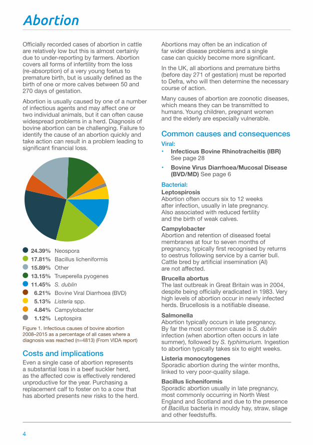
Officially recorded cases of abortion in cattle are relatively low but this is almost certainly due to under-reporting by farmers. Abortion covers all forms of infertility from the loss (re-absorption) of a very young foetus to premature birth, but is usually defined as the birth of one or more calves between 50 and 270 days of gestation.Abortion is usually caused by one of a number of infectious agents and may affect one or two individual animals, but it can often cause widespread problems in a herd. Diagnosis of bovine abortion can be challenging. Failure to identify the cause of an abortion quickly and take action can result in a problem leading to significant financial loss.
24.39% Neospora 17.81% Bacillus licheniformis 15.89% Other 13.15% Trueperella pyogenes 11.45% S. dublin 6.21% Bovine Viral Diarrhoea (BVD) 5.13% Listeria spp. 4.84% Campylobacter 1.12% Leptospira
Figure 1. Infectious causes of bovine abortion 2008–2015 as a percentage of all cases where a diagnosis was reached (n=4813) (From VIDA report)
Costs and implicationsEven a single case of abortion represents a substantial loss in a beef suckler herd, as the affected cow is effectively rendered unproductive for the year. Purchasing a replacement calf to foster on to a cow that has aborted presents new risks to the herd.
Abortions may often be an indication of far wider disease problems and a single case can quickly become more significant.In the UK, all abortions and premature births (before day 271 of gestation) must be reported to Defra, who will then determine the necessary course of action. Many causes of abortion are zoonotic diseases, which means they can be transmitted to humans. Young children, pregnant women and the elderly are especially vulnerable.
Common causes and consequencesViral: • Infectious Bovine Rhinotracheitis (IBR)
See page 28• Bovine Virus Diarrhoea/Mucosal Disease
(BVD/MD) See page 6Bacterial:Leptospirosis Abortion often occurs six to 12 weeks after infection, usually in late pregnancy. Also associated with reduced fertility and the birth of weak calves.Campylobacter Abortion and retention of diseased foetal membranes at four to seven months of pregnancy, typically first recognised by returns to oestrus following service by a carrier bull. Cattle bred by artificial insemination (AI) are not affected.Brucella abortus The last outbreak in Great Britain was in 2004, despite being officially eradicated in 1983. Very high levels of abortion occur in newly infected herds. Brucellosis is a notifiable disease.Salmonella Abortion typically occurs in late pregnancy. By far the most common cause is S. dublin infection (when abortion often occurs in late summer), followed by S. typhimurium. Ingestion to abortion typically takes six to eight weeks.Listeria monocytogenes Sporadic abortion during the winter months, linked to very poor-quality silage.Bacillus licheniformis Sporadic abortion usually in late pregnancy, most commonly occurring in North West England and Scotland and due to the presence of Bacillus bacteria in mouldy hay, straw, silage and other feedstuffs.
Abortion
4

Protozoal:Neospora caninum The most commonly diagnosed cause of bovine abortion, which occurs at five to six months of pregnancy. Infection is carried by dogs and hound packs and is passed on in their faeces.
Fungal:Mycotic (fungal) abortion Sporadic abortion typically in the winter months and most common in the west country following harvesting conditions that promote fungal growth on hay, silage or straw. Usually affects cows in mid-late pregnancy, even if the infection is picked up earlier. May affect up to 10% of the herd in some circumstances.
Early identificationLoss of the foetus before three months, may not be detected until the cow unexpectedly returns to oestrus. If abortion occurs later than three months the foetus may be found, especially when cattle are housed, but retained foetal membranes may be the only evidence, particularly if wild animals have disposed of the foetus.
Prevention and controlBiosecurity and disease-free statusStrict biosecurity to maintain disease-free status is the first priority in minimising the risk of abortion. Abortions must be reported and it is important to isolate aborted cows and dispose of the products of abortion wherever possible to avoid potential vector animals gaining access. It may be advisable to keep samples for analysis.
VaccinationVaccination should be used to maintain the herd’s disease-free status where appropriate. IBR, BVD/MD and S. dublin can all be controlled through vaccination.
Feed qualitySeveral of the organisms causing abortion originate in poor-quality feed (eg Listeria, Bacillus and a host of fungal organisms and mycotoxins). It is therefore essential to apply the highest standards at all times to harvesting and feed storage procedures. If contamination has occurred, be vigilant and remove potentially harmful feeds.
Retained foetal membranes may be the only evidence of abortion
An aborted foetus at three to four months
This in-calf cow drinking surface water close to a dung midden could be at risk of picking up an infection that could cause abortion
Mouldy silage may be a precursor to abortion, but risk assessment is difficult
5

Bovine Viral Diarrhoea (BVD)
Bovine Viral Diarrhoea is a widespread infectious disease of cattle that is usually transmitted through direct cattle-to-cattle contact. It is caused by a virus and lingers in herds through persistently infected (PI) animals (which can appear clinically normal) but will produce more persistently infected animals if they go on to calve. It can also develop into the fatal condition mucosal disease after a temporary infection or changes in the virus within PI animals. Good vaccines are available and should be used. BVD Type II, which is a common virus on North America, is an emerging risk for the UK. Although not currently confirmed as present, it has been responsible for severe outbreaks of disease in Europe, particularly the Netherlands, Germany and neighbouring states.Caused by a different virus, it is a more acute form of the disease and can cause death in adult cattle. Vaccines with BVD Type II protection are emerging onto the UK market and control should be discussed with the vet.
Costs and implicationsBVD can lead to significant losses resulting from reduced fertility, poor production and increased vulnerability to other infections, especially in young calves. Losses are particularly severe where the virus is introduced to groups of susceptible breeding cattle.Mucosal disease typically causes the death of the animal within five to 10 days, so cases should be culled on diagnosis. Calves born with eye and brain defects due to virus infection during development should also be culled.
Risk factors and susceptibilityAll cattle are potentially susceptible to infection with the BVD virus. The virus can be spread in the semen of PI bulls (or bulls with a temporary infection).High-risk situations:• Introduction of PI cattle into a naïve
(uninfected) and unprotected herd• Introduction of naïve cattle into an
infected herd
Early identificationExposure to the BVD virus to non-pregnant cattle causes a temporary infection before protective antibodies are produced within three to four weeks. This infection may temporarily
lower immunity to other infectious diseases, such as salmonella, respiratory infections and coccidiosis, leading to more severe symptoms or ill thrift particularly in young calves. However, animals may show no obvious clinical signs.For cattle in early pregnancy, exposure to the BVD virus during the first 110 days can cause the following problems:• Low pregnancy rates• Embryo death and return to heat• Foetal death/abortion• Mummified foetuses• Birth defects of nervous system and eyes• Weak/premature calves• Live PI calvesMucosal disease occurs when the BVD virus in PI animals changes to a cytopathic (cell killing) virus, typically in 6–18 month old calves, causing these symptoms:• Depression, salivation and fever• Anorexia• Mouth and muzzle ulcers• Pus discharges from eyes and nostrils• Severe diarrhoea with blood and shreds
of gut lining
Prevention and controlVaccinationWhere all breeding females are vaccinated, the disease is controlled by preventing BVD infection of developing foetuses during pregnancy and stopping the production of PI calves. BVD vaccines typically involve two doses, three to four weeks apart before first service, followed by annual booster vaccinations.
EradicationIdentification of PI animals is key. Whole herd blood testing and elimination of all PI carrier animals can effectively eradicate BVD from the herd. This must be backed up by strict biosecurity measures to prevent re-introduction to the herd. Several countries have successfully eradicated BVD (for example Norway) and have shown the production and economic benefits. The BVDFree England scheme, launched in 2016, is working to eradicate BVD in England by 2022.
6
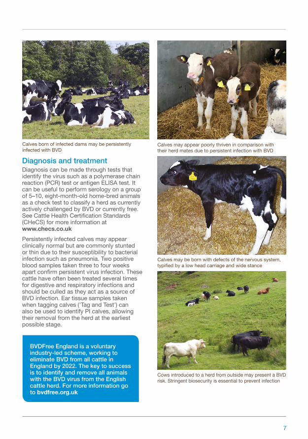
Diagnosis and treatmentDiagnosis can be made through tests that identify the virus such as a polymerase chain reaction (PCR) test or antigen ELISA test. It can be useful to perform serology on a group of 5–10, eight-month-old home-bred animals as a check test to classify a herd as currently actively challenged by BVD or currently free. See Cattle Health Certification Standards (CHeCS) for more information at www.checs.co.ukPersistently infected calves may appear clinically normal but are commonly stunted or thin due to their susceptibility to bacterial infection such as pneumonia. Two positive blood samples taken three to four weeks apart confirm persistent virus infection. These cattle have often been treated several times for digestive and respiratory infections and should be culled as they act as a source of BVD infection. Ear tissue samples taken when tagging calves ('Tag and Test') can also be used to identify PI calves, allowing their removal from the herd at the earliest possible stage.
Calves may appear poorly thriven in comparison with their herd mates due to persistent infection with BVD
Calves may be born with defects of the nervous system, typified by a low head carriage and wide stance
Cows introduced to a herd from outside may present a BVD risk. Stringent biosecurity is essential to prevent infection
Calves born of infected dams may be persistently infected with BVD
BVDFree England is a voluntary industry-led scheme, working to eliminate BVD from all cattle in England by 2022. The key to success is to identify and remove all animals with the BVD virus from the English cattle herd. For more information go to bvdfree.org.uk
7

Diarrhoea (calf scours)
Severe diarrhoea or calf scours is one of the most costly disease issues affecting beef enterprises. It is responsible for 50% of calf mortality and leads to significant financial losses due to the severe growth check in recovering calves.The following are the more common and important variants of calf scours:Rotavirus infection Infection can cause a range of clinical signs, from no observed abnormality to severe diarrhoea and dehydration with high mortality.Coronavirus diarrhoea Infection can progress rapidly to weakness, recumbency, severe dehydration and death.Enterotoxigenic E. coli Incidence is low (1% of scouring calves) but losses can be high. Sudden onset of scour is accompanied by a bloated appearance.Cryptosporidiosis Diarrhoea is caused by the physical loss of absorptive area from the small intestine and increases the severity of other potential infections. Dehydration tends to be mild, but calves lose condition over two to five days and have a dull, tucked-up appearance.Coccidiosis Caused by single-celled parasites, called coccidia. Coccidial species that cause disease damage the cell lining of the large intestine, resulting in diarrhoea.Salmonella Most commonly S. dublin is associated with producing acute or sub-acute illness in calves.
Costs and implicationsFinancial losses from calf scours can be crippling. Not only due to high mortality rates, but the cost of treatment (labour, drugs, etc) can be significant along with the impact of growth checks on calves that survive.
Risk factors and susceptibilityAll young calves are potentially at risk of infection, but the following will increase the likelihood of disease occurring:• History of specific infection on the unit• Replacements from another unit• Inadequate colostrum• Poor standards of hygiene• Rotavirus most commonly affects calves
at 4–21 days (but can affect older calves)• Coronavirus causes diarrhoea in calves up
to 21 days old• Enterotoxigenic E. coli typically affects
calves aged one to three days• Cryptosporidiosis is most common in calves
4–21 days old• Coccidiosis is particularly common in calves
between three weeks and six months old• Salmonella usually affects calves two
to six weeks of age
Early identificationEarly signs for the main diseases are:Rotavirus Reluctance to stand and suck, mild depression, salivation, quickly followed by acute onset of diarrhoea (watery yellow/green faeces).Coronavirus Depression, reluctance to suck and faeces containing mucus and milk curds.Enterotoxigenic E. coli Profuse yellow/white diarrhoea causing rapid and severe dehydration. Calves quickly become recumbent and bloated.Cryptosporidiosis Profuse yellow/green diarrhoea with mucus present.Coccidiosis A watery diarrhoea often accompanied by straining, mucus and blood, depression, lack of appetite and weight loss.Salmonella A pasty diarrhoea often with blood and shreds of mucus from the intestine with an offensive odour. Calves can rapidly become dehydrated, collapse and die.
8

Prevention and controlManagementThe risk of all forms of calf scours can be minimised by ensuring calving areas are clean and well-bedded, preferably mucked-out between calvings.Calves need a first feed of three litres within two hours of birth, followed by another similar sized feed within six to 12 hours of birth.In the case of cryptosporidiosis, where the parasite can remain in the environment for months, it is important to avoid using the same fields for calving and to move newborn animals immediately to clean pasture.VaccinationAnnual vaccination of pregnant cows with a combined rotavirus, coronavirus and E. coli K99 vaccine provides valuable insurance. Protective antibodies are passed on in the colostrum so sufficient colostrum ingestion is key.
Diagnosis and treatmentDiagnosis from symptoms outlined above and/or through laboratory analysis of faeces (ideally from untreated animals) should be carried out by the vet to determine the cause(s) of diarrhoea.Treatment of severely scouring calves:• Isolate in a well-bedded pen• Feed one to two litres, four to six times
a day, allowing four to eight litres of fluid to be given daily (stomach tube once and consult vet if calves will not suck through a teat within two to four hours). Alternate electrolyte fluids and milk, allowing at least two hours between feeds
• If dehydrated calves cannot stand unaided, intravenous fluids should be administered by the vet
• Antibiotic injections should be used for concurrent infections (eg navel ill)
• Offer fluids by teat as active sucking is an indicator of improvement
Mild diarrhoea or calf scour
Moderate diarrhoea requires oral rehydration solution to correct dehydration
Severe diarrhoea
Affected calves rapidly become dehydrated and recumbent
9

Eye conditions
Infectious Bovine Keratoconjunctivitis (IBK), also known as Pink Eye or New Forest Disease, is a highly contagious disease caused by the bacteria Moraxella bovis (M.bovis). It spreads rapidly during the summer and is more commonly seen in young stock than adults.Bovine Iritis, also known as ‘Silage Eye’, is a common cause of inflammation of the middle layer of the eye. It can occur in cattle of all ages that are fed baled silage/haylage.Cancer Eye (Ocular Squamous Cell Carcinoma) is uncommon in northern Europe. These cancerous growths typically develop from the third eyelid (the membrane of the lower eyelid) following prolonged exposure to sunlight.
Costs and implications Lesions in and around the eye are very painful and will disrupt grazing, causing poor performance and weight loss. They can also cause temporary blindness with affected animals wandering about aimlessly.
Risk factors and susceptibilityFlies act as mechanical vectors for M. bovis.Foreign bodies such as dust, grass awns or small bits of ensiled grass stalk, can cause Bovine Iritis.
Early identificationIBK The first signs of IBK are obvious tear staining of the face, with pus matting the lashes and hair below the affected eye. Animals find it painful to be in direct sunlight.Spontaneous recovery may occur in mild cases after three to five days and the animals will be better after two weeks. In severe cases, ulceration may lead to perforation of the surface of the eye (cornea).
Bovine Iritis Initial signs include excessive tear staining, blinking and forced closure of the eyelids.A blueish-white opacity is usually seen on the cornea within two or three days. This can turn yellow as pus accumulates beneath. These lesions can take several weeks to clear without treatment.
Cancer eye Cancerous growths may irritate the eye surface, leading to secondary corneal ulceration and infection, leading to eyelid closure. There is usually a discharge due to mechanical irritation of the eye.White-faced cattle are more prone to cancer eye due to the lack of pigment around the eyes. Cancer eye is most common in cattle aged seven to eight years. It is not common in cattle under three years of age.
Bovine Iritis is characterised by a blueish-white opacity on the surface of the eyeball
Cancer eye in right eye
10
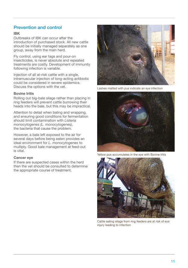
Prevention and controlIBKOutbreaks of IBK can occur after the introduction of purchased stock. All new cattle should be initially managed separately as one group, away from the main herd.Fly control, using ear tags and pour-on insecticides, is never absolute and repeated treatments are costly. Development of immunity following infection is variable.Injection of all at-risk cattle with a single, intramuscular injection of long-acting antibiotic could be considered in severe epidemics. Discuss the options with the vet.
Bovine IritisRolling out big-bale silage rather than placing in ring feeders will prevent cattle burrowing their heads into the bale, but this may be impractical.Attention to detail when baling and wrapping, and ensuring good conditions for fermentation should limit contamination with Listeria monocytogenes (L. monocytogenes), the bacteria that cause the problem.However, a bale left exposed to the air for several days before being eaten provides an ideal environment for L. monocytogenes to multiply. Good bale management at feed-out is vital.
Cancer eyeIf there are suspected cases within the herd then the vet should be consulted to determine the appropriate course of treatment.
Yellow pus accumulates in the eye with Bovine Iritis
Cattle eating silage from ring feeders are at risk of eye injury leading to infection
Lashes matted with pus indicate an eye infection
11
Hypomagnesaemia (grass staggers, grass tetany)
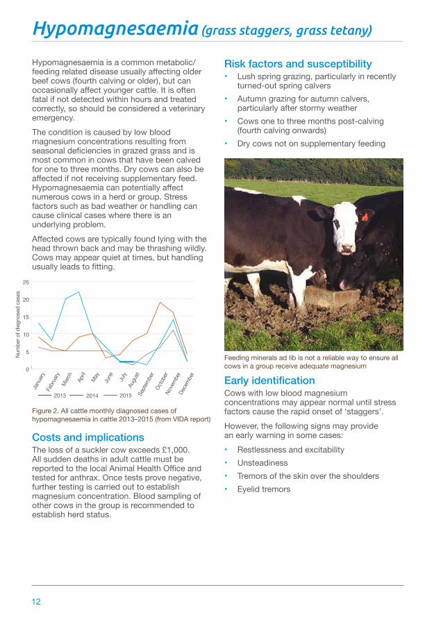
Hypomagnesaemia is a common metabolic/feeding related disease usually affecting older beef cows (fourth calving or older), but can occasionally affect younger cattle. It is often fatal if not detected within hours and treated correctly, so should be considered a veterinary emergency.The condition is caused by low blood magnesium concentrations resulting from seasonal deficiencies in grazed grass and is most common in cows that have been calved for one to three months. Dry cows can also be affected if not receiving supplementary feed. Hypomagnesaemia can potentially affect numerous cows in a herd or group. Stress factors such as bad weather or handling can cause clinical cases where there is an underlying problem.Affected cows are typically found lying with the head thrown back and may be thrashing wildly. Cows may appear quiet at times, but handling usually leads to fitting.
Figure 2. All cattle monthly diagnosed cases of hypomagnesaemia in cattle 2013–2015 (from VIDA report)
Costs and implicationsThe loss of a suckler cow exceeds £1,000. All sudden deaths in adult cattle must be reported to the local Animal Health Office and tested for anthrax. Once tests prove negative, further testing is carried out to establish magnesium concentration. Blood sampling of other cows in the group is recommended to establish herd status.
Risk factors and susceptibility• Lush spring grazing, particularly in recently
turned-out spring calvers• Autumn grazing for autumn calvers,
particularly after stormy weather• Cows one to three months post-calving
(fourth calving onwards)• Dry cows not on supplementary feeding
Early identificationCows with low blood magnesium concentrations may appear normal until stress factors cause the rapid onset of ‘staggers’.However, the following signs may provide an early warning in some cases:• Restlessness and excitability• Unsteadiness• Tremors of the skin over the shoulders• Eyelid tremors
Feeding minerals ad lib is not a reliable way to ensure all cows in a group receive adequate magnesium0
5
10
15
20
25
Num
ber o
f dia
gnos
ed c
ases
2013
Janu
ary
Febr
uary
Octo
ber
Marc
hAp
rilM
ay
Dece
mbe
r
June July
Augu
stSe
ptem
ber
Nove
mbe
r
2014 2015
12

Prevention and controlEnsure daily intake of magnesiumThis is essential as cows are unable to store magnesium in the body. Feeding cattle concentrates containing an appropriate magnesium supplement (1–2kg/cow/day) is the most reliable method of ensuring cattle receive enough magnesium.Home-mixed rations should be supplemented with magnesium as many straights are typically deficient.Ad-lib minerals are not a reliable source of daily magnesium for all cows in a group. The most reliable method of control is to give a magnesium bolus.
Supplement grazing with strawGood-quality barley straw fed ad lib helps slow the flow of lush grass through the gut and aids magnesium absorption.
Avoid potassium fertilisersPotassium interferes with magnesium absorption and should not be included in compound fertilisers spread onto grassland during risk periods.
Diagnosis and treatmentDiagnosis is based on clinical signs and can be confirmed through blood sampling.Symptoms of hypomagnesaemia should be treated as a veterinary emergency. Fitting cows can be sedated first by the attending vet. Treatment will typically involve intravenous injection (by vet) administered slowly, followed by further injections at several sites, using a mixture containing small amounts of magnesium in a calcium solution.If hypomagnesaemia is suspected or confirmed, it is essential to deal with the risk to the rest of the group.
If not detected within hours of onset, hypomagnesaemia is often fatal
Adverse weather is one of the stress factors that can lead to hypomagnesaemia
Hypomagnesaemia is a veterinary emergency
Turn-out onto lush pasture is a common factor involved in hypomagnesaemia
13

Johne’s disease (paratuberculosis)
Johne’s disease is a slow progressing gut inflammation in adult cattle caused by a subspecies (paratuberculosis) of the organism Mycobacterium avium (MAP). It is characterised by progressive weight loss and chronic diarrhoea, but diagnosis and control are particularly difficult.
Figure 3. Johne’s disease in cattle 2008–2015 (from VIDA report)
Costs and implicationsInfected herds may suffer annual culling or mortality rates of 1–5%.Losses due to sub-clinical disease, including weight loss and poor fertility, are also substantial.There is no effective treatment and animals must be culled as soon as a diagnosis is confirmed.
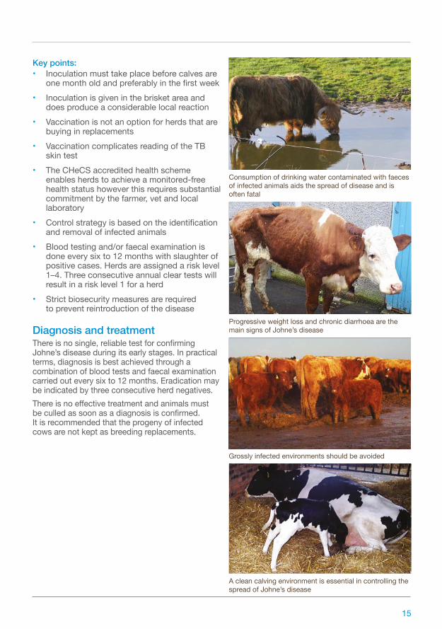
Risk factors and susceptibilityClinical cases of Johne’s disease are not usually seen in cattle until they are three to five years old, although cases in younger animals are possible. The organism is passed onto younger cattle by older infected cattle and there is then a long incubation period.The disease can be transmitted by being:• Picked up from infected faeces found on
contaminated teats, feedstuffs or water troughs
• Passed to newborn calves in the colostrum of infected dams
• Passed from heavily infected dams to developing calves in the uterus
Infected animals may shed causative organisms in their faeces for over a year before clinical signs appear.
Early identificationDiarrhoea and weight loss in cattle three to five years old, despite no loss of appetite and an absence of fever.Signs can appear following a stressful event, such as calving or transportation.
Prevention and controlJohne’s disease is difficult to control due to the long incubation period and the fact that animals will pass on infection long before they show clinical signs. Diagnosis is also unreliable, particularly in the early stages of the disease.Practical control measures to limit losses include:• Rapid culling of diseased animals• Reducing the risks of faecal contamination
of food, water and pasture, eg raise feed or water troughs, avoid use of surface or pond water for drinking, avoid spreading farmyard manure on pastures and maintain good hygiene around calving
• Not using calves from infected dams as replacements
• Only restocking from accredited herds, especially when restocking bulls. Avoid co-grazing sheep and cattle where possible as the disease can pass between the two species
• Vaccination may be a cost-effective option for commercial beef herds rearing their own replacements, as it does reduce clinical cases and overall losses. However, it will not eradicate the disease and a vaccine would need to be imported into the UK under licence
0
500
1000
1500
2000
2500
3000
3500
4000
2008 2009 2010 2011 2012 2013 2014 2015
Num
ber o
f dia
gnos
ed c
ases
14
Key points:• Inoculation must take place before calves are
one month old and preferably in the first week• Inoculation is given in the brisket area and
does produce a considerable local reaction• Vaccination is not an option for herds that are
buying in replacements• Vaccination complicates reading of the TB
skin test• The CHeCS accredited health scheme
enables herds to achieve a monitored-free health status however this requires substantial commitment by the farmer, vet and local laboratory
• Control strategy is based on the identification and removal of infected animals
• Blood testing and/or faecal examination is done every six to 12 months with slaughter of positive cases. Herds are assigned a risk level 1–4. Three consecutive annual clear tests will result in a risk level 1 for a herd
• Strict biosecurity measures are required to prevent reintroduction of the disease
Diagnosis and treatmentThere is no single, reliable test for confirming Johne’s disease during its early stages. In practical terms, diagnosis is best achieved through a combination of blood tests and faecal examination carried out every six to 12 months. Eradication may be indicated by three consecutive herd negatives.There is no effective treatment and animals must be culled as soon as a diagnosis is confirmed. It is recommended that the progeny of infected cows are not kept as breeding replacements.
Consumption of drinking water contaminated with faeces of infected animals aids the spread of disease and is often fatal
Progressive weight loss and chronic diarrhoea are the main signs of Johne’s disease
Grossly infected environments should be avoided
A clean calving environment is essential in controlling the spread of Johne’s disease
15
Lameness
Lameness resulting from pain and/or incapacity in the feet can result from a number of interacting factors in the environment of cattle. It is most problematic when affecting a stud bull or breeding cows during the breeding season and is a serious welfare issue in all livestock.Foot lameness can be caused by the following conditions:• ‘Foul of the foot’ and digital dermatitis
(bacterial infection between the claws)• Hyperplasia (excess skin growth between
the claws, which is hereditary)• Sole abscesses/white line disease/solar
ulceration (impaction by dirt, small stones, foreign bodies)
• Sandcrack (vertical fracture of the hoof wall)
• Overgrowth (elongation of the foot)
• Corkscrew claw (hereditary)
Costs and implicationsLame cattle are unproductive cattle, whether breeding animals or fattening stock. Weight loss is a common consequence in grazing cattle, with delayed heat and poor conception a possibility in suckler cows.Infertility is likely to be the single biggest cost implication.
Common causes and consequencesFoul of the foot/digital dermatitisThis typically occurs as a sudden onset of lameness and the animal only ‘toes’ its foot to the ground. It is usually the result of wet dung and mud softening the interdigital tissue and sharp stones causing wounds that allow bacteria to infect the deeper tissues. Reservoirs of infection can survive in wet areas around gateways.The lesion appears as a widening of the interdigital space with swelling progressing up the leg as far as the fetlock joint. There is usually a break of the skin with damage of tissue between the claws.Digital dermatitis is thought to be caused by a type of bacteria called a treponeme. It typically produces a red, swollen lesion between the claws of the heel, (‘hairy heel warts’) but can infect and complicate the healing of many other
types of lameness. It is a major problem in the dairy industry, which is also emerging in beef herds.
HyperplasiaThis hereditary condition appears as excess skin growth at the front of the interdigital space and is most commonly seen in the hind legs of bulls. Lameness can result from superficial infection.
Sole abscesses/white line disease /solar ulcerAny sharp object can penetrate the sole and cause an abscess that is typically recognised as a black mark overlying the pus.Impaction of dirt and small stones in the white line can result in white line abscesses. Lesions are usually found in the outer claw of the hind foot, on the outside border close to the junction with the heel, as this is where there is most physical stress.If left untreated, infection can extend up the hoof wall and erupt at the coronary band.Sole ulcers can be caused by excessive standing on concrete, especially in the post-calving period. Although more of an issue in the dairy industry, it can also be a problem in cubicle-housed beef cattle.
SandcrackThis is a vertical fracture of the hoof wall of variable degrees between the coronary band and the bottom of the wall. The depth of the lesion varies and pus may or may not be present.Excessive drying-out of the hoof horn during summer months is thought to be the cause of this condition, though this is uncertain.
OvergrowthThis is an elongation of the foot, especially affecting the hind feet. The condition reduces the weight-bearing surface of the foot and in the later stages, the toe bends upwards and no longer touches the ground.Overgrowth is typically caused by a lack of natural wear when cattle are housed in straw yards.
Corkscrew clawA heritable condition typically causing the outer claw of the hind leg to be twisted.
16

TreatmentAll lame cows should be attended to promptly, with veterinary treatment required when the cause of the lameness cannot be determined and/or when the lameness persists after treatment.
Foul of the foot/digital dermatitisIt is essential to lift the animal’s foot to check for impacted foreign bodies, clean the foot and spray the wound with oxytetracycline aerosol. Antibiotics may be required, consult your vet to determine the most effective treatment.Prevention is by regular footbathing and improved hygiene. Disinfection of hoof knives by foot-trimmers to prevent inter-herd spread is important, as is footbathing of purchased animals.
HyperplasiaTreat the infection with oxytetracycline and if necessary, injectable antibiotics. Surgically remove the growths as a last resort.
Abscesses/white line disease/solar ulcerPare down with a hoof knife to release the pus and remove all under-run horn – there should be no bleeding. Avoid damage to the sensitive corium, as this will delay the healing process. Antibiotic treatment should not be necessary. A shoe block may be used to relieve weight from the sensitive claw where a large area of the sole has been pared off.
SandcrackRemove all affected horn by paring out a shallow ‘V’. Antibiotic treatment is not necessary.
Overgrowth/Corkscrew clawCorrective foot trimming is required.
In cases of foul of the foot, the swelling can progress up the leg as far as the fetlock joint
A break in the interdigital skin caused by the impact of a sharp stone
Water-logged gateways with old bricks and stone provide an ideal environment for bacteria and damage animals’ feet, leading to foul of the foot
When abscesses are correctly pared out, there should be pus, but no blood
17

Liver fluke (Fasciolosis)
A bovine liver with enlarged bile ducts due to fluke infection
This disease is a common and devastating condition, commonly found in wet grazing land. It results from the infestation of the liver by the flatworm Fasciola hepatica (liver fluke).
1. Eggs pass to pasture via the dung2. Eggs hatch into water-borne larvae, which infect water snails3. Larvae develop into young fluke before leaving the snail while still in the water4. Young fluke are eaten by livestock grazing wetlandsFigure 4. Liver fluke life cycle
Costs and implicationsSeverely affected cattle become weak, emaciated and unable to stand, with sudden death a possibility.Even in less severe cases, weakness caused by liver damage may lead to an increased incidence of metabolic and infectious diseases, particularly in cows in late pregnancy. Birth of weak calves is likely, particularly in cows on marginal winter rations. Fluke has been estimated to cost around £200/head in cattle.
Risk factors and susceptibilityAll grazing cattle are susceptible to liver fluke.Eggs from an infected animal can complete the life cycle and reinfect grazing animals in a minimum of 20 weeks.Risk tends to be associated with wet pastures, where there is sufficient moisture or standing water to support the snail.Exceptionally wet weather, or successive wet summers can increase the risk in pastures not previously identified with the disease.
Infested sheep brought onto a farm for over-winter grazing will contaminate pasture and increase the risk to cattle during the following summer/autumn.
Early identificationThe following signs may indicate liver fluke infection:• Persistent diarrhoea• Chronic weight loss despite
adequate feeding• Anaemia in severe casesLiver fluke is commonly confused with:• Poor nutrition (where it is a whole herd
or group problem)• Johne’s disease (several cows in a group
or herd)• Salmonellosis (several cows in the group
or herd)• Parasitic gastroenteritis • Rumen fluke – less common than liver fluke
but can cause diarrhoea and ill thrift in young stock. It is important to differentiate as treatment options are different
1.4.
2.3.
18

Prevention and controlFencing off wet areas or carrying out drainage to restrict snail habitats are two solutions, but this may be impractical – particularly in extensively farmed areas. It may also be prevented by environmental stewardship protocols.Control using effective flukicide treatments may therefore be the best solution. In areas where liver fluke is endemic, strategic flukicide treatments should be given in accordance with the veterinary herd health plan.In high-risk years, treat at-risk cattle in January and again in October/November.In low-risk years, a single annual treatment in January should be sufficient.
Diagnosis and treatmentDiagnosis in the early stages may be carried out if high-risk conditions are prevalent and is based on raised liver enzymes in blood samples analysed by the vet.The chronic condition is identified from symptoms outlined above and may be confirmed through the identification of fluke eggs in dung samples, though these may be scarce and difficult to find. Specific antibody tests for liver fluke can be carried out, but do not necessarily indicate current infection, as antibodies can persist from the previous year. The faecal coproantigen test detects fluke secretions in faeces so can indicate infection before the production of fluke eggs or antibodies.Effective treatment of all stages of fluke is achieved with triclabendazole. Nitroxynil and oxyclosanide are less effective against young flukes and should be used in the treatment of adult flukes (chronic disease). Resistance to triclabendazole is emerging and so repeated doses with adult flukicides are often more appropriate for strategic control of fluke in beef cattle. Improved nutrition of affected cattle is essential.
Liver fluke is more commonly encountered in beef cows grazing poor, wet pasture
Diagnosis of liver fluke is not simple. Does this bull have liver fluke, Johne’s disease, or something else? Consult the vet
Persistent diarrhoea is a major visible symptom of liver fluke
Chronic weight loss and very poor body condition despite an adequate ration is a common indication of liver fluke
19

Lungworm (husk or hoose)
Husk (or hoose) is caused by infestation of the bronchial tubes by white thread-like worms known as lungworm (Dictyocaulus viviparous). The condition is characterised by persistent coughing and breathing difficulties, with lung damage potentially developing into secondary pneumonia.
Cattle become infected by ingesting lungworm larvae while grazing. These infective larvae penetrate the intestinal wall and pass via the lymphatic system and blood stream into the lungs. In the lungs, these larvae develop into adults, which cause the problem.Female lungworms lay vast numbers of eggs that develop into minute larvae. These are carried up the windpipe in mucus where they are then swallowed and consequently passed out in the animals dung onto the pasture. The cycle then begins again.
Figure 5. Monthly diagnosed cases of lungworm in cattle 2013–2015 (from VIDA report)
Costs and implicationsIn its most severe form, lungworm can result in sudden death. More typically, affected cattle will suffer marked loss of body condition (up to 10% of body weight), with growing cattle potentially losing 20–40kg. With long recovery periods and the possibility of secondary pneumonia requiring antibiotic treatment, losses from a severe outbreak in growing cattle can average £50 per head.
Risk factors and susceptibilityYoung (first grazing) cattle are at greatest risk if grazing pastures have been recently stocked with older cattle. Adult cattle can develop a natural immunity through grazing moderately infested pastures. However, if they have not been previously exposed to infection they will also be at risk.Autumn grazing conditions are typically most favourable to the development and survival of heavy lungworm larvae infestations on pasture.
Early identificationThe following signs may indicate lungworm infection:• Panting• Frequent coughing, especially after short
periods of exercise• Reluctance to move• Standing with head down and neck
extended, often gasping for breath
Young cattle grazing for the first time are at risk from lungworm
White thread-like worms infesting the lung
0
10
20
30
40
50
Num
ber o
f dia
gnos
ed c
ases
2013
Janu
ary
Febr
uary
Octo
ber
Marc
hAp
rilM
ay
Dece
mbe
r
June July
Augu
stSe
ptem
ber
Nove
mbe
r
2014 2015
20

Prevention and controlCattle exposed to lung infection through grazing moderately infested pastures allows the animal to develop immunity through natural exposure. This must be combined with strategic worming treatment during the grazing period to prevent the disease developing. This is a risky control strategy and is not recommended.Vaccination prior to first grazing should provide immunity for six months. This costs £10–£15 per animal and in combination with good management practice, is the best insurance against lungworm.Worming relies on a low dose early in the season, which stimulates immunity afresh each year. Avoid over-worming early in the season as lungworm can also strike during late grazing. Discuss a strategy with the vet each year.
Diagnosis and treatmentDiagnosis is based on clinical signs outlined above and may also be confirmed through laboratory analysis of dung showing the presence of larvae or by a blood test.When the disease is identified, prompt worming treatment is essential, using a product recommended by the vet. Antibiotic treatment may be required where secondary infection has occurred.
Severe lungworm infection. Note the extended neck and painful expression
Severe lungworm infection and secondary pneumonia
Respiratory distress is a welfare concern in cases of severe lungworm infection
Note the absence of discharge from the eyes and nostrils in cases of lungworm infection. Frequent coughing is the most important clinical sign
21

Midge-borne diseases
Over recent years, several viruses transmitted by Culicoides midges have emerged in European livestock, perhaps associated with climate change, leading to significant economic losses. The two main diseases to affect beef cattle are Bluetongue virus (BTV) and more recently Schmallenberg virus (SBV).
All cattle are susceptible to the disease if they come into contact with the biting midge responsible for carrying the virus. Actions to prevent both these diseases have shown little impact. However, the following are suggested measures:• Identify and destroy midge breeding sites
(dung heaps, damp areas, etc)• Use mesh (impregnated with insecticide) to
prevent midges entering buildings• Apply pour-on insecticides to cattle• Strict biosecurity and quarantine of all
livestock brought onto the farm are essential in the control and prevention of all diseases
Bluetongue virus (BTV)Bluetongue virus is a viral disease of sheep and cattle that is characterised by lameness and fever and can result in serious production losses and mortality. Spread of the disease is dependent upon the presence of the Culicoides midge host.Bluetongue is widespread in the USA and outbreaks are being reported in Europe, with different strains emerging. The BTV 8 virus was identified in Belgium and the Netherlands in 2006 and spread rapidly to central and western European countries. The first incidence of BTV 8 in the UK was reported in East Anglia in September 2007, which supports the theory that transport of midges on air currents from continental Europe is a potential threat. This outbreak was controlled by the strategic use of vaccination but other strains of BTV continue to be a threat. For example, in 2007, BTV 1 spread from the Maghreb in Northern Africa to Spain and was subsequently detected in France and Portugal. BTV 8 was detected again in cattle in Northern France in May 2017, but has currently not been found in the UK.
Schmallenberg virusSchmallenberg virus (SBV) was identified in Germany, in November 2011. Schmallenberg virus primarily infects domestic and wild ruminants and causes clinical signs including diarrhoea, moderate hypothermia, decrease in milk production and anorexia in adult cattle. Sheep and goats can be mildly affected. Infection of bovine foetuses by SBV is associated with abortions, premature births and stillbirths, diverse congenital malformations and abnormalities of the central nervous system. Schmallenberg virus has spread very rapidly from Germany and the Netherlands to the UK, France and other European countries.Although SBV is currently less apparent in the UK, there is evidence of continued virus circulation and successful overwintering. It is not known how long natural immunity to SBV may last, but it appears that immunity in herds is patchy, with some animals remaining vulnerable.A commercial vaccine is available and discussion with the vet is important to manage the ongoing risk of SBV.
Costs and implicationsBluetongue is a notifiable disease in the UK and suspected cases must be reported immediately to the local animal health office.The symptoms – delayed recovery, susceptibility to secondary bacterial infections and potential mortality of cattle – mean that significant production losses are inevitable.The costs of BTV have been estimated at up to £19/sheep and £17/cow on affected farms from:• Deaths• Loss of production• Cost of disposal• Veterinary and medicines• LabourThe cost of schmallenberg depends on the severity of the outbreak.
22
Early identificationThe following symptoms are most likely to occur in affected cattle:• Fever (temperatures over 39.5ºC)• Stiffness and reluctance to move, due to
swelling of the coronary band at the top of the hooves
• Nasal discharge and ulcerations of the muzzle
• Lacrimation (discharge from the tear ducts) but no obvious eye lesions
Prevention and controlVaccines are available through the vet. Always follow the manufacturer’s guidelines. In the case of an outbreak of bluetongue, Defra protocols should be implemented on infected premises.
Diagnosis and treatment• Diagnosis is based upon clinical signs
and/or isolation of the bluetongue virus or schmallenberg virus
• There is no fully effective treatment for clinically affected animals. Treatment is limited to antibiotic therapy to control secondary bacterial infections
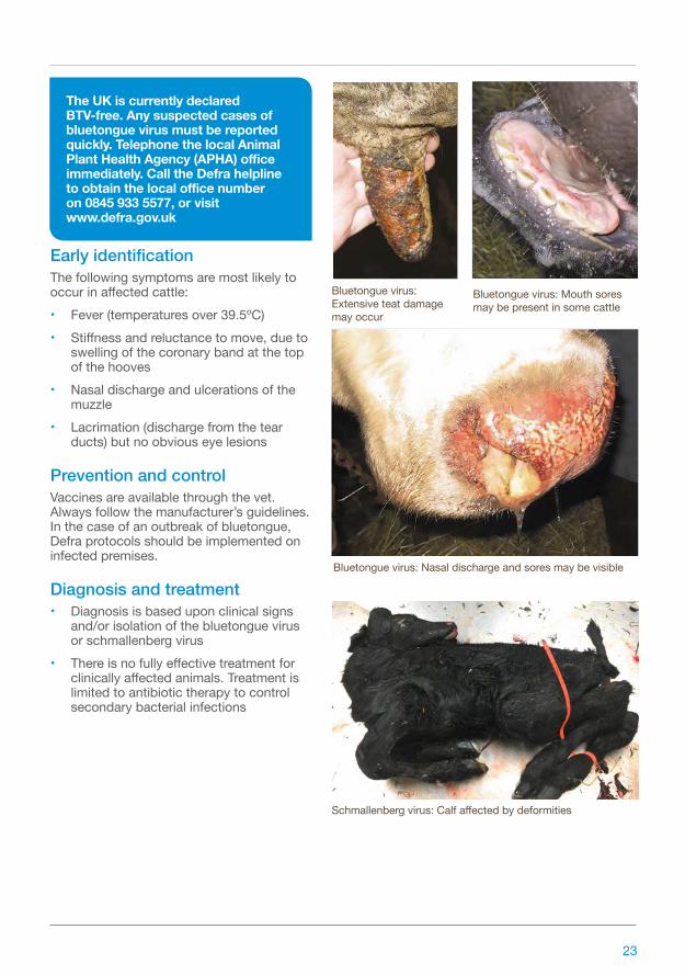
Bluetongue virus: Extensive teat damage may occur
Bluetongue virus: Nasal discharge and sores may be visible
Bluetongue virus: Mouth sores may be present in some cattle
Schmallenberg virus: Calf affected by deformities
The UK is currently declared BTV-free. Any suspected cases of bluetongue virus must be reported quickly. Telephone the local Animal Plant Health Agency (APHA) office immediately. Call the Defra helpline to obtain the local office number on 0845 933 5577, or visit www.defra.gov.uk
23
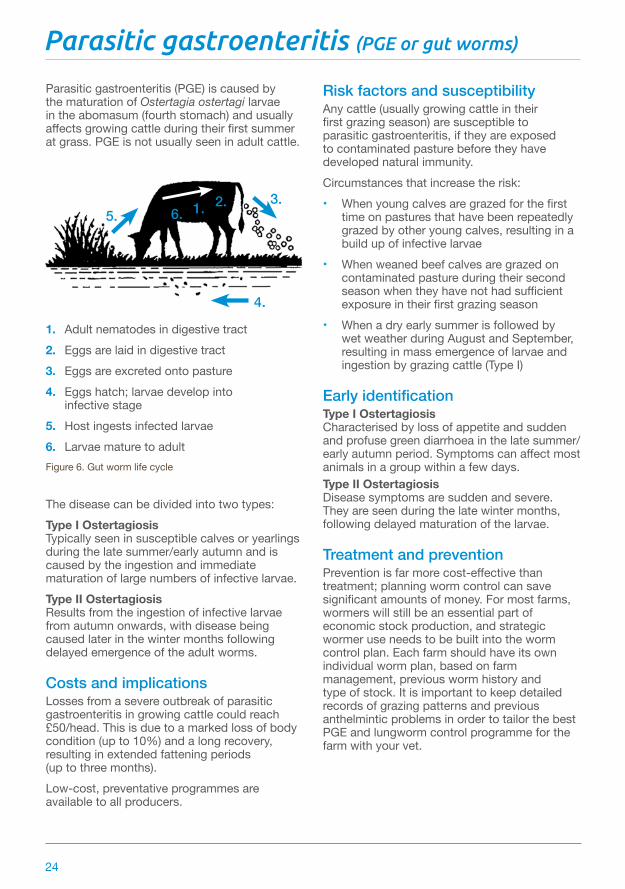
Parasitic gastroenteritis (PGE or gut worms)
Parasitic gastroenteritis (PGE) is caused by the maturation of Ostertagia ostertagi larvae in the abomasum (fourth stomach) and usually affects growing cattle during their first summer at grass. PGE is not usually seen in adult cattle.
1. Adult nematodes in digestive tract2. Eggs are laid in digestive tract3. Eggs are excreted onto pasture4. Eggs hatch; larvae develop into infective stage5. Host ingests infected larvae6. Larvae mature to adultFigure 6. Gut worm life cycle
The disease can be divided into two types:Type I Ostertagiosis Typically seen in susceptible calves or yearlings during the late summer/early autumn and is caused by the ingestion and immediate maturation of large numbers of infective larvae.Type II Ostertagiosis Results from the ingestion of infective larvae from autumn onwards, with disease being caused later in the winter months following delayed emergence of the adult worms.
Costs and implicationsLosses from a severe outbreak of parasitic gastroenteritis in growing cattle could reach £50/head. This is due to a marked loss of body condition (up to 10%) and a long recovery, resulting in extended fattening periods (up to three months).Low-cost, preventative programmes are available to all producers.
Risk factors and susceptibilityAny cattle (usually growing cattle in their first grazing season) are susceptible to parasitic gastroenteritis, if they are exposed to contaminated pasture before they have developed natural immunity.Circumstances that increase the risk:• When young calves are grazed for the first
time on pastures that have been repeatedly grazed by other young calves, resulting in a build up of infective larvae
• When weaned beef calves are grazed on contaminated pasture during their second season when they have not had sufficient exposure in their first grazing season
• When a dry early summer is followed by wet weather during August and September, resulting in mass emergence of larvae and ingestion by grazing cattle (Type I)
Early identificationType I Ostertagiosis Characterised by loss of appetite and sudden and profuse green diarrhoea in the late summer/early autumn period. Symptoms can affect most animals in a group within a few days.Type II Ostertagiosis Disease symptoms are sudden and severe. They are seen during the late winter months, following delayed maturation of the larvae.
Treatment and preventionPrevention is far more cost-effective than treatment; planning worm control can save significant amounts of money. For most farms, wormers will still be an essential part of economic stock production, and strategic wormer use needs to be built into the worm control plan. Each farm should have its own individual worm plan, based on farm management, previous worm history and type of stock. It is important to keep detailed records of grazing patterns and previous anthelmintic problems in order to tailor the best PGE and lungworm control programme for the farm with your vet.
3.6. 1. 2.
5.
4.
24

There are several factors to bear in mind when developing the plan:• Use pasture effectively so that cattle avoid
grazing contaminated pasture during the peak season. This can be as simple as moving cattle onto fresh, ungrazed pasture (such as silage aftermath) just before the summer rise in larval numbers
• Reduce routine worming by monitoring faecal egg counts (FEC) and animal growth rates. This will save money and reduce the risk of resistance developing on the farm
• Worm stock susceptible to hibernating larvae at housing
• Don't forget lungworm control as this is an increasing problem on many farms. Use vaccination to control lungworm as other measures such as pasture management are less effective than for gut worms
Larvae mature to adult in the digestive tract
In spring-calving beef herds, early season pasture contamination is ingested by immune adult cows
Problems with PGE can arise in beef cattle when weaned calves graze contaminated pasture during their second season
Symptoms of PGE include a loss of appetite with sudden and profuse green diarrhoea
25

Plant poisoning
Most plants are not poisonous to cattle, but a few notable exceptions will cause serious problems if ingested.Ragwort High awareness and careful management of pastures means ragwort poisoning is relatively rare in the UK.Incidences usually occur following ingestion of the wilted/dried plant in hay or silage.Yew A common ornamental tree often found in churchyards. Ingestion usually leads to rapid death.Bracken Ingestion of bracken over several weeks when pasture is sparse can be fatal to cattle. Death results from bone marrow suppression, causing loss of blood cells and clotting factors.Ingestion of bracken over many months, particularly when used as bedding material, can lead to tumours in the bladder, oesophagus and rumen.Acorns These can present a serious problem on pastures that have oak trees, after autumn storms. Tannins in the acorns cause serious, often fatal, kidney damage.Water dropwort Dry, hot weather can drive cattle to graze marginal areas in search of food, where they encounter toxic plants they would normally leave alone.One of the most important is Water dropwort, which is very common in the west and south of England.Cattle are particularly at risk after ditches have been cleared out, which exposes the poisonous roots, often referred to as ‘dead man’s fingers’.
Costs and implicationsPlant poisoning in cattle is usually fatal, incurring the loss of productive animals and the associated costs of bringing in or rearing replacements.
Risk factors and susceptibilityPasture management should reduce the risks of plant poisoning as much as possible, eg by digging out ragwort, spraying bracken, ensuring a plentiful supply of nutritious grasses.
Early identificationRagwort• Chronic weight loss and diarrhoea• Jaundice• Accumulation of fluid under the jaw
and brisket caused by liver diseaseYew• No early signs. Death rapidly follows
ingestionBracken• Weight loss and weakness• Blood haemorrhaging from the nasal
passages and vagina• Death within several daysAcorns• Constipation/straining to defecate• Diarrhoea• Anorexia• Bloat due to the rumen not working• Renal failure• Death within four to seven daysWater dropwort• Salivation and dilated pupils• Breathing difficulty• Collapse and spasmodic convulsions• Most affected cattle die
Prevention and controlRagwort Control ragwort on pasture by spraying with selective herbicides, or by digging out the whole plant.Yew Prevent access by cattle by fencing off yew trees and maintaining perimeter fences so they cannot escape.
26

Bracken Many hill farms have substantial areas of bracken where fencing, burning or herbicide treatments would be uneconomic. Adequate feeding should ensure cattle do not graze bracken.Acorns Remove cattle from pasture where oak trees are present, especially after autumn storms or heavy acorn falls.Water dropwort Supplement cattle on bare pastures during drought to prevent them grazing marginal areas. Move cattle out of fields where ditches have been cleared.
Diagnosis and treatmentRagwort Diagnosis is based upon clinical evidence of liver disease with known exposure to ragwort.There is no effective treatment once clinical signs appear. Remove contaminated feed and destroy.Yew Cause of death can only be confirmed by examining rumen contents at post-mortem.There is no treatment.Bracken Diagnosis is based upon clinical signs such as loss of appetite, bloody diarrhoea and a high temperature. Secondary infection is very common. In acute cases, treatment with broad-spectrum antibiotics is generally unsuccessful.Acorns Diagnosis is based upon clinical signs and exposure to acorns. Confirmation by post-mortem.There is no specific treatment to cure the problem. Supportive treatment includes giving large volumes of intravenous fluids, which are prohibitively expensive.Water dropwort Diagnosis is based upon evidence of plants having been grazed or roots exposed by ditching. Confirmation by post-mortem. There is no specific treatment. If poisoning is suspected, remove all cattle from areas where the plant grows.
Cattle that eat ragwort, bracken or acorns will suffer excessive weight loss
Long-term ingestion of bracken can be fatal to cattle
Diarrhoea is a sign of possible ragwort poisoning
Remove cattle from pastures with oak trees in the autumn
27
Respiratory disease (pneumonia)
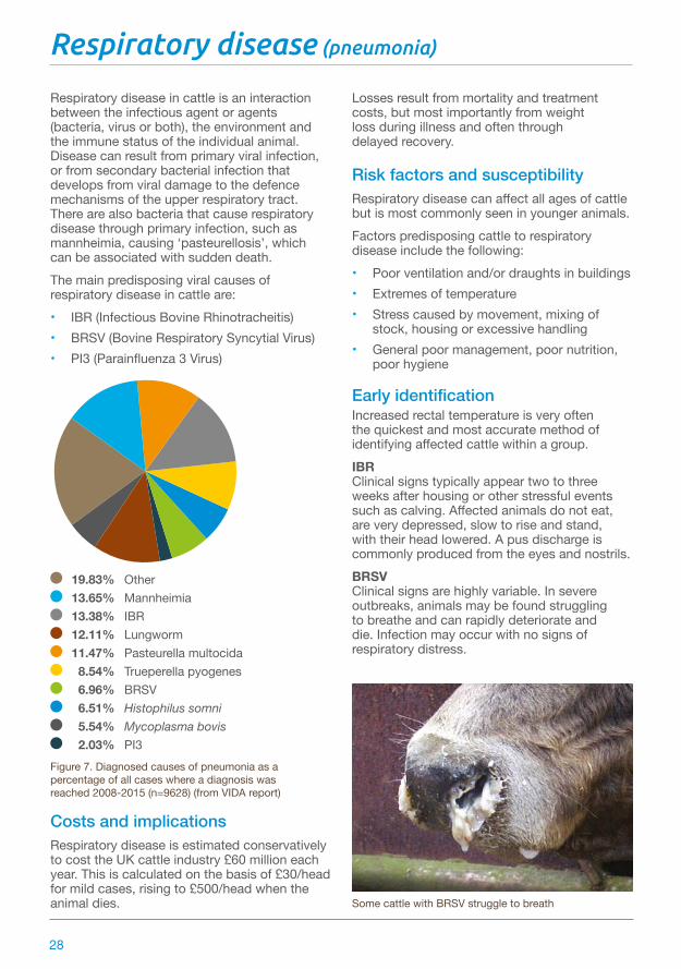
Respiratory disease in cattle is an interaction between the infectious agent or agents (bacteria, virus or both), the environment and the immune status of the individual animal. Disease can result from primary viral infection, or from secondary bacterial infection that develops from viral damage to the defence mechanisms of the upper respiratory tract. There are also bacteria that cause respiratory disease through primary infection, such as mannheimia, causing ‘pasteurellosis’, which can be associated with sudden death.The main predisposing viral causes of respiratory disease in cattle are:• IBR (Infectious Bovine Rhinotracheitis)• BRSV (Bovine Respiratory Syncytial Virus)• PI3 (Parainfluenza 3 Virus)
19.83% Other 13.65% Mannheimia 13.38% IBR 12.11% Lungworm 11.47% Pasteurella multocida 8.54% Trueperella pyogenes 6.96% BRSV 6.51% Histophilus somni 5.54% Mycoplasma bovis 2.03% PI3
Figure 7. Diagnosed causes of pneumonia as a percentage of all cases where a diagnosis was reached 2008-2015 (n=9628) (from VIDA report)
Costs and implicationsRespiratory disease is estimated conservatively to cost the UK cattle industry £60 million each year. This is calculated on the basis of £30/head for mild cases, rising to £500/head when the animal dies.
Losses result from mortality and treatment costs, but most importantly from weight loss during illness and often through delayed recovery.
Risk factors and susceptibilityRespiratory disease can affect all ages of cattle but is most commonly seen in younger animals.Factors predisposing cattle to respiratory disease include the following: • Poor ventilation and/or draughts in buildings• Extremes of temperature• Stress caused by movement, mixing of
stock, housing or excessive handling• General poor management, poor nutrition,
poor hygiene
Early identificationIncreased rectal temperature is very often the quickest and most accurate method of identifying affected cattle within a group.IBR Clinical signs typically appear two to three weeks after housing or other stressful events such as calving. Affected animals do not eat, are very depressed, slow to rise and stand, with their head lowered. A pus discharge is commonly produced from the eyes and nostrils.BRSV Clinical signs are highly variable. In severe outbreaks, animals may be found struggling to breathe and can rapidly deteriorate and die. Infection may occur with no signs of respiratory distress.
Some cattle with BRSV struggle to breath
28

Prevention and controlHusbandryAttention to general husbandry is the first important control measure in relation to respiratory disease, especially ensuring building ventilation design provides a suitable environment. Reducing stocking density will usually reduce the disease pressure when an outbreak occurs.
VaccinationVaccines are available to prevent IBR and BRSV. All beef units should seek veterinary advice on the strategic use of vaccination as part of their herd health programmes. Laboratory confirmation of cause(s) may be necessary before embarking on a vaccination protocol.
Diagnosis and treatmentRapid and accurate diagnosis of the cause(s) of respiratory disease is essential so that steps can be taken to prevent further and future incidence.Choice of antibiotic treatment is critical and will be determined by the vet.As well as antibiotic treatment it is important to minimise long-term lung damage through the use of anti-inflammatory drugs.
Primary bacterial respiratory disease caused by Histophilus somni and Mycoplasma bovis is emerging as a major problem in the UK. There are no vaccines currently licensed against these pathogens and good hygiene and management regarding ventilation and stocking rates are important.
A calf suffering from respiratory disease caused by BRSV; it appeared normal 12 hours earlier
Selecting cattle for treatment on the basis of raised rectal temperature is often the most cost effective way to deal with a respiratory disease outbreak
Lung damage commonly impairs future performance in non-fatal cases
Inadequate ventilation in cattle buildings is common and increases the risks of respiratory disease
29

Septicaemia (blood poisoning)
Septicaemia is a bacterial infection that threatens beef calves within the first six days of life. Joint ill often develops later in calves that survive the initial infection. Calves are vulnerable when born into a contaminated environment. Bacteria typically infect the calf via the tonsil, upper airway or gut and, although the navel is not a major entry point for bacteria, local infection will cause navel ill, which may develop into peritonitis (widespread abomasum infection). There is an incubation period of around 24 hours between first infection and outward signs and the disease can lead to death within as little as eight hours.
Figure 8. Monthly diagnosed cases of joint ill cattle 2013–2015 (from VIDA report)
Costs and implicationsThe loss of young calves will potentially undermine the profitability of any beef enterprise. Avoidance, by ensuring adequate colostrum intake, is therefore a highly cost-effective use of time.Newborn calf infections such as joint ill and navel ill are extremely painful and are easily prevented.
Risk factors and susceptibilityThe risks of infection are heightened by poor hygiene around calving time and failure to ensure the calf receives adequate colostrum within the first six hours of life.Insufficient colostrum intake, which may result from any of the following:• Small, weak and sickly calves eg, premature,
twins• Calves rendered weak following a difficult
calving • Cows with insufficient or poor-quality
colostrum due to poor nutrition or disease• Downer cows after calving, eg milk fever
Early identificationThe following signs are an indication that calves may be infected:• Calves are initially dull and lethargic• Calves have failed to suck and cows
become anxious (bellowing, udder full with milk)
• Cold extremities• Salivation and yellow mucoid diarrhoea
Prevention and controlCalves must ingest 10% of their body weight of colostrum (usually about three litres) within the first six hours of life, otherwise they are susceptible to infection.Ensure adequate colostrum intake through:• Added vigilance around calving time,
including a thorough check that newborn calves have sucked well
• Keeping a supply of frozen colostrum for use when necessary. If using colostrum from another cow, be sure she is not a source of other diseases such as Johne’s disease. Colostrum should ideally be fed with a bottle and teat in preference to a stomach tube
0
5
10
15
20
25
30
35
Num
ber o
f dia
gnos
ed c
ases
2013
Janu
ary
Febr
uary
Octo
ber
Marc
hAp
rilM
ay
Dece
mbe
r
June July
Augu
stSe
ptem
ber
Nove
mbe
r
2014 2015
30

Testing for colostrum intake:• The veterinary surgeon may be able
to offer tests for passive antibody ingestion (colostrum intake) through the practice laboratory
Strict hygiene around calving is an essential management factor in the control and prevention of septicaemia and associated conditions.Maintain the highest levels of hygiene by:• Regularly cleaning out calving boxes
and using sufficient clean bedding straw• Avoiding outdoor calving in potentially
wet and muddy conditions• Fully immerse calves’ navels in strong
veterinary iodine solution soon after birth and again after four to six hours when checking colostrum intake
• Moving cows and newborn calves away from heavily used calving paddocks as soon as possible
Diagnosis and treatmentSepticaemia can be diagnosed by the vet upon clinical examination.Joint ill appears as a hot, painful, swollen joint(s) with obvious lameness affecting one or more legs. Calves may be unwilling to stand when more than one leg is affected.Rapid detection and early veterinary attention is vital if treatment is to be effective. Antibiotic therapy will depend on the likely bacterial cause. Anti-inflammatory drugs may also be prescribed.
All calves must suck sufficient good-quality colostrum within the first six hours of life
Large udders or teats often become contaminated and can be a source of bacterial infection to a newborn calf
If calves require artificial feeding, a bottle and teat is preferable to a stomach tube (oesophageal feeder)
Joint ill is one of the potential consequences if calves do not receive adequate colostrum
31
Skin conditions
Skin conditions are usually caused by ectoparasites. Producers need to identify the specific ectoparasite infection affecting their cattle to treat it appropriately.The parasites that cause most damage to cattle are lice (pediculosis), sarcoptic mange, psoroptic mange, chorioptic mange, ticks and midges.There are five species of louse that infest cattle, classified as either biting or sucking.Sarcoptic and psoroptic mange occur worldwide but are rare in the UK. Since 2007, there have been a few cases of the latter reported in this country, where infestations have been brought in on imported cattle.Vigilance is important as new cases are occurring in the UK and treatments are not very effective.Chorioptic mange, caused by infestation with Chorioptes bovis, is commonly seen in adult cattle in the UK, towards the end of winter housing.Ticks (Ixodes ricinus) are not a significant problem in this country, although they can act as vectors for the occasional case of redwater (Babesia spp.) and tick-borne fever (Ehrlichia phagocytophila).
Costs and implicationsEctoparasites induce production losses due to reduced or disrupted feeding caused by irritation and damaged hides.Sarcoptic mange can lead to weight loss, progressing to weakness and incapacity in neglected cattle. It can also be transferred to humans.
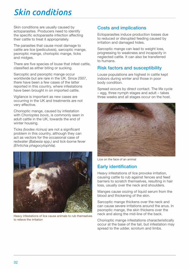
Risk factors and susceptibilityLouse populations are highest in cattle kept indoors during winter and those in poor body condition.Spread occurs by direct contact. The life cycle – egg, three nymph stages and adult – takes three weeks and all stages occur on the host.
Early identificationHeavy infestations of lice provoke irritation, causing cattle to rub against fences and feed barriers to scratch themselves, resulting in hair loss, usually over the neck and shoulders.Manges cause oozing of liquid serum from the blood and thickening of the skin.Sarcoptic mange thickens over the neck and can cause severe irritations around the anus. In psoroptic mange, the skin thickens over the neck and along the mid-line of the back.Chorioptic mange infestations characteristically occur at the base of the tail, but infestation may spread to the udder, scrotum and limbs.
Heavy infestations of lice cause animals to rub themselves to relieve the irritation
Lice on the face of an animal
32
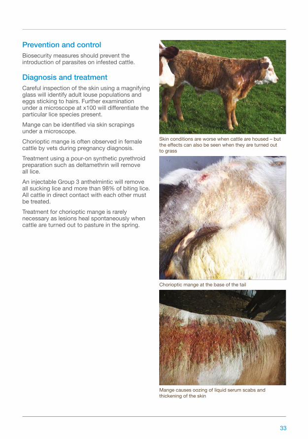
Skin conditions are worse when cattle are housed – but the effects can also be seen when they are turned out to grass
Chorioptic mange at the base of the tail
Mange causes oozing of liquid serum scabs and thickening of the skin
Prevention and controlBiosecurity measures should prevent the introduction of parasites on infested cattle.
Diagnosis and treatmentCareful inspection of the skin using a magnifying glass will identify adult louse populations and eggs sticking to hairs. Further examination under a microscope at x100 will differentiate the particular lice species present.Mange can be identified via skin scrapings under a microscope. Chorioptic mange is often observed in female cattle by vets during pregnancy diagnosis.Treatment using a pour-on synthetic pyrethroid preparation such as deltamethrin will remove all lice.An injectable Group 3 anthelmintic will remove all sucking lice and more than 98% of biting lice. All cattle in direct contact with each other must be treated.Treatment for chorioptic mange is rarely necessary as lesions heal spontaneously when cattle are turned out to pasture in the spring.
33

Summer mastitis
Summer mastitis usually occurs in non-lactating cows and heifers during summer. It occasionally appears in the rudimentary udders of young heifers, bulls and steers. There is wide regional variation in incidence across the UK. In beef cows, summer mastitis is often seen where barren, spring-calving cows are kept for breeding later in the year, ie transferred from a spring- to autumn-calving herd.The disease can be present in beef cows that have stopped lactating before their calf has been removed.A range of bacteria cause the infection, including Arcanobacterium pyogenes, Peptostreptococcus indolicus and Streptococcus dysgalactiae.Infection is transmitted by headflies (Hydrotea irritans), which live in bushes and trees. They only fly during mild, humid conditions and in low wind speeds. Cases of summer mastitis tend to be associated with ‘problem fields’ next to woods and high hedges.
Costs and implicationsLoss of an affected quarter of the udder reduces future milk production by around 10%, so the suckling calf will suffer and will not grow as well as expected. Affected cows may lose up to 100kg liveweight and generally command poor sale prices when sold.
Risk factors and susceptibilityCattle are at risk when grazing low-lying fields surrounded by trees or permanent pasture, where more flies tend to hatch. Some animals will also have a higher genetic susceptibility than others and be more likely to suffer.
Early identificationSupervision of maiden and in-calf heifers and dry cows at pasture may be sporadic during summer and mastitis can be well-advanced before clinical signs are noted.During the early stages, there is gradual enlargement both in length and diameter of the teats of the affected quarters before the heifer/cow becomes sick. Often, large numbers of flies cluster around the affected teat opening, causing considerable irritation and the animal will kick frequently.Obvious swelling accompanies more generalised signs of illness, including isolation from the group, stiffness and reluctance to walk, lack of grazing and rapid loss of body condition.The affected quarter is swollen, hard, painful and hot. The udder secretion is thick and clotted with foul-smelling green/yellow pus. Affected animals can abort and may die without prompt treatment.The affected quarter is usually permanently damaged and the cows are likely to give birth to weak calves that often die. Those that survive should be given colostrum from another cow.
The calves of dams with mastitis may have to be bottle fed to ensure they receive enough milk
Flies often cluster around the affected teat
34

It is important to strip out the lumpy milk if possible
Prevention and controlReduce exposure to flies by grazing cows away from susceptible fields in summer. Move them to higher, more exposed ground, away from clumps of trees or high hedges.Employ fly control measures (usually synthetic pyrethroids), such as impregnated fly tags, pour-on preparations and sprays.Dry-cow therapy remains the most effective means of preventing summer mastitis in cows at weaning and in susceptible pregnant heifers.Use long-duration, dry-cow antibiotic preparations with veterinary advice.Take care when infusing intramammary antibiotic preparations in heifers to prevent teat damage. Cattle should not be tubed in wet weather or in unhygienic conditions.Sealing the teat canal, with physical barriers such as micropore/adhesive tape and external teat sealants can be used to good effect.Remove any affected cows from other cows to prevent the spread of infection.
Diagnosis and treatmentDiagnosis is based on spotting swollen udders as soon as possible.Veterinary drugs, including antibiotics given as injections and intramammary tubes, are required to treat the infection.Stripping the lumpy milk out of the affected quarter should be undertaken as often as is practical, but may be resented by the animal due to the pain, so there is a high risk of being kicked.
Obvious swelling accompanies more general signs of distress
Dry-cow therapy is the most effective way to prevent summer mastitis
35

Best practice
Subcutaneous and intramuscular injectionsSubcutaneous injections are administered in areas where the skin is loose; mainly the neck or behind the shoulder. Grasp a fold of skin and slide the needle through the skin parallel to the animal’s neck. This method will avoid penetration of underlying muscle. The needle should be inserted several inches from the operator’s hand to avoid accidental self-injection. The plunger of the syringe should always be pulled back to ensure that the needle is not located within a blood vessel.The main site for intramuscular injection is the muscle mass of the neck, for which the animal must be adequately restrained to avoid head-butting or kicking. Draw up the solution for injection into the syringe. Disconnect the needle and hold the hub firmly between thumb and middle finger. Insert the needle into the muscle to the hub with a sharp slap action. Connect the syringe to the needle, draw back to check for the absence of blood and then slowly inject the contents of the syringe over 10 seconds. Do not inject too quickly as this may cause pain to the animal.Never insert the needle when connected to the syringe, as this makes it more difficult to insert to the correct depth with a single movement. The syringe hub is the weakest point and will often snap if the animal moves, rendering the contents of the syringe useless and creating potential animal welfare and meat safety issues.
Intravenous injectionThis is the fastest route for drug administration, bypassing absorption. However, this is a vet procedure only. Drugs administered intravenously include some antimicrobials, non-steroidal, anti-inflammatory drugs and mineral solutions including calcium, magnesium and phosphorus.
DrenchingSmall volumes of liquid (less than 50ml) can be administered by mouth using a drenching gun. The animal needs to be suitably restrained in a cattle crush. The animal’s head is held with the chin up and the liquid slowly squirted into the mouth. The animal’s head is released once it has swallowed the liquid.Larger volumes can be administered by stomach tube, most often in calves to administer colostrum or oral rehydration solution by oesophageal feeder. These feeders are designed so that the bulbous end cannot be mistakenly passed into the windpipe. The tube is passed slowly through the animal’s mouth and advanced when the calf swallows. Do not force the tube. After repeated use, the plastic tube may become kinked and it is necessary to pass it through hot water to soften the plastic and enable passage.
The main site for intramuscular injection is the muscle mass of the neck
Correct drenching procedure involves facing the same way as the cow and placing your back against the cow’s chin with one arm over the cow’s nose
A mouth gag will help pass a stomach tube in an adult cow
36

In adult cattle, select a stomach tube with the correct diameter for the cow. A tube with a narrow gauge may increase the risk of passage into the windpipe. Measure the stomach tube against the side of the cow following the contour of the lower neck to a point 30cm behind the point of the cow’s elbow, which is the entry point into the rumen. Face the same way as the cow and place your back against the cow’s chin, with one arm over the cow’s nose with your hand at the mouth. With your other hand, pass the stomach tube into the mouth and guide the tip over the cow’s tongue with your other hand advancing the tube into the rumen. Gas is often released as the tube enters.The animal should not struggle vigorously and rarely cough – such reaction may indicate that the tube has entered the windpipe. A mouth gag or an outer protective metal tube will prevent the cow chewing the stomach tube.
Key points• The volume injected at a single site must not
exceed that stated in the data sheet• Only administer medicines by the stated
route(s)• An accurate liveweight measurement must
be used either by weigh crate or weigh band. Underestimates of body weight may lead to under-dosing and the medicine not being wholly effective. This can potentially lead to the build-up of resistance. This situation is most likely when treating a group of animals with a wide range of weights, where the average body weight is selected, eg drenching growing animals with an anthelmintic
• When using medicines that are suspensions, thorough mixing is essential before administration
• When using the same bottle of medicine multiple times, a needle should be inserted through the rubber stopper and left in place with syringes attached to this needle
Veterinary medicinesStorage• Store in accordance with the manufacturer’s
instructions• Refrigeration must be available and
maintained between 2–8°C. Refrigerators should be fitted with a max/min thermometer to allow monitoring of the temperature
• The designated storage area should not be accessible to the public
• Storage areas should be clean and well ventilated, with eating/drinking forbidden
• Dates of delivery should be logged and marked on products. For multi-use products, date of first use should be marked on the product
Handling• Direct contact between the skin of the person
dispensing the drug and the drug itself should be avoided. This can be achieved through wearing protective clothing, such as disposable gloves
• Always consult the data sheet
Disease prevention and biosecurityLivestock movements are one of the biggest factors in the spread of infectious disease. Always know the health status of animals being bought and moved and ensure adequate quarantine periods apply.Biosecurity reduces/prevents the introduction of new diseases onto a farm from outside sources and is the first line of defence.Biocontainment measures aim to limit the spread of disease within a herd if biosecurity has been breached and a new disease has been inadvertently introduced onto the property.
Weigh cattle to administer correct dosage of medicines
37

Disease spreadInfectious diseases can be introduced by:• Diseased animals• Animals incubating disease• Apparently healthy animals that have
recovered from disease but are now carriers• Vehicles, equipment, clothing and footwear
of people such as vets, contractors, other farmers, salesmen etc, who move between herds
• Feedstuffs, especially those which could be contaminated with faeces
• Contaminated water from surface water, streams and rivers etc
• Manure handling, especially by outside contractors
• Other species such as dogs, cats, wildlife, rodents, birds and insects
Biosecurity principlesBiosecurity control has four major components:1. Selection2. Isolation3. Movement control4. SanitationSelection Purchase animals from known sources that have a health status equal to, or higher than the existing herd.• Know the health history of the herds from
which cattle are purchased• Know the health status of animals brought
onto the farm• Never bring in animals without knowing their
vaccination history• Limit purchases to maiden heifers and bullsIsolationStrict isolation prevents contact between animals after arrival on farm to reduce the risk of spread of infectious agents.• Quarantine all new arrivals for at least
30 days• Cattle must not share community pastures/
common grazings
• Cattle must not share fence lines with a neighbour’s cattle
• Do not use hire bulls from other farmsMovement controlThis includes all vehicle, animals and people traffic that could introduce infection onto the farm.Record all visitors to the farm – both human and domestic animals.
SanitationThis addresses the disinfection of materials, people and equipment entering the farm and the cleanliness of the people and equipment on farm.• Prevent manure contamination of feed and
feeding equipment• Use different equipment to feed and muck
out pens or disinfect between use• Never step in the feed bunker• Transport animals in clean vehicles only• Site the lorry-loading area at the perimeter
of the farm
Best practice
Keep cattle separate between neighbouring farms with double fencing
38

Other steps to maintain biosecurity• Maintain a closed herd wherever possible,
using artificial insemination to introduce new genetics.
• Oestrus synchronisation programmes can be very successful in groups of beef cattle, especially heifers
• Control diseases by vaccination• Maintain perimeter fences• Do not impulse-buy animals from unknown
sources at market or dispersal sales. They may bring new and devastating disease onto the farm
Do not unload muck where cattle will graze as it can spread disease
Top 10 herd health tips: 1 Maintain biosecurity – do not buy in disease2 Monitor herd health status. Check what diseases are present by blood sampling youngstock3 Cull infected animals, eg those animals persistently infected with BVD, or carrying IBR or Johne’s disease4 Control diseases that cannot be eradicated. Vaccination policies save many times their cost5 Implement effective treatment protocols
where disease cannot be completely prevented. Limit damage by treating the disease as quickly and effectively as possible
6 Improve management practices, eg batch calving, all-in, all-out policies
7 Improve the environment, eg improve ventilation in stock buildings8 Record reproductive performance. Beef production starts with the birth of live calves9 Record growth rates. Find out if the animals are responding to their feed input 10 Take a team approach to reducing
disease and the costs associated with preventing, managing and treating it. Involve stockmen, vets and other advisors
Discuss the herd health plan, disease surveillance programme and disease respsonse actions with the vet on a regular basis.
39
Produced for you by:Better Returns Programme AHDB Beef & Lamb Stoneleigh Park Kenilworth Warwickshire CV8 2TL
T 024 7647 8834 E [email protected] W beefandlamb.ahdb.org.uk
@AHDB_BeefLambIf you no longer wish to receive this information, please email us on [email protected]
While the Agriculture and Horticulture Development Board seeks to ensure that the information contained within this document is accurate at the time of printing, no warranty is given in respect thereof and, to the maximum extent permitted by law, the Agriculture and Horticulture Development Board accepts no liability for loss, damage or injury howsoever caused (including that caused by negligence) or suffered directly or indirectly in relation to information and opinions contained in or omitted from this document.
© Agriculture and Horticulture Development Board 2018. All rights reserved.