Minimally Invasive Esophageal Surgery Benign: enucleation, myotomy, anti-reflux op.

BENIGN ESOPHAGEAL DISEASESRichard C. Golding, M.D., M.P.H.
Introduction
2
Outline
Esophageal Anatomy
Clinical Presentation of Benign Esophageal Diseases
Structural Lesions of the Esophagus
Esophagitis
Gastroesophageal Reflux Disease (GERD)
Motility Disorders of the Esophagus
Esophageal Symptoms in Patients After Bariatric Surgery
3

ESOPHAGEAL ANATOMY
4
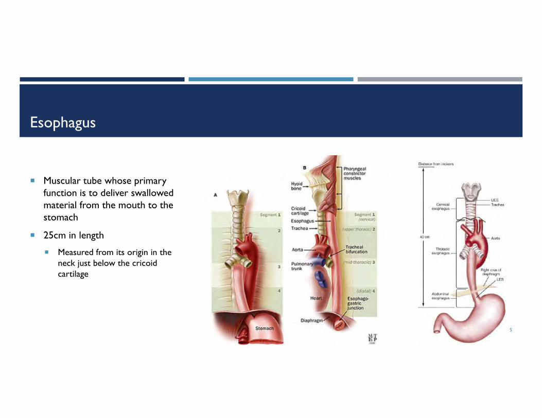
Esophagus
5
Muscular tube whose primary function is to deliver swallowed material from the mouth to the stomach
25cm in length
Measured from its origin in the neck just below the cricoid cartilage
Muscles of Esophagus
6
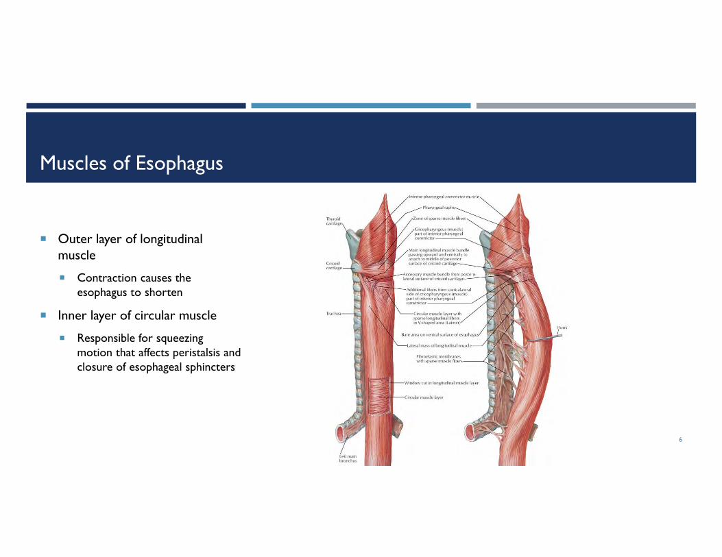
Outer layer of longitudinal muscle
Contraction causes the esophagus to shorten
Inner layer of circular muscle
Responsible for squeezing motion that affects peristalsis and closure of esophageal sphincters
Upper Esophageal Sphincter (UES)
7
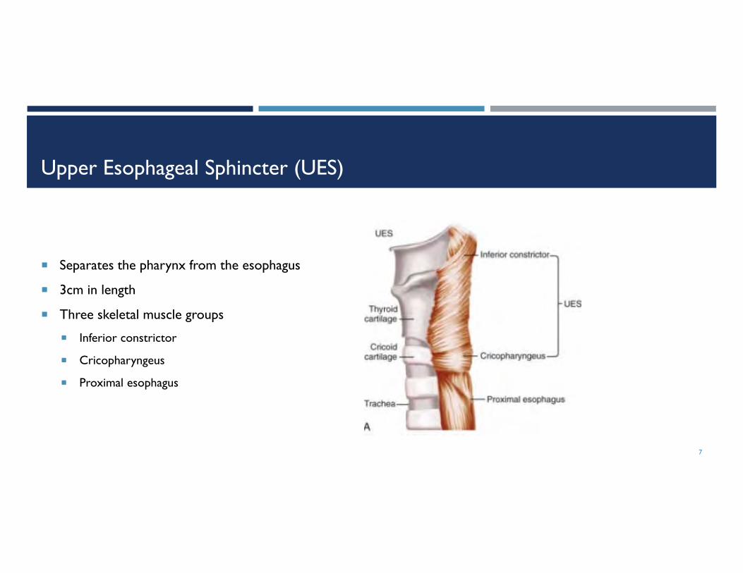
Separates the pharynx from the esophagus
3cm in length
Three skeletal muscle groups
Inferior constrictor
Cricopharyngeus
Proximal esophagus
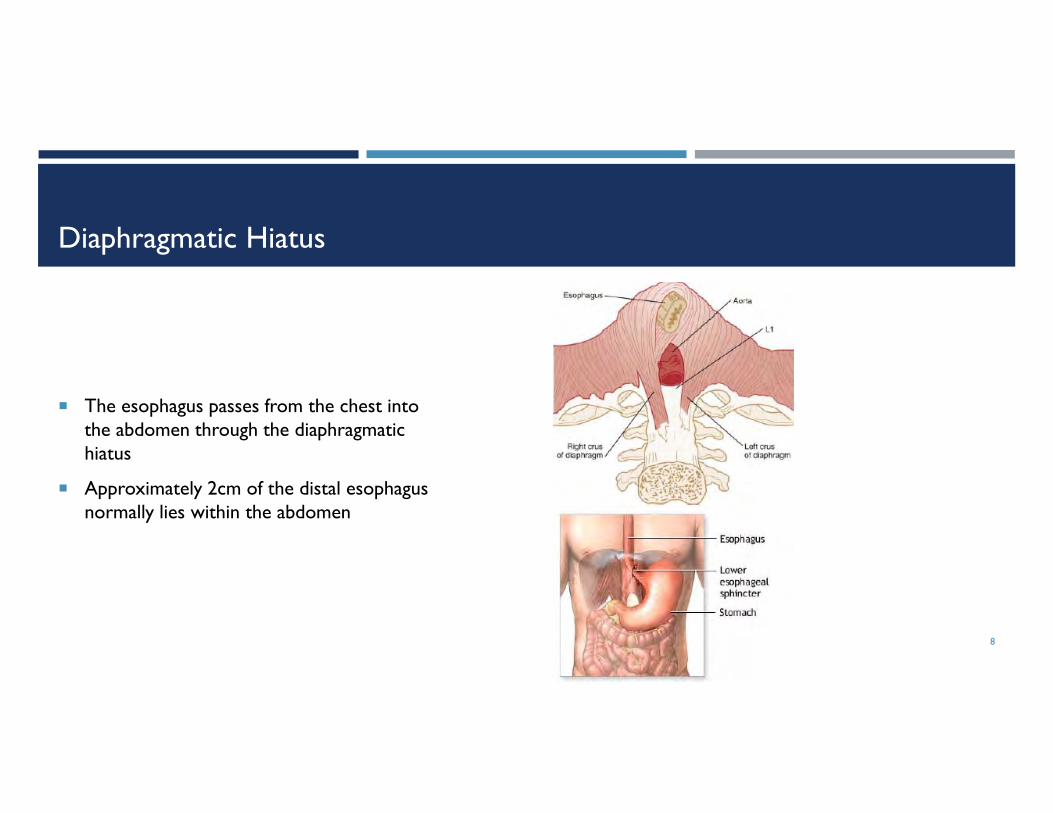
Diaphragmatic Hiatus
8
The esophagus passes from the chest into the abdomen through the diaphragmatic hiatus
Approximately 2cm of the distal esophagus normally lies within the abdomen
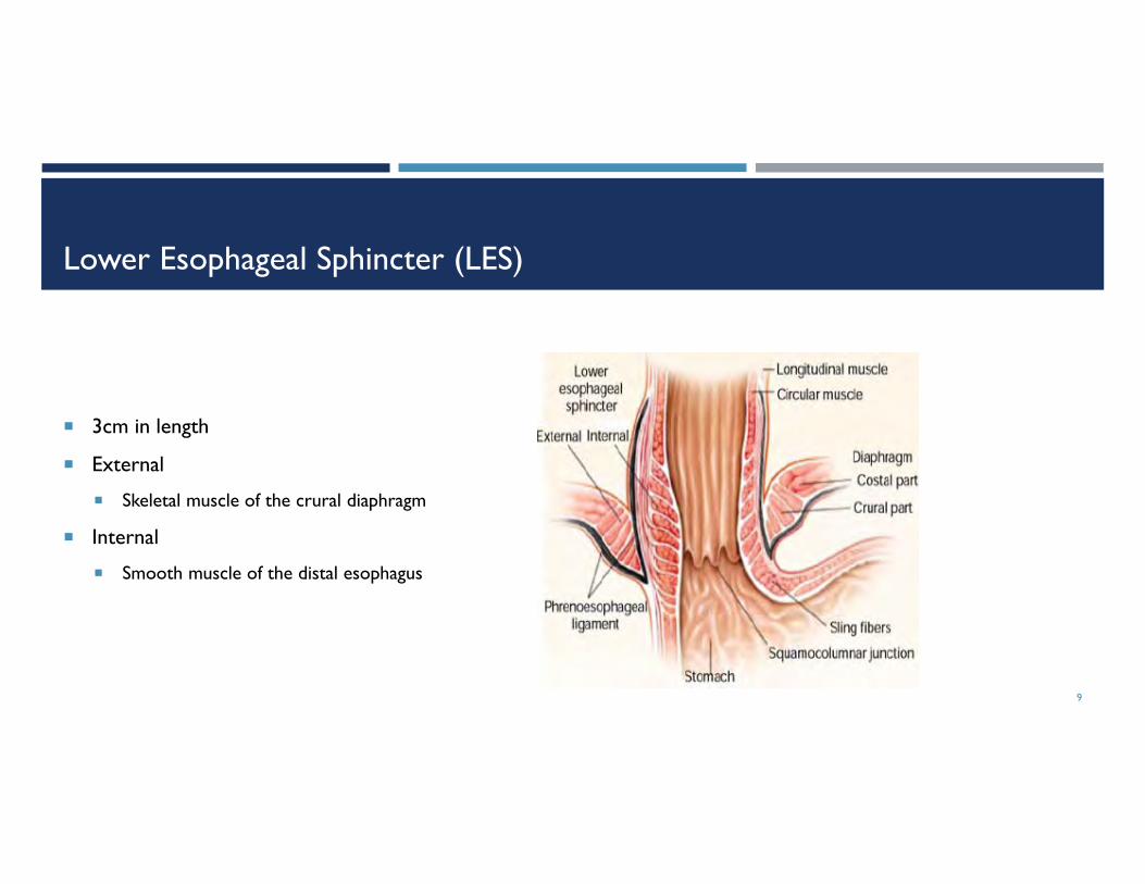
Lower Esophageal Sphincter (LES)
9
3cm in length
External
Skeletal muscle of the crural diaphragm
Internal
Smooth muscle of the distal esophagus
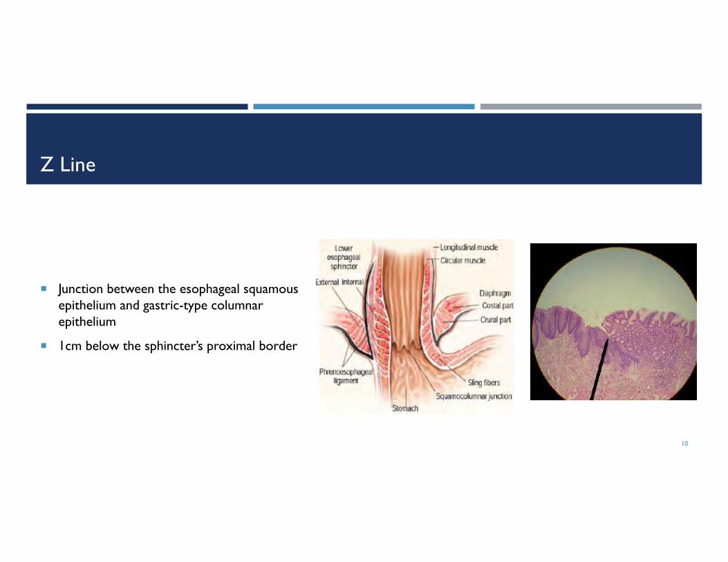
Z Line
10
Junction between the esophageal squamous epithelium and gastric-type columnar epithelium
1cm below the sphincter’s proximal border
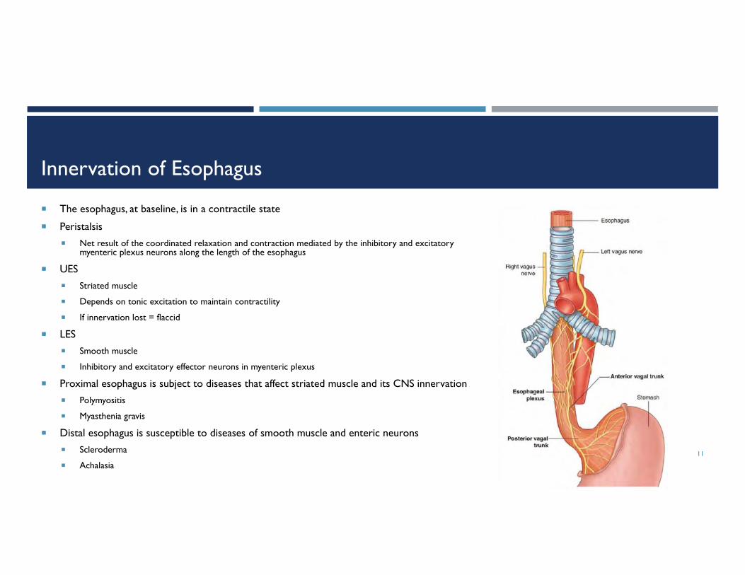
Innervation of Esophagus
11
The esophagus, at baseline, is in a contractile state
Peristalsis Net result of the coordinated relaxation and contraction mediated by the inhibitory and excitatory
myenteric plexus neurons along the length of the esophagus
UES Striated muscle
Depends on tonic excitation to maintain contractility
If innervation lost = flaccid
LES Smooth muscle
Inhibitory and excitatory effector neurons in myenteric plexus
Proximal esophagus is subject to diseases that affect striated muscle and its CNS innervation Polymyositis
Myasthenia gravis
Distal esophagus is susceptible to diseases of smooth muscle and enteric neurons Scleroderma
Achalasia

CLINICAL PRESENTATION OF ESOPHAGEAL DISEASES
12
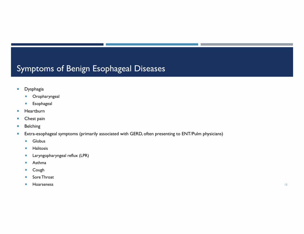
Symptoms of Benign Esophageal Diseases
Dysphagia
Oropharyngeal
Esophageal
Heartburn
Chest pain
Belching
Extra-esophageal symptoms (primarily associated with GERD, often presenting to ENT/Pulm physicians)
Globus
Halitosis
Laryngopharyngeal reflux (LPR)
Asthma
Cough
Sore Throat
Hoarseness 13

Evaluation Of Benign Esophageal Diseases
Barium esophagram
Cervical and thoracic with barium tablet (13mm)
Symptomatic usually with lumen less than 13 mm
EGD
Modified barium swallow (MBS)
Esophageal manometry
pH studies
14
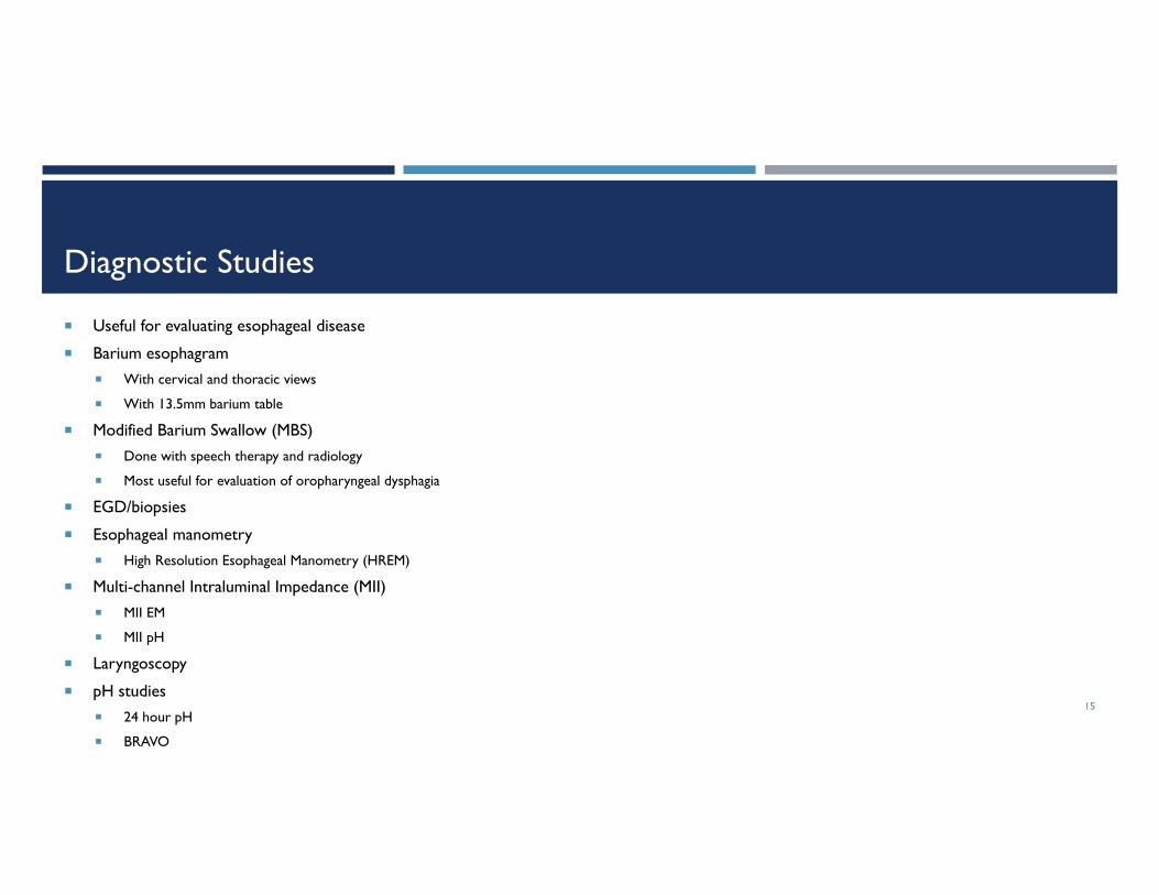
Diagnostic Studies
Useful for evaluating esophageal disease
Barium esophagram With cervical and thoracic views
With 13.5mm barium table
Modified Barium Swallow (MBS) Done with speech therapy and radiology
Most useful for evaluation of oropharyngeal dysphagia
EGD/biopsies
Esophageal manometry High Resolution Esophageal Manometry (HREM)
Multi-channel Intraluminal Impedance (MII) MII EM
MII pH
Laryngoscopy
pH studies 24 hour pH
BRAVO
15
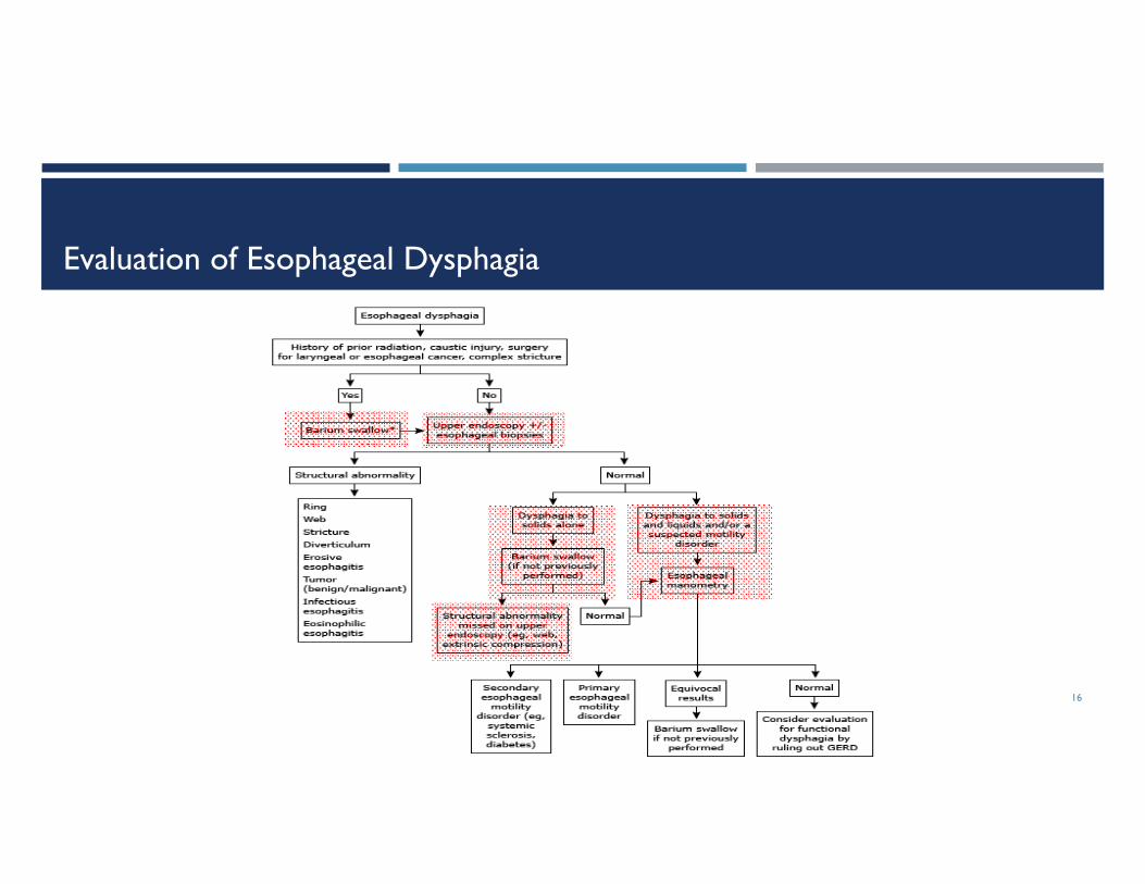
Evaluation of Esophageal Dysphagia
16
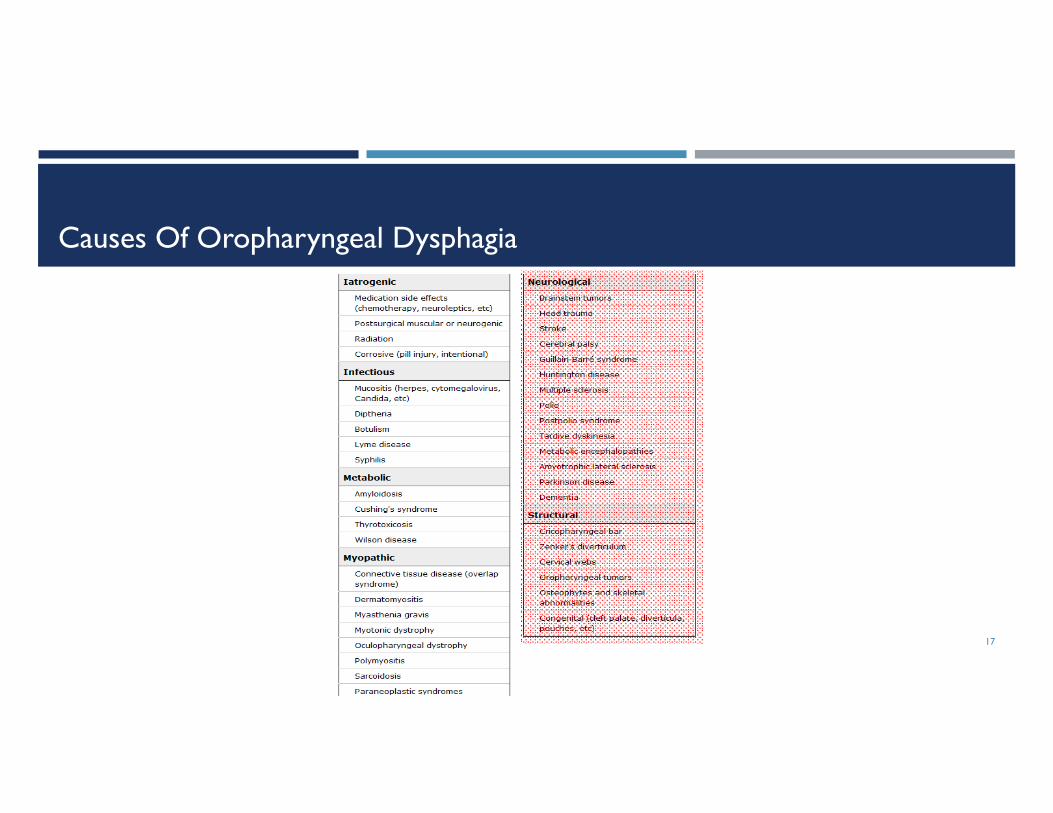
Causes Of Oropharyngeal Dysphagia
17
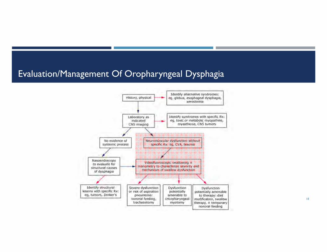
Evaluation/Management Of Oropharyngeal Dysphagia
18
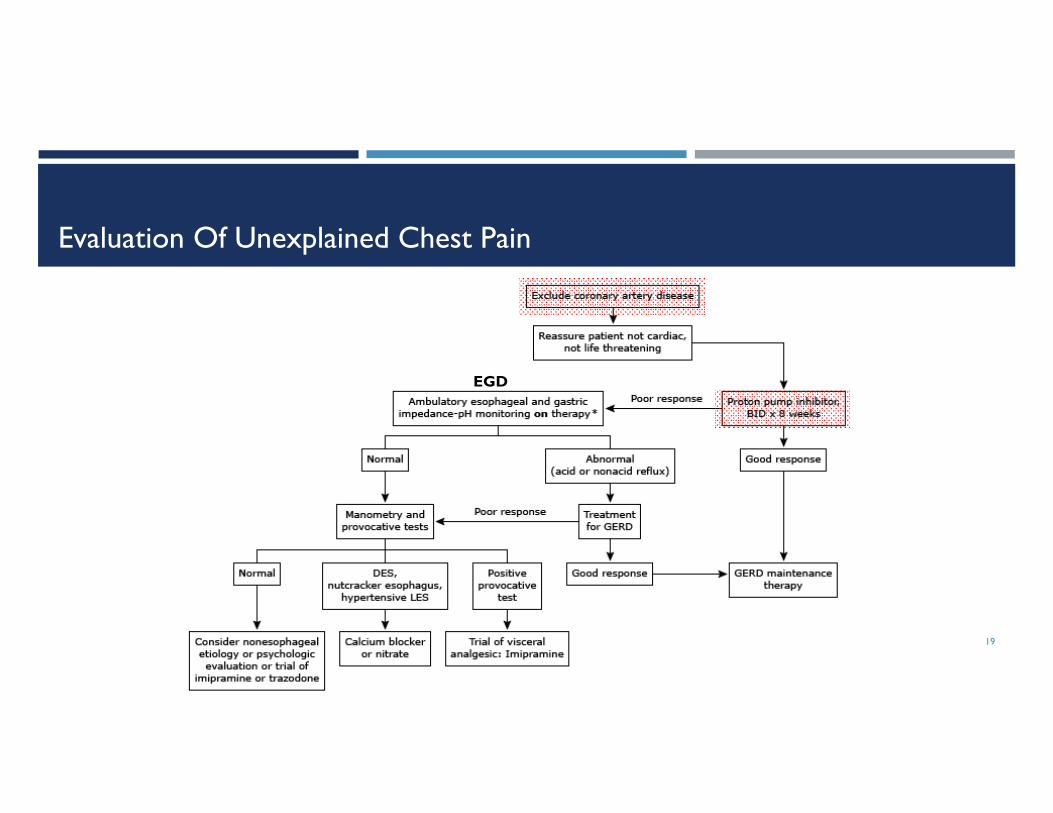
Evaluation Of Unexplained Chest Pain
19
EGD
STRUCTURAL LESIONS OF THE ESOPHAGUS
20
Structural Lesions
Associated with anatomic narrowing
Usually presenting with dysphagia
When luminal diameter <13 mm
Hernias Hiatal
Paraesophageal
Rings
Webs
Food impaction
Foreign bodies
Diverticulum
21
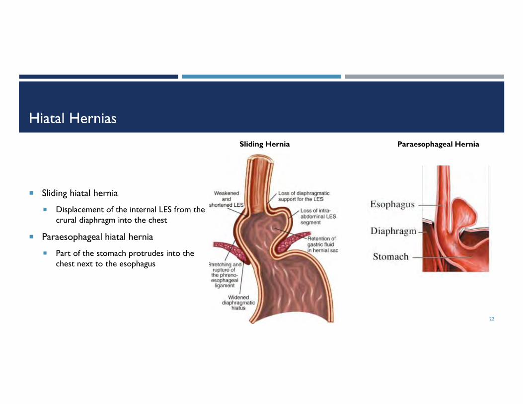
Hiatal Hernias
Sliding hiatal hernia
Displacement of the internal LES from the crural diaphragm into the chest
Paraesophageal hiatal hernia
Part of the stomach protrudes into the chest next to the esophagus
22
Sliding Hernia Paraesophageal Hernia
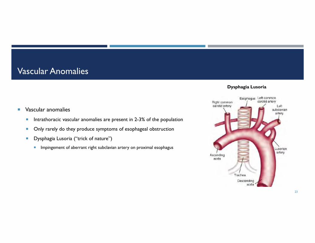
Vascular Anomalies
Vascular anomalies
Intrathoracic vascular anomalies are present in 2-3% of the population
Only rarely do they produce symptoms of esophageal obstruction
Dysphagia Lusoria (“trick of nature”)
Impingement of aberrant right subclavian artery on proximal esophagus
23
Dysphagia Lusoria
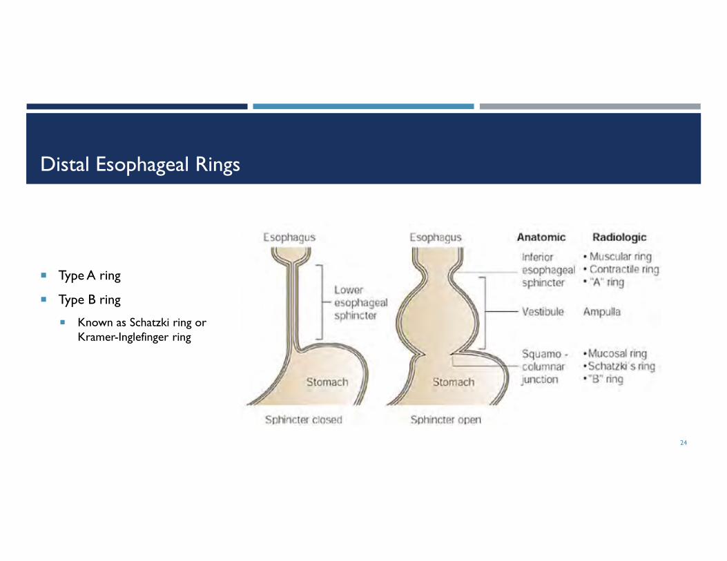
Distal Esophageal Rings
Type A ring
Type B ring
Known as Schatzki ring or Kramer-Inglefinger ring
24
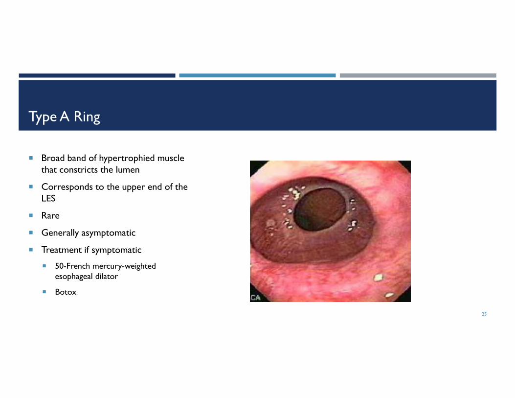
Type A Ring
Broad band of hypertrophied muscle that constricts the lumen
Corresponds to the upper end of the LES
Rare
Generally asymptomatic
Treatment if symptomatic
50-French mercury-weighted esophageal dilator
Botox
25
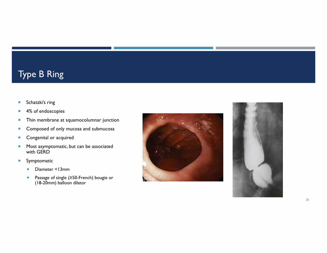
Type B Ring
Schatzki’s ring
4% of endoscopies
Thin membrane at squamocolumnar junction
Composed of only mucosa and submucosa
Congenital or acquired
Most asymptomatic, but can be associated with GERD
Symptomatic
Diameter <13mm
Passage of single (≥50-French) bougie or (18-20mm) balloon dilator
26
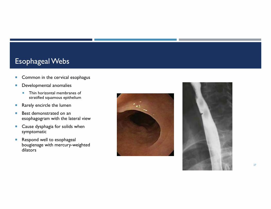
Esophageal Webs
Common in the cervical esophagus
Developmental anomalies
Thin horizontal membranes of stratified squamous epithelium
Rarely encircle the lumen
Best demonstrated on an esophagogram with the lateral view
Cause dysphagia for solids when symptomatic
Respond well to esophageal bougienage with mercury-weighted dilators
27
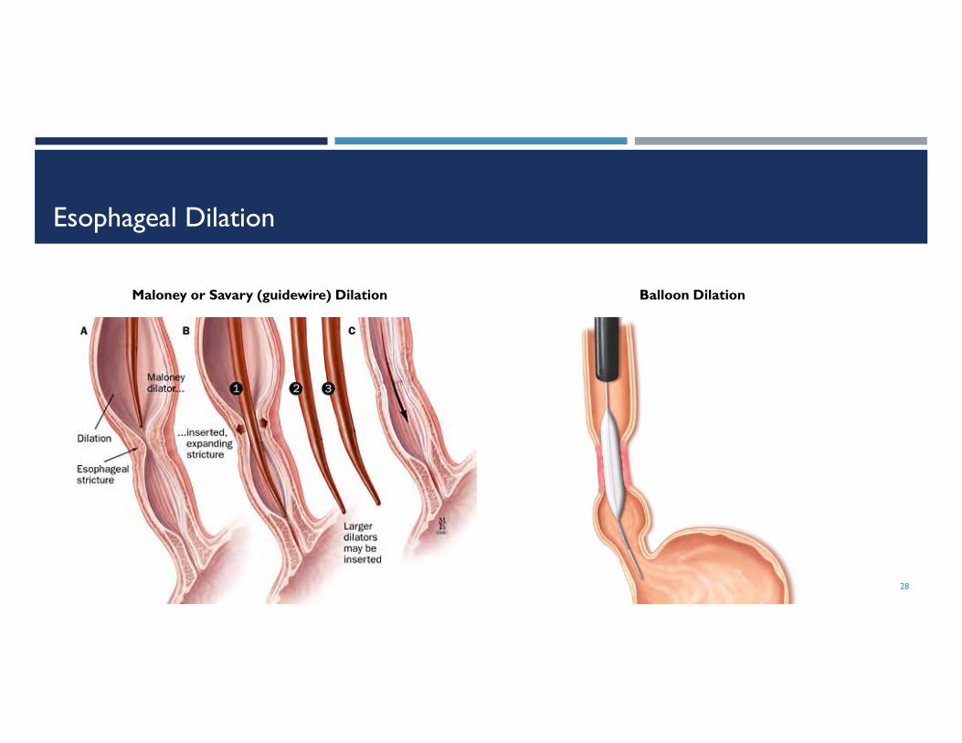
Esophageal Dilation
28
Maloney or Savary (guidewire) Dilation Balloon Dilation
Plummer-Vinson (Paterson-Kelly) Syndrome
Characterized by:
Cervical esophageal webs
Dysphagia
Iron deficiency anemia
Primarily in women
Associated with celiac disease
Increased risk for squamous carcinoma of the pharynx and esophagus
Correction of iron deficiency may result in resolution of the dysphagia and disappearance of the web
29
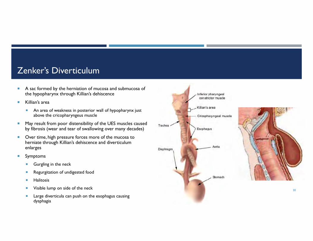
Zenker’s Diverticulum
A sac formed by the herniation of mucosa and submucosa of the hypopharynx through Killian’s dehiscence
Killian’s area
An area of weakness in posterior wall of hypopharynx just above the cricopharyngeus muscle
May result from poor distensibility of the UES muscles caused by fibrosis (wear and tear of swallowing over many decades)
Over time, high pressure forces more of the mucosa to herniate through Killian’s dehiscence and diverticulum enlarges
Symptoms
Gurgling in the neck
Regurgitation of undigested food
Halitosis
Visible lump on side of the neck
Large diverticula can push on the esophagus causing dysphagia
30
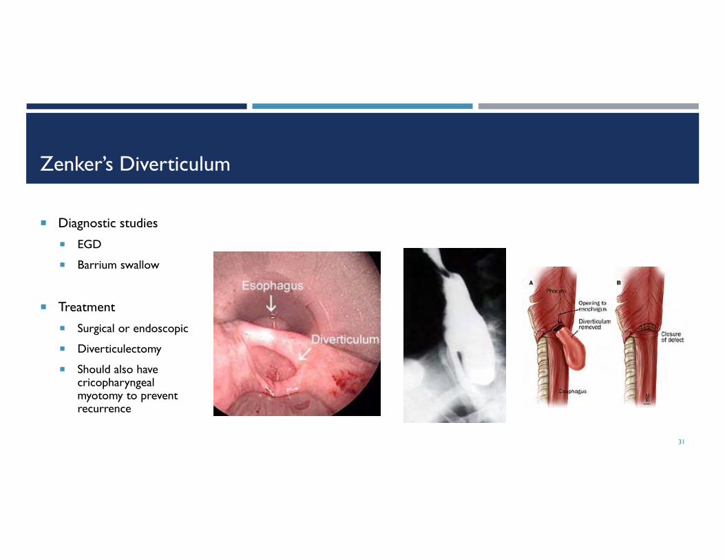
Zenker’s Diverticulum
Diagnostic studies
EGD
Barrium swallow
Treatment
Surgical or endoscopic
Diverticulectomy
Should also have cricopharyngealmyotomy to prevent recurrence
31
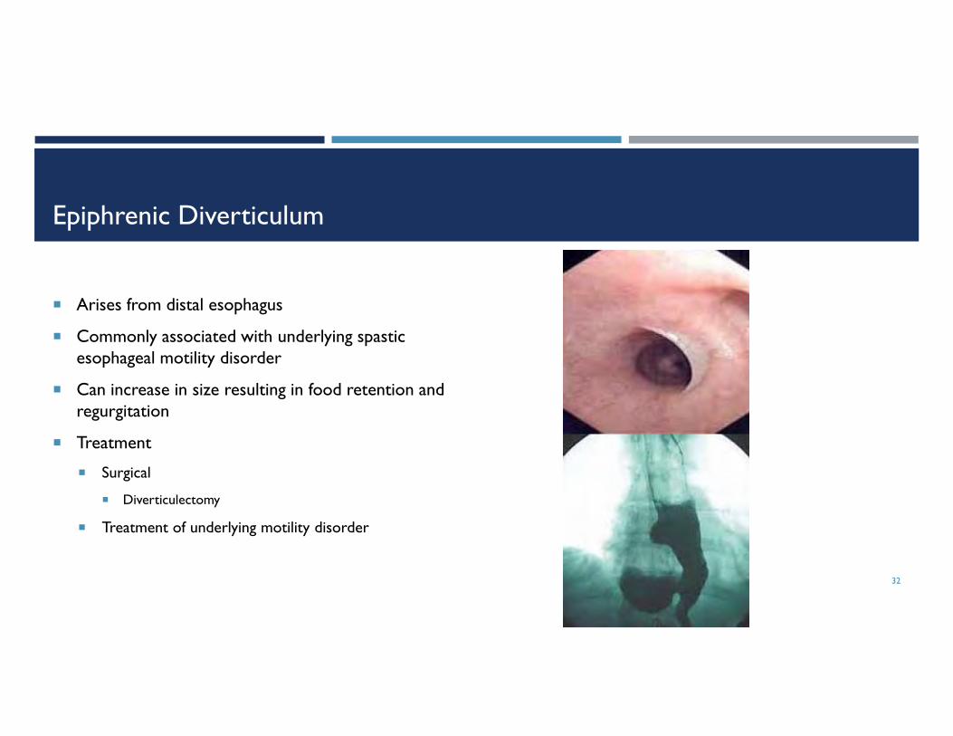
Epiphrenic Diverticulum
Arises from distal esophagus
Commonly associated with underlying spastic esophageal motility disorder
Can increase in size resulting in food retention and regurgitation
Treatment
Surgical
Diverticulectomy
Treatment of underlying motility disorder
32
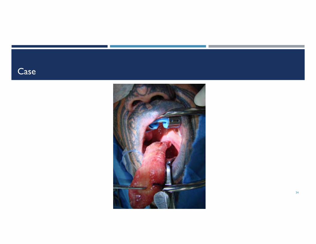
Case
34 yo male reports difficulty swallowing solids and a sense of fullness in his throat
On a recent date, he chocked on a piece of steak and his girlfriend was frightened to see a fleshy tube snap out of his mouth and then snap back
She ran away in horror and never came back
33
Case
34
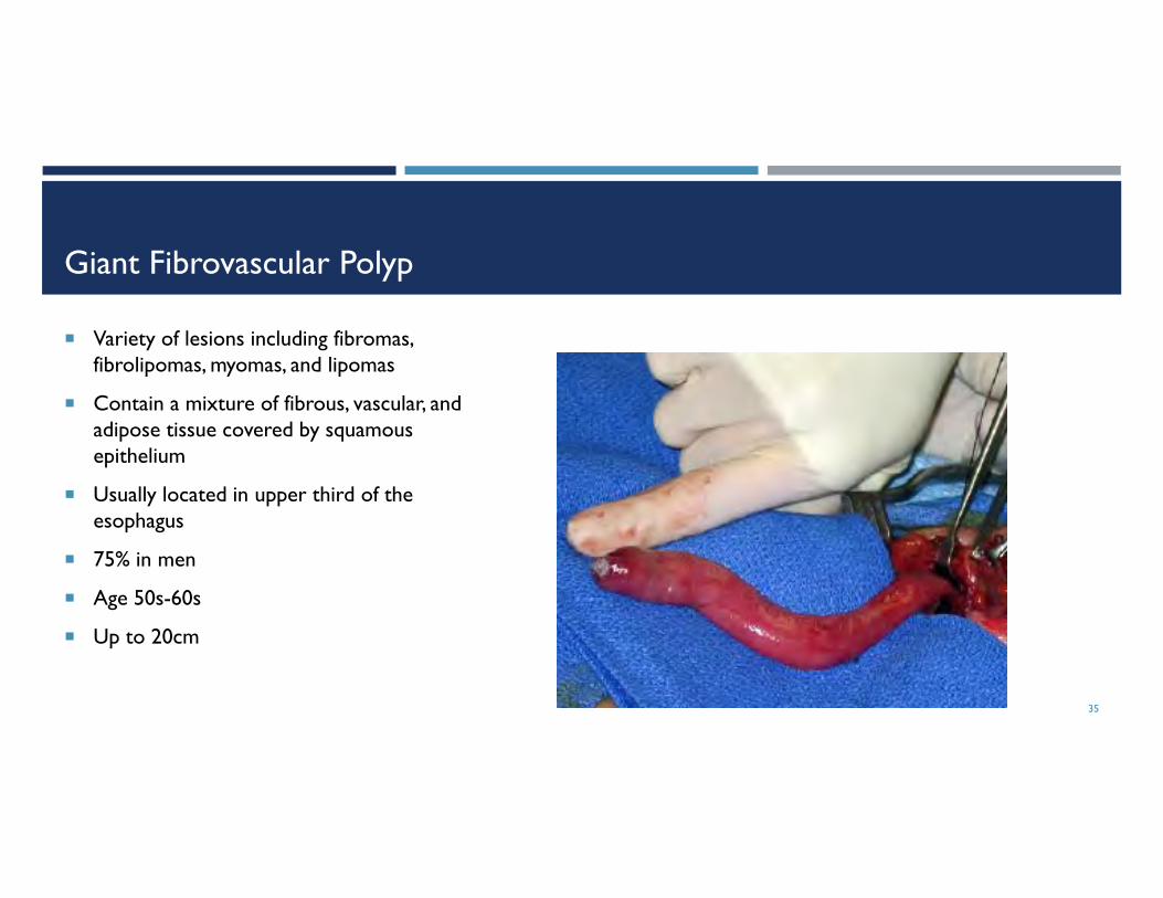
Giant Fibrovascular Polyp
Variety of lesions including fibromas, fibrolipomas, myomas, and lipomas
Contain a mixture of fibrous, vascular, and adipose tissue covered by squamous epithelium
Usually located in upper third of the esophagus
75% in men
Age 50s-60s
Up to 20cm
35
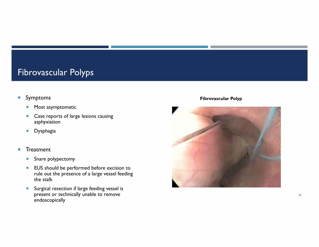
Fibrovascular Polyps
Symptoms
Most asymptomatic
Case reports of large lesions causing asphyxiation
Dysphagia
Treatment
Snare polypectomy
EUS should be performed before excision to rule out the presence of a large vessel feeding the stalk
Surgical resection if large feeding vessel is present or technically unable to remove endoscopically
36
Fibrovascular Polyp
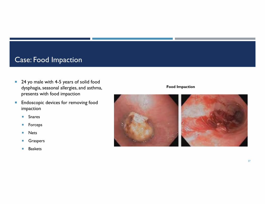
Case: Food Impaction
24 yo male with 4-5 years of solid food dysphagia, seasonal allergies, and asthma, presents with food impaction
Endoscopic devices for removing food impaction
Snares
Forceps
Nets
Graspers
Baskets
37
Food Impaction
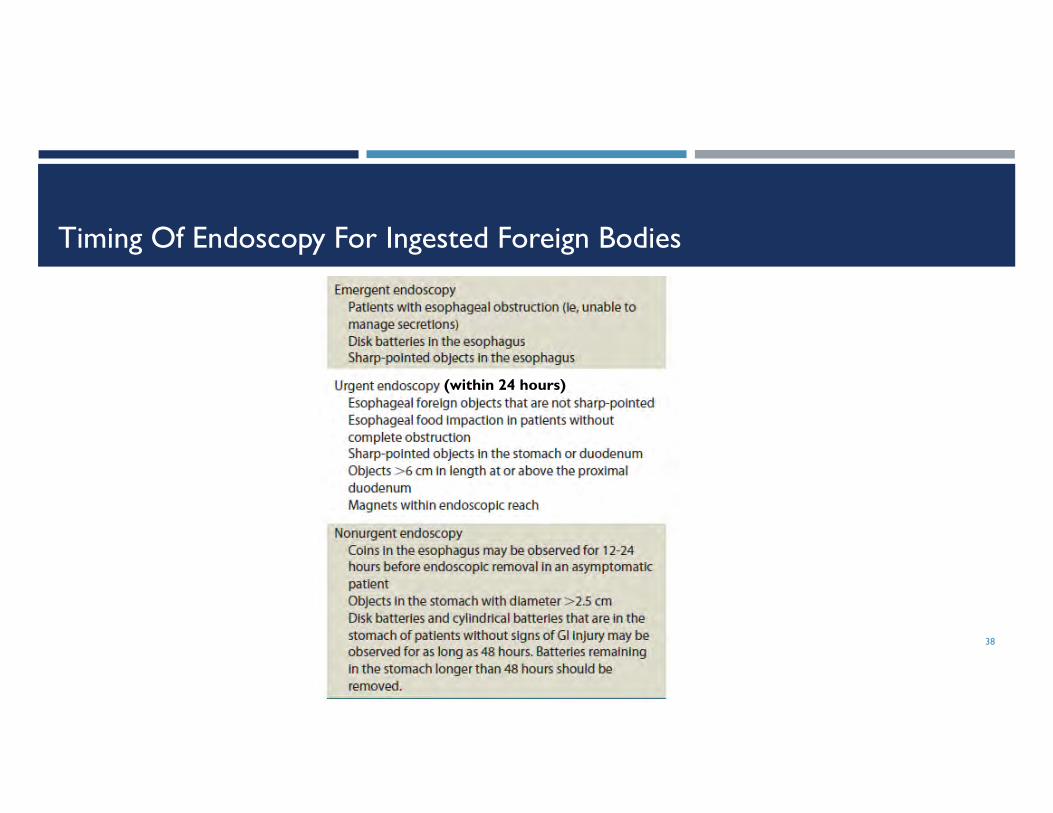
Timing Of Endoscopy For Ingested Foreign Bodies
38
(within 24 hours)

Decisions Regarding Foreign Body Management
Airway protection
Intubation may be required for upper esophageal obstructions
Overtube
Endoscopic hood for sharp objects
Radiologic localization prior to extraction
Thoracic surgery or ENT referral for foreign bodies not amenable to endoscopic removal
39

ESOPHAGITIS
40
CAUSES OF ESOPHAGITIS
GERD
Pills/medication related
Caustic ingestion
Acids
Alkalis
Causing severe esophagitis with long strictures
Radiation
Usually mediastinal
Infections
CMV
Herpes simplex
HIV
Candida/fungal
Graft vs. host disease – BMT patients
Eosinophilic esophagitis (EoE)
Pemphigus
41
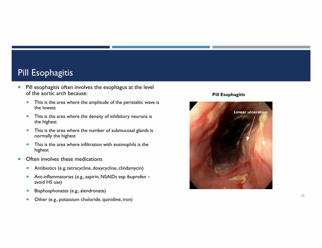
Pill Esophagitis Pill esophagitis often involves the esophagus at the level
of the aortic arch because:
This is the area where the amplitude of the peristaltic wave is the lowest
This is the area where the density of inhibitory neurons is the highest
This is the area where the number of submucosal glands is normally the highest
This is the area where infiltration with eosinophils is the highest
Often involves these medications
Antibiotics (e.g, tetracycline, doxycycline, clindamycin)
Ant-inflammatories (e.g., aspirin, NSAIDs esp ibuprofen –avoid HS use)
Bisphosphonates (e.g., alendronate)
Other (e.g., potassium choloride, quinidine, iron)42
Pill Esophagitis
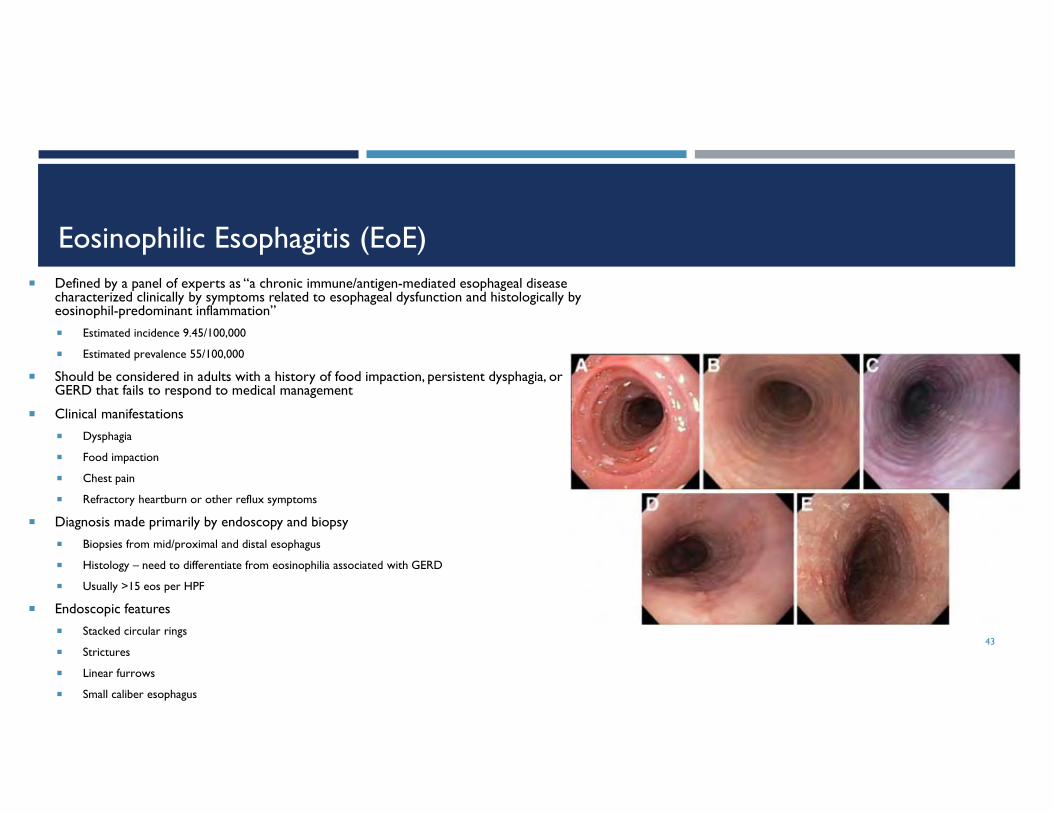
Eosinophilic Esophagitis (EoE) Defined by a panel of experts as “a chronic immune/antigen-mediated esophageal disease
characterized clinically by symptoms related to esophageal dysfunction and histologically by eosinophil-predominant inflammation”
Estimated incidence 9.45/100,000
Estimated prevalence 55/100,000
Should be considered in adults with a history of food impaction, persistent dysphagia, or GERD that fails to respond to medical management
Clinical manifestations
Dysphagia
Food impaction
Chest pain
Refractory heartburn or other reflux symptoms
Diagnosis made primarily by endoscopy and biopsy
Biopsies from mid/proximal and distal esophagus
Histology – need to differentiate from eosinophilia associated with GERD
Usually >15 eos per HPF
Endoscopic features
Stacked circular rings
Strictures
Linear furrows
Small caliber esophagus
43

Eosinophilic Esophagitis Treatment
Dietary therapy Elimination diets
Allergy/immunology evaluation
Pharmacologic Topical steroids
Swallowed corticosteroids (fluticasone spray swallowed – adults 440-880mcg BID for 2 months)
Systemic steroids (if failed topical steroids)
PPIs (for 2 months)
Endoscopic Narrow caliber esophagus requires more careful dilatation over a guide wire
Perforation rate 3/1000, similar to non-EoE strictures
44
High Resolution Manometry In EoE
32% of EoE patients demonstrated pan-esophageal pressurization events with higher volume bolus challenge
Esophageal pressurization may reflect reduced distensibility/compliance of the esophagus in EoE
45

46
GASTROESOPHAGEAL REFLUX DISEASE (GERD)
47
GERD Anatomy/physiology
Clinical manifestations and presentation Esophageal symptoms
Extra-esophageal symptoms
Diagnosis Barium esophagram
EGD
pH studies (24 hour vs. BRAVO)
Esophageal motility
Complications Non-erosive reflux disease (NERD)
Acid vs. bile reflux
Treatment Medications
Medication side effects and complications
The refractory patient
Medical treatment (including baclofen)
Surgical
Endoscopic
48
GERD Introduction
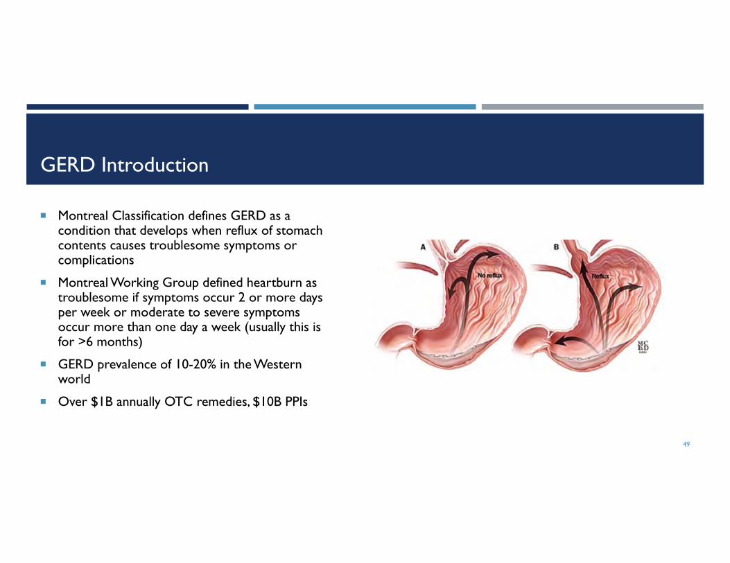
Montreal Classification defines GERD as a condition that develops when reflux of stomach contents causes troublesome symptoms or complications
Montreal Working Group defined heartburn as troublesome if symptoms occur 2 or more days per week or moderate to severe symptoms occur more than one day a week (usually this is for >6 months)
GERD prevalence of 10-20% in the Western world
Over $1B annually OTC remedies, $10B PPIs
49
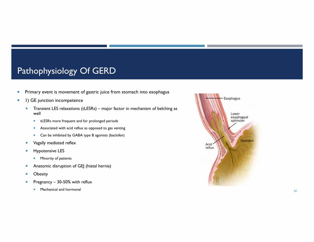
Pathophysiology Of GERD
Primary event is movement of gastric juice from stomach into esophagus
1) GE junction incompetence
Transient LES relaxations (tLESRs) – major factor in mechanism of belching as well
tLESRs more frequent and for prolonged periods
Associated with acid reflux as opposed to gas venting
Can be inhibited by GABA type B agonists (baclofen)
Vagally mediated reflex
Hypotensive LES
Minority of patients
Anatomic disruption of GEJ (hiatal hernia)
Obesity
Pregnancy – 30-50% with reflux
Mechanical and hormonal 50
Pathophysiology Of GERD
Factors which reduce LES pressure Gastric distension
CCK
Foods (fat, chocolate, alcohol, caffeine)
Smoking
Drugs (e.g., nitrates, CCBs, narcotics, benzos, progesterone)
2) Esophageal acid clearance Prolonged with esophagitis and can be prolonged with hiatal hernia
3) Esophageal emptying Peristaltic dysfunction
Intra-esophageal reflux
4) Salivation If reduced, can contribute to GERD
5) Esophageal sensitivity Non-erosive reflux disease (NERD)
51
GERD Barrier
GEJ forms anti-reflux barrier
Dependent on:
GEJ complex
Changes with gastric distension
Esophageal motility
Intra-abdominal pressure
Gravity
52

Diagnosis Of GERD
Presumptive diagnosis with typical symptoms of heartburn and regurgitation Can treat empirically with a PPI
Belching as a primary symptom often not GERD related
Patients with non-cardiac chest pain suspected due to GERD should have cardiac cause excluded before GI evaluation
These studies/procedures NOT required to diagnose GERD in the presence of typical GERD symptoms: Barium radiographs
Upper endoscopy
Biopsies from distal esophagus
Esophageal manometry
Ambulatory reflux monitoring
Screening for H. pylori infection
Endoscopy recommended for patients with alarm symptoms and for screening patients at high risk for complications For patient who are acid suppressant dependent
Especially men over age 50
Repeat endoscopy not indicated for patients without Barrett’s esophagus in absence of new symptoms
53
Extraesophageal Manifestations Of GERD
GERD can be considered a potential co-factor in patients with:
Asthma
Chronic cough
Laryngitis
PPI trial is recommended to treat extraesophageal symptoms in patients who also have typical GERD symptoms
54
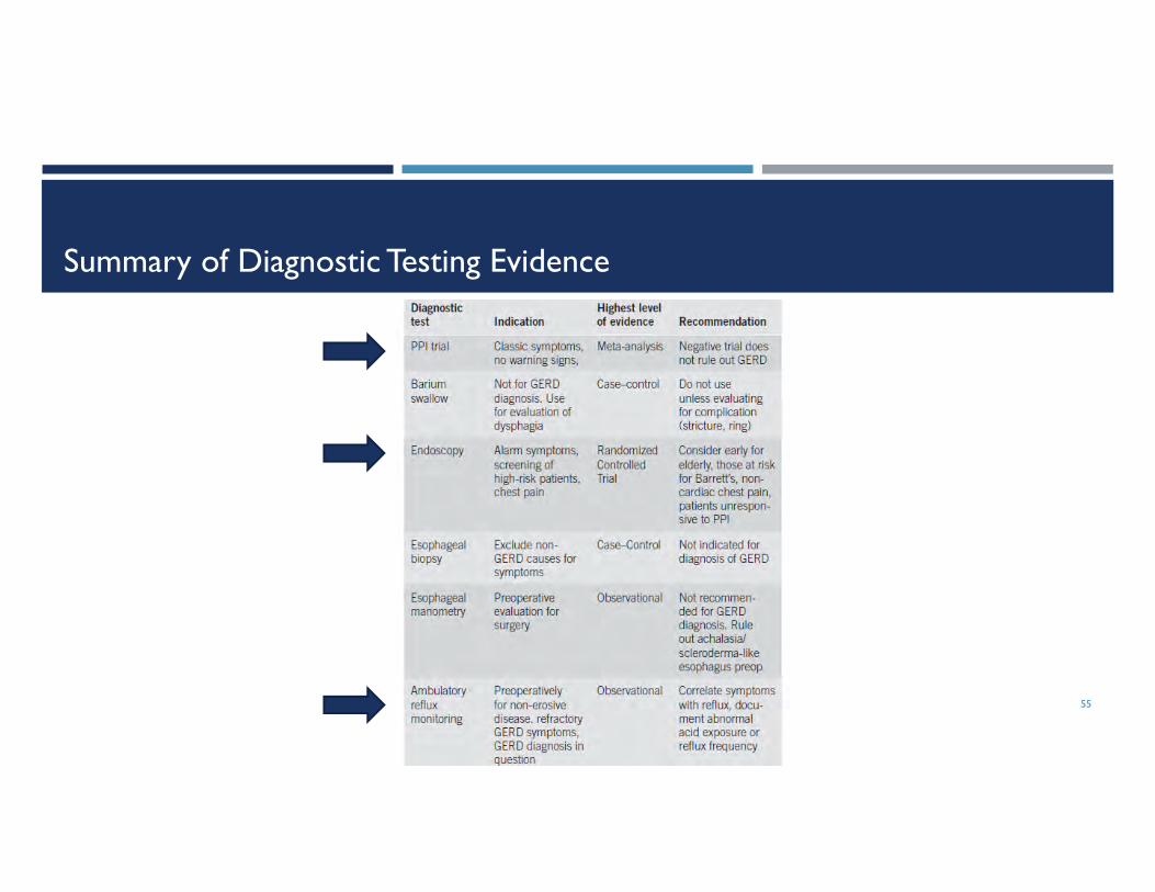
Summary of Diagnostic Testing Evidence
55

Continuous pH Monitoring
Acid-sensitive catheter is placed in the esophagus and is attached to a small monitoring device
Changes in esophageal pH are recorded over an extended period of time (up to 24 hours)
Provides information on the severity and pattern of reflux
Considered the best test for the diagnosis of GERD, however there is a 10-20% false negative rate
If intra-esophageal pH is < 4 for more than 10% of the time, patient is considered to have pathologic reflux
56
Physiologic Reflux
Pathologic Reflux
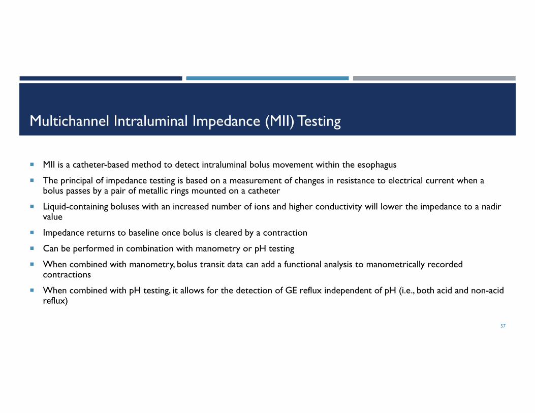
Multichannel Intraluminal Impedance (MII) Testing
MII is a catheter-based method to detect intraluminal bolus movement within the esophagus
The principal of impedance testing is based on a measurement of changes in resistance to electrical current when a bolus passes by a pair of metallic rings mounted on a catheter
Liquid-containing boluses with an increased number of ions and higher conductivity will lower the impedance to a nadir value
Impedance returns to baseline once bolus is cleared by a contraction
Can be performed in combination with manometry or pH testing
When combined with manometry, bolus transit data can add a functional analysis to manometrically recorded contractions
When combined with pH testing, it allows for the detection of GE reflux independent of pH (i.e., both acid and non-acid reflux)
57
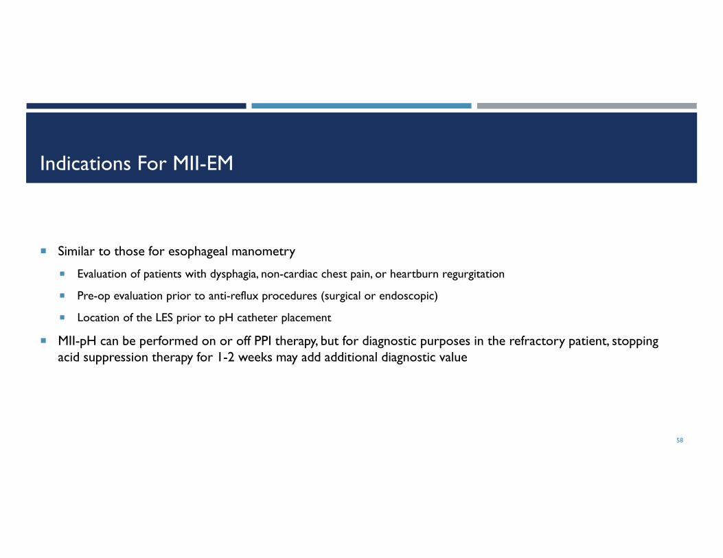
Indications For MII-EM
Similar to those for esophageal manometry
Evaluation of patients with dysphagia, non-cardiac chest pain, or heartburn regurgitation
Pre-op evaluation prior to anti-reflux procedures (surgical or endoscopic)
Location of the LES prior to pH catheter placement
MII-pH can be performed on or off PPI therapy, but for diagnostic purposes in the refractory patient, stopping acid suppression therapy for 1-2 weeks may add additional diagnostic value
58
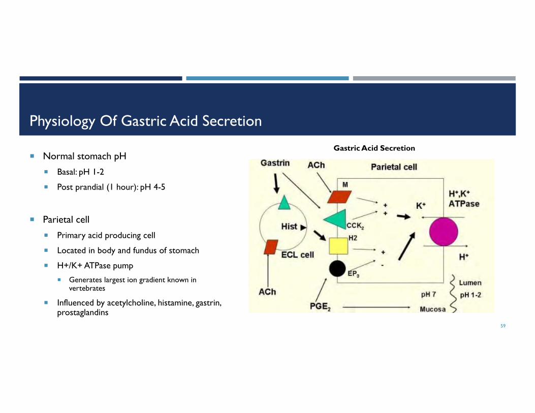
Physiology Of Gastric Acid Secretion
Normal stomach pH
Basal: pH 1-2
Post prandial (1 hour): pH 4-5
Parietal cell
Primary acid producing cell
Located in body and fundus of stomach
H+/K+ ATPase pump
Generates largest ion gradient known in vertebrates
Influenced by acetylcholine, histamine, gastrin, prostaglandins
59
Gastric Acid Secretion
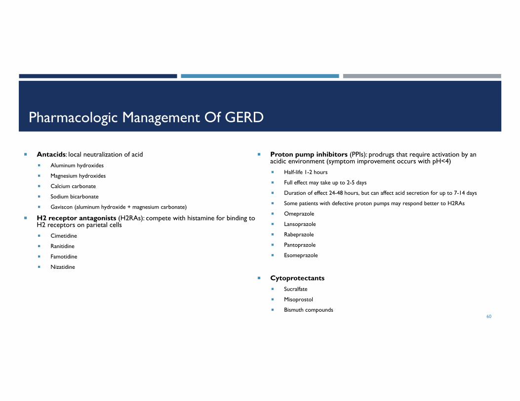
Pharmacologic Management Of GERD
Antacids: local neutralization of acid
Aluminum hydroxides
Magnesium hydroxides
Calcium carbonate
Sodium bicarbonate
Gaviscon (aluminum hydroxide + magnesium carbonate)
H2 receptor antagonists (H2RAs): compete with histamine for binding to H2 receptors on parietal cells
Cimetidine
Ranitidine
Famotidine
Nizatidine
Proton pump inhibitors (PPIs): prodrugs that require activation by an acidic environment (symptom improvement occurs with pH<4)
Half-life 1-2 hours
Full effect may take up to 2-5 days
Duration of effect 24-48 hours, but can affect acid secretion for up to 7-14 days
Some patients with defective proton pumps may respond better to H2RAs
Omeprazole
Lansoprazole
Rabeprazole
Pantoprazole
Esomeprazole
Cytoprotectants Sucralfate
Misoprostol
Bismuth compounds60
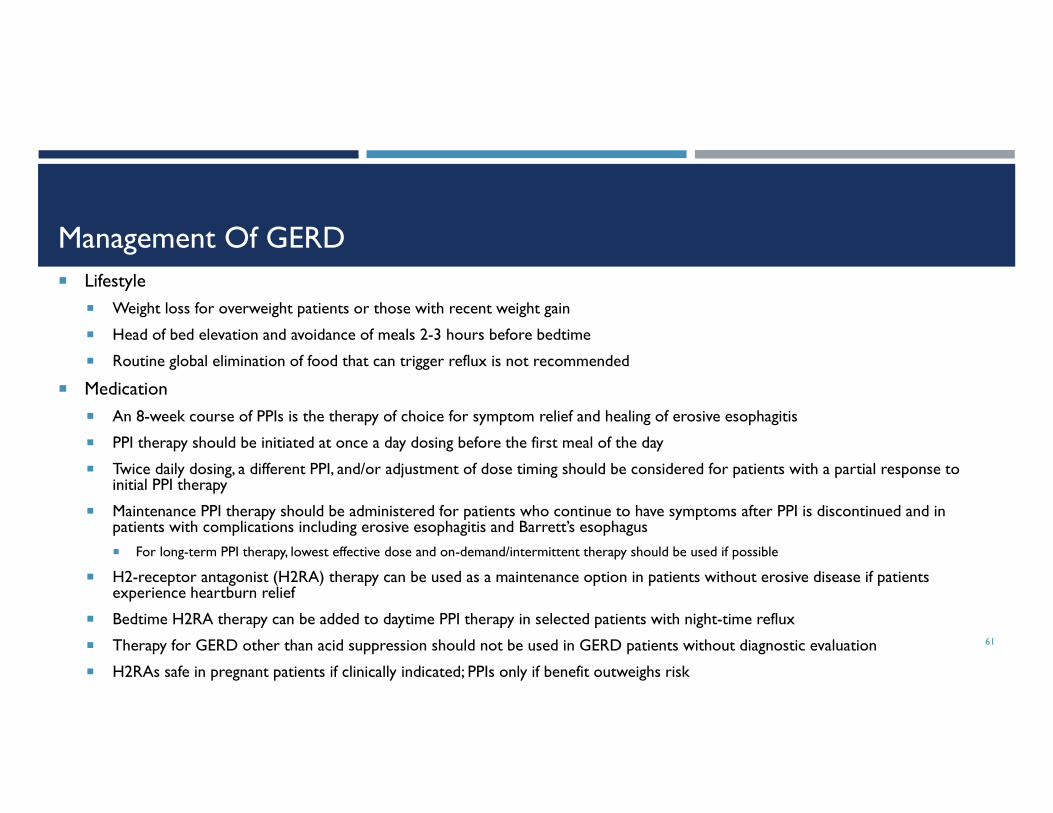
Management Of GERD Lifestyle
Weight loss for overweight patients or those with recent weight gain
Head of bed elevation and avoidance of meals 2-3 hours before bedtime
Routine global elimination of food that can trigger reflux is not recommended
Medication An 8-week course of PPIs is the therapy of choice for symptom relief and healing of erosive esophagitis
PPI therapy should be initiated at once a day dosing before the first meal of the day
Twice daily dosing, a different PPI, and/or adjustment of dose timing should be considered for patients with a partial response to initial PPI therapy
Maintenance PPI therapy should be administered for patients who continue to have symptoms after PPI is discontinued and in patients with complications including erosive esophagitis and Barrett’s esophagus For long-term PPI therapy, lowest effective dose and on-demand/intermittent therapy should be used if possible
H2-receptor antagonist (H2RA) therapy can be used as a maintenance option in patients without erosive disease if patients experience heartburn relief
Bedtime H2RA therapy can be added to daytime PPI therapy in selected patients with night-time reflux
Therapy for GERD other than acid suppression should not be used in GERD patients without diagnostic evaluation
H2RAs safe in pregnant patients if clinically indicated; PPIs only if benefit outweighs risk
61
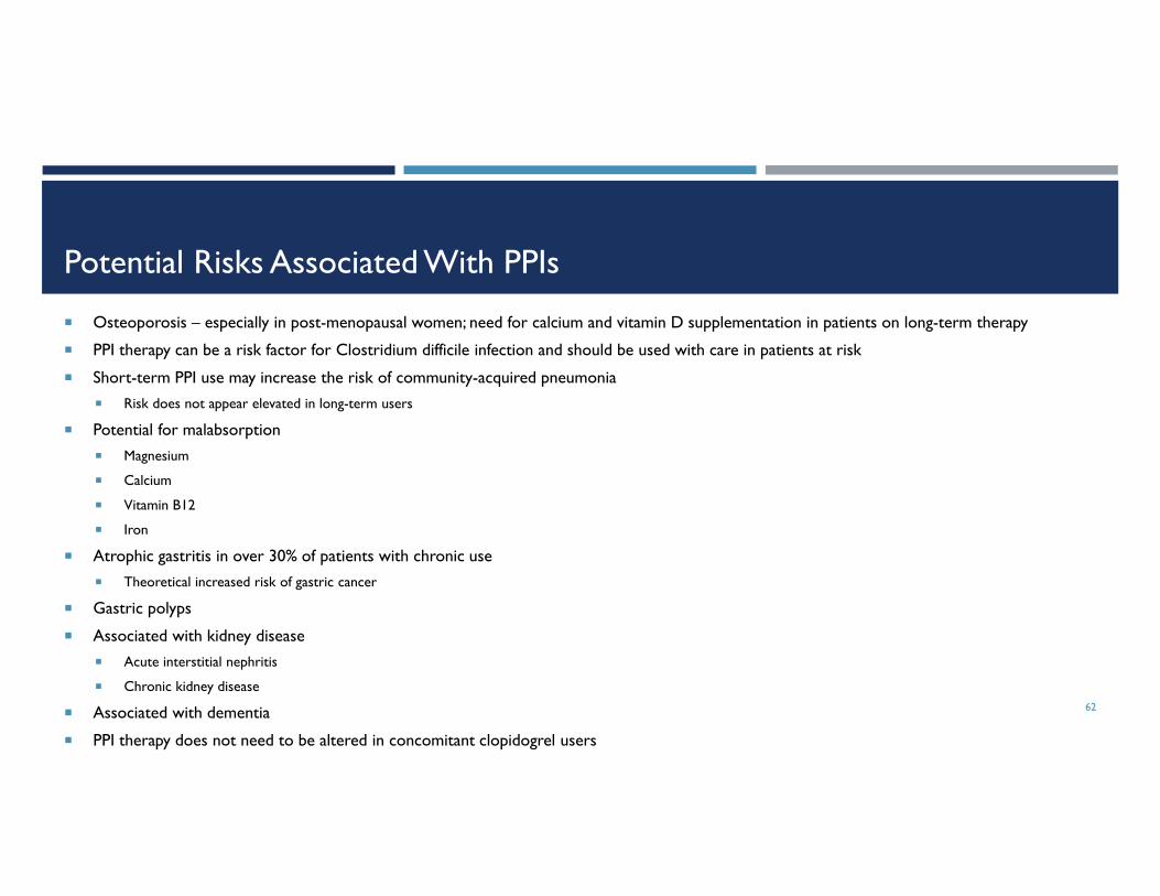
Potential Risks Associated With PPIs
Osteoporosis – especially in post-menopausal women; need for calcium and vitamin D supplementation in patients on long-term therapy
PPI therapy can be a risk factor for Clostridium difficile infection and should be used with care in patients at risk
Short-term PPI use may increase the risk of community-acquired pneumonia Risk does not appear elevated in long-term users
Potential for malabsorption Magnesium
Calcium
Vitamin B12
Iron
Atrophic gastritis in over 30% of patients with chronic use Theoretical increased risk of gastric cancer
Gastric polyps
Associated with kidney disease Acute interstitial nephritis
Chronic kidney disease
Associated with dementia
PPI therapy does not need to be altered in concomitant clopidogrel users
62
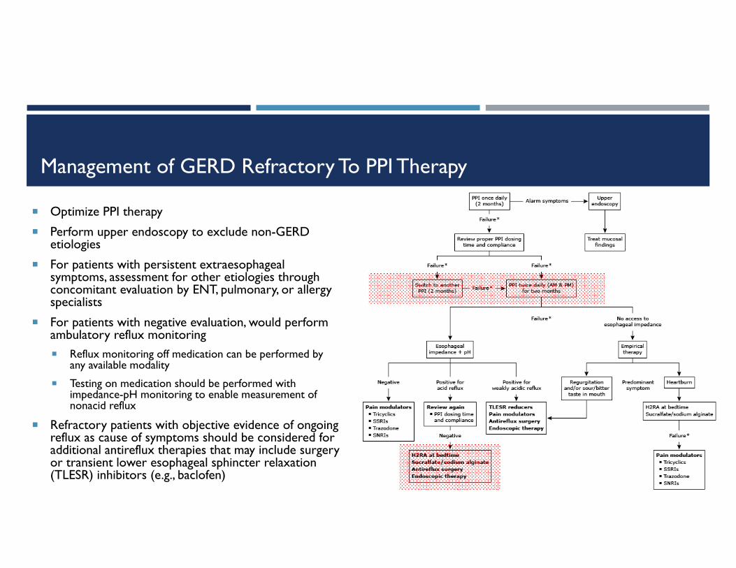
Management of GERD Refractory To PPI Therapy
Optimize PPI therapy
Perform upper endoscopy to exclude non-GERD etiologies
For patients with persistent extraesophagealsymptoms, assessment for other etiologies through concomitant evaluation by ENT, pulmonary, or allergy specialists
For patients with negative evaluation, would perform ambulatory reflux monitoring Reflux monitoring off medication can be performed by
any available modality
Testing on medication should be performed with impedance-pH monitoring to enable measurement of nonacid reflux
Refractory patients with objective evidence of ongoing reflux as cause of symptoms should be considered for additional antireflux therapies that may include surgery or transient lower esophageal sphincter relaxation (TLESR) inhibitors (e.g., baclofen)
63
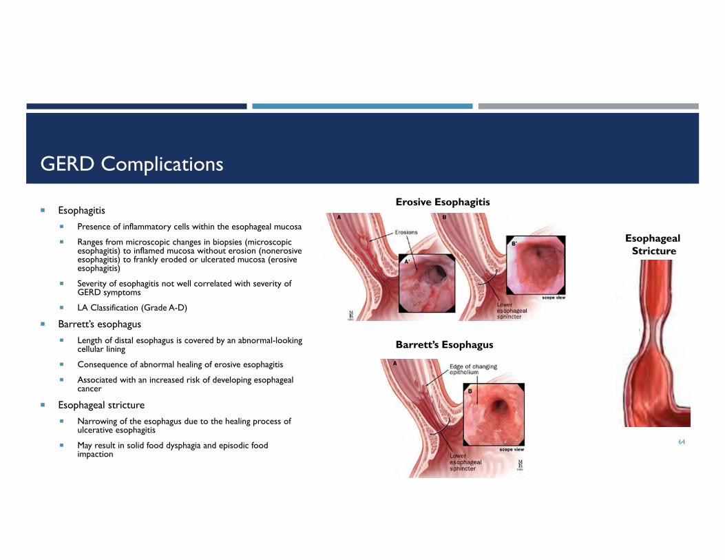
GERD Complications
Esophagitis Presence of inflammatory cells within the esophageal mucosa
Ranges from microscopic changes in biopsies (microscopic esophagitis) to inflamed mucosa without erosion (nonerosiveesophagitis) to frankly eroded or ulcerated mucosa (erosive esophagitis)
Severity of esophagitis not well correlated with severity of GERD symptoms
LA Classification (Grade A-D)
Barrett’s esophagus Length of distal esophagus is covered by an abnormal-looking
cellular lining
Consequence of abnormal healing of erosive esophagitis
Associated with an increased risk of developing esophageal cancer
Esophageal stricture Narrowing of the esophagus due to the healing process of
ulcerative esophagitis
May result in solid food dysphagia and episodic food impaction
64
Erosive Esophagitis
Barrett’s Esophagus
Esophageal Stricture
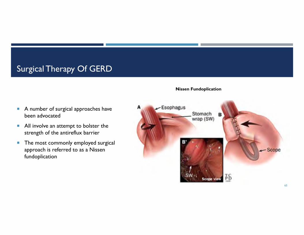
Surgical Therapy Of GERD
A number of surgical approaches have been advocated
All involve an attempt to bolster the strength of the antireflux barrier
The most commonly employed surgical approach is referred to as a Nissenfundoplication
65
Nissen Fundoplication
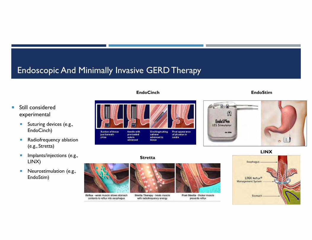
Endoscopic And Minimally Invasive GERD Therapy
Still considered experimental
Suturing devices (e.g., EndoCinch)
Radiofrequency ablation (e.g., Stretta)
Implants/injections (e.g., LINX)
Neurostimulation (e.g., EndoStim)
66
EndoCinch EndoStim
StrettaLINX
Extraesophageal Manifestations of GERD - Cough
Cough
GERD is often reported to be the 2nd or 3rd most common cause of persistent cough (and most common in some reports)
Heartburn or sour taste in mouth absent in more than 40% of patients in whom cough is due to reflux
Several factors potentially responsible for cough due to GERD
Stimulation of receptors in the upper respiratory tract
Aspiration of gastric contents (acid, pepsin), leading to stimulation of receptors in the lower respiratory tract
Esophageal-tracheobronchial cough reflex induced by acid reflux into the distal esophagus
GERD can also contribute to asthma symptoms
67
Laryngopharyngeal Reflux (LPR)
Retrograde movement of gastric contents into the laryngopharynx leading to symptoms referable to larynx/hypopharynx
Symptoms include dysphonia/hoarseness, globus, mild dysphagia, chronic cough, nonproductive throat clearing
Primarily an UES problem that mainly occurs in upright position during periods of physical exertion (e.g., bending over, Valsava, exercise)
Distinct clinical entity from GERD (GERD mainly a problem of the LES)
Much less acid exposure is necessary to create LPR compared to GERD
Most patients relatively unaware of LPR, with only 35% reporting heartburn
68
LPR Treatment
Drug therapy
Acid suppression
PPIs
H2 Blockers
Antacids
Neuromodulating agents
Tricyclic antidepressants
Nortriptylane
Gabapentin
Pregabalin
69
Extraesophageal Manifestations of GERD - Globus
Globus sensation
Functional esophageal disorder characterized by a sensation of a lump or foreign body in the throat
Also referred to as globus pharyngeus or globus hystericus
Unclear pathogenesis, but etiologies include:
Visceral hypersensitivity
Abnormalities of the UES
Psychologic and psychiatric disorders
GERD
70
Treatment Of Globus
Conservative therapy
Reassurance that globus is a benign disorder
Acid suppression
6-8 weeks of PPI therapy
1/3 of patients experience partial relief
Antidepressants (e.g., amitriptyline)
Other
Gabapentin
Relaxation therapy
71
Additional Evaluation Of Globus
Additional evaluation warranted in patients with recurrent or persistent symptoms despite conservative management or those with alarm features (e.g., pain, lateralization of symptoms, dysphagia, odynophagia, weight loss, change in voice, neck mass, unexplained cervical adenopathy)
Modalities include:
Nasoendoscopy
Videofluoroscopy
Barium swallow with solid bolus (e.g., barium tablet)
Esophageal manometry
Esophageal pH and impedance
Upper endoscopy
72

MOTILITY DISORDERS OF THE ESOPHAGUS
73
Introduction
74
Motility Disorders
Presentation
Dysphagia
Reflux
Cough
Choking
Oropharyngeal dysphagia
Causes
Neurologic and neuromuscular disorders
Cricopharyngeal dysfunction
Disorders that affect the esophageal body
75
Major Disorders Of Esophageal Peristalsis
Achalasia
Hypertensive LES/EGJ outflow obstruction
Hypertensive peristaltic disorders
Nutcracker esophagus
Jackhammer esophagus (“spastic nutcracker”)
Distal esophageal spasm (DES)
Ineffective esophageal motility (IEM)
Non-specific motor disorders (e.g., secondary to diabetes)
76

Achalasia
Most common esophageal motor disorder
Incidence 1.6 cases/100,000
Prevalence 10 cases/100,000
Results from inflammation and degeneration of neurons (myenteric plexus in esophageal wall) – possible viral etiology
Loss of inhibitory neurons in esophagus results in increased LESP (not required) and more importantly inability of LES to relax to baseline
Aperistalsis
Dysphagia mainly result of defect in LES relaxation
77
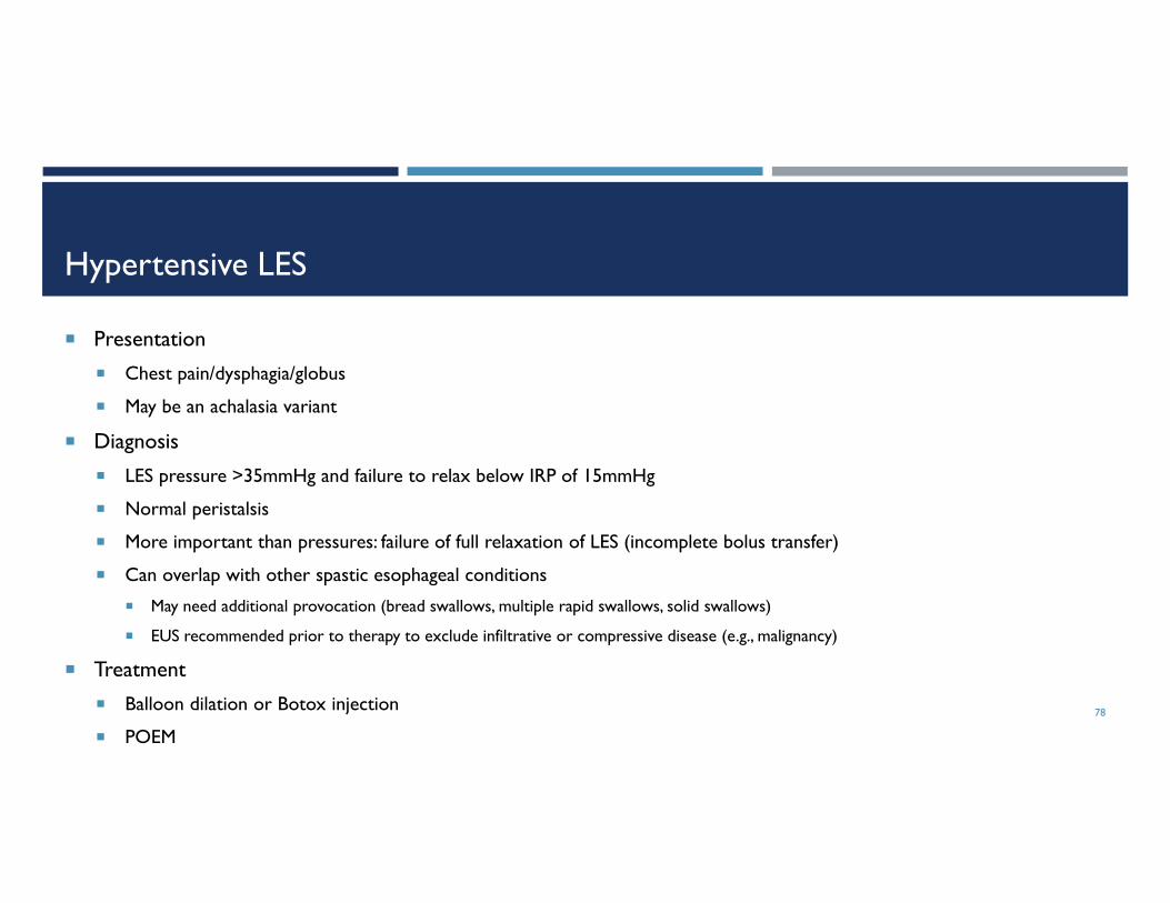
Hypertensive LES
Presentation
Chest pain/dysphagia/globus
May be an achalasia variant
Diagnosis
LES pressure >35mmHg and failure to relax below IRP of 15mmHg
Normal peristalsis
More important than pressures: failure of full relaxation of LES (incomplete bolus transfer)
Can overlap with other spastic esophageal conditions
May need additional provocation (bread swallows, multiple rapid swallows, solid swallows)
EUS recommended prior to therapy to exclude infiltrative or compressive disease (e.g., malignancy)
Treatment
Balloon dilation or Botox injection
POEM78
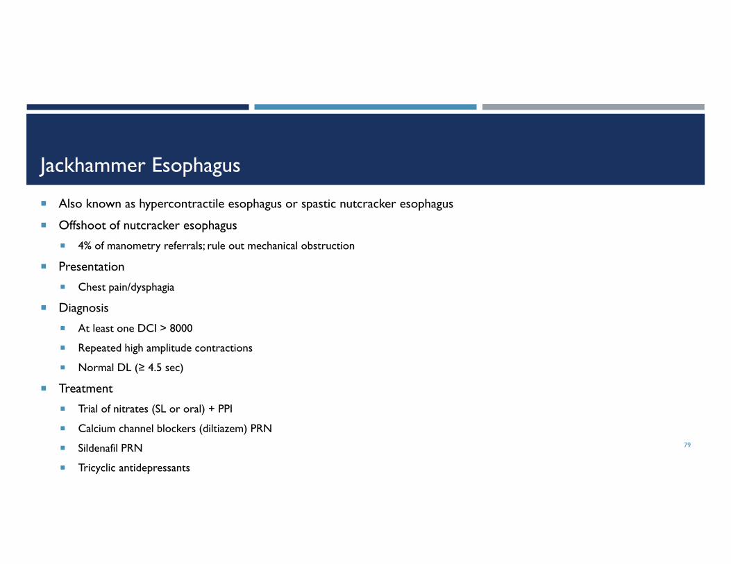
Jackhammer Esophagus
Also known as hypercontractile esophagus or spastic nutcracker esophagus
Offshoot of nutcracker esophagus
4% of manometry referrals; rule out mechanical obstruction
Presentation
Chest pain/dysphagia
Diagnosis
At least one DCI > 8000
Repeated high amplitude contractions
Normal DL (≥ 4.5 sec)
Treatment
Trial of nitrates (SL or oral) + PPI
Calcium channel blockers (diltiazem) PRN
Sildenafil PRN
Tricyclic antidepressants
79
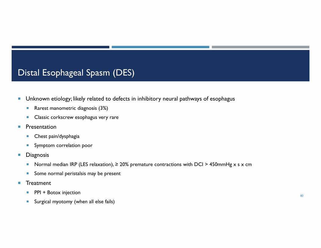
Distal Esophageal Spasm (DES)
Unknown etiology; likely related to defects in inhibitory neural pathways of esophagus
Rarest manometric diagnosis (3%)
Classic corkscrew esophagus very rare
Presentation
Chest pain/dysphagia
Symptom correlation poor
Diagnosis
Normal median IRP (LES relaxation), ≥ 20% premature contractions with DCI > 450mmHg x s x cm
Some normal peristalsis may be present
Treatment
PPI + Botox injection
Surgical myotomy (when all else fails)80
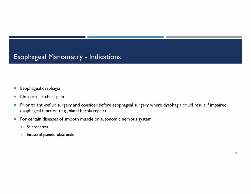
Esophageal Manometry - Indications
Esophageal dysphagia
Non-cardiac chest pain
Prior to anti-reflux surgery and consider before esophageal surgery where dysphagia could result if impaired esophageal function (e.g., hiatal hernia repair)
For certain diseases of smooth muscle or autonomic nervous system
Scleroderma
Intestinal pseudo-obstruction
81
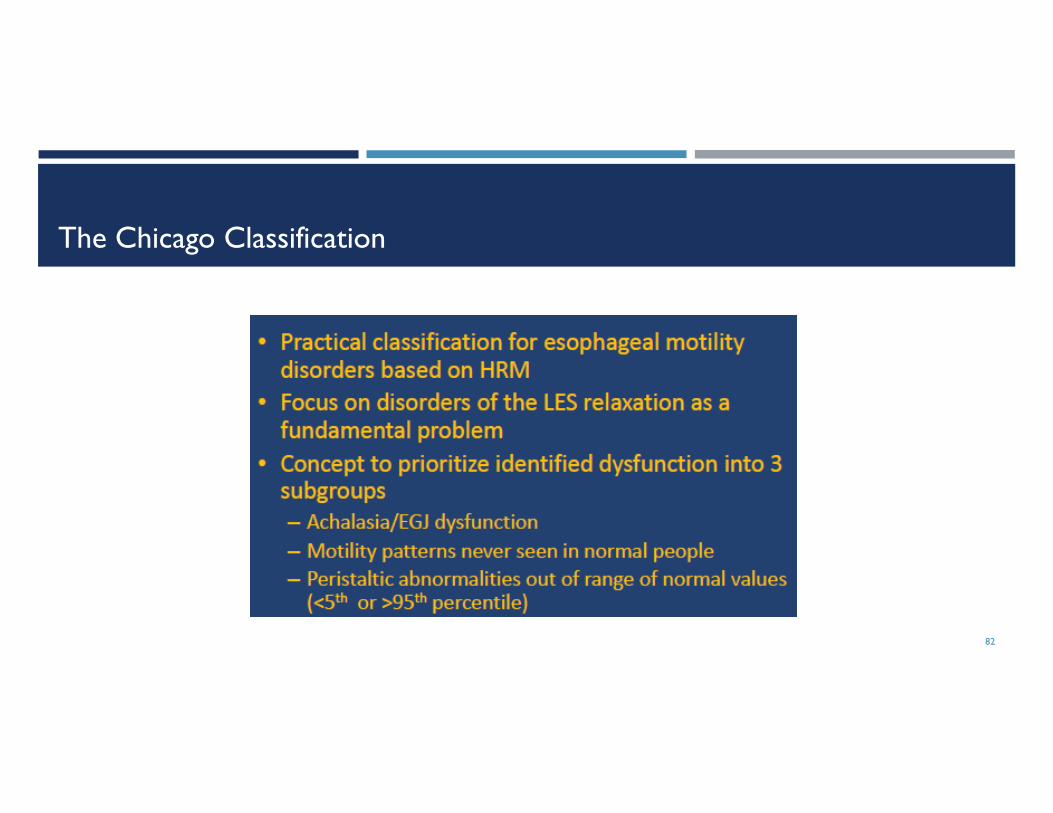
The Chicago Classification
82
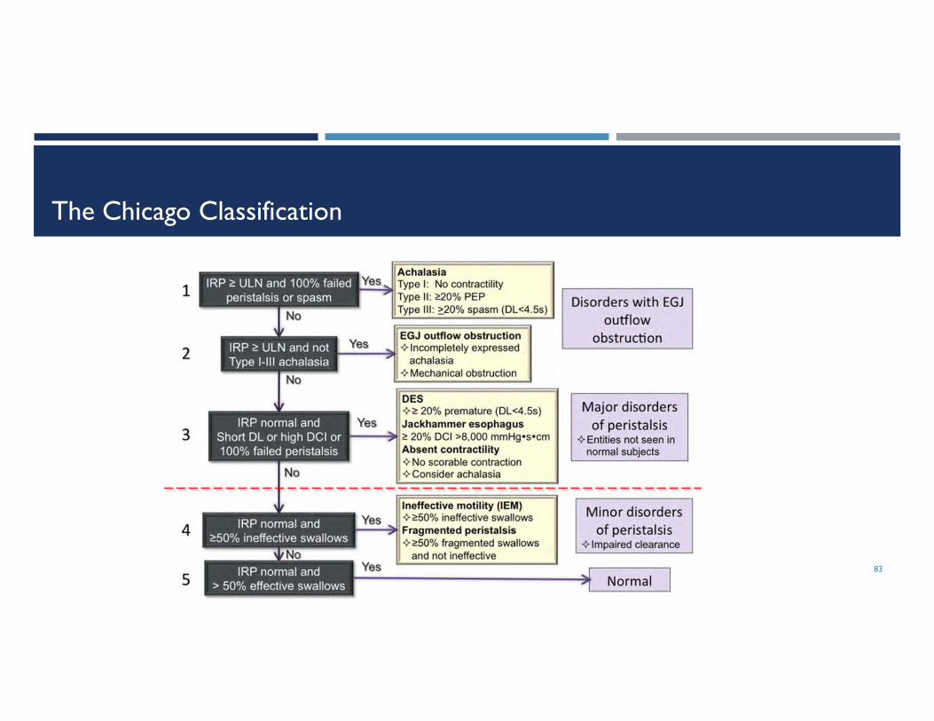
The Chicago Classification
83
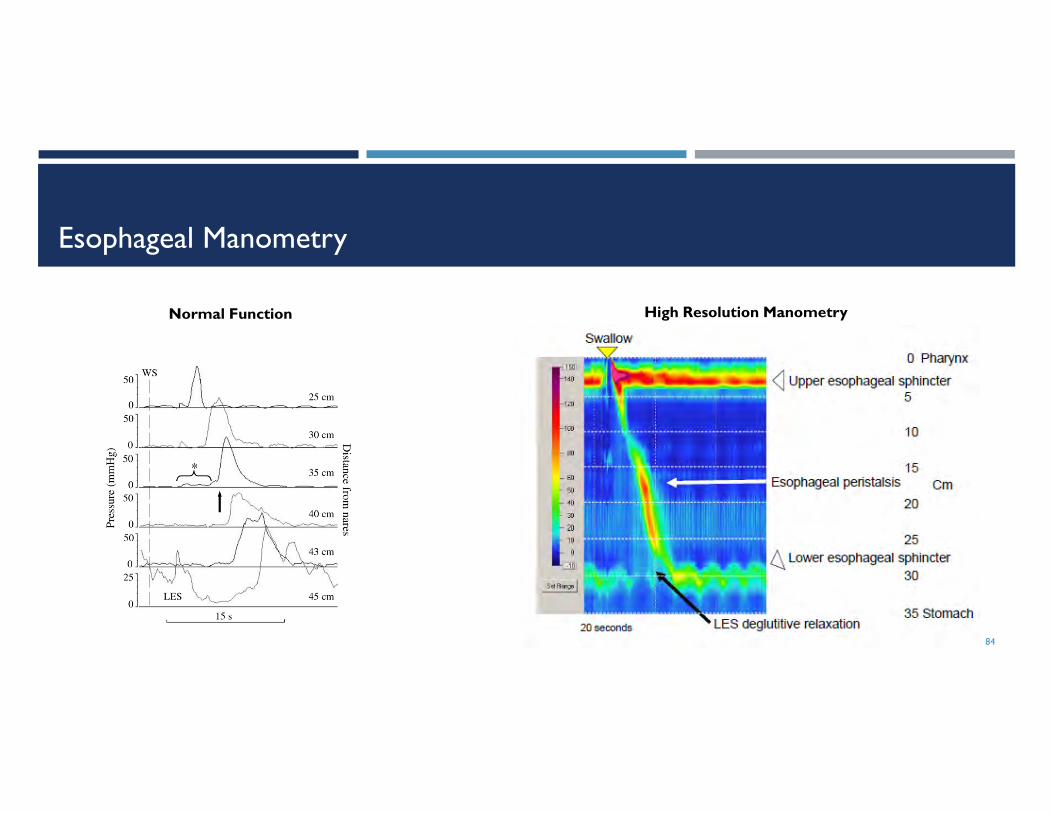
Esophageal Manometry
84
High Resolution ManometryNormal Function
High Resolution Manometry
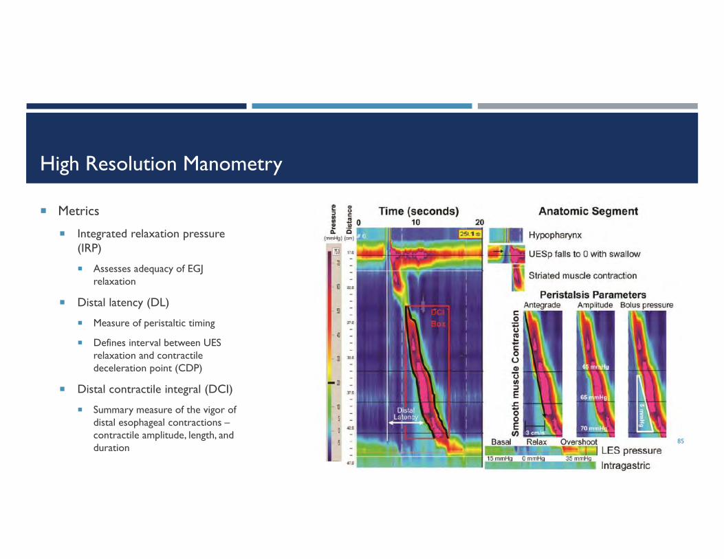
Metrics
Integrated relaxation pressure (IRP)
Assesses adequacy of EGJ relaxation
Distal latency (DL)
Measure of peristaltic timing
Defines interval between UES relaxation and contractile deceleration point (CDP)
Distal contractile integral (DCI)
Summary measure of the vigor of distal esophageal contractions –contractile amplitude, length, and duration
85
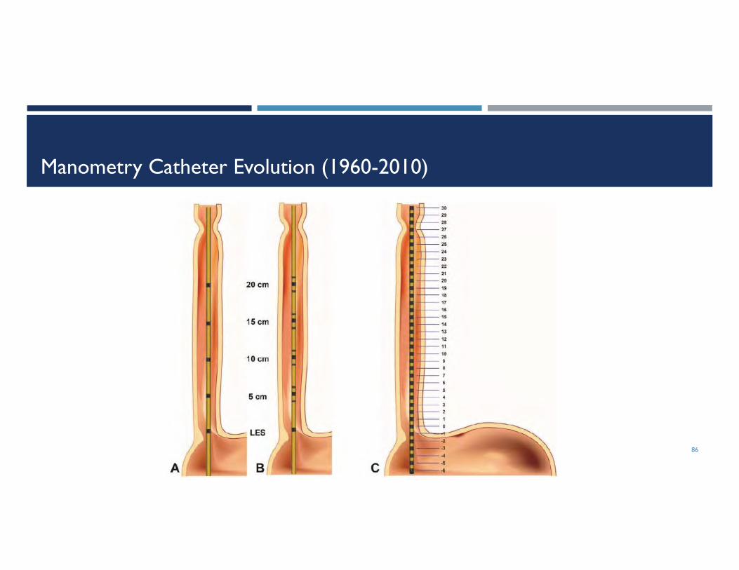
Manometry Catheter Evolution (1960-2010)
86
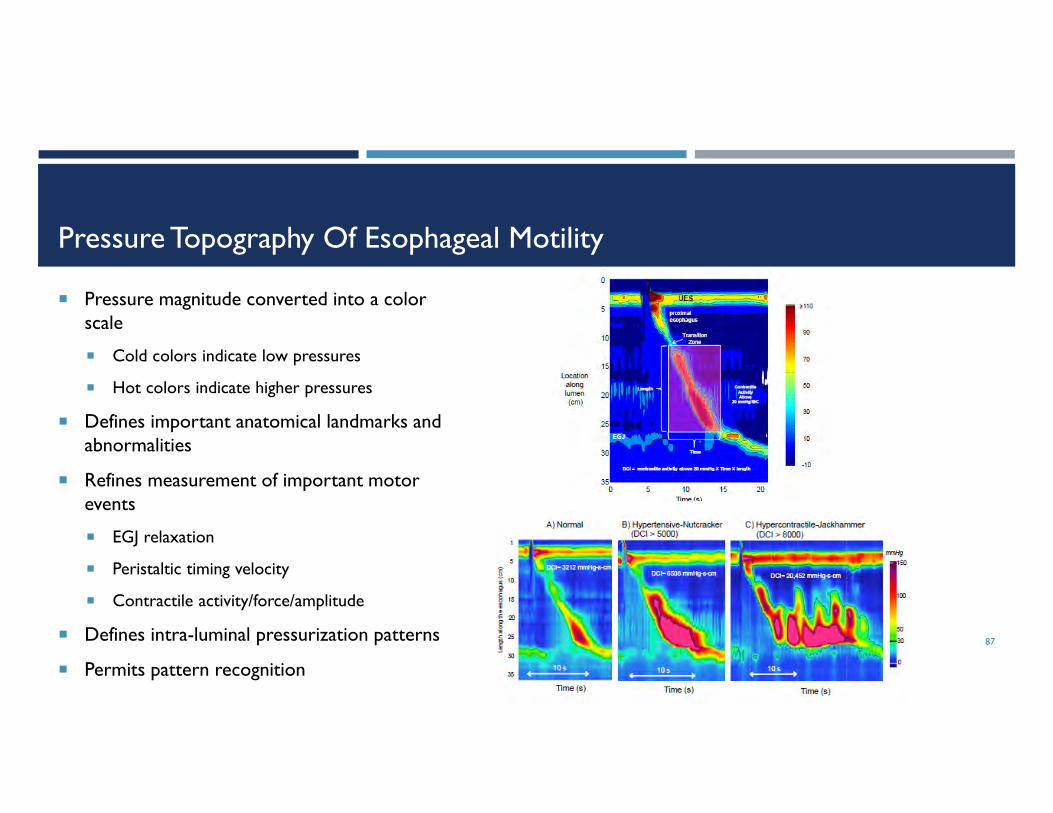
Pressure Topography Of Esophageal Motility
Pressure magnitude converted into a color scale
Cold colors indicate low pressures
Hot colors indicate higher pressures
Defines important anatomical landmarks and abnormalities
Refines measurement of important motor events
EGJ relaxation
Peristaltic timing velocity
Contractile activity/force/amplitude
Defines intra-luminal pressurization patterns
Permits pattern recognition
87
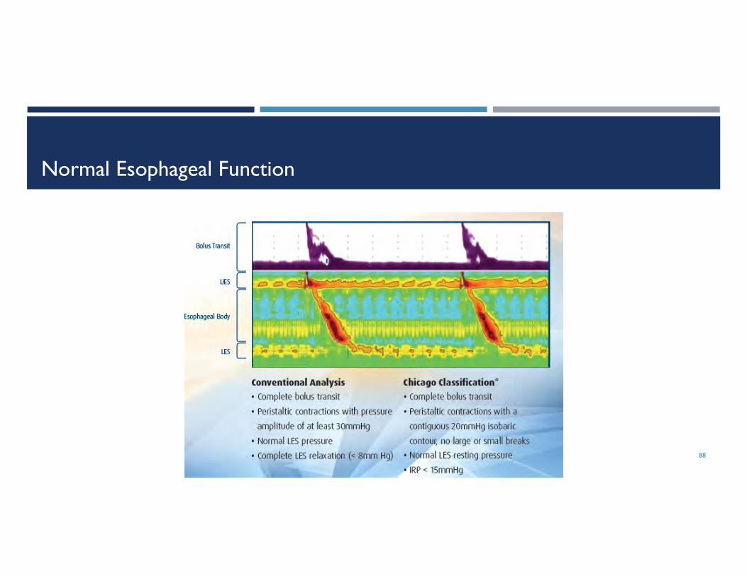
Normal Esophageal Function
88
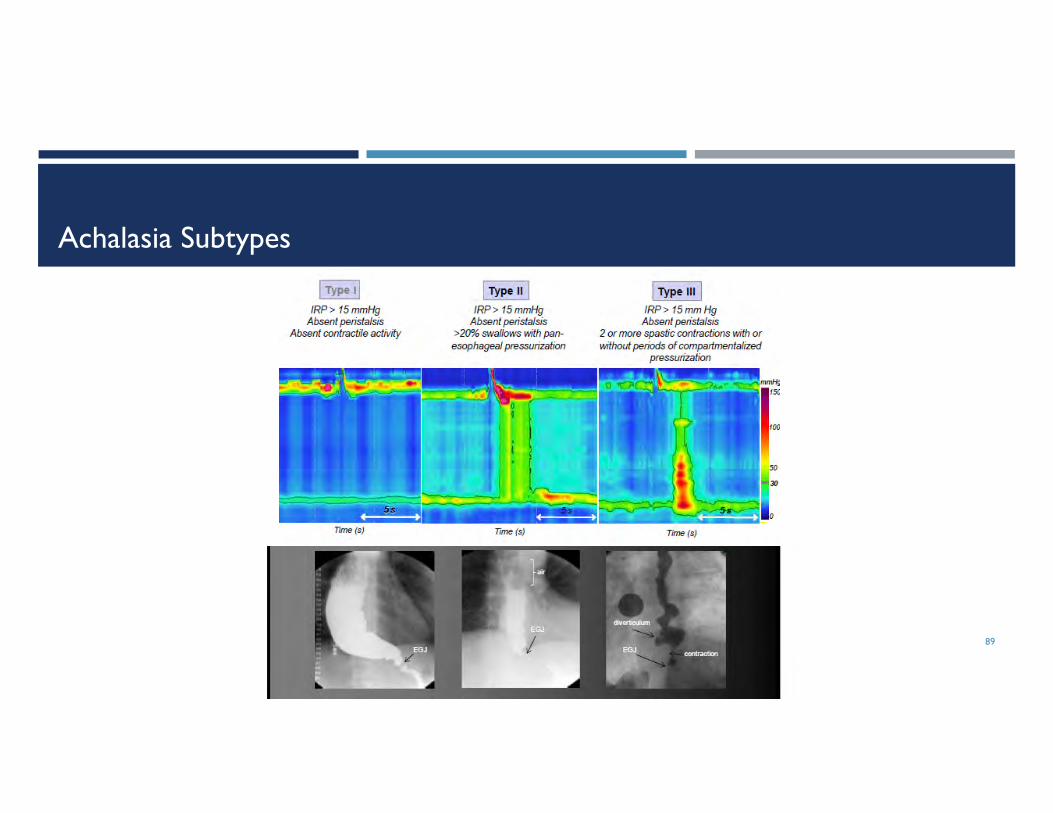
Achalasia Subtypes
89
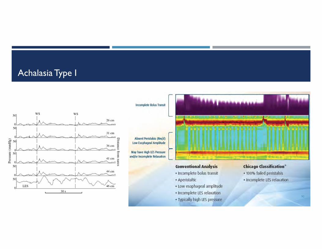
Achalasia Type I
90
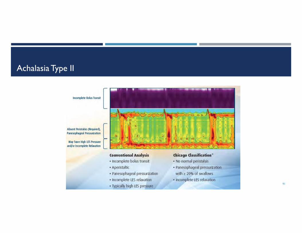
Achalasia Type II
91
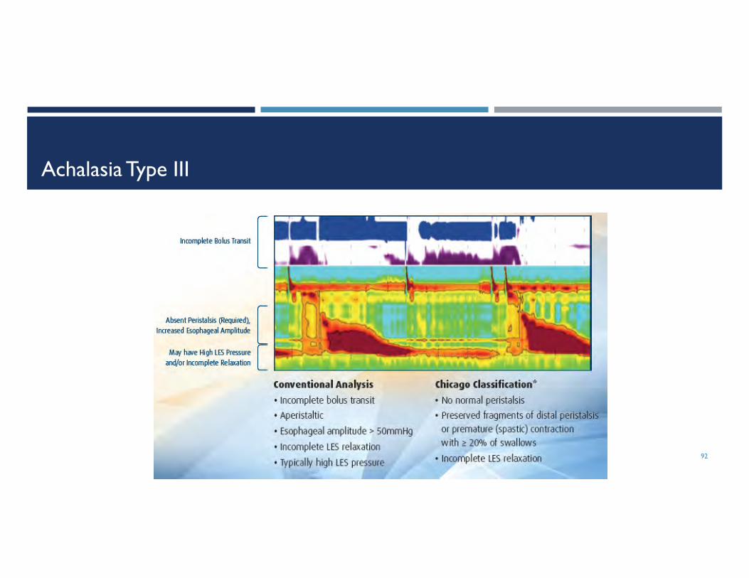
Achalasia Type III
92
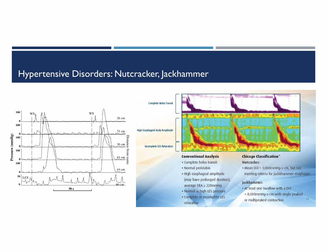
Hypertensive Disorders: Nutcracker, Jackhammer
93
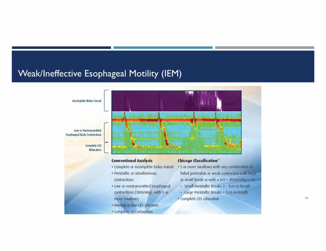
Weak/Ineffective Esophageal Motility (IEM)
94
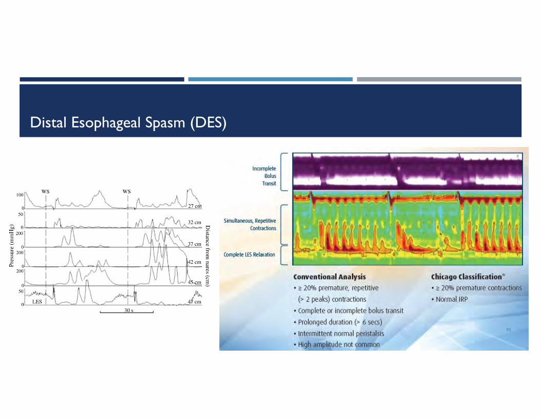
Distal Esophageal Spasm (DES)
95
Scleroderma
96
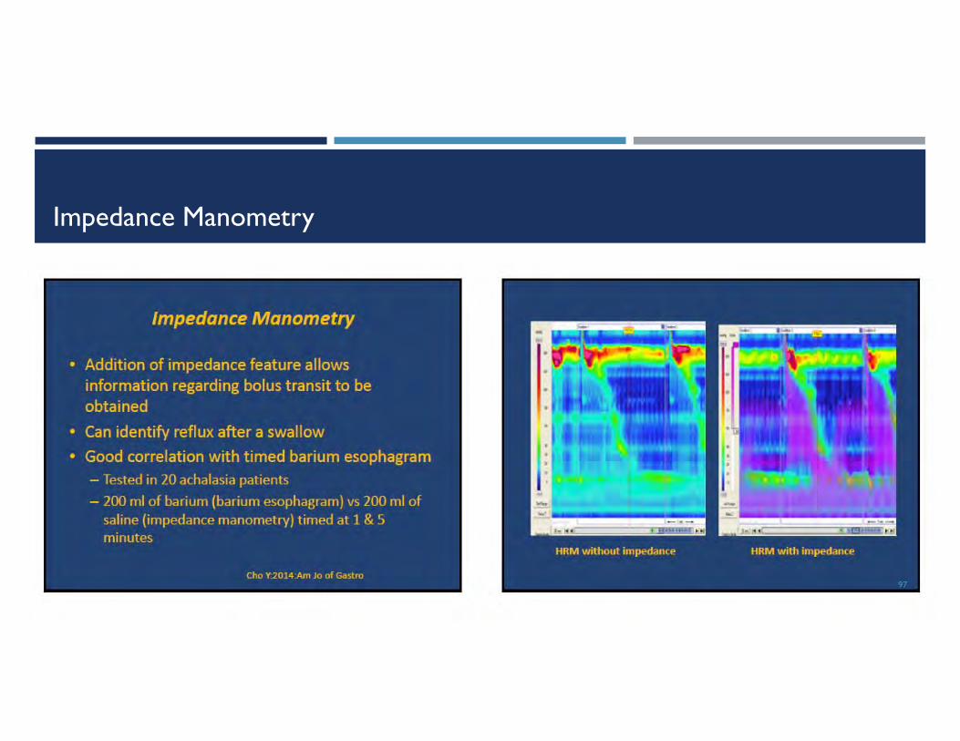
Impedance Manometry
97
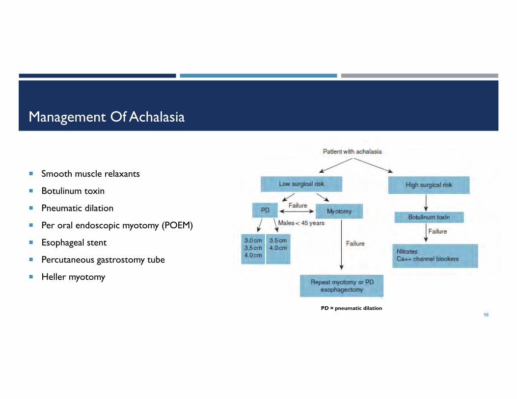
Management Of Achalasia
Smooth muscle relaxants
Botulinum toxin
Pneumatic dilation
Per oral endoscopic myotomy (POEM)
Esophageal stent
Percutaneous gastrostomy tube
Heller myotomy
98PD = pneumatic dilation
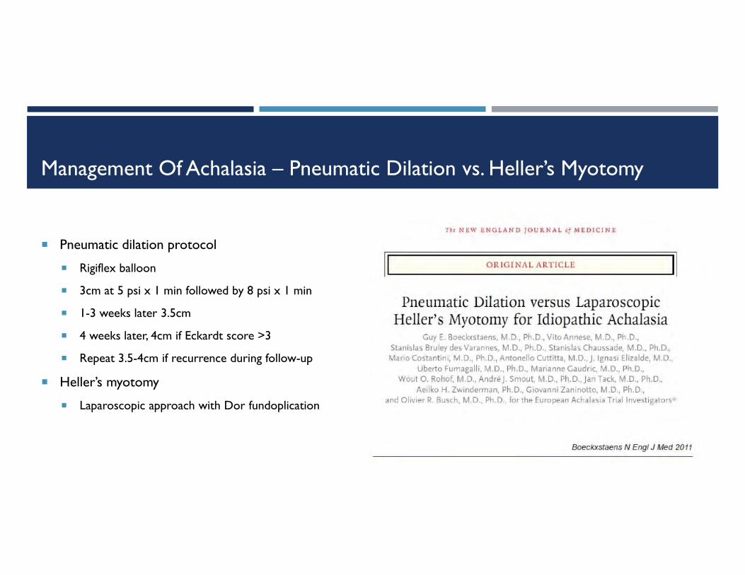
Management Of Achalasia – Pneumatic Dilation vs. Heller’s Myotomy
Pneumatic dilation protocol
Rigiflex balloon
3cm at 5 psi x 1 min followed by 8 psi x 1 min
1-3 weeks later 3.5cm
4 weeks later, 4cm if Eckardt score >3
Repeat 3.5-4cm if recurrence during follow-up
Heller’s myotomy
Laparoscopic approach with Dor fundoplication
99
Management Of Achalasia – Pneumatic Dilation vs. Heller’s Myotomy
Complications of treatment
Perforation
Pneumatic dilation (PD)
Esophageal perforation 4%
3 perforations with 30mm, 1 with 35mm
2 underwent surgery, 2 conservative care
Heller’s myotomy (HM)
Mucosal tear in 13/106 (12%)
Repaired during initial surgery
GERD
Increased acid exposure similar: 15% PD, 23% HM
Erosive esophagitis similar: 19% PD, 21% HM
Conclusion
Effectiveness of PD is comparable to laparoscopic HM if allow for repeated dilations and accept risk of esophageal perforation
100
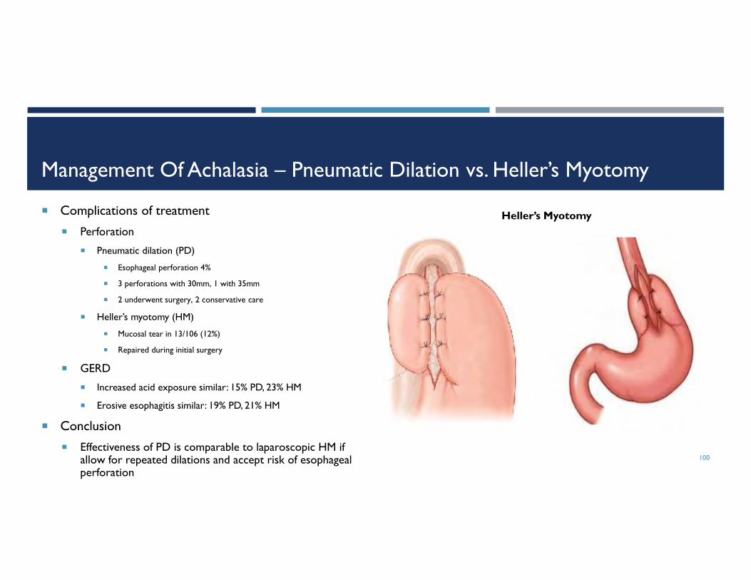
Heller’s Myotomy
Management Of Achalasia – Per Oral Endoscopic Myotomy (POEM)
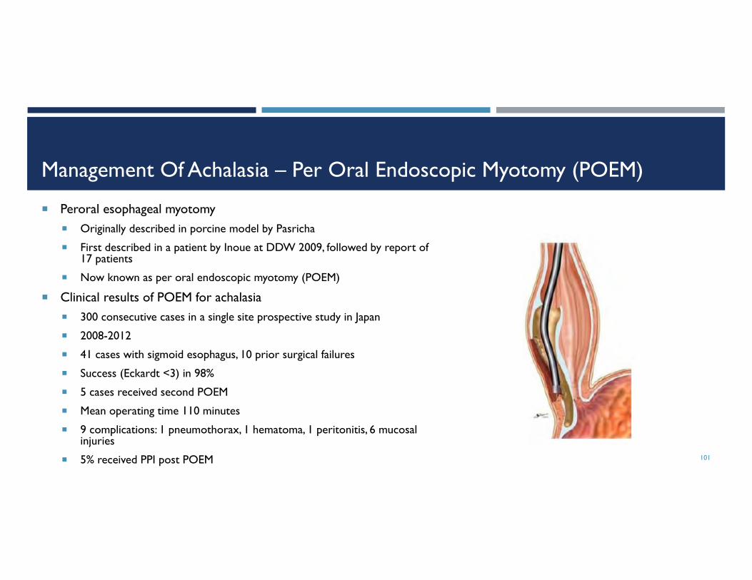
Peroral esophageal myotomy Originally described in porcine model by Pasricha
First described in a patient by Inoue at DDW 2009, followed by report of 17 patients
Now known as per oral endoscopic myotomy (POEM)
Clinical results of POEM for achalasia 300 consecutive cases in a single site prospective study in Japan
2008-2012
41 cases with sigmoid esophagus, 10 prior surgical failures
Success (Eckardt <3) in 98%
5 cases received second POEM
Mean operating time 110 minutes
9 complications: 1 pneumothorax, 1 hematoma, 1 peritonitis, 6 mucosal injuries
5% received PPI post POEM 101
Management Of Achalasia – Endoscopic vs. Surgical Myotomy
Prospective multicenter study of POEM Compared to retrospective cohort of lap Heller’s myotomy
Primary outcome of symptom relief at 3 months
70 patients underwent POEM
Mean operative time 105 minutes (54-240 min)
No conversions to open or lap surgery
Treatment success in 97% with POEM
POEM had significantly better 3 month symptom scores (1 vs. 1.4) and LES pressure (9 vs. 12 mmHg) compared to review of HM
Reflux esophagitis higher in POEM but not statistically significant (41% vs. 28%)
Conclusions Excellent outcomes of POEM are comparable to HM and reproducible in multiple centers
GERD complications may not be as significant as feared, perhaps due to avoidance of hiatal dissection
Growing experience supports effectiveness of POEM
POEM avoids surgical alteration of the EGJ morphology
POEM may become primary approach to treatment of achalasia102
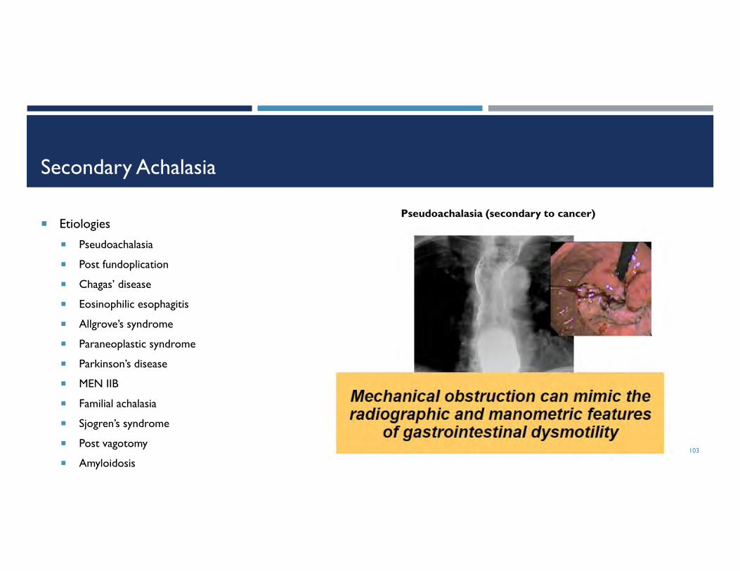
Secondary Achalasia
Etiologies
Pseudoachalasia
Post fundoplication
Chagas’ disease
Eosinophilic esophagitis
Allgrove’s syndrome
Paraneoplastic syndrome
Parkinson’s disease
MEN IIB
Familial achalasia
Sjogren’s syndrome
Post vagotomy
Amyloidosis103
Pseudoachalasia (secondary to cancer)
ESOPHAGEAL SYMPTOMS IN PATIENTS AFTER BARIATRIC SURGERY
104
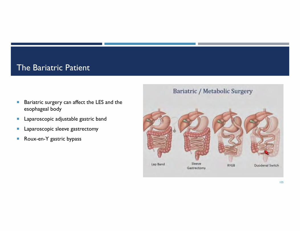
The Bariatric Patient
Bariatric surgery can affect the LES and the esophageal body
Laparoscopic adjustable gastric band
Laparoscopic sleeve gastrectomy
Roux-en-Y gastric bypass
105
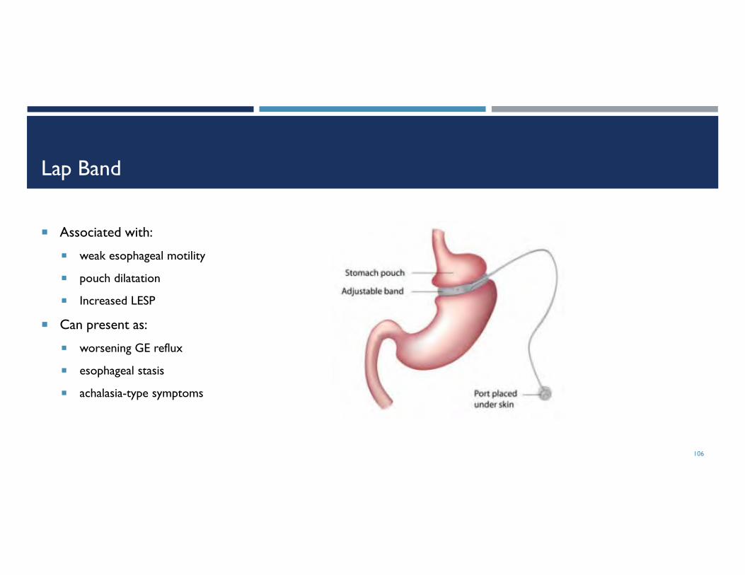
Lap Band
Associated with:
weak esophageal motility
pouch dilatation
Increased LESP
Can present as:
worsening GE reflux
esophageal stasis
achalasia-type symptoms
106
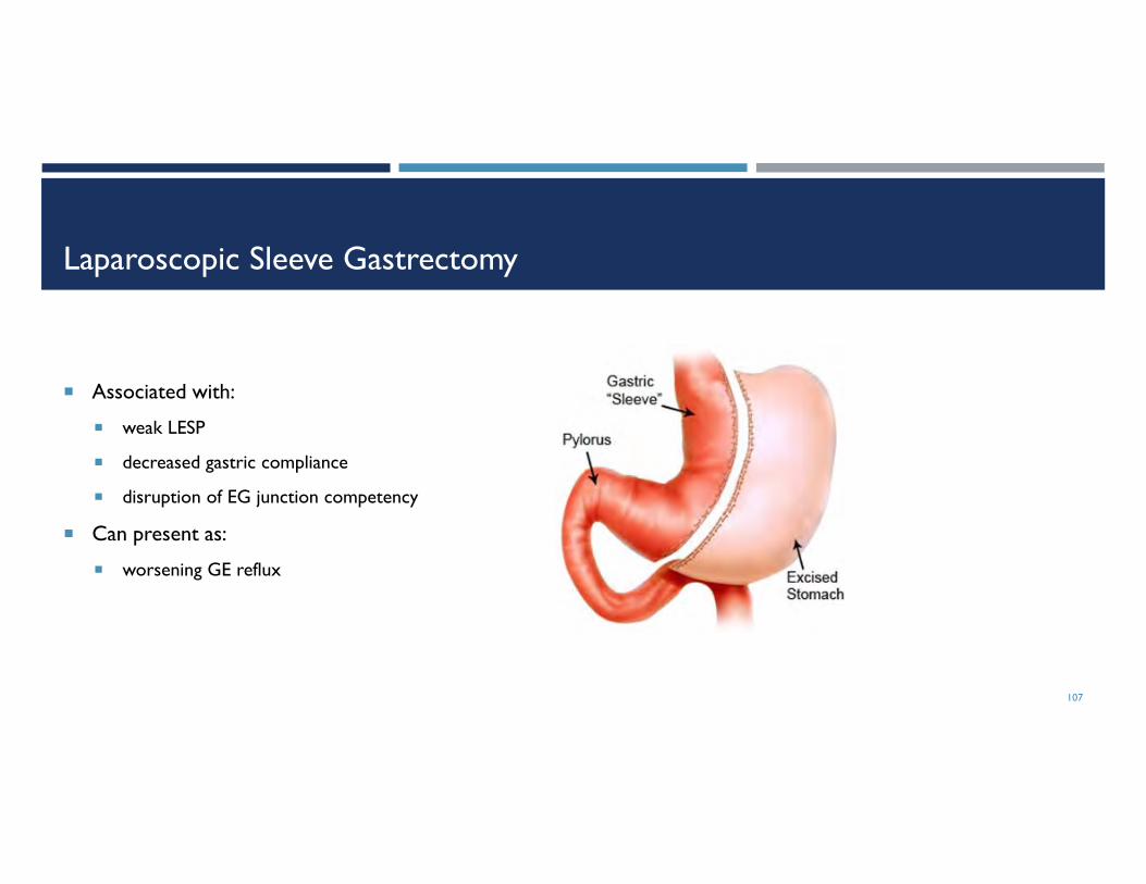
Laparoscopic Sleeve Gastrectomy
Associated with:
weak LESP
decreased gastric compliance
disruption of EG junction competency
Can present as:
worsening GE reflux
107
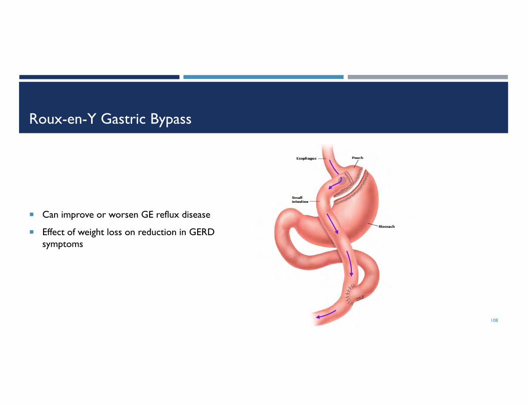
Roux-en-Y Gastric Bypass
Can improve or worsen GE reflux disease
Effect of weight loss on reduction in GERD symptoms
108

109Live Long and Prosper

Q&A
110


![Endoscopic incisional therapy for benign esophageal ... · caustic strictures and radiation strictures are known to be complex strictures[2]. Dilatation by bougie or balloon dilators](https://static.fdocuments.net/doc/165x107/5f80c75354e157596f1a7ef6/endoscopic-incisional-therapy-for-benign-esophageal-caustic-strictures-and-radiation.jpg)