BEAMing (Beads, Emulsion, Amplification, Magnetics) with ...
2
BEAMing (Beads, Emulsion, Amplification, Magnetics) Digital PCR with Microfluidics Ayato Tagawa 1 , Koya Yamawaki 1 , Yasuko Kawamoto 1 , Tsuyoshi Nakano 1 1 Central Research Laboratories, Sysmex Corporation 4-4-4 Takatsukadai, Nishi-ku, Kobe 651-2271, Japan Phone: +81-78-992-5988 E-mail: [email protected] Abstract Circulating tumor DNA (ctDNA) is expected to be a new generation of cancer biomarker and BEAMing Dig- ital PCR is one of the ultra-sensitive ctDNA detection technologies. We are developing automated microfluidic system of BEAMing for large-scale clinical trials and report the results of fundamental evaluation. 1. Introduction BEAMing is one of the ultra-high sensitive ctDNA detec- tion technologies [1] and carries out each process of Pre-amplification / Dilution / Bead-emulsification / Bead-emulsion PCR / Emulsion break / Hybridization / Flow cytometry sequentially by manual handling [2]. Large-scale clinical trials of BEAMing, and its automation process are required. Recently droplet based microfluidics are proposed to detect single DNA molecule [3][4], single cell [5] and single antigen [6], and we are developing micro- fluidic system to realize automated all process of BEAMing as shown in Fig. 1. In this report, we evaluate flow focusing bead-droplets microfluidics and continuous flow bead-emulsion PCR to replace conventional manual assays. 2. Experiment Bead-emulsification We developed a dedicated flow focusing microfluidics of Polydimethylsiloxane (PDMS) on a glass substrate. It’s necessary to encapsulate single DNA molecule and a magnetic bead coated with primers in a droplet as shown in Fig. 2(a) with dNTPs and polymerase. In consideration of Poisson distribution, ten droplets are required for a DNA or a primer coated magnet bead. We are targeting to generate more than 80 million mono-dispersed droplets which are smaller than 7.8 µm diameter each from 20 µL DNA/PCR master mix solution. The width / height of the cross channel of microfluidics were designed 20 µm / 20 µm and droplets are generated with shear stress of oil carrier as shown in Fig. 2(b). The droplets were collected every 2.5 min for 20 min and cap- tured with microscopy (BZ-X700, Keyence) to analyze the diameter [µm] , CV [%] and the ratio of beads / droplets [%] with imaging software (Image-pro plus, Media Cybernetics). Bead-emulsion PCR Using glass microfluidics, we evaluated continuous flow bead-emulsion PCR. The width / height of the cross channel of the microfluidics are 450 µm / 520 µm and placed on hotplates to generate thermal gradient as shown in Fig. 3(a). The bead-emulsion with 1 % mutant DNA / 99 % wild type DNA were made by pipetting and flowed through in the serpentine channel with 50 cycles as shown in Fig. 3(b). After the bead-emulsion PCR, we conducted Emulsion break / Hybridization sequentially by manual handling and the fluorescent labeled beads were analyzed with flow cy- tometer (ACCURI C6, Becton Dickinson) and data analysis software (FlowJo, FLOWJO LCC). The fluorescent probes of mutant / wild type DNA on beads were excited by 488 nm / 640 nm lasers and detected with photomultiplier tube (PMT) at 533 nm / 675 nm respectively. Universal fluores- cent probe of all DNA on beads was excited by 488 nm laser and detected with PMT at 585 nm. 3. Results and discussion Bead-emulsification The flow rates of water / oil phases are set at 2 µL/min (5800 mbar) / 70 µL/min (8743 mbar) respectively using pressure pumps. Monodisperse bead-droplets are sampled as shown in Fig. 4. The average of droplet diameter (CV [%]) and ratio [%] of beads / droplets are 7.45 µm (CV = 10.4 %) as shown Fig. 5(a)(b) and 7.25 % as shown in Fig. 5(c). Approximately 92.4 million droplets were generated and 6.7 million beads were encapsulated for ten minutes. Clogging at 20 µm cross junction was not observed. The bead -droplets are broken into pieces with alcohol by pipetting and the beads are washed with a magnet plate and hybridized by manual handling. We confirmed BEAMing was normally conducted and can be replaced manual emul- sification to the flow focusing bead-droplets microfluidics. Bead-emulsion PCR Bead-emulsion is flowed inside serpentine channel with 50 cycles emulsion PCR in 63 min. The result of flowcytomery is shown in Fig. 6. Fig. 6(a) shows side scatter and forward scatter signals of 1000,000 counts. 65.4 % single beads are gated to Fig. 6(b). Fig. 6(b) shows the fluorescent intensity of universal and mutant probes. 10 % universal signals are gated to Fig. 6(c). Fig. 6(c) shows the ratio of 1 % mutant signal / 99 % wild type signals. 4. Conclusions We demonstrated microfluidics based bead-emulsification and bead-emulsion PCR were successfully worked. Further results of evaluation of all parts of BEAMing with micro- fluidics will be reported at the conference. H-5-03 Extended Abstracts of the 2016 International Conference on Solid State Devices and Materials, Tsukuba, 2016, pp405-406 - 405 -
Transcript of BEAMing (Beads, Emulsion, Amplification, Magnetics) with ...

BEAMing (Beads, Emulsion, Amplification, Magnetics) Digital PCR
with Microfluidics
Ayato Tagawa
1, Koya Yamawaki
1, Yasuko Kawamoto
1, Tsuyoshi Nakano
1
1 Central Research Laboratories, Sysmex Corporation 4-4-4 Takatsukadai, Nishi-ku, Kobe 651-2271, Japan
Phone: +81-78-992-5988 E-mail: [email protected]
Abstract
Circulating tumor DNA (ctDNA) is expected to be a
new generation of cancer biomarker and BEAMing Dig-
ital PCR is one of the ultra-sensitive ctDNA detection
technologies. We are developing automated microfluidic
system of BEAMing for large-scale clinical trials and
report the results of fundamental evaluation.
1. Introduction
BEAMing is one of the ultra-high sensitive ctDNA detec-
tion technologies [1] and carries out each process of
Pre-amplification / Dilution / Bead-emulsification /
Bead-emulsion PCR / Emulsion break / Hybridization /
Flow cytometry sequentially by manual handling [2].
Large-scale clinical trials of BEAMing, and its automation
process are required. Recently droplet based microfluidics
are proposed to detect single DNA molecule [3][4], single
cell [5] and single antigen [6], and we are developing micro-
fluidic system to realize automated all process of BEAMing
as shown in Fig. 1. In this report, we evaluate flow focusing
bead-droplets microfluidics and continuous flow
bead-emulsion PCR to replace conventional manual assays.
2. Experiment
Bead-emulsification
We developed a dedicated flow focusing microfluidics of Polydimethylsiloxane (PDMS) on a glass substrate.
It’s necessary to encapsulate single DNA molecule and a magnetic bead coated with primers in a droplet as shown in Fig. 2(a) with dNTPs and polymerase. In consideration of Poisson distribution, ten droplets are required for a DNA or a primer coated magnet bead. We are targeting to generate more than 80 million mono-dispersed droplets which are smaller than 7.8 µm diameter each from 20 µL DNA/PCR master mix solution.
The width / height of the cross channel of microfluidics were designed 20 µm / 20 µm and droplets are generated with shear stress of oil carrier as shown in Fig. 2(b). The droplets were collected every 2.5 min for 20 min and cap-tured with microscopy (BZ-X700, Keyence) to analyze the diameter [µm] , CV [%] and the ratio of beads / droplets [%] with imaging software (Image-pro plus, Media Cybernetics).
Bead-emulsion PCR
Using glass microfluidics, we evaluated continuous flow bead-emulsion PCR. The width / height of the cross channel of the microfluidics are 450 µm / 520 µm and placed on
hotplates to generate thermal gradient as shown in Fig. 3(a). The bead-emulsion with 1 % mutant DNA / 99 % wild
type DNA were made by pipetting and flowed through in the serpentine channel with 50 cycles as shown in Fig. 3(b).
After the bead-emulsion PCR, we conducted Emulsion break / Hybridization sequentially by manual handling and the fluorescent labeled beads were analyzed with flow cy-tometer (ACCURI C6, Becton Dickinson) and data analysis software (FlowJo, FLOWJO LCC). The fluorescent probes of mutant / wild type DNA on beads were excited by 488 nm / 640 nm lasers and detected with photomultiplier tube (PMT) at 533 nm / 675 nm respectively. Universal fluores-cent probe of all DNA on beads was excited by 488 nm laser and detected with PMT at 585 nm.
3. Results and discussion
Bead-emulsification
The flow rates of water / oil phases are set at 2 µL/min
(5800 mbar) / 70 µL/min (8743 mbar) respectively using
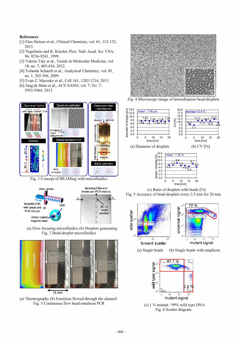
pressure pumps. Monodisperse bead-droplets are sampled as
shown in Fig. 4.
The average of droplet diameter (CV [%]) and ratio [%]
of beads / droplets are 7.45 µm (CV = 10.4 %) as shown Fig.
5(a)(b) and 7.25 % as shown in Fig. 5(c). Approximately
92.4 million droplets were generated and 6.7 million beads
were encapsulated for ten minutes. Clogging at 20 µm cross
junction was not observed.
The bead -droplets are broken into pieces with alcohol by
pipetting and the beads are washed with a magnet plate and
hybridized by manual handling. We confirmed BEAMing
was normally conducted and can be replaced manual emul-
sification to the flow focusing bead-droplets microfluidics.
Bead-emulsion PCR
Bead-emulsion is flowed inside serpentine channel with 50 cycles emulsion PCR in 63 min. The result of flowcytomery is shown in Fig. 6. Fig. 6(a) shows side scatter and forward scatter signals of 1000,000 counts. 65.4 % single beads are gated to Fig. 6(b). Fig. 6(b) shows the fluorescent intensity of universal and mutant probes. 10 % universal signals are gated to Fig. 6(c). Fig. 6(c) shows the ratio of 1 % mutant signal / 99 % wild type signals.
4. Conclusions
We demonstrated microfluidics based bead-emulsification and bead-emulsion PCR were successfully worked. Further results of evaluation of all parts of BEAMing with micro-fluidics will be reported at the conference.
H-5-03Extended Abstracts of the 2016 International Conference on Solid State Devices and Materials, Tsukuba, 2016, pp405-406
- 405 -
References
[1] Elen Heitzer et al., Clinical Chemistry, vol. 61, 112-123,
2015.
[2] Vogelstein and K. Kinzler, Proc. Natl. Acad. Sci. USA,
96, 9236-9241, 1999.
[3] Valerie Taly et al., Trends in Molecular Medicine, vol.
18, no. 7, 405-416, 2012.
[4] Yolanda Schaerli et al., Analytical Chemistry, vol. 81,
no. 1, 302-306, 2009.
[5] Evan Z. Macosko et al., Cell 161, 1202-1214, 2015.
[6] Jung-jk Shim et al., ACS NANO, vol. 7, No. 7,
5955-5964, 2013.
Fig. 1 Concept of BEAMing with microfluidics
(a) Flow focusing microfluidics (b) Droplets generating
Fig. 2 Bead-droplet microfluidics
(a) Thermography (b) Emulsion flowed through the channel
Fig. 3 Continuous flow bead-emulsion PCR
Fig. 4 Microscopy image of monodisperse bead-droplets
(a) Diameter of droplets (b) CV [%]
(c) Ratio of droplets with beads [%]
Fig. 5 Accuracy of bead-droplets every 2.5 min for 20 min
(a) Single beads (b) Single beads with amplicon
(c) 1 % mutant / 99% wild type DNA
Fig. 6 Scatter diagram
7.56
7.82
7.5
7.25
7.54
7.28
7.13
7.54
0.0
2.0
4.0
6.0
8.0
10.0
12.0
14.0
0 5 10 15 20dia
me
ter
of d
rop
let [µ
m]
time [min]
mean : 7.45 µm
time [min]
CV
[%
]
8.2
7.7
11.3
11.3
11.7
10.9
12.3
9.6
0.02.55.07.5
10.012.515.017.520.0
0 5 10 15 20
average 10.4 %
7.3
7.38.6
7.0
7.06.5
8.16.2
0.0
2.0
4.0
6.0
8.0
10.0
12.0
14.0
0 5 10 15 20
be
ad
s / d
rop
lets
[%
] mean : 7.25 %
time [min]
- 406 -