BE SURE OF THE CURE WHEN TREATING ONYCHOMYCOSIS · long-established tinea pedis infection....
3
BACKGROUND Onychomycosis is a common clinical condition affecting between 2% and 8% of adults in the western world. 1 Infection of the nail plate with dermatophytes can lead to discolouration and changes to the consistency of the nail plate. The effect of the disease on a patient’s quality of life has been well documented. 2-5 Subsequently, patients frequently attend clinics seeking treatment for the condition. Pharmacological agents (taken systemically or applied topically) continue to be the mainstay of treatment for this condition. Systemic agents work by growing antifungal agents into the emerging nail to eradicate fungal elements whilst topical antifungal agents work by absorption through the affected nail to directly destroy or prevent fungal growth. DIAGNOSIS The success of drugs in treating onychomycosis traditionally has been measured by the presence or absence of dermatophytes in the affected nail. This is best carried out by microbiological testing to show if any viable fungus is present in the nail sample. Before a treatment is instigated, a positive diagnosis of onychomycosis is required. First of all, it is important to remember that onychomycosis is only responsible for 40-50% of all nail dystrophies, 6 so technically in clinic only about half of the nails seen may be mycotic based on that figure. Clinicians will sometimes look at the clinical picture and make a diagnosis by the visual appearance alone. Visual diagnosis has been reported to be reasonably accurate, 7 with one study demonstrating that up to 67% of nails could be correctly diagnosed by experts on appearance alone. 8 However, this figure suggests that still more than 30% of cases may be incorrectly diagnosed. Ethically, it is best to establish a positive diagnosis of onychomycosis before treatment is instigated otherwise the patient may be undertaking (and in private practice paying for) a treatment they do not need. In addition, knowledge of pathogen can guide antifungal therapy. Moreover, where there is a risk of side effects, a clear diagnosis should be sought for both ethical and medico-legal reasons. Current UK guidelines from the British Association of Dermatologists 9 and NICE 10 strongly recommend that any patient with suspected onychomycosis should have laboratory confirmation before treatment - particularly if oral antifungal therapy is being considered. For the practising podiatrist, there are a number of ways in which the diagnosis may be reached. Visual diagnosis, as mentioned above, may not be sufficient or accurate enough for clinical use. A nail sample may be sent off to the laboratory for testing. This is normally a two-stage process. First, samples are visually analysed under the microscope after application of potassium hydroxide and a stain of calcofluor white observed under UV light, 11 to identify the presence (or absence) of fungal elements. The results of this can be obtained within a few days but this test only identifies the presence of a fungus, not the specific species. In addition, it cannot determine if the observed hyphae are dead or alive. The second part of the test is the culture where the nail sample is placed on a dextrose agar plate (to inhibit bacteria) and cultured at 37 o centigrade to encourage growth of any fungus present. If this is the case, then the species may be identified and reported back to the clinician. This can take 2-3 weeks. The result is considered positive for dermatophytes if either the culture or microscopy is positive, although some argue that culture enables a more solid diagnosis as it confirms viable fungus exists in the nail which can be readily identified. 12 Unlike bacterial testing, susceptibility testing is not generally required. Occasionally, the test may reveal non-dermatophyte mold (NDM). These are often considered to be contaminants secondary to dermatophytes already present in the nail, and therefore are considered positive if both microscopy and culture are positive on two separate occasions. 10 The downside to culture, as many clinicians know, is the high false-negative rate – suggested to be around 30% or higher which, coupled with the time involved, makes this test less attractive for regular clinical use. Culture failure can be due to a number of reasons. In one study of four podiatrists’ sampling technique, positive culture results ranged from 25 to 60%. 13 The test also relies on sufficient amounts of nail sample to be made available to the laboratory for testing. Subsequent studies have also shown that proximal sampling of suspect nail yields a higher culture positive rate than nail samples taken more distally. 14 The Periodic Schiff Stain is an BE SURE OF THE CURE WHEN TREATING ONYCHOMYCOSIS IVAN BRISTOW PROGRAMME LEAD BSC(HONS) PODIATRY, UNIVERSITY OF SOUTHAMPTON PODIATRY NOW / JANUARY 2017 [ 14 ] CLINICAL
Transcript of BE SURE OF THE CURE WHEN TREATING ONYCHOMYCOSIS · long-established tinea pedis infection....

BACKGROUNDOnychomycosis is a common clinical condition affecting between 2% and 8% of adults in the western world.1 Infection of the nail plate with dermatophytes can lead to discolouration and changes to the consistency of the nail plate. The effect of the disease on a patient’s quality of life has been well documented.2-5 Subsequently, patients frequently attend clinics seeking treatment for the condition. Pharmacological agents (taken systemically or applied topically) continue to be the mainstay of treatment for this condition. Systemic agents work by growing antifungal agents into the emerging nail to eradicate fungal elements whilst topical antifungal agents work by absorption through the affected nail to directly destroy or prevent fungal growth.
DIAGNOSIS
The success of drugs in treating onychomycosis traditionally has been measured by the presence or absence of dermatophytes in the affected nail. This is best carried out by microbiological testing to show if any viable fungus is present in the nail sample. Before a treatment is instigated, a positive diagnosis of onychomycosis is required. First of all, it is important to remember that onychomycosis is only responsible for 40-50% of all nail dystrophies,6 so technically in clinic only about half of the nails seen may be mycotic based on that figure. Clinicians will sometimes look at the clinical picture and make a diagnosis by the visual appearance alone. Visual diagnosis has been reported to
be reasonably accurate,7 with one study demonstrating that up to 67% of nails could be correctly diagnosed by experts on appearance alone.8 However, this figure suggests that still more than 30% of cases may be incorrectly diagnosed.
Ethically, it is best to establish a positive diagnosis of onychomycosis before treatment is instigated otherwise the patient may be undertaking (and in private practice paying for) a treatment they do not need. In addition, knowledge of pathogen can guide antifungal therapy. Moreover, where there is a risk of side effects, a clear diagnosis should be sought for both ethical and medico-legal reasons. Current UK guidelines from the British Association of Dermatologists9 and NICE10 strongly recommend that any patient with suspected onychomycosis should have laboratory confirmation before treatment - particularly if oral antifungal therapy is being considered.
For the practising podiatrist, there are a number of ways in which the diagnosis may be reached. Visual diagnosis, as mentioned above, may not be sufficient or accurate enough for clinical use. A nail sample may be sent off to the laboratory for testing. This is normally a two-stage process. First, samples are visually analysed under the microscope after application of potassium hydroxide and a stain of calcofluor white observed under UV light,11 to identify the presence (or absence) of fungal elements. The results of this can be obtained within a few days but this test only identifies the presence of a fungus, not the specific species. In addition, it cannot determine if the observed hyphae are dead or alive.
The second part of the test is the culture where the nail sample is placed on a dextrose agar plate (to inhibit bacteria) and cultured at 37o centigrade to encourage growth of any fungus present. If this is the case, then the species may be identified and reported back to the clinician. This can take 2-3 weeks. The result is considered positive for dermatophytes if either the culture or microscopy is positive, although some argue that culture enables a more solid diagnosis as it confirms viable fungus exists in the nail which can be readily identified.12 Unlike bacterial testing, susceptibility testing is not generally required. Occasionally, the test may reveal non-dermatophyte mold (NDM). These are often considered to be contaminants secondary to dermatophytes already present in the nail, and therefore are considered positive if both microscopy and culture are positive on two separate occasions.10
The downside to culture, as many clinicians know, is the high false-negative rate – suggested to be around 30% or higher which, coupled with the time involved, makes this test less attractive for regular clinical use. Culture failure can be due to a number of reasons. In one study of four podiatrists’ sampling technique, positive culture results ranged from 25 to 60%.13 The test also relies on sufficient amounts of nail sample to be made available to the laboratory for testing. Subsequent studies have also shown that proximal sampling of suspect nail yields a higher culture positive rate than nail samples taken more distally.14
The Periodic Schiff Stain is an
BE SURE OF THE CURE WHEN TREATING ONYCHOMYCOSIS
IVAN BRISTOW PROGRAMME LEAD BSC(HONS) PODIATRY, UNIVERSITY OF SOUTHAMPTON
P O D I AT R Y N O W / J A N U A R Y 2 0 17
[ 14 ] C
LIN
ICA
L
ivanb
Typewritten text
DOWNLOADED FROM WWW.FOOT.EXPERT. PREVIOUSLY PUBLISHED IN PODIATRY NOW JOURNAL
alternative test that may be used to identify the presence of fungi in a nail sample. The stain has been used since the 1950s but has been shown more recently to have advantages in laboratory diagnosis of fungal nail infection. In studies it has shown consistently high rates of specificity and sensitivity,15, 16 although the test has limitations. It is not able to identify specific species of fungi or discriminate between viable and non-viable hyphae. It also may be confounded in the presence of starch in the nail sample – for example in psoriatic nails.17 The test is also more expensive than the traditional methods. For this reason, the test is generally used as a supplementary technique alongside microscopy and culture.
MYCOLOGICAL OR CLINICAL CURE?
The use of diagnostic techniques before and after treatment can help the clinician establish if the fungus has been eradicated. A patient who begins treatment with identifiable fungus in their nail will, at the conclusion, hopefully not have fungus evident upon repeat of the laboratory testing. This is termed a ‘mycological’ cure. Clinicians may therefore judge the effectiveness of their treatment on that basis. But key to the treatment is does this equate to a physical improvement in the look of the nail? The answer is a definitive ‘no’.
A patient who has a ‘mycological cure’ following a treatment may have expectations that are somewhat different from the podiatrist treating them, as their previously fungal nail looks much the same to them as it did at the start. This phenomenon has long been recognised in studies and subsequently a secondary term has evolved – the ‘clinical cure’. This essentially measures if there is an aesthetic improvement in the nail itself.
Along with mycological testing, some studies also measure the visible clearance of discolouration within the nail or clear nail growth (using a percentage figure, or some specific indices).18-20 For most studies a clearance of 90-100% of discolouration is considered to be a ‘clinical’ cure. It is the ‘clinical’ cure that the patient inevitably seeks. Most patients are not so interested in whether the fungus has gone but rather that their nails look visibly better. Needless to say, in virtually all studies, mycological cure rates are much higher than clinical cure rates. The lowest cure rate of all is the ‘complete’ cure rate (requiring both a mycological and clinical cure to be given this highest accolade).
Putting this into context, it highlights why results in onychomycosis studies are often very disappointing - some have argued the criteria for a ‘cure’ are too stringent.21 However, another factor requiring consideration is why does the nail not look better after the fungus has gone? The answer lies in the nail’s history.
Virtually all onychomycosis stems from a chronic fungal foot infection of the skin and has been highlighted as a significant risk factor for the development of onychomycosis.22 Studies suggest that around a third of patients with recurrent tinea pedis will go on to develop onychomycosis.23 Chronic moccasin infection from the plantar surface eventually spreads onto the volar surfaces of the toes and around the digits. From here there is the potential for the fungus to spread into the nails, particularly by the distal subungual or lateral route. This is because the most common causative agent, Trichophyton rubrum,24 does not possess enzymes sufficient to establish pure nail infection but typically spreads along the surface of the nail bed, under the hyponychium, causing inflammation of the nail bed and nail lifting (onycholysis). Onychomycosis is an infective condition but it does not always spread that easily. It has been observed that an individual’s nails may be infected for years without further spread into adjacent nails.25
The question is then, why is it that some nails are affected
and others are not? Studies have observed that the nails most frequently affected are the halluces,26 with sufferers having three affected toes on average. Another study has demonstrated that dystrophy of the third or fifth toenails or the first and fifth nails on the same foot are most predictive of onychomycosis.6 So what makes this the case? The answer is most likely trauma. The nail, like the skin, when healthy and intact is generally a formidable barrier to infection. However, like the skin, when weakened, it becomes more vulnerable to infective agents.
For the nail, trauma to the toes probably represents the biggest threat to nail integrity, making it more vulnerable to fungal infection.27 When looking at the risk factors for fungal nail infection, papers have investigated various factors such as presence of systemic diseases or immunosuppression, age and smoking habits,28-30 but less often trauma is mentioned. This perhaps is because it is the variable that is hard to quantify or measure objectively. However, it has been discussed. Scher & Baran31 highlight how it can play a major role in disease and recurrence, with damaged nails being more susceptible to infection. However, probably the most interesting paper on the subject is the work of Murray & Dawber32 Published in 2002, the paper looks at foot and toe function as a precursor to nail damage and subsequent secondary invasion by opportunistic dermatophytes. Therefore, nail dystrophy arising from abnormal foot or toe shape/function may occur as the first stage, rendering the nail susceptible to fungal invasion.33
What does this mean clinically? Consider onychomycosis for most to be secondary to trauma, with probably a pre-existing, long-established tinea pedis infection. Therefore, when a patient acquires fungal nail infection, the fungus is being opportunistic as the dystrophic nail is a vulnerable nail, and thus the infection occurs. At this point, after the fungal nail disease has established, the patient may consult the podiatrist for treatment as their nails have changed colour.
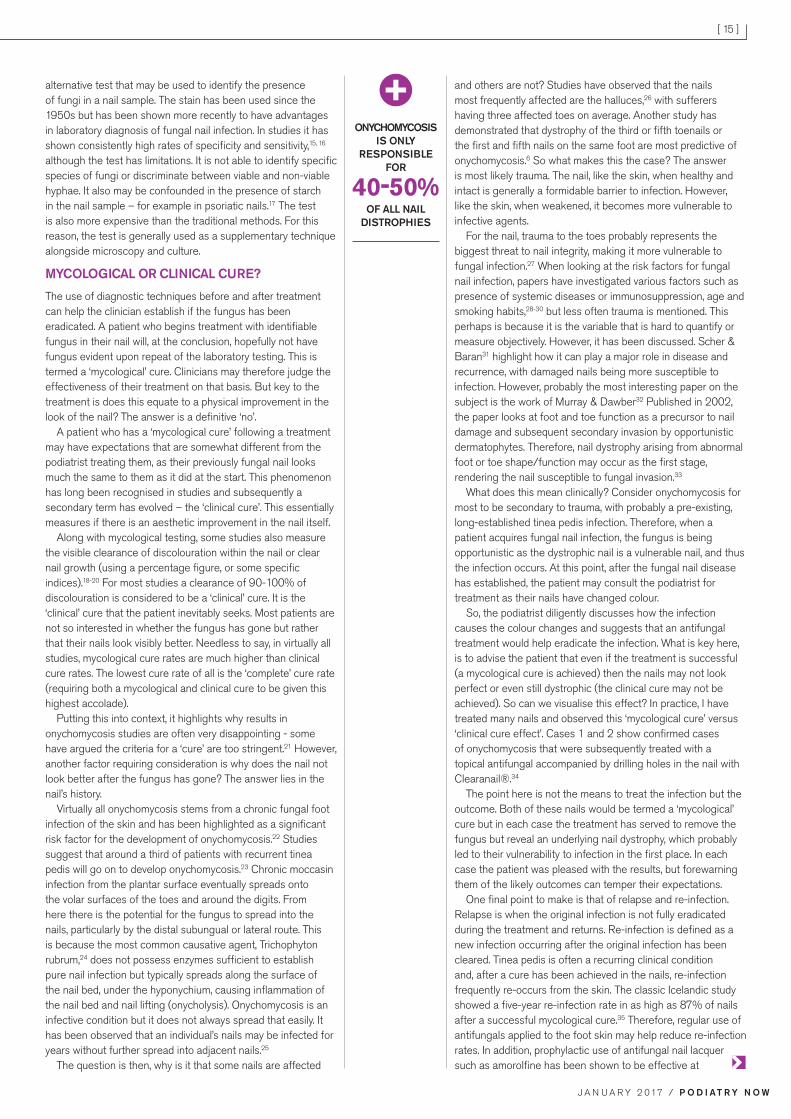
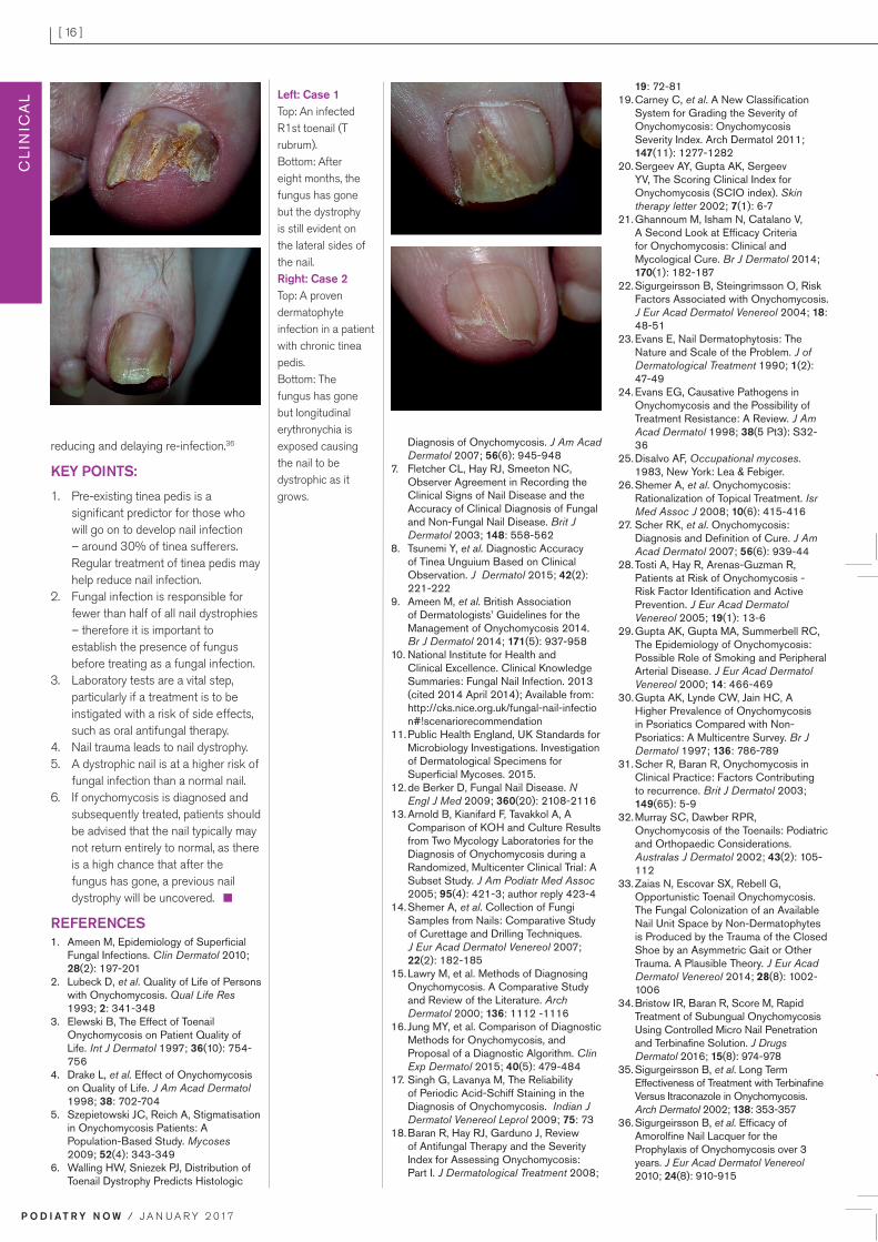
So, the podiatrist diligently discusses how the infection causes the colour changes and suggests that an antifungal treatment would help eradicate the infection. What is key here, is to advise the patient that even if the treatment is successful (a mycological cure is achieved) then the nails may not look perfect or even still dystrophic (the clinical cure may not be achieved). So can we visualise this effect? In practice, I have treated many nails and observed this ‘mycological cure’ versus ‘clinical cure effect’. Cases 1 and 2 show confirmed cases of onychomycosis that were subsequently treated with a topical antifungal accompanied by drilling holes in the nail with Clearanail®.34
The point here is not the means to treat the infection but the outcome. Both of these nails would be termed a ‘mycological’ cure but in each case the treatment has served to remove the fungus but reveal an underlying nail dystrophy, which probably led to their vulnerability to infection in the first place. In each case the patient was pleased with the results, but forewarning them of the likely outcomes can temper their expectations.
One final point to make is that of relapse and re-infection. Relapse is when the original infection is not fully eradicated during the treatment and returns. Re-infection is defined as a new infection occurring after the original infection has been cleared. Tinea pedis is often a recurring clinical condition and, after a cure has been achieved in the nails, re-infection frequently re-occurs from the skin. The classic Icelandic study showed a five-year re-infection rate in as high as 87% of nails after a successful mycological cure.35 Therefore, regular use of antifungals applied to the foot skin may help reduce re-infection rates. In addition, prophylactic use of antifungal nail lacquer such as amorolfine has been shown to be effective at
ONYCHOMYCOSIS
IS ONLY RESPONSIBLE
FOR 40-50%
OF ALL NAIL DISTROPHIES
J A N U A R Y 2 0 17 / P O D I AT R Y N O W
[ 15 ]
reducing and delaying re-infection.36
KEY POINTS:
1. Pre-existing tinea pedis is a significant predictor for those who will go on to develop nail infection – around 30% of tinea sufferers. Regular treatment of tinea pedis may help reduce nail infection.
2. Fungal infection is responsible for fewer than half of all nail dystrophies – therefore it is important to establish the presence of fungus before treating as a fungal infection.
3. Laboratory tests are a vital step, particularly if a treatment is to be instigated with a risk of side effects, such as oral antifungal therapy.
4. Nail trauma leads to nail dystrophy.5. A dystrophic nail is at a higher risk of
fungal infection than a normal nail.6. If onychomycosis is diagnosed and
subsequently treated, patients should be advised that the nail typically may not return entirely to normal, as there is a high chance that after the fungus has gone, a previous nail dystrophy will be uncovered.
REFERENCES 1. Ameen M, Epidemiology of Superficial
Fungal Infections. Clin Dermatol 2010; 28(2): 197-201
2. Lubeck D, et al. Quality of Life of Persons with Onychomycosis. Qual Life Res 1993; 2: 341-348
3. Elewski B, The Effect of Toenail Onychomycosis on Patient Quality of Life. Int J Dermatol 1997; 36(10): 754-756
4. Drake L, et al. Effect of Onychomycosis on Quality of Life. J Am Acad Dermatol 1998; 38: 702-704
5. Szepietowski JC, Reich A, Stigmatisation in Onychomycosis Patients: A Population-Based Study. Mycoses 2009; 52(4): 343-349
6. Walling HW, Sniezek PJ, Distribution of Toenail Dystrophy Predicts Histologic
Diagnosis of Onychomycosis. J Am Acad Dermatol 2007; 56(6): 945-948
7. Fletcher CL, Hay RJ, Smeeton NC, Observer Agreement in Recording the Clinical Signs of Nail Disease and the Accuracy of Clinical Diagnosis of Fungal and Non-Fungal Nail Disease. Brit J Dermatol 2003; 148: 558-562
8. Tsunemi Y, et al. Diagnostic Accuracy of Tinea Unguium Based on Clinical Observation. J Dermatol 2015; 42(2): 221-222
9. Ameen M, et al. British Association of Dermatologists’ Guidelines for the Management of Onychomycosis 2014. Br J Dermatol 2014; 171(5): 937-958
10. National Institute for Health and Clinical Excellence. Clinical Knowledge Summaries: Fungal Nail Infection. 2013 (cited 2014 April 2014); Available from: http://cks.nice.org.uk/fungal-nail-infection#!scenariorecommendation
11. Public Health England, UK Standards for Microbiology Investigations. Investigation of Dermatological Specimens for Superficial Mycoses. 2015.
12. de Berker D, Fungal Nail Disease. N Engl J Med 2009; 360(20): 2108-2116
13. Arnold B, Kianifard F, Tavakkol A, A Comparison of KOH and Culture Results from Two Mycology Laboratories for the Diagnosis of Onychomycosis during a Randomized, Multicenter Clinical Trial: A Subset Study. J Am Podiatr Med Assoc 2005; 95(4): 421-3; author reply 423-4
14. Shemer A, et al. Collection of Fungi Samples from Nails: Comparative Study of Curettage and Drilling Techniques. J Eur Acad Dermatol Venereol 2007; 22(2): 182-185
15. Lawry M, et al. Methods of Diagnosing Onychomycosis. A Comparative Study and Review of the Literature. Arch Dermatol 2000; 136: 1112 -1116
16. Jung MY, et al. Comparison of Diagnostic Methods for Onychomycosis, and Proposal of a Diagnostic Algorithm. Clin Exp Dermatol 2015; 40(5): 479-484
17. Singh G, Lavanya M, The Reliability of Periodic Acid-Schiff Staining in the Diagnosis of Onychomycosis. Indian J Dermatol Venereol Leprol 2009; 75: 73
18. Baran R, Hay RJ, Garduno J, Review of Antifungal Therapy and the Severity Index for Assessing Onychomycosis: Part I. J Dermatological Treatment 2008;
19: 72-8119. Carney C, et al. A New Classification
System for Grading the Severity of Onychomycosis: Onychomycosis Severity Index. Arch Dermatol 2011; 147(11): 1277-1282
20. Sergeev AY, Gupta AK, Sergeev YV, The Scoring Clinical Index for Onychomycosis (SCIO index). Skin therapy letter 2002; 7(1): 6-7
21. Ghannoum M, Isham N, Catalano V, A Second Look at Efficacy Criteria for Onychomycosis: Clinical and Mycological Cure. Br J Dermatol 2014; 170(1): 182-187
22. Sigurgeirsson B, Steingrimsson O, Risk Factors Associated with Onychomycosis. J Eur Acad Dermatol Venereol 2004; 18: 48-51
23. Evans E, Nail Dermatophytosis: The Nature and Scale of the Problem. J of Dermatological Treatment 1990; 1(2): 47-49
24. Evans EG, Causative Pathogens in Onychomycosis and the Possibility of Treatment Resistance: A Review. J Am Acad Dermatol 1998; 38(5 Pt3): S32-36
25. Disalvo AF, Occupational mycoses. 1983, New York: Lea & Febiger.
26. Shemer A, et al. Onychomycosis: Rationalization of Topical Treatment. Isr Med Assoc J 2008; 10(6): 415-416
27. Scher RK, et al. Onychomycosis: Diagnosis and Definition of Cure. J Am Acad Dermatol 2007; 56(6): 939-44
28. Tosti A, Hay R, Arenas-Guzman R, Patients at Risk of Onychomycosis - Risk Factor Identification and Active Prevention. J Eur Acad Dermatol Venereol 2005; 19(1): 13-6
29. Gupta AK, Gupta MA, Summerbell RC, The Epidemiology of Onychomycosis: Possible Role of Smoking and Peripheral Arterial Disease. J Eur Acad Dermatol Venereol 2000; 14: 466-469
30. Gupta AK, Lynde CW, Jain HC, A Higher Prevalence of Onychomycosis in Psoriatics Compared with Non-Psoriatics: A Multicentre Survey. Br J Dermatol 1997; 136: 786-789
31. Scher R, Baran R, Onychomycosis in Clinical Practice: Factors Contributing to recurrence. Brit J Dermatol 2003; 149(65): 5-9
32. Murray SC, Dawber RPR, Onychomycosis of the Toenails: Podiatric and Orthopaedic Considerations. Australas J Dermatol 2002; 43(2): 105-112
33. Zaias N, Escovar SX, Rebell G, Opportunistic Toenail Onychomycosis. The Fungal Colonization of an Available Nail Unit Space by Non-Dermatophytes is Produced by the Trauma of the Closed Shoe by an Asymmetric Gait or Other Trauma. A Plausible Theory. J Eur Acad Dermatol Venereol 2014; 28(8): 1002-1006
34. Bristow IR, Baran R, Score M, Rapid Treatment of Subungual Onychomycosis Using Controlled Micro Nail Penetration and Terbinafine Solution. J Drugs Dermatol 2016; 15(8): 974-978
35. Sigurgeirsson B, et al. Long Term Effectiveness of Treatment with Terbinafine Versus Itraconazole in Onychomycosis. Arch Dermatol 2002; 138: 353-357
36. Sigurgeirsson B, et al. Efficacy of Amorolfine Nail Lacquer for the Prophylaxis of Onychomycosis over 3 years. J Eur Acad Dermatol Venereol 2010; 24(8): 910-915
Left: Case 1 Top: An infected R1st toenail (T rubrum).Bottom: After eight months, the fungus has gone but the dystrophy is still evident on the lateral sides of the nail. Right: Case 2 Top: A proven dermatophyte infection in a patient with chronic tinea pedis.Bottom: The fungus has gone but longitudinal erythronychia is exposed causing the nail to be dystrophic as it grows.
Discounts are available for group booking, please ask one of the events team for details.
For further details or to bookOnline booking and website http://tinyurl.com/hlpav4f
Email [email protected]
Telephone +44 (0) 114 225 9143
Directorate of Podiatric Medicine 6th Annual Podiatric Sports Medicine Conference in Association with European College of Sports and Exercise Physicians 2017
“Inspiring Clinical Excellence - Thinking Beyond Biomechanics”
Thursday 13th and Friday 14th July 2017
The Royal College of Physicians, Regents Park, London
Conference Chairmen: Dr Nat Padhiar and Dr Nikos Malliaropoulos
Places are limited and will be offered on a first come first served basis.Two day conference will include lectures, workshops and informal
discussions delivered by experts from across Europe and Australia:
• Blood tests – Focus on Vitamin B12
• Ultrasound Scan - Clinician’s stethoscope and guidance system
• Platelet Rich Plasma (PRP)
• Extra-corporeal Shock Wave Therapy (ESWT)
• Ultrasound Tendon Characterisation (UTC)
• Knee problems in sport - Basic examination
• Laser therapy
• Physical activity, inactivity and exercise prescription
• Interpreting plain radiographs
• Gut dysbiosis and musculoskeletal problems
Course FeesRegister by 31st March 2017
Early bird 1 day rate £150.00
Early bird 2 day rate £275.00
Register after 31st March 2017
1 day rate £180.00
2 day rate £330.00
Postgraduate rate
1 day rate £90.00
2 day rate £180.00
Undergraduates
Limited free places are available
to undergraduates. Terms and
conditions apply.
P O D I AT R Y N O W / J A N U A R Y 2 0 17
[ 16 ] C
LIN
ICA
L