Basics of Pressure Injury Prevention and Wound Care
128
Basics of Pressure Injury Prevention and Wound Care UNM- Health Sciences Center Department of Pediatrics Continuum of Care Vera Asplund, BSN, RN, CDDN September 29, 2017
Transcript of Basics of Pressure Injury Prevention and Wound Care

Basics of Pressure Injury Prevention and Wound Care
UNM- Health Sciences CenterDepartment of PediatricsContinuum of Care
Vera Asplund, BSN, RN, CDDN
September 29, 2017

Learning Outcome Statement
• The learner will be made aware of issues related to pressure injuries including assessment, prevention, staging, and various treatments of pressure injuries in persons with I/DD

Skin
• The skin is the largest organ of the body and provides the interface between the body and the rest of the world
• The skin provides the first line of defense and protects the integrity and functioning of internal organ systems
• The psychosocial aspect of skin appearance is extremely important to a person’s well-being

Skin Characteristics and Functions
• Skin thickness ranges from 1/50 of an inch over the eyelids to 1/3 of an inch on the palms of the hands and the soles of the feet
• Specialized skin cells harden to form nails and elongate to form hair
• The pH of skin normally ranges from 4.5 to 5.5, thus providing the protective mantle of the skin, which serves to maintain the skin’s normal flora

Vital Functions of the Skin
• Regulating body temperature
• Transmitting such sensations as touch, pressure, and pain
• Preventing excessive loss of body fluids
• Acting as an excretory organ
• Providing an interface between the body and its environment
• Protecting the inner tissues from invasion

Skin Layers- Epidermis
• Outermost layer of skin, which is thin and avascular
• Further divided into five structurally and functionally distinct layers
• Stratum corneum
• Stratum lucidum
• Stratum granulosum
• Stratum spinosum
• Stratum germinativum (basal layer)

Skin LayersDermal-Epidermal Junction
• The dermal-epidermal junction provides structural support and allows exchange of fluids and cells between the skin layers
• The epidermis has an irregular surface, with downward fingerlike projections known as rete ridges or pegs
• These pegs of epidermis interface with upward projections of the dermis anchoring the epidermis to the dermis


Skin LayersDermal-Epidermal Junction
• As the skin ages, this dermal-epidermal junction tends to flatten, as the contacting surfaces of epidermis and dermis decrease by one-third
• This loss increases the potential for dermal-epidermal separation and places older people at risk for skin tears

Skin LayersDermis
• The layer of skin lying beneath the epidermis. It is highly vascular, tough connective tissue, containing nerves, lymphatics, sebaceous glands, and hair follicles.

Skin LayersSubcutaneous Tissue• This layer is made up of dense connective and
adipose tissue
• It houses major blood vessels, lymphatics, and nerves; acts as a heat insulator; and provides a nutritional depot that is used during illness or starvation
• The subcutaneous fat also acts as a mechanical shock absorber and helps the skin move easily over the underlying structures
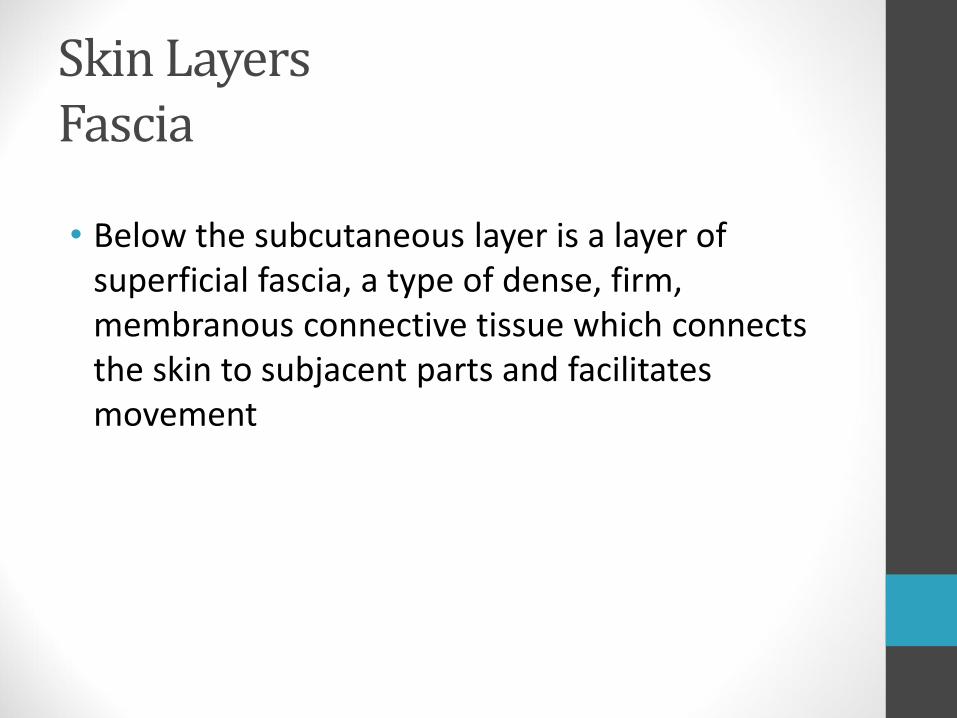
Skin LayersFascia
• Below the subcutaneous layer is a layer of superficial fascia, a type of dense, firm, membranous connective tissue which connects the skin to subjacent parts and facilitates movement

Normal Skin

Blood Supply
• The vasculature of the dermis is the most expansive of any organ system
• The main purpose of this vast blood supply to the skin is to regulate body temperature
• The skin is oversupplied with blood when compared with its metabolic needs
• Muscle and fatty tissue do not tolerate ischemia or hypoxia, and are more susceptible to the effects of pressure than are the dermis and epidermis

Age-Related Changes
• Sweat glands diminish in number
• Epithelial and fatty layers of tissue atrophy and become thin
• Thickness of subcutaneous fat on the legs or forearms diminishes, even if abdominal or hip fat remains abundant
• Collagen and elastin shrink and degenerate

Age-Related Changes
• One result of the general loss of fat from the subcutaneous tissue is the relative prominence of the bony protuberances of the thorax, scapula, trochanters, and knees. The loss of this valuable padding contributes to the development of pressure injuries.

Age-Related Changes
• Collagen content of the skin decreases by approximately 1% per year throughout adult life. The net effect of all these changes is thin, dry, and inelastic skin that is increasingly susceptible to separation of dermis and epidermis as minor friction or shearing forces cause an injury known as skin tear.
Change in Terminology
• In April 2016 the National Pressure Ulcer Advisory Panel announced a change in terminology from pressure ulcer to pressure injury
• They also changed from Roman numerals to Arabic numbers
• The change more accurately describes pressure injuries to both intact and ulcerated skin

Etiology of Pressure Injuries
• Pressure injuries usually occur in soft tissue over bony prominences that remain in contact with compressing surfaces
• Many other factors—primarily shear, friction, excessive moisture, and possibly infection—interact to mechanically damage soft tissue
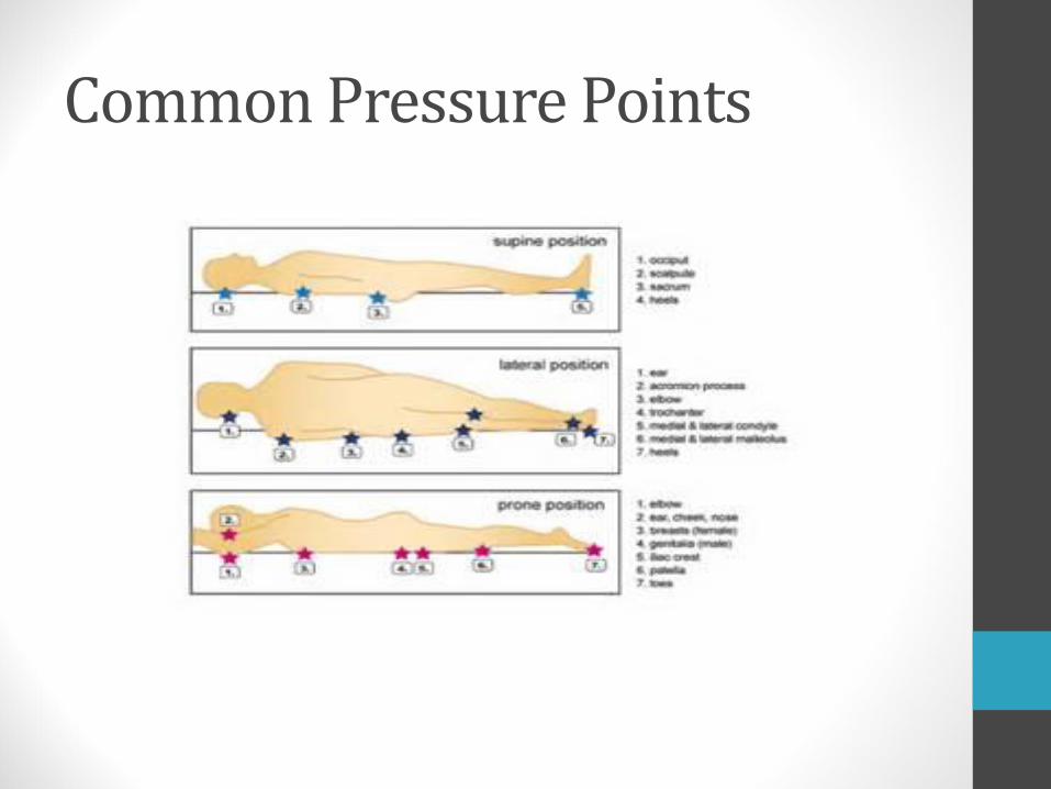
Common Pressure Points

Pressure
• Muscle is more sensitive to compression than skin
• The deeper muscle tissue may be necrotic before damage to the overlying skin is apparent
• The force of pressure increases as the affected body surface area decreases

Time-Pressure
• The normal response to prolonged pressure is a change in body position before tissue ischemia occurs
• Low pressure endured for long periods of time is believed to be more significant in producing pressure ulcers than higher pressure of short duration
• If the time-pressure threshold is reached or exceeded, tissue damage continues even after pressure is released

Time-Pressure
• Pressure injury can result from one period of sustained pressure
• Most pressure injuries occur secondary to repeated ischemic events without adequate time for recovery

Bony Prominences
• Sacrum
• Coccyx
• Ischial tuberosities
• Greater trochanters
• Elbows
• Heels
• Scapulae
• Occipital bone
• Sternum
• Ribs
• Iliac crests
• Patellae
• Lateral malleoli
• Medial malleoli
• Pressure ulcers can form over any bony prominence or any area of soft tissue that is subjected to prolonged pressure


Contractures
• Untreated contractures may cause pressure ulceration
• A contracted limb may exert pressure on adjacent areas other than bony prominences
• A contracted limb may exert more pressure on the mattress than does a non-contracted limb

Pressure Gradient
• When blood vessels, muscle, subcutaneous fat, and skin are compressed between bone and the surface where an individual is lying or sitting, pressure is transmitted from the body surface toward the bone, and the bone exerts counter pressure. These opposing forces result in a cone-shaped pressure gradient.

Pressure Gradient
• Pressure affects all of the tissue between the external surface and the skeletal anatomy, but the greatest tissue destruction is at the bony interface
• The wound one observes may be just the tip of the “iceberg.”
• Because fat and muscle have little tolerance for decreased blood flow, they are less resistant than skin to pressure

Pressure Gradient• Destruction in the subcutaneous tissues and
muscle may be far worse than the surface damage indicates
• Assessment of pressure ulcer size must take into consideration the presence of unseen necrosis in the area of the pressure gradient

Shear
• Shear is a mechanical force that is parallel rather than perpendicular to an area with the main effect impacting deep tissues
• Elevating the head of the bed increases shear and pressure in the sacral an coccygeal areas

Shear• The mechanical forces can obstruct or tear and
stretch blood vessels
• Minimizing shearing forces involves raising the head of the bed to no more than a 30 degreeangle, except for short periods of time (meals, meds, etc.)
• Shearing forces decreases the time tissue can remain under pressure—with shear present, vascular occlusion may occur at half the usual amount of pressure

Shear- helps to relieve
• When head of bed is elevated, elevate the knee
• Repositioning of bed or patient in bed should be followed by shear relieving rocking of the patient, being cautious not to induce additional shear

Shear & Friction
• Shear injury will not be seen at the skin level because it happens beneath the skin
• Shear and friction go hand in hand—one rarely occurs without the other
• Friction is the force of two surfaces moving across one another
• Friction injury will be visible

Friction
• Erosion of surface tissue increases the potential for deeper tissue damage because friction is the precursor of shear
• Those at risk for friction injuries:
• Individuals who have spastic conditions
• Patients who wear braces or appliances that rub against the skin
• The elderly

Excessive Moisture
• Moist skin is five times as likely to become ulcerated as dry skin
• Constant exposure to wetness can waterlog or macerate the skin
• Macerated epidermis is easily eroded
• Wet skin surfaces increase the risk of friction as the patient is moved across the surface of the bed linen

Excessive Moisture
• Excessive moisture may be the result of perspiration, wound drainage, soaking during bathing, and fecal and/or urinary incontinence
• Maceration

Wound Healing
• The process of wound healing begins at the moment of injury and may continue for years
• No matter how trivial or extensive the wound, healing always includes three overlapping phases: inflammation, proliferation, and differentiation
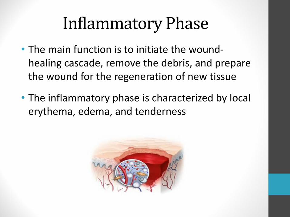
Inflammatory Phase
• The main function is to initiate the wound-healing cascade, remove the debris, and prepare the wound for the regeneration of new tissue
• The inflammatory phase is characterized by local erythema, edema, and tenderness

Proliferative Phase• The proliferative phase of wound healing
overlaps the inflammatory phase
• It begins 2-4 days after wounding and lasts for approximately 15 or 16 days
• The main events during this phase are deposition of connective tissue and collagen cross-linking which fills the wound with collagen and provides strength

Proliferative PhaseGranulation• During collagen production, new capillaries are forming as
budlike structures
• Capillaries penetrate the wound and carry nutrients to the newly generating tissue
• Granulation tissue, when kept moist, provides good tissue for advancing epithelial cells
• As matures, the synthesis of collagen decreases, the new vascular channels regress, the wound transforms to a comparatively avascular, and cell-free scar tissue composed of dense collagen bundles

Epithelization• Epithelial cells migrate from the wound margins across
the wound surface to creating a watertight seal
• Fibrin strands functions as a scaffolding over which the cells creep
• Sheets of new epithelial cells continue to grow until they come into contact with others moving across the wound from other directions
• If a scab is covering the wound surface, the epithelial cells must migrate underneath the scab

Differentiation Phase• The wound matures and the collagen in the scar
undergoes repeated degradation and resynthesis
• This is the longest phase of wound healing
• The tensile strength of the scar increases
• Between the 1st and the 14th day, tissues regain approximately 30% to 50% of their original strength
• Tensile strength continues to increase to approximately 80% of normal tissue strength
• Wounds never completely regain the tensile strength of unwounded tissue

Factors Affecting Healing
• Healing is influenced by systemic conditions or by local conditions in the wound
• Tissue oxygenation
• Stress
• Advanced age
• Nutrition
• Infection
• Compromised Immune System

Tissue Oxygenation
• Oxygen is essential for wound healing
• Blood flow supplies the wound with oxygen and nutrients
• Blood flow removes carbon dioxide and metabolic by-products
• Any condition that reduces blood flow to a wound, such as arterial occlusion, vasoconstriction, or external pressure impedes healing

Stress• Sympathetic nervous system and adrenal
responses to stress (i.e. neural, hormonal, or metabolic changes) can impair wound healing
• A plan that provides sleep and rest for the patient with a pressure injury will promote wound healing

Advanced Age
• Aging affects almost all aspects of the healing response
• Slowing epidermal turnover and increasing skin fragility together reduce wound healing by a factor of four
• The repair rate declines with: falling rates of cell proliferation, lack of development of wound tensile strength, impaired collagen deposition and wound contraction

Advanced Age
• Medical conditions occur in many elderly persons which adversely affect healing
• The elderly tend to be malnourished and poorly hydrated, and have compromised respiratory and immune functions
• Loss of dermal and subcutaneous mass increases the risk for pressure-induced tissue injury

Malnutrition
• Wound healing and the immune response both require an adequate supply of various nutrients, including protein, vitamins, and minerals
• Loss of more than 15% of lean body mass interferes with wound healing
• Individuals with chronic wounds may need more protein and calories than the recommended daily allowances and may require dietary supplements

Protein
• Low serum albumin levels are a late manifestation of protein deficiency
• Serum concentrations below 3.0 g/dl are an indicator of poor nutritional status
• Serum concentrations below 2.5 g/dl reflect severe protein depletion

Vitamins & Minerals• Vitamin C
• Deficiency is associated with impaired fibroblastic function and decreased collagen synthesis which delay healing and contribute to breakdown of old wounds
• Deficiency causes loss of resistance to infection
• Is water-soluble and cannot be stored in the body

Vitamins & Minerals• Vitamin A
• Associated with retarded epithelialization and decreased collagen synthesis
• Deficiency is uncommon because it is fat-soluble and is stored in the liver
• Other vitamins, such as thiamine and riboflavin, are also necessary for collagen organization and the resultant tensile strength of the wound
• Various minerals, such as iron, copper, manganese, and magnesium play a role in wound healing

Barriers to Healing
• Corticosteroids—
• Suppress the inflammatory response; inflammation is necessary to trigger the wound-healing cascade
• Steroid therapy begun after the inflammatory phase of healing (usually 4-5 days after wounding) has a minimal effect on wound healing

Barriers to Healing• Smoking
• Nicotine interferes with blood flow:
• Is a vasoconstrictor
• It increases platelet adhesiveness—causing clot formation
• Cigarette smoke is a vasoconstrictor, and contains carbon monoxide and hydrogen cyanide

Barriers to Healing - Diabetes
• High levels of glucose compete with transport of ascorbic acid, which is necessary for the deposition of collagen, into cells
• Tensile strength and connective tissue production are significantly lower in diabetics
• Arterial occlusive disease can impair healing
• Reduced sensation may leave wounds undetected
• Patients with diabetes have more difficulty resisting infection and their wounds heal more slowly than non-diabetic patients
Infection• Infectious complications of pressure injuries
include sepsis and osteomyelitis
• Debridement, drainage, and removal of the necrotic tissue alone controls most infections
• Open wounds do not have to be sterile to heal
• Healing cannot proceed until all necrotic tissue has been removed from the wound
• Parenteral antibiotics are indicated only when signs and symptoms suggest cellulitis, sepsis, or osteomyelitis

Wound Dehydration
• Wound healing occurs more rapidly when dehydration is prevented
• Epidermal cells migrate faster and cover the wound surface sooner in a moist environment

Evaluation of Healing
• Use a systematic and consistent method to record wound assessments
• Examination should include:
• Measurement of the wound’s length, width, and depth measured in centimeters or millimeters
• Observation of inflammation, wound contraction, granulation, and epithelialization

Wound Healing
• Whenever possible, the body should be allowed to heal itself
• The best treatment is to support conditions that promote optimum healing—such as protection from trauma and maintaining a moist environment

Assessing Risk
• Number and type of medical diagnoses
• Presence of chronic health problems
• Chronologic age
• Immobility/ability to move independently
• Mental status/level of consciousness
• Nutritional status
• Incontinence
• Presence of infection
• Adequacy of circulation

Risk Factors- Device Related
• A localized injury to the skin or underlying tissue resulting from sustained pressure caused by a medical device, such as a brace, splint, cast, respiratory mask or tubing, tracheostomy tube collar or strap, feeding tube or a Foley tubing.

Risk Factors- Device Related
• Choose the correctly sized medical device for the individual
• Cushion and protect the skin with dressing in high-risk areas
• Remove or move the device daily to assess skin
• Avoid placing the device over the site of a previous or existing pressure injury
• Be aware of edema under the device and the potential for skin breakdown
• Confirm that the device isn’t placed directly under a patient who is bedridden or immobile
• Educate staff
Risk Factors -Nutritional
• Dental health
• Oral and GI history
• Chewing and swallowing ability
• Quality and frequency of foods eaten
• Involuntary weight loss or gain
• Serum albumin levels
• Nutritionally pertinent medications
• Psychosocial factors affecting nutritional intake

Risk Factors -Nutritional
• Laboratory tests—depressed serum protein, serum albumin, and transferrin levels together indicate poor nutritional status
• Body weight—
• At-risk patients should be weighed weekly
• Notify a physician, nurse, or dietitian if there is an unintended loss of 10 pounds or more during any 6-month period
• A change of 5% of body weight is predictive of a drop in serum albumin

Risk Assessment Scales
• Braden Scale—The Braden Scale has six subscales divided into two categories
• Intensity and duration of pressure: mobility, activity, and sensory perception
• Tissue tolerance: moisture, nutrition, and friction/shear
See Handouts

Skin Inspection• Blanchable erythema—In light-skinned individuals
• Compressing the reddened area causes the color to blanch or turn pale
• Redness returns immediately after compression is relieved
• Causes no long-term effect on the tissue; should return to its normal color within 24 hours
• In dark-skinned patients
• Comparing skin on the contralateral side can help in assessment of subtle color changes
• Palpate for increased warmth, a feeling of tightness, and areas of hardness under the skin

Terms• Induration: Tissue firmness that may occur around a
wound margin
• Erythema: An inflammatory redness of the skin due to engorged capillaries
• Maceration: Softening of a tissue by soaking until the connective tissue fibers are so weakened that the tissue components can be teased apart

Terms• Undermining: a tunneling effect or pocket
occurring under the pressure ulcer edges or margins
• Slough: Nonviable tissue is loosely attached and characterized by string-like, moist, necrotic debris; yellow, green, or gray in color

Terms
• Eschar: Nonviable (dead) wound tissue characterized by a leathery, black crust covering an underlying necrotic process
• Granulation: Formation in wounds of soft, pink, fleshy projections consisting of new capillaries surrounded by fibrous collagen

Assessment of the Pressure Injury• History: etiology,
duration, prior treatment
• Anatomic location
• Stage
• Size: length, width, and depth measured in centimeters
• Extent: edges, sinus tracts, undermining, tunneling
• Exudate or drainage
• Necrotic tissue: slough and eschar
• Granulation tissue
• Epithelialization or new skin growth
• See Handouts
Measuring Wounds
• Use a consistent technique every time you measure
• Document all measurements in centimeters as L x W x D
• Most common type of measurement—clock method
• Longest length, greatest width, and greatest depth
• Use the body as the face of an imaginary clock
Clock Method
• The body
• The head is always at 12 o’clock
• The feet are always at 6 o’clock
• The feet
• The heels are always at 12 o’clock
• The toes are always at 6 o’clock

Exudate
• Exudate (drainage), is a liquid produced by the body in response to tissue damage, and is present in wounds as they heal
• Accurate assessment of exudate is important throughout the healing process because the color, consistency, odor, and amount change as a result of various physiologic processes and underlying complications

Exudate Assessment- Type
• Serous- thin, clear, watery plasma
• Sanguineous- bloody drainage
• Serosanguineous-thin, watery, pale red to pink plasma with red blood cells
• Purulent- thick, opaque drainage that is tan, yellow, green, or brown. Purulent exudate is never normal and is often associated with infection or high bacteria levels

Exudate Assessment- Amount
• None– Wound tissues are dry
• Scant—Wound tissues are moist, but there is no measurable drainage
• Small/minimal—Wound tissues are moist or wet; the drainage covers less than 25% of the dressing
• Moderate—Wound tissues are wet; the drainage involves more than 25% to 75% of the dressing
• Large or copious—Wound tissues are filled with fluid that involves more than 75% of the dressing

Exudate Assessment-Consistency/Odor
• Consistency
• Low viscosity—thin, runny
• High viscosity—thick or sticky; doesn’t flow easily
• Odor
• No odor noted
• Strong, foul, pungent, fecal, musty, or sweet

Exudate Assessment-Condition of dressing• Dry—primary dressing is unmarked by exudate;
the dressing may adhere to the wound
• Moist—small amount of exudate are visible when the dressing is removed
• Saturated—the primary dressing is wet and strike through occurs
• Leaking—the dressings are saturated, and exudate is leaking from primary and secondary dressing onto the patient’s clothes

Stage 1 Pressure Injury• Non-blanchable erythema of intact skin
• This stage may be difficult to detect in a patient with darkly pigmented skin tones — Assess the surrounding area to observe differences in skin color
• Also assess the area for:• Change in sensation• Warmth or coolness as compared with adjacent
tissue• Firmness or softness as compared with adjacent
tissue• Pain

Treatment: Stage 1
• Remove pressure
• Do not rub or massage prominence
• Do not use donuts
• Protect from moisture
• Monitor
• No dressings required
• Treat pain if present

Stage 1
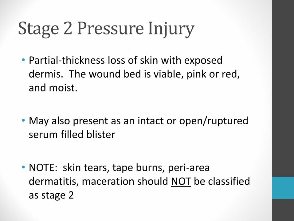
Stage 2 Pressure Injury
• Partial-thickness loss of skin with exposed dermis. The wound bed is viable, pink or red, and moist.
• May also present as an intact or open/ruptured serum filled blister
• NOTE: skin tears, tape burns, peri-area dermatitis, maceration should NOT be classified as stage 2

Treatment: Stage 2
• Remove pressure
• Keep clean
• Keep fluid filled blister intact if possible
• Cover with light dressing if ulcer is open
• Example: non-adherent gauze dressing changed every day

Stage 2
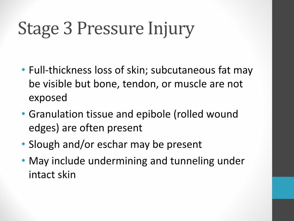
Stage 3 Pressure Injury
• Full-thickness loss of skin; subcutaneous fat may be visible but bone, tendon, or muscle are not exposed
• Granulation tissue and epibole (rolled wound edges) are often present
• Slough and/or eschar may be present
• May include undermining and tunneling under intact skin

Treatment: Stage 3
• Remove pressure
• Eliminate slough
• Autolytic, or sharp debridement
• Manage exudate
• Foam, alginate
• Monitor for infection
• Treat pain

Stage 3

Stage 4 Pressure Injury
• Full-thickness skin and tissue loss with exposed or directly palpable fascia, muscle, tendon, ligament, cartilage or bone in the ulcer
• Slough & eschar may be present on some parts of the wound bed
• Often epibole (rolled edges), undermining and/or tunneling occur

Treatment: Stage 4
• Remove pressure
• Eliminate slough or eschar
• Manage exudate
• Treat pain
• Monitor for infection
• Osteomyelitis
• Septicemia
• Cellulitis
• Abscess

Stage 4

Stage 4

Unstageable Pressure Injury
• Full thickness skin and tissue loss in which the extent of tissue damage within the ulcer cannot be confirmed because it is obscured by slough (yellow, tan, gray, green or brown) and/or eschar (tan, brown or black) in the wound bed
• Note: Until enough slough and/or eschar is removed to expose the base of the wound, the true depth, and therefore stage, cannot be determined

Treatment: Unstageable• Remove pressure
• Eliminate slough and/or eschar
• Hydrogel application to soften, sharp debridement
• Never debride dry, stable heel ulcer
• Restage once all slough and/or eschar has been removed
• Manage exudate
• Monitor for infection
• Treat pain

Unstageable

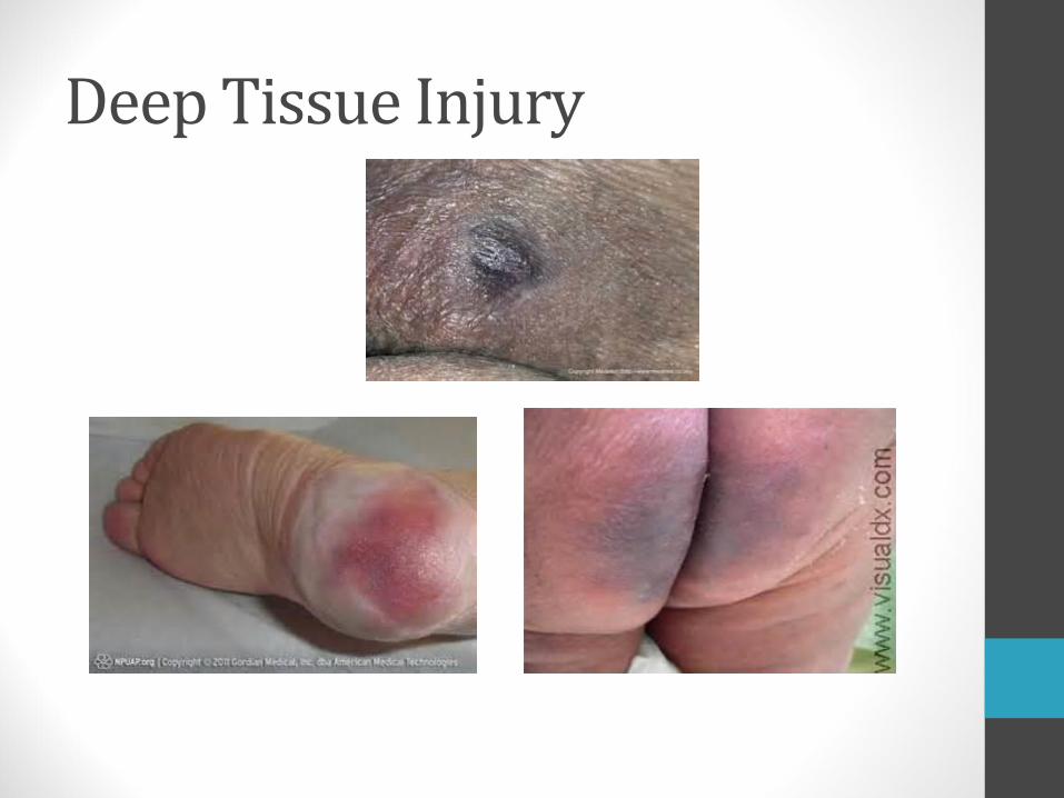
Deep Tissue Injury
• Intact or non-intact skin with localized area of persistent non-blanchable deep red, maroon, purple discoloration or epidermal separation revealing a dark wound bed or blood filled blister
• Pain and temperature change often precede skin color changes
• Injury results from intense and/or prolonged pressure and shear forces at the bone-muscle interface

Treatment: Deep Tissue Injury
• Remove pressure
• Monitor for opening of wound
• Treat pain
• Protect from moisture
Deep Tissue Injury

Staging
• Pressure injuries are NOT restaged at each assessment. They are staged only once unless a deeper layer of tissue becomes exposed

Wound-cleansing
• Normal saline solution is the preferred cleansing agent—isotonic, cost effective and available in many different formats
• Other:
• Commercial cleansers—ingredients may include surfactants, wetting agents, moisturizers, and/or antimicrobials
• Lactated Ringer’s solution—provides sodium, potassium, and calcium chloride to the wound; be careful if used for continuous irrigation or allowed to dwell inside body cavities as may be absorbed into the bloodstream leading to circulatory overload
• Potable tap water—can be used if there is no other alternative
Skin cleansers should never be used on open wounds—formulated to remove fecal matter, and are toxic to wound tissues

Wound-cleansing
• The cleansing solution should be at room temperature or slightly warmer
• It can take up to 40 minutes after cleaning the wound to regain its original temperature, and up to 3 hours for miotic cell division and leukocyticactivity to return to normal
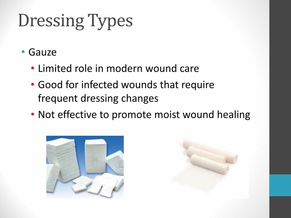
Dressing Types
• Gauze
• Limited role in modern wound care
• Good for infected wounds that require frequent dressing changes
• Not effective to promote moist wound healing

Dressing Types
• Transparent Films
• Allow O2 to penetrate wound and release wound moisture
• Helps with autolytic debridement
• Good for partial thickness wounds stage I & II
• Not suitable for heavy draining wounds

Dressing Types
• Foam
• Non-occlusive dressing
• Highly absorbent
• Less frequent dressing changes—up to 7 days
• Use on draining stage II-IV
• Don’t use on dry wounds

Dressing Types
• Hydrocolloids
• Contain gelatin or pectin that swells with exudate
• Waterproof—helps with autolytic debridement
• Use on shallow stage II pressure ulcers
• Can trap moisture under the dressing causing maceration
• Particles of the dressing can become lodged in the wound bed

Dressing Types
• Silicone
• Easy to apply
• Atraumatic to the wound and surrounding skin upon removal
• Comfortable to wear
• Can lift and adjust
• May remain in place for several days
• Can be used on moderately exuding wounds

Dressing Types
• Hydrogel
• Viscous amorphous gels
• Applied to base of the wound to soften eschar
• Use in wounds that are dry, contain hard eschar
• Provide some soothing, pain relieving properties
• Consists mostly of hypertonic saline
• Require secondary dressing

Dressing Types
• Alginates
• Seaweed based woven fibers form a gel like material when they come in contact with exudate
• Highly absorbent
• Can be left in wound bed for several days
• Require a secondary dressing
• Good on highly draining stage III and IV ulcer
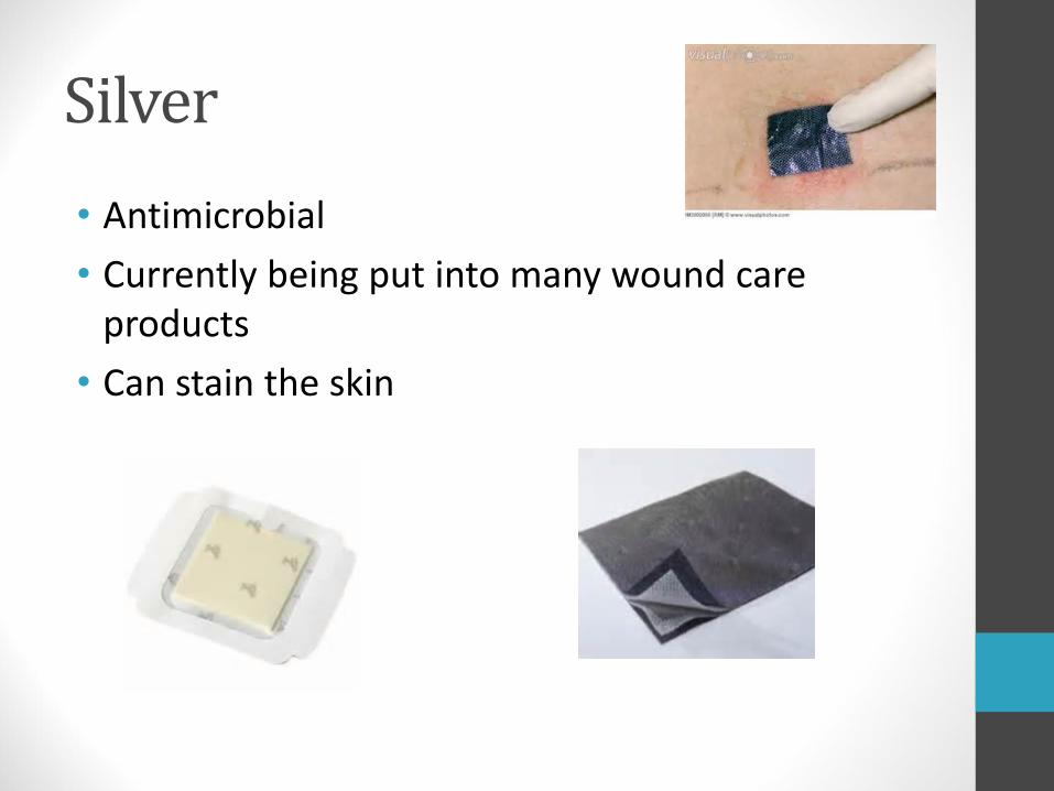
Silver
• Antimicrobial
• Currently being put into many wound care products
• Can stain the skin

Honey
• Medical grade honey
• Promotes moist wound healing
• Supports autolytic debridement
• Helps to lower pH of a wound which can increase healing
• It is antimicrobial

Dressing changes
• Remove tape by pulling parallel to the skin, towards the dressing, pull slowly and keep your hand low
• If tape is over hairy areas, remove it in the direction of hair growth
• Mimimize wound manipulation
• Minimize the number of dressing changes
• Soak dried dressings before trying to remove
• Avoid overpacking the wound
• Use low-adhesive dressings

Assessing Complications
• Complications can delay healing and may become life-threatening
• All pressure ulcers are colonized with bacteria
• Debridement and adequate cleansing prevent the ulcer from becoming infected in most cases
• Swab cultures of the wound surface should not be used to identify infecting organisms

Assessing Complications
• Ulcer infection
• Is recognized by the classic signs of redness, fever, pain, edema and odor
• The cardinal sign is advancing cellulitis
• Sepsis
• May originate from infected pressure injuries of any stage
• Blood culture is the only way to identify the pathogen

Assessing Complications• Osteomyelitis (infection involving the bone)
• Is likely in stage IV ulcers
• Delays healing, causes extensive tissue damage, and is associated with a high mortality rate
• A bone biopsy and culture are necessary for diagnosis
• If the patient’s white blood cell count, erythrocyte sedimentation rate, and plain X-ray are all positive, osteomyelitis is likely

Prevention
• Nutritional management
• Managing pressure
• Skin care
• Monitoring changes in risk status

Nutritional Management• The nutritional goal is a diet containing adequate
nutrients to maintain tissue integrity
• Monitor for signs of vitamin and mineral deficiencies—provide a daily high-potency vitamin and mineral supplement
• Supplement or support the intake of protein and calories—healthy adults need one to two 3-ounce servings of meat, milk, cheese or eggs each day; a malnourished patient may require as much as 2 grams of protein per kilogram of body weight daily

Manage Pressure
• Pressure management entails the awareness of proper body positioning, recognizing the importance of turning and repositioning , and choosing suitable support surfaces for sleeping and sitting

Body Positioning• In Bed
• Do not position an individual on skin that is already reddened by pressure
• Donut-shaped products reduce the blood flow to an even wider area of tissue
• Pillow placement and bridging can help reduce pressure
• Do not place an individual directly on the greater trochanter
• Heels should be suspended to avoid pressure
• The head of the bed should be raised as little as possible (no more than 30˚)
Turning and Repositioning
• Healthy people change position as frequently as every 15 minutes
• Those unable to reposition themselves should be repositioned frequently enough to allow any reddened areas of skin to recover from pressure
• Repositioning should happen at least every 2 hours while in bed and at least every hour when in a wheelchair
• Never sit on personal items such as keys, pens, phone, etc.

Turning and Repositioning
• To avoid effects of friction and shear forces
• Lift rather than drag individuals across the bed surface
• Have the individuals wear socks and long sleeves to protect heels and elbows

Turning and Repositioning• Sitting—carries the greatest risk of pressure
ulcers.
• Good body posture and alignment helps minimize the pressure on susceptible surfaces
• Thighs should be horizontal so the weight is evenly distributed
• If the knees are higher than the hips, body weight concentrates on the ischial tuberosities
• Adequate support of the ankles, elbows, forearms, and wrist in a neutral position reduces risk
• Separate knees so they do not rub together

Support Surfaces
• Using pillows to bridge vulnerable areas is an effective way to eliminate pressure.
• Using pillows to bridge vulnerable areas is an effective way to eliminate pressure

Support Surfaces
• Many beds, mattresses, and cushions are available to help reduce the intensity of pressure
• Pressure reducing surfaces include:
• Foam, gel, water, and air mattresses
• Alternating pressure pads
• Low-air-loss, high-air-loss, and oscillating beds
• Turning frames

Skin Care
• Massaging reddened areas of skin over bony prominences may reduce blood flow and cause tissue damage
• With older adults, gentle handling can reduce the likelihood of skin tears
• Advancing age is closely associated with skin dryness. Central or room humidifiers can significantly reduce the detrimental effect of low humidity

Cleansing the Skin
• Frequent bathing may remove the natural barrier and increase skin dryness
• The temperature of bath water should be slightly warm
• Use gentle washing with a soft cloth and patting the skin dry with a soft towel

Moisturizing the Skin• It is important to keep the skin well lubricated
• Topical agents relieve the signs and symptoms of dry skin
• Lotions—highest water content, evaporate the most quickly and, need to be reapplied the most frequently
• Creams—preparations of oil in water; more occlusive than lotions; need to be applied about four times daily for maximum effectiveness.
• Ointments—mixtures of water in oil, the most occlusive, and provide the longest lasting effect on skin moisture
Summary
• Healthy skin requires a holistic approach
• Pressure must be managed
• Routine skin inspection is a must
• If a pressure injury develops, one must first find the source and relieve the pressure
• Stage and manage any wound
• Use a team approach
• Monitor

Thank You

Bibliography
Demarco, Sharon. “Wound and Pressure Ulcer Management.” Johns Hopkins Medicine. 11 March 2014.
Maklebust, JoAnn and Sieggreen, Mary. Pressure Ulcers – Guidelines for Prevention and Management. Springhouse, Pennsylvania, Sprighhouse Corporation, 2001. Print.
Morgan, N. RN, BSN, MBA, WOC, DWC, OMS. “How to assess wound exudate.” Apple Bites. Assessment 2014 Journal Vol3 No2, 2014.
Morgan, N. “Measuring Wounds.” https://woundccareadvisor.com. Accessed September 15, 2017.
• “National Pressure Ulcer Advisory Panel (NPUAP) announces a change in terminology from pressure ulcer to pressure injury and updates the stages of pressure injury,” Hot Topics, Press Releases, [email protected]. April 13, 2016.
• Sardina, D. (ed.) Wound Care Advisor. https://woundcareadvisor.com/best-practice. Vol1-no3/ “Ouch! That hurts!”
• Sardina, D. (ed.). “Device—related pressure ulcers: Avoidable or not?” Pressure Injury 2014 Journal Vol3 No4, wound care, Wound Care Advisor.
• Sardina, D. (ed.). “Is your wound-cleansing practice up to date?” https://woundcareadvisor.com/is-your-wound-cleansing-practice-up-to-date_vol2_no3. Accessed September 15, 2017.
• Wilhelmy, Jennifer, RN, CWCN, CNP. “Save our Skin Heal our Holes: The basics of pressure ulcer prevention and wound care.” DDNA National Conference 2014.

• National Pressure Ulcer Advisory Panel
• www.npuap.org