Basics of Diagnostic Laboratory Tests for Leptospirosis
32
Basics of Diagnostic Laboratory Tests for Leptospirosis Presentation made by: Elizabeth Perez; Student Intern at PAHO Reviewed by: Martha Maria Pereira, Ph.D. from Fiocruz, WHO Colaborating Center Internship oriented by: Maria Cristina Schneider, D.V.M., M.Sc., Sc.D. Disclaimer: The content of this presentation is for information purposes only and is not an official PAHO/WHO guideline document.
Transcript of Basics of Diagnostic Laboratory Tests for Leptospirosis

Basics of Diagnostic Laboratory
Tests for Leptospirosis Presentation made by: Elizabeth Perez; Student Intern at PAHO
Reviewed by: Martha Maria Pereira, Ph.D. from Fiocruz, WHO Colaborating Center
Internship oriented by: Maria Cristina Schneider, D.V.M., M.Sc., Sc.D.
Disclaimer: The content of this presentation is for information purposes only and is not an official
PAHO/WHO guideline document.
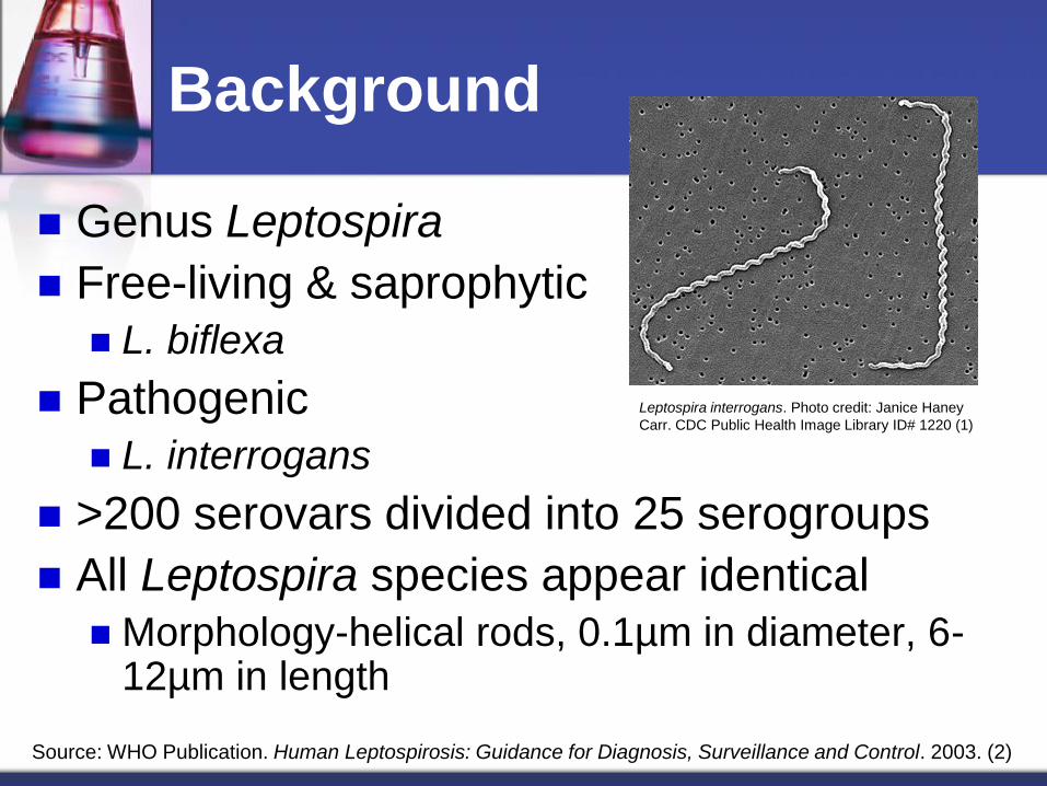
Genus Leptospira
Free-living & saprophytic
L. biflexa
Pathogenic
L. interrogans
>200 serovars divided into 25 serogroups
All Leptospira species appear identical
Morphology-helical rods, 0.1µm in diameter, 6-12µm in length
Background
Leptospira interrogans. Photo credit: Janice Haney
Carr. CDC Public Health Image Library ID# 1220 (1)
Source: WHO Publication. Human Leptospirosis: Guidance for Diagnosis, Surveillance and Control. 2003. (2)
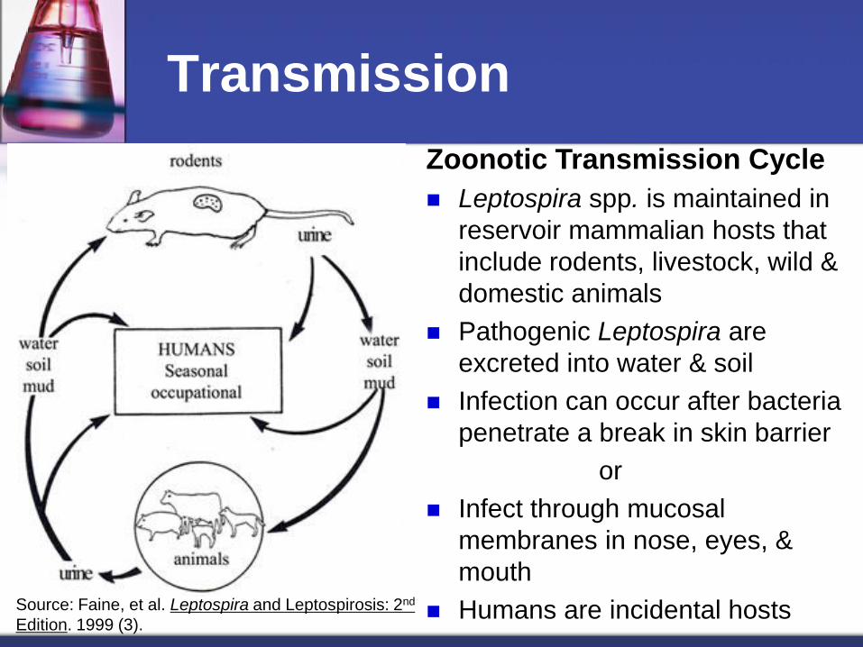
Zoonotic Transmission Cycle
Leptospira spp. is maintained in
reservoir mammalian hosts that
include rodents, livestock, wild &
domestic animals
Pathogenic Leptospira are
excreted into water & soil
Infection can occur after bacteria
penetrate a break in skin barrier
or
Infect through mucosal
membranes in nose, eyes, &
mouth
Humans are incidental hosts
Transmission
Source: Faine, et al. Leptospira and Leptospirosis: 2nd
Edition. 1999 (3).

Leptospirosis: Clinical Symptoms
Phase I: Leptospiremic Anicteric form: 90% of cases
Symptoms: Sudden onset of fever, intense myalgia (calves & thighs), conjunctival suffusion, and severe headache
Lasts for 4-9 days
Brief afebrile period
Phase II: Immune Phase or “Weil’s Disease” Icteric Form: 5-10% of cases progress to serious disease
Symptoms: Hepatic & renal dysfunction, jaundice, circulatory collapse (shock)
From 6-12th day
Mortality of 5-30% (WHO)
Sources: I. WHO 2003 (2).
II. Levett, P. N. “Leptospirosis” Clin Microbiol Rev. 2001(4).
III. Heymann, DL. American Public Health Association. 2004 (5).

Clinical Symptoms
Source: Levett, P.N., “Leptospirosis.” Clin Mircobiol Rev. Apr. 2001 p. 296-326 (4)

Differential Diagnosis
Illnesses with symptoms that are similar to leptospirosis:
Dengue
Yellow Fever
Influenza
Broad spectrum of symptoms presents a challenge for healthcare workers
Clinical Symptoms
Diagnosis is based on the entire clinical picture
Clinical Symptoms
Comprehensive Patient History: occupation
recreation activities
lifestyle
seasonal fluctuations in rainfall
recent climate/disaster event (flooding or hurricane)
Laboratory confirmation variety of available methods
Laboratory Tests
Direct Assays
Detection of Leptospira pathogen
Acute leptospiremic stage first 10 days of
illness
Indirect Assays
Detection of antibodies produced in response
to Leptospira infection
After 5th day of illness and can last for years

Laboratory Tests
Laboratory Diagnostic
Methods for Leptospirosis
Direct
Detection of Leptospira
pathogen
Indirect
Detection of Leptospira
specific antibodies
Microscopy Culture PCR MAT RDTs ELISA

Direct Laboratory Tests
Microscopy Dark field & phase-contrast
Silver & Fluorescence staining
Pros Early detection
Variety of patient specimens
Cons Artifact
Low sensitivity & specificity
Requires sophisticated microscopes
Silver Stain
Fluorescent Stain
Photo credit: Dr. Martin Hicklin. CDC Public Health
Image Library ID# 2769-Silver Stain of Kidney (6).
Photo credit: Mildred Galton. CDC Public Health Image
Library ID# 1346-Fluorescent Stain of Liver Smear (7).

Direct Laboratory Tests
Culture
Incubation at 28-30°C for 4-6 weeks
Semi-solid and liquid culture media
Pro Definitive ID of infecting serovar
Cons Delayed results due to slow growth rate of Leptospira
Cumbersome to maintain cultures for extended time periods
Culture is not warranted for acute clinical diagnosis
Source: WHO Publication. Human Leptospirosis: Guidance for Diagnosis, Surveillance and Control. 2003. (2)

Direct Laboratory Tests
Culture Specimens Blood Samples
Collect within 10 days of illness onset
Transport in tube with heparin at room temp (refrigeration or freezing is detrimental to
pathogenic leptospires)
CSF Samples
Collect between 5-10 days after onset of symptoms
Urine Samples
Collect between 10-30 days of illness onset
Limited survival of leptospires in urine & must be processed within 2 hours to avoid loss of viability
Post Mortem Samples
Collect tissue aseptically and as soon as possible after death
Transport in sterile container at + 4°C to prevent autolysis of cells
Source: WHO Publication. Human Leptospirosis: Guidance for Diagnosis, Surveillance and Control. 2003. (2)
Direct Laboratory Tests
Polymerase Chain Reaction-PCR Use of nucleic acid amplification of Leptospira specific
target to detect pathogen from patient serum sample
Pro Rapid results - presence of leptospires can be detected
before development of antibodies
Cons Not extensively evaluated in clinical applications and
should only be performed on an experimental basis
According to the Royal Tropical Institute (WHOCC) in Amsterdam “A Real-time PCR has been developed and is in the process of validation”
Sources: WHO 2003 (2) & KIT 2012 (8)

Laboratory Tests
Laboratory Diagnostic
Methods for Leptospirosis
Direct
Detection of Leptospira
pathogen
Indirect
Detection of Leptospira
specific antibodies
Microscopy Culture PCR MAT RDTs ELISA

Indirect Laboratory Tests
Serology
Detection of an antibody (either IgM or IgG) in
blood after seroconversion has occurred
IgM-biomarker of current or recent infection
IgG-biomarker of past infection
Detectable titers of antibodies appear in the
blood approx. 6–10 days after the onset of
disease
All rapid diagnostic tests (RDTs) utilize
serological principles to detect antibodies Source: WHO Publication. Human Leptospirosis: Guidance for Diagnosis, Surveillance and Control. 2003. (2)
Indirect Laboratory Tests
Microscopic Agglutination Test (MAT)
Panel of live cell suspensions mixed with diluted
patient sample to test for serum antibodies
Examine agglutination reactions for the
presence of clumps
Positive Result = Four-fold rise in titer between
acute and convalescent phase sera run in
parallel
Gold standard used in reference laboratories
Source: WHO Publication. Human Leptospirosis: Guidance for Diagnosis, Surveillance and Control. 2003. (2)
Source: WHO Publication: Human Leptospirosis: Guidance for Diagnosis, Surveillance and Control. 2003. (2)
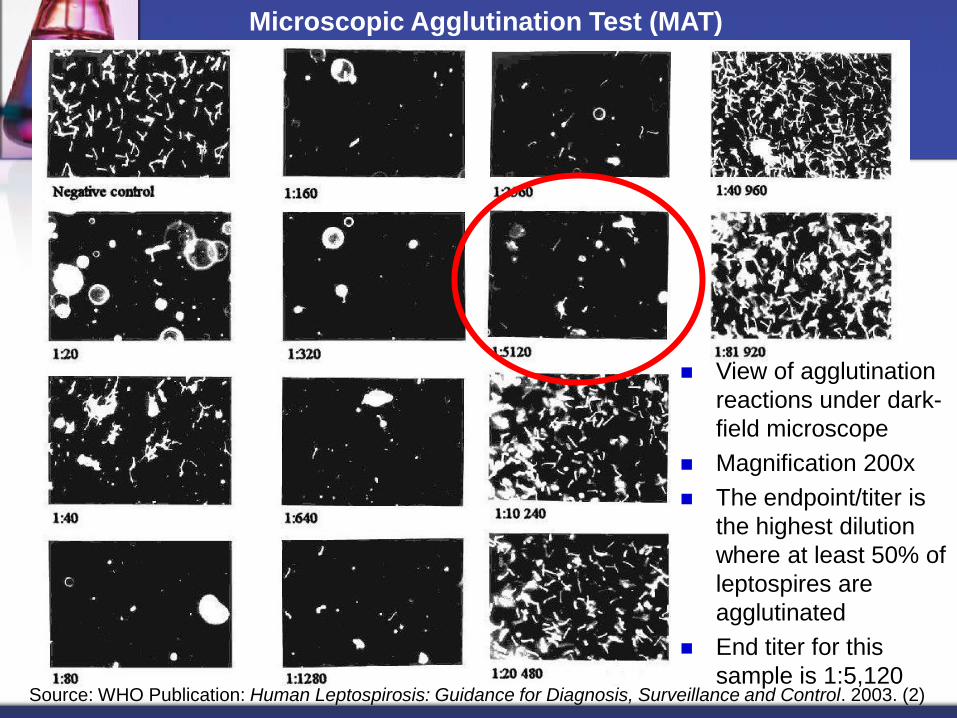
Microscopic Agglutination Test (MAT)
View of agglutination
reactions under dark-
field microscope
Magnification 200x
The endpoint/titer is
the highest dilution
where at least 50% of
leptospires are
agglutinated
End titer for this
sample is 1:5,120

Indirect Laboratory Tests
Microscopic Agglutination Test (MAT)
Pros
High specificity and sensitivity
Cons
Diagnosis is relative when acute and convalescent
serum samples are collected in intervals less than 10
days
Labor intensive-maintenance of living cultures
including reference and local strains
Source: WHO Publication: Human Leptospirosis: Guidance for Diagnosis, Surveillance and Control. 2003. (2)

Indirect Laboratory Tests
ELISA: Enzyme-Linked Immunosorbent Assay
Broadly reactive antigen is used for
detection of Leptospira specific antibodies
The antibody complex is visualized by a
colormetric change that is measured by a
spectrophotometer
Assays for either IgM or IgG and both
IgM/IgG can be performed on patient serum

Indirect Laboratory Tests
ELISA
Positive
Controls
Negative
Controls
Sample 1
Sample 2
Sample 3
Sample 4
Serum Dilution Series
Source: Paul, W. E. (ed.) Fundamental
Immunology, 6th ed. 2008 (9).
Indirect Laboratory Tests
ELISA
Pros
Only one serum specimen is required for diagnosis
Automated process that yields results within a few hours
Antigen coated plates are stable at room temperature
Earlier detection of Leptospira specific antibody-as early
as 6-8 days after illness onset
Cons
Requires local standardization
Genus specific antigens commonly indicated for
screening prior to complementary tests Source: WHO Publication: Human Leptospirosis: Guidance for Diagnosis, Surveillance and Control. 2003. (2)

Indirect Laboratory Tests
Rapid Diagnostic Tests (RDTs)
Results are available within minutes, or at most
2 hours
Samples for RDTs require little or no processing
Result interpretation is straightforward
Simple to use and require minimal facilities,
equipment, & training
Stable reagents may be stored under extreme
conditions
Source: RDT Info: Current Information on Rapid Diagnostic Tests. United States Agency for International
Development (USAID). 2008. (10)

Indirect Laboratory Tests
RDTs
Variety of different RDT technologies
Lateral-flow: tests where the user adds the specimen directly onto the strip and reads the results after a specified amount of time has elapsed
Flow through: kits of individual cassettes with extraction, wash buffers and a “reveal” reagent to obtain results
Agglutination: tests based on agglutination of particles in a sample after the addition of antigenic reagent; agglutination reaction can be visualized with the naked eye
Pros Potential for point-of-care diagnostics for quick results
Easy to use in low-resource settings and in the field
Cons Cross-reactive antibodies also have been described in patients with syphilis,
relapsing fever, Lyme disease, and legionellosis
Patients that are immunocompromised, malnourished, or have immune system defects may yield false negative results
Values for % Sensitivity and % Specificity vary between different RDTs
Sources: USAID 2008 (10) & MD Consult 2011 (11).

Laboratory Tests
Laboratory Diagnostic
Methods for Leptospirosis
Direct
Detection of Leptospira
pathogen
Indirect
Detection of Leptospira
specific antibodies
Microscopy Culture PCR MAT RDTs ELISA
Gold
standard
IgM
IgG
Both
Lateral Flow
Flow Through
Agglutination

Specimen Collection & Transport
Microscopic Agglutination Test (MAT)
Sample: Minimum of two clotted blood or serum samples
Container: Sterile tube
When to obtain MAT specimens:
First sample: at the first clinical care
Second sample: about 10 days after the first sample
Storage and transportation of MAT specimens:
Separation of serum from whole blood must be conducted
before dispensing serum into a sterile plastic freezing vial.
Serum must be transported between 0°C to 4°C
Serum should be stored at 4°C for short term or at - 20°C if
samples are stored for long time periods
Source: WHO Publication. Human Leptospirosis: Guidance for Diagnosis, Surveillance and Control. 2003. (2)

Enzyme Linked Immunosorbent Assay (ELISA)
Sample: Clotted blood or serum sample
Container: Sterile tube
When to obtain an ELISA specimens: approximately 6-8 days after
the onset of clinical symptoms
Storage and transportation of ELISA specimens:
Separation of serum from whole blood must be conducted
before dispensing serum into a sterile plastic freezing vial.
Serum must be transported between 0°C to 4°C
Serum should be stored at 4°C for short term or at - 20°C if
samples are stored for long time periods
Source: WHO Publication. Human Leptospirosis: Guidance for Diagnosis, Surveillance and Control. 2003. (2)
Specimen Collection & Transport

Specimen Collection & Transport
Culture Specimens Revisited Blood Samples
Collect within 10 days of illness onset
Transport in tube with heparin at room temp (refrigeration or freezing is detrimental to pathogenic leptospires)
CSF Samples
Collect between 5-10 days after onset of symptoms
Urine Samples
Collect within 10-30 days of illness onset
Survival of leptospires is limited and must be processed within 2 hours of voiding
Post Mortem Samples
Collect tissue aseptically and as soon as possible after death
Transport in sterile container at + 4°C to prevent autolysis of cells
Source: WHO Publication. Human Leptospirosis: Guidance for Diagnosis, Surveillance and Control. 2003. (2)

Specimen Collection & Transport
The following data must be recorded and
accompany any specimen sent for lab tests:
Date of sample collection
Specimen type
Date of illness onset
Date of Antibiotic treatment (if any)
Type of Antibiotic treatment (if any)

Example: Form Requesting
Laboratory Testing for
Leptospirosis
Source: WHO Publication. Human
Leptospirosis: Guidance for Diagnosis,
Surveillance and Control. 2003. (2)

Conclusions
Phase of illness determines the appropriate lab test for successful diagnosis of Leptospirosis
Leptospiremic Phase in the first week – direct lab methods
Immune Phase after first week – indirect lab methods
Every laboratory test has both advantages and limitations
A negative RDT result does not rule out leptospirosis and must be confirmed using the gold standard of MAT
Proper specimen collection and transport is essential to yielding accurate laboratory results

References
1. Photo Credit: Janice Haney Carr. CDC Public Health Image Library. ID#:1220: Scanning electron micrograph of Leptospira interrogans.
2. World Health Organization (WHO). Human Leptospirosis: Guidance for Diagnosis, Surveillance and Control. Geneva. 2003.
3. Source: Faine, et al. Leptospira and Leptospirosis: 2nd Edition. CRC Press. Boca Raton. 1999
4. Levett, P. N. 2001. “Leptospirosis” Clin Microbiol Rev 296-326.
5. Control of Communicable Diseases Manual: 18th Edition. Heymann, DL. American Public Health Association. Washington DC 2004.
6. Photo Credit: Dr. Martin Hicklin. CDC Public Health Image Library ID# 2769. Silver Stain of Kidney tissue.
7. Photo Credit: Mildred Galton. CDC Public Health Image Library ID# 1346. Leptospira bacteria in liver impression smear. FA stain.
8. Royal Tropical Institue/WHO Collaborating Center for Reference and Research on Leptospirosis. “Leptospirosis reference and diagnostic services.” Accessed online: 16 Nov 2012.
<URL: http://www.kit.nl/kit/Leptospirosis-reference-and-diagnostic-services>
9. Paul, W. E. (ed.) Fundamental Immunology, 6th ed. Lippincott Williams & Wilkins, Philadelphia, PA. 2008.
10. RDT Info. Current Information on Rapid Diagnostic Tests. United States Agency for International Development (USAID) 2008.
URL < http://www.rapid-diagnostics.org/index.htm >
11. Fraser, T, Walsh SR., Harper, W. Leptospirosis. First Consult, MD Consult Web site. URL: <http://www.mdconsult.com.proxygw.wrlc.org/das/pdxmd/body/382400869-8/0?type=med&eid=9-u1.0-_1_mt_1014555#2378586>. Posted: 9 Aug 2011. Accessed online: 16 Nov 2012.
12. Photo Credit: Janice Haney Carr. CDC Public Health Image Library. ID#: 138: Scanning electron micrograph of Leptospira sp.
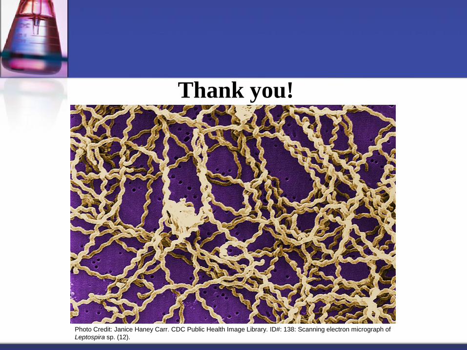
Thank you!
Photo Credit: Janice Haney Carr. CDC Public Health Image Library. ID#: 138: Scanning electron micrograph of
Leptospira sp. (12).