Basic Principles of Cell Culture
20
1 Basic Principles of Cell Culture R. Ian Freshney Centre for Oncology and Applied Pharmacology, Cancer Research UK Beatson Laboratories, Garscube Estate, Bearsden, Glasgow G61 1BD, Scotland, UK, [email protected] 1. Introduction ...................................................... 4 2. Types of Cell Culture .............................................. 4 2.1. Primary Explantation Versus Disaggregation .................... 4 2.2. Proliferation Versus Differentiation ............................ 4 2.3. Organotypic Culture ......................................... 7 2.4. Substrates and Matrices ...................................... 9 3. Isolation of Cells for Culture ....................................... 9 3.1. Tissue Collection and Transportation .......................... 9 3.2. Biosafety and Ethics .......................................... 10 3.3. Record Keeping ............................................. 11 3.4. Disaggregation and Primary Culture ........................... 11 4. Subculture ........................................................ 11 4.1. Life Span .................................................... 12 4.2. Growth Cycle ............................................... 12 4.3. Serial Subculture ............................................. 14 5. Cryopreservation ................................................. 14 6. Characterization and Validation ..................................... 16 6.1. Cross-Contamination ........................................ 16 6.2. Microbial Contamination ..................................... 16 6.3. Characterization ............................................. 18 6.4. Differentiation ............................................... 18 Sources of Materials ................................................... 20 References ........................................................... 21 Culture of Cells for Tissue Engineering, edited by Gordana Vunjak-Novakovic and R. Ian Freshney Copyright 2006 John Wiley & Sons, Inc. 3
Transcript of Basic Principles of Cell Culture
�
1Basic Principles of Cell Culture
R. Ian Freshney
Centre for Oncology and Applied Pharmacology, Cancer Research UK BeatsonLaboratories, Garscube Estate, Bearsden, Glasgow G61 1BD, Scotland, UK,[email protected]
1. Introduction . . . . . . . . . . . . . . . . . . . . . . . . . . . . . . . . . . . . . . . . . . . . . . . . . . . . . . 42. Types of Cell Culture . . . . . . . . . . . . . . . . . . . . . . . . . . . . . . . . . . . . . . . . . . . . . . 4
2.1. Primary Explantation Versus Disaggregation . . . . . . . . . . . . . . . . . . . . 42.2. Proliferation Versus Differentiation . . . . . . . . . . . . . . . . . . . . . . . . . . . . 42.3. Organotypic Culture . . . . . . . . . . . . . . . . . . . . . . . . . . . . . . . . . . . . . . . . . 72.4. Substrates and Matrices . . . . . . . . . . . . . . . . . . . . . . . . . . . . . . . . . . . . . . 9
3. Isolation of Cells for Culture . . . . . . . . . . . . . . . . . . . . . . . . . . . . . . . . . . . . . . . 93.1. Tissue Collection and Transportation . . . . . . . . . . . . . . . . . . . . . . . . . . 93.2. Biosafety and Ethics . . . . . . . . . . . . . . . . . . . . . . . . . . . . . . . . . . . . . . . . . . 103.3. Record Keeping . . . . . . . . . . . . . . . . . . . . . . . . . . . . . . . . . . . . . . . . . . . . . 113.4. Disaggregation and Primary Culture . . . . . . . . . . . . . . . . . . . . . . . . . . . 11
4. Subculture . . . . . . . . . . . . . . . . . . . . . . . . . . . . . . . . . . . . . . . . . . . . . . . . . . . . . . . . 114.1. Life Span . . . . . . . . . . . . . . . . . . . . . . . . . . . . . . . . . . . . . . . . . . . . . . . . . . . . 124.2. Growth Cycle . . . . . . . . . . . . . . . . . . . . . . . . . . . . . . . . . . . . . . . . . . . . . . . 124.3. Serial Subculture . . . . . . . . . . . . . . . . . . . . . . . . . . . . . . . . . . . . . . . . . . . . . 14
5. Cryopreservation . . . . . . . . . . . . . . . . . . . . . . . . . . . . . . . . . . . . . . . . . . . . . . . . . 146. Characterization and Validation . . . . . . . . . . . . . . . . . . . . . . . . . . . . . . . . . . . . . 16
6.1. Cross-Contamination . . . . . . . . . . . . . . . . . . . . . . . . . . . . . . . . . . . . . . . . 166.2. Microbial Contamination . . . . . . . . . . . . . . . . . . . . . . . . . . . . . . . . . . . . . 166.3. Characterization . . . . . . . . . . . . . . . . . . . . . . . . . . . . . . . . . . . . . . . . . . . . . 186.4. Differentiation . . . . . . . . . . . . . . . . . . . . . . . . . . . . . . . . . . . . . . . . . . . . . . . 18
Sources of Materials . . . . . . . . . . . . . . . . . . . . . . . . . . . . . . . . . . . . . . . . . . . . . . . . . . . 20References . . . . . . . . . . . . . . . . . . . . . . . . . . . . . . . . . . . . . . . . . . . . . . . . . . . . . . . . . . . 21
Culture of Cells for Tissue Engineering, edited by Gordana Vunjak-Novakovic and R. Ian FreshneyCopyright 2006 John Wiley & Sons, Inc.
3
�
� �

�
1. INTRODUCTION
The bulk of the material presented in this book assumes background knowledgeof the principles and basic procedures of cell and tissue culture. However, it isrecognized that people enter a specialized field, such as tissue engineering, frommany different disciplines and, for this reason, may not have had any formaltraining in cell culture. The objective of this chapter is to highlight those prin-ciples and procedures that have particular relevance to the use of cell culturein tissue engineering. Detailed protocols for most of these basic procedures arealready published [Freshney, 2005] and will not be presented here; the emphasiswill be more on underlying principles and their application to three-dimensionalculture. Protocols specific to individual tissue types will be presented in subsequentchapters.
2. TYPES OF CELL CULTURE
2.1. Primary Explantation Versus DisaggregationWhen cells are isolated from donor tissue, they may be maintained in a number
of different ways. A simple small fragment of tissue that adheres to the growthsurface, either spontaneously or aided by mechanical means, a plasma clot, oran extracellular matrix constituent, such as collagen, will usually give rise to anoutgrowth of cells. This type of culture is known as a primary explant, and thecells migrating out are known as the outgrowth (Figs. 1.1, 1.2, See Color Plate 1).Cells in the outgrowth are selected, in the first instance, by their ability to migratefrom the explant and subsequently, if subcultured, by their ability to proliferate.When a tissue sample is disaggregated, either mechanically or enzymatically (SeeFig. 1.1), the suspension of cells and small aggregates that is generated will con-tain a proportion of cells capable of attachment to a solid substrate, forming amonolayer. Those cells within the monolayer that are capable of proliferation willthen be selected at the first subculture and, as with the outgrowth from a primaryexplant, may give rise to a cell line. Tissue disaggregation is capable of generatinglarger cultures more rapidly than explant culture, but explant culture may still bepreferable where only small fragments of tissue are available or the fragility of thecells precludes survival after disaggregation.,
2.2. Proliferation Versus DifferentiationGenerally, the differentiated cells in a tissue have limited ability to prolifer-
ate. Therefore, differentiated cells do not contribute to the formation of a primaryculture, unless special conditions are used to promote their attachment and pre-serve their differentiated status. Usually it is the proliferating committed precursorcompartment of a tissue (Fig. 1.3), such as fibroblasts of the dermis or the basalepithelial layer of the epidermis, that gives rise to the bulk of the cells in a
4 Chapter 1. Freshney
�
� �

�
EXPLANT CULTURE
DISSOCIATED CELL CULTURE
ORGANOTYPIC CULTURE
ORGAN CULTURE
Tissue at gas-liquid interface; histologicalstructure maintained
Tissue at solid-liquidinterface; cells migrate
to form outgrowth
Disaggregated tissue;cells form monolayer at solid-liquid interface
Different cells co-cultured with or without matrix; organotypic
structure recreated
Figure 1.1. Types of culture. Different modes of culture are represented from left to right. First, an organculture on a filter disk on a triangular stainless steel grid over a well of medium, seen in section in thelower diagram. Second, explant cultures in a flask, with section below and with an enlarged detail in sectionin the lowest diagram, showing the explant and radial outgrowth under the arrows. Third, a stirred vesselwith an enzymatic disaggregation generating a cell suspension seeded as a monolayer in the lower diagram.Fourth, a filter well showing an array of cells, seen in section in the lower diagram, combined with matrixand stromal cells. [From Freshney, 2005.]
(a) (b)
Figure 1.2. Primary explant and outgrowth. Microphotographs of a Giemsa-stained primary explant fromhuman non-small cell lung carcinoma. a) Low-power (4× objective) photograph of explant (top left) andradial outgrowth. b) Higher-power detail (10× objective) showing the center of the explant to the right andthe outgrowth to the left. (See Color Plate 1.)
primary culture, as, numerically, these cells represent the largest compartmentof proliferating, or potentially proliferating, cells. However, it is now clear thatmany tissues contain a small population of regenerative cells which, given thecorrect selective conditions, will also provide a satisfactory primary culture, whichmay be propagated as stem cells or mature down one of several pathways toward
Basic Principles of Cell Culture 5
�
� �

�
Totipotent stem cell; embryonal, bone marrow, or other
Tissue stem cell; uni-, pluri-, or multipotent
Transit amplifying progenitor, or precursor (TAP), cells
Differentiation
Restricted in propagated cell lines in favor of cell proliferation
Source of bulk of cell mass in cultured cell lines
May be present in primary cultures and cell lines as
minority; may self-renew or progress
to TAP cells
Need enrichment (>107?) and inhibition
of progression to create cell line
Attenuation: LIF, TGF-β, MIP-1α
Amplification: EGF, FGF, PDGF
Figure 1.3. Origin of cell lines. Diagrammatic representation of progression from totipotent stem cell,through tissue stem cell (single or multiple lineage committed) to transit amplifying progenitor cell com-partment. Exit from this compartment to the differentiated cell pool (far right) is limited by the pressure onthe progenitor compartment to proliferate. Italicized text suggests fate of cells in culture and indicates thatthe bulk of cultured cells probably derive from the progenitor cell compartment, because of their capacityto replicate, but accepts that stem cells may be present but will need a favorable growth factor environmentto become a significant proportion of the cells in the culture. [From Freshney, 2005.]
differentiation. This implies that not only must the correct population of cells beisolated, but the correct conditions must be defined to maintain the cells at anappropriate stage in maturation to retain their proliferative capacity if expansionof the population is required. This was achieved fortuitously in early culture offibroblasts by the inclusion of serum that contained growth factors, such as platelet-derived growth factor (PDGF), that helped to maintain the proliferative precursorphenotype. However, this was not true of epithelial cells in general, where serumgrowth factors such as transforming growth factor β (TGF-β) inhibited epithelialproliferation and favored differentiation. It was not until serum-free media weredeveloped [Ham and McKeehan, 1978, Mather, 1998, Karmiol, 2000] that thiseffect could be minimized and factors positive to epithelial proliferation, such asepidermal growth factor and cholera toxin, used to maximum effect.
Although undifferentiated precursors may give the best opportunity for expansionin vitro, transplantation may require that the cells be differentiated or carry thepotential to differentiate. Hence, two sets of conditions may need to be used, one forexpansion and one for differentiation. The factors required to induce differentiationwill be discussed later in this chapter (See Section 7.4) and in later chapters. Ingeneral, it can be said that differentiation will probably require a selective mediumfor the cell type, supplemented with factors that favor differentiation, such as
6 Chapter 1. Freshney
�
� �
�
retinoids, hydrocortisone, and planar-polar compounds, such as sodium butyrate(NaBt). In addition, the correct matrix interaction, homotypic and heterotypic cellinteraction, and, for epithelial cells, the correct cellular polarity will need to beestablished, usually by using an organotypic culture. This assumes, of course, thattissue replacement will require the graft to be completely or almost completelydifferentiated, as is likely to be the case where extensive tissue repair is carriedout. However, there is also the option that cell culture will only be required toexpand a precursor cell type and the process of implantation itself will then inducedifferentiation, as appears to be the case with stem cell transplantation [Greco andLecht, 2003].
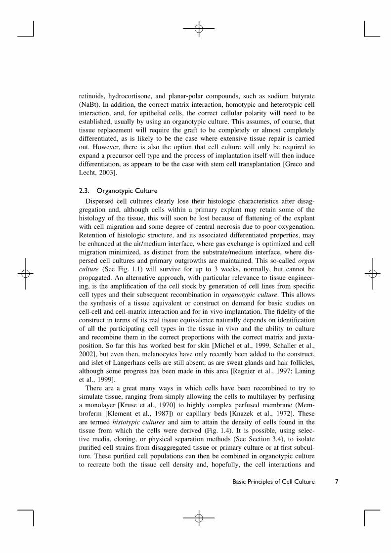
2.3. Organotypic CultureDispersed cell cultures clearly lose their histologic characteristics after disag-
gregation and, although cells within a primary explant may retain some of thehistology of the tissue, this will soon be lost because of flattening of the explantwith cell migration and some degree of central necrosis due to poor oxygenation.Retention of histologic structure, and its associated differentiated properties, maybe enhanced at the air/medium interface, where gas exchange is optimized and cellmigration minimized, as distinct from the substrate/medium interface, where dis-persed cell cultures and primary outgrowths are maintained. This so-called organculture (See Fig. 1.1) will survive for up to 3 weeks, normally, but cannot bepropagated. An alternative approach, with particular relevance to tissue engineer-ing, is the amplification of the cell stock by generation of cell lines from specificcell types and their subsequent recombination in organotypic culture. This allowsthe synthesis of a tissue equivalent or construct on demand for basic studies oncell-cell and cell-matrix interaction and for in vivo implantation. The fidelity of theconstruct in terms of its real tissue equivalence naturally depends on identificationof all the participating cell types in the tissue in vivo and the ability to cultureand recombine them in the correct proportions with the correct matrix and juxta-position. So far this has worked best for skin [Michel et al., 1999, Schaller et al.,2002], but even then, melanocytes have only recently been added to the construct,and islet of Langerhans cells are still absent, as are sweat glands and hair follicles,although some progress has been made in this area [Regnier et al., 1997; Laninget al., 1999].
There are a great many ways in which cells have been recombined to try tosimulate tissue, ranging from simply allowing the cells to multilayer by perfusinga monolayer [Kruse et al., 1970] to highly complex perfused membrane (Mem-broferm [Klement et al., 1987]) or capillary beds [Knazek et al., 1972]. Theseare termed histotypic cultures and aim to attain the density of cells found in thetissue from which the cells were derived (Fig. 1.4). It is possible, using selec-tive media, cloning, or physical separation methods (See Section 3.4), to isolatepurified cell strains from disaggregated tissue or primary culture or at first subcul-ture. These purified cell populations can then be combined in organotypic cultureto recreate both the tissue cell density and, hopefully, the cell interactions and
Basic Principles of Cell Culture 7
�
� �

�
Heterogeneous primary culture
Isolate by cloning,
selective media, and/or cell
sorting
Perfused multilayer from monolayer
Sponge or scaffold
Perfused capillary bed
Spheroid or organoid
Filter well insert
Propagate and seed on desired substrate. Grow to high cell density with
medium change' stirring, or perfusion
Expand each purified line separately
Combine in filter well inserts, transmembrane,
with or without matrix
Combine in three-dimensional array in concentric capillaries
ORGANOTYPIC CULTURE
HISTOTYPIC CULTURE
Figure 1.4. Histotypic and organotypic culture. Indicates the heterogeneity of a primary culture (top left),how this might be purified to give defined cell populations, which, if expanded and seeded into appropriateconditions can give high-density cultures of one cell type in perfused multilayers (top right), spheroidsor organoids in stirred suspension (second top right), three-dimensional multilayers in perfused capillaries(third top right), or monolayers or multilayers in filter well inserts (bottom right). Expansion of purifiedpopulations and recombination can generate organotypic cultures, in filter well inserts (bottom left) or onconcentric microcapillaries (bottom center). This last seems to be suggested by the architecture of the device(CellGro Triac), but the author has no knowledge of its use in this capacity.
8 Chapter 1. Freshney
�
� �
�
matrix generation found in the tissue (See Fig. 1.4). Filter well inserts provide thesimplest model system to test such recombinants, but there are many other pos-sibilities including porous matrices, perfused membranes, and concentric doublemicrocapillaries (Triac hollow fiber modules, [www.spectrapor.com/1/1/9.html]).
2.4. Substrates and MatricesInitially, cultures were prepared on glass for ease of observation, but cells may
be made to grow on many different charged surfaces including metals and manypolymers. Traditionally, a net negative charge was preferred, such as found on acid-washed glass or polystyrene treated by electric ion discharge, but some plastics arealso available with a net positive charge (e.g., Falcon Primaria), which is claimed toadd some cell selectivity. In either case, it is unlikely that the cell attaches directlyto synthetic substrates and more likely that the cell secretes matrix products thatadhere to the substrate and provide ligands for the interaction of matrix receptorssuch as integrins. Hence it is a logical step to treat the substrate with a matrixproduct, such as collagen type IV, fibronectin, or laminin, to promote the adhesionof cells that would otherwise not attach.
The subject of scaffolds will be dealt with in detail in later chapters (See Part II).Suffice it to say at this stage that scaffolds have the same requirements as conven-tional substrates in terms of low toxicity and ability to promote cell adhesion, oftenwith the additional requirement of a three-dimensional geometry. If the polymer orother material does not have these properties, derivatization and/or matrix coatingwill be required.
Most studies suggest that cell cultivation on a three-dimensional scaffold isessential for promoting orderly regeneration of engineered tissues in vivo andin vitro. Scaffolds investigated to date vary with respect to material chemistry(e.g., collagen, synthetic polymers), geometry (e.g., gels, fibrous meshes, poroussponges, tubes), structure (e.g., porosity, distribution, orientation, and connectivityof the pores), physical properties (e.g., compressive stiffness, elasticity, conductiv-ity, hydraulic permeability), and degradation (rate, pattern, products).
In general, scaffolds should be made of biocompatible materials, preferentiallythose already approved for clinical use. Scaffold structure determines the transportof nutrients, metabolites, and regulatory molecules to and from the cells, whereasthe scaffold chemistry may have an important role in cell attachment and differ-entiation. The scaffold should biodegrade at the same rate as the rate of tissueassembly and without toxic or inhibitory products. Mechanical properties of thescaffold should ideally match those of the native tissue being replaced, and themechanical integrity should be maintained as long as necessary for the new tissueto mature and integrate.
3. ISOLATION OF CELLS FOR CULTURE
3.1. Tissue Collection and TransportationThe first, and most important, element in the collection of tissue is the cooper-
ation and collaboration of the clinical staff. This is best achieved if a member of
Basic Principles of Cell Culture 9
�
� �

�
the surgical team is also a member of the culture project, but even in the absenceof this, time and care must be spent to ensure the sympathy and understanding ofthose who will provide the clinical material. It is worth preparing a short handoutexplaining the objectives of the project and spending some time with the per-son likely to be most closely involved with obtaining samples. This may be thechief surgeon (who will need to be informed anyway), or it may be a more juniormember of the team willing to set up a collaboration, one of the nursing staff, orthe pathologist, who may also require part of the tissue. Whoever fulfils this roleshould be identified and provided with labeled containers of culture medium con-taining antibiotics, bearing a contact name and phone number for the cell culturelaboratory. A refrigerator should be identified where the containers can be stored,and the label should also state clearly DO NOT FREEZE! The next step is bestcarried out by someone from the laboratory collecting the sample personally, but itis also possible to leave instructions for transportation by taxi or courier. If a thirdparty is involved, it is important to ensure that the container is well protected [See,for example, www.ehs.ucsf.edu/Safety%20Updates/Bsu/Bsu5.pdf], preferably dou-ble wrapped in a sealed polythene bag and an outer padded envelope provided withthe name, address, and phone number of the recipient at the laboratory. Refrigera-tion during transport is not usually necessary, as long as the sample is not allowedto get too warm, but if delivery will take more than an hour or two, then one ortwo refrigeration packs, such as used in picnic chillers, should be included but keptout of direct contact.
If the tissue sample is quite small, a further tissue sample (any tissue) or a bloodsample should be obtained for freezing. This will be used ultimately to corroboratethe origin of any cell line that is derived from the sample by DNA profiling. Acell line is the culture that is produced from subculture of the primary, and everyadditional subculture after this increases the possibility of cross-contamination,so verification of origin is important (See Section 6). In addition, the possibil-ity of misidentification arises during routine subculture and after recovery fromcryopreservation (See Section 5).
3.2. Biosafety and EthicsAll procedures involved in the collection of human material for culture must be
passed by the relevant hospital ethics committee. A form will be required for thepatient to sign authorizing research use of the tissue, and preferably disclaiming anyownership of any materials derived from the tissue [Freshney, 2002, 2005]. Theform should have a brief layman’s description of the objectives of the work and thename of the lead scientist on the project. The donor should be provided with a copy.
All human material should be regarded as potentially infected and treated withcaution. Samples should be transported securely in double-wrapped waterproofcontainers; they and derived cultures should be handled in a Class II biosafetycabinet and all discarded materials autoclaved, incinerated, or chemically disin-fected. Each laboratory will its own biosafety regulations that should be adheredto, and anyone in any doubt about handling procedures should contact the local
10 Chapter 1. Freshney
�
� �

�
safety committee (and if there is not one, create it!). Rules and regulations varyamong institutions and countries, so it is difficult to generalize, but a good reviewcan be obtained in Caputo [1996].
3.3. Record KeepingWhen the sample arrives at the laboratory, it should be entered into a record
system and assigned a number. This record should contain the details of the donor,identified by hospital number rather than by name, tissue site, and all informa-tion regarding collection medium, time in transit, treatment on arrival, primarydisaggregation, and culture details, etc. [Freshney, 2002, 2005]. This informationwill be important in the comparison of the success of individual cultures, and if along-term cell line is derived from the culture, this will be the first element in thecell line’s provenance, which will be supplemented with each successive manipu-lation or experimental procedure. Such records are best maintained in a computerdatabase where each record can be derived from duplication of the previous recordwith appropriate modifications. There may be issues of data protection and patientconfidentiality to be dealt with when obtaining ethical consent.
3.4. Disaggregation and Primary CultureDetailed information on disaggregation as a method for obtaining cells is pro-
vided in the appropriate chapters. Briefly, the tissue will go through stages ofrinsing, dissection, and either mechanical disaggregation or enzymatic digestion intrypsin and/or collagenase. It is often desirable not to have a complete single-cellsuspension, and many primary cells survive better in small clusters. Disaggregatedtissue will contain a variety of different cell types, and it may be necessary to gothrough a separation technique [See Chapter 15, Freshney 2005], such as densitygradient separation [Pretlow and Pretlow, 1989] or immunosorting by magnetizablebeads (MACS), using a positive sort to select cells of interest [Carr et al., 1999] ora negative sort to eliminate those that are not required [Saalbach et al., 1997], orby using fluorescence-activated cell sorting (FACS) [See, e.g. Swope et al., 1997].The cell population can then be further enriched by selection of the correct medium(e.g., keratinocyte growth medium (KGM) or MCDB 153 for keratinocytes [Peehland Ham, 1980]), many of which are now available commercially (See Sources ofMaterials), and supplementing this with growth factors. Survival and enrichmentmay be improved in some cases by coating the substrate with gelatin, collagen,laminin, or fibronectin [Freshney, 2005].
4. SUBCULTURE
Frequently, the number of cells obtained at primary culture may be insufficient tocreate constructs suitable for grafting. Subculture gives the opportunity to expandthe cell population, apply further selective pressure with a selective medium, andachieve a higher growth fraction and allows the generation of replicate cultures for
Basic Principles of Cell Culture 11
�
� �
�
characterization, preservation by freezing, and experimentation. Briefly, subcultureinvolves the dissociation of the cells from each other and the substrate to generatea single-cell suspension that can be quantified. Reseeding this cell suspension ata reduced concentration into a flask or dish generates a secondary culture, whichcan be grown up and subcultured again to give a tertiary culture, and so on. Inmost cases, cultures dedifferentiate during serial passaging but can be induced toredifferentiate by cultivation on a 3D scaffold in the presence of tissue-specificdifferentiation factors (e.g., growth factors, physical stimuli). However, the cell’sability to redifferentiate decreases with passaging. It is thus essential to determine,for each cell type, source, and application, a suitable number of passages duringsubculture. Protocols for subculture of specific cell types are given in later chapters,and a more general protocol is available in Chapter 13, Freshney [2005].
4.1. Life SpanMost normal cell lines will undergo a limited number of subcultures, or passages,
and are referred to as finite cell lines. The limit is determined by the number ofdoublings that the cell population can go through before it stops growing becauseof senescence. Senescence is determined by a number of intrinsic factors regulatingcell cycle, such as Rb and p53 [Munger and Howley, 2002], and is accompanied byshortening of the telomeres on the chromosomes [Wright and Shay, 2002]. Once thetelomeres reach a critical minimum length, the cell can no longer divide. Telomerelength is maintained by telomerase, which is downregulated in most normal cellsexcept germ cells. It can also be higher in stem cells, allowing them to go througha much greater number of doublings and avoid senescence. Transfection of thetelomerase gene hTRT into normal cells with a finite life span allows a smallproportion of the cells to become immortal [Bodnar et al., 1998; Protocol 18.2,Freshney, 2005], although this probably involves deletion or inactivation of othergenes such as p53 and myc [Cerni, 2000].
4.2. Growth CycleEach time that a cell line is subcultured it will grow back to the cell density that
existed before subculture (within the limits of its finite life span). This process canbe described by plotting a growth curve from samples taken at intervals throughoutthe growth cycle (Fig. 1.5), which shows that the cells enter a latent period of nogrowth, called the lag period, immediately after reseeding. This period lasts froma few hours up to 48 h, but is usually around 12–24 h, and allows the cells torecover from trypsinization, reconstruct their cytoskeleton, secrete matrix to aidattachment, and spread out on the substrate, enabling them to reenter cell cycle.They then enter exponential growth in what is known as the log phase, during whichthe cell population doubles over a definable period, known as the doubling timeand characteristic for each cell line. As the cell population becomes crowded whenall of the substrate is occupied, the cells become packed, spread less on the sub-strate, and eventually withdraw from the cell cycle. They then enter the plateau orstationary phase, where the growth fraction drops to close to zero. Some cells may
12 Chapter 1. Freshney
�
� �

�
0 2 4 6 8 10104
105
106
PlateauLogLag
Cel
ls /
ml
Days from subculture
Medium change
Next subculture
Subculture & seeding
0 2 4 6 8 10
Cel
ls m
l−1
104
105
104
105
104
Cells cm
−2
Saturation density
Doubling time
Lag
(a)
(b)
Figure 1.5. Growth curve. Increase in cell number on a log scale plotted against days from subculture.a) Defines the lag, log (exponential), and plateau phases, and when culture should be fed and subculturedafter the indicated seeding time. b) Shows the kinetic parameters that can be derived from the growth curve:lag from the intercept between a line drawn through the points on the exponential phase and the horizontalfrom the seeding concentration; doubling time from the time taken, in the middle of the exponential phase,for the cell population to double; saturation density from the maximum (stable) cell density achieved bythe culture, under the prevailing culture conditions. This is determined in cells/cm2 (cell density ratherthan cell concentration) and is not absolute, as it will vary with culture conditions. It is best determined(as characteristic of the cell type) in conditions that are nonlimiting for medium, e.g., a small area ofhigh-density cells in a large reservoir of medium (such as a coverslip, or a filter well insert, in a non-tissueculture-grade dish) or under continuous perfusion of medium. [Adapted from Freshney, 2005.]
differentiate in this phase; others simply exit the cell cycle into G0 but retain viabil-ity. Cells may be subcultured from plateau, but it is preferable to subculture beforeplateau is reached, as the growth fraction will be higher and the recovery time (lagperiod) will be shorter if the cells are harvested from the top end of the log phase.
Reduced proliferation in the stationary phase is due partly to reduced spreadingat high cell density and partly to exhaustion of growth factors in the medium athigh cell concentration. These two terms are not interchangeable. Density impliesthat the cells are attached, and may relate to monolayer density (two-dimensional)
Basic Principles of Cell Culture 13
�
� �
�
or multilayer density (three-dimensional). In each case there are major changes incell shape, cell surface, and extracellular matrix, all of which will have signifi-cant effects on cell proliferation and differentiation. A high density will also limitnutrient perfusion and create local exhaustion of peptide growth factors [Stoker,1973; Westermark and Wasteson, 1975]. In normal cell populations this leads to awithdrawal from the cycle, whereas in transformed cells, cell cycle arrest is muchless effective and the cells tend to enter apoptosis.
Cell concentration, as opposed to cell density, will exert its main effect throughnutrient and growth factor depletion, but in stirred suspensions cell contact-mediated effects are minimal, except where cells are grown as aggregates. Cellconcentration per se, without cell interaction, will not influence proliferation, otherthan by the effect of nutrient and growth factor depletion. High cell concentrationscan also lead to apoptosis in transformed cells in suspension, notably in myelomasand hybridomas, but in the absence of cell contact signaling this is presumably areflection of nutrient deprivation.
4.3. Serial SubcultureEach time the culture is subcultured the growth cycle is repeated. The number
of doublings should be recorded (Fig. 1.6) with each subculture, simplified byreducing the cell concentration at subculture by a power or two, the so-called splitratio. A split ratio of two allows one doubling per passage, four, two doublings,eight, three doublings, and so on (See Fig. 1.6). The number of elapsed doublingsshould be recorded so that the time to senescence (See Section 4.1) can be predictedand new stock prepared from the freezer before the senescence of the existingculture occurs.
5. CRYOPRESERVATION
If a cell line can be expanded sufficiently, preservation of cells by freezingwill allow secure stocks to be maintained without aging and protect them fromproblems of contamination, incubator failure, or medium and serum crises. Ideally,1 × 106 –1 × 10⁷ cells should be frozen in 10 ampoules, but smaller stocks can beused if a surplus is not available. The normal procedure is to freeze a token stockof one to three ampoules as soon as surplus cells are available, then to expandremaining cultures to confirm the identity of the cells and absence of contamination,and freeze down a seed stock of 10–20 ampoules. One ampoule, thawed from thisstock, can then be used to generate a using stock. In many cases, there may not besufficient doublings available to expand the stock as much as this, but it is worthsaving some as frozen stock, no matter how little, although survival will tend todecrease below 1 × 10⁶ cells/ml and may not be possible below 1 × 10⁵ cells/ml.
Factors favoring good survival after freezing and thawing are:
(i) High cell density at freezing (1 × 10⁶ –1 × 10⁷ cells/ml).
(ii) Presence of a preservative, such as glycerol or dimethyl sulfoxide (DMSO)at 5–10%.
14 Chapter 1. Freshney
�
� �

�
0 5 10 15 20 25104
105
106
Split ratio= 8
Subculture interval
2x
987654321Generation number
321Passage number
Cel
ls/c
m2
Days from subculture
Figure 1.6. Serial subculture. Recurring growth curves during serial subculture, not necessarily recordedby daily cell counts, but predicted from one or two detailed growth curve analyses of these cells. Eachcycle should be a replicate of the previous one, such that the same terminal cell density is achieved aftersubculture at the same seeding concentration. The lower number represents the passage number, i.e., thenumber of times the culture has been subcultured. The upper numbers represent the generation number,i.e., the number of times the population has doubled. In this example, the cell population doubles threetimes between each subculture, suggesting that the culture should be split 1:8 to regain the same seedingconcentration each time. [From Freshney, 2005.]
(iii) Slow cooling, 1 ◦C/min, down to −70 ◦C and then rapid transfer to a liquidnitrogen freezer.
(iv) Rapid thawing.
(v) Slow dilution, ∼20-fold, in medium to dilute out the preservative.
(vi) Reseeding at 2- to 5-fold the normal seeding concentration. For example,if cells are frozen at 5 × 10⁶ cells in 1 ml of freezing medium with 10%DMSO and then thawed and diluted 1:20, the cell concentration will still be2.5 × 105 cells/ml at seeding, higher than the normal seeding concentrationfor most cell lines, and the DMSO concentration will be reduced to 0.5%,which most cells will tolerate for 24 h.
(vii) Changing medium the following day (or as soon as all the cells haveattached) to remove preservative. Where cells are more sensitive to thepreservative, they may be centrifuged after slow dilution and resuspendedin fresh medium, but this step should be avoided if possible as centrifugationitself may be damaging to freshly thawed cells.
There are differences of opinion regarding some of the conditions for freezingand thawing, for example, whether cells should be chilled when DMSO is added ordiluted rapidly on thawing, both to avoid potential DMSO toxicity. In the author’sexperience, chilling diminishes the effect of the preservative, particularly withglycerol, and rapid dilution reduces survival, probably due to osmotic shock. Cul-turing in diluted DMSO after thawing can be a problem for some cell lines if they
Basic Principles of Cell Culture 15
�
� �

�
respond to the differentiating effects of DMSO, for example, myeloid leukemiacells, neuroblastoma cells, and embryonal stem cells; in these cases it is preferableto centrifuge after slow dilution at thawing or use glycerol as a preservative.
6. CHARACTERIZATION AND VALIDATION
6.1. Cross-ContaminationThere has been much publicity about the very real risks of cross-contamination
when handling cell lines [Marcovic and Marcovic, 1998; Macleod et al., 1999;Masters et al., 2001; van Bokhoven et al., 2001; Masters, 2002], particularly con-tinuous cell lines. This is less of a problem with short-term cultures, but therisk remains that if there are other cell lines in use in the laboratory, they cancross-contaminate even a primary culture, or misidentification can arise duringsubculture or recovery from the freezer. If a laboratory focuses on one particularhuman cell type, superficial observation of lineage characteristics will be inade-quate to ensure the identity of each line cultured. Precautions must be taken toavoid cross-contamination:
(i) Do not handle more than one cell line at a time, or, if this is impractical,do not have culture vessels and medium bottles for more than one cell lineopen at one time, and never be tempted to use the same pipette or otherdevice for different cell lines.
(ii) Do not share media or other reagents among different cell lines.
(iii) Do not share media or reagents with other people.
(iv) Ensure that any spillage is mopped up immediately and the area swabbedwith 70% alcohol.
(v) Retain a tissue or blood sample from each donor and confirm the identity ofeach cell line by DNA profiling: (a) when seed stocks are frozen, (b) beforethe cell line is used for experimental work or transplantation.
(vi) Keep a panel of photographs of each cell line, at low and high densities,above the microscope, and consult this regularly when examining cells dur-ing maintenance. This is particularly important if cells are handled over anextended period, and by more than one operator.
(vii) If continuous cell lines are in use in the laboratory, handle them after handingother, slower-growing, finite cell lines.
6.2. Microbial ContaminationAntibiotics are often used during collection, transportation, and dissection of
biopsy samples because of the intrinsic contamination risk of these operations.However, once the primary culture is established, it is desirable to eliminate
16 Chapter 1. Freshney
�
� �
�
antibiotics as soon as possible. If the culture grows well, then antibiotics canbe removed from the bulk of the stocks at first subculture, retaining one culturein antibiotics as a precaution if necessary. Antibiotics can lead to lax aseptic tech-nique, can inhibit some eukaryotic cellular processes, and can hide the presence ofa microbial contamination. If a culture is contaminated this must become apparentas soon as possible, either to indicate that the culture should be discarded before itcan spread the contamination to other cultures or to indicate that decontaminationshould be attempted. The latter should only be used as a last resort; decontamina-tion is not always successful and can lead to the development of antibiotic-resistantorganisms.
Most bacterial, fungal, and yeast infections are readily detected by regular care-ful examination with the naked eye (e.g., by a change in the color of culturemedium) and on the microscope. However, one of the most serious contamina-tions is mycoplasma, which is not visible by routine microscopy. Any cell culturelaboratory should have a mycoplasma screening program in operation, but thosecollecting tissue for primary culture are particularly at risk. The precautions thatshould be observed are as follows:
(i) Treat any new material entering the laboratory from donors or from otherlaboratories as potentially infected and keep it in quarantine. Ideally, a sep-arate room should be set aside for receiving samples and imported cultures.If this is not practicable, handle separately from other cultures, preferablylast in the day and in a designated hood, and swab the hood down after usewith 2% phenolic disinfectant in 70% alcohol. Use a separate incubator, andadhere strictly to the rules given above regarding medium sharing.
(ii) Screen new cultures as they arrive, and existing stocks at regular intervals,e.g., once a month. There are a number of tests available, but the mostreliable and sensitive are fluorescence microscopy after staining with Hoechst33258 [Chen et al., 1977; Protocol 19.2, Freshney, 2005] or PCR with a pan-mycoplasma primer [Uphoff and Drexler, 2002a; Protocol 19.3, Freshney,2005]. The latter is more sensitive but depends on the availability of PCRtechnology, whereas the former is easier, will detect any DNA-containingcontamination, but requires a fluorescence microscope. Both techniques arebest performed with so-called indicator cultures. The test culture is refreshedwith antibiotic-free medium and, after 3–5 days, the medium is transferredto an antibiotic-free, 10% confluent indicator culture of a cell line such as3T6 or A549 cells, which are well spread and known to support mycoplasmagrowth. After a further 3 days (the indicator cells must not be allowed toreach confluence) the indicator culture is fixed in acetic methanol and stainedwith Hoechst 33258, or harvested by scraping for PCR (trypsinization mayremove the mycoplasma from the cell surface).
(iii) Discard all contaminated cultures. If the culture is irreplaceable, decontam-ination may be attempted (under strict quarantine conditions) with agents
Basic Principles of Cell Culture 17
�
� �

�
such as Mycoplasma Removal Agent (MRA), ciprofloxacin, or BM-cycline[Uphoff and Drexler, 2002b]. Briefly, the culture is rinsed thoroughly, tryp-sinized (wash by centrifuging three times after trypsinization), and subcul-tured into antibiotic-containing medium. This procedure should be repeatedfor three subcultures and then the culture should be grown up antibiotic-free and tested after one, two, and four further antibiotic-free subcultures,whereupon the culture reenters the routine mycoplasma screening program.
6.3. CharacterizationMost laboratories will have, as an integral part of the research program, pro-
cedures in place for the characterization of new cultures. Species identification(in case the cells are misidentified or cross-contaminated) will be unnecessary ifDNA profiling is being used to confirm cell line identity; otherwise, chromosomeanalysis or isoenzyme electrophoresis [Hay et al., 2000] can be used. The lineageor tissue or origin can be determined by using antibodies to intermediate filamentproteins, for example, cytokeratins for epithelial cells, vimentin for mesodermalcells such as fibroblasts, endothelium, and myoblasts, desmin for myocytes, neu-rofilament protein for neuronal and some neuroendocrine cells, and glial fibrillaryacidic protein for astrocytes. Some cell surface markers are also lineage specific,for example, EMA in epithelial cells, A2B5 in glial cells, PECAM-1 in endothelialcells, and N-CAM in neural cells, and have the additional advantage that they canbe used in cell sorting by magnetic sorting or flow cytometry. Morphology canalso be used, but can be ambivalent as similarities can exist between cells of verydifferent origins.
Spontaneous transformation is unlikely in normal cells of human origin, butindicators are a more refractile appearance under phase contrast with a lowercytoplasmic/nuclear ratio, piling up of the cells and loss of contact inhibition anddensity limitation of growth, increased clonogenicity in agar, and the ability toform tumors in immune-deprived hosts, such as the Nude or SCID mouse. Wheretransformation is detected, it is more likely to be due to cross-contamination,although it is possible that the tissue sample may have contained some preneoplasticcells that have then progressed in culture.
6.4. DifferentiationAs stated above, a prerequisite for sustained growth in culture is the ability for
the cells to proliferate, and this may preclude differentiation. If differentiation isrequired, then it is generally necessary for the cells to withdraw from the cell cycle.This can be achieved by removing, or changing, the growth factor supplementa-tion; for example, the O2A common precursor of astrocytes and oligodendrocytesremains as a proliferating precursor cell in PDGF and bFGF, whereas combin-ing bFGF with ciliary neurotropic factor (CNTF) results in differentiation into atype 2 astrocyte [Raff, 1990], and embryonal stem cells, which remain as prolif-erating primitive cells in the presence of leukemia inhibitory factor (LIF), will
18 Chapter 1. Freshney
�
� �
�
differentiate in the absence of LIF and in the presence of a positively acting factorsuch as phorbol myristate acetate (PMA, also known as TPA) [Rizzino, 2002].
There are four main parameters governing the entry of cells into differentiation:
(i) Soluble factors such as growth factors (e.g., EGF, KGF, TGF-β and HGF,NGF), cytokines (IL-6, oncostatin-M, GM-CSF, interferons), vitamins (e.g.,retinoids, vitamin D3, and vitamin K) and calcium [Table 16.1, Freshney,2000], and planar polar compounds (e.g., DMSO and NaBt) [Tables 17.1,17.2, Freshney, 2005].
(ii) Interaction with matrix constituents such as collagen IV, laminin, and pro-teoglycans. Heparan sulfate proteoglycans (HSPGs), in particular, have asignificant role not only in binding to cell surface receptors but also inbinding and translocating growth factors and cytokines to high-affinity cellsurface receptors [Lopez-Casillas et al., 1993; Filla, 1998].
(iii) Enhanced cell-cell interaction will also promote differentiation. Homotypiccontact interactions can act via gap junctions, which tend to coordinate theresponse among many like cells in a population by allowing free intercellularflow of second messenger molecules such as cyclic adenosine monophos-phate (cAMP) and via cell adhesion molecules such as E-cadherin or N-CAM, which signal via anchorage to the cytoskeleton [Juliano, 2002]. Het-erotypic interactions, in solid tissues at least, will tend to act across a basallamina and are less likely to involve direct cell-cell contact. Signaling isachieved by, on the one hand, modification of the matrix by the mutual con-tribution of both cell types, and, on the other, by reciprocal transmission ofcytokines and growth factors across the basal lamina, such as the transfer ofKGF and GM-CSF from dermal fibroblasts to the basal layer of the epider-mis in response to IL-1α and -β diffusing from the epidermis to the dermalfibroblasts [Maas-Szabowski et al., 2002].
(iv) The position, shape, and polarity of the cells may induce, or at least makethe cells permissive for the induction of, differentiation. Epidermal ker-atinocytes [Maas-Szabowski et al., 2002] and bronchial epithelial cells [Petraet al., 1993] require to be close to the air/liquid interface, presumably toenhance oxygen availability, and secretory cells, such as thyroid epithelium,need the equivalent of the acinar space, that is, no direct access to nutri-ent or hormones, above them in a thin fluid space [Chambard et al., 1983,1987]. When cells are grown on collagen at this location, and the collagengel is allowed to retract, a shape change can occur, for example, from aflat squamous or cuboidal cell into a more columnar morphology, and this,combined with matrix interaction, allows the establishment of polarity inthe cells, such that secretory products are released apically and signalingreceptors and nutrient transporters locate basally.
Combining these effects in vitro may require strict attention to culture geometry,for example, by growing cells in a filter well insert on a matrix incorporating
Basic Principles of Cell Culture 19
�
� �
�
stromal fibroblasts, and providing differentiation inducers basally in a defined,nonmitogenic medium. Similar conditions may be created in a perfused capillarybed or scaffold.
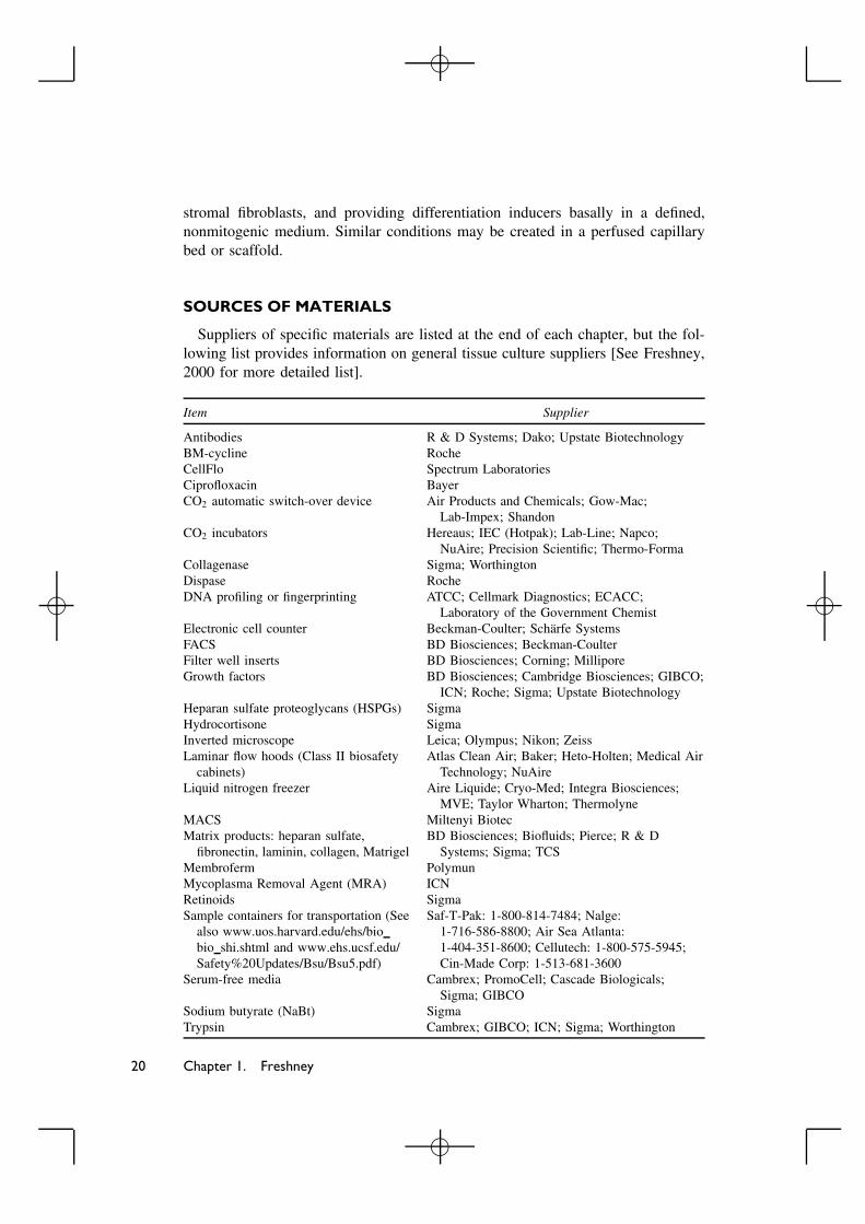
SOURCES OF MATERIALS
Suppliers of specific materials are listed at the end of each chapter, but the fol-lowing list provides information on general tissue culture suppliers [See Freshney,2000 for more detailed list].
Item Supplier
Antibodies R & D Systems; Dako; Upstate BiotechnologyBM-cycline RocheCellFlo Spectrum LaboratoriesCiprofloxacin BayerCO2 automatic switch-over device Air Products and Chemicals; Gow-Mac;
Lab-Impex; ShandonCO2 incubators Hereaus; IEC (Hotpak); Lab-Line; Napco;
NuAire; Precision Scientific; Thermo-FormaCollagenase Sigma; WorthingtonDispase RocheDNA profiling or fingerprinting ATCC; Cellmark Diagnostics; ECACC;
Laboratory of the Government ChemistElectronic cell counter Beckman-Coulter; Scharfe SystemsFACS BD Biosciences; Beckman-CoulterFilter well inserts BD Biosciences; Corning; MilliporeGrowth factors BD Biosciences; Cambridge Biosciences; GIBCO;
ICN; Roche; Sigma; Upstate BiotechnologyHeparan sulfate proteoglycans (HSPGs) SigmaHydrocortisone SigmaInverted microscope Leica; Olympus; Nikon; ZeissLaminar flow hoods (Class II biosafety
cabinets)Atlas Clean Air; Baker; Heto-Holten; Medical Air
Technology; NuAireLiquid nitrogen freezer Aire Liquide; Cryo-Med; Integra Biosciences;
MVE; Taylor Wharton; ThermolyneMACS Miltenyi BiotecMatrix products: heparan sulfate,
fibronectin, laminin, collagen, MatrigelBD Biosciences; Biofluids; Pierce; R & D
Systems; Sigma; TCSMembroferm PolymunMycoplasma Removal Agent (MRA) ICNRetinoids SigmaSample containers for transportation (See
also www.uos.harvard.edu/ehs/biobio shi.shtml and www.ehs.ucsf.edu/Safety%20Updates/Bsu/Bsu5.pdf)
Saf-T-Pak: 1-800-814-7484; Nalge:1-716-586-8800; Air Sea Atlanta:1-404-351-8600; Cellutech: 1-800-575-5945;Cin-Made Corp: 1-513-681-3600
Serum-free media Cambrex; PromoCell; Cascade Biologicals;Sigma; GIBCO
Sodium butyrate (NaBt) SigmaTrypsin Cambrex; GIBCO; ICN; Sigma; Worthington
20 Chapter 1. Freshney
�
� �

�
REFERENCES
Bodnar, A.G., Ouellette, M., Frolkis, M., Holt, S.E., Chiu, C.-P., Morin, G.B., Harley, C.B.,Shay, J.W., Lichsteiner, S., Wright, W.E. (1998) Extension of life-span by introduction oftelomerase into normal human cells. Science 279: 349–352.
Caputo, J.L. (1996) Safety Procedures. In Freshney, R.I., Freshney, M.G., eds., Culture of Im-mortalized Cells. New York, Wiley-Liss, pp. 25–51.
Carr, T., Evans, P., Campbell, S., Bass, P., Albano, J. (1999) Culture of human renal tubularcells: positive selection of kallikrein-containing cells. Immunopharmacology 44: 161–167.
Cerni, C. (2000) Telomeres, telomerase, and myc. An update. Mutat Res. 462: 31–47.Chambard, M., Vemer, B., Gabrion, J., Mauchamp, J., Bugeia, J.C., Pelassy, C., Mercier, B.
(1983) Polarization of thyroid cells in culture; evidence for the basolateral localization ofthe iodide “pump” and of the thyroid-stimulating hormone receptor-adenyl cyclase complex.J. Cell Biol. 96: 1172–1177.
Chambard, M., Mauchamp, J., Chaband, O. (1987) Synthesis and apical and basolateral secre-tion of thyroglobulin by thyroid cell monolayers on permeable substrate: Modulation bythyrotropin. J. Cell Physiol. 133: 37–45.
Chen, T.R. (1977) In situ detection of mycoplasma contamination in cell cultures by fluorescentHoechst 33258 stain. Exp. Cell Res. 104: 255.
Filla, M.S., Dam, P., Rapraeger, A.C. (1998) The cell surface proteoglycan syndecan-1 mediatesfibroblast growth factor-2 binding and activity. J. Cell Physiol. 174: 310–321.
Freshney, R.I. (2002) Cell line provenance. Cytotechnology 39: 3–15.Freshney, R.I. (2005) Culture of Animal Cells, a Manual of Basic Technique, 5th Ed. Hoboken
NJ, John Wiley & Sons.Greco, B., Recht, L. (2003) Somatic plasticity of neural stem cells: Fact or fancy? J. Cell.
Biochem. 88: 51–56.Ham, R.G., McKeehan, W.L. (1978) Development of improved media and culture conditions
for clonal growth of normal diploid cells. In Vitro 14: 11–22.Hay, R.J., Miranda-Cleland, M., Durkin, S., Reid, Y.A. (2000) Cell line preservation and authen-
tication. In Masters, J.R.W, ed., Animal Cell Culture, 3rd Ed. Oxford, Oxford University Press,pp. 69–103.
Juliano, R.L. (2002) Signal transduction by cell adhesion receptors and the cytoskeleton: func-tions of integrins, cadherins, selectins, and immunoglobulin-superfamily members. Annu. Rev.Pharmacol. Toxicol. 42: 283–323.
Karmiol, S. (2000) Development of serum-free media. In Masters, J.R.W, ed., Animal Cell Cul-ture, 3rd Ed. Oxford, Oxford University Press, pp. 105–121.
Klement, G., Scheirer, W., Katinger, H.W. (1987) Construction of a large scale membrane reac-tor system with different compartments for cells, medium and product. Dev Biol Stand., 66:221–226.
Knazek, R.A., Gullino, P., Kohler, P.O., Dedrick, R. (1972) Cell culture on artificial capillaries.An approach to tissue growth in vitro. Science 178: 65–67.
Kruse, P.F., Jr., Keen, L.N., Whittle, W.L. (1970) Some distinctive characteristics of high den-sity perfusion cultures of diverse cell types. In Vitro 6: 75–78.
Laning, J.C., DeLuca, J.E., Hardin-Young, J. (1999) Effects of immunoregulatory cytokines onthe immunogenic potential of the cellular components of a bilayered living skin equivalent.Tissue Eng. 5: 171–181.
Lopez-Casillas, F., Wrana, J.L., Massague, J. (1993) Betaglycan presents ligand to the TGF-βsignalling receptor. Cell 73: 1435–1444.
Maas-Szabowski, N., Stark, H.-J., Fusenig, N.E. (2002) Cell interaction and differentiation. InFreshney. R.I. and Freshney. M.G., eds, Culture of Epithelial Cells, 2nd Ed. New York, JohnWiley & Sons, pp. 31–63.
MacLeod, R.A., Dirks, W.G., Matsuo, Y., Kaufmann, M., Milch, H., Drexler, H.G. (1999)Widespread intraspecies cross-contamination of human tumor cell lines arising at source.Int. J. Cancer 83: 555–63.
Marcovic, O., Marcovic, N. (1998) Cell cross-contamination in cell cultures: the silent andneglected danger. In Vitro Cell Dev Biol. 34: 108.
Basic Principles of Cell Culture 21
�
� �

�
Masters, J.R. (2002) HeLa cells 50 years on: the good, the bad and the ugly. Nat. Rev. Cancer2: 315–9.
Masters, J.R.W., Thomson, J.A., Daly-Burns, B., Reid, Y.A., Dirks, W.G., Packer, P., Toji,L.H., Ohno, T., Tanabe, H., Arlett, C.F., Kelland, L.R., Harrison, M., Virmani, A., Ward,T.H., Ayres, K.L., Debenham, P.G. (2001) STR profiling provides an international reference
standard for human cell lines. Proc. Natl. Acad. Sci. USA 98: 8012–8017.Mather, J.P. (1998) Making informed choices: medium, serum, and serum-free medium. How to
choose the appropriate medium and culture system for the model you wish to create. MethodsCell Biol. 57: 19–30.
Michel, M., L’Heureux, N., Pouliot, R., Xu, W., Auger, F.A., Lucie, G. (1999) Characterizationof a new tissue-engineered human skin equivalent with hair. In Vitro Cell. Dev. Biol. Anim.35: 318–326.
Munger, K., Howley, P.M. (2002) Human papillomavirus immortalization and transformationfunctions. Virus Res. 89(2): 213–228.
Peehl, D.M., Ham, R.G. (1980) Clonal growth of human keratinocytes with small amounts ofdialysed serum. In Vitro 16: 526–540.
Petra, M., de Jong, P.M., van Sterkenburg, A.J.A., Kempenaar, J.A., Dijkman, J.H., Ponec, M.(1993) Serial culturing of human bronchial epithelial cells derived from biopsies. In Vitro CellDev. Biol. 29A: 379–387.
Pretlow, T.G., and Pretlow, T.P. (1989) Cell separation by gradient centrifugation methods.Methods Enzymol. 171: 462–482.
Raff, M.C. (1990) Glial cell diversification in the rat optic nerve. Science 243: 1450–1455.Regnier, M., Staquet, M.J., Schmitt, D., Schmidt, R. (1997) Integration of Langerhans cells into
a pigmented reconstructed human epidermis. J. Invest. Dermatol. 109: 510–512.Rizzino, A. (2002) Embryonic stem cells provide a powerful and versatile model system. Vitam
Horm. 64: 1–42.Saalbach, A., Aust, G., Haustein, U.F., Herrmann, K., Anderegg, U. (1997) The fibroblast-
specific MAb AS02: A novel tool for detection and elimination of human fibroblasts. CellTissue Res. 290: 593–599.
Schaller,. M., Mailhammer, R., Grassl, G., Sander, C.A., Hube, B., Korting, H.C. (2002) Infec-tion of human oral epithelia with Candida species induces cytokine expression correlated tothe degree of virulence. J. Invest. Dermatol. 118: 652–657.
Stoker, M.G.P. (1973) Role of diffusion boundary layer in contact inhibition of growth. Nature246: 200–203.
Swope, V.B., Supp, A.P., Cornelius, J.R., Babcock, G.F., Boyce, S.T. (1997) Regulation of pig-mentation in cultured skin substitutes by cytometric sorting of melanocytes and keratinocytes.J, Invest. Dermatol. 109: 289–295.
Uphoff, C.C., Drexler, H.G. (2002a) Comparative PCR analysis for detection of mycoplasmainfection in continuous cell lines. In Vitro Cell. Dev. Biol. Anim. 38: 79–85.
Uphoff, C.C., and Drexler, H.G. (2002b) Comparative antibiotic eradication of mycoplasmainfections from continuous cell lines. In Vitro Cell. Dev. Biol. Anim. 38: 86–89.
van Bokhoven, A., Varella-Garcia, M., Korch, C., Hessels, D., Miller, G.J. (2001). Widely usedprostate carcinoma cell lines share common origins. Prostate 47: 36–51.
Westermark, B., Wasteson, A. (1975) The response of cultured human normal glial cells togrowth factors. Adv. Metab. Disord. 8: 85–100.
Wright, W.E., Shay, J.W. (2002) Historical claims and current interpretations of replicativeaging. Nat. Biotechnol. 20: 682–688.
22 Chapter 1. Freshney
�
� �