Basic fibroblast growth factor reduces scar by inhibiting ...
13
RESEARCH Open Access Basic fibroblast growth factor reduces scar by inhibiting the differentiation of epidermal stem cells to myofibroblasts via the Notch1/Jagged1 pathway Peng Wang 1† , Bin Shu 1† , Yingbin Xu 1 , Jiayuan Zhu 1 , Jian Liu 1 , Ziheng Zhou 1 , Lei Chen 1 , Jingling Zhao 1 , Xusheng Liu 1 , Shaohai Qi 1 , Kun Xiong 2 and Julin Xie 1* Abstract Background: Basic fibroblast growth factor (bFGF) plays an important role in promoting wound healing and reducing scar, but the possible molecular mechanisms are still unclear. Our previous studies have found that activating the Notch1/Jagged1 pathway can inhibit the differentiation of epidermal stem cells (ESCs) to myofibroblasts (MFB). Herein, we document that bFGF reduces scar by inhibiting the differentiation of ESCs to MFB via activating the Notch1/Jagged1 pathway. Methods: In in-vitro study, ESCs were isolated from 10 neonatal SD rats (1–3 days old), cultured in keratinocyte serum-free medium, and divided into six groups: bFGF group, bFGF + SU5402 group, bFGF + DAPT group, siJagged1 group, bFGF + siJagged1 group, and control group. Jagged1 of the ESCs in the siJagged1 group and bFGF + siJagged1 group was knocked down by small-interfering RNA transfection. Expression of ESC markers (CK15/CK10), MFB markers (α-SMA, Collagen I, Collagen III), and Notch1/Jagged1 components (Jagged1, Notch1, Hes1) was detected by FCM, qRT-PCR, and western blot analysis to study the relationships of bFGF, ESCs, and Notch1/Jagged1 pathway. In in-vivo study, the wound healing time and scar hyperplasia were observed on rabbit ear scar models. The quality of wound healing was estimated by hematoxylin and eosin staining and Masson staining. Expression of ESC markers, MFB markers and Notch1/Jagged1 components was elucidated by immunohistochemistry, immunofluorescence, and western blot analysis. Results: The in-vitro study showed that bFGF could significantly upregulate the expression of ESC markers and Notch1/Jagged1 components, while downregulating the expression of MFB markers at the same time. However, these effects could be obviously decreased when we knocked down Jagged1 or added DAPT. Similarly, in in-vivo study, bFGF also exhibited its functions in inhibiting the differentiation of rabbit ESCs to MFB by activating the Notch1/Jagged1 pathway, which improved the wound healing quality and alleviated scar significantly. Conclusion: These results provide evidence that bFGF can reduce scar by inhibiting the differentiation of ESCs to MFB via the Notch1/Jagged1 pathway, and present a new promising potential direction for the treatment of scar. Keywords: Scar, Basic fibroblast growth factor, Epidermal stem cells, Myofibroblasts, Notch1/Jagged1 pathway * Correspondence: [email protected] † Equal contributors 1 Department of Burn Surgery, First Affiliated Hospital of Sun Yat-Sen University, No. 58, 2nd Zhongshan Road, Yuexiu District, Guangzhou, Guangdong Province 510080, People’s Republic of China Full list of author information is available at the end of the article © The Author(s). 2017 Open Access This article is distributed under the terms of the Creative Commons Attribution 4.0 International License (http://creativecommons.org/licenses/by/4.0/), which permits unrestricted use, distribution, and reproduction in any medium, provided you give appropriate credit to the original author(s) and the source, provide a link to the Creative Commons license, and indicate if changes were made. The Creative Commons Public Domain Dedication waiver (http://creativecommons.org/publicdomain/zero/1.0/) applies to the data made available in this article, unless otherwise stated. Wang et al. Stem Cell Research & Therapy (2017) 8:114 DOI 10.1186/s13287-017-0549-7
Transcript of Basic fibroblast growth factor reduces scar by inhibiting ...

RESEARCH Open Access
Basic fibroblast growth factor reduces scarby inhibiting the differentiation ofepidermal stem cells to myofibroblasts viathe Notch1/Jagged1 pathwayPeng Wang1† , Bin Shu1†, Yingbin Xu1, Jiayuan Zhu1, Jian Liu1, Ziheng Zhou1, Lei Chen1, Jingling Zhao1,Xusheng Liu1, Shaohai Qi1, Kun Xiong2 and Julin Xie1*
Abstract
Background: Basic fibroblast growth factor (bFGF) plays an important role in promoting wound healing andreducing scar, but the possible molecular mechanisms are still unclear. Our previous studies have found thatactivating the Notch1/Jagged1 pathway can inhibit the differentiation of epidermal stem cells (ESCs) tomyofibroblasts (MFB). Herein, we document that bFGF reduces scar by inhibiting the differentiation of ESCs to MFBvia activating the Notch1/Jagged1 pathway.
Methods: In in-vitro study, ESCs were isolated from 10 neonatal SD rats (1–3 days old), cultured in keratinocyteserum-free medium, and divided into six groups: bFGF group, bFGF + SU5402 group, bFGF + DAPT group,siJagged1 group, bFGF + siJagged1 group, and control group. Jagged1 of the ESCs in the siJagged1 group andbFGF + siJagged1 group was knocked down by small-interfering RNA transfection. Expression of ESC markers(CK15/CK10), MFB markers (α-SMA, Collagen I, Collagen III), and Notch1/Jagged1 components (Jagged1, Notch1,Hes1) was detected by FCM, qRT-PCR, and western blot analysis to study the relationships of bFGF, ESCs, andNotch1/Jagged1 pathway. In in-vivo study, the wound healing time and scar hyperplasia were observed on rabbitear scar models. The quality of wound healing was estimated by hematoxylin and eosin staining and Massonstaining. Expression of ESC markers, MFB markers and Notch1/Jagged1 components was elucidated byimmunohistochemistry, immunofluorescence, and western blot analysis.
Results: The in-vitro study showed that bFGF could significantly upregulate the expression of ESC markers andNotch1/Jagged1 components, while downregulating the expression of MFB markers at the same time. However,these effects could be obviously decreased when we knocked down Jagged1 or added DAPT. Similarly, in in-vivostudy, bFGF also exhibited its functions in inhibiting the differentiation of rabbit ESCs to MFB by activating theNotch1/Jagged1 pathway, which improved the wound healing quality and alleviated scar significantly.
Conclusion: These results provide evidence that bFGF can reduce scar by inhibiting the differentiation of ESCs toMFB via the Notch1/Jagged1 pathway, and present a new promising potential direction for the treatment of scar.
Keywords: Scar, Basic fibroblast growth factor, Epidermal stem cells, Myofibroblasts, Notch1/Jagged1 pathway
* Correspondence: [email protected]†Equal contributors1Department of Burn Surgery, First Affiliated Hospital of Sun Yat-SenUniversity, No. 58, 2nd Zhongshan Road, Yuexiu District, Guangzhou,Guangdong Province 510080, People’s Republic of ChinaFull list of author information is available at the end of the article
© The Author(s). 2017 Open Access This article is distributed under the terms of the Creative Commons Attribution 4.0International License (http://creativecommons.org/licenses/by/4.0/), which permits unrestricted use, distribution, andreproduction in any medium, provided you give appropriate credit to the original author(s) and the source, provide a link tothe Creative Commons license, and indicate if changes were made. The Creative Commons Public Domain Dedication waiver(http://creativecommons.org/publicdomain/zero/1.0/) applies to the data made available in this article, unless otherwise stated.
Wang et al. Stem Cell Research & Therapy (2017) 8:114 DOI 10.1186/s13287-017-0549-7

BackgroundScar is one of the most common complications ofwound healing, which not only traps patients into disfig-urement, functional impairment, and physical and men-tal agony, but also makes them a heavy burden for theirfamilies and society [1]. Although current studies havediscovered some treatments, such as pressure therapy,topical silicone gel, and so forth, the curative effects arestill unsatisfactory [2]. Therefore, further research on themechanism of scar is extremely crucial.It has been widely confirmed that the main causes of
scar are the lack of epidermal stem cells (ESCs) and theexcessive hyperplasia of myofibroblasts (MFB) [3, 4]. Lo-cated in the basal layer of the epidermis and the folliclebugle of hair, ESCs possess a strong proliferation anddifferentiation potential [5]. In physiological conditions,ESCs keep the normal structure and function of skinand repair damage by proliferation, migration, and differ-entiation [6]. Theoretically, all skin lesions can be repairedby ESCs. However, in the case of severe skin appendagedamage, such as follicle and sweat gland injury, the scartissue, which lacks hair and sweat glands, is widespread[7]. We also found that the number of ESCs in scar wassignificantly lower than in normal skin, while MFB weresignificantly higher [8]. This phenomenon implied thatthe proliferation decrease and abnormal differentiation ofESCs can be an important reason for scar.The proliferation and differentiation of ESCs mainly
depend on the regulation of stem cell niches, which in-clude growth factor, extracellular matrix and signalingpathways, and so forth [9]. Among these, the Notchpathway mediated by Jagged1 plays an important role[10]. Our previous research found that activating theNotch1/Jagged1 pathway can promote ESC proliferationand inhibit the cells’ differentiation to MFB [11, 12]. Thespecific mechanism is as follows. With Jagged1 bindingto Notch1 receptors, the Notch intracellular domain(NICD) can be released and translocated to the nucleusand can induce the activating of target genes Hairy andenhancer of split-1 (Hes1), which play a key role in pro-moting proliferation and inhabiting differentiation ofstem cells [13].Years of clinical observation have documented that
basic fibroblast growth factor (bFGF) can promotewound healing and reduce scarring [14], but how it reg-ulates the proliferation and differentiation of ESCs andthe possible molecular mechanism remains unclear. Inour previous study, we found that bFGF could promoteESC proliferation and inhibit the cells’ differentiation toMFB [15]. These effects are consistent with the Notch1/Jagged1 pathway. Therefore, we implemented the studyto determine whether bFGF could regulate the prolifera-tion and differentiation of ESCs by activating theNotch1/Jagged1 pathway and ultimately promote wound
healing and reduce scarring. The results of our study notonly indicate a relation of bFGF, the Notch1/Jagged1pathway, and ESCs in wound healing, but also reveal theformation of scar in a new view, which may help us toexplore new approaches to prevent and treat scar.
MethodsAnimal sources and ethics statementAll animal experiments were approved by the InstitutionalAnimal Care and Use Committee at Sun Yat-Sen Univer-sity and were performed according to National Institutesof Health guidelines. Sprague–Dawley (SD) rats (fetal, age:1–3 days, weight: 8–10 g, grade: SPF) and New Zealandrabbits (female, age: 8–10 weeks, weight: 2–2.5 kg, grade:clean) were obtained from the Experimental AnimalCenter of Sun Yat-Sen University (license: SYXK 2016-0112) and kept in standard conditions according to theregulation of ethics committee of the Medical SciencesDepartment.
Isolation and culture of ESCsESCs were isolated from the back of newborn SD rats’skin. Briefly, the skin samples were taken from the backof fetal SD rats and cut into pieces (approximately 0.3 ×0.3 cm2). After incubation in 0.5% Dispase II (17105041;Gibco) in PBS at 4 °C overnight, the epidermal sheetswere carefully separated from the dermis and digested in0.25% trypsin (25200-056; Gibco) at 37 °C for 20 minutes.The trypsin was inactivated in Dulbecco’s modified Eagle’smedium (DMEM, 12100-046; Gibco) containing 10% FBS.Followed by filtering and centrifuge, the cells were resus-pended in keratinocyte serum-free medium (K-SFM,17005042; Gibco) and seeded at a density of 105 cells/cm2
in flasks coated with 100 μg/ml collagen IV (ab6586;Abcam) to adhere for 15 minutes at 37 °C. The rapidlyadhering cells were collected and cultured in K-SFMmedium at 37 °C in 5% CO2. When the culture reached70–80% confluence, the cells were digested and passagedat a ratio of 1:2 [16]. Meanwhile, the cells were identifiedto be ESCs with integrin-α6bri (3750S; CST, BOS, USA)and CD71dim (553264; BD) by immunofluorescencestaining [17].
Cell treatmentESCs of SD rats were redivided into groups of bFGF(10 ng/ml, PHG0264; Gibco), bFGF + SU5402 (10 μmol/L,PK-CA577-1645-05; PromoCell, Germany), bFGF +DAPT(20 μmol/L, ab120633; Abcam, UK), siJagged1, bFGF +siJagged1, and control. The control group was treated withK-SFM medium only. The siJagged1 group was transfectedby specific small-interfering RNA (siRNA) for Jagged1, toknock down Jagged1 ligand [18]. SU5402 and DAPT arethe specific inhibitors of bFGF receptor and Notch signal-ing respectively [19, 20].
Wang et al. Stem Cell Research & Therapy (2017) 8:114 Page 2 of 13

Transient transfection with siRNA and Jagged1knockdownThe sequences of siRNA for Jagged1 (5′-CAGCGAAUUGAGGAAUCUGTT-3′) and the negative control (5′-UUCUCCGAACGUGUCACGUTT-3′) were designed andsynthesized by RiboBio (Guangzhou, China). Beforetransfection, the cells (3 × 10⁵ cells/well) were plated in asix-well plate (Nest Biotech, Shanghai, China) and cul-tured in fresh K-SFM medium for 24 hours. At approxi-mately 50% cell confluence, the designed siRNA wastransfected to cells by TurboFect siRNA TransfectionReagent (Fermentas, Vilnius, Lithuania) according to themanufacturer’s protocol. The knock-down efficiency ofJagged1 was detected by qRT-PCR and western blot(Fig. 2a, b). The transfected cells were harvested at72 hours post transfection for protein extraction andfurther study.
Flow cytometry analysisESCs (1 × 10⁶/ml) were trypsinized and suspended in 2%BSA/PBS (16000-044; Gibco) after 10 days of culture.After centrifuge and resuspension, the cells were incu-bated for 2 hours at room temperature with the followingprimary antibodies: anti-CK10 (1:100, ab9026; Abcam),anti-CK15 (1:100, ab52816; Abcam), and anti-α-SMA(1:20, ab32575; Abcam). Followed by centrifuge andwashing, the resuspended cells were added in FITC-la-beled secondary antibody IgG (1:500, ab6785; Abcam),and incubated for 30 minutes. The expression of CK10,CK15, and α-SMA was detected by BD Accuri C6 (BD,USA). Independent experiments were done in triplicate.
Quantitative real-time PCRTotal RNA was extracted from the rats’ ESCs by TrizolReagent (Invitrogen, CA, USA), and transcribed intocDNA using the PrimeScript RT reagent Kit (TaKaRa,Dalian, China). Quantitative real-time PCR (qRT-PCR)was performed with SYBR Premix ExTaq (TaKaRa, Dalian,China) and the primer sequences presented in Table 1.The qRT-PCR reactions were processed in the StratageneMx3000P real-time PCR system (Agilent Technologies,CA, USA). GAPDH (ab9485; Abcam) was used as internal
control for mRNA quantification. The relative expressionratio of mRNA was calculated by the 2−ΔΔCT method.PCR reactions for each gene were repeated three times.
Protein extraction and western blot analysisTotal proteins of rats’ ESCs or rabbits’ tissues were ex-tracted with the ProteoPrep® Total Protein ExtractionKit (PROTTOT-1KT; Sigma), and the protein concen-tration was determined by the BCA Assay Kit (23225;Pierce). After boiling for 10 minutes, equal amounts ofprotein extract (50 μg) were subjected to electrophoresisin 10% SDS-PAGE gels at 100 V for 2 hours, and thentransferred to PVDF membranes (Millipore) at 100 V for90 minutes. The membranes were blocked with 5% non-fat milk in TBST (0.1 M, pH 7.4) and incubated over-night at 4 °C with one of the following primary rabbitanti-mouse antibodies: anti-α-SMA (1:1000, ab32575;Abcam), anti-Collagen I (1:1000, ab34170; Abcam), anti-Collagen III (1:1000, ab7778; Abcam), anti-Jagged1(1:500, ab7771; Abcam), anti-Notch1 (1:1000, ab52627;Abcam), and anti-Hes1 (1:1000, ab71559; Abcam). Afterwashing with TBS/Tween-20 solution, the membraneswere incubated with peroxidase-conjugated secondaryantibody IgG (1:2000, ab6721; Abcam). Finally, proteinbands were detected by the Odyssey infrared imagingsystem (LI-COR Biosciences) and analyzed by ImagePro-Plus 6.0 software (Media Cybernetics). Quantitativewestern blot measurements of target protein were nor-malized by corresponding measures of GAPDH derivedfrom the same samples in each blot. Independent experi-ments were done in triplicate.
Animal studyTo study the function of ESCs in scar, the rabbit ear scarmodel was adopted [21]. Before injury, 10 New Zealandrabbits (8–10 weeks old) were injected intraperitoneallywith 50 mg/kg 5-bromodeoxyuridine (BrdU, B-9285;Sigma) four times every 12 hours to identify ESCs. Be-cause ESCs possess a longer period for division, theyshould be the only cells in skin to retain the BrdU labelafter a 60-day chase period [22]. Two full-thicknesswounds with a diameter of 2 cm were then made in the
Table 1 Primer sequences for quantitative real-time PCR
Gene name Forward Reverse
α-SMA 5′-CATCACCAACTGGGACGACA-3′ 5′-TCCGTTAGCAAGGTCGGATG-3′
Collagen I 5′-GTACATCAGCCCAAACCCCA-3′ 5′-CAGGATCGGAACCTTCGCTT-3′
Collagen III 5′-ATATGTGTCTGCGACTCGGG-3′ 5′-GGGCAGTCTAGTGGCTCATC-3′
Jagged1 5′-TGAGGACTACGAGGGCAAGA-3′ 5′-GCACCCCTTCAGGAGTATCG-3′
Notch1 5′-CAATGGCACAGGGGCTATGA-3′ 5′-TTAGCGGGTTGTACTGGCTG-3′
Hes1 5′-AGCGCTACCGATCACAAAGT-3′ 5′-ACGTCCCCTTTACTTGGCTT-3′
GAPDH 5′-GGGGCTCTCTGCTCCTCCCTG-3′ 5′-CGGCCAAATCCGTTCACACCG-3′
Wang et al. Stem Cell Research & Therapy (2017) 8:114 Page 3 of 13

skin of each ear and the rabbits were divided into fourgroups randomly. bFGF (10 ng/ml), bFGF (10 ng/ml) +DAPT (20 μmol/L), and TGF-β1 (10 ng/ml) were ap-plied respectively to the wound daily until wound clos-ure, while the control group was only cleaned by saline.At the same time, we recorded the wound healing time,photographed the wound, measured the wound areasand the thickness of scar tissue using National Institutesof Health ImageJ software, and calculated the residualwound area rate and scar index regularly. The formulafor calculating the residual wound area rate is [22]:
Residual wound area rate ¼ day n areað Þ = day 0 areað Þ½ � � 100%n ¼ 0; 7; 14; 21; or 30ð Þ
:
The formula for calculating the scar index is:
Scar index ¼ ½ðthickness of the scar –thickness of the adjacent normal skinÞ =thickness of adjacent normal skinð Þ�� 100%:
The wound tissues at 0, 7, 14, 30, and 60 days wereharvested and separated into two halves across the center:one half was processed for histological analysis and im-munofluorescence analysis, and the other was rapidlyfrozen in liquid nitrogen for protein analysis.
Histological analysis and immunohistochemistry stainingFor further histological study, skin tissue samples wereroutinely fixed with formalin, embedded in paraffin, andsectioned at 4 μm thick. The sections of each group at 7,14, 30, and 60 days were deparaffinized and stained withhematoxylin and eosin (H&E) and Masson, and wereexamined under blindfold conditions with standardlight microscopy (OLYMPUS, Japan) to observe the skinepidermis, dermis, accessories, inflammation, and scartissue.The paraffin-embedded fixed tissue sections of each
group were deparaffinized and rehydrated. Followingantigen retrieval, the sections were blocked with 2% goatserum in PBS for 20 minutes and then incubated withmouse monoclonal anti-α-SMA (1:50, ab7817; Abcam)overnight at 4 °C in a humidified container. After washingin PBS, the sections were incubated with an HRP-conjugated secondary antibody (1:2000, ab97051; Abcam)for 1 hour at room temperature. The sections were furtherincubated with 2,4-diaminobenzidine substrate and coun-terstained with hematoxylin.
Immunofluorescence analysisAfter washing in PBS, the sections of each group wereblocked in 10% goat serum (16210064; Gibco) for30 minutes at 37 °C. For double labeling, two compat-ible primary mouse anti-rabbit antibodies were added:anti-BrdU (1:250, ab8152; Abcam) and anti-α-SMA
(1:200, ab7817; Abcam), anti-Collagen I (1:1000, ab90395;Abcam), anti-Collagen III (1:500, ab6310; Abcam), anti-Jagged1 (1:500, ab89663; Abcam), rabbit anti-Notch1(1:500, ab128076; Abcam), and anti-Hes1 (1:100, ab119776;Abcam). After incubating at 4 °C overnight, the sectionswere washed with 3% BSA/PBS and incubated with the fol-lowing secondary antibodies for 1 hour: goat anti-mouseIgG labeled with Alex Fluor 488 (1:200, ab150113; Abcam)and goat anti-mouse IgG labeled with Alexa Fluor 594(1:200, ab150116; Abcam). Sections were documented witha fluorescence microscope (OLYMPUS, Japan).
Statistical analysisData were analyzed with PRISM5.0 software (GraphPad,CA, USA). Values were expressed as the mean ± standarddeviation (SD) unless otherwise indicated. Comparisons ofexpression difference between control and experimentalgroups were conducted by Student’s t test. The differencesbetween multiple groups were compared using one-wayanalysis of variance (ANOVA), followed by a Bonferronipost-hoc test for pairwise comparisons. All statisticalanalyses were performed by SPSS 19.0 software (SPSS,Chicago, IL, USA), and P < 0.05 indicates that the dif-ference were statistically significant.
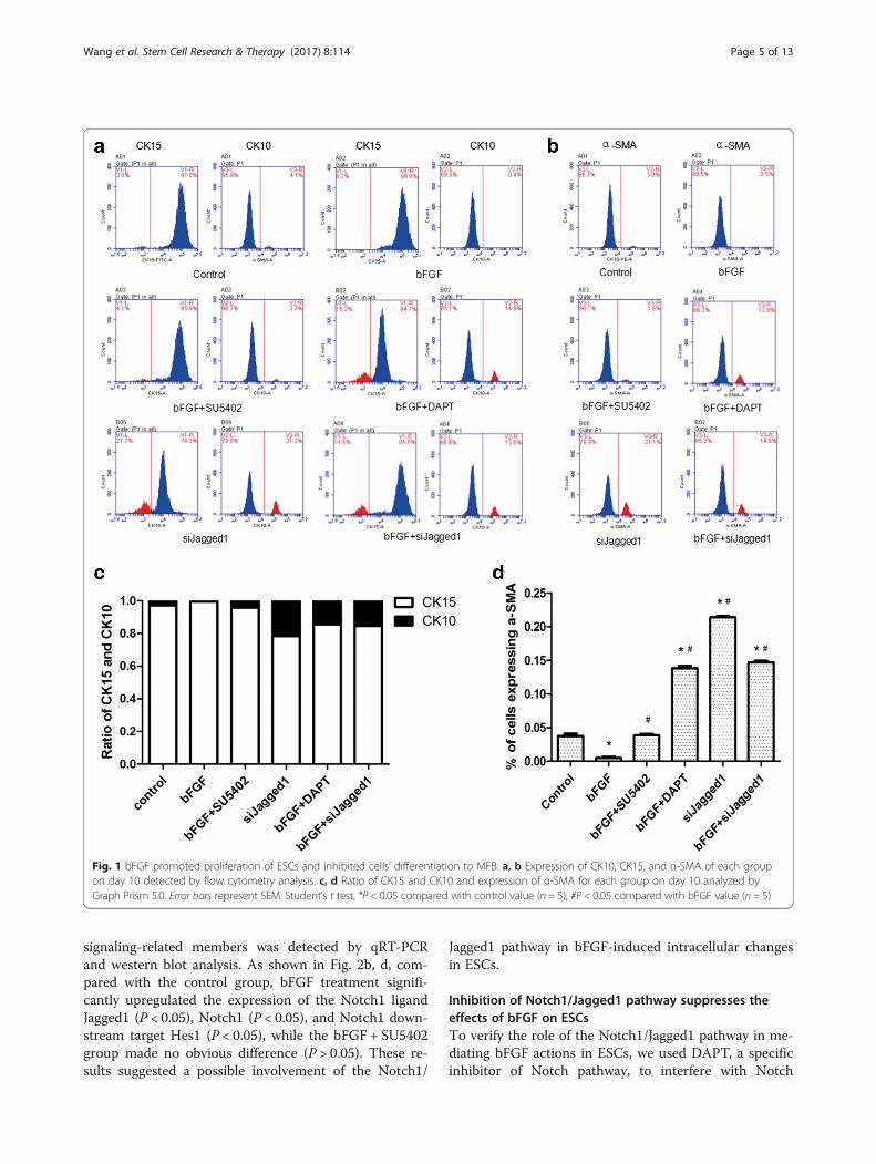
ResultsbFGF inhibits the differentiation of ESCs to MFB in vitroAfter treatment for 10 days, the ESCs of different groupswere collected for further experiments. To confirm theeffect of bFGF on ESC differentiation to MFB, we testedthe expression of α-SMA, CK10, and CK15 by FCM, anddetected the expression of α-SMA, Collagen I (Col I),and Collagen III (Col III) by qRT-PCR and western blotanalysis. The ratio of CK15 and CK10 reflects the purityof ESCs: the higher the ratio, the higher the purity [23, 24].α-SMA is a specific marker of MFB and Col I and Col IIIare metabolites of MFB, and they were used to show thedifferentiation of ESCs to MFB [25, 26].As FCM results showed, compared with the control
group, the ratio of CK15 and CK10 in the bFGF groupwas significantly higher (P < 0.05; Fig. 1a, c), while theexpression of α-SMA was obviously lower (P < 0.05;Fig. 1b, d). Similarly, in qRT-PCR and western blot de-tection, the expression of α-SMA, Col I, and Col III wassignificantly lower in the bFGF group (P < 0.05; Fig. 2a, c),while the bFGF + SU5402 group made no obvious differ-ence with the control group (P > 0.05; Fig. 2a, c). These re-sults indicated that bFGF could inhibit the differentiationof ESC to MFB in vitro.
bFGF enhances the expression of Notch1/Jagged1signaling of ESCs in vitroTo investigate the underlying mechanism of the effect ofbFGF on ESCs, the expression of Notch1/Jagged1
Wang et al. Stem Cell Research & Therapy (2017) 8:114 Page 4 of 13
signaling-related members was detected by qRT-PCRand western blot analysis. As shown in Fig. 2b, d, com-pared with the control group, bFGF treatment signifi-cantly upregulated the expression of the Notch1 ligandJagged1 (P < 0.05), Notch1 (P < 0.05), and Notch1 down-stream target Hes1 (P < 0.05), while the bFGF + SU5402group made no obvious difference (P > 0.05). These re-sults suggested a possible involvement of the Notch1/
Jagged1 pathway in bFGF-induced intracellular changesin ESCs.
Inhibition of Notch1/Jagged1 pathway suppresses theeffects of bFGF on ESCsTo verify the role of the Notch1/Jagged1 pathway in me-diating bFGF actions in ESCs, we used DAPT, a specificinhibitor of Notch pathway, to interfere with Notch
Fig. 1 bFGF promoted proliferation of ESCs and inhibited cells’ differentiation to MFB. a, b Expression of CK10, CK15, and α-SMA of each groupon day 10 detected by flow cytometry analysis. c, d Ratio of CK15 and CK10 and expression of α-SMA for each group on day 10 analyzed byGraph Prism 5.0. Error bars represent SEM. Student’s t test, *P < 0.05 compared with control value (n = 5), #P< 0.05 compared with bFGF value (n = 5)
Wang et al. Stem Cell Research & Therapy (2017) 8:114 Page 5 of 13

signaling in ESCs. Furthermore, we also suppressedJagged1 with specific siRNA siJagged1 to block theNotch1/Jagged1 pathway of ESCs. qRT-PCR and westernblot analysis showed that siJagged1 was able to effectivelyknock down the expression of Jagged1 in ESCs (Fig. 2a, b).As shown in Fig. 2, compared with the control group,
expression of α-SMA, Col I, and Col III in the siJagged1group was significantly higher (P < 0.05). Compared withthe bFGF group, expression of α-SMA, Col I, and Col IIIin the bFGF +DAPT group and bFGF + siJagged1 groupwas significantly higher (P < 0.05). These results suggestedbFGF might inhibit the differentiation of ESCs to MFB by
Fig. 2 bFGF inhibited differentiation of ESCs to MFB by activating the Notch1/Jagged1 pathway in vitro. a Representative immunoblot and results ofdensitometric analysis of blots showing relative protein levels of Jagged1 in control, negative control (ESCs transfected with a scrambled siRNA forJagged1), and siJagged1 (ESCs transfected with siRNA specific for Jagged1). GAPDH used as a loading control. b Representative qRT-PCR analysisshowing relative mRNA levels of Jagged1 in control, negative control, and siJagged1. c Representative immunoblot and results of densitometricanalysis of blots showing relative protein levels of α-SMA, Col I, and Col III for each group on day 10. d Representative immunoblot and results ofdensitometric analysis of blots showing relative protein levels of Jagged1, Notch1, and Hes1 for each group on day 10. e Representative qRT-PCRanalysis showing relative mRNA levels of α-SMA, Col I, and Col III for each group on day 10. f Representative qRT-PCR analysis showing relative mRNAlevels of Jagged1, Notch1, and Hes1 for each group on day 10. a, b *P < 0.05. d–f Error bars represent SEM. Student’s t test, *P< 0.05 compared withcontrol value (n = 5), #P< 0.05 compared with bFGF value (n = 5). Con control NC negative control, bFGF basic fibroblast growth factor, Col collagen
Wang et al. Stem Cell Research & Therapy (2017) 8:114 Page 6 of 13

activating the Notch1/Jagged1 pathway. What is more, wealso found that expression of α-SMA, Col I, and Col III inthe bFGF + siJagged1 group was higher than in the bFGF+DAPT group (P < 0.05), which indicated that Jagged1 lig-and might play an important role in the effects of bFGFon ESCs.
bFGF accelerates wound closure and alleviates scar in vivoTo investigate the role of bFGF in vivo, we conductedour experiments using the rabbit ear scar model. Afterlabeling and random grouping, we treated the woundwith bFGF, bFGF + DAPT, TGF-β1, and saline respect-ively. Meanwhile, we recorded the wound healing time,photographed the wound, measured the wound areasand the thickness of scar tissues, and calculated the re-sidual wound area rate and scar index regularly. As Fig. 3shows, compared with the control group, the bFGF groupshowed a significantly shorter healing time (P < 0.05;
Fig. 3a), lower residual wound area (P < 0.05; Fig. 3b), andlower scar index (P < 0.05; Fig. 3c), while the bFGF +DAPT group presented an obvious healing delay andhigher scar index (P < 0.05; Fig. 3). What is more, the GF-β1 group showed a relative shorter wound healing time(P > 0.05; Fig. 3a, b) and an obviously higher scar index(P < 0.05; Fig. 3c). These results suggested that bFGFcould promote wound healing and alleviate scar, whileadding DAPT or using TGF-β1 might aggravate scareventually.
bFGF promotes re-epithelialization, skin attachmentregeneration, and collagen reassignmentTo further evaluate the wound healing quality and thescar hyperplasia, we selected the scar tissue specimen atspecific time points (7, 14, 30, and 60 days), and observedre-epithelialization, skin appendage regeneration, andcollagen reassignment by H&E and Masson staining. As
Fig. 3 Pharmacological effect of bFGF, TGF-β1, and bFGF + DAPT in repairing wound healing and scaring. Full-thickness dermal wounds wereinduced in rabbit ears and treated by saline (control), bFGF, bFGF + DAPT, and TGF-β1 respectively. a Representative rabbit ear from eachgroup taken on post-injury days 0, 7, 14, 30, and 60. b Wound areas for each group. The computation was that the indicated area was dividedby the initial area. Results represent means ± SEM. *P < 0.05 compared with control value (n = 10). c Scar indexes for each group. The computationwas that the thickness difference of the scar minus the adjacent normal skin was divided by the adjacent normal skin. Results represent means ± SEM.*P < 0.05 compared with control value (n = 10). d days, bFGF basic fibroblast growth factor
Wang et al. Stem Cell Research & Therapy (2017) 8:114 Page 7 of 13

shown in Fig. 4, the wound healing quality of the bFGFgroup was significantly better than that of the controlgroup, with more cell layers, more epidermal ridges,more formation of primitive hair follicle and sweatgland structures, and more regular and ordered collagen ar-rangement. However, when the Notch1/Jagged1 pathwaywas inhibited in the bFGF +DAPT group and TGF-β1group, the re-epithelialization, skin appendage regeneration,and collagen reassignment were significantly impaired(Fig. 4a, b). These results suggests that bFGF has an activefunction in promoting re-epithelialization, skin appendageregeneration, and collagen reassignment, and may mainlywork through the Notch1/Jagged1 pathway.
bFGF inhibits differentiation of ESCs to MFB in vivo viathe Notch1/Jagged1 pathwayBy immunohistochemistry staining (Fig. 4c), we foundthat bFGF can improve the quality of wound healing byinhibiting the expression of α-SMA significantly. Tofurther study its mechanism and verify our hypothesisin vivo, we labeled ESCs with BrdU and detected therelative expression levels of Notch1/Jagged1 signalingcomponents and α-SMA at the same time by double-immunofluorescence staining. As shown in Fig. 5, com-pared with the control group, the expression of BrdU/Jagged1 (Fig. 5a), BrdU/Notch1 (Fig. 5b), and BrdU/Hes1 (Fig. 5c) double-positive cells in the bFGF group
Fig. 4 Histological features and expression of α-SMA of the rabbit ear wounds in each group. a, b Skin tissue sections stained with H&E and Massonshowing histological features in rabbit ears treated with saline (control), bFGF, bFGF + DAPT, and TGF-β1 on post-injury days 7, 14, 30, and 60.bFGF-treated ears exhibited significantly higher quality wound healing, with more cell layers, more epidermal ridges, more formation of primitivehair follicle and sweat gland structures, and more regular and ordered collagen arrangement. Ears treated with bFGF + DAPT and TGF-β1 exhibitedpoor-quality wound healing compared with control ears. c Immunohistochemical staining for α-SMA was performed using skin tissue sections in rabbitears treated with saline (control), bFGF, bFGF + DAPT, and TGF-β1 on post-injury days 7, 14, 30, and 60. bFGF-treated ears exhibited a significantlyhigher quality wound healing with a significant decrease of α-SMA, while ears treated with bFGF + DAPT and TGF-β1 exhibited poor-quality woundhealing with increase of α-SMA. Scale bar, 100 μm. d days, bFGF basic fibroblast growth factor
Wang et al. Stem Cell Research & Therapy (2017) 8:114 Page 8 of 13

was significantly higher (P < 0.05), while expression ofBrdU/α-SMA (Fig. 5d) was obviously lower (P < 0.05).The double-positive cells were mainly detected in hairfollicle cell nucleus and skin basal cell nucleus. On thecontrary, expression of BrdU/Jagged1, BrdU/Notch1,and BrdU/Hes1 double-positive cells in the bFGF +DAPT group was significantly lower (P < 0.05) than thecontrol group, while expression of BrdU/α-SMA wasobviously higher (P < 0.05). What is more, to test our
results further we examined these indexes by westernblot analysis, and consequently found similar results(Fig. 6). These results suggested that bFGF could inhibitESC differentiation to MFB by activating the Notch1/Jagged1 pathway in vivo.
DiscussionAs a common complication of wound healing, scar hasseriously impacted the life of patients for a long time,
Fig. 5 Relationships of bFGF and the Notch1/Jagged1 pathway and differentiation of ESCs in healed skin analyzed by immunofluorescence.a, b, c Representative Brdu/Jagged1, Brdu/Notch1, and Brdu/Hes1 double-positive cells in healed skin and the percentage of the positivecells to total cells in healed skin of each group on post-injury day 60. d Representative Brdu/α-SMA double-positive cells in healed skin andthe percentage of the positive cells to total cells in healed skin of each group on post-injury day 60. Error bars represent SEM. Student’s t test,*P < 0.05 compared with control value (n = 10). Scale bar, 50 μm. bFGF basic fibroblast growth factor
Wang et al. Stem Cell Research & Therapy (2017) 8:114 Page 9 of 13

but there is still no effective treatment [27]. bFGF hasbeen reported to promote wound healing and reducescarring, but the underlying mechanisms remain unclear[14]. With relevant research going deep, it has been con-firmed that the decrease of ESCs and the hyperplasia ofMFB are the main causes of scar [28, 29]. In our previ-ous study, we found that bFGF could promote the prolif-eration and migration of ESCs in vitro. In this study, wedemonstrated that bFGF can also inhibit the differenti-ation of ESCs to MFB by activating the Notch1/Jagged1pathway. We firstly blocked the Notch1/Jagged1 pathwayby adding the relevant inhibitor DAPT and knockingdown Jagged1 to confirm our conjecture in vitro, andthen verified our results using the rabbit ear scar models.Our results provide the evidence that bFGF reduces scarby inhibiting the differentiation of ESCs to MFB via theNotch1/Jagged1 pathway.Although the mechanism of scar is still unclear, the
hyperplasia of MFB has so far been confirmed to be the
most important factor [30]. Early in 2009, Ishiguro et al.[31] identified bFGF as a potent stimulator for the re-duction of the myofibroblastic area in vivo, presumablybecause of its effects on the downregulation of α-SMAexpression as well as rapid induction of apoptosis inmyofibroblasts. A recently study in cell therapy alsofound that bFGF could inhibit the differentiation of car-diac stem cells to MFB, which improved extracellularmatrix dysregulation post myocardial infarction [32].While these findings show that that bFGF could reducethe formation of MFB, its effects on ESCs, a key role inwound healing and re-epithelialization, are still unknown[33]. Herein, we speculated that bFGF might inhibit thedifferentiation of ESCs to MFB analogously. In our in-vitro study, we detected the expression of α-SMA, whichis a specific marker of MFB, to show the differentiationof ESCs to MFB directly. At the same time, we also ex-amined the expression of Col I and Col III, which aremetabolites of MFB, to reflect the differentiation of ESCs
Fig. 6 Relationships of bFGF and the Notch1/Jagged1 pathway and differentiation of ESCs in healed skin analyzed by western blot. a, b Representativeimmunoblot and results of densitometric analysis of blots showing relative protein levels of α-SMA, Col I, and Col III for each group on post-injury day 60.c, d. Representative immunoblot and results of densitometric analysis of blots showing relative protein levels of Jagged1, Notch1, and Hes1 for eachgroup on post-injury day 60. Error bars represent SEM. Student’s t test, *P< 0.05 compared with control value (n= 10), #P< 0.05 compared with bFGFvalue (n= 10). bFGF basic fibroblast growth factor, Col collagen
Wang et al. Stem Cell Research & Therapy (2017) 8:114 Page 10 of 13

to MFB indirectly. The results from FCM, RT-qPCR,and western blot analysis consistently showed that ex-pression of α-SMA, Col I, and Col III in the bFGF groupwas significantly lower than in other groups, suggestingthat bFGF could inhibit ESC differentiation to MFB.The Notch1/Jagged1 pathway has been confirmed to
play an important role in stem cells [34]. As shown inresearch conducted by Guiu et al. [35], Hes1 repressorsare essential regulators of hematopoietic stem cell devel-opment downstream of Notch signaling. By activating itsdownstream gene Hes1, the Notch1/Jagged1 pathwaymainly promotes stem cell proliferation and inhibits theirdifferentiation [36]. In our previous study, we found thatactivating the Notch1/Jagged1 pathway contributed topromoting ESC proliferation and inhibiting the cells’ dif-ferentiation [11], which is consistent with the effects ofbFGF on ESCs. Therefore we supposed that bFGF mightregulate ESCs by activating the Notch1/Jagged1 pathway.In our present in-vitro study, we first detected the expres-sion of Jagged1, Notch1, and Hes1 for each group, andfound this was much higher in the bFGF groups than inother groups. We then adopted two ways to interfere withthe Notch1/Jagged1 pathway: adding DAPT and knockingdown Jagged1. As a γ-secretase inhibitor, DAPT can in-hibit the Notch pathway by blocking the cleavage ofNICD, which is necessary for activation of transcription ofdownstream target genes [20]. To further detect the func-tion of the Notch1/Jagged1 pathway on ESCs, we knockeddown Jagged1 ligands by transfecting the correspondingsiRNA into ESCs [37]. By inhibiting the Notch1/Jagged1pathway, we observed that the effects of bFGF on ESCswere significantly weakened. What is more, this effect de-crease was much more obvious in the bFGF + siJagged1group than in the bFGF +DAPT group, suggesting thatbFGF might work mainly by activating Jagged1. However,in our present study we found that the siJagged1 group ex-hibited a significant difference from the bFGF +DAPTgroup and bFGF + siJagged1 group, which indicates thatthere is another signaling pathway existing between bFGFand the differentiation of ESCs. This result is consistentwith our previous findings that both the Wnt and Notchpathways are important to wound healing [22].Since Morris et al. [21] found that rabbits’ ears could
be used for studying scar, the rabbit ear wound modelhas been an ideal model to date [38]. In order to verifyour findings in vivo, we conducted our animal researchon the rabbit ear wound models. By observing thewound healing time and measuring the wound areas andthe thickness of scar tissues, we found that bFGF couldobviously promote wound healing and reduce the scar.These results are consistent with the function of bFGFin clinical observation. As we all know, normal skin tis-sue is constituted by the epidermis, dermis, and skin ap-pendages [39]. At the junction of epidermal and dermal
tissue, we often see a clear papillary structure and basalcells, in which the ESCs reside [40]. In our in-vivo study,we collected the tissues of different groups in 60 daysfor pathology detection, and found that bFGF significantlypromoted re-epithelialization, skin attachment regener-ation, and collagen reassignment, with clearer cell layers,more epidermal ridges, and more reformation of hairfollicle and sweat gland structures. Recent studies haveshown that the Notch1/Jagged1 pathway is involved inregulating investigation of hair follicles into the dermisand maintaining postnatal hair homeostasis [41] andthat DAPT could modulate human hair follicle stemcell proliferation and differentiation [42], and we foundthat the Notch1/Jagged1 pathway also played an im-portant role in the process of bFGF on wound healing.When we inhibited the Notch signal pathway by DAPT,the function of bFGF on wound healing was weakenedsignificantly. This result was analogous to the study ofChen et al. [43], which confirmed that Notch1 signalinginhibits apoptosis of human dental follicle stem cellsvia both the cytoplasmic mitochondrial pathway andnuclear transcription regulation.In order to study the effect of bFGF on ESCs in vivo,
we labeled ESCs by injecting BrdU. Because ESCs pos-sess a longer period between divisions, they should bethe only cells retaining the BrdU label after a long chaseperiod [22]. The specific mechanism for retaining thelabel is that stem cells normally remain in a relativelystatic state of long-term, slow differentiation, in whichBrdU dilutes much more slowly than in other cells.Therefore, after labeling for a certain period of time,only slowly proliferating cells such as ESCs retain theBrdU marker [44]. In our present study, we collected thescar tissue specimens of different groups in 60 days anddetected the double-positive cells of BrdU/Jagged1,BrdU/Notch1, BrdU/Hes1, and BrdU/α-SMA simultan-eously by double-immunofluorescence staining. In linewith our expectation, we found that bFGF could obviouslyactivate the Notch1/Jagged1 pathway, while inhibiting thedifferentiation of ESCs to MFB at the same time. More-over, we further detected these indices by western blotanalysis and observed the same results.Additionally, we discovered that TGF-β1 played an ab-
solutely opposite role with bFGF during wound healingand scar formation in rabbit ears. Although the TGF-β1/Smad3 pathway has been widely accepted to be a majorfactor leading to scar [45], our study showed that theNotch1/Jagged1 pathway was obviously suppressed inthe TGF-β1 group, which led to an overdeposition ofMFB and eventually aggravated the scar. In recent re-search, Luo [46] stated that signaling crosstalk exists be-tween TGF-β1/Smad and other signaling pathways,which could properly account for our results. This dis-covery not only indicates that TGF-β1 might increase
Wang et al. Stem Cell Research & Therapy (2017) 8:114 Page 11 of 13

the scar by inhibiting the Notch1/Jagged1 pathway, butalso suggests that there is a close relationship betweenvarious cytokines and signaling pathways during woundhealing and scar hyperplasia, which remains to be stud-ied further.
ConclusionIn summary, this work provides the first evidence thatbFGF can reduce scar by promoting the proliferation ofESCs and inhibiting its differentiation to MFB by activat-ing the Notch1/Jagged1 pathway. Moreover, we demon-strate that TGF-β1 might aggravate the scar by inhibitingthe Notch1/Jagged1 pathway. This work not only builds abridge between the signaling pathway and ESCs in woundhealing, but also reveals the formation of scar in a newview, which may help us explore new approaches to pre-vent and treat scar. However, our work just begins. Therelation of the Notch1/Jagged1 pathway with other cyto-kines such as TGF-β1 and its crosstalk with differentpathways remain to be studied further.
AbbreviationsbFGF: Basic fibroblast growth factor; Brdu: 5-Bromodeoxyuridine; DAPT: N-[N-(3,5-Difluorophenacetyl)-L-alanyl]-S-phenylglycine t-butyl ester; ESC: Epidermalstem cell; FCM: Flow cytometry; FITC: Fluorescein isothiocyanate;GAPDH: Glyceraldehyde-3-phosphate dehydrogenase; H&E: Hematoxylin andeosin; Hes: Hairy and enhancer of split; HRP: Horseradish peroxidase;K-SFM: Keratinocyte serum-free medium; NICD: Notch intracellular domain;qRT-PCR: Quantitative real-time polymerase chain reaction; SD rats: Sprague–Dawley rats; TGF-β1: Transforming growth factor-β1; WB: Western blot;α-SMA: α-Smooth muscle actin
AcknowledgementsThe authors thank Ronghua Yang PhD for his expert technical assistance.
FundingThis article was supported by the National Natural Science Foundation ofChina (Grant No. 81372062) and Science and Technology Planning Project ofGuangdong Province, China (Grant No. 2014A020212055).
Availability of data and materialsThe data supporting the conclusions of this article are included within thearticle.
Authors’ contributionsPW participated in study design and drafted the manuscript. BS participatedin the design of the study and helped to draft the manuscript. YbXparticipated in the design of the study. JyZ carried out the FCM analysis. JLcarried out the isolation and culture of ESCs. ZhZ carried out theimmunoassays. LC performed the animal study. ShQ participated in thewestern blot analysis. XsL participated in the wound analysis. JlZ performedthe statistical analysis. KX participated in the liposome transfection and geneknockdown. JlX conceived of the study, participated in study design andcoordination, and helped to draft the manuscript. All authors read andapproved the final manuscript.
Competing interestsThe authors declare that they have no competing interests.
Consent for publicationAll authors have contributed to, read, and approved the final manuscript forsubmission.
Ethics approvalAll animal experiments in this study were approved by the InstitutionalAnimal Care and Use Committee at Sun Yat-Sen University and performedaccording to National Institutes of Health guidelines. SD rats and New Zea-land rabbits were obtained from the Experimental Animal Center of Sun Yat-Sen University (license: SYXK 2016-0112) and kept in standard conditions ac-cording to the regulation of ethics committee of the Medical SciencesDepartment.
Publisher’s NoteSpringer Nature remains neutral with regard to jurisdictional claims inpublished maps and institutional affiliations.
Author details1Department of Burn Surgery, First Affiliated Hospital of Sun Yat-SenUniversity, No. 58, 2nd Zhongshan Road, Yuexiu District, Guangzhou,Guangdong Province 510080, People’s Republic of China. 2Department ofAnatomy and Neurobiology, School of Basic Medical Sciences, Central SouthUniversity, Changsha, Hunan Province 410013, People’s Republic of China.
Received: 9 January 2017 Revised: 28 February 2017Accepted: 31 March 2017
References1. Han CM. Wound healing is still a game of “blind men and an elephant”.
Zhonghua Shao Shang Za Zhi. 2016;32:580–1.2. Berman B, Maderal A, Raphael B. Keloids and hypertrophic scars: pathophysiology,
classification, and treatment. Dermatol Surg. 2017;43 Suppl 1:S3–18.3. Zhang GY, Li X, Chen XL, Li ZJ, Yu Q, Jiang LF, Ding J, Gao WY. Contribution
of epidermal stem cells to hypertrophic scars pathogenesis. Med Hypotheses.2009;73:332–3.
4. Li-Tsang CW, Feng B, Huang L, Liu X, Shu B, Chan YT, Cheung KK. Ahistological study on the effect of pressure therapy on the activities ofmyofibroblasts and keratinocytes in hypertrophic scar tissues after burn.Burns. 2015;41:1008–16.
5. Charruyer A, Ghadially R. What’s new in dermatology: epidermal stem cells.G Ital Dermatol Venereol. 2011;146:57–67.
6. Blanpain C, Fuchs E. Epidermal homeostasis: a balancing act of stem cells inthe skin. Nat Rev Mol Cell Biol. 2009;10:207–17.
7. Doupe DP, Jones PH. Interfollicular epidermal homeostasis: a response toGhadially, '25 years of epidermal stem cell research'. J Invest Dermatol. 2012;8:2096–7.
8. Zhou S, Cai J, Niu F, Zong X, Xu J, Du L, Chen G. Comparison of biologicalcharacteristics and quantity of epidermal stem cells from hypertrophicscar skin and normal skin of human beings. Zhonghua Yi Xue Za Zhi.2014;94:1097–100.
9. Choi HR, Byun SY, Kwon SH, Park KC. Niche interactions in epidermal stemcells. World J Stem Cells. 2015;7:495–501.
10. Watt FM, Estrach S, Ambler CA. Epidermal Notch signalling: differentiation,cancer and adhesion. Curr Opin Cell Biol. 2008;20:171–9.
11. Yang RH, Qi SH, Shu B, Ruan SB, Lin ZP, Lin Y, Shen R, Zhang FG, Chen XD,Xie JL. Epidermal stem cells (ESCs) accelerate diabetic wound healing viathe Notch signalling pathway. Biosci Rep. 2016;36:e00364.
12. Liu J, Sato C, Cerletti M, Wagers A. Notch signaling in the regulation of stemcell self-renewal and differentiation. Curr Top Dev Biol. 2010;92:367–409.
13. Yeo SY, Chitnis AB. Jagged-mediated Notch signaling maintains proliferatingneural progenitors and regulates cell diversity in the ventral spinal cord.Proc Natl Acad Sci U S A. 2007;104:5913–8.
14. Xie JL, Bian HN, Qi SH, Chen HD, Li HD, Xu YB, Li TZ, Liu XS, Liang HZ, Xin BR,et al. Basic fibroblast growth factor (bFGF) alleviates the scar of the rabbit earmodel in wound healing. Wound Repair Regen. 2008;16:576–81.
15. Xie J, Qi S, Xu Y, Tang J, Li T, Liu X, Shu B, Liang H, Huang B. Effects of basicfibroblast growth factors on hypertrophic scarring in a rabbit ear model. JCutan Med Surg. 2008;12:155–62.
16. Reiisi S, Esmaeili F, Shirazi A. Isolation, culture and identification of epidermalstem cells from newborn mouse skin. In Vitro Cell Dev Biol Anim. 2010;46:54–9.
17. Eckert RL, Adhikary G, Balasubramanian S, Rorke EA, Vemuri MC, Boucher SE,Bickenbach JR, Kerr C. Biochemistry of epidermal stem cells. BiochimBiophys Acta. 2013;1830:2427–34.
18. Liu GY, Gao ZH, Li L, Song TT, Sheng XG. Expression of Jagged1 mRNA inhuman epithelial ovarian carcinoma tissues and effect of RNA interference
Wang et al. Stem Cell Research & Therapy (2017) 8:114 Page 12 of 13

of Jagged1 on growth of xenograft in nude mice. Zhonghua Fu Chan KeZa Zhi. 2016;51:448–53.
19. Kaftan H, Reuther L, Miehe B, Hosemann W, Beule A. Inhibition of fibroblastgrowth factor receptor 1: influence on tympanic membrane wound healingin rats. Eur Arch Otorhinolaryngol. 2012;269:87–92.
20. Li S, Zyang X, Wang Y, Ji H, Du Y, Liu H. DAPT protects brain against cerebralischemia by down-regulating the expression of Notch 1 and nuclear factorkappaB in rats. Neurol Sci. 2012;33:1257–64.
21. Morris DE, Wu L, Zhao LL, Bolton L, Roth SI, Ladin DA, Mustoe TA. Acuteand chronic animal models for excessive dermal scarring: quantitativestudies. Plast Reconstr Surg. 1997;100:674–81.
22. Shi Y, Shu B, Yang R, Xu Y, Xing B, Liu J, Chen L, Qi S, Liu X, Wang P, et al.Wnt and Notch signaling pathway involved in wound healing by targetingc-Myc and Hes1 separately. Stem Cell Res Ther. 2015;6:120.
23. Bose A, Teh MT, Mackenzie IC, Waseem A. Keratin k15 as a biomarker ofepidermal stem cells. Int J Mol Sci. 2013;14:19385–98.
24. Guo A, Jahoda CA. An improved method of human keratinocyte culturefrom skin explants: cell expansion is linked to markers of activated progenitorcells. Exp Dermatol. 2009;18:720–6.
25. Yang JP, Su DJ, Li SN, Gao L. The expression of alpha-smooth muscle actinin primary cultural fibroblasts of rats. Zhongguo Ying Yong Sheng Li Xue Za Zhi.2009;25:339–43.
26. Al-Qattan MM, Abd-Elwahed MM, Hawary K, Arafah MM, Shier MK.Myofibroblast expression in skin wounds is enhanced by collagen IIIsuppression. Biomed Res Int. 2015;2015:958695.
27. Rabello FB, Souza CD, Farina JJ. Update on hypertrophic scar treatment.Clinics (Sao Paulo). 2014;69:565–73.
28. Quaggin SE, Kapus A. Scar wars: mapping the fate of epithelial-mesenchymal-myofibroblast transition. Kidney Int. 2011;80:41–50.
29. Jia CY. Awareness of present status of study on hypertrophic scar.Zhonghua Yi Xue Za Zhi. 2011;91:2597–9.
30. Liu Y, Cen Y, Ying T, Xu X. The role of myofibroblast in the development ofpathological scar. Zhongguo Xiu Fu Chong Jian Wai Ke Za Zhi. 2005;19:39–41.
31. Ishiguro S, Akasaka Y, Kiguchi H, Suzuki T, Imaizumi R, Ishikawa Y, Ito K, Ishii T.Basic fibroblast growth factor induces down-regulation of alpha-smoothmuscle actin and reduction of myofibroblast areas in open skin wounds.Wound Repair Regen. 2009;17:617–25.
32. Fedak PW, Bai L, Turnbull J, Ngu J, Narine K, Duff HJ. Cell therapy limitsmyofibroblast differentiation and structural cardiac remodeling: basicfibroblast growth factor-mediated paracrine mechanism. Circ Heart Fail.2012;5:349–56.
33. Xie J, Bian H, Qi S, Xu Y, Tang J, Li T, Liu X. Effects of basic fibroblast growthfactor on the expression of extracellular matrix and matrixmetalloproteinase-1 in wound healing. Clin Exp Dermatol. 2008;33:176–82.
34. Sancho R, Cremona CA, Behrens A. Stem cell and progenitor fate in themammalian intestine: Notch and lateral inhibition in homeostasis anddisease. Embo Rep. 2015;16:571–81.
35. Guiu J, Shimizu R, D’Altri T, Fraser ST, Hatakeyama J, Bresnick EH, Kageyama R,Dzierzak E, Yamamoto M, Espinosa L, et al. Hes repressors are essentialregulators of hematopoietic stem cell development downstream of Notchsignaling. J Exp Med. 2013;210:71–84.
36. Zhang Z, Yan R, Zhang Q, Li J, Kang X, Wang H, Huan L, Zhang L, Li F, Yang S,et al. Hes1, a Notch signaling downstream target, regulates adult hippocampalneurogenesis following traumatic brain injury. Brain Res. 2014;1583:65–78.
37. Wang J, Wang C, Meng Q, Li S, Sun X, Bo Y, Yao W. siRNA targeting Notch-1decreases glioma stem cell proliferation and tumor growth. Mol Biol Rep.2012;39:2497–503.
38. Ju-Lin X, Shao-Hai Q, Tian-Zeng L, Bin H, Jing-Ming T, Ying-Bin X, Xu-Sheng L,Bin S, Hui-Zhen L, Yong H. Effect of asiaticoside on hypertrophic scar in therabbit ear model. J Cutan Pathol. 2009;36:234–9.
39. Dahl MV. Stem cells and the skin. J Cosmet Dermatol. 2012;11:297–306.40. Senoo M. Epidermal stem cells in homeostasis and wound repair of the
skin. Adv Wound Care (New Rochelle). 2013;2:273–82.41. Vagnozzi AN, Reiter JF, Wong SY. Hair follicle and interfollicular epidermal
stem cells make varying contributions to wound regeneration. Cell Cycle.2015;14:3408–17.
42. Jiang J, Miao Y, Xiao S, Zhang Z, Hu Z. DAPT in the control of human hairfollicle stem cell proliferation and differentiation. Postepy Dermatol Alergol.2014;31:201–6.
43. Chen X, Li S, Zeng Z, Gu Z, Yu Y, Zheng F, Zhou Y, Wang H. Notch1 signallinginhibits apoptosis of human dental follicle stem cells via both the cytoplasmic
mitochondrial pathway and nuclear transcription regulation. Int J Biochem CellBiol. 2017;82:18–27.
44. Dunnwald M, Chinnathambi S, Alexandrunas D, Bickenbach JR. Mouse epidermalstem cells proceed through the cell cycle. J Cell Physiol. 2003;195:194–201.
45. Zunwen L, Shizhen Z, Dewu L, Yungui M, Pu N. Effect of tetrandrine on theTGF-beta-induced smad signal transduction pathway in humanhypertrophic scar fibroblasts in vitro. Burns. 2012;38:404–13.
46. Luo K. Signaling cross talk between TGF-beta/Smad and other signalingpathways. Cold Spring Harb Perspect Biol. 2017;9:a022137.
• We accept pre-submission inquiries
• Our selector tool helps you to find the most relevant journal
• We provide round the clock customer support
• Convenient online submission
• Thorough peer review
• Inclusion in PubMed and all major indexing services
• Maximum visibility for your research
Submit your manuscript atwww.biomedcentral.com/submit
Submit your next manuscript to BioMed Central and we will help you at every step:
Wang et al. Stem Cell Research & Therapy (2017) 8:114 Page 13 of 13