
Basalt fiber-reinforced polylactic acid composite · A novel basalt fiber-reinforced polylactic...
9
A novel basalt fiber-reinforced polylactic acid composite for hard tissue repair This article has been downloaded from IOPscience. Please scroll down to see the full text article. 2010 Biomed. Mater. 5 044104 (http://iopscience.iop.org/1748-605X/5/4/044104) Download details: IP Address: 121.249.16.167 The article was downloaded on 27/12/2010 at 01:34 Please note that terms and conditions apply. View the table of contents for this issue, or go to the journal homepage for more Home Search Collections Journals About Contact us My IOPscience
Transcript of Basalt fiber-reinforced polylactic acid composite · A novel basalt fiber-reinforced polylactic...

A novel basalt fiber-reinforced polylactic acid composite for hard tissue repair
This article has been downloaded from IOPscience. Please scroll down to see the full text article.
2010 Biomed. Mater. 5 044104
(http://iopscience.iop.org/1748-605X/5/4/044104)
Download details:
IP Address: 121.249.16.167
The article was downloaded on 27/12/2010 at 01:34
Please note that terms and conditions apply.
View the table of contents for this issue, or go to the journal homepage for more
Home Search Collections Journals About Contact us My IOPscience

IOP PUBLISHING BIOMEDICAL MATERIALS
Biomed. Mater. 5 (2010) 044104 (8pp) doi:10.1088/1748-6041/5/4/044104
A novel basalt fiber-reinforced polylacticacid composite for hard tissue repairXi Chen, Yan Li and Ning Gu1
Jiangsu Laboratory for Biomaterials and Devices, State Key Laboratory of Bioelectronics, School ofBiological Science and Medical Engineering, Southeast University, Nanjing, 210096, People’s Republicof China
E-mail: [email protected]
Received 26 February 2010Accepted for publication 12 May 2010Published 3 August 2010Online at stacks.iop.org/BMM/5/044104
AbstractA basalt fiber (BF) was, for the first time, introduced into a poly(L-lactic acid) (PLLA) matrixas innovative reinforcement to fabricate composite materials for hard tissue repair. Firstly,BF/PLLA composites and pure PLLA were produced by the methods of solution blending andfreeze drying. The results showed that basalt fibers can be uniformly dispersed in the PLLAmatrix and significantly improve the mechanical properties and hydrophilicity of the PLLAmatrix. The presence of basalt fibers may retard the polymer degradation rate and neutralizethe acid degradation from PLLA. Osteoblasts were cultured in vitro to evaluate thecytocompatibility of the composite. An MTT assay revealed that osteoblasts proliferated wellfor 7 days and there was little difference found in their viability on both PLLA and BF/PLLAfilms, which was consistent with the alkaline phosphatase (ALP) activity results. A fluorescentstaining observation showed that osteoblasts grew well on the composites. SEM imagesdisplayed that osteoblasts tended to grow along the fiber axis. The formation of mineralizednodules was observed on the films by Alizarin red S staining. These results suggest that thepresence of basalt fibers does not noticeably affect osteoblastic behavior and the designedcomposites are osteoblast compatible. It is concluded that basalt fibers, as reinforcing fibers,may have promising applications in hard tissue repair.
(Some figures in this article are in colour only in the electronic version)
1. Introduction
A variety of bioabsorbable polymers have been usedfor scaffolds and implant devices in hard tissue repair.Bioabsorbable devices offer two major advantages overconventional metallic implants: firstly, no retrieval of thedevices is needed after fracture healing. Secondly, theelastic modulus of bioabsorbable devices is closer to thatof bone, which could minimize stress concentrations nearthe extremities of the implant and reduce, if not eliminate,stress protection atrophy of the bone [1, 2]. Polyesters suchas poly(lactic acid) (PLA), poly(glycolic acid) (PGA) andpoly(lactic acid-co-glycolic acid) (PLGA) are the most popularsynthetic polymers considered for scaffold applications [3].Among these polymers, poly(L-lactide) (PLLA) is of special
1 Author to whom any correspondence should be addressed.
interest. Structurally, PLLA is a type of polyester formed fromcondensation of L-lactic acid or ring-opening polymerizationof the cyclic diester (L-lactide). It is susceptible to enzymaticand hydrolytic degradation to form L-lactic acid, a naturallyoccurring metabolite in the human body [4, 5]. However,the low mechanical strength of pure PLLA cannot meet therequirement of the repairing of weight bearing bones, whichlimits its application [1, 6]. Accordingly, a PLLA matrixis usually reinforced with high strength fibers. One kind ofsuitable reinforcing fiber is an organic fiber, such as chitosanfiber [7], chitin fiber [8] and poly(glycolic acid) (PGA) fiber[9]. Another kind is an inorganic fiber, such as carbon fiber[10] and glass fiber [11].
More recently, basalt fiber (BF), a kind of highperformance silicate-based fiber [12], is increasingly beingused as an efficient reinforcing fiber for polymer materials [13].
1748-6041/10/044104+08$30.00 1 © 2010 IOP Publishing Ltd Printed in the UK

Biomed. Mater. 5 (2010) 044104 X Chen et al
Compared with carbon fiber, glass fiber or other traditionalreinforcement counterparts, basalt fiber is characterized byits favorable cost performance. It has better mechanicalstrength than an E-glass fiber [14], greater failure strainthan a carbon fiber, as well as good resistance to chemicalattack [15]. Nowadays, basalt fiber-reinforced composites arebeing successfully used as structural and functional materialsin different fields [15, 16]. The increasing applicationof basalt fiber raised the question whether basalt fiber isharmful to health. Kogan et al [17] made rats inhale aircontaining asbestos and basalt fibers for 6 months. In thecase of asbestos fibers at a dose of 1.7 g kg−1 (referredto the body weight of the rat), one third of the animalsdied, while a dose of 2.7 g kg−1 killed all the rats. Inthe case of the basalt fiber, all the animals survived evenwhen the dose reached the 10 g kg−1 concentration. Similarinvestigations were conducted by McConnell et al [18] andthey also concluded that basalt fibers pose no risk to humanbeings. The favorable price–performance ratio of basalt fiber[12, 13] encourages new potential applications. It couldbe envisaged as alternative reinforcement in load-bearingbiomedical materials. Particularly, it stands a good chance ofprocessing three-dimensional scaffolds for hard tissue repairsuch as bone defect repair and oral orthopedics through theintegration of basalt fibers into biopolymers.
From the first published paper of the study on the basaltfiber as a possible polymer reinforcing material in 1980 [19],a lot of research work on the properties of basalt fiber wascarried out to find out the useful properties and advantages ofthe basalt fiber. However, few researchers paid attention tothe application of the basalt fiber in biomedical materials. Inthis paper, a preliminary approach was attempted to producebasalt fiber-reinforced PLLA composites possessing improvedmechanical properties to jointly preserve biocompatibility.In particular, the BF/PLLA composite was produced bothin the form of a thin film and a porous scaffold. Themorphological features and mechanical properties of thescaffold were investigated, and short-term osteoblast responsesto the composite film were also evaluated in vitro.
2. Materials and methods
2.1. Preparation of BF/PLLA composites
Briefly, PLLA (Shandong Institute of Medical Instruments,China) of molecular weight 100 000 Da was first dissolved in1,4-dioxane to produce a ratio of polymer weight to solventvolume of 4%. Basalt fibers (Shanghai Russia & Gold BasaltFiber Co. Ltd. (GBF), China) used in the experiment were cutinto an average length of 2 mm with a diameter of 8 μm.Determined amounts of basalt fibers, calculated to resultin final proportions of 5, 10 and 15 wt% of fibers inthe composites, were dispersed with the PLLA 1,4-dioxanesolution by stirring uniformly. Each mixture was sonicated for15 min in order to eliminate air bubbles, and then subjected totwo different procedures designed to produce either thin filmsor scaffolds.
A thermally induced phase-separation method wasdeveloped to fabricate a basalt fiber-reinforced PLLA scaffold.
Each solution with different BF/PLLA weight ratios wasrapidly poured into a designed polytetrafluoroethylene mold(�8 mm × 20 mm), followed by quenching the solution ina freezer at a preset temperature (−20 ◦C) to solidify andinduce a solid–liquid phase separation, and then evaporatingthe ice crystals under a high vacuum (0.03 mbar) in a freeze-dryer system (LY-3-FM, Snijders Scientific, Tilburg, TheNetherlands) for 12 h to remove ice crystals of dioxaneand produce a porous structure simultaneously [20, 21].Alternatively, the composite films with different fiber loadingswere prepared by casting the solution into petri dishesand dried extensively at room temperature [22]. A rangeof BF/PLLA composites, both scaffolds and films, weresubsequently dried in a vacuum oven for 72 h to remove thesolvent completely.
2.2. Scaffold morphological characterization
The microstructures of the scaffolds were studied usinga scanning electron microscope (SEM, JSM 5610 LV,JEOL, Tokyo, Japan) with a 15 kV acceleration voltage.Raw composite specimens were fractured parallel andperpendicular to the surface using a blade, the resulting crossand vertical sections including the outer surface were sputter-coated with gold. The distribution of basalt fibers in scaffoldsand microstructural features were visualized.
2.3. Mechanical properties
The compressive mechanical properties of scaffolds weremeasured with an electronic universal testing machine(CMT5305, SANS, Shenzhen, China) with a 1 kN load cell.According to the literature [23], the specimens were made intoa cylindrical shape with 8 mm diameter and 15 mm length.The crosshead speed was set at 1 mm min−1, and load wasapplied until the scaffold was compressed to approximately30% of its original length. The compressive modulus wascalculated as the slope of the initial linear portion of the stress–strain curve. The compressive strength was determined as themaximum point of the stress–strain curve. Five specimens ofeach scaffold were tested and the mean values and standarderrors were reported.
2.4. Hydrophilicity of composites
Contact angles of the BF/PLLA films and basalt fiber fabric(GBF, Shanghai, China) were measured through the sessiledrop method using a contact angle meter (CAM200, KSV,Helsinki, Finland). A water droplet (distilled water, about5 μl) was placed on the samples and its contact angles weremeasured. Three samples of each material were used and threemeasurements were carried out for each sample.
2.5. In vitro degradation study
The BF/PLLA composite scaffolds with fiber weight ratiosof 0%, 5% 10 and 15% were immersed in a 10 ml phosphatebuffer solution (PBS) with the initial pH value of 7.40 at 37 ◦C.The PBS was replaced every 7 days, and pH values were tested
2
Biomed. Mater. 5 (2010) 044104 X Chen et al
at fixed time intervals. After degradation, the microstructuresof the scaffolds were observed under an SEM.
2.6. Osteoblast culture and seeding
Osteoblasts were isolated via sequential digestions of neonatalrat calvarias according to established procedures [24]. Thecells were cultured in Dulbecco’s Modified Eagle Medium(DMEM, Hyclone, Beijing, China) supplemented with 15%fetal calf serum (FCS, Sijiqing, Hangzhou, China) and1% penicillin–streptomycin (KeyGEN, Nanjing, China) in ahumidified incubator at 37 ◦C, with 5% CO2. The 15 wt%BF/PLLA films and neat PLLA films were used to observethe effect of basalt fibers on the cellular responses. Prior tocell seeding, the specimens were cut into 10 mm × 10 mmsquare films and �6 circle films, then sterilized for 2 h with75% ethanol and pretreated by immersion in DMEM for 12 h.The osteoblasts from passage 3 were seeded on the films. �6circle films (5 × 10³ cells/well) in 96-well plates were forthe MTT assay and 10 mm × 10 mm square films (2 × 10⁴
cells/well) in 24-well plates were for the other tests. Thecells plated on tissue culture polystyrene served as controls.All plates were incubated at 37 ◦C in 5% CO2/95% air. Theculture medium was changed every 3 days.
2.7. MTT assay
The MTT assay was used as a relative measure of cell viability.At each end of the cell incubation period of 1, 2, 3, 5 and 7days, 180 μl serum-free media and 20 μl of MTT (5 mg ml−1)were added to each well, and the plates were incubated at37 ◦C for 4 h. The blue formazan reaction product was thendissolved by adding 150 μl DMSO. The absorbance at 570 nmwas measured by a microplate reader (Model 680, BioRAD,Berkeley, USA).
2.8. Alkaline phosphatase assay
The alkaline phosphatase (ALP) activity of the cells wasmeasured to observe their functional activity [25]. After 1,2, 3, 5 and 7 days of cultivation, the cell layers were gatheredand disrupted by treating them with 0.3% Triton X-100 and afurther cyclic freezing/thawing process. The supernatant ofthe cell lysates was collected for the ALP assay with an alkalinephosphatase assay kit (keyGEN, Nanjing, China) according tothe manufacturer’s protocol. The absorbance of the productobtained was measured with a spectrophotometer (UV3600,SHIMADZU, Kyoto, Japan) at 520 nm and converted to theALP activity level, which was expressed as King unit of ALPper 100 ml.
2.9. Cell morphology
After the growth for 1, 3, 5 and 7 days, osteoblasts onthe different films were fixed, stained with acridine orange(AO) and observed by a fluorescence inverted microscope(Axiovert200, ZEISS, Gottingen, Germany). Also, themorphology of the cells grown on the samples for 7 days wasobserved with an SEM (JSM-6300, JEOL, Tokyo, Japan), afterfixing and dehydrating the cells, followed by gold coating.
2.10. Mineralization
Whether mineralized nodules were formed on the BF/PLLAfilms or not was determined by using the alizarin red S staining.It is a common histochemical technique used to detect calciumdeposits in mineralized cultures [26]. The cells cultured on thefilms at day 15 post-seeding were fixed in 2.5% glutaraldehydeand stained with 0.1% alizarin red in Tris–HCl (pH 8.3) for30 min at 37 ◦C. Films were then washed with a PBS solutionand viewed under a light microscope.
2.11. Statistical analysis
Multiple samples were collected in each measurement(n � 3) and expressed as mean ± standard deviations (SD).One-way analysis of variance (ANOVA) was applied to assessthe statistical significance of the results obtained. A value ofp < 0.05 was considered to be statistically significant.
3. Results and discussion
3.1. Scaffold morphology
An ideal scaffold should act as a three-dimensional (3D)template for in vitro and in vivo bone growth. Therefore, itmust consist of a porous network with a modal interconnectedpore diameter of at least 100 μm to allow cell migrationand promote cell adhesion and activity [23, 27]. TheSEM images of the neat PLLA scaffolds and the BF/PLLAcomposites with 15 wt% BF content are presented infigure 1. The representative morphology of a neat PLLAscaffold (figures 1(a)–(c)) showed a typical microstructure ofa polymeric scaffold prepared by thermally induced phaseseparation. At 15 wt% fiber content, the BF/PLLA compositesshowed a microporous structure with pore diameters of above100 μm in cross and vertical sections (figures 1(d) and (e)),similar to that observed in the neat PLLA scaffold. Theporous structure of the scaffolds prepared in this paper willbe qualified to allow the migration or entry of cultured cellsinto the scaffolds and guarantee the exchange and transport ofnutrients and metabolites. Basalt fibers were well distributedboth inside the polymer matrices (figures 1(d) and (e)) andon the surface of the composite (figure 1(f )), exhibiting veryfew large agglomerates of non-dispersed fibers. The porousstructures were maintained without distortion by the inclusionof high fiber concentration. This result suggests that, up to15 wt% fiber content, basalt fibers do not influence themechanism of pore formation via crystallization and phaseseparation. Therefore, the preparation process of BF/PLLAscaffolds is proved to be feasible and convenient.
3.2. Mechanical properties
Mechanical properties are important in determining thepotential performance of new materials expected to be used inbiomedical applications, especially for hard tissue [28]. Therelationship between compressive properties and basalt fibercontent is summarized in figure 2. The compressive strengthand modulus of the scaffolds were improved significantly with
3

Biomed. Mater. 5 (2010) 044104 X Chen et al
(a)
(d) (e) (f )
(b) (c)
Figure 1. SEM photographs of the neat PLLA scaffold: (a) cross section, (b) vertical section and (c) outer surface, as well as the porousBF/PLLA scaffold with 15 wt% BF content: (d) cross section, (e) vertical section and (f ) outer surface.
0
2
4
6
8
compressive modulus
510 105Fiber content(wt%)
Co
mp
ressiv
e m
od
ulu
s(M
pa)
0.10
0.15
0.20
0.25
0.30
0.35
0.40
0.45
0.50
compressive strengthC
om
pre
ssiv
e s
tren
gth
(MP
a)
Figure 2. Dependence of the compressive modulus andcompressive strength on the fiber content in the composites. Datarepresent the mean ± SD, n = 5.
the increase in the amount of basalt fibers in the composites.The elastic modulus of the 15 wt% BF scaffold reached upto 6.7 MPa, threefold higher than that of the unfilled PLLAof 1.8 MPa. Simultaneously, the compressive strength of thecomposite increased from 0.11 MPa to 0.31 MPa as the fibercontent increased from 0 to 15 wt%. Meanwhile, the highcontent of basalt fibers did not destabilize or cause rupture ofthe PLLA matrix.
Since the bonding force between basalt fibers and apolymer matrix can directly contribute to the mechanicalproperties of composites, a succession of further investigationswill be carried out to examine in detail how basalt fiberinteracts with a polymer matrix and how to strengthen thebonding force. In this manner, the study of specific interactionsof basalt fiber and different biopolymers is another area ofinterest, as this will expand the range of basalt fiber-containingcomposites in biomedical applications.
3.3. Hydrophilicity of composites
As can be concluded from the results in table 1, thehydrophilicity of the BF/PLLA films was improved
(a) (b)
(c) (d )
Figure 3. Images obtained during measurement of contact angleson each of the surfaces studied: (a) neat PLLA, (b) 5 wt% BF,(c) 10 wt% BF, (d) 15 wt% BF.
Table 1. Hydrophilicity of the films expressed as contact anglemeasurement (means ± SD, n = 9).
Samples Contact angle (deg)
PLLA 80.41 ± 0.885 wt% BF 75.18 ± 0.7310 wt% BF 65.74 ± 1.0215 wt% BF 54.03 ± 3.08
prominently by the inclusion of basalt fibers. The pure PLLAsurface showed a relatively high contact angle of 80.41◦. Sucha hydrophobic polymer has been known to be unfavorablefor cell attachment [29, 30]. When basalt fibers (0, 5, 10and 15 wt%) were added in the composites, the contactangle decreased from the original 80.41◦ to 54.03◦. Figure 3is representative of the typical water droplets observed ondifferent films by optical microscopy. What is more, a liquiddrop was completely spread out on the neat basalt fiber fabricwith a water contact angle close to 0◦.
The coating of a silicone coupling agent on basalt fibersdetermined by the production process may be responsible fortheir high hydrophilicity. Thus, on one hand, the improvedhydrophilicity of the PLLA matrix could be attributed to theincorporation of the hydrophilic fibers; on the other hand, thetextured surface of composite films have a better wettabilitythan flat films. All these efforts could enhance cell affinity ofcomposite materials.
4
Biomed. Mater. 5 (2010) 044104 X Chen et al
0 1 2 3 47.25
7.30
7.35
7.40
7.45
Time (week)
pH
PLLA 5% BF 10% BF 15% BF
Figure 4. Changes in pH values of PBS during the degradation ofscaffolds with different fiber contents.
3.4. In vitro degradation
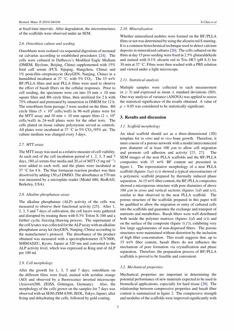
In order to study the effect of fiber content on the degradabilityof the BF/PLLA composite scaffolds, in vitro degradation testswere carried out in PBS (pH 7.4) at 37 ◦C. The degradationprocess was monitored by pH variation and scaffold poremorphology in the PBS medium. pH variation of PBS mediawith different incubation times is presented in figure 4. Afterthe first week of incubation, the pH of the incubation mediumdecreased from the initial value (7.4) in the unfilled PLLAscaffolds and in the composite filled with 5 and 10 wt% basaltfibers. In contrast, the pH of the medium slightly increased tovalues �7.4 for the 15 wt% basalt fiber composites. Fourweeks later, the pH of the medium decreased to values�7.3 in the unfilled PLLA scaffolds, because acidic groupsresulting from the degradation of PLLA decreased the pHvalue of medium. But the decreasing extent of pH variationwas much smaller for the 15 wt% BF/PLLA composites.Such differences may be correlated with the alkaline oxideconstituents of basalt fibers, such as CaO, MgO, K2O orNa2O, which could compensate for the acidification of themedium due to acidic products of the polymer degradation.It has been considered to be another benefit of using basaltfibers in composite scaffolds with the aim to avoid possibleinflammatory responses due to acidic degradation of thepolymers.
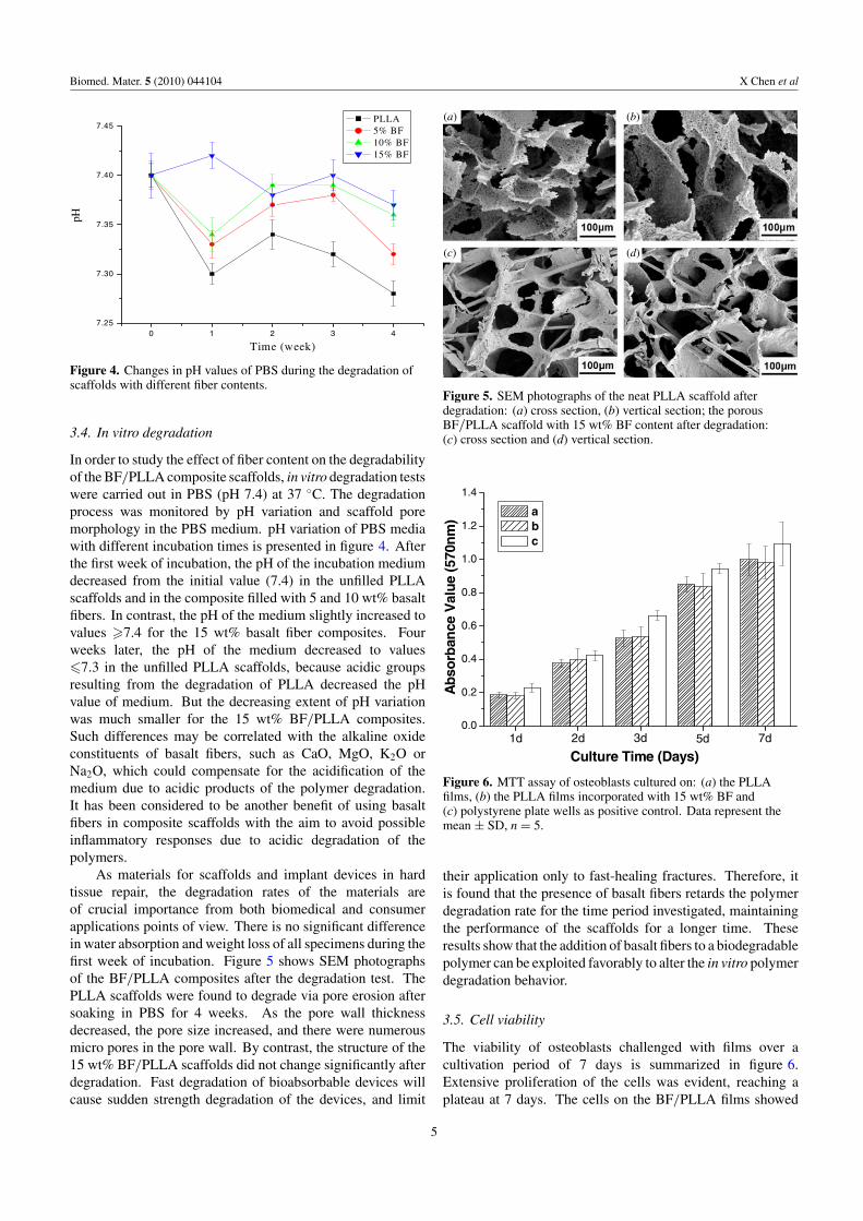
As materials for scaffolds and implant devices in hardtissue repair, the degradation rates of the materials areof crucial importance from both biomedical and consumerapplications points of view. There is no significant differencein water absorption and weight loss of all specimens during thefirst week of incubation. Figure 5 shows SEM photographsof the BF/PLLA composites after the degradation test. ThePLLA scaffolds were found to degrade via pore erosion aftersoaking in PBS for 4 weeks. As the pore wall thicknessdecreased, the pore size increased, and there were numerousmicro pores in the pore wall. By contrast, the structure of the15 wt% BF/PLLA scaffolds did not change significantly afterdegradation. Fast degradation of bioabsorbable devices willcause sudden strength degradation of the devices, and limit
(a)
(c) (d)
(b)
Figure 5. SEM photographs of the neat PLLA scaffold afterdegradation: (a) cross section, (b) vertical section; the porousBF/PLLA scaffold with 15 wt% BF content after degradation:(c) cross section and (d) vertical section.
0.0
0.2
0.4
0.6
0.8
1.0
1.2
1.4
7d5d3d2d1d
Culture Time (Days)
Ab
so
rban
ce V
alu
e (
570n
m)
a
b
c
Figure 6. MTT assay of osteoblasts cultured on: (a) the PLLAfilms, (b) the PLLA films incorporated with 15 wt% BF and(c) polystyrene plate wells as positive control. Data represent themean ± SD, n = 5.
their application only to fast-healing fractures. Therefore, itis found that the presence of basalt fibers retards the polymerdegradation rate for the time period investigated, maintainingthe performance of the scaffolds for a longer time. Theseresults show that the addition of basalt fibers to a biodegradablepolymer can be exploited favorably to alter the in vitro polymerdegradation behavior.
3.5. Cell viability
The viability of osteoblasts challenged with films over acultivation period of 7 days is summarized in figure 6.Extensive proliferation of the cells was evident, reaching aplateau at 7 days. The cells on the BF/PLLA films showed
5

Biomed. Mater. 5 (2010) 044104 X Chen et al
0.0
0.8
1.6
2.4
3.2
4.0
4.8
7d5d3d2d1d
Culture Time (Days)
AL
P a
cti
vit
y
(Kin
g-A
rmstr
on
g u
nit
/100m
L) a
b
c
Figure 7. ALP activity of osteoblasts cultured on: (a) the PLLAfilms, (b) the PLLA films incorporated with 15 wt% BF and (c)polystyrene plate wells as positive control. Data represent themean ± SD, n = 5.
high viability values, similar to that on the neat PLLA films(p > 0.05), even if the cells on tissue culture polystyrenedisplayed significantly higher values (p < 0.05). Althoughfurther studies need to be carried out to confirm the long-termcell viability, the fact that the cells can survive normally for aweek in close culture with basalt fibers is encouraging.
3.6. Alkaline phosphatase activity
After 1, 2, 3, 5 and 7 days of culture, osteoblasts on thefilms were subjected to an ALP assay. Data normalized to thestandard sample provided in the kit are presented in figure 7.All substrates exhibited sustainable growth of ALP activitiesto some extent, among which, the expression of ALP on theBF/PLLA composite and the neat PLLA followed a similarprofile (p > 0.05). These results are consistent with thoseobtained by the MTT assay. It is indicated that the presenceof BF does not noticeably harm osteoblastic activity and the
(a)
(e) (f) (g) (h)
(b) (c) (d)
Figure 8. Morphology evaluation of osteoblasts on the BF/PLLA films (a), (b), (c) and (d) and the PLLA films (e), (f ), (g) and (h) byfluorescence microscopy: most cells were adherent to their substrates after 1 day: (a) and (e); cells enlarged and entered the splitting phaseafter 3 days: (b) and (f ); 5 days later they were fully spread (c) and (g), with cell sizes ranging from 50 to 70 μm in diameter; and 7 dayslater cells grew up to the formation of a complete monolayer (d) and (h) (scale bar = 50 μm).
designed composites are osteoblast compatible for at least aweek in culture.
3.7. Cell morphology
Through fluorescent staining observation over the periodof the cell growth, the cells were found to be uniformlydistributed over all the films and markedly proliferated withthe advance of incubation intervals (figure 8). Dense andalmost confluent cells on films suggest the absence of atoxic leachable substance from the composites. SEM images(figure 9(a)) at low magnification displayed that osteoblastsgrew favorably on the BF/PLLA films and some cells mergedinto each other. Basalt fibers were found to be embedded inthe PLLA matrix, indicating a strong degree of interactionbetween the fibers and the matrix. Numerous pseudopodiaproviding intercellular connections were evident in highermagnification images (figures 9(b) and (c)).
Moreover, cells could actively sense and respond to theirenvironment. Czarnecki et al [31] noted that osteoblastsgrown on carbon fiber-reinforced phenolic composites alignedparallel to the carbon fibers. Similar results were obtainedin our previous experiment. We observed that the fibroblastsgrown on basalt fiber fabrics grew along the fiber axis andwere mainly elongated and spindle shaped, a process usuallyreferred to as contact guidance [32]. So, we may predictthat the osteoblast orientation and growth can be regulatedby a preferred basalt fiber arrangement. Nevertheless, furtherexperiments are needed to confirm the effect of osteoblastalignment induced by the basalt fibers.
3.8. Mineralization
Positive alizarin red staining for mineralized nodules is oneof the most reliable variables for evaluating the functionalbehavior of osteoblasts [26]. A large mineralized nodule wasstained heavily with alizarin red (figure 10(a)), suggesting themaintenance of osteoblastic function on the composites. The
6

Biomed. Mater. 5 (2010) 044104 X Chen et al
(a) (b) (c)
(d) (e) (f)
Figure 9. SEM images of osteoblasts after 7 days of cultivation on the BF/PLLA composite films (a), (b) and (c) as well as the PLLA films(d), (e) and (f ).
(a) (b)
Figure 10. Light micrograph of mineralized nodules from(a) BF/PLLA film and (b) neat PLLA film stained with alizarin redafter 15 days of culture (scale bar = 50 μm).
nodule that formed on the neat PLLA film was also stainedpositively (figure 10(b)).
4. Conclusion
In summary, the present study tested short basaltfibers as alternative reinforcing fillers in PLLA-basedcomposites. BF/PLLA scaffolds with interconnected poreswere successfully developed by freeze drying. Within thiscomposite, basalt fibers were found to be well incorporatedand distributed uniformly, and the presence of 15 wt%BF significantly augmented mechanical and hydrophilicproperties. The presence of basalt fibers may retard thepolymer degradation rate and neutralize the acid degradationfrom PLLA. Also, the inclusion of basalt fibers in the PLLAfilms does not noticeably affect osteoblast viability and growth.These findings are indicative of the potential and feasibility ofinnovative BF/PLLA composites in applications to hard tissuerepair. Further investigation into the osteoblast response to 3Dscaffolds is being carried out to validate the biological responseof the proposed composite scaffolds for application in hardtissue repair, and the in vivo biocompatibility and mechanicalperformance are needed and well warranted in view of the
increasing amount of interest in applying BF for biomedicalpurposes.
Acknowledgments
This work is supported by the National Basic ResearchProgram of China (2006CB933206, 2006CB705600) and theInternational Cooperation Program awarded by the Ministry ofthe Science and Technology of China (2008DFA51180). Theauthors gratefully acknowledge GBF for kindly gifting basaltfibers.
References
[1] Wan Y Z, Wang Y L, Xu X H and Li Q Y 2001 In vitrodegradation behavior of carbon fiber-reinforced PLAcomposites and influence of interfacial adhesion strengthJ. Appl. Polym. Sci. 82 150–8
[2] Slivka M A, Chu C C and Adisaputro I A 1997 Fiber-matrixinterface studies on bioabsorbable composite materials forinternal fixation of bone fractures: I. Raw materialevaluation and measurement of fiber-matrix interfacialadhesion J. Biomed. Mater. Res. 36 469–77
[3] Ramakrishna S, Mayer J, Wintermantel E and Leong K W2001 Biomedical applications of polymer-compositematerials: a review Compos. Sci. Technol. 61 1189–224
[4] Shen L, Yang H, Ying J, Qiao F and Peng M 2009 Preparationand mechanical properties of carbon fiber-reinforcedhydroxyapatite/polylactide biocomposites J. Mater. Sci.,Mater. Med. 20 2259–65
[5] Zhang D H, Kandadai M A, Cech J, Roth S and Curran S A2006 Poly(L-lactide) (PLLA)/multiwalled carbon nanotube(MWCNT) composite: characterization andbiocompatibility evaluation J. Phys. Chem. B 110 12910–5
[6] Takayama T, Todo M and Takano A 2009 The effect ofbimodal distribution on the mechanical properties ofhydroxyapatite particle filled poly(L-lactide) compositesJ. Mech. Behav. Biomed. Mater. 2 105–12
[7] Wang J, Qu L J, Meng X C, Gao J, Li H B and Wen G W 2008Preparation and biological properties of PLLA/beta-TCPcomposites reinforced by chitosan fibers Biomed. Mater.3 025004
7

Biomed. Mater. 5 (2010) 044104 X Chen et al
[8] Li X M, Liu X H, Dong W, Feng Q L, Cui F Z, Uo M,Akasaka T and Watari F 2009 In vitro evaluation of porouspoly(L-lactic acid) scaffold reinforced by chitin fibersJ. Biomed. Mater. Res. B 90 503–9
[9] Slivka M A and Chu C C 1997 Fiber–matrix interface studieson bioabsorbable composite materials for internal fixationof bone fractures: 2. A new method using laser scanningconfocal microscopy J. Biomed. Mater. Res. 37 353–62
[10] Chomyszyn-Gajewska M, Czajkowska B, Blazewicz M,Pamula E and Ptak M 2002 In vitro response ofmacrophages to a new carbon–polylactide compositefor the treatment of periodontal diseases Biomaterials23 463–70
[11] Ahmed I, Cronin P S, Abou Neel E A, Parsons A J,Knowles J C and Rudd C D 2009 Retention of mechanicalproperties and cytocompatibility of a phosphate-based glassfiber/polylactic acid composite J. Biomed. Mater. Res. B89 18–27
[12] Botev M, Betchev H, Bikiaris D and Panayiotou C 1999Mechanical properties and viscoelastic behavior of basaltfiber-reinforced polypropylene J. Appl. Polym. Sci.74 523–31
[13] Friedrich K, Fakirov S and Zhang Z 2005 Polymer Compositesfrom Nano- to Macro-scale (New York: Springer)pp 309–28
[14] Wang M C, Zhang Z G, Li Y, Li M and Sun Z J 2008Chemical durability and mechanical properties ofalkali-proof basalt fiber and its reinforced epoxy compositesJ. Reinf. Plast. Compos. 27 393–407
[15] Sim J, Park C and Moon D Y 2005 Characteristics of basaltfiber as a strengthening material for concrete structuresComposites B 36 504–12
[16] Wang G J, Liu Y W, Guo Y J, Zhang Z X, Xu M X andYang Z X 2007 Surface modification and characterizationsof basalt fibers with non-thermal plasma Surf. Coat.Technol. 201 6565–8
[17] Kogan F M and Nikitina O V 1994 Solubility of chrysotileasbestos and basalt fibers in relation to their fibrogenic andcarcinogenic action Environ. Health Perspect. 102 205–6
[18] McConnell E E, Kamstrup O, Musselman R, Hesterberg TW,Chevalier J, Miller W C and Thevenaz P 1994 Chronicinhalation study of size-separated rock and slag woolinsulation fibers in Fischer 344/N rats Inhal. Toxicol.6 571–614
[19] Subramanian R V and Austin H F 1980 Silane coupling agentsin basalt-reinforced polyester composites Int. J. Adhes.Adhes. 1 50–4
[20] Hong Z K, Reis R L and Mano J F 2008 Preparation and invitro characterization of scaffolds of poly(L-lactic acid)containing bioactive glass ceramic nanoparticles ActaBiomater. 4 1297–306
[21] Todo M, Kuraoka H, Kim J, Taki K and Ohshima M 2008Deformation behavior and mechanism of porous PLLAunder compression J. Mater. Sci. 43 5644–6
[22] Chen Y, Mak A F T, Wang M, Li J S and Wong M S 2008In vitro behavior of osteoblast-like cells on PLLA filmswith a biomimetic apatite or apatite/collagen compositecoating J. Mater. Sci., Mater. Med. 19 2261–8
[23] Liao S S, Cui F Z, Zhang W and Feng Q L 2004 Hierarchicallybiomimetic bone scaffold materials: nano-HA/collagen/PLA composite J. Biomed. Mater. Res. B69 158–65
[24] Ajami-Henriquez D, Rodriguez M, Sabino M, Castillo R V,Muller A J, Boschetti-de-Fierro A, Abetz C, Abetz Vand Dubois P 2008 Evaluation of cell affinity onpoly(L-lactide) and poly(epsilon-caprolactone) blends andon PLLA-b-PCL diblock copolymer surfaces J. Biomed.Mater. Res. A 87 405–17
[25] Kim H W, Song J H and Kim H E 2006 Bioactive glassnanofiber-collagen nanocomposite as a novel boneregeneration matrix J. Biomed. Mater. Res. A 79 698–705
[26] Gough J E, Jones J R and Hench L L 2004 Nodule formationand mineralisation of human primary osteoblasts culturedon a porous bioactive glass scaffold Biomaterials25 2039–46
[27] Jones J R, Ehrenfried L M and Hench L L 2006 Optimisingbioactive glass scaffolds for bone tissue engineeringBiomaterials 27 964–73
[28] Wahl D A and Czernuszka J T 2006 Collagen–hydroxyapatitecomposites for hard tissue repair Eur. Cells Mater. 11 43–56
[29] Mattii L et al 2008 Gelatin/PLLA sponge-like scaffolds allowproliferation and osteogenic differentiation of humanmesenchymal stromal cells Macromol. Biosci. 8 819–26
[30] Zhu N, Cui F Z, Hu K and Zhu L 2007 Biomedicalmodification of poly(L-lactide) by blending with lecithinJ. Biomed. Mater. Res. A 82 455–61
[31] Czarnecki J S, Lafdi K and Tsonis P A 2008 A novel approachto control growth, orientation, and shape of humanosteoblasts Tissue Eng. A 14 255–65
[32] Wiernann M, Bingmann D, Franzka S, Hartmann N, Urch Hand Epple M 2007 Oriented growth of osteoblast-like cellson two-dimensionally structured films of functionalizedcalcium phosphate nanoparticles on a silicon substrate Adv.Eng. Mater. 9 1077–81
8


![Flexural Behaviour of Basalt Fiber Reinforced Concrete ... · Basalt rock can also make basalt rock, chopped basalt fiber, basalt fabrics and continuous filament wire [9]. Basalt](https://static.fdocuments.net/doc/165x107/5e8d373fa059ea2b69053027/flexural-behaviour-of-basalt-fiber-reinforced-concrete-basalt-rock-can-also.jpg)


