BasaloidSquamousCellCarcinoma of the Esophagus ... · Keywords:basaloidsquamouscellcarcinoma,...
7
354 J Nippon Med Sch 2008; 75 (6) ―Case Reports― Basaloid Squamous Cell Carcinoma of the Esophagus: Report of Two Cases Ichiro Akagi, Masao Miyashita, Hiroshi Makino, Tsutomu Nomura, Keiichi Ohkawa and Takashi Tajiri Surgery for Organ Function and Biological Regulation, Graduate School of Medicine, Nippon Medical School Abstract Basaloid squamous cell carcinoma is an uncommon malignancy of the esophagus. We present two cases of basaloid squamous carcinoma of the esophagus. Both tumors histologically consisted of solid cell nests and displayed focal immunoreactivity for type IV collagen. The nests comprised pseudoglandular structures containing myxoid matrix. Transthoracic esophagectomy with lymph node dissection was performed in both patients. The patients had uneventful postoperative courses. One patient showed no evidence of recurrence or metastasis in the 6-month postoperative period, and the other patient died of lung metastasis 28 months after the primary treatment. (J Nippon Med Sch 2008; 75: 354―360) Key words: basaloid squamous cell carcinoma, esophagus, operation Introduction Basaloid squamous cell carcinoma (BSC) is a rare histological tumor type that was first described by Wain et al. in 1986 and which occurs predominantly in the upper respiratory and digestive tracts 1 . However, BSC of the esophagus is a rare tumor, accounting for only 0.068% to 4.0% of all esophageal carcinomas 2―4 , and, to date, only 60 cases of esophageal BSC have been reported in Japan 5 . BSC is characterized by a poor degree of differentiation, high proliferative activity 6 , high biological malignancy 3,7,8 , high incidence of distant metastases, and a high rate of spontaneous apoptosis 6,7 . In general, BSC of the esophagus has a poor prognosis; however, a recent report has suggested that the prognosis after curative resection might not differ significantly from that of typical squamous cell carcinoma 3,6―8 . Herein, we report two cases of esophageal BSC that were treated with curative resection. Case 1 A 61-year-old man was admitted to our department because of an abnormality of the esophagus detected on esophagography during a medical checkup. Physical examination revealed no abnormalities, and the laboratory data were also within normal limits, except for elevation of serum squamous cell carcinoma (SCC) antigen level (1.7 ng! mL). Esophagography showed a protruding lesion 5.0 cm in diameter with a nodular surface in the Correspondence to Ichiro Akagi, MD, Department of Surgery for Organ Function and Biological Regulation, Nippon Medical School, 1―1―5 Sendagi, Bunkyo-ku, Tokyo 113―8603, Japan E-mail: [email protected] Journal Website (http:!! www.nms.ac.jp! jnms! )
Transcript of BasaloidSquamousCellCarcinoma of the Esophagus ... · Keywords:basaloidsquamouscellcarcinoma,...

354 J Nippon Med Sch 2008; 75 (6)
―Case Reports―
Basaloid Squamous Cell Carcinoma of the Esophagus:
Report of Two Cases
Ichiro Akagi, Masao Miyashita, Hiroshi Makino,Tsutomu Nomura, Keiichi Ohkawa and Takashi Tajiri
Surgery for Organ Function and Biological Regulation, Graduate School of Medicine, Nippon Medical School
Abstract
Basaloid squamous cell carcinoma is an uncommon malignancy of the esophagus. Wepresent two cases of basaloid squamous carcinoma of the esophagus. Both tumorshistologically consisted of solid cell nests and displayed focal immunoreactivity for type IVcollagen. The nests comprised pseudoglandular structures containing myxoid matrix.Transthoracic esophagectomy with lymph node dissection was performed in both patients.The patients had uneventful postoperative courses. One patient showed no evidence ofrecurrence or metastasis in the 6-month postoperative period, and the other patient died oflung metastasis 28 months after the primary treatment.(J Nippon Med Sch 2008; 75: 354―360)
Key words: basaloid squamous cell carcinoma, esophagus, operation
Introduction
Basaloid squamous cell carcinoma (BSC) is a rarehistological tumor type that was first described byWain et al. in 1986 and which occurs predominantlyin the upper respiratory and digestive tracts1.However, BSC of the esophagus is a rare tumor,accounting for only 0.068% to 4.0% of all esophagealcarcinomas2―4, and, to date, only 60 cases ofesophageal BSC have been reported in Japan5.
BSC is characterized by a poor degree ofdifferentiation, high proliferative activity6, highbiological malignancy3,7,8, high incidence of distantmetastases, and a high rate of spontaneousapoptosis6,7. In general, BSC of the esophagus has apoor prognosis; however, a recent report has
suggested that the prognosis after curative resectionmight not differ significantly from that of typicalsquamous cell carcinoma3,6―8. Herein, we report twocases of esophageal BSC that were treated withcurative resection.
Case 1
A 61-year-old man was admitted to ourdepartment because of an abnormality of theesophagus detected on esophagography during amedical checkup. Physical examination revealed noabnormalities, and the laboratory data were alsowithin normal limits, except for elevation of serumsquamous cell carcinoma (SCC) antigen level (1.7 ng�mL). Esophagography showed a protruding lesion 5.0cm in diameter with a nodular surface in the
Correspondence to Ichiro Akagi, MD, Department of Surgery for Organ Function and Biological Regulation, NipponMedical School, 1―1―5 Sendagi, Bunkyo-ku, Tokyo 113―8603, JapanE-mail: [email protected] Website (http:��www.nms.ac.jp�jnms�)

Basaloid SCC of the Esophagus
J Nippon Med Sch 2008; 75 (6) 355
Fig. 1 The esophagogram shows a protruding, 8.0-cm-diameter lesion with a nodular surface at the posterior side of the middle esophagus.
posterior wall of the middle third of the esophagus(Fig. 1). Upper gastrointestinal endoscopy showed atumorous lesion occupying approximately half of theesophageal lumen, located 25 cm from the incisors(Fig. 2A, B). The tumor was reddish, with anindistinct margin. Endoscopic ultrasonographyrevealed that the tumor was a semicircumferentiallesion and suggested that the tumor might haveinvaded the muscularis propria (Fig. 2C). Histologicalexamination of biopsy specimens from the tumorsurface suggested poorly differentiated SCC.Computed tomography revealed a thickening of theesophageal wall but no evidence of invasion to thesurrounding organs.
After admission, curative resection withtransthoracic esophagectomy and lymphaticdissection of the neck, mediastinum, and abdomenwas performed, followed by cervicalesophagogastrostomy with a gastric tube pulled viathe retromediastinal route. Grossly, the tumor had
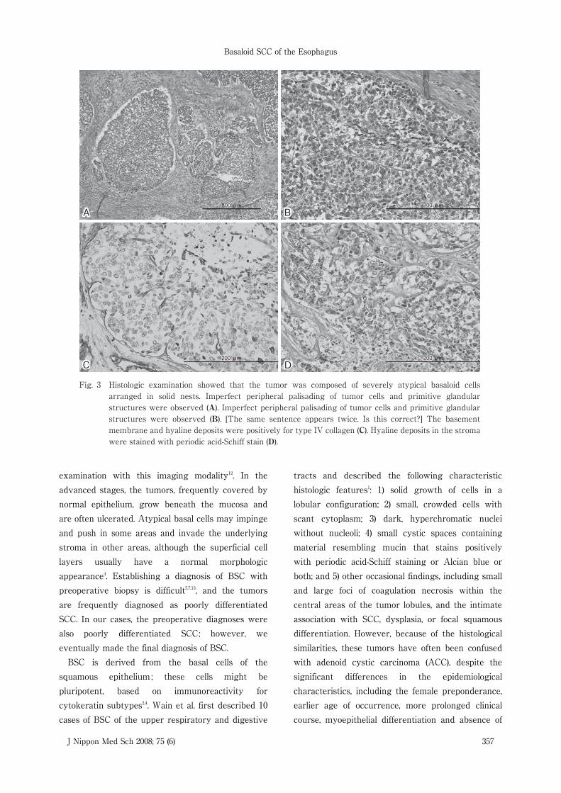
an infiltrative, cauliflower-like appearance. Itmeasured 45 × 35 mm and was located in the middlethird of the thoracic esophagus. Microscopicexamination of the tumor showed severely atypicalbasaloid cells arranged in solid nests. Imperfectperipheral palisading of tumor cells and primitiveglandular structures were observed (Fig. 3A, B).The carcinoma had infiltrated the lamina propriauntil the deeper parts of the submucosal layer, andalthough the tumor strongly impinged on themuscularis propria, there was no evidence ofinvasion of this layer. The regional lymph nodeswere negative for metastasis. Histochemical studiesshowed that the tumor cells expressed type IVcollagen and were positive for periodic acid-Schiffstaining but were not positive for chromogranin A,Elastica Masson Goldman (EMG), synaptophysin, orCongo red (Fig. 3C, D). The postoperative coursewas uneventful. No chemotherapy or radiotherapywas performed postoperatively, and the regularfollow-up has revealed no evidence of recurrence ormetastasis until the time of writing, 6 months afterthe surgery.
Case 2
A 60-year-old woman was admitted to ourdepartment because of an abnormality of theesophagus detected on esophagography during amedical checkup. Physical examination revealed noabnormalities, and the laboratory data were alsowithin normal limits. Esophagography showed anirregularly shaped lesion 3.0 cm in diameter in theposterior wall of the middle of the esophagus (Fig.4). Upper gastrointestinal endoscopy showed anulcerated lesion that was located 25 cm from theincisors and was negative for iodine staining.Endoscopic ultrasonography suggested that thetumor might have partially invaded the muscularispropria. Histological examination of biopsyspecimens of the tumor surface suggested poorlydifferentiated SCC. Computed tomography revealedno evidence of invasion or metastasis.
After admission, curative resection withtransthoracic esophagectomy and lymphaticdissection of the neck, mediastinum, and abdomen

I. Akagi, et al
356 J Nippon Med Sch 2008; 75 (6)
Fig. 2 Upper gastrointestinal endoscopy showed a elevated leision 25 cm from the incisors which occluded the esophageal lumen (A). The tumor area on endoscopy is not stained with lugol (B). Endoscopic ultrasonography showed the tumor occupying half of the esophageal lumen and suggested that the tumor had invaded the muscularis propria (C).
A B
C
was performed, followed by cervicalesophagogastrostomy with a gastric tube pulled viathe retromediastinal route. The postoperative coursewas uneventful. The ulcerated tumor measured 21 ×20 mm and was located in the middle third of theesophagus (Fig. 5). Histologic examination showedthat the tumor was composed of basaloid cells (Fig.6A). The carcinoma had infiltrated the muscularispropria, and the regional lymph nodes were positivefor metastasis. Basaloid components wereimmunohistochemically positive for type IV collagenand were stained with periodic acid-Schiff stain (Fig.6B, C). The postoperative course was uneventful. Nochemotherapy or radiotherapy was performedpostoperatively. The patient died of lung metastasis28 months after the primary treatment.
Discussion
BSC of the esophagus is a rare epithelialmalignancy of the esophagus2―4. The epidemiologicalcharacteristics of BSC do not significantly differ
from those of SCC in that BSC also occurspredominantly in the middle third of the esophagusin men aged 60 years6,7. The growth pattern of BSCis downward and expansive even at an early stage,and intraepithelial growth is rare5. On radiologicalexamination, BSC is characterized by a smoothnodular surface and gentle lines representing thegutters between the nodules, differing in appearancefrom SCC9. Superficial BSC often assumes a formsimilar to that of submucosal tumors10, or the 0―Itype tumors in the endoscopic classification based onthe guidelines for Clinical and Pathological Studies ofthe Japanese Society for Esophageal Disease11.Recently, magnifying endoscopy has becomeavailable at some institutions, and this modality isexpected to contribute to the early diagnosis of BSC.Using magnifying endoscopy, Tomori et al reportedavascular areas on the surface of BSCscorresponding to solid cell nests and unusual vesselssurrounding the avascular areas corresponding tothe vessels between the BSC nests, which mayrepresent characteristic findings of BSC on
Basaloid SCC of the Esophagus
J Nippon Med Sch 2008; 75 (6) 357
200200 μm200 μm 200200 μm200 μm
200200 μm200 μm50000 μm500 μm
Fig. 3 Histologic examination showed that the tumor was composed of severely atypical basaloid cells arranged in solid nests. Imperfect peripheral palisading of tumor cells and primitive glandular structures were observed (A). Imperfect peripheral palisading of tumor cells and primitive glandular structures were observed (B). [The same sentence appears twice. Is this correct?] The basement membrane and hyaline deposits were positively for type IV collagen (C). Hyaline deposits in the stroma were stained with periodic acid-Schiff stain (D).
A B
C D
examination with this imaging modality12. In theadvanced stages, the tumors, frequently covered bynormal epithelium, grow beneath the mucosa andare often ulcerated. Atypical basal cells may impingeand push in some areas and invade the underlyingstroma in other areas, although the superficial celllayers usually have a normal morphologicappearance4. Establishing a diagnosis of BSC withpreoperative biopsy is difficult5,7,13, and the tumorsare frequently diagnosed as poorly differentiatedSCC. In our cases, the preoperative diagnoses werealso poorly differentiated SCC; however, weeventually made the final diagnosis of BSC.
BSC is derived from the basal cells of thesquamous epithelium ; these cells might bepluripotent, based on immunoreactivity forcytokeratin subtypes2,4. Wain et al. first described 10cases of BSC of the upper respiratory and digestive
tracts and described the following characteristichistologic features1: 1) solid growth of cells in alobular configuration; 2) small, crowded cells withscant cytoplasm; 3) dark, hyperchromatic nucleiwithout nucleoli; 4) small cystic spaces containingmaterial resembling mucin that stains positivelywith periodic acid-Schiff staining or Alcian blue orboth; and 5) other occasional findings, including smalland large foci of coagulation necrosis within thecentral areas of the tumor lobules, and the intimateassociation with SCC, dysplasia, or focal squamousdifferentiation. However, because of the histologicalsimilarities, these tumors have often been confusedwith adenoid cystic carcinoma (ACC), despite thesignificant differences in the epidemiologicalcharacteristics, including the female preponderance,earlier age of occurrence, more prolonged clinicalcourse, myoepithelial differentiation and absence of

I. Akagi, et al
358 J Nippon Med Sch 2008; 75 (6)
Fig. 4 The esophagogram showed an irregularly shaped, 3.0-cm-diameter lesion in the posterior wall of the middle of the esophagus (arrows).
Fig. 5 The ulcerated tumor measured 21 × 20 mm and was located in the middle third of the esophagus.
association with SCC in the case of ACCs1,14―17. On theother hand, Kobayashi et al examined thehistological features of 36 cases of BSC and reportedthat none of the cases showed the ACC (cribriform)pattern exclusively and that all contained, in variousdegrees, the heterogeneous components of BSC.Therefore, they suggested that ACC may representa subtype of BSC showing differentiation toward thestructure of the esophagus18. Considering thesereports, the criteria for confirming the diagnosis ofBSC of the esophagus are still being debated.
Immunohistochemical examination is useful forconfirming the diagnosis of BSC3,6―8. Increasedexpression of type IV collagen in the basementmembrane is a common finding and indicatessufficient adhesibility19. Moreover, Takubo et al. havesuggested that staining with antibodies againstcollagen and laminin is useful for identifying thebasement membrane rich in these substances3. It has
also been reported that BSC has mucinous contentswhich are stained with periodic acid-Schiff stain. Inour cases, immunoreactivity for type IV collagen andperiodic acid-Schiff staining were observed.
In general, BSC has been reported to be poorlydifferentiated, to have high proliferative activity, tobe biologically highly malignant3,5,7,13,20, and to be morefrequently associated with hematogenous recurrencethan with local or lymphatic recurrence7,13,20. Themost frequent sites of metastasis are the lung andliver3; however, metastasis to the thoracic spine orgingiva has also been reported3,7. Therefore, it hasbeen suggested that BSC has a poor prognosis3,7,8,and the estimated overall 3-year survival rate is28.5%21.
In contrast, other reports suggest that for thesame clinical stage, site of origin, and treatment, theprognosis of BSC does not differ significantly fromthat of the more common SCC of the esophagus6,13. Ina comparison of 17 cases of esophageal BSC with 133cases of esophageal SCC, Sarbia et al. found that theprognosis of patients with BSC did not differsignificantly from that of patients with SCC whencurative resection was performed6. In our case 1,until the time of writing, 6 months after the surgery,there has been no evidence of recurrence ormetastasis. However, in our case 2, the patient had apoor outcome.
Postoperative radiotherapy and chemotherapyhave been performed in some institutions4,6,13,20.However, no consensus has been reached regardingthe efficacy or usefulness of such treatmentsbecause of the limited number of cases. Larner et al.have reported that radiation therapy, either alone or

Basaloid SCC of the Esophagus
J Nippon Med Sch 2008; 75 (6) 359
Fig. 6 Histologic examination showed that the tumor was composed of bundles of basaloid cells (A). The basaloid components were immunohistochemically positive for type IV collagen and were stained with periodic acid-Schiff stain (B, C).
A B
C
in combination with surgery, achieves a local controlrate of 80%20, but that distant metastases are,however, not controlled in 60% of patients. Luna etal. have suggested that adjuvant chemotherapy maybe warranted because of the high incidence ofdistant metastases13. Tsuchiya et al. have reported acase of esophageal BSC that was effectively treatedby combined chemotherapy22. On the other hand,Sarbia et al. have suggested that the role ofchemotherapy in the treatment of BSC of theesophagus remains to be established6. Therefore,BSC is currently treated like typical SCC, i.e., bysurgery, radiotherapy, or a combination of both. Forour patients, we did not perform adjuvant therapy.Further accumulation of cases is necessary toestablish effective and concrete protocols for thetreatment of this cancer.
In summary, we have reported two cases of BSCof the esophagus, which is rare disease.Immunohistochemical study of the resected tumor
was useful for establishing the final diagnosis.
References
1.Wain SL, Vollmer RT, Bossen EH: Basaloid-squamous carcinoma of the tongue, hypopharynx,and larynx. Human Pathology 1986; 17: 1158―1166.
2.Rubio CA: The histogenesis of the microinvasivebasal cell carcinoma of the esophagus. Pathol ResPract 1990; 186: 223―227.
3.Takubo K, Mafune K, Tanaka Y, Miyama T, FujitaK: Basaloid-squamous carcinoma of the esophaguswith marked deposition of basement membranesubstance. Acta Pathol Jpn 1991; 411: 59―64.
4.Abe K, Sasano H, Itakura Y, Nishihira T, Mori S,Nagura H: Basaloid-squamous carcinoma of theesophagus. A clinicopathologic, DNA ploidy, andimmunohistochemical study of seven cases. Am JSurg Pathol 1996; 204: 453―461.
5.Kato T, Morita T, Fujita M, et al.: Basaloid-squamouscarcinoma of the esophagus: report of a case. SurgToday 2000; 302: 163―167.
6.Sarbia M, Verreet P, Bittinger F, et al.: Basaloidsquamous cell carcinoma of the esophagus: diagnosisand prognosis. Cancer 1997; 79: 1871―1878.
7.Ide F, Shimoyama T, Haga H, Horie N: Basaloid

I. Akagi, et al
360 J Nippon Med Sch 2008; 75 (6)
squamous cell carcinoma of the esophagusmetastatic to the gingiva: a case report. Oral SurgOral Med Oral Pathol Oral Radiol Endod 1997; 835:584―587.
8.Koide N, Koike S, Adachi W, Amano J, Usuda N,Nagata T: Immunohistochemical expression of bcl-2protein in squamous cell carcinoma and basaloidcarcinoma of the esophagus. Surg Today 1997; 278:685―691.
9.Imai Y, Nagashima R, Makuuchi H, Shimada H:Special Histopathologic Type of EsophagealCarcinoma: Point of View from the Radiologicalfindings. Stomach and Intestine 2005; 403: 301―309.
10.Kuwano H, Watanabe M, Yasuda M, Nozoe T,Sugimachi K: Oesophageal cancer composed ofmixed histological types. Eur J Surg Oncol 1996; 22:225―231.
11.Noguchi H, Naomoto Y, Kondo H: Evaluation ofendoscopic mucosal resection for superficialesophageal carcinoma. Surg Laparosc EndoscPercutan Tech 2000; 10: 343―350.
12.Tomori A: BSC with Characteristic Vascular Patternby Magnifying Endoscopy, Report of a Case.Stomach and Intestine 2005; 403: 397―402.
13.Luna MA, Parichatikanond P, Weber RS, BatsakisJG: Basaloid squamous carcinoma of the upperaerodigestive tract. Clinicopathologic and DNA flowcytometric analysis. Cancer 1990; 66: 537―542.
14.Batsakis JG: Basaloid-squamous carcinomas of theupper aerodigestive tracts. Ann Otol RhinolLaryngol 1989; 17: 1158―1166.
15.Tsang WY, Chan JK, Lee KC, Leung AK, Fu YT:Basaloid-squamous carcinoma of the upperaerodigestive tract and so-called adenoid cysticcarcinoma of the oesophagus: the same tumour
type? Histopathology 1991; 191: 35―46.16.Banks ER, Mills SE, George E, Zarbo RJ, Swanson
PE: Basaloid squamous cell carcinoma of the headand neck. A clinicopathologic andimmunohistochemical study of 40 cases. Am J SurgPathol 1992; 16: 939―946.
17.Barnes L, Altavilla G, MacMillan C, Rinaldo A,Doglioni C: Basaloid squamous cell carcinoma of thehead and neck: Clinicopathologic features anddifferential diagnosis. Ann Otol Rhinol Laryngol 1996;105: 75―82.
18.Kobayashi Y, Nakanishi Y, et al.: Wide HistrogicalSpectrum of Basaloid Squamous Cell Carcinoma ofthe Esophagus. Stomach and Intestine 2005; 403:371―379.
19.Ohashi K, Horiguchi S, Moriyama S, et al.: Superficialbasaloid squamous carcinoma of the esophagus. Aclinicopathological and immunohistochemical studyof 12 cases. Pathol Res Pract 2003; 199: 713―721.
20.Larner JM, Mills SE, Frierson HF Jr, Banks ER,Levine PA: Radiotherapy for basaloid squamous cellcarcinoma of the head and neck. Head Neck 1993; 15:249―252.
21.Ereno C, Sanchez JM, Toledo JD: Basaloid-squamouscell carcinoma od the larynx and hypopharynx: aclinicopathologic study of 7 cases. Pathol Res Pract1994; 1902: 186―193.
22.Tsuchiya Y, Onda M, Miyashita M, et al.: Combinedmodality therapy for basaloid squamous carcinomaof the esophagus. Hepatogastroenterology 1999; 46:2868―2871.
(Received,(Accepted,
JulyAugust
4, 2008)20, 2008)