Ballistocardiography as a Technique ... - spo.nmfs.noaa.govspo.nmfs.noaa.gov/mfr364/mfr3644.pdf ·...
6
Suwa. K., and H. H. Be ndix en. 1972. Pul- monary gas exchange in a tid a ll y ventilat- ed single alveolus mode l. J. Appl. Ph y iol. 32:834-84 1. Tenney. S. M ., and J. E. Remmers. 1963. Comparative quantitative morphology of the mamma li an lung : diffusion area. Nat ur e 197 :54-56. Wah on. E. H. and (1. H . I "''1e\ 19t-2 Gro\\th and deHlopment "I ,hold.en 4th ed., Year Boo" l\Iedll.:al Pur.l"he. Inc Ch .eago. 384 p. MFR Paper 1045. Fr om Marine Fisheries Review, Vol. 36, N o.4 , April 1974. Copies of this paper , in limited numbers , are available from 08 3, Technical Inf ormation Divi sion , Env ironmental S cience Informati on Center, NOAA , Washington , DC 20235. MFR PAPER 1046 Ballistocardiography as a Technique for Comparative Physiology N. TY SMITH and ERIC A. WAHRENBROCK ABSTRACT Th e IIltra low -fr e qu en cy ballist ocar di og ram was recorded on a YO llng Califor- nia gray whale. Th e Iracing is remarkabl y s imilar 10 Ih ose obtained from man and m ouse, bOlh in a mplill/de and in forll1. Th e IJ all1pliludes for 1I101lse. man. and whal e were 2.6, 4.3, a nd 4.6 c m/sec 2 . We co ncilld e Ihal grearer are ca ll sed by p oo r reco rdin g l ec hniqu e o r by di sease Ihan by species differences. The major inlerspecies diff eren ces we re see n in Ihe liming of cardiac el'ents. s ll ch as pr eejec lion or ejec lion lim e. Th ese differenc es could he caused hy differenc es in hearl size. The ba lli stocardiograp h (Bcg) is a dev ice for eva lu a tin g th e m ec ha nic al fu ncti on of the h eart. It h as been recor ded in an i ncr ed i bl e a rr ay of a nim a ls, rangin g from egg embryos to ca ttle . One of the more i nteresti ng facts to a ri se from these recordi ngs is th at the tracin gs a re remarkabl y s imilar among species , particularl y ma mmals . Thi s s imil ar ity holds both in form a nd in amp litud e. It was ther efore an exce ll e nt o pp o rtunit y to N. Ty Smith is an Associat e Professor of Anesthesia at the University of California at San Diego, Veterans Administration Hospital , San Diego , CA 92161, and Eric A. Wahrenbrock is an Assistant Professor of Anesthesia at the University of California, San Diego. exte nd these observations to Gigi. an a nim al with an entirely different ma ss a nd configu r ation from other mamm a ls previously u sed. Th e Bcg records the movements of th e body caused by movements of bl ood in the body. First recorded in 1 887. the Bcg has und ergone a series of up s and downs in its attempts to become a usefu I tool for measuri ng card i ovascular f un ction noninva ivel). Not un til the 1950's when ph) icists and engineers entered the field. did the Bcg finally re-emerge a an accurate, relat ively simple technique . Essentially, the Bcg v. or\...s on the principle that an attempted shift in the center of mass of a floating hod) IS compensated for by a mO\'ement of the body in the opposite direction. so that the center of remain con tant in 9 relation to a fixed pOint. Thu,. II hlond moves in one direction alter e.lectlt1n by the left ventricle. the hOlh \\ ill move in the opposite direction fhe,e movement are quite small. hut the reader ha s certainl) noticed a ,light bodily movement as he qLl1etl) on a bed or a s li ght 1110\ emenl \)1 the point er on a weighing ,calc . movement ""Ith the heart beat. Thi minute bod) n1\)\e- ment can be recorded dl,pl.lce ment, velocity. or acceleration f-igure I shows examples of normal tracing in man. The important fact to note is that the major componenh l)f the Bcg occur during ejection of bl\)\)d, parti cularly during the early portion. METHODS When the ph) the field. the) standards for ical scientl\ts entered laid dov. n certain recording the lk g, standards which were to con\ert bal- listocardiography from a haphazard technique to a precise one. The Ilr I requirement is that a light hed necessar). in contrast to the hea\\ one formerl) used A. rat 10 \)1 I (I I for ubjecl.bed 1\ mlnlm.ll 'nJ coupling. or binding . 01 suhJecl 1\) bed must be as tight a, Pl) "hie. fhlrd . coupling to ground 11lu\t he nllnlmal. so that amhlent \ibrall\)n can be attenuated. The Beg I an e tremel\ In,trument. Pea" til pi u:- ment IS a bl1ut I ()Op. pea" dCLek·r.lt Ion a fe\\ 11lillig ·. g being the ,lcLc\era- tlon of gra\Jt\ \\ IIh Ida In trument \Jbratll1n from a truL" out Ide the buddtng ahle to de Ir \ a bal-
Transcript of Ballistocardiography as a Technique ... - spo.nmfs.noaa.govspo.nmfs.noaa.gov/mfr364/mfr3644.pdf ·...
Suwa. K. , and H. H . Bendixen. 1972. Pulmonary gas exchange in a tida lly ventil ated single alveolus model. J . Appl. Phy iol. 32:834-84 1.
T enney. S. M ., and J. E. Remmers. 1963. Comparative quantitative morphology of the mammali an lung : diffusion area. N ature 197:54-56.
Wahon. E. H . and (1. H . I "''1e\ 19t-2 Gro\\th and deHlopment "I ,hold.en 4th ed., Year Boo" l\Iedll.:al Pur.l"he. Inc Ch .eago. 384 p.
MFR Paper 1045. From Marine Fisheries Review, Vol . 36, No.4, April 1974. Copies o f this paper, in limited numbers , are available from 083, Technical Information Division , Environmental Science Information Center, NOAA , Washington , DC 20235.
MFR PAPER 1046
Ballistocardiography as a Technique for Comparative Physiology
N. TY SMITH and ERIC A. WAHRENBROCK
ABSTRACT
Th e IIltra low-frequ ency ballistocardiogram was recorded on a YOllng California gray whale. Th e Iracing is remarkably similar 10 Ih ose obtained from man and m ouse, bOlh in amplill/de and in forll1. Th e IJ all1pliludes for 1I101lse. man. and whale were 2.6, 4.3, and 4.6 cm/sec2 . W e concillde Ihal grearer difference~ are ca ll sed by poor recording lechniqu e o r by disease Ihan by species differences. The major inlerspecies differences were seen in Ihe liming of cardiac el'ents. sll ch as preejec lion or ejeclio n lim e. Th ese differences could he caused hy differences in hearl size.
The ba lli stocardiograph (Bcg) is a device for eva lua tin g th e mecha nica l fu ncti on of th e heart. It has been recorded in an i ncredi bl e a rray of anima ls, rangin g from egg embryos to cattle . One of the more i nteresti ng facts to a ri se from these recordi ngs is th at the tracin gs a re remarkabl y similar among species , particularl y mammals . This s imil ar it y ho lds bo th in form a nd in amplitude. It was th erefore a n excell e nt o ppo rtunit y to
N. Ty Smith is an Associate Professor of Anesthesia at the University of California at San Diego, Veterans Administration Hospital, San Diego, CA 92161, and Eric A. Wahrenbrock is an Assistant Professor of Anesthesia at the University of California, San Diego.
extend these observations to Gigi. a n a nim a l with an entirely different mass a nd configu ration from other mammals previously used.
The Bcg records the movements of the body caused by movements of bl ood in the body. First recorded in 1887. the Bcg has undergone a series of ups and downs in its attempts to become a usefu I tool for measuri ng card iovascular function noninva ivel). Not un til the 1950's when ph) icists and engineers entered the field. did the Bcg finally re-emerge a an accurate, re lat ively simple technique .
Essentially, the Bcg v. or\...s on the principle that an attempted shift in the center of mass of a floating hod) IS compensated for by a mO\'ement of the body in the opposite direction. so that the center of ma~s remain con tant in
9
relation to a fixed pOint. Thu,. II hlond moves in one direction alter e.lectlt1n by the left ventricle. the hOlh \\ ill move in the opposite direction fhe,e movement are quite small. hut the reader has certainl) noticed a ,light bodily movement as he lie~ qLl1etl) on a bed or a s li ght 1110\ emenl \)1 the pointer on a weighing ,calc. eae~ movement synchronou~ ""Ith the heart beat. Thi minute bod) n1\)\ement can be recorded a~ dl,pl.lce ment, velocity. or acceleration f-igure I shows examples of normal tracing in man. The important fact to note is that the major componenh l)f the Bcg occur during ejection of bl\)\)d, particularly during the early portion.
METHODS
When the ph) the field. the) standards for
ical scientl\ts entered laid dov. n certain
recording the lk g, standards which were to con\ert ballistocardiography from a haphazard technique to a precise one. The Ilr I
requirement is that a \er~ light hed necessar). in contrast to the hea\\
one formerl) used A. rat 10 \)1 I (I I for ubjecl.bed 1\ mlnlm.ll ~LL 'nJ coupling. or binding. 01 suhJecl 1\) bed must be as tight a, Pl) "hie. fhlrd . coupling to ground 11lu\t he nllnlmal. so that amhlent \ibrall\)n can be attenuated. The Beg I an e tremel\ sen~ltJ\e In,trument. Pea" til pi u:ment IS a bl1ut I ()Op. pea" dCLek·r.lt Ion a fe\\ 11lillig·. g being the ,lcLc\eratlon of gra\Jt\ \\ IIh Ida In trument \Jbratll1n from a truL" out Ide the buddtng \~ere ahle to de Ir \ a bal-

A
v
0
Phono
EKG
L' Htor'
R! Hoor!
listocardiographic recording . Finally, the natural freq uency of the entire system should be as low as possible-0 .3 Hz or less is mandatory. These four requirements imply that the ideal Bcg system is one in which subject and bed float as a unit in space .
Several ingenious systems , some simple, some complex , have been assembled to accomplish the above requirements . Beds have been con-tructed from aluminum and canvas,
styrofoam, bal a, or a luminum honey-
Figure 1. - Examples of normal ballistocardiographic tracings in man . From top to bottom are recorded acceleration (A) , velocity (V), and displacement (0) . In addition, the EKG and the major events of the cardiac cycle are given as reference pOints . (From Scarborough et aI. , Am . J . Cardiol. 2:613-641, 1958 .)
I t 1 1.0 m
I -.L
-..-20,u
1..
0.1
He
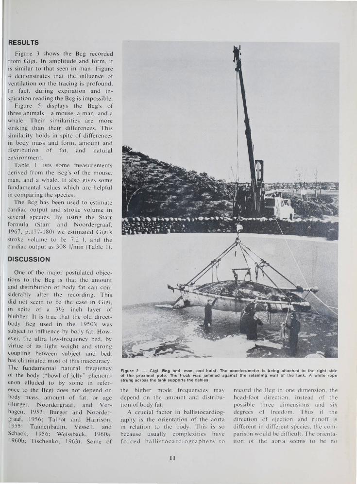
comb, and suspended by wires or floated on mercu ry or ai r. The si mplest and origi nal bed i based on the pendu lum , and was the type used in this study. The Bcg bed was the ame stretcher used to weigh Gigi (Figure 2). The stretche r was constructed from canvas and two 20-foot heavy wal l, galvan ized steel pipes 3 inches in diameter. The total weight of 227 kg may seem large to most ballistocardiogra phers, but Gigi's weight at the time was 4,500 kg, and th e whale:
10
bed ratio of 20: I wa more than adequate. Six rope supported the poles, four at the ends, each 13 feet in length , a nd two in the middle . A board inserted between the two middle ropes prevented injury to the animal. The six ropes were su pended by a ingle cable from a crane . During the
recording the cable was 71/2 meters from pulley to hook, giving a natural frequency of about 0 . 18 Hz. The crane was part of a truck hoi st, which was ideal for iso lation from ground because of the pneumatic lift and the rubber tires.
Most of the water was drained from Gigi's tank to reduce her mobility and to enhance our own . She was reluctant to lie on the bed , and had to be coaxed. The coaxing process took 45 minutes. Once on the bed , he became surprisingly quiet, which was fortunate, since she cou ld easily have demolished our fragi Ie accelerometer. One readju tment of the relative position of whale and bed was required to level the bed .
Acceleration wa transduced \0
the head-foot direction with an Endevco 1 piezo-re istive accelerometer clamped to one of the steel poles with a large C clamp. The accelerometer was calibrated with a pendulum, accordi ng to the method of Moss (196 I). Lead two of the ECG was recorded using 4 inch 18 g spinal needles. All electrical cables were supported by a rope stretched across the tank . A 60 Hz passive notch filter and a 50 Hz low pass Butterworth filter were used on both the ECG and Bcg to eliminate unwanted noise and at the same time preserve timing relations . Data were recorded on a Hewlett-Packard oscilloscope and an Ampex FM tape recorder .
'Use of trade names In thiS publlcallon does not Imply endorsement of com merCia l products by the Nallonal Marine Fisheries Service .
RESULTS
Figure 3 shows the Bcg recorded from Gigi. In amplitude and form, it is similar to that seen in man . Figure 4 demonstrates that the influence of ve ntilation on the tracing is profound . In fact. during expiration and inspiration reading the Bcg is imposs ibl e.
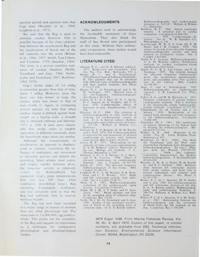
Figure 5 displays the Bcg's of t hree a nimals-a mouse, a man , a nd a whale. Their simil a riti es are mo re strikin g th an their differences. This similarit y ho lds in spite of differences in body mass and form , amount a nd distribution of fat, and natural environment.
Table I li sts some measure ments derived from the Bcg' of th e mouse, man, and a whale . It a lso gives some fundamental va lues which are help ful in comparing the species.
The Bcg has been used to estim ate cardiac output and stroke volume in several pecies . By using th e Starr formula (Starr and N oordergraaf. 1967, p .I77-180) we estimated Gigi 's troke volume to be 7 .2 I. and th e
cardiac output as 308 I/min (Tabl e I).
DISCUSSION
One of the major postulated objections to the Bcg is th at the amount and distribution of body fat can considerabl y alter th e recording. This did not seem to be the case in Gigi, in spite of a 3Y2 inch layer of blubber . It is true that the old directbody Bcg used in the 1950's wa subject to influence by body fat. However , the ultra low-frequency bed, by virtue of its li ght weight and strong couplin g between subject a nd bed, has elimin ated most of thi s inaccuracy . The fundamental natural frequency of the body ("bowl of jelly" phenomenon alluded to by some in reference to the Bcg) does not depend on body mass, amount o f fat. or age (Burger, Noordergraaf. a nd Verhagen , 1953 ; Burger a nd N oordergraaf. 1956; Talbot and H arri son. 1955; Tannenbaum, Vessell. a nd Schack. 1956; Weiss back , 1960a. 1960b; Tischenko, 1963). Some of
Figure 2. - Gigi , Beg bed , man , and hoist. The accelerometer is being attached to the right side of the proximal pole . The truck was jammed against the retaining wall of the tank. A white rope strung across the tank supports the cables .
the higher mode frequencies may depend on the amount and distribution of body fat.
A crucial factor in ballistocardiography is the orientation of the aorta in relation to the body. This is so because usually complexities have forced ballistocardiographers to
11
record the Bcg in one dimension. the head-foot direction, instead of the possible three dimensions and six degrees of freedom. Thus if the direction of ejection and runoff is different in different species, the compari on would be difficult. The orientation of the aorta eem to be no
...
• ., 1
EeG - II r"\, "---"-- '"' '---'-. . ......., ~ "" "---"- "\ ............,. '""\ ~ , ,. .
ULFBC90 J~~\V~~V\~~V~ ~] I ' •
TI ME
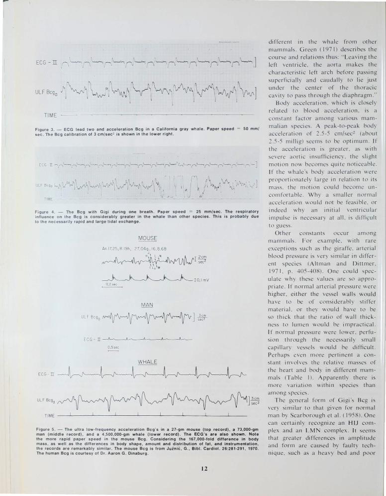
Figure 3. - ECG lead two and acceleration Bcg in a Californ ia gray whale . Paper speed sec . The Bcg calibration of 3 cm/sec2 is shown in the lower right.
50 mm/
'1 t ~ \,,"¥ ' t,~.. 1 /. '\.,J " .. - . , .... J
j '. . \ I
-~ TIME
Figure 4. The Bcg with Gigi during one breath . Paper speed = 25 mm/sec. The respiratory inlluence o n t he Bcg is considerably greater in the whale than other species. This is probably due to the necessari ly rapid and large tidal exchange .
MOUSE
An.17.25, R 19h,
12cm ~
10.1 mV 0.2 sec
MAN
ULF 8cgo ] 3cm seer
ECG - IT
O.55ec
WHALE
ECG - II
ULF 8cgo ] 3cm SeC2
TIME __ ~~ ____ ~ ____ ~ ______ ~ ____ -L ____ ~ ______ ~ ____ -L ____ _
Figure 5. - The ultra low-frequency acceleration Bcg 's in a 27-gm mouse (top record) , a 73 ,OOO-gm man (middle record) , and a 4,500,OOO-gm whale (lower record) . The ECG 's are also shown . Note the more rapid paper speed in the mouse Bcg. Consideri ng t he 167,000-fold difference in body mass, as well as the differences in body shape, amount and d istr ibution of fat , and instrumentation, the records are remarkably similar . The mouse Bcg is from Jui ni!: , G., Bib l. Cardiol. 26:281 -291 , 1970. The human Bcg is courtesy of Or. Aaron G. Oinaburg .
12
different in the wha le from other mammals . Green (1971) describes the course and relations thus: "Leaving the left ventricle. the aorta makes the characteristic left arch before pas i ng superficially and caudally to lie just under the center of the thoracic cavity to pass through the diaphragm ."
Body acceleration. which i clo ely related to blood acceleration. is a co n tant factor among various mammalian specie . A peak-to-peak body acceleration of 2 .5-5 cm/sec2 (about 2 .5-5 millig) seems to be optimum . If the acceleration is greater. as with severe aortic in ufficiency . the slight motion now becomes quite noticeable. If the whale' body acceleration were pro portionatel y large in relation to it mass. the motion could become unco mfortabl e. Wh y a smaller no rmal acceleration would not be fea ible. or indeed why an initial ventricula r impulse is nece sary at all. is diffi~ult to gues .
Other constants occur among mammals. For example . with rare exceptions such a the gi raffe . arterial blood pressure is very si mi lar in di fferent species (Altman and Dittmer. 197 I. p. 405-408) . One cou Id speculate ""hy these value are so appropriate . I f normal arterial pressure were higher. either the ve sel walls would have to be of considerably stiffer material. or they would have to be so thick that the ratio of wall thickne s to lumen wou Id be impractical. I f normal pressure were lower. perfusion through the nece sarily small capillary vessels would be difficult. Perhaps even more perti nent a constant involves the relative ma ses of the heart and body in differen t mammal (Table I). Apparently there is more variation within species than among specie .
The general form of Gigi's Bcg is very simi lar to that given fo r norma l man by Scarborough et al. ( 1958). One can certai nl y recog nize an HIJ complex a nd an LM complex . It seems that greater d iffe rences in amplit ude and form a re caused by fa ulty technique . uc h as a heavy bed a nd poor

couplin g . o r by di ~ea~e sta tes . tha n by diffe re n ce~ in pec ies. F igure 6 give,> a n exa mpl e o f thi s . It compa res a virtua ll y norm a l Bcg in a dog with th e Bcg in a d og at th e terminal stage o f rejecti on. The latte r tracin g is o bviously grossly a bn o rm al a nd demo nstra te the ex treme in Bcg a bno rm alit y. Oth e r conditi on which ca n cause a g reater ba lli tocardi o gra phic vari a ti o n within th a n a mo ng species include angina l att acks, severe co ro nary a rt e ry di ea e, hype rth yro idi m, aorti c va lvul a r insuffi c iency, a nd congesti ve hea rt fa ilure (Sta rr and N oord e rgraaL 1967). Eve n a program o f ph y ica l conditi o nin g ove r seve ra l m o nth ca n a lte r an indi vidua l's Bcg to a great a n ex tent as th e di ffc rences ee n am o ng speci es (E lsbach e t al .. 1970. H o ll oszy et a l .. 1964).
The major d iffe re nce a mo ng th e Bcg's o f va ri o us m a mmals see ms to be o ne o f timing of th e systo li c wave fo rms. As body ize inc reases. th e o nset o f th e systo li c compl ex is de layed (QH int e rva l) and the compl ex spreads out (HJ and H L int erva ls. T a bl e I). If we con ide r th e tip o f the H wave as th e o nset of ejecti o n . we sha ll a t worst li ghtl y un de restim a te th e ca rdi ac pre-ejecti o n pe ri od . Cert a inl y the re la ti ve va lues a mo ng spec ie can be estim a ted by th e Q H inte rva l . Sim il a rl y. ejec ti o n time ca n be e tim ated by th e HL inte rva l. T hi s interva l di d not seem to be so re la ti ve ly prolo nged in G igi as th e Q H . The cont ributi o n of prolo nged condu c ti o n t ime in hea rt s of diffe rent sizes to the int erspecies difference in systolic time int erva ls is probabl) considerable. as is -ho\\ n by th e PR a nd Q RS interval. in Table I .
In gene ra l. heart ra te and ejection tim e a re in ve rse l) re la ted. Thu~ part of th e diffe rences in ys to li c time inte r als i du e to hea rt ra te di fference . But hea rt ra te cann ot e'\p lain all o f th e di ffe rence. G ig i's heart ra te of 43 bea ts/min was not as ~IO\\ a e'\ pcctcd and occurred pre~umabl) because ~h e \\ as e '\ ci ted . n athlete \\i th a heart ra te of 40-4: beat /min doc~ not \ho\\ the prolonged pre-
Tabl e 1.-Some comparative values among mouse, man , and whale
M ouse
27 Welghl (gm) Lenglh (cm) Hearl/body mass
65-9.5 ( 125-20) + (I) 041-051 (2)
(gm/l00 gm) Heart rate (beats/min) Bcg IJ amplItude
(cm/sec2 )
Bcg IJ amplItude (corrected . cm/sec 2 )
Bcg IJ amplItude (dynes)
CardIac output (I/mln) CardIac Index
(MI/mln/kg) Stroke volume (ml) Stroke Index (ml/kg) PR Interval (msec) ORS Interval (msec) OH Interval (msec)
" Pre -e jecti on pert od" HJ Interval (msec) HL Interval (msec)
" Ejecti on time "
300-700 P) 26(")
3.4 •
73.0
42 (11) 22 ( 11 )
27
43 64
Corrected for mass o f bed IJ Total Mass + With tall ()
• Measured In G Igl B ody Mass
I Walker . et al . 1968 2 Allman and Dittmer . 1971 . p 240 3 Allman and Dillmer , 1971 , p 236-7 I Allman and Dittmer , 1971 , p . 239
ECG - II
PG
Man Wt">ale
73.000 4500000 180 760
044-057( 032050 I
60-80 43 43(6) 48
4 7 ' 5 O'
250X 10 (. H) 227 10
50-8.0 308 70-90 (" 1<1) 684
70-90 (" III) 7150 09-1 2 (" W) 16
180-200 294' (320) (II J 80-100 10~' (90-120) (1'1
90- 110 (7.H) 320
140 (7.H) 205 320 (7 H) 490
Allman and Dittmer 1971 p 340 6 JUlniC 1970
Starr and Noordergraaf 1967 H Moss 1961 " Cullen el al 1970 10 Altman and DIttmer 1971 p 323-4 11 A ltman and Dittmer 971 p 2.8
In second~
013 18
15
3" 000
70 12 ~ 119
48 7 7
Figure 6a . - Th ese tw o tracings are from a conscious dog after cardiac autotransplantation The Beg is essentially normal. PG = Pneumogram (Whitney gauge) .
EeG - II --J\---.J~---------J ----r--------'--...J"---------
PG
T m~ n e .... ~ond~
Figure 6b . - These tracIngs are from a dog In lhe lermonal slages of rejection aft ... c8tdlac allotransplantation . The dIfference between the balhslocardlograph lc recordl from the two dogl I
o bviously greater than that between the tracong' from a whale and mouse I FI\JUre 5)
13
ejection period and ejection ti me that G igi does (Weiss ler et a\. . 1960: Leighton et a \.. 197 I).
We said that the Bcg is used to estimate cardiac fun ction . This is po sible because of th e close relati onship between the accelerat ion Bcg and the acceleration of blood out o f the left ventricle in to th e ao rt a (Winter et a\. , 1966, 1967: Smi th , Van C itters, and Verdouw, 1970: Deucha r. 1966) . The latter is a proven sensiti ve indi cator of ca rdiac functi on ( o bl e, Trenchard, and G u z. 1966: o bl e. G abe. and Tre ncha rd , 196 7: Rushme r. 1964, 1970).
Gigi ' stroke index of 1.6 ml/kg is somewhat greater th an that o f man, about I ml/kg . H owever, sin ce the heart ra te was slower in Gi gi, the ca rdiac index was close r to that o f man (Table I). Again. in comparin g several species. we note th at when ca rd iac output is pl ott ed aga i nst body weight on a log-l og sca le. a stra ight line IS obtained (A lt ma n and Dittmer, 1971. p. 320) . It does seem reasonable that stroke index is roughl y eqUivalen t in diffe rent mam mals. since the heart/body mass ratios a re si mi la r.
A lth ough the measureme nt of acceleratio n. a opposed to di splaceme nt or velocit) , minimize the in flue nce of ve ntil ati o n. any move ment or muscul ar acti vit) can distu rb th e recording. Si nce ~ hales mu t expire and mspl re rapidl ) between dives. the muscular activit) is relatively violent As Wahrenbrock has mea ured. Gigl's peak in tantaneous fl o~ rate ~as 285 II ec. T hus venttl ation demoli hed Gi gi's Bcg recording . Fortunatel) . ventil ato ry rate ~as e:\tremel) slow so that the Bcg had suffi Ci ent time to recove r bet\\een breath .
The Bcg has nov\ been reco rded in a \\Ider range of mas es In animal than an) other ph)s iologic test. Th e ma~~ ratio I~ I :6.000 .000. egg em br) 0 :
\\ ha le Thl~ point out the ver atilit ) o f the Bcg and ~ugge ts it importance a~ a technique for comparati ve ph) \Iologlcal and ph armacological \tUJ IC~
ACKNOWLEDGMENTS
The authors wish to acknowledge the invaluable assistance of Gary Maruschak . They also thank the taff of Sea World who participated
in thi s study. Without their enthu iastic cooperation. these studies would have been impossible .
LITERATURE CITED
Altma n, P. L. , and D. S. Dittmer (editors). 197 1. Respira tion and circul atio n. Fed . Am . Soc . Exp. Bi o\. , Betheseda, 93 0 p.
Burger . H. c.. and A . oordergraaf. 195 6. Phys ical basis of ba llistocardi ography. I!. The qua nties th at can be measured with different types of ballistoca rdiogra phs and their mutua l relations. Am . Heart J . 5 1: 127- 139.
Burger , H . c., A. Noordergraaf, and A . M. W. Verhagen. 1953. Phy ical basis of th e low-frequency ba llistoca rdiograph. Am. H ear t J . 46:7 1-83.
Cu llen , B. F., E. I. Eger, II , . T. Smit h. D . C. Sawyer , G. A. Gregory, and T. A. Joas. 1970. C ardi ovascula r effects of fluroxe ne in man. Anesthes iology 32: 2 18-230 .
Deuchar , D. C. 1966 . The relat ions of ba llistocardi ography to other methods of studying the cardiovascul ar system in ma n. 111 A. A . Kn oop (edi tor), Ballistocardiography and cardiovascul ar dynam ics, p. 225-236 . Willi am & W ilkins Co ., Baltimore .
Elsbach, H ., F. A . Rod rigo, H. W. H. Weeda, and J . Poo\. 1970. Th e ball istoca rdiogram in se lection for and assessme nt of phys ical condi ti oning in pa tients with ischem ic heart disease . Bib\. Card io\. 26: 49-5 I.
Green, R. F . 197 I. Observati ons on th e anatomy of some ce tacea ns and pinnipeds. III S. H. Ridgway (edito r). Mammals of the sea . Biology and medicine. C. C. Thomas , Springfie ld , p. 269 .
Holloszy, J . 0 ., J . S. Skinner, A. J. Ba rry, and T . K . Cureton. 1964 . Effect of physica l conditio ning on cardi ovascula r functi on. Am. J . Cardio \. 14:76 1-770.
Juznic , G . 1970. The ultra-l ow frequency ba llistocardi ogram of th e mouse . Bib\. Cardio\' 26:280-29 1.
Leighton , R. F ., A . M. W eiss ler , P . B. Weinstein, and C. F . Wooley. 197 1. Right and left ventricul ar systo lic time inte rvals. Am. J . Cardi o\' 27 :66-n.
Moss. A. J . 196 1. Ballistocardi ographic evaluati on of th e ca rdiovascular aging process . Circul ation 23 :434-45 1.
oble, M. 1. M., I. T. Gabe, and D. Trenchard . 1967 . Blood pressure and flow in t he ascending aorta of conscious dogs. Cardiovasc. Res. 1:9-20. oble, M. 1. M., D. Trench ard , and A. Guz. 1966. Studies of th e max imum acce leration of blood in th e ascending ao rt a of conscious dogs. I II A. A . Knoop (edi to r),
Ballistocardiography and cardiovascular dynamics , p. 243-247. William & Wilkins C o., Baltimore .
Rushmer, R. F. 1964. Initi al ventricular impulse . A potential key to cardiac eva luation . Circulation 29 :268-283 .
__ ----,-__ .,,- . 1970. Cardiovascular dy-namic . 3rd ed. W. B. Saunders Co .. Philadelphia , p. 110.
Sca rborough. W . R., E. F. Folk, HI , P. M. Smith , and J . H. Co ndon . 1958. The nature of records from ultra-low frequency ba lli locardi ographic systems and their rela tion to circulatory events. Am. J . C ardio\. 2:613-64l.
Smith , . T. In press. Ballistoca rdi ography. III A. M. Weissler (editor ). oninvasive meth ods in ca rdi ac evaluation . Grune and Stratton, ew York.
Smith , . T. , R. L. Van Citters, and P. D. V erdouw . 1970. The relation bet ween the ultra-low frequency ballistocardiogram. the acceleration pneumocardiogra m, and ascending aortic fl ow acceleratio n in the baboon. Bib\. Cardio\' 26: 198-205.
ta rr , I., and A. oordergraaf. 1967. Ballistocardi ograph y 10 cardiovascula r research . J . B. Lippincott Co .. Phila ., 438 p.
Ta lbot , S. A. , and W. K. Harrison, Jr. 195 5 . Dynamic compar ison of current ba llistocardi ogra phic meth ods. Part I: Artefacts in the dynamically simple ba llistocardi ographic methods. Circul atio n 12: 577-587.
Tannenbaum . 0 ., H. Vessell , and J. A . Schack. 1956. Rel ationship of th e natura l body damping and body frequency 10 the ba llistocardiogram . Circul ation 13:404-409.
Ti chenko, M. 1. 1963. The significance of natural vibrati ons of the hum an body in shaping the balli tocardiogram . Biophysics 8:311-319.
Walker , E. P. 1968 . Mammals o f th e world . Vo\. II. J ohns Hopkins Press. Baltim ore, p. n I.
Weissbach , G . H.-J . 1960a. Die Rolle der Tischm asse und der Wert einer zusatzlichen Dampfung beim Elongationsba llistokardiographen . [I n Ger.] Pfluegers Arch. ges. Physio \. 270:529-535 .
__ -;---;---;-_. 1960b. Die registrier-technischen Eigenschaft en des direkte n ba llistokardiographen . Z. Kreislaufforsch . 49:626-630.
WeissIer , A. M., R. G. Peeler, W. H. Roehll . Jr .. and . C. Durham . 1960. Relationships between left ventricular ejection time , stroke volume, and heart rate in norm al individuals and patients with cardiovascular disease. Am. Heart J . 62:367-378 .
Winter, P. J ., D . C. Deuch ar, M. . oble . and A. Guz. 1966. The ballistocardiogram and left ventricular ejection in the dog. In A. A. Knoop (editor) , Ballistocardiography and cardiovascular dynamics, p. 248-254 . Williams & Wilkins Co., Balti more .
Winter , P. J ., D. C. Deuchar, . \. M. oble, et a\. 1967 . Relationship between the ballistoca rdiogram and the movement of blood from the left ventric le in the dog. Cardiova c . Re . I: 194-200.
MFR Paper 1046. From Marine Fisheries Review, Vol . 36, No . 4, April 1974. Copies of this paper, in limited numbers, are available from 083, Technical Information Division , Env ironmental Science Information Center , NOAA , Washington, DC 20235.
14