Lederberg j , hunter l artificial intelligence and molecular biology (1993)

BACTERIAL REPRODUCTION*
7 JOSHUA LEDERBERG
Professor of Genetics, Uhersity of Wirconsifz, Madison, Wisconsin
I N contemporary biological research on growth and reproduc- tion, bacteria now play an outstanding role. They are believed
to be the simplest complete organisms-complete in the sense that they can sustain themselves and proliferate in chemically simple habitats. By contrast, for example, the structurally simpler viruses must rely upon the metabolic machinery of another cell. What- ever the validity of this belief, either as physiology or phylogeny, the bacteria and their viruses have proved to be remarkably handy tools for the analysis of reproduction with the ultimate aim of unifying generation with metabolism.
The simplest aspect of reproduction is also the most elusive: the mechanism of replication of like from like. An earlier genera- tion of microscopists saw cell division as a simple splitting of a lump of protoplasm, but we no longer credit so naive an outlook on the complexity of the bacterial cell (Dubos, 1945). Instead we are led to examine the parts of the bacterium which have to be individually copied before the cell as a whole can divide. For this analysis, exact replication tells us little more than its own bare fact, and most of our present understanding depends on treasured exceptions from the rule (mutation) and especially on those other modes of reproduction in which the information from more than
* Lecture delivered December 19, 1957. As originally delivered, additional il- lustrative material was included. However, this has been amply reviewed else- where (Lederberg and Tatum, 1953; Stocker et al., 1953; Lederberg, 1955a, 1956a, b, 1957a; Lederberg and Lederberg, 1956; Lederberg and St. Clair, 1958) so as to preclude its repetition here. The studies at Wisconsin have been aided by research grants from the National Cancer institute (C-2157), United States Public Health Service, from the National Science Foundation, and from the Research Committee of the Graduate School, University of Wis- consin, from funds allocated by the Wisconsin Alumni Research Foundation. Present address: Department of Genetics, Stanford University, Stanford, Cali- fornia.

70 JOSHUA LEDERBERG
one cell is redistributed to the progeny, that is to say, genetic recombination. *
During the past dozen years geneticists have discovered a formerly unthought of variety of reproductive techniques among various bacteria, but their greatest significance is their homology with the genetic processes of other organisms. The ubiquity of chromosomes and sexuality is as plausible as that of adenine, arginine, and deoxyribose-genetics and biochemistry both testi- fying to the common heritage of terrestrial life.
These advances in bacterial genetics have not gone unnoticed in scientific reviews (Cold Spring Harbor Symposia, 1946, 195 I, 1953, 1956; McElroy and Glass, 1957; Braun, 1953; Symposium on Genetic Recombination, 1955; Lederberg, 1956~)) which should be sampled to complement tonight’s brief encounter with the phenomena of sexuality in Escherichid co/i. Historically, this analysis has proceeded in reverse order as compared, say, to NezlroJporct or fruit flies. A convenient starting point was Tatum’s (1945) search for useful genetic markers which he undertook with no stronger encouragement than his own faith in their ulti- mate use in biochemical, genetic, and life-cycle investigations. This judgment was justified in due course by the observation of the recombination of these markers in mixed cultures (Tatum and Lederberg, 1947). This occurred at a very low rate ( 1O-6) neces- sitating rigorous selection to find recombinants, which impeded linkage analysis and frustrated the microscopic confirmation of sexuality. However, the conditions under which recombinants occurred and a statistical analysis of the various types that were produced could show that recombination entailed cell-to-cell union, and that the whole genetic material was organized into a single, linear linkage group (Lederberg, 1947). The life cycle could also be outlined: the vegetable cells are multinucleate, but haploid, and the hypothetical diploid zygote has an abbreviated life span, undergoing prompt segregation to return to the normal haploid state. (This is a “haplobiontic” cycle similar to that of most lower fungi and algae. It contrasts with the “diplobiontic”
* Hopefully, this remark may begin to be superseded by the content of the following Harvey Society Lecture by Professor A. Kornberg on the enzymatic synthesis of DNA.

BACTERIAL REPRODUCTION 71
cycle of higher plants and animals where the somatic phase is diploid, and an abbreviated haploid phase is represented only in the gametes or gametophytes.)
_’ By 1949, exceptional clones proving to be persistent diploids were isolated among the progeny of certain crosses (and were since found to represent about one per thousand of the sexual progeny of most crosses). Since single cells of these diploid clones carried a complement of genetic markers from each parent, they were a tangible representation of the hitherto hypothetical zygotes and furnished additional evidence of the normal life cycle. It was soon found that in these diploids, the contribution of genetic material from the two parents was unequal. The same is true for the regular haploid progeny also, but this aberration had been partly obscured by the need for selective isolation of specific classes of recombinants. It therefore had to be assumed that some of the genetic material of either parent was eliminated in the course of the sexual cycle before the recombinants emerged. (Lederberg, 1949; Lederberg et A., 1951.) There was, however, no clue as to the source of the bias in elimination: why the markers of one parent should be retained in preference to the other’s, and why some markers were affected and not others. This and the equally abstruse question of when this elimination occurs, before or after fertilization, have played a central role in further analysis.
Sexual differentiation-the distinction of males and females- is one of the most obvious aspects of the life cycle of higher organisms, but it was almost the last to be recognized and verified in E. coli. One of the first leads was Hayes’ finding (1952) that some cultures were more susceptible than others to sex& sterili- L zation by streptomycin. He therefore supposed a unilateral fertili- zation in which the streptomycin-insensitive male gamete might even be extruded from the cell. Concurrently, Cavalli et al. (1953) discovered self- and intersterile clones which they classi- fied in an F- mating type, the original wild type and most of its progeny being F’. We were at first reluctant (awaiting further evidence) to specify these mating types as, female ( o ) and male ( 8 ) respectively, but Hayes’ supposition to this effect has since been fully justified, the 8 function being less susceptible

72 JOSHUA LEDERBERG
to streptomycin than the o . However both the 8 donor and the 0 recipient cells remain intact.
Crosses of F+ x F+ and F+ x F- are both fertile, the latter more so, while F- x F- is completely sterile. In the light of - further work, we can then designate the F- mating type as being restricted by its genotype to function as o while F+ cells can potentially act either as 8 or ? , Obligate 8 clones, such as have arisen by mutational loss of female capacity in other hermaphro- ditic fungi (Hansen and Snyder, 1943; Wheeler, 1954) have not yet been seriously looked for.
Why was this elaborate system of sexual determination over- looked for so long? It was obscured mainly by the ambivalent capacity of the wild type strain and its derivatives, which were therefore self-fertile.” It was concealed further by the remarkable fact that maleness in E. coli is highly contagious, so that Q (F-) cells exposed to ~3 (F+) rapidly become, like them, $‘. Thus, the progeny of F+ x F- crosses are regularly F+ and do not show a segregation of sexual capacity.
Many kinds of experiments on E. coli mating are frustrated by the very low fertility of the indicated F+ x F- crosses. In 1951, however, Cavalli fortunately discovered the first of a series of Hfr mutants. These mutants, which derive from F+ strains, are much more fertile in crosses with F- than is the standard F+, so that recombinants occur with a frequency as high as 10 per cent of the input 6 cells. These Hfr strains have made it possible to visualize the mating process as illustrated in Fig. 1 and to conduct precise kinetic analyses of the various stages of mating. That the pairwise combination of cells represented in Fig. 1 does represent the mating process has been verified by the isolation of single pairs with a micromanipulator and the analysis of the exconjugant clones derived from each of the two mates (Leder- berg, 1956b, 1957b; Anderson and Maze, 1957).
The recent kinetic studies by Wollman and associates (Woll- man and Jacob, 1955; Wollman et al., 1956) have done much
* Tatum’s choice of strain (E. coli K12) was a remarkable stroke of luck. Later surveys (Lederberg, 1951), have shown that only a few per cent of E. coti strains are self-fertile, and this was a necessary condition for the sue- cessful outcome of the initial trials.

BACTERIAL REPRODUCTION 73
to elucidate the individual steps of the mating process. The first step is conjugal pairing to form complexes of 8 and P cells
c similar to those of Fig. 1. This process takes place very quickly after the collision of the competent cells, as complexes which are stable to dilution form within a minute of mixing the two parent cultures. It is, however, more than a passive colloidal agglutination, since effective pairs do not accumulate at low temperatures or in the presence of metabolic inhibitors (Nelson, 1956; Fisher, 1957a, b). This step might well be called cytogamy. Then follows fertilization in the sense of the transfer
FIG. 1. Conjugal pairing in E. co/i. From Lederberg (Ig%b) with permis- sion of the publishers of the Journal of Bacteriology.
of the genetic material from the 8 to the P cell. This process remains to be studied by standard cytological techniques. Genetic analysis, together with the unimpaired viability of exconjugant 8 cells, supports the view that each parent cell contains several nuclei and that fertilization transfers the substance of one of them from 8 to o cell via a conjugation canal. Remarkable stereoscopic electron micrographs of the structures have been published by Anderson et al. (l957), and these would indicate that the bridge is in fact rather smaller than indicated by Fig. 1,
the preparation of which is subject to obvious artifacts of flat- tening and drying.
The most far-reaching finding of Jacob and associates was that fertilization is progressive and can be interrupted in mid-course by shearing the mating pairs by turbulence in a Waring blendor.

7~1 JOSHUA LEDERBERG
By thus interrupting pairs at various times after the first contact of the two parents, these authors were able to show that the recovery of various markers was regularly progressive in time. For example, using the Hfvr culture isolated by Hayes (1953) as a male strain, they found that of various markers from the $ parent, T+L+ was first recoverable when interruption was con- ducted at least 8 minutes after mating; Lac+ could be recovered after 12 minutes and Gal’ only after 25 minutes. The incidence of a given marker from the 5 parent among the progeny could be plotted as a function of time before interruption. By extrapo- lating these curves to zero incidence these authors could infer “time of initial entry” for each of a series of markers, giving a linear time sequence. This corresponded perfectly with the linear linkage maps that had been previously established by more conventional procedures.
The most plausible interpretation of these results is that fertili- zation comprises the progressive movement of a linear chromo- some from one cell to another beginning at a specific point and allowing for the progressive transfer of more distant markers as time goes on. The separation of a mating pair while the chromosome was still in mid-transit would cut the chromosome at that point and allow for the recovery of only those markers that had already entered the o cell. The over-all chance of recovery of later (more distant) markers proved to be less and less efficient: fertilization might be subject to accidents of spon- taneous interruption during transfer which render the survival of distant markers less and less likely. In addition, the pairing of the gamete chromosomes must begin at the point of initial entry and become less and less perfect down their length. This picture thus furnishes a reasonable interpretation of the unequal contribution to the sexual progeny of the paternal markers, especially the most distal markers (see Fig. 2) .
The final stage of the recombination process is the assimilation of genetic information from the gametic chromosomes into a viable recombinant. As with crossing over in higher forms, we have little detailed knowledge of this stage, though it must be preceded by the point-to-point synapsis of homologous parts. This might be followed either by physical interchange or, more inter-

BACTERIAL REPRODLJCTION 75
estingly, by copy-choice alternation of templates in the construc- tion of a daughter chromosome. Since the paternal chromosome suffers a loss of distal genes which must be haploid-lethal, no
- paternal marker can be recovered without an exchange between
FIG. 2. Schematic representation of mating in E. coli. Above: An interrupted mating experiment with interruption at 20 minutes, according to two hy- potheses. The time scale for the two versions is the same and they have the same end results. Below: Formation of paternal-deficient versus m.iternal- deficient diploid hcterozygotes. On the assumption that a broken fragment is
J present in the primary zygote, the two types are produced by exchange to the left and to the right of the breakage point, respectively. The chromosomes are schematized by a smooth line for the paternal, a beaded string for the ma- ternal, respectively. For simplicity these chromosomes are shown fully ex- tended and the several other nuclear units in each parental bacterium are omitted.
it and the point of breakage. Thus, the recovery of a marker depends both on its entry and on a crossover which integrates the marker into an intact chromosome. The quantitative role of these two factors is not easily assessed.

76 JOSHUA LEDERBERG
Several functional criteria may be proposed for recognizing the transfer of a given paternal marker apart from its synapsis and integration with the maternal homolog. These include the induction of prophage, the synthesis of inducible enzymes, and ’ the magnitude of the paternal contribution to persistent hetero- zygotes. The experimental results now at hand (Jacob and Woll- man, 1957a, b, and unpublished; Lederberg and colleagues, un- published) lend great weight to the necessity of progressive trans- fer as at least one element in the development of each of these functions after fertilization. Other criteria suggested but not yet tried include the development of resistance phenotypes (Hayes, 1957) and unilinear clones that might follow abortive recombination (cf. Stocker, 1956; Lederberg, 1956a). However, for each of these criteria a plausible case might be made out for synapsis as another requisite to the functioning of a paternal factor, viz., in prophage or enzyme synthesis, and even in the replication of the distal segments of the paternal chromosome. The clearing up of this uncertainty will therefore help not only to solve this particular problem of the sexual cycle but also to shed light on an important aspect of gene physiology.
Fuerst and associates (1956) have also succeeded in measuring a linkage distance in nucleotide units by correlating the lethal effect of a Ps2 label with the number of paternal markers re- covered in the progeny. The calculated rate of transfer (cf. also Fuerst and Stent, 1956) then approximates a thousand nucleo- tide pairs or 0.3 p of extended polynucleotide per second. By another, more direct, labeling procedure Garen and Skaar (1958) could verify the unilateral transfer of P”‘-labeled DNA from $ to o cells in amounts averaging 10 per cent of the DNA per 8 cell per mating and therefore presumably less than a single
nuclear equivalent. The time seems to be ripe for a combination of interrupted mating with Levinthal’s (1956) star-counting technique for measuring the radioactivity of individual micro- scopic particles.
Interrupted progressive fertilization is now one of the most powerful experimental techniques for the genetic analysis of EJcher-ichh co/i. The elegant simplicity of Jacob and Wollman’s experimental results lends great weight to progressive transfer as

BACTERIAL REPRODUCTION 77
the most plausible model of bacterial sexuality at this time, and its standing is not necessarily impaired by exceptions which involve at most a few per cent of the recombinant progeny. The
* most serious present obstacle to a completely unified interpreta- tion comes from the behavior of the persistent heterozygotes. As already discussed, these heterozygotes regularly lack a segment of genetic material from one of the parents. It soon became clear (Nelson and Lederberg, 1954) that this deficiency was usually for part of the paternal genome. These puternd-deficient diploids can be easily accounted for on the model of incomplete or inter- rupted fertilization, the missing segment simply being what was left behind in the 6 cell. However, a substantial proportion of the heterozygotes are not puternuj- but muternl-deficient for these markers. This anomaly cannot be accounted for by an incomplete paternal contribution. We were therefore obliged to invoke another process of genetic elimination which occurs after fertiliza- tion, and can also result in the loss of maternal markers.
Postzygotic elimination by itself can furnish an alternative model of the mating process (Lederberg, 1955b). A more reasonable inference from present evidence is to abandon this formalistic approach in favor of a more eclectic model. To account for Garen and Skaar’s and Jacob and Wollman’s results, our working hypothesis might admit progressive transfer as the primary source of the peculiarities of segregation in E. coli. We would superimpose an additional mechanism of posttygotic loss of segments to reconcile this with the behavior of diploids. The two modes of loss can in fact be unified in this way: the inter- ruption of mating causes the scission of the chromosome. In a proportion of matings, however, the scission either does not become effective until after synapsis, or the distal piece is still capable of being transferred. The distal segment is doomed to be eliminated, but if this is preceded by crossing over with the homologous section of the maternal chromosome, paternal mark- ers distal to the crossover will be conserved, and the homologous maternal markers will be the ones ejected (see Fig. 2). The tentative nature of these proposals should be stressed.
Different Hfr strains show characteristically different orders of entry of various markers. For example, Hfrz shows the pro-
78 JOSHUA LEDERBERG
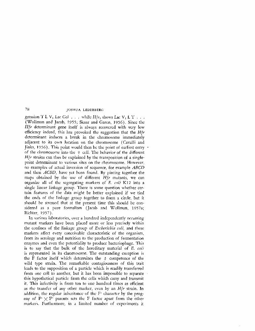
gression T L V, Lac Gal . . . while Hfrl shows Lac V, L T . . . (Wollman and Jacob, 1955; Skaar and Garen, 1956). Since the Hfr determinant gene itself is always recovered with very low efficiency indeed, this has provoked the suggestion that the Hfr determinant induces a break in the chromosome immediately adjacent to its own location on the chromosome (Cavalli and Jinks, 1956). Th’ 1 IS >oint would then be the point of earliest entry + of the chromosome into the o cell. The behavior of the different Hfv strains can thus be explained by the transposition of a single- point determinant to various sites on the chromosome. However, no examples of actual inversion of sequence, for example ABCD and then ACBD, have yet been found. By piecing together the maps obtained by the use of different Hfr mutants, we can organize all of the segregating markers of E. coli K12 into a single linear linkage group. There is some question whether cer- tain features of the data might be better explained if we tied the ends of the linkage group together to form a circle, but it should be stressed that at the present time this should be con- sidered as a pure formalism (Jacob and Wollman, 1957a; Richter, 1957).
In various laboratories, over a hundred independently occurring mutant markers have been placed more or less precisely within the confines of the linkage group of Escberichia coli, and these markers affect every conceivable characteristic of the organism, from its serology and nutrition to the production of fermentation enzymes and even the potentiality to produce bacteriophage. This is to say that the bulk of the hereditary material of E. coli is represented in its chromosome. The outstanding exception is the F factor itself which determines the $ competence of the wild type strain. The remarkable contagiousness of this trait leads to the supposition of a particle which is readily transferred from one cell to another, but it has been impossible to separate ’ this hypothetical particle from the cells which carry and transmit it. This infectivity is from ten to one hundred times as efficient as the transfer of any other marker, even by an Hfr strain. In addition, the regular inheritance of the F’ character by the prog- eny of F+ x F- parents sets the F factor apart from the other markers. Furthermore, in a limited number of experiments it

BACTERIAL REPRODL!CTION 79
has been found that the entire exconjugant clone from an F?/F interaction acquires the F* trait, while as is already known, the transmittal of other markers in Hfr x Fm crosses shows a
h segregation which is accounted for at least in part by the separa- tion of fertilized from unfertilized nuclei. No linkage of the F
, marker can be discerned with any other marker, nor have hetero- zygotes which segregate F+ :F- been observed. The introduction of a few F+ cells into a mass culture of F- leads to the rapid spread of the F + trait through the population. Finally, it has recently been discovered by Hirota and Iijima (1957) that the F agent can be regularly removed from F+ cells by exposing them to acridine dyes. These results taken together are most satisfac- torily explained by the assumption that the s -determining parti. cle is an extrachromosomal, cytoplasmic element which is readily transferred from one cell to another by brief contact. This element would have to be capable of reproducing autonomously and more rapidly than the other genetic elements of the cell.
On the other hand, there is strong evidence that the 6 determinant of Hfr strains occupies particular chromosomal sites. Hff mutants have always been derived from F+ strains (in the course of their isolation losing their F infectivity) and some Hfr strains are more or less readily revertible to the F+ condition. We can most plausibly interpret the Hfr mutants as representing the fixation of the F particle to a particular chromosomal site. A consequence of the fixation might then be some type of inter- ference between the chromosomal and the extrachromosomal par- ticles so that the latter would disappear and thus account for the noninfectivity of the male character in Hfr strains. In the Hfr
_ cell, where it occupies a chromosomal site, the F particle is no Longer infective nor is it accessible to the disinfecting action of the acridine dyes. The two states of the F particle, extrachromo- somal and chromosomal might be considered to be analogous to vegetative phage and prophage, respectively.
A somewhat different view has been offered by Wollman et d. (1956), namely that F+ differs from Hfr only in the position of the determinant of the chromosome, this being at the apex in F+ strains and the first marker to enter in the course of mating. The infertility of an F’ strain is then ascribed to the inevitability of

80 JOSHUA LEDERBERG
breakage between the apical F determinant and the other markers. In fact, they suggest that all of the fertility of an F+ strain should be accounted for by the incidence of Hfr mutants arising in the culture, these being transpositions of the apical F+ segment to ’ other loci. It is difficult to reconcile this hypothesis with the features enumerated in the previous paragraph, but since HEY
mutants do occur, there is hardly any doubt but that they do contribute in a variable measure to the fertility of some F’ cultures. In fact, taking account of the graded differences in stability of the Hfy mutants that have been isolated and the fact that some of them have an extreme tendency to revert to F+, it may be possible to reconcile the two points of view in the following manner: Fertility of a cell in an F-‘- culture may depend on a chromosomal location, at least for the time being, of the F determinant. In some cases, this represents a more or less stable fixation that can be recognized as an Hfr mutant, in others it is a transient event which does not influence the infectivity of the cell nor the character of its clonal progeny.
I hope the complexity of the data now available for recombi- nation in E. coli will convey the impression that we are getting down to prime numbers in the analysis of bacterial heredity, and an approach to it in chemical terms. There is not time here to outline the specific application of these techniques to the detailed analysis of genetic factors which control enzyme formation and bacteriophage production. The power of these methods was already illustrated by the brilliant application of P3?-labeling methods by Jacob and his colleagues. Their scope may be sug- gested by the fact that a simple operation of mixing two mutant cultures on a few selective agar plates can generate information on 10” incidents of recombination-an easy match for the repro- ductive potentiality of the entire human species. But we are still hindered by having to use a 8 bacterium as a vehicle for injecting its DNA into the o cell, that is, if we are concerned with the pervasive problem of contemporary genetics: its translation into polynucleotide chemistry. Our colleagues in bacterial genetics are all eager to find that figurative hybrid of Escherichia co/i X Diplococctls pneumoniae which will combine the suitability for large-scale but precise genetic analysis of the one and the amena-

BACTERIAL REPRODUCTION 81
bility to chemical extraction and self-propelled input of DNA of the other (Hotchkiss, 1955 ) . I rather doubt that any of us has had the courage even to try such a cross; if, against our best in- tuitive judgments, it happened to work, it would doubtless pro- duce something like Raphanobrassica, the famous hybrid that so uncooperatively grows the leaves of a radish and the roots of a cabbage.
This conclusion is not altogether artless if it suggests what I consider to have been the major significance of these studies: the unity that they afford to the biologist’s outlook on the living world. But this stage is now well over, and our expectation for their future role is of fundamental knowIedge of processes under- lying the reproduction of all organisms but nowhere more accessible to experiment than among the bacteria.
REFERENCES
Anderson, T. F., and Maze, R. (1957). Ann. irut. Pasteur 93, 194-198. Anderson2 T. F., Wollman, E. L., and Jacob, F. (1957). AIZ~Z. iwt. Pasteur
93, 45&455. Braun, W. (1953). “Bacterial Genetics.” W. B. Saunders, Philadelphia. Cavalli, L. L., and links, J. L. (1956). J. Genet. 54, 87-112. Cavalli, L. L., Lederberg, J., and Lederberg, E. M. (1953). J. Gen. Microbial.
8, 89-103. Cold Spring Harbor Symposia QURN~. Biol. (1946, 1951, 1953, 1956). Vols.
11, 16, 18, 21. Biological Laboratory, Cold Spring Harbor, L. I., New York.
Dubos, R. J. (1945). “The Bacterial Cell.” Harvard Univ. Press, Cambridge, Massachusetts.
Fisher, K. W. (1957a). J, Gels. Microhiol. 16, 120-135. Fisher, K. W. (19576). J. Gen. Microbial. 16, 136145. Fuerst, C. R., and Stent, G. S. (1956). J. Gen. Physisiol. 40, 73-90. Fuerst, C. R., Jacob, F., and Wollman, E. L. (1956). Corn/~/. umd. 243, 2162-
2164. Garen, A., and Skaar, P. D. (1958). Biorhim. et Biophyr. Art;t 27, 457--463. Hansen, H. N., and Snyder, W. C. (1943). Am. J. Botnny 30,419-422. Hayes, W. (1952). A’aiuve 169, 118-119. Hayes, W. (1953). Cold Spring Harbor Sympo.ria Quant. Biol. 18, 75-93. Hayes, W. (1957). J. Gen. Mirrobiol. 16, 97-119. Hirota, Y., and Iijimg T. (1957). Nature 180, 655-656. Hotchkiss, R. D. (1955). Harvey Lertures Ser. 49, 124-144. Jacob, F., and Wollman, E. L. (1957a). Symposia Snc. Exptl. Biol. No. 12, 75%
92.

82 JOSHUA LEDERBERG
Jacob, F., and Wollman, E. I.. (1957b). IIZ “Symposium on the Chemical Basis of Heredity” (W. D. McElroy and B. Glass, eds.), pp. 46%499. Johns Hopkins Press, Baltimore, Maryland.
Lederberg, J. (1947). (;~~nrtjcr 32, 505~-525. Lederberg, J. (1949). Pmt. NatI. Acad. Sri. l,‘. S. 35, 178-184. Lrderberg, J. (1951). Scienrp 114, 68-69. Lederberg, J. (1955a). 1. Cellulw Camp. PhyJiol. 45, Suppl. 2, 75-107. Lederberg, J. ( 1955b). Scirucr 122, 920. Lederberg, J. (1956a). Genetics 41, 845-871. Lederberg, J. (1956b). J, BLtcteriol. 71, 497-498. Lederberg, J. (1956~). Am. Srieztht 44, 264-280. Lederberg, J. (1957a). Bartepiol. Revs. 21, 133-139. Lederberg, J. (1957b). Pvor. Nc2tl. Artid. Sri. U. S. 43, 1060-1065. Lederberg, J., and Lederberg, E. M. (1956). 1~ “Cellular Mechanisms in Dif-
ferentiation and Growth,” 14th Symposium Sot. Growth and Development (D. Rudnick, ed.), pp. 101-124, Princeton Univ. Press, Princeton, New Jersey.
Lederberg, J., and St. Clair, J. (1958). J. Bateviol. 75, 143-160. Lederberg, J., and Tatum, E. L. (1953). Scienre 118, 169-175. Lederberg, J., Lederberg, E. M., Zinder, N. D., and Lively, E. R. (1951).
Cold Spring Harbor SympojiJ Qumzt. Biol. 16, 413-443. Levinthal, C. (1956). Ptoc. Nail. Acdd. Sri. U. S. 42, 394-404. McElroy, W. D., and Glass, B. (eds.) (1957). “Symposium on the Chemical
Basis of Heredity.” Johns Hopkins Press. Baltimore, Maryland. Nelson, T. C. (1956). J. Cellular Camp. Physiol. 48, 271-292. Nelson, T. C., and Lederberg, J. (1954). Proc. Nntl. Acad. Sri. U. S. 40,
415-419. Richter, A. A. (1957). Genetic recombination in E.rrherichln coli K-12. M. S.
Thesis, University of Wisconsin, Madison. Skaar, P. D., and Garen, A. (1956). Puoc. Nrrtl. Ac.td. Sri. C. S. 42, 619-624. Stocker, B. A. D. (1956). J. Gen. Microhiol. 15, 575-598. Stocker, 8. A. D., Zinder, N. D., and Lederberg, J. (1953). J. Gen. Micro-
biol. 9, 410-433. Symposium on Genetic Recombination (1955). J. Crllzh Con/p. Pl,ytio/. 45,
Suppl. 2. Tatum, E. L. (1945). Proc. Nirtl. Ac‘id. Sri. U. S. 31, 215-219. Tatum, E. L., and Lederberg, J. (1947). 1. B.uieriol. 53, 673-684. Wheeler, H. E. (1954). Ph.~op~rthology 44, 342-345. Wollman, E. L., and Jacob, F. (1955). Compt. rrud. 240, 2449-2451. Wollman, E. L., Jacob, F., and Hayes, W. (1956). Cold Spring Habor Slm-
pnrjd Qamf. Rjol, 21, 141-163.