Pili ialah alat yang mengawal pengaliran air keluar. Apa itu pili ?

RESEARCH ARTICLE SUMMARY◥
BACTERIAL PILI
Architecture of the type IVapilus machineYi-Wei Chang, Lee A. Rettberg, Anke Treuner-Lange, Janet Iwasa,Lotte Søgaard-Andersen, Grant J. Jensen*
INTRODUCTION: Type IVa pili are bacterialcell surface structures that perform criticalfunctions in motility, surface adhesion, viru-lence, and biofilm formation. Type IVa pili areanchored in the cell envelope and pull cellsforward through cycles of extension, adhesionto surfaces, and retraction, all powered by thetype IVa pilus machine (T4PM). Although thestructures and connectivities of the 10 coreT4PM proteins and minor pilins have alreadybeen determined, the overall architecture of the
T4PM and its extension and retraction mech-anisms have not.
RATIONALE:To elucidate the architecture ofthe intact T4PM, we directly imaged T4PMswithin intact Myxococcus xanthus cells by cryo–electron tomography. Mutants that either lackedT4PM components or contained individual T4PMproteins fused to a tag were then imaged. Differ-ence maps revealed the locations of all compo-nents of the T4PMmachine. Hypothetical models
were then built by fitting the known atomic struc-tures of the components together in their rela-tive positions.
RESULTS:Both piliated and nonpiliated T4PMsaremultilayered structures that span the entirecell envelope. T4PMs includeanoutermembranepore, three interconnectedperiplasmic ring struc-tures andanother in the cytoplasm, a cytoplasmicdisc and dome, and a periplasmic stem. ThePilQ secretin forms the outer membrane pore;TsaP formsa periplasmic ring around PilQ; peri-
plasmic domains of PilQtogether with PilP consti-tute the mid-periplasmicring; and the globular do-mains of PilO and PilNconstitute the lower peri-plasmic ring and connect
via coiled coils across the inner membrane toPilM, which forms the cytoplasmic ring. Thecytoplasmic domains of the inner membraneprotein PilC form the cytoplasmic dome on theT4PM axis inside the PilM ring. The short stemin the nonpiliated state is composed of minorpilins and PilA, the major subunit of the pilus.In the piliated state, the pilus extends from thecell exterior through the PilQ pore and the peri-plasmic rings to PilC in the inner membrane. Inthe piliated structure, the hexameric adenosinetriphosphatases (ATPases) PilB andPilT bind in amutually exclusive manner to the base of theT4PM,where they appear as the cytoplasmic discduring extensions and retractions, respectively.Next, we asked whether the known atomic
structures of the proteins could be fit within themapwhere our imaging results indicated,whilestill satisfying all known constraints of size,connectivities, and interfaces. This successfuleffort resulted in “pseudo-atomic”workingmod-els of both states of the T4PM. The models sug-gest that through ATP hydrolysis, PilB rotatesPilC, incrementally moving it into positions thatfacilitate incorporation of new PilA subunits oneby one from the innermembrane onto the base ofthe growing helical pilus. Pilus retraction is drivenbyreplacementofPilBwithPilT,whichrotatesPilCinto positions that promotePilAdeparture fromthe base of the pilus back into the membrane.
CONCLUSION:Wedetermined the architectureof theT4PMin thepiliated andnonpiliated statesand mapped all known components onto thisarchitecture, producing a complete structuralmap of the T4PM. The results illustrate how thestructure and function ofmacromolecular com-plexes that defy purification and traditional struc-tural approaches cannonetheless be interrogatedthrough cryo–electron tomography of intact cellsand model building.▪
RESEARCH
SCIENCE sciencemag.org 11 MARCH 2016 • VOL 351 ISSUE 6278 1165
The list of author affiliations is available in the full article online.*Corresponding author. E-mail: [email protected] this article as Y.-W. Chang et al., Science 351, aad2001(2016). DOI: 10.1126/science.aad2001
Bacterial type IVa pilus machine (T4PM).Two T4PMs are depicted on the left, spanning theenvelope of a Gram-negative bacterial cell. T4PMs extend and retract pili to pull cells forward.The structural data presented here support the hypothesis that ATP hydrolysis in the cytoplasmcauses an adapter protein in the inner membrane to rotate, facilitating the transfer of pilin subunitsfrom the inner membrane onto the growing pilus. The process is reversed during retraction.
ON OUR WEB SITE◥
Read the full articleat http://dx.doi.org/10.1126/science.aad2001..................................................
on M
arch
17,
201
6D
ownl
oade
d fr
om o
n M
arch
17,
201
6D
ownl
oade
d fr
om o
n M
arch
17,
201
6D
ownl
oade
d fr
om o
n M
arch
17,
201
6D
ownl
oade
d fr
om o
n M
arch
17,
201
6D
ownl
oade
d fr
om o
n M
arch
17,
201
6D
ownl
oade
d fr
om o
n M
arch
17,
201
6D
ownl
oade
d fr
om o
n M
arch
17,
201
6D
ownl
oade
d fr
om

RESEARCH ARTICLE◥
BACTERIAL PILI
Architecture of the type IVapilus machineYi-Wei Chang,1,2 Lee A. Rettberg,2 Anke Treuner-Lange,3 Janet Iwasa,4
Lotte Søgaard-Andersen,3 Grant J. Jensen1,2*
Type IVa pili are filamentous cell surface structures observed in many bacteria. Theypull cells forward by extending, adhering to surfaces, and then retracting. We usedcryo–electron tomography of intact Myxococcus xanthus cells to visualize type IVa piliand the protein machine that assembles and retracts them (the type IVa pilus machine,or T4PM) in situ, in both the piliated and nonpiliated states, at a resolution of 3 to4 nanometers. We found that T4PM comprises an outer membrane pore, four interconnectedring structures in the periplasm and cytoplasm, a cytoplasmic disc and dome, and aperiplasmic stem. By systematically imaging mutants lacking defined T4PM proteins orwith individual proteins fused to tags, we mapped the locations of all 10 T4PM corecomponents and the minor pilins, thereby providing insights into pilus assembly, structure,and function.
Type IVa pilusmachines (T4PMs) are part of asuperfamily of bacterial and archaeal multi-protein assemblies that also include type IVbpilus machines, type II secretion systems(T2SS), DNA uptake systems, and archaeal
flagella (1, 2). T4PMs are involved in cell motility(3), host adhesion (4), predation (5),DNAuptake (6),biofilm formation (7, 8), and protein secretion (9).T4PMs consist of an extracellular pilus fiber (T4P)and a cell envelope–spanning complex that we re-fer to here as the basal body. A key feature of T4Psis their ability to undergo cycles of extension andretraction (10, 11). PoweredbyaT4P-assembly aden-osine triphosphatase (ATPase), the basal body ex-tracts pilinmonomers from the innermembrane(IM) and appends them onto the base of the heli-cal pilus fiber, pushing the fiber outward. The fiberhas a diameter of ~6 nm, can extend up to severalmicrometers in length, and can adhere to specificsurfaces (12). Subsequently, the basal body, pow-ered by a T4P-disassembly ATPase, extracts mono-mers from the pilus base back into themembrane(13). T4PMs retract at rates up to 1 mm/s andgenerate forces as high as 150 pN to pull the cellforward (14, 15), making T4PMs the strongest mo-lecularmotors known. T4Ps have been identifiedas important virulence factors in several hu-man pathogens, includingNeisseria gonorrhoeae,N.meningitidis, and Pseudomonas aeruginosa (16).
T4PMs exhibit a multilayered structurespanning the cell envelope of M. xanthus
To determine the structure of the T4PM in vivo,we used cryo–electron tomography (also known
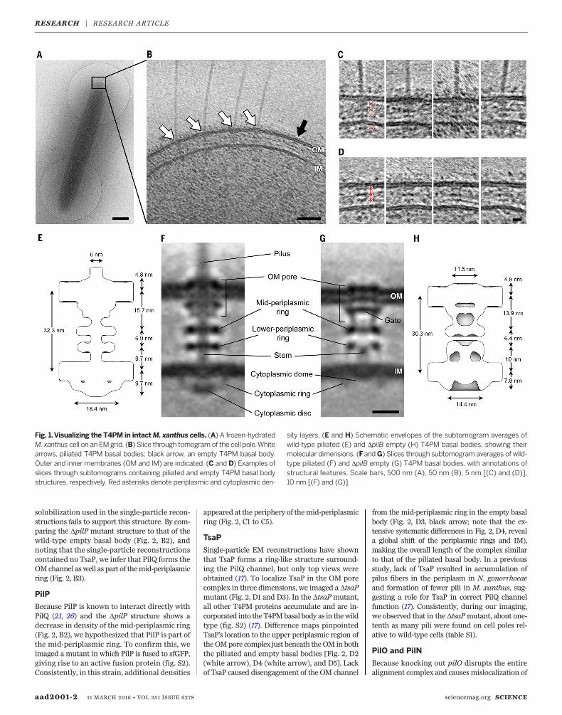
as electron cryotomography) to image the cellpoles of Myxococcus xanthus, a ubiquitous rod-shaped soil-residing predatory bacterium (Fig. 1A).(Examples of a tilt series and a cryotomogramare shown in movies S1 and S2, respectively).T4Ps with diameters of ~6 nm were easily rec-ognized on the cell surface with their basalbodies spanning the cell envelope (Fig. 1B, whitearrows). Basal bodies exhibited three layers ofdensities in the periplasm and a fourth in thecytoplasm (Fig. 1C). In the vicinity of piliatedT4PMs, we also observed “empty” nonpiliatedbasal bodies with similar structures but withoutlong fibers attached (Fig. 1B, black arrow, andFig. 1D).To reveal details, we generated subtomogram
averages of the piliated T4PM and nonpiliatedbasal bodieswith local resolution between~2.5 nmand 4.5 nm (Fig. 1F and fig. S1, A and B). We laterdetermined that a DpilBmutant (which lacks thePilB T4P-assembly ATPase and cannot assembleT4P) produced better-quality images of the emptybasal body (likely due to the basal bodies beingstalled in a “pilus preassembly” state and there-fore more structurally homogeneous than in thewild type; see below), sowe used the average fromthis strain for subsequent structural interpreta-tion (Fig. 1G).We found that the structure of the piliated
T4PM basal body comprises an outer membrane(OM)–spanning pore including a ring immedi-ately below the OM, two distinct rings in the peri-plasm, another ring in the cytoplasm surroundinga disc-like structure, and a long stem originatingat the IM and passing through the periplasmicrings and OM pore (Fig. 1F). The structure of theempty basal body shows several similar structuralfeatures, including an OM pore with a ring imme-diately below theOM, twoperiplasmic rings, and acytoplasmic ring, aswell as a gatedensity in theOM
pore, connections between the OM pore and themid-periplasmic ring, connections between thelower periplasmic ring and the IM, amuch shorterstem, and no cytoplasmic disc (Fig. 1G). In theabsence of the cytoplasmic disc, a cytoplasmicdome is also apparent. Aligning the averages ofpiliated and empty basal bodies with the IM re-veals clear conformational changes upon piliation(movie S3). Going from the nonpiliated to thepiliated state, the OMpore is ~2 nm farther awayfrom the mid-periplasmic ring (13.9 to 15.7 nm).Also, the diameter of the cytoplasmic ring in thepiliated state is greater by ~4 nm (18.4 versus14.4 nm), possibly because of its association withthe cytoplasmic disc (Fig. 1, E and H).
Mapping components in the molecularenvelope by imaging T4PM mutants
Ten highly conserved proteins are known to con-stitute the T4PM (16, 17). PilA, the major pilinprotein, contains an N-terminal hydrophobic ahelix and alternates between being anchored in-dividually in the IM or bundled with other PilAN-terminal a helices to form the central pilus core(18). The remaining nine proteins are divided intothree subgroups according to their location andfunction: the OM pore complex (PilQ and TsaP);the alignment complex (PilM, PilN, PilO, and PilP);and the motor complex (PilB, PilT, and PilC) (19).To systematically localize each component withinthe basal body, we imaged a series ofM. xanthusmutants with individual T4PM proteins eithermissing or fused to a superfolder green fluorescentprotein (sfGFP) (20) tag. Differencemaps betweenthe resulting subtomogram averages of the T4PMmutants and the wild-type structures were thencalculated (Fig. 2). Combined with information al-ready available about the accumulation, subcellularlocalization, and incorporation of individual T4PMproteins into the T4PM basal bodies of these mu-tants (fig. S2) and their connectivities and struc-tures (fig. S3) (17, 18, 21–40), these maps allowedus to pinpoint each component within the T4PM(Figs. 2 and 3A).
PilQ
A previous study showed that knocking out pilPaccelerates degradation of PilM, PilN, and PilOand causes mislocalization of PilC, whereas PilQand TsaP remain stable and at the cell pole, form-ing a rudimentary T4PM basal body consistingonly of PilQ and TsaP (fig. S2) (21). This suggeststhat PilP is crucial for stabilizing and linking othercomponents to the OM pore complex. We there-fore first imaged the DpilPmutant to obtain thestructure of theOMpore complex (PilQ and TsaP)alone (Fig. 2, B1). The average showeda cylindricalchannel in the OM with a clear gate and a largeperiplasmic vestibule. Theoverall structure is remi-niscent of single-particle reconstructions of secre-tin channel complexes in T4PM, T2SS, and type IIIsecretion systems (T3SS) (41–44) (fig. S4). Super-posing the highly conserved gate and periplasmicvestibule regions, our in vivo structure was seento be markedly longer in the transmembrane re-gion than the structures generated from single-particle analyses, probably because the detergent
RESEARCH
SCIENCE sciencemag.org 11 MARCH 2016 • VOL 351 ISSUE 6278 aad2001-1
1California Institute of Technology, Pasadena, CA 91125, USA.2Howard Hughes Medical Institute, Pasadena, CA 91125,USA. 3Max Planck Institute for Terrestrial Microbiology,35043 Marburg, Germany. 4University of Utah, Salt LakeCity, UT 84112, USA.*Corresponding author. E-mail: [email protected]
solubilization used in the single-particle recon-structions fails to support this structure. By com-paring the DpilP mutant structure to that of thewild-type empty basal body (Fig. 2, B2), andnoting that the single-particle reconstructionscontained no TsaP, we infer that PilQ forms theOMchannel aswell as part of themid-periplasmicring (Fig. 2, B3).
PilP
Because PilP is known to interact directly withPilQ (21, 26) and the DpilP structure shows adecrease in density of the mid-periplasmic ring(Fig. 2, B2), we hypothesized that PilP is part ofthe mid-periplasmic ring. To confirm this, weimaged a mutant in which PilP is fused to sfGFP,giving rise to an active fusion protein (fig. S2).Consistently, in this strain, additional densities
appeared at the periphery of themid-periplasmicring (Fig. 2, C1 to C5).
TsaP
Single-particle EM reconstructions have shownthat TsaP forms a ring-like structure surround-ing the PilQ channel, but only top views wereobtained (17). To localize TsaP in the OM porecomplex in three dimensions, we imaged a DtsaPmutant (Fig. 2, D1 and D3). In the DtsaPmutant,all other T4PM proteins accumulate and are in-corporated into the T4PMbasal body as in thewildtype (fig. S2) (17). Difference maps pinpointedTsaP’s location to the upper periplasmic region oftheOMpore complex just beneath theOM inboththe piliated and empty basal bodies [Fig. 2, D2(white arrow), D4 (white arrow), and D5]. Lackof TsaP caused disengagement of the OM channel
from the mid-periplasmic ring in the empty basalbody (Fig. 2, D3, black arrow; note that the ex-tensive systematic differences in Fig. 2, D4, reveala global shift of the periplasmic rings and IM),making the overall length of the complex similarto that of the piliated basal body. In a previousstudy, lack of TsaP resulted in accumulation ofpilus fibers in the periplasm in N. gonorrhoeaeand formation of fewer pili in M. xanthus, sug-gesting a role for TsaP in correct PilQ channelfunction (17). Consistently, during our imaging,we observed that in the DtsaPmutant, about one-tenth as many pili were found on cell poles rel-ative to wild-type cells (table S1).
PilO and PilN
Because knocking out pilO disrupts the entirealignment complex and causesmislocalization of
aad2001-2 11 MARCH 2016 • VOL 351 ISSUE 6278 sciencemag.org SCIENCE
Fig. 1.Visualizing the T4PM in intactM. xanthus cells. (A) A frozen-hydratedM. xanthus cell on an EMgrid. (B) Slice through tomogramof the cell pole.Whitearrows, piliated T4PM basal bodies; black arrow, an empty T4PM basal body.Outer and inner membranes (OM and IM) are indicated. (C and D) Examples ofslices through subtomograms containing piliated and empty T4PM basal bodystructures, respectively. Red asterisks denote periplasmic and cytoplasmic den-
sity layers. (E and H) Schematic envelopes of the subtomogram averages ofwild-type piliated (E) and DpilB empty (H) T4PM basal bodies, showing theirmolecular dimensions. (FandG) Slices through subtomogram averages of wild-type piliated (F) and DpilB empty (G) T4PM basal bodies, with annotations ofstructural features. Scale bars, 500 nm (A), 50 nm (B), 5 nm [(C) and (D)],10 nm [(F) and (G)].
RESEARCH | RESEARCH ARTICLE

SCIENCE sciencemag.org 11 MARCH 2016 • VOL 351 ISSUE 6278 aad2001-3
Fig. 2. Mapping T4PM components. First and third columns: Central slicesof subtomogram averages of piliated and empty T4PM basal bodies, respec-tively, from different M. xanthus strains. Second and fourth columns: Differ-ences in the T4PM mutant structures versus the wild type (red and yellowcolors respectively denote addition and omission of densities, with opacities of10%, 20%, 30%, 40%, and 50% corresponding to density differences of 1, 1.5,2, 2.5, and 3 standard deviations, respectively, overlaid on the wild-type sub-tomogram averages). White arrows indicate the component locations iden-tified by the difference maps. Fifth column: Schematic representations ofpiliated (left) and empty (right) T4PM basal bodies showing each identifiedcomponent location. Scale bar in A2, 10 nm (applies to columns 1 through 4).
RESEARCH | RESEARCH ARTICLE

PilC, leaving only PilQ andTsaP in the basal body(fig. S2) (21), we imaged a mutant with an activePilO-sfGFP fusion protein to map the location ofPilO (fig. S2). In the piliated structure, we ob-served additional density in the lower periplasmicring (Fig. 2, E1 and E2), but in the empty basalbody structure, we observed decreased density inthis ring (Fig. 2, E3 and E4). Both results suggestthat PilO localizes to the lower periplasmic ring
(Fig. 2, E5), because the sfGFP tag likely addeddensity to the ring in the piliated form but per-turbed the ring in the empty basal body. As withpilO, a DpilN mutant also disrupts the entirealignment complex and also causes mislocaliza-tion of PilC, leaving only PilQ and TsaP in thebasal body (fig. S2) (21). So far, we have beenunable to generate a functional PilN protein fusedto a tag. Therefore, the samemethod could not be
used to localize PilN in the T4PM basal body.Nonetheless, the two structural homologs PilOand PilN interact directly (21, 30), likely formingheterodimers (30). Therefore, we assume thatPilN is also located in the lower periplasmic ring.
PilA and minor pilins
In the wild-type piliated basal body structure, weobserved a rod-like stem structure that passes up
aad2001-4 11 MARCH 2016 • VOL 351 ISSUE 6278 sciencemag.org SCIENCE
Fig.3. Architecturalmodels of theT4PM. (A) Summary schematics showingthe component locations identified in the piliated and empty T4PM basal bodystructures. (B and C) Central slices of the architectural models of piliated andempty T4PM basal bodies, respectively, in which atomic models of T4PM com-ponents are placed in the in vivo envelopes according to the component mapsin (A) and previously reported constraints and filtered to 3-nm resolution. (Theprocess of how each component was placed is detailed in Movie 1 and thesupplementary materials.) Models of each component are colored as in (A),with the transmembrane segments of PilN and PilO shown as cylinders; “x3”indicates three AMIN domains per PilQ monomer, only one of which is shown.
Note that the empty T4PM basal body is shown with five PilA major pilin sub-units in the short stem; however, the short stem likely also containsminor pilins.(D) Top view of the PilP HR domains and the PilQ N0 and N1 domains in thearchitectural model [colored as in (B) and (C)], with PG model (colored green)as background; 36 AMIN (b) domain models from 12 PilQ proteins are ran-domly placed on PG and connected by long flexible linkers (black) with lengthswithin 20 nm between b1 and b2 domains (70 residues), 12 nm between b2 andb3 domains (40 residues), and 12 nmbetween b3 andN0domains (40 residues).(E) Overall architectural models of piliated (left) and empty (right) T4PM basalbodies. For clarity, the PilQ AMIN domains displayed in (D) are not shown.
RESEARCH | RESEARCH ARTICLE

from the IM through the lower periplasmic ring,mid-periplasmic ring, and OM pore. This stemhas the same diameter as the PilA helical poly-mer (18) (~6 nm) and is directly connected to thepilus outside of the cell. Also, this long stem ismissing in the empty basal body structure. Theseobservations suggest that the stem is the part ofthe pilus fiber located in the periplasm and is as-sociated with the basal body. In the empty basalbody, a short stem is also present between thelower periplasmic ring and the IM. To investigatewhether this remaining short stem is formed byPilA or by other T4PM components, we first im-aged a DpilA mutant that lacks the major pilinPilA. As expected (45), no T4P formed in thismutant.In agreement with the observation that the
T4PM assembles in a DpilAmutant (fig. S2) (21),we were able to identify empty basal body struc-tures in the cells and generate an average of theDpilAmutant, which clearly lacked any stem [Fig.2, F1 and F2 (white arrow)]. Because the minorpilins in P. aeruginosa were recently suggestedto form a complex that primes pilus assembly (46)and the four minor pseudopilins in the T2SS arethought to prime pseudopilus formation by themajor pseudopilin (47, 48), we generatedmutantslacking as many as nine of the 10 minor pilinsencoded in the M. xanthus genome (fig. S5A).Lack of nine of the minor pilins abolished T4P-dependent motility, and T4P did not assemble;however, all 10 core components of the T4PMaccumulated atwild-type levels in total cell extracts(fig. S5, B to D). Consistently, when we imaged themutant deleted for nine of the minor pilin genes,we did not detect T4P (table S1). We did, however,detect empty basal body structures, and theyclearly lacked the short stem [Fig. 2, G1 and G2(white arrow)].On the basis of these observations, we conclude
that the extended stem is made of the major pilinPilA (Fig. 2, F3) and that the short stem is com-posed of an assembly-priming complex consistingof minor pilins and PilA (Fig. 2, F3 andG3). In thePilA andminor pilin mutants, the structure of thelower periplasmic ringwasperturbedand the shortstem was absent (Fig. 2, F2 and G2, black arrows).Because it is unlikely for PilA to withdraw its hy-drophobic a helix from the IM or pilus fiber toparticipate in the lowerperiplasmic ring, the simul-taneous changes of the short stem and the lowerperiplasmic ring suggest a structural/functionallinkage between them (see below).
PilC, PilM, PilB, and PilT
The T4PM components PilC, PilM, PilB, and PilTall have folded cytoplasmic domains and are there-fore the candidates for the cytoplasmic ring, disc,and dome. Within the set of single-gene knock-outs of these four proteins, only empty basal bodieswere found on the DpilC, DpilM, and DpilB cells,as expected (Fig. 2, H1, I1, and J2), and onlypiliated T4PM basal bodies were found on theDpilT cells (Fig. 2, J1). In these four mutants, allthe remaining T4PM proteins accumulated (fig.S2). Because the cytoplasmic ring was missing inthe DpilM mutant (Fig. 2, I1) but appeared un-
perturbed in the DpilC, DpilB, and DpilT mu-tants, we conclude that the ring is composed ofPilM (Fig. 2, I3). Because the ring and domewereretained in theDpilBmutant but not the disc, thedisc must be PilB (Fig. 2, J2 and J4). The fact thatthe cytoplasmic disc had a size corresponding tothat of a hexameric secretion or traffic ATPase(37–40) also strengthens this conclusion (seebelow). The cytoplasmic dome and disc were bothmissing in the DpilC mutant (Fig. 2, H2), but thecytoplasmic ring was still present (Fig. 2, H1, blackarrows),which suggests that thedome is composedof PilC and that the PilB disc does not localize inits absence (Fig. 2, H3). Notably, the short stemstructure was alsomissing in the DpilC structure,revealing that PilC is required to stabilize it.With the identification of the ring as PilM, the
dome as PilC, and the disc as PilB, it follows that(i) the PilM ring assembles in the absence of thePilC dome or PilB disc, (ii) the PilC dome andPilB disc both require the PilM ring, and (iii) thePilC dome assembles without the PilB disc, butthe PilB disc requires the PilC dome. These inter-dependenciesmatch previous observations (fig. S2)except in one regard: Earlier immunofluorescenceexperiments demonstrated that PilC can be in-corporated into the T4PM independently of PilM(21) (fig. S2). Our observation that in the DpilMmutant all cytoplasmic densities and the shortstem are missing in averages (Fig. 2, I2) clarifiesthat the PilM cytoplasmic ring is important forconsistent incorporation of the other cytoplasmicproteins and formation of the short stem.After the cytoplasmic ring, dome, and disc had
been assigned to PilM, PilC, and PilB, respectively,no additional cytoplasmic density was availableto interpret as PilT. The most likely explanationis that PilT occupies the same location as PilB.When generating subtomogramaverages, becausewe could not tell whether any particular pilus wasin the extension or retraction state, we averagedall identified particles. The fact that the DpilTmutant displayedmore T4Ps than wild-type cellsin our images (table S1) confirmed that the T4Pswere actively extending and retracting in ourwild-type sample. Hence, the wild-type piliatedstructure was an average of both PilB- and PilT-bound states. The DpilT piliated structure (Fig. 2,J1), however, showed no clear addition or loss ofcytoplasmic densities relative to the wild type(fig. S1, B and D), which suggests that the differ-ences between PilB- and PilT-bound particles arenot visible at this resolution. We conclude thatboth PilB and PilT form cytoplasmic disc struc-tures (Fig. 2, J4) and bind to the basal body in amutually exclusive manner.To test this idea, we generated a DpilB DpilT
double mutant. As expected, this mutant lackedT4P-dependent motility and all other T4PM pro-teins accumulated as in the wild type (fig. S6).When we imaged the DpilB DpilT doublemutant(Fig. 2, J3), no clear differences were observed onits empty basal body relative to that of DpilB andwild-type strains (Fig. 2, A2 and J2), confirmingour assignment of both PilB and PilT ATPases tothe same cytoplasmic disc. The ATPase densitywas located ~2 nm away from the IM, preventing
any direct interaction between the ATPases andpilin subunits. We therefore conclude that PilC,which forms the cytoplasmic dome structure, liesbetween the ATPases and the stem and trans-duces force generated byATPhydrolysis into pilusextension and retraction. In agreement with thismodel, the N-terminal cytoplasmic domain ofPilC was recently shown to interact directly withPilB, and the C-terminal cytoplasmic domain wassuggested to interact with PilT (49).
Placing available component structuresinto the overall molecular envelope
Because atomic structures are available for homo-logs of >90% of the domains of the T4PM,we nextsought to test the plausibility of our componentmaps by trying to place these structures into theoverall molecular envelope in a way that wouldsatisfy the maps and all known constraints, in-cluding domain sizes, structures, and connectiv-ities (see supplementary materials, in particularfigs. S7 to S11, and for details, and Movie 1 for apresentation of the process in three dimensions).Remarkably, in this process, the sizes and shapesof all domains analyzed fitted well into the mo-lecular envelopes and resulted in a hypotheticalworking model of the piliated and nonpiliatedbasal bodies (fig. S12). Because the 2- to 4-nmresolution of the subtomogram averages was nothigh enough to reveal the orientation of each com-ponentwithin theEMenvelope,wenext generatedmodels with most components filtered to 3-nmresolution (Fig. 3, B to E). The process of position-ing and connecting the domains in the context ofa full hypothetical structural model nonethelessrevealed important relationships that rationalizenumerous previous observations and suggest in-sights into T4PM assembly, structure, and func-tion, as described below and shown in Movie 2.
Overall architecture and assembly
All T4PM components except the pilus and PilCappear to form rings. Each of the OM, peptido-glycan (PG), and IM cell envelope layers is nego-tiated or engaged by a proteinaceous ring, andthere is an additional “floating” lower periplasmicring. Each ring is linked to the rings above andbelow to create an integrated but flexible structurespanning the entire cell envelope. The subtomogramaverages of assembled T4PM subcomplexes indifferent mutants provide snapshots of the as-sembly pathway and support the sequence [PilQ,TsaP] → [PilP, PilN, PilO] → [PilM, PilC, PilA,minor pilins]→ [PilB, PilT] (fig. S13). The coiled-coil domains of PilN and PilO form a cage-likecompartment above and within the IM. Althoughthe exact number of subunit monomers in eachring remains unknown, the 1:1 connectivities be-tween PilQ-PilP, PilP-PilN-PilO, and PilN-PilM sug-gest that all the rings have the same stoichiometry.We found that 12 copies of the ring componentsfit best into the EM density, in agreement withobservations of secretin channels of T4PM andT2SS in multiple species (41, 42, 50). The entirebasal body is therefore robustly anchored to thePG by ~12 TsaP N-terminal LysM domains and~36 PilQ AMIN domains arranged irregularly (for
SCIENCE sciencemag.org 11 MARCH 2016 • VOL 351 ISSUE 6278 aad2001-5
RESEARCH | RESEARCH ARTICLE

instance, as in Fig. 3D). PilQ in turn links to PilP,which links to the PilN-PilO ring, which links toPilM, which binds PilB (and PilT). Unless one ofthese known connectivities is only transitory invivo, the only components free to rotate are PilCand potentially the pilus.
Pilus assembly and disassembly by arotating PilC
The structure of the pilus can be thought of aseither a 3-start left-handed helix or a 1-start or4-start right-handed helix. Because no substantialpilus rotation has been observed during pilus re-traction or extension, three different assemblystructures or mechanisms can be imagined (18):(i) a fixed structure with three active sites addingpilins at each of the three sites needed to extenda 3-start helix; (ii) a fixed structure with four ac-tive sites adding pilins at each of the four sitesneeded to extend a 4-start helix; or (iii) a rotatingstructure with one or a few active sites addingpilins one at a time as it rotates around the axisof the fiber, extending the fiber as a 1-start helix(3, 51). As described above, the OMpore complex,alignment complex, and PilM-ATPase complexesare all directly or indirectly linked and anchoredto the PG. The only T4PM component able torotate is PilC. We found that the space inside thePilM-PilN-PilO “cage” can accommodate no morethan one PilC dimer. It is unlikely that a PilC di-mer would have three or four active sites for inter-acting with different PilA molecules; therefore,ourmodels point to a 1-start assemblymechanismin which PilC rotates as it assembles the helicalpilus fiber. It is also known thatN. gonorrhoeaeT4Ps extend and retract in increments smallerthan the length of one helical turn (52), which ismost easily explained by amechanismwithmorethan one step per turn, corresponding to the 1-startassembly mechanism. Another reason to favormodels that involve rotation of some componentsuch as PilC is that the homologous archaeal fla-gellar motor clearly begins to rotate its flagellar fil-ament once this filament has been assembled (53).In our architectural model of the piliated basal
body, the PilC dimer rests on top of (and is pre-sumed to interact with) two opposing PilB sub-units in the ATPase hexamer, and the sixNTDs ofPilB in the ATPase hexamer are clamped in placeby interacting with every second PilM subunit inthe cytoplasmic ring (fig. S12A). We therefore pro-pose that ATP hydrolysis by the pairs of opposingPilB subunits that contact the PilC dimer causesPilB NTD movements that rotate the asymme-trical PilC dimer. We predict the rotation of PilCto have two consequences: (i) It “scoops”new PilAsubunits one at a time out of the membrane andonto the base of the pilus, and (ii) PilC is trans-ferred to the next pair of opposing PilB subunits.The transmembrane segments of PilC likelymatewith the tapered tip of the helical pilus fiber insuch a way as to extend and anchor the pilus tothe basal body, thereby creating a complete bind-ing pocket for the hydrophobic tail of the nextPilA subunit to be incorporated. Once a new PilAsubunit is incorporated, PilB drives PilC arounda fraction of a turn, pushing the pilus up a frac-
tion of the length of a pilin subunit and recre-ating the binding pocket in the next availableposition.One problem with this model is that the hex-
americ nature of PilB would predict elementaryturns of 60° or 120°; neither would result in theexactly 3.6 subunits per turn needed to extendthe pilus without any rotation. Perhaps the pro-cess does involve some slight rotations or Brown-ian motions of the proteins that would causesmall slips in register. Pilus retraction would beaccomplished by a switch fromPilB to PilT. Thus,although the exact structure of the PilC dimerand its orientation in theT4PMremain uncertain,the architecture alone implies that both PilB andPilT act as ratchets biasing the assembly or disas-sembly process:While PilB holds the PilC-tip pock-et on the next empty position of the pilus fiberuntil it is filled, and then quickly rotates to pre-vent subsequent dissociation, PilT holds the PilC-tip pocket on the last subunit of the tip until itdissociates back into the membrane, after whichit quickly rotates to prevent reassociation.
The PilN-PilO and PilM rings likely sensepilus retraction signals and guideATPase selection
Previous studies have shown that pilus retractionis induced by adding pilin-binding substrates (54)or pulling on the pilus directly (52). Recent studieswith antibodies showed that tension induces con-formational changes in the pilus itself (55), whichcould propagate into the basal body. Substratebinding may also induce similar conformationalchanges. Our results point to amodel inwhich thealignment complex is an IM-crossing transmissionmodule: Pilus retraction signals carried by the pi-lus itself into the basal body could be sensed in theperiplasm by the PilN-PilO ring and then trans-mitted via the coiled-coil domains through theIM tomodulate the conformation of the PilM ring,which in turn governs which ATPase is bound.Thismodel rationalizes the recent report that con-formational changes are required in the coiled-coil domains of PilN-PilO during the transitionbetween T4P extension and retraction, which sug-gests that the alignment complex is not simply astatic connector between IM andOM components,but insteadplays a critical role inT4Pdynamics (56).
The power of combined structuralapproaches to dissect complicatedmolecular machines
Solving the structures of large macromolecularmachines is challenging. Traditional structuralmethods suchas x-ray crystallography, nuclearmag-netic resonance spectroscopy, and single-particlecryo–electronmicroscopy can deliver near-atomicresolution, but they all rely on purified samples.Many important biological structures such asflagellar motors (57), chemoreceptor arrays (58),and the T4PM studied here, however, may neverbe purifiable in a native state; we found that eventhe PilQ (fig. S4), PilC (fig. S10), andPilM-PilN-PilO(fig. S14) subcomplexes lose their native struc-ture when purified. As a result, only structures ofisolated subunits and small subcomplexes have
been determined. Cryo–electron tomography canreveal at least the architectures of these large ma-chines in situ, as ourwork has shown.When thereis sufficient additional information about thestructures and connectivities of the components,working models can be built that provide a pathtoward structural understanding, where both thestructural relationships and the new mechanisticinsights they suggest can then be tested.
REFERENCES AND NOTES
1. K. V. Korotkov, M. Sandkvist, W. G. J. Hol, The type IIsecretion system: Biogenesis, molecular architecture andmechanism. Nat. Rev. Microbiol. 10, 336–351 (2012).pmid: 22466878
2. K. F. Jarrell, S.-V. Albers, The archaellum: An old motilitystructure with a new name. Trends Microbiol. 20, 307–312(2012). doi: 10.1016/j.tim.2012.04.007; pmid: 22613456
3. J. S. Mattick, Type IV pili and twitching motility. Annu. Rev.Microbiol. 56, 289–314 (2002). doi: 10.1146/annurev.micro.56.012302.160938; pmid: 12142488
4. L. Craig, M. E. Pique, J. A. Tainer, Type IV pilus structure andbacterial pathogenicity. Nat. Rev. Microbiol. 2, 363–378(2004). doi: 10.1038/nrmicro885; pmid: 15100690
5. K. J. Evans, C. Lambert, R. E. Sockett, Predation byBdellovibrio bacteriovorus HD100 requires type IV pili.J. Bacteriol. 189, 4850–4859 (2007). doi: 10.1128/JB.01942-06; pmid: 17416646
6. I. Chen, D. Dubnau, DNA uptake during bacterialtransformation. Nat. Rev. Microbiol. 2, 241–249 (2004).doi: 10.1038/nrmicro844; pmid: 15083159
7. M. Klausen, A. Aaes-Jørgensen, S. Molin, T. Tolker-Nielsen,Involvement of bacterial migration in the development ofcomplex multicellular structures in Pseudomonas aeruginosabiofilms. Mol. Microbiol. 50, 61–68 (2003). doi: 10.1046/j.1365-2958.2003.03677.x; pmid: 14507363
8. G. A. O’Toole, R. Kolter, Flagellar and twitching motility arenecessary for Pseudomonas aeruginosa biofilm development.Mol. Microbiol. 30, 295–304 (1998). doi: 10.1046/j.1365-2958.1998.01062.x; pmid: 9791175
9. A. J. Hager et al., Type IV pili-mediated secretion modulatesFrancisella virulence. Mol. Microbiol. 62, 227–237 (2006).doi: 10.1111/j.1365-2958.2006.05365.x; pmid: 16987180
10. A. J. Merz, M. So, M. P. Sheetz, Pilus retraction powersbacterial twitching motility. Nature 407, 98–102 (2000).doi: 10.1038/35024105; pmid: 10993081
11. J. M. Skerker, H. C. Berg, Direct observation of extension andretraction of type IV pili. Proc. Natl. Acad. Sci. U.S.A. 98,6901–6904 (2001). doi: 10.1073/pnas.121171698;pmid: 11381130
12. L. Craig, J. Li, Type IV pili: Paradoxes in form and function.Curr. Opin. Struct. Biol. 18, 267–277 (2008). doi: 10.1016/j.sbi.2007.12.009; pmid: 18249533
13. P. C. Morand et al., Type IV pilus retraction in pathogenicNeisseria is regulated by the PilC proteins. EMBO J. 23,2009–2017 (2004). doi: 10.1038/sj.emboj.7600200;pmid: 15103324
14. B. Maier et al., Single pilus motor forces exceed 100 pN.Proc. Natl. Acad. Sci. U.S.A. 99, 16012–16017 (2002).doi: 10.1073/pnas.242523299; pmid: 12446837
15. M. Clausen, V. Jakovljevic, L. Søgaard-Andersen, B. Maier,High-force generation is a conserved property of type IV pilussystems. J. Bacteriol. 191, 4633–4638 (2009). doi: 10.1128/JB.00396-09; pmid: 19429611
16. V. Pelicic, Type IV pili: E pluribus unum? Mol. Microbiol. 68,827–837 (2008). doi: 10.1111/j.1365-2958.2008.06197.x;pmid: 18399938
17. K. Siewering et al., Peptidoglycan-binding protein TsaPfunctions in surface assembly of type IV pili. Proc. Natl. Acad.Sci. U.S.A. 111, E953–E961 (2014). doi: 10.1073/pnas.1322889111; pmid: 24556993
18. L. Craig et al., Type IV pilus structure by cryo-electronmicroscopy and crystallography: Implications for pilusassembly and functions. Mol. Cell 23, 651–662 (2006).doi: 10.1016/j.molcel.2006.07.004; pmid: 16949362
19. L. L. Burrows, Pseudomonas aeruginosa twitching motility:Type IV pili in action. Annu. Rev. Microbiol. 66, 493–520(2012). doi: 10.1146/annurev-micro-092611-150055;pmid: 22746331
20. J.-D. Pédelacq, S. Cabantous, T. Tran, T. C. Terwilliger,G. S. Waldo, Engineering and characterization of a superfolder
aad2001-6 11 MARCH 2016 • VOL 351 ISSUE 6278 sciencemag.org SCIENCE
RESEARCH | RESEARCH ARTICLE

green fluorescent protein. Nat. Biotechnol. 24, 79–88 (2006).doi: 10.1038/nbt1172; pmid: 16369541
21. C. Friedrich, I. Bulyha, L. Søgaard-Andersen, Outside-inassembly pathway of the type IV pilus system in Myxococcusxanthus. J. Bacteriol. 196, 378–390 (2014). doi: 10.1128/JB.01094-13; pmid: 24187092
22. I. Bulyha et al., Regulation of the type IV pili molecular machineby dynamic localization of two motor proteins. Mol. Microbiol.74, 691–706 (2009). doi: 10.1111/j.1365-2958.2009.06891.x;pmid: 19775250
23. M. Georgiadou, M. Castagnini, G. Karimova, D. Ladant,V. Pelicic, Large-scale study of the interactions betweenproteins involved in type IV pilus biology in Neisseriameningitidis: Characterization of a subcomplex involved in pilusassembly. Mol. Microbiol. 84, 857–873 (2012). doi: 10.1111/j.1365-2958.2012.08062.x; pmid: 22486968
24. C. Li, R. A. Wallace, W. P. Black, Y. Z. Li, Z. Yang, Type IV pilusproteins form an integrated structure extending from thecytoplasm to the outer membrane. PLOS ONE 8, e70144(2013). doi: 10.1371/journal.pone.0070144; pmid: 23922942
25. S. Tammam et al., PilMNOPQ from the Pseudomonasaeruginosa type IV pilus system form a transenvelope proteininteraction network that interacts with PilA. J. Bacteriol. 195,2126–2135 (2013). doi: 10.1128/JB.00032-13; pmid: 23457250
26. S. V. Balasingham et al., Interactions between the lipoproteinPilP and the secretin PilQ in Neisseria meningitidis. J. Bacteriol.189, 5716–5727 (2007). doi: 10.1128/JB.00060-07;pmid: 17526700
27. M. Ayers et al., PilM/N/O/P proteins form an inner membranecomplex that affects the stability of the Pseudomonasaeruginosa type IV pilus secretin. J. Mol. Biol. 394, 128–142(2009). doi: 10.1016/j.jmb.2009.09.034; pmid: 19857645
28. S. Tammam et al., Characterization of the PilN, PilO and PilPtype IVa pilus subcomplex. Mol. Microbiol. 82, 1496–1514(2011). doi: 10.1111/j.1365-2958.2011.07903.x; pmid: 22053789
29. S. Gu et al., Solution structure of homology region (HR)domain of type II secretion system. J. Biol. Chem. 287,9072–9080 (2012). doi: 10.1074/jbc.M111.300624;pmid: 22253442
30. L. M. Sampaleanu et al., Periplasmic domains of Pseudomonasaeruginosa PilN and PilO form a stable heterodimeric complex.J. Mol. Biol. 394, 143–159 (2009). doi: 10.1016/j.jmb.2009.09.037; pmid: 19857646
31. V. Karuppiah, J. P. Derrick, Structure of the PilM-PilN innermembrane type IV pilus biogenesis complex from Thermusthermophilus. J. Biol. Chem. 286, 24434–24442 (2011).doi: 10.1074/jbc.M111.243535; pmid: 21596754
32. K. V. Korotkov et al., Structural and functional studies on theinteraction of GspC and GspD in the type II secretion system.PLOS Pathog. 7, e1002228 (2011). doi: 10.1371/journal.ppat.1002228; pmid: 21931548
33. V. Karuppiah, R. F. Collins, A. Thistlethwaite, Y. Gao,J. P. Derrick, Structure and assembly of an inner membraneplatform for initiation of type IV pilus biogenesis. Proc. Natl.Acad. Sci. U.S.A. 110, E4638–E4647 (2013). doi: 10.1073/pnas.1312313110; pmid: 24218553
34. V. Karuppiah, D. Hassan, M. Saleem, J. P. Derrick, Structureand oligomerization of the PilC type IV pilus biogenesis proteinfrom Thermus thermophilus. Proteins 78, 2049–2057 (2010).pmid: 20455262
35. J. Abendroth et al., The three-dimensional structure of thecytoplasmic domains of EpsF from the type 2 secretion systemof Vibrio cholerae. J. Struct. Biol. 166, 303–315 (2009).doi: 10.1016/j.jsb.2009.03.009; pmid: 19324092
36. J. Abendroth, P. Murphy, M. Sandkvist, M. Bagdasarian,W. G. J. Hol, The X-ray structure of the type II secretionsystem complex formed by the N-terminal domain of EpsE andthe cytoplasmic domain of EpsL of Vibrio cholerae. J. Mol. Biol.348, 845–855 (2005). doi: 10.1016/j.jmb.2005.02.061;pmid: 15843017
37. A. Yamagata, J. A. Tainer, Hexameric structures of the archaealsecretion ATPase GspE and implications for a universalsecretion mechanism. EMBO J. 26, 878–890 (2007).doi: 10.1038/sj.emboj.7601544; pmid: 17255937
38. A. M. Misic, K. A. Satyshur, K. T. Forest, P. aeruginosa PilTstructures with and without nucleotide reveal a dynamic typeIV pilus retraction motor. J. Mol. Biol. 400, 1011–1021 (2010).doi: 10.1016/j.jmb.2010.05.066; pmid: 20595000
39. K. A. Satyshur et al., Crystal structures of the pilus retractionmotor PilT suggest large domain movements and subunitcooperation drive motility. Structure 15, 363–376 (2007).doi: 10.1016/j.str.2007.01.018; pmid: 17355871
40. C. Lu et al., Hexamers of the type II secretion ATPase GspEfrom Vibrio cholerae with increased ATPase activity. Structure21, 1707–1717 (2013). doi: 10.1016/j.str.2013.06.027;pmid: 23954505
41. J.-L. Berry et al., Structure and assembly of a trans-periplasmic channel for type IV pili in Neisseria meningitidis.PLOS Pathog. 8, e1002923 (2012). doi: 10.1371/journal.ppat.1002923; pmid: 23028322
42. S. L. Reichow, K. V. Korotkov, W. G. J. Hol, T. Gonen, Structureof the cholera toxin secretion channel in its closed state. Nat.Struct. Mol. Biol. 17, 1226–1232 (2010). doi: 10.1038/nsmb.1910; pmid: 20852644
43. J. Kowal et al., Structure of the dodecameric Yersiniaenterocolitica secretin YscC and its trypsin-resistant core.Structure 21, 2152–2161 (2013). doi: 10.1016/j.str.2013.09.012;pmid: 24207124
44. T. Tosi et al., Structural similarity of secretins from type II andtype III secretion systems. Structure 22, 1348–1355 (2014).doi: 10.1016/j.str.2014.07.005; pmid: 25156426
45. S. S. Wu, D. Kaiser, Genetic and functional evidence that TypeIV pili are required for social gliding motility in Myxococcusxanthus. Mol. Microbiol. 18, 547–558 (1995). doi: 10.1111/j.1365-2958.1995.mmi_18030547.x; pmid: 8748037
46. Y. Nguyen et al., Pseudomonas aeruginosa minor pilins primetype IVa pilus assembly and promote surface display of thePilY1 adhesin. J. Biol. Chem. 290, 601–611 (2015).doi: 10.1074/jbc.M114.616904; pmid: 25389296
47. N. Sauvonnet, G. Vignon, A. P. Pugsley, P. Gounon, Pilusformation and protein secretion by the same machinery inEscherichia coli. EMBO J. 19, 2221–2228 (2000). doi: 10.1093/emboj/19.10.2221; pmid: 10811613
48. D. A. Cisneros, P. J. Bond, A. P. Pugsley, M. Campos,O. Francetic, Minor pseudopilin self-assembly primes type IIsecretion pseudopilus elongation. EMBO J. 31, 1041–1053(2012). doi: 10.1038/emboj.2011.454; pmid: 22157749
49. H. K. Takhar, K. Kemp, M. Kim, P. L. Howell, L. L. Burrows,The platform protein is essential for type IV pilus biogenesis.J. Biol. Chem. 288, 9721–9728 (2013). doi: 10.1074/jbc.M113.453506
50. M. Chami et al., Structural insights into the secretin PulD andits trypsin-resistant core. J. Biol. Chem. 280, 37732–37741(2005). doi: 10.1074/jbc.M504463200; pmid: 16129681
51. D. Nunn, Bacterial type II protein export and pilus biogenesis:More than just homologies? Trends Cell Biol. 9, 402–408(1999). doi: 10.1016/S0962-8924(99)01634-7;pmid: 10481178
52. M. Clausen, M. Koomey, B. Maier, Dynamics of type IV pili iscontrolled by switching between multiple states. Biophys. J. 96,1169–1177 (2009). doi: 10.1016/j.bpj.2008.10.017; pmid: 19186152
53. S.-V. Albers, K. F. Jarrell, The archaellum: How Archaea swim.Front. Microbiol. 6, 23 (2015). doi: 10.3389/fmicb.2015.00023;pmid: 25699024
54. Y. Li et al., Extracellular polysaccharides mediate pilus retractionduring social motility of Myxococcus xanthus. Proc. Natl. Acad.Sci. U.S.A. 100, 5443–5448 (2003). pmid: 12704238
55. N. Biais, D. L. Higashi, J. Brujić, M. So, M. P. Sheetz, Force-dependent polymorphism in type IV pili reveals hiddenepitopes. Proc. Natl. Acad. Sci. U.S.A. 107, 11358–11363(2010). doi: 10.1073/pnas.0911328107; pmid: 20534431
56. T. L. Leighton, N. Dayalani, L. M. Sampaleanu, P. L. Howell,L. L. Burrows, Novel role for PilNO in type IV pilus retractionrevealed by alignment subcomplex mutations. J. Bacteriol. 197,2229–2238 (2015). doi: 10.1128/JB.00220-15
57. S. Chen et al., Structural diversity of bacterial flagellar motors.EMBO J. 30, 2972–2981 (2011). doi: 10.1038/emboj.2011.186;pmid: 21673657
58. A. Briegel et al., New insights into bacterial chemoreceptorarray structure and assembly from electron cryotomography.Biochemistry 53, 1575–1585 (2014). doi: 10.1021/bi5000614
ACKNOWLEDGMENTS
We thank C. Oikonomou and D. Ortega for discussions. Supportedby NIH grant R01 GM094800B (G.J.J.), the Howard HughesMedical Institute, the Max Planck Society, and the DeutscheForschungsgemeinschaft within the framework of the CollaborativeResearch Center (SFB) 987 “Microbial Diversity in EnvironmentalSignal Response.” The 18 subtomogram averages of T4PMsreported in this study have been deposited in the ElectronMicroscopy Data Bank with accession numbers EMD-3247(wild type, piliated); EMD-3248 (wild type, empty);EMD-3249 (DpilP, empty); EMD-3250 (PilP-sfGFP, piliated);EMD-3251 (PilP-sfGFP, empty); EMD-3252 (DtsaP,piliated); EMD-3253 (DtsaP, empty); EMD-3254 (PilO-sfGFP,piliated); EMD-3255 (PilO-sfGFP, empty); EMD-3256 (DpilC,empty); EMD-3257 (DpilA, empty); EMD-3258 (DpilV pilW fimU1+2+3, empty); EMD-3259 (DpilT, piliated); EMD-3260 (DpilB,empty); EMD-3261 (DpilB DpilT, empty); EMD-3262 (DpilM,empty); EMD-3263 (DpilQb1-b2, piliated); and EMD-3264(DpilQb1-b2, empty). The coordinates of the hypothetical T4PMworking models have been deposited in the Protein Data Bank withaccession numbers 3JC8 (piliated) and 3JC9 (empty), respectively.Author contributions: Y.-W.C. collected the cryo–electrontomography data, which were analyzed by Y.-W.C. andL.A.R.; Y.-W.C. built the T4PM models and generated the moviedescribing the modeling process; A.T.-L. and L.S.-A. provided theM. xanthus strains and characterized their motility and T4PMcomponent accumulation and localization; J.I. producedthe animation of T4PM dynamics; and Y.-W.C., L.S.-A.,and G.J.J. wrote the paper.
SUPPLEMENTARY MATERIALS
www.sciencemag.org/content/351/6278/aad2001/suppl/DC1Materials and MethodsFigs. S1 to S14Tables S1 to S5Movies S1 to S3References (59–98)
10 August 2015; accepted 13 January 201610.1126/science.aad2001
SCIENCE sciencemag.org 11 MARCH 2016 • VOL 351 ISSUE 6278 aad2001-7
RESEARCH | RESEARCH ARTICLE

DOI: 10.1126/science.aad2001, (2016);351 Science
et al.Yi-Wei ChangArchitecture of the type IVa pilus machine
This copy is for your personal, non-commercial use only.
clicking here.colleagues, clients, or customers by , you can order high-quality copies for yourIf you wish to distribute this article to others
here.following the guidelines
can be obtained byPermission to republish or repurpose articles or portions of articles
): March 17, 2016 www.sciencemag.org (this information is current as of
The following resources related to this article are available online at
/content/351/6278/aad2001.full.htmlversion of this article at:
including high-resolution figures, can be found in the onlineUpdated information and services,
/content/suppl/2016/03/09/351.6278.aad2001.DC1.html can be found at: Supporting Online Material
/content/351/6278/aad2001.full.html#ref-list-1, 41 of which can be accessed free:cites 98 articlesThis article
/cgi/collection/biochemBiochemistry
subject collections:This article appears in the following
registered trademark of AAAS. is aScience2016 by the American Association for the Advancement of Science; all rights reserved. The title
CopyrightAmerican Association for the Advancement of Science, 1200 New York Avenue NW, Washington, DC 20005. (print ISSN 0036-8075; online ISSN 1095-9203) is published weekly, except the last week in December, by theScience
on M
arch
17,
201
6D
ownl
oade
d fr
om