
Preventing Chronic Disease in Diverse Communities: Looking ...

Bacterial Communities of Diverse Drosophila Species:Ecological Context of a Host–Microbe Model SystemJames Angus Chandler1.*, Jenna Morgan Lang1,2,3,4., Srijak Bhatnagar2, Jonathan A. Eisen1,2,3,4, Artyom
Kopp1
1Center for Population Biology, Department of Evolution and Ecology, University of California Davis, Davis, California, United States of America, 2UC Davis Genome
Center, University of California Davis, Davis, California, United States of America, 3Department of Medical Microbiology and Immunology, School of Medicine, University
of California Davis, Davis, California, United Stated of America, 4United States Department of Energy Joint Genome Institute, Walnut Creek, California, United States of
America
Abstract
Drosophila melanogaster is emerging as an important model of non-pathogenic host–microbe interactions. The genetic andexperimental tractability of Drosophila has led to significant gains in our understanding of animal–microbial symbiosis.However, the full implications of these results cannot be appreciated without the knowledge of the microbial communitiesassociated with natural Drosophila populations. In particular, it is not clear whether laboratory cultures can serve as anaccurate model of host–microbe interactions that occur in the wild, or those that have occurred over evolutionary time. Tofill this gap, we characterized natural bacterial communities associated with 14 species of Drosophila and related generacollected from distant geographic locations. To represent the ecological diversity of Drosophilids, examined speciesincluded fruit-, flower-, mushroom-, and cactus-feeders. In parallel, wild host populations were compared to laboratorystrains, and controlled experiments were performed to assess the importance of host species and diet in shaping bacterialmicrobiome composition. We find that Drosophilid flies have taxonomically restricted bacterial communities, with 85% ofthe natural bacterial microbiome composed of only four bacterial families. The dominant bacterial taxa are widespread andfound in many different host species despite the taxonomic, ecological, and geographic diversity of their hosts. Both naturalsurveys and laboratory experiments indicate that host diet plays a major role in shaping the Drosophila bacterialmicrobiome. Despite this, the internal bacterial microbiome represents only a highly reduced subset of the externalbacterial communities, suggesting that the host exercises some level of control over the bacteria that inhabit its digestivetract. Finally, we show that laboratory strains provide only a limited model of natural host–microbe interactions. Bacterialtaxa used in experimental studies are rare or absent in wild Drosophila populations, while the most abundant associates ofnatural Drosophila populations are rare in the lab.
Citation: Chandler JA, Morgan Lang J, Bhatnagar S, Eisen JA, Kopp A (2011) Bacterial Communities of Diverse Drosophila Species: Ecological Context of a Host–Microbe Model System. PLoS Genet 7(9): e1002272. doi:10.1371/journal.pgen.1002272
Editor: Harmit S. Malik, Fred Hutchinson Cancer Research Center, United States of America
Received March 21, 2011; Accepted July 18, 2011; Published September 22, 2011
This is an open-access article distributed under the terms of the Creative Commons Public Domain declaration which stipulates that, once placed in the publicdomain, this work may be freely reproduced, distributed, transmitted, modified, built upon, or otherwise used by anyone for any lawful purpose.
Funding: This work was supported by NSF grant IOS-0815141 to AK (www.nsf.gov), a Laboratory Directed Research and Development Program grant from theLawrence Berkeley National Laboratory to JAE (www.lbl.gov), a Gordon and Betty Moore Foundation grant #1660 to JAE (www.moore.org), and the UC DavisCenter for Population Biology (www.cpb.ucdavis.edu). Some work was conducted at the U.S. Department of Energy Joint Genome Institute, which is supported bythe Office of Science of the U.S. Department of Energy under contract No. DE-AC02-05CH11231 (www.jgi.doe.gov). The funders had no role in study design, datacollection and analysis, decision to publish, or preparation of the manuscript.
Competing Interests: The authors have declared that no competing interests exist.
* E-mail: [email protected]
. These authors contributed equally to this work.
Introduction
The genetic and experimental tractability of Drosophila melano-gaster often overshadows the phenotypic, evolutionary andecological diversity of its relatives. Over 3000 species of Drosophilaand related genera inhabit all continents except Antarctica, occurin practically every type of habitat, and show a great variety ofmorphological, behavioral, and life-history traits [1]. In particular,the feeding and breeding substrates vary tremendously within theDrosophilids. While the well-known cosmopolitan species areconsidered generalists, as decaying fruit of many different plantsmakes for an acceptable substrate, dietary specialization hasevolved many times within Drosophila. A well-known example is D.sechellia, which specializes on the Morinda fruit, a resource that istoxic to most other animals [2]. Other Drosophila species useflowers, mushrooms, sap fluxes, cambium, decaying vegetation,
and cacti as feeding and breeding sites [3,4]. Importantly, dietaryshifts have occurred numerous times within the genus, and closelyrelated species are known to utilize different types of food sources[5,6,7]. At the same time, it is common to find phylogeneticallydistant species using the same food source. In almost all of thesecases, the biotic environment that Drosophila are interacting with,especially the microbial communities associated with these flies, isunknown.The importance and ubiquity of microbial associates of animals
is only beginning to be appreciated. Although most attention hasbeen devoted to pathogenic bacteria, pathogens are a smallminority of animal symbionts. Bacteria can play beneficial, andoften essential, roles in the lives of their hosts. In animals that carryvertically transmitted, intracellular bacteria, the host and itssymbiont community form an inseparable holobiont with sharedmetabolism and evolutionary fate [8,9]. However, symbionts need
PLoS Genetics | www.plosgenetics.org 1 September 2011 | Volume 7 | Issue 9 | e1002272

not be intracellular or completely dependent on the host to shapehost physiology and evolution. Most animal-microbial interactionsare flexible and facultative, where the symbionts can exist withoutthe host and the host can carry different symbionts at differenttimes. It is likely that every animal is associated with a complexand ever-changing microbial community that consists predomi-nantly of non-pathogenic, free-living bacteria [10]. Nowhere is thismore evident than in intestinal microbiology. In humans, bacterialgut fauna is composed of more than a thousand taxa and certainaspects of human health, such as obesity, are associated with analtered intestinal community [11]. Bacterial gut symbionts areequally prevalent in other mammals [12] and in insects [13,14]. Inmany insects, gut symbionts are essential for survival and form thecore of host physiology and ecological adaptation [15,16,17]. Evenwhen not strictly essential for survival, experimental evidencesuggests that insect gut fauna affects many aspects of hostphenotype [18] and can mediate interactions between the hostand potential pathogens [19].The composition of bacterial symbiont communities is shaped
both by host genotype and its diet. In mice and fruit flies,mutations in a single host gene can be sufficient to altermicrobiome composition [20,21]. Reciprocal transplants ofintestinal microbiomes between zebrafish and mice reveal thatthe gut habitat of these hosts selects for different communities [22].These differences are smaller at shorter evolutionary time scales, asspecies that are more closely related often share more similarbacterial communities. This trend has been observed in stinkbugs[23], termites [24], and mammals [12]. Diet also plays animportant role in shaping the intestinal bacterial microbiome inmany systems. When humans are shifted onto a low carbohydrate,low fat diet, their intestinal communities shift towards a higherpercentage of the phylum Bacteroidetes [11]. The gut communi-ties of European and African human populations are shaped, atleast in part, by their different diets [25].D. melanogaster is naturally emerging as a model of host-microbe
interactions. Genetic experiments have identified some of thegenes contributing to intestinal community homeostasis. The gene
PIMS actively suppresses immune response when flies are exposedto commensal, non-pathogenic intestinal communities [26].Similarly, downregulation of caudal significantly alters this bacterialcommunity, allowing normally rare bacteria to increase inabundance [21]. However, little is known about the effects ofgut bacteria on Drosophila physiology. Axenic strains of D.melanogaster are viable, at least on rich media. Although somestudies suggested that gut symbionts increase life span in D.melanogaster [27], other studies failed to replicate this effect [21,28].Commensal bacteria can even affect mate choice in D. melanogasterin the lab [29], although the evolutionary significance of this effectin the wild is not clear.In contrast to our increasing understanding of Drosophila-
microbe interactions in the lab, little is known about themicrobial communities associated with natural Drosophila popu-lations. In other insects, laboratory-reared larvae have beenshown to harbor significantly less diverse bacterial microbiomesthan their wild counterparts [30,31]. Laboratory strains of D.melanogaster have been reported to carry the bacterial generaLactobacillus, Acetobacter and Enterococcus [21,27,28,32]. Althoughthese taxa are present in most studies, there is also a possible‘‘lab effect’’ where different labs have different bacteria [28].Many of the same bacterial genera (although not always thesame species) were found in natural D. melanogaster populations inthe eastern United States [32,33]. However, given the worldwidedistribution of Drosophila and the tremendous variation inDrosophila ecology, these taxa may represent only a small fractionof the bacterial communities associated with flies in the wild. Abetter knowledge of these communities is necessary to under-stand the role of symbiosis in Drosophila physiology, ecology, andevolution.To explore the bacterial communities associated with this
speciose and ecologically diverse lineage, and to identify thefactors shaping these communities, we surveyed natural popula-tions of 14 species of Drosophila and two closely related genera(Scaptodrosophila and Microdrosophila). Although we acknowledgethat non-bacterial microbes such as archaea and yeasts are likelyassociated with these hosts, we focused our survey on thebacterial portion of the microbiome because of its knownimportance to animal and Drosophila biology. We shall use theterm ‘‘bacterial microbiome’’ to refer to what was sampled in thisstudy. We used culture-independent 16S ribosomal DNA (rDNA)polymerase chain reaction (PCR) amplification and sequencing tocharacterize the bacterial communities associated with eachpopulation. To sample the widest spectrum of fly-associatedbacteria, collections were selected from as large a swath ofDrosophila ecology, phylogeny, and geography as possible. Flieswere collected directly from their natural feeding substratesincluding rotting fruit, flowers, mushrooms, and cacti, without theuse of any artificial baits, from locations on both coasts of NorthAmerica, Hawaii, Australia, Southeast Asia, and Seychelles,Africa. In addition to the natural survey, controlled laboratoryexperiments were performed to further determine the role ofenvironment and host species in shaping the bacterial commu-nities. This combined approach allows us to address severalpreviously unexplored questions. Do the bacterial communitiesassociated with Drosophila exhibit the same diversity as their hosts?What factors are most important in shaping the differencesbetween symbiont communities of different host species? Howdoes the composition and structure of these communitiescompare to the bacterial microbiomes of other taxa, particularlymammals? Finally, is the bacterial microbiome of lab strains usedin experimental research representative of natural bacterialcommunities?
Author Summary
All animals are associated with large consortia of non-pathogenic microbes. Most of these ‘‘microbiomes’’ arenot well characterized despite their importance for manyaspects of host biology including human and animalhealth and the agricultural impact of pest species. The fruitfly Drosophila melanogaster provides a powerful experi-mental model for investigating the dynamics and conse-quences of animal–microbial interactions. However, it isnot clear whether the model bacteria studied in the lab arerepresentative of natural microbial consortia. To establishan ecological and comparative background for experi-mental studies, we have conducted a global survey ofbacterial communities associated with natural populationsof 14 species of Drosophila and related genera. Despite thetaxonomic and ecological diversity of these species, wefind that they are associated with the same dominantbacterial groups. Based on our results, we propose amodel of microbiome assembly where its composition iscircumscribed by host diet and physiology but, withinthose limits, is highly dependent on chance environmentalencounters. Consistent with this model, the microbiomesof wild flies differ significantly from those of laboratorystrains, suggesting that experimental studies should beextended to include the bacteria that are most prevalent innatural communities.
The Microbiome of Diverse Drosophila Species
PLoS Genetics | www.plosgenetics.org 2 September 2011 | Volume 7 | Issue 9 | e1002272

Results
Data summaryDrosophila samples were collected with the help of many
colleagues around the world (see Acknowledgments, Table 1 andDataset S1). Flies were either washed in sterile water (to removecuticular bacterial cells) or were dissected to obtain just their cropsand digestive tracts. After DNA extraction, rDNA PCR amplifi-cation was done with bacterial specific primers. The 16S rDNAamplicons were cloned, transformed, and Sanger sequenced fromboth ends. For 50% of the clones, the two reads did not overlapand therefore a concatenated read was made by inserting gapcharacters in the space between the two reads.After all preliminary filtering, our dataset consisted of 3243
nearly full-length high quality sequences representing 39 hostsamples (which we refer to as libraries) (Table 1 and Dataset S1).This dataset excluded 421 clones that were only sequenced fromone end, 65 sequences with fewer than 300 non-gap characters, 76sequences that were identified as chimeric, 9 that appeared to bechimeric based on conflicting taxonomy assignments of the 39 and59 reads, 3 chloroplast sequences, and 351 sequences of likelyendosymbionts such as Wolbachia and Spiroplasma (which will beaddressed in a separate section). Because small sample sizes canlead to inaccurate diversity measures [34], two libraries containinga total of 28 sequences were removed completely. The 39remaining libraries vary in size from 26 to 223 sequences, with anaverage of 83.2 6 37.4. Most libraries (29 of 39) contain between63 and 97 sequences. 20 libraries containing 1850 total sequencesare from wild-caught hosts, while the remaining libraries andsequences came from laboratory samples and experiments. Fulltables containing each library’s identifier, size, the host speciesfrom which it was collected, location and date of collection, andother information are given in Table 1 and Dataset S1.Clustering with mothur [35] using the average neighbor
algorithm with 0.03 cutoff (corresponding to 97% sequencesimilarity) creates 139 operational taxonomic units (OTUs), thelargest of which contains 638 sequences. 66 OTUs are singletons(i.e., there is only a single sequence in the OTU) and 110 OTUscontain 10 or fewer sequences. The average OTU contains 23.3sequences (standard deviation = 78).Phylogenetic analysis was performed using FastTree [36].
Included in this analysis (and many other comparisons throughoutthis study) were many previously identified Drosophila-associatedbacteria [21,32,33,37].
Dominant bacterial taxa associated with DrosophilaFour bacterial families representing three orders make up 90%
of all sequences within our dataset. These include Enterobacter-iales: Enterobacteriaceae (60%), Rhodospirillales: Acetobacter-aceae (9%), and Lactobacillales: primarily Lactobacillaceae andEnterococcaceae (21%) (Figure 1A and Table 2). 14 other orderscomprise the remaining 10% of the dataset. All wild populationsare dominated by at least one of the three major clades, andmany Drosophila species carry all three of them (Figure 1B and1C). Although no core bacterial microbiome (a set of taxa presentin all samples) emerges, Enterobacteriaceae and Lactobacillalescome close, being found in 18 and 17 out of 20 wild Drosophilapopulations, respectively (Figure 1B). There is an interestingreciprocal relationship between these two taxa (Figure 1C). Eachof the five host samples which lacks one of these groups isdominated (.84%) by the other one. In only two populations(ELA and SCA) do Lactobacillales and Enterobacteriaceae eachmake up at least 15% of the bacterial microbiome; both ofthese are flower-feeding flies with highly diverse bacterial
microbiomes. In all the other samples, the abundance of themore dominant microbe is, on average, 44 times greater than theother one.
Enterobacteriaceae. The Enterobacteriaceae, representing,60% (1956 out of 3243) of the sequences in our analysis, are alarge family that includes many animal and plant associatedbacteria. They are found as free living associates of many insects,including Drosophila melanogaster [32]. Several lineages areendosymbiotic and required for insect nutrition, defense fromparasites, and tolerance of heat stress [38,39,40,41]. Almost everywild and laboratory host contains some Enterobacteriaceaealthough it is notably absent from both distantly relatedmushroom-feeding species (Figure 1C).1069 of our sequences, or nearly a third of the total dataset,
form a closely related group within the Enterobacteriaceae (FigureS1). The closest type strains in the RDP database are within thefamily Pasteurellaceae, although the entire clade is nested withinthe Enterobacteriaceae. The closest named isolate is Orbus hercyniusgen. nov. sp. nov., which was isolated from the feces of a wild boar[42]. We have thus designated this entire lineage as ‘‘Enterobacte-riaceae Group Orbus’’. Although there is only one instance ofmembers of this clade being previously found with Drosophila [33],it is highly abundant in both laboratory and wild Drosophilasamples (548 and 521 sequences, respectively). Interestingly, thetwo Enterobacteriaceae Group Orbus OTUs with the largest number ofsequences show a reciprocal distribution in the laboratory and wildhost samples: one includes 539 out of 638 sequences in the lab,and the other 389 out of 392 sequences in the wild (Figure S1). Innatural populations, representatives of these OTUs are notrestricted to any single diet type and are found in fruit-, flower-,and cactus-feeding flies (Dataset S2). Many of the related sequencesin Genbank were isolated from bee guts [43,44,45,46]. Finally,despite that fact that 548 of the 1393 sequences isolated fromlaboratory samples belong to Enterobacteriaceae Group Orbus, norepresentatives have been found using standard culturing methods(data not shown).Several of the other Enterobacteriaceae genera associated with
Drosophila are closely related to opportunistic animal pathogens(Figure S2). Species in the genera Providencia, Serratia, and Shigellaare common associates of the animal intestinal tract [47]. SeveralProvidencia and Serratia species are used as model pathogens ofDrosophila, as they elicit an immune response when introduced intothe body cavity [48]. We find that Providencia is the most commongenus found with laboratory flies and is present in 12 samples(Dataset S2). In contrast, Serratia is rare in the Drosophila intestine,but much more abundant on the exterior fly surface (Dataset S2).Flower-feeding flies, such as D. elegans and D. flavohirta, are unusualin having substantial internal Serratia communities. Shigella is lessprevalent overall, and 67% of all Shigella sequences come from asingle sample of wild-caught D. melanogaster (Library MAW;Dataset S2).In addition to animal pathogens, several Enterobacteriaceae
genera such as Erwinia and Pantoea contain plant pathogenicspecies. 109 sequences from 6 Drosophila samples are either Erwiniaor Pantoea (Figure S2, Dataset S2). Interestingly, 98% of thesesequences come from flower-feeding flies. Pantoea is present onboth samples of D. elegans, and is represented by two distinct OTUs(Dataset S2).
Acetobacteraceae. The family Acetobacteraceae containsseveral genera collectively known as the acetic acid bacteria[49]. These obligate aerobic microbes thrive on high-energysubstrates and are usually limited by nutrients other than theirprimary carbon source. They are common in sugary, acidic andalcoholic habitats, such as fruits and flowers. Possibly due to these
The Microbiome of Diverse Drosophila Species
PLoS Genetics | www.plosgenetics.org 3 September 2011 | Volume 7 | Issue 9 | e1002272

Table 1. Bacterial community samples used in this study.
Naturally Collected Flies
Library Name Species Diet Location
ELA D. elegans Alpinia Flowers Hsinchu, Taiwan
ELD D. elegans Brugmansia Flowers Hsinchu, Taiwan
FLV D. flavohirta Syzigium Flowers NSW, Australia
FNS D. falleni Mushroom, Russula sp. Stony Brook, NY
HCF D. hydei Citrus Fruit Winters, Ca
HPM D. hydei Pomegranate Fruit Winters, Ca
HPP D. hydei Opuntia Fruit Arboretum, Davis, Ca
ICF D. immigrans Citrus Fruit Winters, Ca
IMH D. sp. aff. immigrans Hibiscus flowers Captain Cook, Hawaii
MAG D. sulfurigaster Mango Fruit Waimanu, Hawaii
MAH D. melanogaster Grapes Mahoney Winery, Napa, Ca
MAW D. melanogaster Grapes Mahoney Winery, Napa, Ca
MIC Microdrosophila sp. Shelf Mushroom Malaysia
MOV D. mojavensis + D. arizonae Agria Cactus Sonora, Mexico
POM Unidentified Drosophila Ipomoea Flowers Waimanu, Hawaii
PON Unidentified Drosophila Pandanus Fruit Waimanu, Hawaii
SCA Scaptodrosophila hibiscii Hibiscus Flowers Queensland, Australia
SEC D. sechellia Morinda Fruit Seychelles, Africa
TBB D. melanogaster Citrus Fruit Winters, Ca
TKM D. takahashii Morinda Fruit Captain Cook, Hawaii
Laboratory Collected Flies
Library Name Species Diet Location
CAN D. melanogaster Canton-S Bloomington media Kimbrell lab
ORF D. melanogaster Oregon-R (Females) Bloomington media Kimbrell lab
ORM D. melanogaster Oregon-R (Males) Bloomington media Kimbrell lab
WOB D. melanogaster (WO) Bodies Bloomington media Kopp lab
WOE D. melanogaster (WO) External Wash Bloomington media Kopp lab
WOG D. melanogaster (WO) guts Bloomington media Kopp lab
WOL D. melanogaster (WO) 3rd Instar Larvae Bloomington media Kopp lab
WOP D. melanogaster (WO) Late Pupa Bloomington media Kopp lab
MED Media Sample Bloomington media Kopp lab
Diet experiment (details in text)
XDA D. melanogaster (WO) Agar Kopp lab
XDE D. melanogaster (WO) EtOH Kopp lab
XDM D. melanogaster (WO) Bloomington media (3 days) Kopp lab
XDO D. melanogaster (WO) Bloomington media (Start) Kopp lab
XDS D. melanogaster (WO) Sugar Kopp lab
XDY D. melanogaster (WO) High Yeast Kopp lab
Host species experiment (details in text)
XYE D. elegans High Yeast Kopp lab
XYM D. melanogaster (WO) High Yeast Kopp lab
XYV D. virilis High Yeast Kopp lab
XYX External Wash All 3 species High Yeast Kopp lab
All 20 naturally collected samples were obtained without the use of artificial baits. All samples represent either externally washed whole bodies or dissected intestines,unless otherwise noted. All laboratory samples are from the University of California, Davis. Further details, including media composition, are provided in Dataset S1 andText S1.doi:10.1371/journal.pgen.1002272.t001
The Microbiome of Diverse Drosophila Species
PLoS Genetics | www.plosgenetics.org 4 September 2011 | Volume 7 | Issue 9 | e1002272

Figure 1. Composition and distribution of dominant bacterial taxa within 20 natural populations of Drosophila. A. Pooled samplesacross all species, diets and locations. ‘‘Other taxa’’ represents 34 families with an average abundance of ,0.05% and 18 orders with an averagecoverage of,1%. B. Venn diagram representing the presence of these taxa within the 20 Drosophila populations. The numbers in the circles indicatehow many populations contain at least one member of each of the three dominant bacterial taxa. Note that the Enterobacteriaceae and theLactobacillales are almost universally found, each being found in 18 and 17 different populations, respectively. 10 populations contain all threedominant bacterial taxa. C. Relative abundance of bacterial orders within 20 wild Drosophila populations. Dark red indicates 100% of sample iscomposed of that order and white indicates 0% (exact scale at bottom). Note that each population is dominated by either the Enterobacteriales (allfamily Enterobacteriaceae), the Rhodospirillales (all family Acetobacteraceae), or the Lactobacillales. Diet Key: FRU= Fruit; FLW= Flower;MSH=Mushroom; CCT=Cactus. Location Key: CAL=Northern California; SEY = Seychelles; HAW=Hawaii; TWN= Taiwan; AUS=Australia; MAL=Malaysia; NY=New York; MEX=Mexico. Library identifiers are given in Table 1.doi:10.1371/journal.pgen.1002272.g001
The Microbiome of Diverse Drosophila Species
PLoS Genetics | www.plosgenetics.org 5 September 2011 | Volume 7 | Issue 9 | e1002272
habitat preferences, they are commonly associated with insectsthat consume sugar rich diets [50].Commensalibacter intestini, originally isolated from D. melanogaster
intestines, is a novel Acetobacteraceae species and genus proposedby Roh et al., 2008. In our data, an OTU of 97 sequences isidentical to this cultured microbe (Figure S3; OTU TKM 092).This OTU is found in both wild and lab environments, primarilyin fruit-feeding flies, but also in small amounts in mushroomfeeders (Dataset S2). Most of the top BLAST hits in Genbank arefrom previous wild-caught D. melanogaster studies [32,33]. In theAcetobacter genus, two OTUs represented by 153 and 15 sequencesare found in both wild and laboratory environments, mainly infruit-feeding flies (Dataset S2). The more abundant OTU is.99%identical to A. malorum isolated from D. melanogaster [21] (Figure S3;OTU TBB 298), and many of the top Genbank BLAST hits arefrom wild D. melanogaster [33]. The less abundant OTU is .99%identical to A. pomorum [21] (Figure S3; OTU MAW 008).One apparent difference between our survey and previous
Drosophila studies is the nearly complete lack of Gluconobacter andGluconacetobacter within our samples. Four Gluconobacter sequenceswere found in D. melanogaster feeding on citrus fruit, and these arevery closely related to those discovered in previous Drosophilastudies (Figure S3, OTU TBB 129). D. melanogaster populations inthe eastern US were found to contain a much higher diversity andabundance of Gluconobacter [33] (Figure S3). Gluconacetobacter, whichwas present in small numbers in those studies, is not found in oursamples at all.
Lactobacillales. Lactobacillales (phylum Firmicutes) arewidespread in the environment, and many are associated withanimal hosts and fermenting plants. In our survey, Lactobacillalesare represented mainly by the genus Lactobacillus (251 sequences)and the family Enterococcaceae (365 sequences). Of lesserabundance are the genera Leuconostoc and Lactococcus.
Lactobacilli are Gram-positive, acidophilic bacteria usuallyfound on nutrient-rich resources. Several species, notablyLactobacillus plantarum, are routinely found within the mammaliandigestive tract [51,52]. We recovered two Lactobacillus OTUs thatare .99% similar to cultured isolates of L. plantarum and L. brevisisolated from D. melanogaster in our lab (Figure S4; OTUWOG 027and OTU MAW 097, respectively). These OTUs contained 171and 68 sequences, respectively, and were found in both lab andwild samples, particularly in D. melanogaster collected on rottinggrapes (Dataset S2).Enterococcus is a very common inhabitant of insects, humans, and
other mammals, possibly due to its tolerance of low pHenvironments and the ability to survive both hypotonic andhypertonic conditions [53]. In our survey, Enterococcus was foundalmost exclusively in wild-caught samples. An OTU containing 70sequences was mainly found in a mushroom-feeding species ofMicrodrosophila (Library MIC, Dataset S2). This OTU is 97%identical to E. faecalis, a well-known commensal of mammals that isresponsible for many hospital acquired infections in humans [53](Figure S5; OTU MIC 001). A second Enterococcaceae clade ofinterest is sister to the genus Vagococcus and consists of 272sequences from 3 OTUs (Figure S5; OTUs PON 059, SEC 085and POM 057). The largest of these (210 sequences) is foundalmost exclusively in 3 samples of fruit-feeding flies. Interestingly,these samples came from two very distant sampling sites (Hawaiiand Seychelles). Conversely, a very closely related OTUcontaining 55 sequences is found in only one sample (anunidentified Drosophilid from Ipomoea flowers in Taiwan) (DatasetS2 and Figure S5; OTU POM 057). Several sequences inGenBank closely related to both of these OTUs were isolated fromlarvae of humus-feeding beetles [54].
Candidate endosymbionts. The order Enterobacteriaceaecontains many obligate endosymbiotic bacteria including Buchnera(in aphids), Wigglesworthia (in tsetse flies) and Baumannia (insharpshooters) [8,38,55]. None of the sequences in our surveyfall within the monophyletic clade comprised of theseendosymbionts. Similarly, no close relatives of the facultativedefensive symbionts such as Regiella insecticola and Hamiltonelladefensa [41] were found. This does not preclude the possibility thatnovel endosymbionts are present in the surveyed species, since ourmethods do not allow them to be distinguished from free-livingbacteria.We did observe two well-known Drosophila endosymbiotic
bacteria, Wolbachia and Spiroplasma. 317 Wolbachia sequences wereseen in 9 libraries (Dataset S3). D. melanogaster in NorthernCalifornia were particularly infected withWolbachia, whereas otherpopulations had much lower infection loads (Dataset S3). OTUanalysis places these Wolbachia into two distinct clusters. 312sequences are .99% similar to each other and contain all thesequences from the Northern California populations, as well as 23sequences from D. takahashii from Hawaii. A BLAST searchconfirms this strain as being closely related to Wolbachia pipiensfrom Drosophila simulans at 98% identity [56]. 5 additionalWolbachia sequences came from Scaptodrosophila (Australia) and D.sechellia (Seychelles, Africa) (Dataset S3). We also identified 26Spiroplasma sequences (class Mollicutes, phylum Tenericutes). Asingle sequence from D. hydei is closely related to other Spiroplasmastrains from D. hydei [57]. The remaining 25 sequences alloriginate in D. takahashii, and are similar to a group of male-killingSpiroplasma found in a variety of insects. Several Drosophila species,including D. ananassae and D. atriplex, are infected with closelyrelated Spiroplasma [58].
Other bacterial taxa. In addition to the four dominantfamilies, 31 additional families representing 18 different orders are
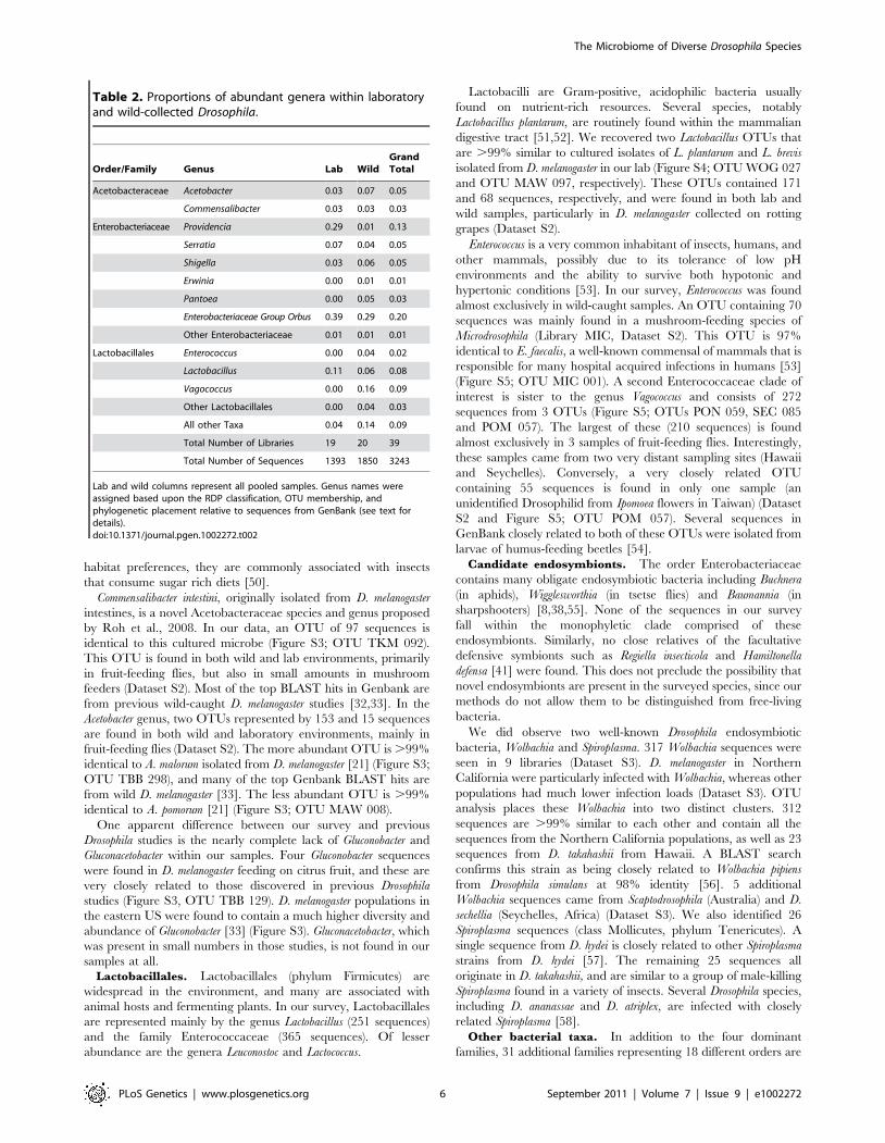
Table 2. Proportions of abundant genera within laboratoryand wild-collected Drosophila.
Order/Family Genus Lab WildGrandTotal
Acetobacteraceae Acetobacter 0.03 0.07 0.05
Commensalibacter 0.03 0.03 0.03
Enterobacteriaceae Providencia 0.29 0.01 0.13
Serratia 0.07 0.04 0.05
Shigella 0.03 0.06 0.05
Erwinia 0.00 0.01 0.01
Pantoea 0.00 0.05 0.03
Enterobacteriaceae Group Orbus 0.39 0.29 0.20
Other Enterobacteriaceae 0.01 0.01 0.01
Lactobacillales Enterococcus 0.00 0.04 0.02
Lactobacillus 0.11 0.06 0.08
Vagococcus 0.00 0.16 0.09
Other Lactobacillales 0.00 0.04 0.03
All other Taxa 0.04 0.14 0.09
Total Number of Libraries 19 20 39
Total Number of Sequences 1393 1850 3243
Lab and wild columns represent all pooled samples. Genus names wereassigned based upon the RDP classification, OTU membership, andphylogenetic placement relative to sequences from GenBank (see text fordetails).doi:10.1371/journal.pgen.1002272.t002
The Microbiome of Diverse Drosophila Species
PLoS Genetics | www.plosgenetics.org 6 September 2011 | Volume 7 | Issue 9 | e1002272

associated with Drosophila populations. Nearly all of these arepresent in amounts of less than 1% of the total bacterialmicrobiome (Dataset S2). Several of these taxa are knownsymbionts of animals [12]. For example, the Clostridiales, theBacteroidales, and the Actinomycetales each make up ,1% of theDrosophila bacterial microbiome. The most widespread of thesetaxa, the Bacteroidales genus Dysgonomonas, is present in 8 separateDrosophila populations and is not restricted to any one locality,species, or diet type (Dataset S2).
Diversity of bacterial communitiesOTU richness, evenness, and overall diversity vary widely
among host samples (Table S1). As many as 30 OTUs werepresent in some samples such as D. falleni collected on Russulamushrooms, while five or fewer OTUs were found in 5 differentsamples. For example, D. hydei collected from either citrus fruit orprickly pear are found with four or less bacterial OTUs, and asingle Enterobacteriaceae OTU represents at least 85% of each ofthese bacterial microbiomes. Similarly, D. sechellia collected onMorinda fruit is dominated by a single Lactobacillales OTU (84%),leading to very low bacterial community richness and evenness(Dataset S2). Rarefaction analysis, which helps determine howclose the sampling effort came to fully describing the community,shows that different host communities differ greatly in richness andwere sampled at different depths (Figure 2A). The least diversesamples are those collected from fruit-feeding hosts, while theflower- and mushroom-feeders tend to have more diverse bacterialcommunities. For the communities that have not been sampled tocompletion, the situation exists in which rare, and potentiallyimportant, taxa have not been identified.Community similarity (beta-diversity) between samples was
calculated for each of the 190 comparisons between the 20 wildpopulations (Dataset S4). In 27% of these comparisons, no OTUsare shared between the two samples. The two Drosophila that sharethe highest proportion of their bacterial microbiomes are D. hydeicollected from citrus fruit and prickly pear fruit (samples HCF andHPP, respectively, Dataset S4).In contrast to the bacterial communities associated with wild
populations, laboratory samples are much less diverse and so weresampled nearly to completion (Figure 2B). Chao1 analysis [59]
predicts an average of 6.3 OTUs per sample, and most librarieshave.80% coverage (Table S2). It is interesting to note that someof the most OTU-rich communities are present on the culturemedia and on the external surfaces of flies (MED and XYX)(Table S2). This suggests that flies are able to exclude many of theexternal microorganisms present on the feeding substrate, allowingonly a subset to persist in their digestive tract.In both wild and lab host samples, most of the bacterial diversity
is found at short phylogenetic distances, since most samples sharethe same dominant orders and families (Figure 1). This distributionproduces a typical ‘‘hockey stick’’ pattern found in many animal-associated microbial communities (Figure S6) [60].
Differences between Drosophila and mammalianbacterial communitiesTo put the Drosophila bacterial microbiome in perspective, we
compared the 20 wild-caught samples to published mammaliandatasets [12] and previous studies of naturally isolated D.melanogaster [33]. These studies are well suited for effectivecomparison to our data because they use culture-independent,long-read Sanger sequencing that allows closely related OTUs tobe resolved, and because they represent a large taxonomic breadthand/or include many samples from a wide geographic area.Principal component analysis (PCA) shows that the Drosophilabacterial microbiome from our study is similar to previous D.melanogaster samples, but is clearly distinct from the microbiomefound in the mammalian orders Artiodactyla, Carnivora, andPrimates (Figure 3B). Despite the relatively tight clustering ofDrosophila samples, some differences between separate studies areapparent (Table S3). Notably, the Enterobacteriaceae, which arethe dominant taxon in our global survey, are almost absent fromtwo previous Drosophila studies [21,33]. Although Enterobacteria-ceae comprise a large proportion of the bacterial microbiomewithin a single Massachusetts population [32], the dominantgenera in that sample were Enterobacter and Klebsiella, which are notpresent in our survey. The high abundance of Acetobacteraceae inthe Massachusetts population may be caused by the fruit bait usedduring sample collection in that study [32].The dominant bacterial order in all three mammalian orders is
the strictly anaerobic Clostridiales, which is rarely found in
Figure 2. Rarefaction analysis of observed richness within Drosophila. All calculations were performed using mothur [35]. OTUs were definedat the 3% divergence threshold using the average neighbor clustering algorithm. Library identifiers are given in Table 1. Note the different scales ofthe Y-axis in panels A and B. A. Rarefaction analysis of wild populations of Drosophila. B. Rarefaction analysis of laboratory collected samples.doi:10.1371/journal.pgen.1002272.g002
The Microbiome of Diverse Drosophila Species
PLoS Genetics | www.plosgenetics.org 7 September 2011 | Volume 7 | Issue 9 | e1002272

Drosophila (Table S4). The Enterobacteriaceae are not found or areminimal residents of the Artiodactyla and Primate guts, respec-tively. While this family is present in high amounts within theCarnivora, the dominant genera, Escherichia and Shigella, are notcommon in flies (Dataset S2). A similar pattern is found for theLactobacillales. This order is found in relatively high numbers inthe Carnivora and Primates (20% and 9% respectively) [12], butthe major genus in mammals (Streptococcus) is found at less than0.5% abundance in wild flies (Dataset S2). Finally, Acetobacter-aceae are not present in any of the three mammalian orders [12].The only bacterial genus present in appreciable numbers in bothmammals and Drosophila is Lactobacillus. This genus is found inArtiodactyla (2%), Carnivora (3%), Primates (2%), and Drosophila(3%) [12] (Dataset S2).
Flies also differ from mammals in the overall patterns of bacterialmicrobiome diversity. The richness of Drosophila bacterial commu-nities is dramatically lower than in mammals, although communityevenness is comparable (Table 3). Additionally, we find that manyOTUs are present in taxonomically and ecologically diverseDrosophila populations (Dataset S2) and that the proportion ofbacterial OTUs that are unique to a single host sample isconsistently lower in Drosophila than in mammals (Figure S7).
Effect of host diet on the composition of natural bacterialcommunitiesTo estimate the role of host diet in shaping bacterial
microbiome composition, we compared taxonomically diverseDrosophila species collected from different types of food sources.
Figure 3. Principle component analysis of the natural Drosophila microbiome. All sequences were aligned and trimmed as described in thetext. A single rooted tree for each PC analysis was generated using FastTree [36]. PC analysis was done with the FastUniFrac web application [62]. A:Comparison of the Drosophila microbiome with respect to diet type. All 20 naturally collected samples are included along with the laboratorysamples from adult Drosophila feeding on rich Bloomington media (Text S1). B: Comparison of the natural Drosophila bacterial microbiome and themammalian bacterial microbiome. D. melanogaster data is from Corby-Harris et al., 2007 [33]. Selected mammalian orders are from Ley et al., 2008a[12].doi:10.1371/journal.pgen.1002272.g003
Table 3. Average diversity measurements for Drosophila and mammals.
Global SurveyLaboratoryDrosophila D. melanogaster Artiodactyla Carnivora Primates
Chao1 Richness Average 17.13 5.86 19.06 542.68 87.31 307.20
SD 14.75 2.66 11.52 360.89 56.47 133.76
Shannon Diversity Average 1.38 0.84 2.03 3.94 2.18 3.59
SD 0.77 0.52 0.51 1.08 1.10 0.73
Shannon evenness Average 0.58 0.54 0.88 0.87 0.62 0.80
SD 0.20 0.24 0.05 0.20 0.25 0.12
All calculations were performed using mothur [35]. OTUs were defined at the 3% divergence threshold using the average neighbor clustering algorithm. D. melanogasterdata is from Corby-Harris et al., 2007 [33]. Selected mammalian orders are from Ley et al., 2008a [12]. Details regarding calculations can be found at http://www.mothur.org/wiki/Calculators.doi:10.1371/journal.pgen.1002272.t003
The Microbiome of Diverse Drosophila Species
PLoS Genetics | www.plosgenetics.org 8 September 2011 | Volume 7 | Issue 9 | e1002272
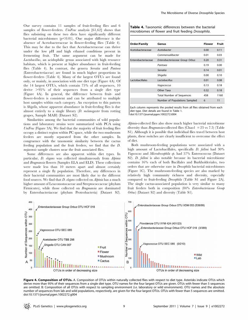
Our survey contains 11 samples of fruit-feeding flies and 6samples of flower-feeders. UniFrac analysis [61,62] shows thatflies subsisting on these two diets have significantly differentbacterial microbiomes (p,0.01). One major difference is theabsence of Acetobacteraceae in flower-feeding flies (Table 4).This may be due to the fact that Acetobacteraceae can thriveunder the low pH and high ethanol conditions present infermenting fruit. The same argument can be made forLactobacillus, an acidophilic genus associated with high resourcehabitats, which is present at higher abundance in fruit-feedingflies (Table 4). In contrast, the genera Serratia and Pantoea(Enterobacteriaceae) are found in much higher proportions inflower-feeders (Table 4). Many of the largest OTUs are foundonly, or mainly, in association with one diet type (Figure 4A). Ofthe 14 largest OTUs, which contain 75% of all sequences, 10derive .95% of their sequences from a single diet type(Figure 4A). In general, the difference between fruit- andflower-feeders is consistent and can be attributed to multiplehost samples within each category. An exception to this patternis Shigella, whose apparent abundance in fruit-feeding flies is duealmost entirely to a single library (D. melanogaster from rottinggrapes, Sample MAH) (Dataset S2).Similarities among the bacterial communities of wild popula-
tions and laboratory strains were summarized with PCA usingUniFrac (Figure 3A). We find that the majority of fruit feeding fliesoccupy a distinct region within PC space, while the two mushroomfeeders are mostly separated from the other samples. Incongruence with the taxonomic similarity between the cactusfeeding population and the fruit feeders, we find that the D.mojavensis sample clusters near the fruit associated flies.Some differences are also apparent within diet types. In
particular, D. elegans was collected simultaneously from Alpiniaand Brugmansia flowers (Samples ELA and ELD). These collectionswere made less than 10 meters apart and almost certainlyrepresent a single fly population. Therefore, any differences intheir bacterial communities are most likely due to the differentfood sources. We find that D. elegans collected on Alpinia has a muchhigher amount of Leuconostocaceae and Streptococcaceae (phylumFirmicutes), while those collected on Brugmansia are dominatedby Enterobacteriaceae (phylum Proteobacteria) (Dataset S2).
Alpinia-collected flies also show much higher bacterial microbiomediversity than Brugmansia-collected flies (Chao1 =23 vs 7.5) (TableS1). Although it is possible that individual flies travel between hostplants, these switches are clearly insufficient to overcome the effectof diet.Both mushroom-feeding populations were associated with a
high amount of Lactobacillales, specifically D. falleni had 30%Vagococcus and Microdrosophila sp. had 57% Enterococcus (DatasetS2). D. falleni is also notable because its bacterial microbiomecontains 16% each of both Bacillales and Burkholderiales, twoorders that are otherwise rare in Drosophila bacterial microbiomes(Figure 1C). The mushroom-feeding species are also marked byrelatively high community richness and diversity, especiallycompared to fruit-feeding Drosophila (Table S1 and Figure 2A).The single cactus-associated population is very similar to manyfruit feeders both in composition (84% Enterobacteriaceae GroupOrbus) (Dataset S2) and diversity (Table S1).
Figure 4. Composition of OTUs. A. Composition of OTUs within naturally collected flies with respect to diet type. Asterisks indicate OTUs whichderive more than 95% of their sequences from a single diet type. OTU names for the four largest OTUs are given. OTUs with fewer than 5 sequencesare omitted. B: Composition of all OTUs with respect to sampling environment (i.e. laboratory or wild environment). OTU names and the absolutenumber of sequences from lab and wild populations, respectively, are given for the four largest OTUs. OTUs with fewer than 5 sequences are omitted.doi:10.1371/journal.pgen.1002272.g004
Table 4. Taxonomic differences between the bacterialmicrobiomes of flower and fruit feeding Drosophila.
Order/Family Genus Flower Fruit
Acetobacteraceae Acetobacter 0.00 0.11
Commensalibacter 0.01 0.03
Enterobacteriaceae Enterobacteriaceae Group Orbus 0.20 0.31
Pantoea 0.19 0.00
Serratia 0.14 0.01
Shigella 0.00 0.10
Lactobacillales Lactobacillus 0.01 0.08
Vagococcus 0.13 0.18
Other Taxa 0.32 0.18
Total Number of Sequences 458 1160
Number of Populations Sampled 6 11
Each column represents the pooled results from all flies obtained from eachdiet type. Diet details are found in Table 1.doi:10.1371/journal.pgen.1002272.t004
The Microbiome of Diverse Drosophila Species
PLoS Genetics | www.plosgenetics.org 9 September 2011 | Volume 7 | Issue 9 | e1002272

The bacterial microbiome of laboratory flies has reduceddiversity and distinct compositionA major benefit of the Drosophila model is the experimental
flexibility it provides in a laboratory setting. However, OTUclassification and rarefaction analysis show that lab-raised fliescontain a much lower richness and diversity of bacteria compared towild-caught flies (Figure 2, Table 3, Table S1 and Table S2). At thebroadest level, the wild and laboratory samples are similar in thatboth are composed mainly of Enterobacteriaceae, Lactobacillales,and Acetobacteraceae (Table 2). However, 90 of the 139 totalOTUs are present only in wild samples, while six are found only inlab samples (Figure 4B). Most of these OTUs are rare, so that themajority of sequences in our survey belong to OTUs that are foundin both wild and lab hosts (Figure 4B). The four largest OTUs,which together comprise over half of the entire dataset, arecomposed of both wild and laboratory sequences. It should benoted, however, that each of these four OTUs is composedprimarily (.95%) of either wild or laboratory sequences (Figure 4B).PCA (Figure 3A) further emphasizes the reduced diversity and
distinct composition of the bacterial microbiome of laboratoryflies. We find the laboratory populations in a subset of the total PCspace occupied by the wild populations. Specifically, thelaboratory samples’ PC space is near that of the fruit feedingDrosophila, which could be explained by the nutritional similarity ofthese sugar rich diets.Many of the bacterial strains found in this study are closely
related to those from previous laboratory studies of Drosophila. Fivestrains that are common in our lab samples (Acetobacter malorum, A.pomorum, Commensalibacter intestini, Lactobacillus brevis, and L.plantarum) are .99% identical to previously indentified culturedisolates of D. melanogaster [21] (Figure S3 and Figure S4). A notabledifference between our results and another previous study is thatEnterococcus is virtually absent in our lab samples (Table S3), butcomprises nearly 50% of the laboratory bacterial microbiome inthat study [32].
Experimental analysis of host species and diet effects onthe intestinal bacterial microbiomeOur survey of natural bacterial communities suggests that host
diet may be an important determinant of bacterial microbiomecomposition. We tested this hypothesis using laboratory experi-ments where diet and rearing conditions were carefully controlled.Starting with a large pool of isogenic D. melanogaster, we transferred25 flies each to a different sterile diet and examined the resultingchanges in their gut bacterial communities. We find that the highyeast diet, which is most similar in composition to our standard labmedia, induced a similar bacterial microbiome with a highabundance of Enterobacteriaceae Group Orbus (Table 5). In contrast,the high ethanol and sugar-only diets resulted in a bacterialmicrobiome dominated by Providencia. Flies on the no-nutrient(agar-water) diet contained appreciable levels of both of thesegroups, but a quarter of their bacterial microbiome was composedof Commensalibacter intestini (Table 5). Flies kept on standard labmedia showed little change in their bacterial microbiome afterthree days, suggesting that diet has a consistent effect on thebacterial microbiome. UniFrac analysis confirms a significantoverall effect of diet in this experiment (p,0.01).In a reciprocal experiment, we tested whether different host
species develop different bacterial microbiomes when feeding onthe same diet. Three distantly related Drosophilids that feed ondifferent food sources in the wild, D. melanogaster (fruits), D. elegans(flowers), and D. virilis (sap fluxes and cambium), were rearedtogether on the same media. We found that all three species hadsimilar bacterial microbiomes at the end of this experiment(Table 6). The digestive tracts of each species contained between72% and 94% Providencia. UniFrac analysis does not showsignificant differences between host species (p = 0.54). However,some differences between these species could be masked becausethe strains used in this experiment have been adapting to thelaboratory environment for many generations. Additionally,laboratory Drosophila are likely exposed to a lower overall diversityof possible symbionts than their wild counterparts, further maskingpossible differences between host species.Our study spanned two years and used flies from two different
labs at UC-Davis. The Kimbrell and Kopp lab flies hadsignificantly different bacterial microbiomes, despite obtainingthe same type of media from the same kitchen (p,0.01). The threedominant taxa in the Kopp lab are Enterobacteriaceae Group Orbus,Providencia, and Lactobacillus, while all three are at minimal amountswithin the Kimbrell lab (all three combined equal 9% of thebacterial microbiome within Drosophila from the Kimbrell lab)(Table S5). Conversely, the dominant taxa in the Kimbrell lab areShigella and Variovorax, which are not present in the Kopp lab. Evenwithin the Kopp lab, the bacterial microbiome was different inexperiments performed at different times (p,0.01) (Table S6). Wepropose that these inter- and intra-lab differences are the result of
Table 5. Gut bacterial microbiome composition on different diets.
Lab Media (Start) Lab Media (3 days) Sugar Agar EtOH Yeast
Providencia 0.17 0.10 0.98 0.22 0.85 0.24
Commensalibacter 0.00 0.00 0.01 0.24 0.00 0.09
Enterobacteriaceae Group Orbus 0.82 0.90 0.01 0.50 0.13 0.65
Other Taxa 0.02 0.00 0.00 0.04 0.01 0.03
Total Number of Sequences 173 88 82 72 68 34
doi:10.1371/journal.pgen.1002272.t005
Table 6. Gut bacterial microbiome composition in differentspecies co-cultured on the same media.
D. elegans D. melanogaster D. virilis
Enterobacteriaceae Group Orbus 0.10 0.01 0.00
Lactobacillus 0.07 0.01 0.00
Providencia 0.72 0.94 0.89
Serratia 0.09 0.03 0.05
Other Taxa 0.02 0.00 0.05
Total Number of Sequences 82 90 38
doi:10.1371/journal.pgen.1002272.t006
The Microbiome of Diverse Drosophila Species
PLoS Genetics | www.plosgenetics.org 10 September 2011 | Volume 7 | Issue 9 | e1002272
different sets of environmental communities that inhabit the labsand inoculate the fly stocks during routine maintenance. Theseobservations suggest that some of the conflicting phenotypic resultsreported by different labs [27,28] may be the result of differentbacterial communities.
Intestinal bacterial microbiome differs from theenvironmental bacterial communityThe bacterial microbiome of Drosophila is likely environmentally
acquired since, with the exception of Wolbachia and Spiroplasma, noevidence exists that bacterial communities are transmittedvertically within the egg. To ask whether the gut bacterialmicrobiome differs from the external bacterial community, weexamined external washes of adults and their culture media (TableS7). UniFrac analysis shows a significant difference (p,0.01)between the external and internal samples of D. melanogaster grownon unsterilized media. Larvae also differ significantly from themedia they feed on (p,0.01). The larval bacterial microbiomeconsisted entirely of Enterobacteriaceae Group Orbus, while the mediaalso contained Serratia, Providencia, and Lactobacillus.
Discussion
Drosophila has a taxonomically restricted bacterialmicrobiomeNatural Drosophila populations have a remarkably restricted
bacterial microbiome. Despite the phylogenetic, ecological, andgeographical diversity of the hosts we surveyed, only a fewbacterial clades are associated with all these flies. The familiesEnterobacteriaceae and Acetobacteraceae and the order Lacto-bacillales make up over 85% of natural Drosophila bacterialmicrobiome (Figure 1A). All Drosophila populations are dominatedby at least one of these clades, and many host isolates have allthree of them (Figure 1B). Although we find no strict core bacterialmicrobiome, Enterobacteriaceae and Lactobacillales are found in18 and 17 of the 20 wild Drosophila populations, respectively. Eachof the five samples that lack either of these groups is dominated bythe other, and the two groups generally show a pattern ofreciprocal abundance (Figure 1C). One possible explanation is thatcompetitive interactions between the two groups allow only one ofthem to persist at a detectable level within the host digestive tract.These three bacterial taxa are emerging as common microbial
associates of insects. The Acetobacteraceae (Acetobacter sp.) havebeen found with bees, olive fruit flies, parasitic wasps andmealybugs [44,63,64,65,66]. Likewise, the Lactobacillales (suchas Lactobacillus) are common symbionts of insects, notably bees andbeetles [43,65,67,68]. Finally, the most common Enterobacteria-ceae found with Drosophila (Enterobacteriaceae Group Orbus) has foundwith numerous insect species, but especially bees (Figure S2)[43,44,45,46,69,70,71,72].This taxonomically restricted bacterial microbiome leads to
interesting patterns of bacterial diversity. Many samples have verylow observed and expected (Chao1) species richness (Table S1).These results stand in contrast with the highly diverse bacterialcommunities found in mammals [12] (Table 3). There is animportant difference in sampling procedures: the mammaliansamples each come from a single individual [12], while theDrosophila samples were isolated from multiple individuals.However, this difference would be expected to bias the results inthe opposite direction, since different individuals are likely to carryslightly different bacterial communities.Our laboratory studies show that the intestinal bacterial
microbiome represents only a subset of the external bacterialcommunities (Table S7). This suggests that although the gut
bacterial microbiome is environmentally acquired, the host exertssignificant control over its composition. Since most environmentalsamples are composed of many phyla and are rarely dominated byjust one or two lineages [73], we suggest that the low-diversitycommunities of Drosophila reflect the effects of strong host filtering.Whether this filtering is an adaptive function of the immunesystem or simply a by-product of the physiological conditions inthe gut remains to be determined, but host control has previouslybeen demonstrated in genetic experiments [21,26]. The impor-tance of bacterial microbiome restriction for host fitness is yet to beinvestigated, as well.
The same bacterial lineages are associated with differenthost species, diets, and locationsAnalysis of OTU-level data shows that individual OTUs are not
specific to a single host species, diet type, or location, but aretypically associated with many Drosophila populations. Althoughmost OTUs (91 out of 127) present in wild flies are each found inone host sample, all these OTUs represent only a small percentageof the total fly bacterial microbiome (16%). Conversely, thedominant OTUs from each host population are usually found inother populations as well. In fact, we find that the most commonOTU in 19 out of 20 populations is also found in other, oftengeographically distant, hosts. Several particularly wide-rangingOTUs are found in nearly half of all populations. In comparisonwith mammalian bacterial microbiomes [12], the fraction ofOTUs unique to a single host sample is much lower (Figure S7).The closest relatives of many bacterial lineages found in our
survey were also detected in previous studies of D. melanogaster. Forseveral common taxa (Commensalibacter, A. malorum, A. pomorum, L.plantarum, and L. brevis), the closest sequences in GenBank wereisolated from D. melanogaster. Since few Drosophila-associated 16Ssequences are available in GenBank, compared to the muchgreater number of non-host associated and mammalian-associatedsequences, these similarities imply a pervasive association of theselineages with Drosophila. Overall, these patterns suggest that thebacteria associated with Drosophila display some level of hostspecificity. Since far-flung, ecologically diverse flies are associatedwith a common set of bacteria, ‘‘Drosophila’’ can be considered aselective environment that allows only certain taxa to persist.
Host diet has a greater effect on the bacterialmicrobiome than host speciesPrevious studies have shown that the mammal-associated
bacterial microbiome is shaped by both host phylogeny and hostdiet, while sampling location has little or no effect on communitycomposition [12,74]. Diet has also been shown to influence thebacterial composition of gypsy moth [30] and cotton bollworm[31] larval midguts. We find that host diet plays a substantial rolein shaping bacterial microbiome composition in Drosophila, as well.This conclusion is supported both by the survey of naturalcommunities and by controlled laboratory experiments. Althoughwe were unable to quantify the role of host species in naturalpopulations because many species were only represented by asingle collection, laboratory populations of multiple co-habitatingspecies showed no significant differences between their bacterialmicrobiomes.These results suggest two possible hypotheses regarding the
assembly of Drosophila–associated bacterial communities. Onepossibility is that the guts of different host species inhabiting thesame food source provide suitable environments for the samebacteria. These bacteria could provide specific benefits to theirhosts on that diet, so that phylogenetically distant Drosophila species
The Microbiome of Diverse Drosophila Species
PLoS Genetics | www.plosgenetics.org 11 September 2011 | Volume 7 | Issue 9 | e1002272
evolve to allow the persistence of the same, diet-specific, bacteria.Alternatively, different substrates may harbor different bacterialcommunities and environmental acquisition of these bacteria maysimply overwhelm any potential control by the host. As thesehypotheses suggest different roles for the host (adaptive vs. passive),future experiments should take care to sample the bacterialcommunity of the environment the host is interacting with.If environmental acquisition is indeed the most important factor
determining Drosophila bacterial microbiome composition, thentwo general observations are expected. First, patterns of host andsymbiont co-speciation seen in closely related insect andmammalian groups should not be observed within Drosophila[17,74]. Second, the genetic complementarily commonly found intightly associated symbionts should be harder to evolve [8].
Lab-raised flies are a limited model of natural host–microbe interactionsDrosophila has recently emerged as a powerful model for studying
non-pathogenic host-microbe interactions. Several importantgenes that control host interactions with commensal intestinalbacteria have been identified, including caudal and PIMS (Lhocineet al., 2008; Ryu et al., 2008). Another study has shown thatLactobacillus plantarum can affect mating preferences (Sharon et al.,2010). Cox and Gilmore, 2007, have suggested that D. melanogasteris naturally colonized by the commensal/opportunistic pathogenEnterococcus faecalis, and can serve as a good model for E. faecalispathogenesis. In all these studies, laboratory experiments serve as aproxy for the natural ecology of Drosophila-microbe interactions.However, in order to serve as an ideal model system, the labbacterial microbiome should be a subset of the wild bacterialmicrobiome, and the most common wild taxa should be found inthe lab.We find that these conditions are only partially satisfied. The
putative commensal bacterial genera studied by Ryu et al., 2008are members of the family Acetobacteraceae (Acetobacter, Glucoace-tobacter, Commensalibacter) and the genus Lactobacillus (L. plantarumand L. brevis). Ren et al., 2007 also identified Acetobacter andLactobacillus as commensal bacteria in laboratory-reared flies.While all of these bacteria are present in some Drosophilapopulations, their abundance in wild samples is low and noneare ubiquitous. In D. melanogaster samples L. plantarum and L. breviscomprise 7.7% and 9.7% of the total bacterial microbiome,respectively, whereas Enterococcus, Commensalibacter and Glucoaceto-bacter are not found at all. Only L. plantarum is found in all wild D.melanogaster samples.Drosophila has been used for decades as a model for pathogenic
bacterial infections. In some cases, it was applied to study bacteriathat pose important threats to human health, such as Bacillusanthracis [75], Vibrio cholerae [76,77], Salmonella typhimurium [78–80],Pseudomonas aeuruginosa [81–86] and Burkholderia cepacia [78]. Otherstudies focused on elucidating the molecular mechanisms of flyimmunity using known or suspected entomopathogens orphytopathogens, including species of Serratia [86,87], Erwinia[88,89], Micrococcus [90], and Pseudomonas [91,92]. We find that,collectively, the above 8 microbes make up less than 10% of thetotal Drosophila microbiome, and none constitutes more than 3.5%individually. This indicates that they are relatively rare in wildDrosophila populations on the whole, although we cannot rule outthe existence of some unsampled, heavily infected individuals.While most of the well-studied lab bacteria are rare in natural
populations, the reciprocal is also true – the most commonbacteria in wild populations are not the most abundant Drosophilaassociates in the lab (Figure 4B), and have not been used as modelbacteria in laboratory studies. A single group, Enterobacteriaceae
Group Orbus, represents over 21% of all bacteria present withnatural Drosophila populations and is nearly twice as abundant asthe next most common genus. This clade is present in over half ofall Drosophila populations, but has not been used in any laboratorystudies. The second most common bacterium in wild Drosophila, astrain of Vagococcus (15% of total bacterial microbiome, present in 9populations), has also never been used in Drosophila host-microbestudies.One final consideration for laboratory studies concerns the lab-
and time-dependent variation in bacterial communities. It hasbeen previously suggested that discrepancies between reportedphenotypes may be due to different bacterial communities presentin different labs [28]. Indeed, we find that different laboratories atUC-Davis are home to completely different bacterial communitiesdespite using the same media (Table S5). Even when genus-leveltaxonomies agree (as in Serratia), OTU clustering shows thatdifferent strains are present in different laboratories. Moreover, wefind that bacterial community composition can change in the samelab over time (Table S6).Despite these caveats, laboratory strains of Drosophila can still
serve as a useful model of host-microbe interactions. For example,conclusions from the natural survey mesh well with laboratoryexperiments in highlighting the importance of diet in shaping thebacterial microbiome. We suggest that many experimentalprojects would benefit from determining and monitoring thecomposition of bacterial communities associated with fly strains.Awareness of this important aspect of host biology will lead to abetter understanding of Drosophila physiology, ecology, andevolution.
A model of Drosophila microbiome assemblyOur results suggest a model where the composition of gut
bacterial communities is determined by three separate factors:diet, host physiology, and chance. Since all gut bacteria must firstbe ingested, bacterial taxa that thrive on the feeding substrates ofthe host species will have the greatest chance of colonizing the gut.The aerobic, and often high-nutrient environments frequented byDrosophila may present taxonomically and geographically distantfly populations with similar ‘‘source’’ bacterial communities.Furthermore, the quantitative differences between Drosophilafeeding upon different food sources may be the result of exposureto different diet-specific bacterial communities. Next, within therange of microbes presented by the diet, some properties of theDrosophila intestinal environment determine which bacteria areallowed to persist. These properties may reflect conserved featuresof the Drosophila immune system as well as the physico-chemicalconditions in the gut lumen – such as pH or the simple fact that,unlike the mammalian digestive tract, the Drosophila gut is mostlikely an aerobic environment. This may explain why the closestrelatives of the dominant OTUs in our survey come from otherinsects, and why bacteria commonly associated with flies are veryrare in diverse mammalian species and vice versa. At this time, it isnot clear whether genetic variation between or within species canfurther bias the acquisition of symbionts. Although we do notdetect an effect of host species in our study, it is possible thatdeeper sequencing will uncover quantitative effects of the hostgenotype, especially under controlled environmental conditions.Finally, within the boundaries set by the host diet and subject tohost filtering, the microbiome of each population is likelydetermined by chance environmental encounters between fliesand bacteria. This factor may explain both the lab effect and thechange in bacterial communities over time observed in our labsamples. In the simplest scenario, each individual host wouldcollect a random sample of permissible bacteria available in its
The Microbiome of Diverse Drosophila Species
PLoS Genetics | www.plosgenetics.org 12 September 2011 | Volume 7 | Issue 9 | e1002272

environment. A further level of complexity may be added if oneconsiders the interactions between bacterial taxa or their order ofcolonization. The reciprocal dominance of Enterobacteriaceaeand Lactobacillales in Drosophila samples suggests that one or bothof these processes may be important.This model of microbiome assembly, while consistent with all
our data, remains to be tested by more systematic environmentalsampling and experimental analyses. It is also unclear whether itapplies to other Drosophila-associated microbes such as yeast.Repeated sampling of multiple co-occurring species from the samefeeding sources, analysis of individual variation in naturalpopulations and laboratory settings, and characterization ofbacterial communities native to the diet of each population willall be necessary to determine the relative importance of sourcebacterial communities, host control, and the vagaries of chance inshaping the gut microbiome.The gut bacterial communities of Drosophila are likely to represent
the most common type of animal microbiomes, where symbiontsare free-living and horizontally transmitted and the host-symbiontassociations are flexible and facultative. If this model is confirmed byfuture work, it may serve as a paradigm for the assembly of otheranimal microbiomes in nature. This framework may help usunderstand both the ecology of host-symbiont interactions and thefunctional impact of these interactions on the host.
Materials and Methods
Fly collection, dissection, and DNA extractionDrosophila samples were collected with the help of many
colleagues around the world (see Acknowledgments, Table 1 andDataset S2). All samples were obtained from naturally occurringsubstrates and no artificial baits were used to attract flies. Forcollections done in Northern California, adults were immediatelytransferred to sterile no-nutrient media (agar-water) and trans-ported to UC-Davis for dissection, which occurred within 2 hoursof collection. For more remote field collections, flies were stored in100% ethanol for transport.Freshly collected flies were washed twice in 2.5% bleach and
twice in sterile water. The entire gut was dissected in sterile insectsaline and placed in sterile TES buffer (10 mM Tris-HCl[pH=7,5], 1 mM EDTA, 100 mM NaCl). For flies stored inethanol, dissection was not feasible because weakening of the flytissues caused the gut to fragment. For these samples, the entire flybody was used after three washes with sterile water. To ensureadequate removal of external bacteria, each final wash wasconfirmed to be free of bacterial cells by PCR with universalbacterial primers and by plating onto rich media. In no case didthe final wash show evidence of bacterial contamination. For asingle sample (D. melanogaster reared in the Kopp laboratory), thefirst wash was saved for DNA extraction to characterize theexternal bacterial community. Seven to 20 fly bodies or guts werecombined for most samples. In one exception (D. melanogasterbodies collected from rotting grapes, sample MAW) only a singlebody was used. On a single occasion, the bacterial community oflaboratory media within the Kopp laboratory was sampled using1 ml of media that had been inhabited by D. melanogaster for 7–10days. Further details regarding sample collection dates, locations,and contents can be found in Dataset S1.DNA was extracted from samples using a modification of the
Bead Beater protocol [93]. The tissue was homogenized by grindingand three freeze/thaw cycles on dry ice. Samples were thenincubated with 50 units/ml of lysozyme for 15 minutes. Next,physical disruption was performed in a Bead-Beater (BioSpecProducts, Inc., Bartlesville, OK) on the homogenize setting for three
minutes. An overnight incubation with 1% SDS and 2 mg/mlProteinase K was followed by extraction with an equal volume of25:24:1 phenol:chloroform:isoamyl alcohol. The aqueous phase wasincubated at room temperature for 30 minutes with 2.5 volumes of100% isopropanol and 0.1 volumes of 3 M sodium acetate beforecentrifugation at 16,000 g for 30 minutes at 4uC. The DNA pelletwas washed with cold 70% ethanol and allowed to air dry beforeresuspension in TE (10 mM Tris-HCl pH 7.5, 1 mM EDTA.).
16S library creation and sequencingApproximately 100 ng of DNA was used as template for small-
subunit rDNA (16S) amplification. Bacterial universal primers 27F(59- AGAGTTTGATCCTGGCTCAG) and 1492R (59-GGT-TACCTTGTTACGACTT) were used to amplify a ,1450 bpfragment (Lane, 1991). These primers were chosen for threereasons. First, although they are not truly universal, they arespecific to a region that is conserved in many groups of bacteria[94]. Second, they allow for the amplification of nearly the fulllength of the gene, therefore providing consistent comparisons toprevious studies of 16S rDNA diversity [95]. Finally, both of theseprimers have been used in many similar surveys of bacterialdiversity, including a previous study of bacterial diversity inDrosophila melanogaster [32]. Using these primers allows our resultsto be directly comparable to those previous studies. The PCRconditions were as follows: initial denaturation for 5 minutes at95uC; 30 or 35 cycles at 95uC for 30 seconds, 55uC for 30 seconds,and 72uC for 2 minutes; final extension for 10 minutes at 72uC.These PCR conditions were used for all samples, with anannealing temperature of 55uC chosen from a temperaturegradient study of 48uC to 58uC because it produced the maximumproduct yield. The 16S rDNA amplicons were cloned into thepCR4-TOPO vector using the TOPO TA Cloning Kit. Clones weretransformed chemically into One Shot TOP10 chemicallycompetent E. coli cells or via electroporation into ElectroMAXDH10B E. coli cells (Invitrogen, Carlsbad, CA) and plated ontoagar plates with X-gal and either 50 mg/mL Kanamycin or50 mg/mL Ampicillin. Colony PCR (20 colonies) was used toverify a ,10% insertless rate and ,1.5 kb insert size. Whitecolonies were arrayed into 384-well plates. Prior to sequencing,plasmids were amplified by rolling circle amplification using theTempliPhi DNA Sequencing Amplification Kit (AmershamBiosciences, Piscataway, NJ) and sequenced from both ends usingthe M13 (228 or 240) primers with the BigDye kit (AppliedBiosystems, Foster City, CA). Sequencing reactions were purifiedusing magnetic beads and run on an ABI PRISM 3730 (AppliedBiosystems) sequencing machine.
Sequence quality assuranceVector and primer sequences were removed with cross_match, a
component of the Phrap software package [96,97], and bases with aPHRED quality score of Q.=15 were converted to ‘‘N’’s usingJAZZ, the Joint Genome Institute’s in-house assembly algorithm.When possible, overlapping regions from the forward and reversereads of each clone were used to assemble a single contiguoussequence for each clone. In cases where the overlap was not sufficientfor assembly, custom perl scripts were used to concatenate the forwardand reverse reads with gaps inserted between them (see below). Allsequence data are available via BioTorrents (http://biotorrents.net/details.php?id=143) and have been submitted GenBank under theaccession numbers JN420379 through JN426767.
Sequence alignmentWe used the Infernal 1.0 software [98] to create a single multiple
sequence alignment for all of our samples. Infernal creates a Hidden
The Microbiome of Diverse Drosophila Species
PLoS Genetics | www.plosgenetics.org 13 September 2011 | Volume 7 | Issue 9 | e1002272

Markov Model (HMM) based on a high-quality referencealignment with a fixed length of 1532. 2078 of the 4198 clonesconsisted of non-overlapping paired reads; for those we created a59-alignment (of reads beginning with the 27F primer sequence)and a 39-alignment (of reads ending with the reverse complementof the 1492R primer sequence), and merged the two alignments,inserting gaps into the intervening columns, based on positions inthe reference alignment. The concatenated sequences from this‘‘merged’’ alignment were combined with the successfully-assembled, full-length clones to create a single multiple sequencealignment. This alignment is available via BioTorrents (http://biotorrents.net/details.php?id = 143).For the purposes of OTU (operational taxonomic units [99])
definition and phylogenetic inference, this multiple sequencealignment was further refined to remove column blocks thatcontained .80% gaps. This resulted in the removal of the first 11(1–11) and last 132 (1400–1532) positions, as well as positions 642–806 (164 positions) from the middle of the alignment (whichprimarily corresponded to the regions of non-overlap betweenreads). A custom perl script was used to remove sequences withfewer than 300 remaining nucleotides from this trimmedalignment. Chimeric sequences were identified and removedusing the chimera.slayer function within mothur v.1.11 [35].
Taxonomy prediction and OTU assignmentWe submitted our sequences to the Ribosomal Database Project
(RDP10) Classifier for taxonomic assignment [100] to the genuslevel. We were unable to submit a single, full-length sequence forevery 16S clone that was sequenced, because for 50% of ourclones, there was no overlap between the forward and reversereads. For each clone, we assigned taxonomy independently to the59 read, the 39 read, and to the full-length or concatenated reads(with intervening gaps inserted, as described above), and thenselected a single taxonomy assignment for each 16S clone. Weused the measures of confidence (bootstrap values) that areassociated with the RDP taxonomy predictions to guide theselection process. Most investigators agree that .70% bootstrapsupport is indicative of strong support for a phylogenetic clade[101]. In order to arrive at taxonomy predictions with very highconfidence, we only considered taxonomy assignments that hadbootstrap values of .75% at the genus level, .80% at the familylevel, .95% at the order level, and 100% at the class level.Strongly supported disagreements between the 59, 39, andcombined data sets were rare (72 total sequences). These werehandled in one of two ways: 1) if the conflict was at the level offamily or above, they were considered likely to be chimericsequences and excluded from further analysis, or 2) if the conflictwas within a single family, the genus name was changed to‘‘unclassified’’.We used the mothur program [35] to generate a distance matrix
using our trimmed Infernal alignment of 3243 sequences as input.Using the distance matrix created by mothur, sequences wereclustered using the average neighbor algorithm. Using the 0.03cutoff option (97% sequence similarity), all sequences fell into 139OTUs. The average OTU abundance was 23.3 sequences(Min= 1, Max= 638). A representative sequence from eachOTU was selected using the get.oturep function within mothur. Thisrepresentative sequence and the dist.seqs command in mothur wasused to calculate genetic distances between OTUs and represen-tative sequences throughout this study.Taxonomy predictions generated by RDP were mapped onto
each sequence within an OTU. In many cases, this led to a clearreassignment of ‘‘unclassified’’ sequences to the genus level basedon the dominant genus present in that OTU. In other cases, the
entire OTU was comprised of ‘‘unclassified’’ sequences. TheseOTUs were assigned names based on their phylogenetic positionrelative to the reference sequences included, either from the RDPtype strains, from other Drosophila bacterial microbiome studies, orfrom GenBank.
Representative sequences for tree building, PCA analysis,and diversity comparisonsAll 7448 good quality 16S sequences longer than 1200 bp from
bacterial type strains were downloaded from the RDP website on8/22/10 [100]. These representatives are usually the firstidentified or most fully characterized strains within a bacteriallineage. Although closely related bacteria may differ substantiallyin genome content, inclusion of these type strains providesimportant phylogenetic landmarks during tree building. All 7448strains were aligned using Infernal, and the resulting alignment wastrimmed to remove the first 11 (1–11) and last 132 (1400–1532)positions, as well as positions 642–806 (164 positions) from themiddle of the alignment as described above.All sequences from previous studies of Drosophila bacterial
communities [32,33] (Corby-Harris, unpublished) were download-ed from GenBank. Mothur was used to create a distance matrix,and OTUs were created at a 97% similarity cutoff. The get.oturepfunction was used to pick a representative sequence for eachOTU. Additional Drosophila-associated sequences were alsoincluded [21,37]. Finally, for OTUs in our study that do nothave any closely related sequences within the RDP database (suchas Enterobacteriaceae Group Orbus) the closest BLAST hits fromGenBank were included. A list of the RDP and GenBankaccession numbers for sequences used in the final tree are foundin the Dataset S5.To compare our results to mammalian studies, the 17,504
ultraclean sequences from [12] were analyzed. To obtain a samplethat was roughly equal to our data in taxonomic breadth, only thesequences from Artiodactyla, Carnivora, and Primate samples wereanalyzed. These sequences were aligned and trimmed as aboveand a full PCA analysis was performed using the Fast UniFracInterface [62]. Taxonomic classifications were done with RDP[100].Diversity measurements were calculated for each library from
both Drosophila [33] and mammalian [12] datasets using mothur[35]. The proportion of OTUs unique to each library wascalculated for each group (as in Ley et al., 2008a) [12].
Tree buildingUsing representative sequences from our dataset, previously
identified Drosophila-associated bacteria, representative type strainsfrom the RDP database, and sequences obtained from GenBank(see previous section), a phylogenetic tree was created with FastTree[36]. Default settings with the GTR (generalized time-reversible)model were used. The entire tree was rooted using Thermusthermophilus (RDP identifier S000381199). After an initial run withall 8,407 sequences, many clades were removed from thealignment (for example, bacterial Phyla in which no Drosophilaassociated sequences were present). The remaining 1349 alignedsequences were then re-run on FastTree using the settings describedabove. Final publication quality images were prepared usingDendroscope [102].
UniFrac significance testsTests of significance of differences between samples were
performed using UniFrac and FastUniFrac [61,62]. The low depthof coverage provided by the sequencing method used is sufficient
The Microbiome of Diverse Drosophila Species
PLoS Genetics | www.plosgenetics.org 14 September 2011 | Volume 7 | Issue 9 | e1002272

to find significant results using UniFrac [103]. Because of thecorrection for multiple comparisons, pairwise comparisons foreach library were not feasible with the amount of data collected.We therefore parsed all data into bins representing different hostdiets to estimate the overall effect of this factor. The effect ofdifferent experimental treatments was determined similarly. Allcomparisons of bacterial communities are given in the Text S1.Co-occurrence tests were also performed (as in [104]), butinadequate power precluded the finding of any significant co-occurring pairs of taxa (additional details in Text S1).
Laboratory experimentsUnless explicitly stated, flies were fed unsterilized standard lab
media (Text S1). All transfer steps were performed near a Bunsenburner flame and all surfaces and instruments were frequentlysterilized with 70% ethanol. For bacterial DNA extraction, flieswere washed to remove external bacterial cells and their intestinesdissected as described above. All negative controls were confirmedto be bacteria-free by plating onto MRS media and PCR withuniversal bacterial primers. Separate libraries were created fromadult Canton-S males, Oregon-R males, and Oregon-R femalesfor Deborah Kimbrell’s lab (UC-Davis, CA). In addition, a largepopulation of wild D. melanogaster originally collected from Winters,CA was established in the Kopp lab for use in dietary treatmentexperiments (strain WO).For diet experiments, approximately 25 flies were transferred to
each of 5 separate diets, with one vial per treatment. The dietsincluded standard lab media, high yeast media, high yeastsupplemented with 6% ethanol, sugar-agar, and agar only (seeText S1 for media composition). All media were initially sterilizedin an autoclave, with the exception of the standard lab media.Ethanol was added to the ethanol treatment after media cooledbelow 55uC. To reduce the effect of the media-dwelling bacterialpopulation that arose after contact with non-sterile flies, cultureswere transferred daily to fresh sterile media, with the exception ofthe standard lab diet. These transfers continued for three days onall media except the agar-only diet, where starvation-induceddeath limited the experiment to two days. Four hours after thefinal transfer, the intestines of 10 flies per treatment were dissectedfor analysis.For the multiple species experiment, approximately 25 adults
each of D. melanogaster, D. elegans, and D. virilis were combined onsterilized high yeast media. After three days of daily transfers asabove, 10 males per species were dissected for analysis.
Supporting Information
Dataset S1 A more detailed version of Table 1 describingwhere, when and by whom each sample was collected.(XLS)
Dataset S2 This excel file contains the taxonomy assigned toeach sequence used in this study along with information regardingthe host species, location, environment and other informationregarding the library each sequence belongs to. Additionally, thecomposition of each OTU can be determined using the uniqueOTU identifiers (used in Figures S1, S2, S3, S4, and S5 and in themain text).(XLSX)
Dataset S3 This table describes the samples which Wolbachiaand Spiroplasma were found in. Note that samples TRR and TURwere removed from the overall analysis because, after the removalof their Wolbachia sequences, they each were left with too fewsequences for analysis. Each of these samples was a D. melanogaster/
D. simulans mix collected from citrus fruit at Michael Turelli’orchard in Winters, California.(XLSX)
Dataset S4 This file contains the beta-diversity measurementsfor all possible comparisons between our 20 natural Drosophilasamples.(XLSX)
Dataset S5 This file contains the GenBank accession numbersand the RDP identifiers of all the sequences used for tree buildingin this study.(TXT)
Figure S1 Phylogenetic tree of the Enterobacteriaceae GroupOrbus found with Drosophila. Taxa highlighted in red are OTUsidentified within this study. Each OTU begins with a uniqueidentifier corresponding to a sequence within the FASTA filesavailable on BioTorrents (http://biotorrents.net/details.php?id=143). The number of libraries and the number of sequenceseach OTU represents is also given. For each, this is further dividedinto how many libraries/sequences were found in either laboratoryor wild samples. For example, OTU HCF 018-#libs(2/10)-#seqs(3/389) represents 3 sequences found in 2 laboratorylibraries and 389 sequences found in 10 wild libraries. Taxahighlighted in green are from previous studies of the bacterialcommunities associated with Drosophila. The taxon highlighted inpurple is the cultured isolate that this group is named after (seemain text). Unhighlighted taxa are type strains found within theRibosomal Database Project (RDP) or taxa found in GenBank.Each of these taxa is followed by its GenBank accession number,its RDP identifier, or a unique identifier which corresponds to asequence within the FASTA files available on BioTorrents (http://biotorrents.net/details.php?id = 143). The orange edge in thisfigure corresponds to the orange node in Figure S2. The mainphylogenetic tree of which this tree is a subset was rooted usingThermus thermophilus (RDP identifier S000381199).(PDF)
Figure S2 Phylogenetic tree of the Enterobacteriaceae foundwith Drosophila. Taxa highlighted in red are OTUs identifiedwithin this study. Each OTU begins with a unique identifiercorresponding to a sequence within the FASTA files available onBioTorrents (http://biotorrents.net/details.php?id = 143). Thenumber of libraries and the number of sequences each OTUrepresents is also given. For each, this is further divided into howmany libraries/sequences were found in either laboratory or wildsamples. For example, OTU HCF 018-#libs(2/10)-#seqs(3/389)represents 3 sequences found in 2 laboratory libraries and 389sequences found in 10 wild libraries. Taxa highlighted in green arefrom previous studies of the bacterial communities associated withDrosophila. Unhighlighted taxa are type strains found within theRibosomal Database Project (RDP). Each of these taxa is followedby its GenBank accession number, its RDP identifier, or a uniqueidentifier which corresponds to a sequence within the FASTA filesavailable on BioTorrents (http://biotorrents.net/details.php?id=143). The orange node in this figure corresponds to the orangeedge in Figure S1. The main phylogenetic tree of which this tree isa subset was rooted using Thermus thermophilus (RDP identifierS000381199).(PDF)
Figure S3 Phylogenetic tree of the Acetobacteraceae found withDrosophila. Phylogenetic trees of bacterial groups associated withDrosophila. Taxa highlighted in red are OTUs identified withinthis study. Each OTU begins with a unique identifier correspond-ing to a sequence within the FASTA files available on BioTorrents
The Microbiome of Diverse Drosophila Species
PLoS Genetics | www.plosgenetics.org 15 September 2011 | Volume 7 | Issue 9 | e1002272
(http://biotorrents.net/details.php?id = 143). The number of li-braries and the number of sequences each OTU represents is alsogiven. For each, this is further divided into how many libraries/sequences were found in either laboratory or wild samples. Forexample, OTU HCF 018-#libs(2/10)-#seqs(3/389) represents 3sequences found in 2 laboratory libraries and 389 sequences foundin 10 wild libraries. Taxa highlighted in green are from previousstudies of the bacterial communities associated with Drosophila. Thetaxon highlighted in purple is a cultured isolate that closely relatedOTUs from this study were named after (see main text).Unhighlighted taxa are type strains found within the RibosomalDatabase Project (RDP). Each of these taxa is followed by itsGenBank accession number, its RDP identifier, or a uniqueidentifier which corresponds to a sequence within the FASTA filesavailable on BioTorrents (http://biotorrents.net/details.php?id=143). The main phylogenetic tree of which this tree is a subsetwas rooted using Thermus thermophilus (RDP identifier S000381199).(PDF)
Figure S4 Phylogenetic tree of the Lactobacilli found withDrosophila. Taxa highlighted in red are OTUs identified within thisstudy. Each OTU begins with a unique identifier corresponding to asequence within the FASTA files available on BioTorrents (http://biotorrents.net/details.php?id= 143). The number of libraries andthe number of sequences each OTU represents is also given. Foreach, this is further divided into howmany libraries/sequences werefound in either laboratory or wild samples. For example, OTUHCF 018-#libs(2/10)-#seqs(3/389) represents 3 sequences foundin 2 laboratory libraries and 389 sequences found in 10 wildlibraries. Taxa highlighted in green are from previous studies of thebacterial communities associated with Drosophila. Unhighlightedtaxa are type strains found within the Ribosomal Database Project(RDP). Each of these taxa is followed by its GenBank accessionnumber, its RDP identifier, or a unique identifier whichcorresponds to a sequence within the FASTA files available onBioTorrents (http://biotorrents.net/details.php?id= 143). Themain phylogenetic tree of which this tree is a subset was rootedusing Thermus thermophilus (RDP identifier S000381199).(PDF)
Figure S5 Phylogenetic tree of the Enterococci found withDrosophila. Taxa highlighted in red are OTUs identified within thisstudy. Each OTU begins with a unique identifier corresponding to asequence within the FASTA files available on BioTorrents (http://biotorrents.net/details.php?id= 143). The number of libraries andthe number of sequences each OTU represents is also given. Foreach, this is further divided into howmany libraries/sequences werefound in either laboratory or wild samples. For example, OTUHCF 018-#libs(2/10)-#seqs(3/389) represents 3 sequences foundin 2 laboratory libraries and 389 sequences found in 10 wildlibraries. Taxa highlighted in green are from previous studies of thebacterial communities associated with Drosophila. Unhighlightedtaxa are type strains found within the Ribosomal Database Project(RDP). Each of these taxa is followed by its GenBank accessionnumber, its RDP identifier, or a unique identifier whichcorresponds to a sequence within the FASTA files available onBioTorrents (http://biotorrents.net/details.php?id= 143). Themain phylogenetic tree of which this tree is a subset was rootedusing Thermus thermophilus (RDP identifier S000381199).(PDF)
Figure S6 Number of OTUs as a function of genetic distance.Number of OTUs was calculated at all genetic distances from 0(unique sequences) to 0.37 (the largest distance between any twosequences). Clustering was performed using the average neighboralgorithm in mothur [35].(TIF)
Figure S7 Proportion of OTUs that are unique to a singlelibrary for wild Drosophila and mammals. Calculations done as inLey et al., 2008a [12].(TIF)
Table S1 Diversity of bacterial communities associated with wildflies.(DOC)
Table S2 Diversity of bacterial communities associated withlaboratory samples.(DOC)
Table S3 Comparison of the wild Drosophila samples in this andprevious studies.(DOC)
Table S4 Taxonomic comparison of the dominant bacterialorders found within Drosophila and mammals.(DOC)
Table S5 Gut bacterial microbiome composition in D. melano-gaster strains from different labs.(DOC)
Table S6 Variation in D. melanogaster bacterial microbiome onrich media at different times within the same laboratory.(DOC)
Table S7 Comparison between internal and external bacterialmicrobiome of Drosophila adults (August, 2008) and larvae (July,2008).(DOC)
Text S1 The composition of all the laboratory diets used in thisstudy are described here along with which libraries are includedfor each UniFrac comparison. Additionally, a description of the co-occurrence tests that were attempted is included.(DOC)
Acknowledgments
This work would not be possible without the help of many colleaguesaround the world. Wild Drosophila samples were collected for us by OlgaBarmina, Marie-Louise Cariou, Bill Etges, Delphine Legrand, Nina Ly,Shane McEvey, Chen-Siang Ng, and John True. We thank DeborahKimbrell for providing lab Drosophila strains; Gideon Bradburd and GregPauly for advice on phylogenetic and bioinformatic methods; and HollyHuse, Pamela James, and Olga Barmina for technical help.
Author Contributions
Conceived and designed the experiments: JAC JML JAE AK. Flycollection and dissection and 16S PCR library construction: JAC JML.Performed laboratory experiments: JAC. Sequence processing andalignment: JML SB. Perl scripting: JML SB. Ecological analysis of data:JAC. Wrote the paper: JAC JML JAE AK.
References
1. Markow TA, O’Grady P (2006) Drosophila: A guide to species identificationand use. Oxford, UK: Elsevier.
2. Jones CD (2005) The genetics of adaptation in Drosophila sechellia. Genetica123: 137–145.
The Microbiome of Diverse Drosophila Species
PLoS Genetics | www.plosgenetics.org 16 September 2011 | Volume 7 | Issue 9 | e1002272

3. Markow TA, O’Grady P (2008) Reproductive ecology of Drosophila.Functional Ecology 22: 747–759.
4. Markow TA, O’Grady PM (2005) Evolutionary genetics of reproductivebehavior in Drosophila: Connecting the dots. Annual Review of Genetics 39:263–291.
5. Jaenike J (1985) Parasite Pressure and the Evolution of Amanitin Tolerance inDrosophila. Evolution 39: 1295–1301.
6. Spicer GS, Jaenike J (1996) Phylogenetic analysis of breeding site use andalpha-amanitin tolerance within the Drosophila quinaria species group.Evolution 50: 2328–2337.
7. Magnacca KN, Foote D, O’Grady PM (2008) A review of the endemicHawaiian Drosophilidae and their host plants. Zootaxa. pp 1–58.
8. Wu D, Daugherty SC, Van Aken SE, Pai GH, Watkins KL, et al. (2006)Metabolic complementarity and genomics of the dual bacterial symbiosis ofsharpshooters. PLoS Biol 4: e188. doi:10.1371/journal.pbio.0040188.
9. Moran NA (2002) The ubiquitous and varied role of infection in the lives ofanimals and plants. American Naturalist 160: S1–S8.
10. Turnbaugh PJ, Ley RE, Hamady M, Fraser-Liggett CM, Knight R, et al.(2007) The human microbiome project. Nature 449: 804–810.
11. Ley RE, Turnbaugh PJ, Klein S, Gordon JI (2006) Human gut microbesassociated with obesity. Nature 444: 1022–1023.
12. Ley RE, Hamady M, Lozupone C, Turnbaugh PJ, Ramey RR, et al. (2008)Evolution of mammals and their gut microbes. Science 320: 1647–1651.
13. Brooks MA (1963) The microorganisms of healthy insects. In: Steinhaus EA,ed. Insect Pathology: An Advanced Treatise. London: Academic Press.
14. Dillon RJ, Dillon VM (2004) The gut bacteria of insects: nonpathogenicinteractions. Annu Rev Entomol 49: 71–92.
15. Janson EM, Stireman JO, 3rd, Singer MS, Abbot P (2008) Phytophagousinsect-microbe mutualisms and adaptive evolutionary diversification. Evolution62: 997–1012.
16. Brune A (1998) Termite guts: the world’s smallest bioreactors. TrendsBiotechnol 16: 16–21.
17. Hosokawa T, Kikuchi Y, Nikoh N, Shimada M, Fukatsu T (2006) Strict Host-Symbiont Cospeciation and Reductive Genome Evolution in Insect GutBacteria. PLoS Biol 4: e337. doi:10.1371/journal.pbio.0040337.
18. Kaufman MG, Klug MJ (1991) The Contribution of Hindgut Bacteria toDietary Carbohydrate Utilization by Crickets (Orthoptera, Gryllidae).Comparative Biochemistry and Physiology a-Physiology 98: 117–123.
19. Broderick NA, Raffa KF, Handelsman J (2006) Midgut bacteria required forBacillus thuringiensis insecticidal activity. Proceedings of the NationalAcademy of Sciences of the United States of America 103: 15196–15199.
20. Ley RE, Backhed F, Turnbaugh P, Lozupone CA, Knight RD, et al. (2005)Obesity alters gut microbial ecology. Proceedings of the National Academy ofSciences of the United States of America 102: 11070–11075.
21. Ryu JH, Kim SH, Lee HY, Bai JY, Nam YD, et al. (2008) Innate immunehomeostasis by the homeobox gene Caudal and commensal-gut mutualism inDrosophila. Science 319: 777–782.
22. Rawls JF, Mahowald MA, Ley RE, Gordon JI (2006) Reciprocal gutmicrobiota transplants from zebrafish and mice to germ-free recipients revealhost habitat selection. Cell 127: 423–433.
23. Hosokawa T, Kikuchi Y, Shimada M, Fukatsu T (2007) Obligate symbiontinvolved in pest status of host insect. Proceedings of the Royal Society B-Biological Sciences 274: 1979–1984.
24. Noda S, Kitade O, Inoue T, Kawai M, Kanuka M, et al. (2007) Cospeciationin the triplex symbiosis of termite gut protists (Pseudotrichonympha spp.), theirhosts, and their bacterial endosymbionts. Molecular Ecology 16: 1257–1266.
25. De Filippo C, Cavalieri D, Di Paola M, Ramazzotti M, Poullet JB, et al. (2010)Impact of diet in shaping gut microbiota revealed by a comparative study inchildren from Europe and rural Africa. Proceedings of the National Academyof Sciences of the United States of America 107: 14691–14696.
26. Lhocine N, Ribeiro PS, Buchon N, Wepf A, Wilson R, et al. (2008) PIMSmodulates immune tolerance by negatively regulating Drosophila innateimmune signaling. Cell Host & Microbe 4: 147–158.
27. Brummel T, Ching A, Seroude L, Simon AF, Benzer S (2004) Drosophilalifespan enhancement by exogenous bacteria. Proceedings of the NationalAcademy of Sciences of the United States of America 101: 12974–12979.
28. Ren C, Webster P, Finkel SE, Tower J (2007) Increased internal and externalbacterial load during Drosophila aging without life-span trade-off. CellMetabolism 6: 144–152.
29. Sharon G, Segal D, Ringo JM, Hefetz A, Zilber-Rosenberg I, et al. (2010)Commensal bacteria play a role in mating preference of Drosophilamelanogaster. Proceedings of the National Academy of Sciences of the UnitedStates of America 107: 20051–20056.
30. Broderick NA, Raffa KF, Goodman RM, Handelsman J (2004) Census of thebacterial community of the gypsy moth larval midgut by using culturing andculture-independent methods. Appl Environ Microbiol 70: 293–300.
31. Xiang H, Wei GF, Jia S, Huang J, Miao XX, et al. (2006) Microbialcommunities in the larval midgut of laboratory and field populations of cottonbollworm (Helicoverpa armigera). Can J Microbiol 52: 1085–1092.
32. Cox CR, Gilmore MS (2007) Native microbial colonization of Drosophilamelanogaster and its use as a model of Enterococcus faecalis pathogenesis.Infection and Immunity 75: 1565–1576.
33. Corby-Harris V, Pontaroli AC, Shimkets LJ, Bennetzen JL, Habel KE, et al.(2007) Geographical distribution and diversity of bacteria associated with
natural populations of Drosophila melanogaster. Applied and EnvironmentalMicrobiology 73: 3470–3479.
34. Hill JE, Seipp RP, Betts M, Hawkins L, Van Kessel AG, et al. (2002) Extensiveprofiling of a complex microbial community by high-throughput sequencing.Applied and Environmental Microbiology 68: 3055–3066.
35. Schloss PD, Westcott SL, Ryabin T, Hall JR, Hartmann M, et al. (2009)Introducing mothur: Open-Source, Platform-Independent, Community-Sup-ported Software for Describing and Comparing Microbial Communities.Applied and Environmental Microbiology 75: 7537–7541.
36. Price MN, Dehal PS, Arkin AP (2010) FastTree 2-Approximately Maximum-Likelihood Trees for Large Alignments. PLoS ONE 5: e9490. doi:10.1371/journal.pone.0009490.
37. Juneja P, Lazzaro BP (2009) Providencia sneebia sp nov and Providenciaburhodogranariea sp nov., isolated from wild Drosophila melanogaster.International Journal of Systematic and Evolutionary Microbiology 59:1108–1111.
38. Douglas AE (1998) Nutritional interactions in insect-microbial symbioses:Aphids and their symbiotic bacteria Buchnera. Annual Review of Entomology43: 17–37.
39. Oliver KM, Russell JA, Moran NA, Hunter MS (2003) Facultative bacterialsymbionts in aphids confer resistance to parasitic wasps. Proceedings of theNational Academy of Sciences of the United States of America 100:1803–1807.
40. Montllor CB, Maxmen A, Purcell AH (2002) Facultative bacterial endosym-bionts benefit pea aphids Acyrthosiphon pisum under heat stress. EcologicalEntomology 27: 189–195.
41. Moran NA, Russell JA, Koga R, Fukatsu T (2005) Evolutionary relationships ofthree new species of Enterobacteriaceae living as symbionts of aphids and otherinsects. Applied and Environmental Microbiology 71: 3302–3310.
42. Volkmann M, Skiebe E, Kerrinnes T, Faber F, Lepka D, et al. (2010) Orbushercynius gen. nov., sp. nov., isolated from faeces of wild boar, is most closelyrelated to members of the orders ‘Enterobacteriales’ and Pasteurellales.International Journal of Systematic and Evolutionary Microbiology 60:2601–2605.
43. Jeyaprakash A, Hoy MA, Allsopp MH (2003) Bacterial diversity in workeradults of Apis mellifera capensis and Apis mellifera scutellata (Insecta:Hymenoptera) assessed using 16S rRNA sequences. Journal of InvertebratePathology 84: 96–103.
44. Babendreier D, Joller D, Romeis J, Bigler F, Widmer F (2007) Bacterialcommunity structures in honeybee intestines and their response to twoinsecticidal proteins. Fems Microbiology Ecology 59: 600–610.
45. Yoshiyama M, Kimura K (2009) Bacteria in the gut of Japanese honeybee, Apiscerana japonica, and their antagonistic effect against Paenibacillus larvae, thecausal agent of American foulbrood. Journal of Invertebrate Pathology 102:91–96.
46. Olofsson TC, Vasquez A (2008) Detection and identification of a novel lacticacid bacterial flora within the honey stomach of the honeybee Apis mellifera.Current Microbiology 57: 356–363.
47. Janda JM, Abbott SL, eds (2006) The Enterobacteria. 2nd ed. Washington,DC: ASM Press.
48. Lazzaro BP, Sackton TB, Clark AG (2006) Genetic variation in Drosophilamelanogaster resistance to infection: A comparison across bacteria. Genetics174: 1539–1554.
49. Kersters K, Lisdiyanta P, Komagata K, Swings J (2006) The FamillyAcetobacteraceae: The Genera Acetobacter, Acidomonas, Asaia, Gluconace-tobacter, Gluconobacter, and Kozakia. In: Falkow S, Rosenberg E, Schleifer K-H, Stackebrandt E, Dworkin M, eds. The Prokaryotes. New York, NY:Springer Science+Business Media, LLC. pp 163–200.
50. Crotti E, Rizzi A, Chouaia B, Ricci I, Favia G, et al. (2010) Acetic AcidBacteria, Newly Emerging Symbionts of Insects. Applied and EnvironmentalMicrobiology 76: 6963–6970.
51. Ljungh A, Wadstrom T, eds (2009) Lactobacillus Molecular Biology: FromGenomics to Probiotics. Norfolk, UK: Caister Academic Press.
52. Hammes WP, Hertel C (2006) The Genera Lactobacillus and Carnobacterium.In: Falkow S, Rosenberg E, Schleifer K-H, Stackebrandt E, Dworkin M, eds.The Prokaryotes. New York, NY: Springer Science+Business Media, LLC. pp320–403.
53. Gilmore MS, ed (2002) The Enterococci: pathogenesis, molecular biology, andantibiotic resistance. Washington, DC: ASM Press.
54. Egert M, Wagner B, Lemke T, Brune A, Friedrich MW (2003) Microbialcommunity structure in midgut and hindgut of the humus-feeding larva ofPachnoda ephippiata (Coleoptera: Scarabaeidae). Applied and EnvironmentalMicrobiology 69: 6659–6668.
55. Akman L, Yamashita A, Watanabe H, Oshima K, Shiba T, et al. (2002)Genome sequence of the endocellular obligate symbiont of tsetse flies,Wigglesworthia glossinidia. Nature Genetics 32: 402–407.
56. Klasson L, Westberg J, Sapountzis P, Nasiund K, Lutnaes Y, et al. (2009) Themosaic genome structure of the Wolbachia wRi strain infecting Drosophilasimulans. Proceedings of the National Academy of Sciences of the UnitedStates of America 106: 5725–5730.
57. Kageyama D, Anbutsu H, Watada M, Hosokawa T, Shimada M, et al. (2006)Prevalence of a non-male-killing spiroplasma in natural populations ofDrosophila hydei. Applied and Environmental Microbiology 72: 6667–6673.
The Microbiome of Diverse Drosophila Species
PLoS Genetics | www.plosgenetics.org 17 September 2011 | Volume 7 | Issue 9 | e1002272

58. Haselkorn TS, Markow TA, Moran NA (2009) Multiple introductions of theSpiroplasma bacterial endosymbiont into Drosophila. Molecular Ecology 18:1294–1305.
59. Chao A (1984) Non-parametric estimation of the number of classes in apopulation. Scandinavian Journal of Statistics 11: 6.
60. Bik EM, Long CD, Armitage GC, Loomer P, Emerson J, et al. (2010) Bacterialdiversity in the oral cavity of 10 healthy individuals. Isme Journal 4: 962–974.
61. Lozupone C, Hamady M, Knight R (2006) UniFrac - An online tool forcomparing microbial community diversity in a phylogenetic context. BmcBioinformatics 7: -.
62. Hamady M, Lozupone C, Knight R (2010) Fast UniFrac: facilitating high-throughput phylogenetic analyses of microbial communities including analysisof pyrosequencing and PhyloChip data. Isme Journal 4: 17–27.
63. Kounatidis I, Crotti E, Sapountzis P, Sacchi L, Rizzi A, et al. (2009)Acetobacter tropicalis Is a Major Symbiont of the Olive Fruit Fly (Bactroceraoleae). Applied and Environmental Microbiology 75: 3281–3288.
64. Ashbolt NJ, Inkerman PA (1990) Acetic-Acid Bacterial Biota of the Pink Sugar-Cane Mealybug, Saccharococcus-Sacchari, and Its Environs. Applied andEnvironmental Microbiology 56: 707–712.
65. Mohr KI, Tebbe CC (2006) Diversity and phylotype consistency of bacteria inthe guts of three bee species (Apoidea) at an oilseed rape field. EnvironmentalMicrobiology 8: 258–272.
66. Zouache K, Voronin D, Tran-Van V, Mavingui P (2009) Composition ofBacterial Communities Associated with Natural and Laboratory Populations ofAsobara tabida Infected with Wolbachia. Applied and EnvironmentalMicrobiology 75: 3755–3764.
67. Schloss PD, Delalibera I, Handelsman J, Raffa KF (2006) Bacteria associatedwith the guts of two wood-boring beetles: Anoplophora glabripennis andSaperda vestita (Cerambycidae). Environmental Entomology 35: 625–629.
68. Vasanthakumar A, Delalibera I, Handelsman J, Klepzig KD, Schloss PD, et al.(2006) Characterization of gut-associated bacteria in larvae and adults of thesouthern pine beetle, Dendroctonus frontalis Zimmermann. EnvironmentalEntomology 35: 1710–1717.
69. Lehman R, Lundgren J, Petzke L (2009) Bacterial Communities Associatedwith the Digestive Tract of the Predatory Ground Beetle, Poecilus chalcites,and Their Modification by Laboratory Rearing and Antibiotic Treatment.Microbial Ecology 57: 349–358.
70. Russell JA, Moreau CS, Goldman-Huertas B, Fujiwara M, Lohman DJ, et al.(2009) Bacterial gut symbionts are tightly linked with the evolution of herbivoryin ants. Proceedings of the National Academy of Sciences of the United Statesof America 106: 21236–21241.
71. Burke GR, Normark BB, Favret C, Moran NA (2009) Evolution and Diversityof Facultative Symbionts from the Aphid Subfamily Lachninae. Applied andEnvironmental Microbiology 75: 5328–5335.
72. Latorre A, Lamelas A, Perez-Brocal V, Gomez-Valero L, Gosalbes MJ, et al.(2008) Evolution of the secondary symbiont ‘‘Candidatus Serratia symbiotica’’in aphid species of the subfamily Lachninae. Applied and EnvironmentalMicrobiology 74: 4236–4240.
73. Ley RE, Lozupone CA, Hamady M, Knight R, Gordon JI (2008) Worldswithin worlds: evolution of the vertebrate gut microbiota. Nature ReviewsMicrobiology 6: 776–788.
74. Ochman H, Worobey M, Kuo CH, Ndjango JBN, Peeters M, et al. (2010)Evolutionary Relationships of Wild Hominids Recapitulated by Gut MicrobialCommunities. PLoS Biol 8: e1000546. doi:10.1371/journal.pbio.1000546.
75. Guichard A, Park JM, Cruz-Moreno B, Karin M, Bier E (2006) Anthrax lethalfactor and edema factor act on conserved targets in Drosophila. Proc Natl AcadSci U S A 103: 3244–3249.
76. Blow NS, Salomon RN, Garrity K, Reveillaud I, Kopin A, et al. (2005) Vibriocholerae infection of Drosophila melanogaster mimics the human diseasecholera. PLoS Pathog 1: e8. doi:10.1371/journal.ppat.0010008.
77. Park SY, Heo YJ, Kim KS, Cho YH (2005) Drosophila melanogaster issusceptible to Vibrio cholerae infection. Mol Cells 20: 409–415.
78. Schneider DS, Ayres JS, Brandt SM, Costa A, Dionne MS, et al. (2007)Drosophila eiger mutants are sensitive to extracellular pathogens. PLoS Pathog3: e41. doi:10. 1371/journal.ppat.0030041.
79. Pham LN, Dionne MS, Shirasu-Hiza M, Schneider DS (2007) A specificprimed immune response in Drosophila is dependent on phagocytes. PLoSPathog 3: e26. doi:10.1371/journal.ppat.0030026.
80. Brandt SM, Dionne MS, Khush RS, Pham LN, Vigdal TJ, et al. (2004) Secretedbacterial effectors and host-produced eiger/TNF drive death in a Salmonella-infected fruit fly. PLoS Biol 2: e418. doi:10.1371/journal.pbio.0020418.
81. Fauvarque MO, Bergeret E, Chabert J, Dacheux D, Satre M, et al. (2002) Roleand activation of type III secretion system genes in Pseudomonas aeruginosa-induced Drosophila killing. Microb Pathog 32: 287–295.
82. Lau GW, Goumnerov BC, Walendziewicz CL, Hewitson J, Xiao W, et al.(2003) The Drosophila melanogaster toll pathway participates in resistance toinfection by the gram-negative human pathogen Pseudomonas aeruginosa.Infect Immun 71: 4059–4066.
83. Erickson DL, Lines JL, Pesci EC, Venturi V, Storey DG (2004) Pseudomonasaeruginosa relA contributes to virulence in Drosophila melanogaster. InfectImmun 72: 5638–5645.
84. Apidianakis Y, Mindrinos MN, Xiao W, Lau GW, Baldini RL, et al. (2005)Profiling early infection responses: Pseudomonas aeruginosa eludes hostdefenses by suppressing antimicrobial peptide gene expression. Proc Natl AcadSci U S A 102: 2573–2578.
85. Avet-Rochex A, Bergeret E, Attree I, Meister M, Fauvarque MO (2005)Suppression of Drosophila cellular immunity by directed expression of theExoS toxin GAP domain of Pseudomonas aeruginosa. Cell Microbiol 7:799–810.
86. Kocks C, Cho JH, Nehme N, Ulvila J, Pearson AM, et al. (2005) Eater, atransmembrane protein mediating phagocytosis of bacterial pathogens inDrosophila. Cell 123: 335–346.
87. Flyg C, Kenne K, Boman HG (1980) Insect pathogenic properties of Serratiamarcescens: phage-resistant mutants with a decreased resistance to Cecropiaimmunity and a decreased virulence to Drosophila. J Gen Microbiol 120:173–181.
88. Basset A, Tzou P, Lemaitre B, Boccard F (2003) A single gene that promotesinteraction of a phytopathogenic bacterium with its insect vector, Drosophilamelanogaster. EMBO Rep 4: 205–209.
89. Basset A, Khush RS, Braun A, Gardan L, Boccard F, et al. (2000) Thephytopathogenic bacteria Erwinia carotovora infects Drosophila and activatesan immune response. Proc Natl Acad Sci U S A 97: 3376–3381.
90. Dionne MS, Schneider DS (2008) Models of infectious diseases in the fruit flyDrosophila melanogaster. Dis Model Mech 1: 43–49.
91. Vodovar N, Vinals M, Liehl P, Basset A, Degrouard J, et al. (2005) Drosophilahost defense after oral infection by an entomopathogenic Pseudomonas species.Proc Natl Acad Sci U S A 102: 11414–11419.
92. Liehl P, Blight M, Vodovar N, Boccard F, Lemaitre B (2006) Prevalence oflocal immune response against oral infection in a Drosophila/Pseudomonasinfection model. PLoS Pathog 2: e56. doi:10.1371/journal.ppat.0020056.
93. McOrist AL, Jackson M, Bird AR (2002) A comparison of five methods forextraction of bacterial DNA from human faecal samples. Journal ofMicrobiological Methods 50: 131–139.
94. Baker GC, Smith JJ, Cowan DA (2003) Review and re-analysis of domain-specific 16S primers. Journal of Microbiological Methods 55: 541–555.
95. Schloss PD (2010) The Effects of Alignment Quality, Distance CalculationMethod, Sequence Filtering, and Region on the Analysis of 16S rRNA Gene-Based Studies. PLoS Comput Biol 6: e1000844. doi:10.1371/journal.pcbi.1000844.
96. Ewing B, Hillier L, Wendl MC, Green P (1998) Base-calling of automatedsequencer traces using phred. I. Accuracy assessment. Genome Research 8:175–185.
97. Ewing B, Green P (1998) Base-calling of automated sequencer traces usingphred. II. Error probabilities. Genome Research 8: 186–194.
98. Nawrocki EP, Kolbe DL, Eddy SR (2009) Infernal 1.0: inference of RNAalignments. Bioinformatics 25: 1335–1337.
99. Schloss PD, Handelsman J (2005) Introducing DOTUR, a computer programfor defining operational taxonomic units and estimating species richness.Applied and Environmental Microbiology 71: 1501–1506.
100. Cole JR, Wang Q, Cardenas E, Fish J, Chai B, et al. (2009) The RibosomalDatabase Project: improved alignments and new tools for rRNA analysis.Nucleic Acids Research 37: D141–D145.
101. Soltis DE, Soltis PS (2003) The role of phylogenetics in comparative genetics.Plant Physiology 132: 1790–1800.
102. Huson DH, Richter DC, Rausch C, Dezulian T, Franz M, et al. (2007)Dendroscope: An interactive viewer for large phylogenetic trees. BmcBioinformatics 8: -.
103. Kuczynski J, Costello EK, Nemergut DR, Zaneveld J, Lauber CL, et al. (2010)Direct sequencing of the human microbiome readily reveals communitydifferences. Genome Biology 11: -.
104. Chaffron S, Rehrauer H, Pernthaler J, von Mering C (2010) A global networkof coexisting microbes from environmental and whole-genome sequence data.Genome Research 20: 947–959.
The Microbiome of Diverse Drosophila Species
PLoS Genetics | www.plosgenetics.org 18 September 2011 | Volume 7 | Issue 9 | e1002272


