Bacteria, bile, and the smiall bowel - Gutgut.bmj.com/content/gutjnl/10/12/963.full.pdf ·...
10
Gut, 1969, 10, 963-972 Bacteria, bile, and the smiall bowel SHERWOOD L. GORBACH5 AND SOAD TABAQCHALI2 From the Department of Microbiology and the MRC Intestinal Malabsorption Research Group, Department of Medicine, Royal Postgraduate Medical School, London SUMMARY Microbial populations of the small bowel and bile salt metabolism were studied in 15 patients with lesions of the stomach and small intestine. These types of microorganism could be correlated with the site and extent of stasis in the small bowel and the presence of a normally func- tioning stomach. The presence of obligate anaerobes (bacteroides) and free bile acids could be correlated with areas of stagnation. When these abnormalities were detected throughout the small bowel, steatorrhoea was also noted. However, bacteroides and free bile acids in localized regions of either proximal or distal small bowel were generally associated with normal faecal fat excretion. Vitamin B12 malabsorption appeared to be related to the total number of bacteria colonizing the small bowel rather than to any specific type of microorganisms. The effect of antibiotics on intestinal function and bacteriology was studied in three patients. In one patient, the broad-spectrum antibiotic tetracycline was effective in eradicating an abnormal bacterial flora. In the other two, lincomycin, which is specifically effective in eradicating the anaerobic flora, restored intestinal function to normal. Under normal circumstances the lumen of the upper gastrointestinal tract in man contains low concentra- tions (less than 103 per ml) of predominantly Gram- positive microorganisms (Van der Reis, 1925; Gorbach, Plaut, Nahas, Weinstein, Spanknebel, and Levitan, 1967b). Anaerobic lactobacilli (bifido- bacterium) and Gram-negative organisms, such as coliform and bacteroides, are usually confined to the large bowel, although the distal ileum may also contain such bacteria, possibly derived by retrograde flow from the caecum. These microorganisms, how- ever, are not normally found in the stomach and upper small intestine of healthy individuals. A luxuriant and abnormal intestinal bacterial flora may develop in association with certain diseases of the stomach and small intestine. Faecal-type microorganisms may colonize the upper small intestine in patients with gastrectomy, jejunal diver- ticulosis, blind loops, strictures, resections, and fistulae (Goldstein, Wirts, and Josephs, 1962; Wirts and Goldstein, 1963; Donaldson, 1964; Paulk and Farrar, 1964; Dellipiani and Girdwood, 1964; Tabaqchali and Booth, 1966; Tabaqchali and Booth, 1967). These bacteria may be no more than benign 'Dr Gorbach was the recipient of training grant 5 TIAI-246, Institute of Allergy and Infectious Disease, National Institute of Health, United States PLublic Health Service. Now at the Medical Centre, University of Illinois, Chicago, Illinois, USA. 2Address requests for reprints to: Dr S. Tabaqchali, Royal Post- graduate Medical School, London, W.12, England. commensals but under certain circumstances they may cause malabsorption of fat and vitamin B12 and interfere with the mretabolism of other vitamins and of dietary protein: this situation has been termed the 'stagnant loop syndrome'. The purpose of this investigation was to study the microflora of the gastrointestinal tract in a variety of disorders of the small intestine. Although previous workers have studied the bacterial flora in the jejunum of patients with the stagnant loop syndrome, little or no data are available on the microbial population at different levels of the alimentary tract. Where possible samples of intestinal fluid were therefore obtained by intubation at different levels of the small intestine as described by Gorbach and his colleagues (Gorbach et al, 1967b) and the rela- tionship of the bacterial flora to the absorption of fat and vitamin B12 was studied. Since steatorrhoea may be related to bacterial deconjugation of the bile salts within the lumen of the smal! bowel (Donald- son, 1965; Kim, Spritz, Blum, Terz, and Sherlock, 1966; Tabaqchali and Booth, 1966; Tabaqchali, Hatzioannou, and Booth, 1968), studies of the relationship between the bacteriological findings and bile salt metabolism were carried out simultaneously. MATERIALS AND METHODS INTUBATION OF THE GASTROINTESTINAL TRACT A double- 963 on 21 August 2018 by guest. Protected by copyright. http://gut.bmj.com/ Gut: first published as 10.1136/gut.10.12.963 on 1 December 1969. Downloaded from
Transcript of Bacteria, bile, and the smiall bowel - Gutgut.bmj.com/content/gutjnl/10/12/963.full.pdf ·...

Gut, 1969, 10, 963-972
Bacteria, bile, and the smiall bowelSHERWOOD L. GORBACH5 AND SOAD TABAQCHALI2
From the Department of Microbiology and the MRC Intestinal Malabsorption Research Group,Department of Medicine, Royal Postgraduate Medical School, London
SUMMARY Microbial populations of the small bowel and bile salt metabolism were studied in 15patients with lesions of the stomach and small intestine. These types of microorganism could becorrelated with the site and extent of stasis in the small bowel and the presence of a normally func-tioning stomach. The presence of obligate anaerobes (bacteroides) and free bile acids could becorrelated with areas of stagnation. When these abnormalities were detected throughout the smallbowel, steatorrhoea was also noted. However, bacteroides and free bile acids in localized regionsof either proximal or distal small bowel were generally associated with normal faecal fat excretion.Vitamin B12 malabsorption appeared to be related to the total number of bacteria colonizing thesmall bowel rather than to any specific type of microorganisms.The effect of antibiotics on intestinal function and bacteriology was studied in three patients.
In one patient, the broad-spectrum antibiotic tetracycline was effective in eradicating an abnormalbacterial flora. In the other two, lincomycin, which is specifically effective in eradicating the anaerobicflora, restored intestinal function to normal.
Under normal circumstances the lumen of the uppergastrointestinal tract in man contains low concentra-tions (less than 103 per ml) of predominantly Gram-positive microorganisms (Van der Reis, 1925;Gorbach, Plaut, Nahas, Weinstein, Spanknebel,and Levitan, 1967b). Anaerobic lactobacilli (bifido-bacterium) and Gram-negative organisms, such ascoliform and bacteroides, are usually confined to thelarge bowel, although the distal ileum may alsocontain such bacteria, possibly derived by retrogradeflow from the caecum. These microorganisms, how-ever, are not normally found in the stomach andupper small intestine of healthy individuals.A luxuriant and abnormal intestinal bacterial
flora may develop in association with certain diseasesof the stomach and small intestine. Faecal-typemicroorganisms may colonize the upper smallintestine in patients with gastrectomy, jejunal diver-ticulosis, blind loops, strictures, resections, andfistulae (Goldstein, Wirts, and Josephs, 1962; Wirtsand Goldstein, 1963; Donaldson, 1964; Paulk andFarrar, 1964; Dellipiani and Girdwood, 1964;Tabaqchali and Booth, 1966; Tabaqchali and Booth,1967). These bacteria may be no more than benign'Dr Gorbach was the recipient of training grant 5 TIAI-246, Instituteof Allergy and Infectious Disease, National Institute of Health, UnitedStates PLublic Health Service. Now at the Medical Centre, Universityof Illinois, Chicago, Illinois, USA.2Address requests for reprints to: Dr S. Tabaqchali, Royal Post-graduate Medical School, London, W.12, England.
commensals but under certain circumstances theymay cause malabsorption of fat and vitamin B12 andinterfere with the mretabolism of other vitamins andof dietary protein: this situation has been termed the'stagnant loop syndrome'.The purpose of this investigation was to study the
microflora of the gastrointestinal tract in a variety ofdisorders of the small intestine. Although previousworkers have studied the bacterial flora in thejejunum ofpatients with the stagnant loop syndrome,little or no data are available on the microbialpopulation at different levels of the alimentary tract.Where possible samples of intestinal fluid weretherefore obtained by intubation at different levelsof the small intestine as described by Gorbach andhis colleagues (Gorbach et al, 1967b) and the rela-tionship of the bacterial flora to the absorption offat and vitamin B12 was studied. Since steatorrhoeamay be related to bacterial deconjugation of the bilesalts within the lumen of the smal! bowel (Donald-son, 1965; Kim, Spritz, Blum, Terz, and Sherlock,1966; Tabaqchali and Booth, 1966; Tabaqchali,Hatzioannou, and Booth, 1968), studies of therelationship between the bacteriological findings andbile salt metabolism were carried out simultaneously.
MATERIALS AND METHODS
INTUBATION OF THE GASTROINTESTINAL TRACT A double-
963
on 21 August 2018 by guest. P
rotected by copyright.http://gut.bm
j.com/
Gut: first published as 10.1136/gut.10.12.963 on 1 D
ecember 1969. D
ownloaded from

Sherwood L. Gorbach and Soad Tabaqchali
lumen radioopaque polythene tube (diameter 1-8 mm;length 400 cm) with a weighted tip was passed perorallyunder fluoroscopic control. The distance between thetwo openings at the distal end of the tube was 50 cm.Specimens were obtained from the stomach and multiplesites in the small bowel as previously described (Gorbachet al, 1967b). All samples were obtained without flushingthe tube.
MICROBIAL TECHNIQUES Specimens were taken to thelaboratory and were dispensed in bacteriological mediawithin two to three hours of collection. One ml of thesample was thoroughly mixed with 9 ml of sterile salineand 10-fold tube dilutions were carried to 10-9. Earlierstudies had compared the use of saline diluent with ananaerobic protein-enriched broth (thioglycollate broth.Oxoid) and no significant differences were found. Of theappropriate dilutions, 0-1 ml was spread over the surfacesof the following media: blood agar (aerobic andanaerobic), blood agar with 25 ,ug/ml neomycin (aerobicand anaerobic), MacConkey (Oxoid), Mitis-Salivariusagar (Difco), mannitol salt agar (Oxoid), and Naglar-neomycin agar (anaerobic). Pour plates with 1 ml inoculawere used for Rogosa SL agar (aerobic and anaerobic,Difco) and Sabouraud agar (Oxoid) with chlor-amphenicol. Coliforms were counted on MacConkeyplates; representative lactose and non-lactose fermentingcolonies were subcultured for specification by biochemicalcharacteristics.
Special measures were employed to achieve anaerobicconditions. Culture plates were placed in a stainless steel
anaerobic jar (Baird and Tatlock, London), containinga 'cold catalyst'. An oxygen scavenger composed of steelwool impregnated with copper sulphate was also placedin each jar. The container was evacuated and flushedtwice with 90% hydrogen-10% CO2 and then remainedunopened for the total period of incubation. Blood agarplates (with and without neomycin) were incubatedanaerobically at 37°C for five days. The presence ofbacteroides was confirmed by Gram stain and subculture.Non-sporulating, Gram-negative rods which would notgrow aerobically were classified as bacteroides.
Lactobacilli were identified in Rogosa SL agar afterthree days of aerobic and anaerobic growth. Lecithinase-positive clostridia showed typical colony formation onNaglar medium after 24 to 48 hours of anaerobicincubation.The numbers of specific microorganisms per millilitre
of intestinal fluid are expressed as the logarithim to thebase of 10 of the mean colony counts from plates con-taining 10-1,000 organisms. Sampling error and biologicalvariation in the techniques was approximately 1.0-45 log(Gorbach, Nahas, Lerner, and Weinstein, 1967a).
INTESTINAL FUNCTION STUDIES Faecal fat excretion wasmeasured on two or more consecutive three-day balanceperiods following a five-day equilibrium period. The dietcontained 50-100 g of fat daily. Chromium sesquioxidewas given regularly and used as an intestinal marker(Whitby and Lang, 1960). The faecal fat excretion wasdetermined by the method of Van der Kamer (Van deKamer, Bokkel Huinink, and Weyers, 1949). Control
TABLE ICLINICAL DATA, VITAMIN B12 ABSORPTION, AND FAECAL FAT EXCRETION IN PATIENTS WITH INTESTINAL MALABSORPTION
Group Case Age Sex Vitamin Bl, Absorption' Faecal Fat Excretion Gastric Acid Diagnosis Associated LesionNo. No. (% urinary excretion) (g/day) Secretion
(m-equiv/hour)Before After Before AfterAntibiotics Antibiotics Antibiotics Antibiotics
1 43 M 27 -2 55 M 69 -
1 3 51 M 40 -
4 70 M 25 10-6
5 57 M 7-7 9.42 6 63 F 09 6-6
7 43 F 79 -
F 8 51 F 57 95
9 80 M 30 11-7
3 110 70 M 5-8 -
K11 74 F 2-6 12-2
F12 56 F 2-9 9 4
4 113 53 M 09 25
F14 48 M 03
5 {15 78 M 001
'Test dose was given with 50 mg of intrinsic factor.2Improved with pancreatic extract but not with antibiotics.
3090
30'6-8
13-5 8-6
40 3.54.9 3.3
3017 9
22 4
12-3 8
3.5 -
4.0 -
7
24
0 Polya partial gastrectomy0 Polya partial gastrectomy0 Polya partial gastrectomy Chronic
pancreatitis0 Polya partial gastrectomy Intestinal
adhesions0 Single duodenal diverticulum -0 Single duodenal diverticulum Billroth I
gastrectomy7-4 Single duodenal diverticulum -1.1 Multiple duodenal and Diabetes mellitus
jejunal diverticulosis0 Multiple duodenal and Billroth I
jejunal diverticulosis gastrectomy0 Multiple duodenal and
jejunal diverticulosis0 Multiple duodenal and Polycythaemia
jejunal diverticulosis rubra vera0 Ileal stricture (Crohn's Pernicious
disease) anaemia0 Ileal stricture (Crohn's Resection of 1 ft
disease) of terminal ileum4 9 Ileal resection (3 ft of
terminal ileum)24 Massive resection of small Diabetes mellitus
intestine (14 in. of jejunumremaining)
964
on 21 August 2018 by guest. P
rotected by copyright.http://gut.bm
j.com/
Gut: first published as 10.1136/gut.10.12.963 on 1 D
ecember 1969. D
ownloaded from

Bacteria, bile, and the small bowel
subjects excreted less than 6 g of fat per day with dietaryintakes of up to 150 g of fat.
Vitamin B, 2 absorption was measured using theSchilling technique. The oral test dose was 1 gtg of58Co-labelled vitamin B12. This was given either alone orwith 50 mg of intrinsic factor (Lederle Laboratories Inc).Normal values are greater than 10% of dose excreted in24-hour urine. In some instances studies were repeatedafter treatment with oral antibiotics (either tetracyclineor lincomycin).
THIN-LAYER CHROMATOGRAPHY OF BILE ACIDS Of freshlythawed whole intestinal fluid, 20 g)l was spotted on toKieselgel G thin-layer plates (025 mm in thickness).The conjugated and free bile acids were separated bymeans of the solvent systems described by Hofmann andEneroth (Hofmann, 1962; Eneroth, 1963) and wereidentified by comparing their mobility with that of knownmarkers, and by their colour reactions after sprayingwith 15% phosphomolybdic acid in ethanol (Hofmann,1962) or with 30% sulphuric acid (Eneroth, 1963). Usingthis method amounts greater than 3 ,tg of bile acids canbe detected.
Gastric acidity was measured using the augmentedhistamine test.
PATIENTS STUDIED
Fifteen patients with disorders of the stomach and small
intestine were studied (Table I). Four patients had aPolya partial gastrectomy; one of these patients hadchronic pancreatitis and one other had multiple adhesionsinvolving the ileum. Seven patients had diverticula of theproximal small intestine; three had a single duodenaldiverticulum and the other four had multiple jejunaldiverticulosis. Two patients had lesions of the distalsmall intestine and two others had undergone distalsmall intestinal resections.
RESULTS
DISTRIBUTION OF BACTERIA IN THE LUMEN OF THESMALL INTESTINE The results are set out in somedetail for each group of patients studied.Polya partial gastrectomy The numbers of
bacteria cultured per millilitre of intestinal fluidobtained at different levels of the small intestine inthe four patients with Polya partial gastrectomy areillustrated in Figure 1, which shows the results forcoliforms, bacteroides, streptococci, staphylococci,clostridia and fungi, aerobic and anaerobic lacto-bacilli.
In these four patients, large numbers of coliforms(105 to 109 per ml) were found in all specimensaspirated from the gastric remnants and at thevarious levels of the small intestine (Fig. 1). In
MICROBIAL FLORA IN PATIENTS WITH POLYA PARTIAL GASTRECTOMY
FIG. 1. The microbial flora of the gastrointestinal tract in patients with polya partial gastrectomy.
9.0
:E
z
0
0
-c
6.0
3.0
9.0
6.0
3.0
E E
E E ' - ' ' - V 2XE
jejunum ileum jejunum ileum jejunum ileum jejunum ileum
965
on 21 August 2018 by guest. P
rotected by copyright.http://gut.bm
j.com/
Gut: first published as 10.1136/gut.10.12.963 on 1 D
ecember 1969. D
ownloaded from

Sherwood L. Gorbach and Soad Tabaqchali
MICROBIAL FLORA IN PATIENTS WITH SINGLE DUODENAL DIVERTICULUM
9.0
6.0
3.0
9.0
6.0
COLIFORMS
I
22
_92,j 2unumb A .n , ilu , i
lelunum ileum jejunum ileum jejunlum ileum Ojunum ileum
FIG. 2. The microbialflora of the gastrointestinal tract in patients with single duodenal diverticulum.
lesser concentrations, there were also streptococci,lactobacilli, and, in one patient (case 3), clostridiaand fungi. No bacteroides were present in any of thesamples, however, although there were high concen-trations in the stools.
Single duodenal diverticulum The results in thethree patients with a single duodenal diverticulumare shown in Figure 2. The first of these patients(case 5) had achlorhydria. In this patient, coliforms,aerobic and anaerobic lactobacilli, streptococci andclostridia, were present in relatively high concen-trations throughout the small intestine. Bacteroideswere also present in the samples aspirated fromstomach, duodenum, and jejunum, but were strik-ingly absent from the ileal samples and in thesesamples the concentration of coliforms was lower(Fig. 2). In a second patient (case 6) in whom therewas also achlorhydria, a single sample taken fromthe duodenum revealed bacteroides in similar con-centrations as well as coliforms, streptococci, andanaerobic lactobacilli.The third patient (case 7) had normal gastric
acidity. In this patient the findings were different.No microorganisms were cultured from the stomachand whereas coliforms and streptococci were found
in samples obtained from the duodenum andjejunum, no bacteroides were present at any level(Fig. 2).
Multiple jejunal diverticulosis The findings in thefour patients with multiple jejunal diverticulosis areshown in Figure 3. In tL-ee of these patients (cases8, 9, and 10), coliforms, bacteroides, aerobic andanaerobic lactobacilli, and streptococci were culturedfrom all levels of the small intestine in relatively highconcentrations. In the fourth patient (case 11), how-ever, the bacteriological findings contrasted withthose in the other three patients. Although coliforms,streptococci, and anaerobic lactobacilli were presentin the stomach and upper small bowel, bacteroideswere restricted to the ileum in this patient.
Lesions of the distal small intestine Two patients(cases 12 and 13) with localized stricture of theterminal ileum due to regional enteritis were studied.The bacteriological findings at different levels of thesmall intestine are shown in Figure 4. Althoughcoliforms, anaerobic lactobacilli and streptococciwere present throughout the small intestine in thesepatients, the most striking feature was the absenceof bacteroides from the upper intestine and theirappearance in the area of stasis in the ileum (Fig. 4).
0
0:E
c
0
07-P
oc
ck:0
3.0
966
l\*1
on 21 August 2018 by guest. P
rotected by copyright.http://gut.bm
j.com/
Gut: first published as 10.1136/gut.10.12.963 on 1 D
ecember 1969. D
ownloaded from
Bacteria, bile, and the small bowel 967
9.0 - ,*y LACTOBACLL UDl.IL
- 6.0 /
03.0 D/A
.XS XEXI
STREPTOCOCCI STAPHYLOCOCCI CLOSTRIDIA FUNGI
X- IX Case 8
/ I ~~~~~~~~~~~~~~~~~Case10*-:E 60 Icase 11 a-a2
3.0o
jejunumileum jjunum ileum jejunum ileum jejunum ileum
FIG. 3. The microbial flora of the gastrointestinal tract in patients with multiple duodenal and jejunal diverticuilosis.
COLIFORMS BACTEROIDES ANAEROBIC AEROBIC
9.0 LACTOBAC 111L1 LACTOBACILLI
6.0
g 3.0II
Z STREPTOCOCCI STAPHYLOCOCCI CLOSTRIDIA FUNGI
o 7 ~~~~~~~~~~~~~~~~~~~~~~~~Case 12
6.0 ICsl*-
3.0I
E E E E
c t c 9 c c=
sR &Ef IE 1 R R a9 E g S IEs a32X 3 E t E S E 1 A--v-jejunum ileum jejunum ileum jejunum iieum jejunum iieum
FIG. 4. The microbialflora of the gastrointestinal tract in patients with distal intestinal strictures.
on 21 August 2018 by guest. P
rotected by copyright.http://gut.bm
j.com/
Gut: first published as 10.1136/gut.10.12.963 on 1 D
ecember 1969. D
ownloaded from
Sherwood L. Gorbach and Soad Tabaqchali
9.0
CIY
0
U,
z
00
6.0
3.0
9.0
6.0
3.0 LL*iJJ1 I IE E E E
ESn XX 3 g EX XX 3sE E E_ m-~ _ _ __
jejunum ileum jejunum ileum jejunum ileum jejunum ileum
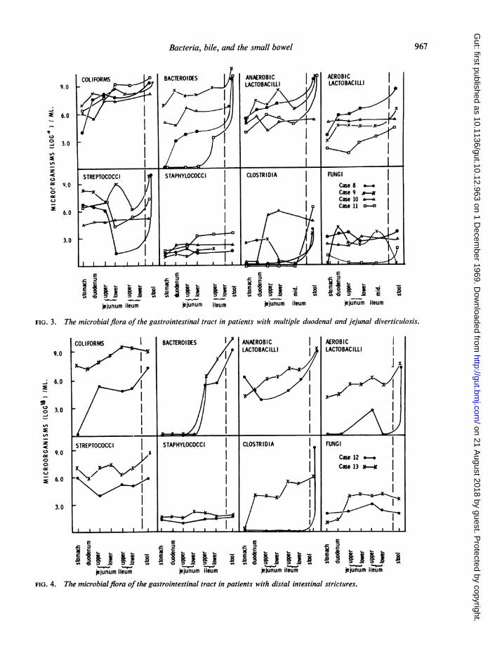
FIG. 5. The microbial flora of the gastrointestinal tract in two patients with distal intestinal resection.
1~ - -.- - - - - - - - Fasting- + + - + + - + + - + + Aftermeal
1010 Colifoms Bacteroide Strepto Staphy-[ cocci lococciio, -e,' ~~~~~~~~0Fastingibs- 0~..pAfter meat
102lou
101
100106lot
10'10?
Aerobicactobacitli
Anaerobiclactobacilli
SoDJ
Clostridia Fungi
S:stomach0-duodenumJs jejunum
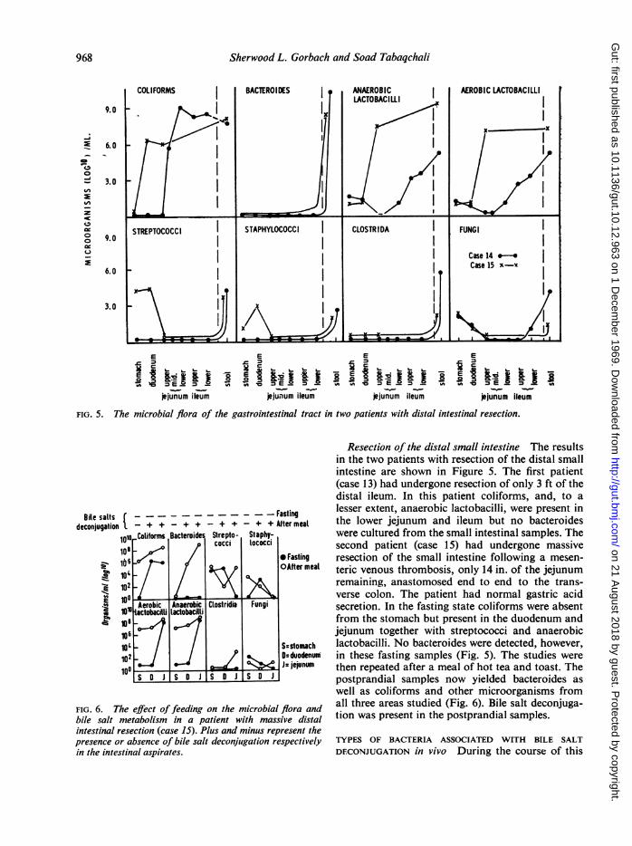
FIG. 6. The effect offeeding on the microbial flora andbile salt metabolism in a patient with massive distalintestinal resection (case 15). Plus and minus represent thepresence or absence of bile salt deconjugation respectivelyin the intestinal aspirates.
Resection of the distal small intestine The resultsin the two patients with resection of the distal smallintestine are shown in Figure 5. The first patient(case 13) had undergone resection of only 3 ft of thedistal ileum. In this patient coliforms, and, to alesser extent, anaerobic lactobacilli, were present inthe lower jejunum and ileum but no bacteroideswere cultured from the small intestinal samples. Thesecond patient (case 15) had undergone massiveresection of the small intestine following a mesen-teric venous thrombosis, only 14 in. of the jejunumremaining, anastomosed end to end to the trans-verse colon. The patient had normal gastric acidsecretion. In the fasting state coliforms were absentfrom the stomach but present in the duodenum andjejunum together with streptococci and anaerobiclactobacilli. No bacteroides were detected, however,in these fasting samples (Fig. 5). The studies werethen repeated after a meal of hot tea and toast. Thepostprandial samples now yielded bacteroides aswell as coliforms and other microorganisms fromall three areas studied (Fig. 6). Bile salt deconjuga-tion was present in the postprandial samples.
TYPES OF BACTERIA ASSOCIATED WITH BILE SALTDFCONJUGATION in vivo During the course of this
Bile saltsdeconjugation
l5 m - a- -m-IF-Mf . . .
11
9,68
CZ,
,,E
on 21 August 2018 by guest. P
rotected by copyright.http://gut.bm
j.com/
Gut: first published as 10.1136/gut.10.12.963 on 1 D
ecember 1969. D
ownloaded from

Bacteria, bile, and the small bowel
TABLE IIMICROORGANISMS ASSOCIATED WITH DECONJUGATION OF BILE SALTS IN SMALL BOWEL FLUID
No. of Samples Isolation of Intestinal Microorganisms in Small Bowel Fluid
Bacteroides Clostridia Bifidobacteria Faecal Streptococci Coliforms
No. Positive No. Positive No. Positive No. Positive No. Positive
29 (94 %) 15 (49 %) 13 (42%) 29 (94 %)1 (2%)
30
study, 81 specimens of small intestinal fluid wereexamined by thin-layer chromatography for evidenceof bile acid deconjugation. Free bile acids (cholicchenodeoxycholic, and deoxycholic acid) werepresent in 31 of these samples but only conjugatedbile acids were found in the remaining 50 specimens.There was a close correlation between the presenceof free bile acids and the anaerobic bacteroides inthe intestinal samples (Table II). Of the 31 samples inwhich free bile acids were found, bacteroides werepresent in 29. In the 50 samples without bile saltdeconjugation, only one contained bacteroides. Nocorrelation was found between the presence orabsence of bile salt deconjugation and other micro-organisms (Table II).
9 (18%) 13 (26%) 30(60%)
24 26 59
31 (100%)
48 (96%)79
these patients had coliforms (104_108 per ml) in thestomach and upper intestine. Furthermore, thosepatients, who had evidence of proximal stasis induodenal or jejunal diverticula, also had bacteroides(103-o108 per ml) associated with bile salt deconjuga-tion. By contrast, no coliforms were cultured fromthe stomach of three patients with normal gastricacid secretion and only small numbers (102_104 perml) of streptococci and anaerobic lactobacilli werefound in the gastric samples. The bacterial flora ofthe proximal small intestine was also different inthese patients. The duodenum and upper jejunumcontained coliforms (10fi-107) but no bacteroideswere present and there was no evidence of bile saltdeconjugation.
GASTRIC ACIDITY, BACTERIOLOGY, AND BILE SALTSThe relationship between gastric acidity and thepresence and type of bacteria in stomach, duodenum,and jejunum was studied in these 15 patients. Twelvepatients had gastrectomy or achlorhydria associatedwith lesions in the small intestine. All but one of
EFFECTS OF ANTIBIOTIC TREATMENT ON BACTERIALFLORA AND INTESTINAL FUNCTION The concentra-tions of microorganisms cultured at different levelsof the small intestine in three patients before andafter treatment with either lincomycin (500 mgeight-hourly for seven days) or tetracycline (250 mg
Coliforms
00E 10
Bacteroides
10.
E
Upper Lower Upper Lower Stooljejunum jejunum ileum ileum
Streptococci
Lactobacilli
Staphylococci
Upper Lower Upper Lower Stooljejunum jejunum ileum ileum
FIG. 7. The effect ofantibiotics onthe microbialflora ofa patient with distalintestinal stricture (case 13).
jO Before therapy A During therapy @ 6 weeks after therapy
Bile Acid
Free bile acids presentFree bile acids absent(all conjugated)
Totals
31
50
81
969
on 21 August 2018 by guest. P
rotected by copyright.http://gut.bm
j.com/
Gut: first published as 10.1136/gut.10.12.963 on 1 D
ecember 1969. D
ownloaded from

Sherwood L. Gorbach and Soad Tabaqchali
TABLE IIIEFFECT OF LINCOMYCIN ON THE MICROBIAL FLORA AND BILE SALT METABOLISM
Case No. Antibiotic Used Site of Sampling Microorganisms Isolated (log 10) (ml or g)
Coliforms Bacteroides Streptococci
Before After Before After Before After
5 Lincomycin 500 mg8 hourly
9 Lincomycin 500 mg8 hourly
A = Aerobacter, E = E. coli, P = Proteus
StomachUpper jejunumLower jejunumUpper ileum
Stool
Duodenum
Upper jejunum
Stool
six-hourly for seven days) are set out in Table IIIand Figure 7.The first patient (case 5), who had a single duo-
denal diverticulum and achlorhydria, was treatedwith lincomycin. Although there was little change inthe concentrations of coliforms, streptococci, andlactobacilli, there was a dramatic fall in the bac-teroides and clostridia which were no longer presentafter antibiotic treatment. At the same time free bileacids disappeared from the intestinal fluid. Faecalfat excretion, which was normal, did not changesignificantly nor did vitamin B12 absorption.The second patient (case 9) had jejunal diverti-
culosis, achlorhydria, and extensive bacterial pro-liferation throughout the small intestine (Fig. 3).After treatment with lincomycin the concentrationsof both bacteroides and clostridia in the jejunumfell dramatically but there was no change in theconcentrations of coliforms, streptococci, and lacto-bacilli. At the same time there was now no bile saltdeconjugation following treatment with lincomycin,and the faecal fat excretion fell from 22 to 4g daily.The third patient (case 13 in Fig. 7) had achlor-
hydria and a stricture of the distal ileum. Aftertreatment with tetlacycline for seven days, there wasa marked fall in the concentration of coliforms,bacteroides, streptococci, and lactobacilli at alllevels of the small intestine but there was an increasein the concentrations of fungi (107-108 per ml) andstaphylococci (104-105 per ml). The faecal fat fellfrom 7.0 to 5.0 g per day but vitamin B12 absorptiondid not change significantly. Six weeks followingcessation- of antibiotics, a repeat intubation studywas performed as demonstrated in Figure 7. Therewas a general return to the pretreatment microflora.The numbers of coliforms and lactobacilli rose toformer levels. The streptococci were generallyincreased although in the lower jejunum and stool,
4-9A8-7A7-2A8-2A7-8P6-5P9-5A
7-2A
8-3A
9-5A
7-8P8-5P8-0A8-7A8-7P8-5P8-3A7-1P8-2E7-6A8-3E7-6A8-7E9-3A
5.3405-60
0000
6-56-56-340
6-46-05.76-5
9.8 < 103 8-5 9.0
7-1 0 85 6-5
7-6 0 7-6 6-0
11-4 <104 10-0 5.8
they were somewhat below the pretreatment values.Similarly, fungi and staphylococci fell to the numbersfound before tetracycline treatment.Two important differences were noted in the
specimens from this patient obtained six weeks aftertreatment. Although the total number of coliformswere the same, approximately 50% were nowAerobacter aerogenes and the remainder were differ-ent biotypes of E. coli. No aerogenes were found inhigher dilutions of pretreatment samples. Further-more, the number of bacteroides in the ileal speci-mens was significantly less than in the pretreatmentspecimens.
DISCUSSION
Previous studies of the intestinal microflora inpatients with disorders of the small intestine haveemphasized that coliform microorganisms weremost frequently found, and these microorganismshave often been incriminated as causing the absorp-tion defects in the stagnant loop syndrome. It wasnot until 1966 that Drasar, Hill, and Shiner (1966)showed as in stools the anaerobic flora might bepresent in the lumen of the gut in the highest con-centrations. Furthermore, on the basis of experi-ments in vitro, they suggested that bacteroides inparticular might be responsible for the steatorrhoeathrough its known ability to deconjugate bile salts,a facility not possessed by coliforms.
Microbial populations of the diseased small bowelhave previously been investigated by sampling theupper jejunum in fasting subjects (Goldstein et al,1962; Wirts and Goldstein, 1963; Donaldson, 1964;Paulk and Farrar, 1964; Dellipiani and Girdwood,1964; Tabaqchali and Booth, 1966). Althoughthis approach has yielded useful information,the results presented in this paper demonstrate
970
on 21 August 2018 by guest. P
rotected by copyright.http://gut.bm
j.com/
Gut: first published as 10.1136/gut.10.12.963 on 1 D
ecember 1969. D
ownloaded from

Bacteria, bile, and the small bowel
TABLE IlII-continuedEFFECT OF LINCOMYCIN ON THE MICROBIAL FLORA AND BILE SALT METABOLISM
Mi(croorganisnms Isolated (log 10) (ml or g)
971
Bile Salt Deconjugation
Staphylococci Aerobic Lactobacilli Anaerobic Lactobacilli
Before After Before After Before After
202.210
02.72.3
3.26.34.7
3.8305.0
46664.7
4.74.769
Clostridia Fungi
Before After Before After
3.4605s8
000
1-5201I5
1.32027
30 45 46 5.3 9.7 84 90 2.1 27 - -
0 20 64 5.7 68 66 57 0 23 49 0
1.0 20 30 4.9 5.5 5s 26 0 0 43 + 0
20 22 4.7 51 78 5s0 3.3 0 10 17 + 0
0 27 65 65 100 64
that the microflora of the small intestine is bestrevealed by multiple sampling at various levels sincethe bacterial flora in different patients may be relatedto the site and extent of the anatomical abnormalityof the intestine. The validity of this technique hasbeen demonstrated by comparative studies with a
closed capsule (Kalser, Cohen, Arteaga, Yawn,Mayoral, Hoffert, and Frazier, 1966), and by a directsurgical approach in man (Anderson and Langford,1958), and in experimental animals (Gorbach et al,1967b).The results of these studies at multiple levels of
the small intestine show that although many patientshad an extensive growth of microorganisms, such as
coliforms and lactobacilli, throughout the smallbowel irrespective of the causative lesion, thepresence of anaerobic microorganisms and free bileacids was found to be related to local anatomicaldefects. For example, bacteroides and bile saltdeconjugation generally occurred only in relation toareas of stasis. Patients with duodenal and upper
jejunal diverticula might show these abnormalitiesin proximal sites (Figs. 2 and 3), while stagnationof the ileum was associated with bacteroides andbile salt splitting in distal regions (Fig. 4). In normalsubjects or patients with uncomplicated gastricresection, microorganisms of the bacteroides group
are generally absent in the small bowel, althoughsmall numbers are occasionally found in the distalileum. It appears, therefore, that the micro-environ-ment necessary for growth ofthis fastidious anaerobe,that is, low oxidation-reduction potential, is availablein areas of intestinal stasis. This requirement appears
to be fulfilled in the colon or terminal ileum ofnormal subjects and in the stagnant area of smallbowel in patients with diverticula, strictures, or
blind loops.
0 20 - -
Although a variety of intestinal microorganismsare able to deconjugate bile salts in vitro, ie, clostridiafaecal streptococci, anaerobic lactobacilli, and bac-teroides (Norman and Grubb,1955 ;Drasar et al,1966;Midtvedt and Norman, 1967; Hill and Drasar,1968), it has been uncertain which species are
responsible for this reaction in vivo. The resultspresented in this paper suggest that deconjugationof bile salts occurs in areas of stasis where condi-tions favour the growth of an anaerobic flora andthat bacteroides may be the major bacterial speciesresponsible for deconjugation. In patients withpartial gastrectomy, for example, there were rela-tively high concentrations of coliform organismsbut no bacteroides or bile salt deconjugation. Inpatients with ileal stricture, although coliforms andother organisms were present throughout the smallintestine, bacteroides were restricted to the area ofstasis in the ileum and bile salt deconjugation onlyoccurred in the intestinal samples obtained from thisregion. However, the evidence for incriminatingbacteroides in deconjugation of bile salts in vivo islargely circumstantial and it is possible that thismicroorganism may only be serving as a marker forthe physicochemical environment of a stagnant area
which allows the deconjugation reaction to proceedby a variety of other bacteria. The importance ofbacteroides in the aetiology of the blind loop syn-drome has been emphasized by Polter, Boyle, Miller,and Finegold (1968). These authors studied a singlepatient with jejunal diverticulosis in whom they usedseveral antibiotics, including lincomycin, anddemonstrated that malabsorption of fat and vitaminB12 was related to the anaerobic flora in the jejunumof their patient.The results of treatment with lincomycin reported
in this paper (Table JII) support the hypothesis that
Before After
t
00
on 21 August 2018 by guest. P
rotected by copyright.http://gut.bm
j.com/
Gut: first published as 10.1136/gut.10.12.963 on 1 D
ecember 1969. D
ownloaded from

972 Sherwood L. Gorbach and Soad Tabaqchali
bacteroides appears to be responsible for bile saltdeconjugation in such patients, since eradication ofthese microorganisms by lincomycin, without affect-ing the aerobic bacterial flora, led to a reduction insteatorrhoea and disappearance of free bile acidsfrom the intestinal fluid. Lincomycin may, therefore,be of particular value in the treatment of patientswith malabsorption due to bacterial contaminationof the small bowel.
Extensive deconjugation of bile salts throughoutthe small bowel was associated with steatorrhoea.This correlates with quantitative bile salt studies inseveral patients in this series (Tabaqchali et al,1968), which have shown that defective absorptionwas associated with a reduction in the level of theconjugated bile salts below the critical concentra-tion for micellar formation. On the other hand,when deconjugation was limited to either jejunumor ileum, steatorrhoea was not present; under thesecircumstances there may be sufficient concentrationsof conjugated bile salts to facilitate normal fatabsorption even though high concentrations of freebile acids may be present (Tabaqchali et al, 1968).Those patients with extensive bacterial deconjuga-
tion of bile salts in the small bowel and steatorrhoeaare examples of the stagnant loop syndrome (cases8, 9, and 10). However, steatorrhoea was alsoobserved in the absence of small bowel stasis or bilesalt deconjugation. In two such patients withgastrectomy and one with pancreatic insufficiency,the concentration of conjugated bile salts wasnormal and defective fat absorption was apparentlyrelated to other causes. The patient with extensivedistal intestinal resection (case 15), on the otherhand, had a reduced concentration of conjugatedbile salts in the jejunum, presumably as a result ofimpaired reabsorption in the ileum (Austad, Lack,and Tyor, 1967). The effect of feeding on the smallbowel microflora in this patient (case 15) was striking(Fig. 6). Fasting specimens had a sparse microfloraand there was no bile salt deconjugation, whereasthe postprandial specimens contained large numbersof coliforms, bacteroides, and lactobacilli (Fig. 6),and free bile acids were also demonstrated. It ispossible that these microorganisms had passedretrograde from the colon into the small intestinesince this patient had no ileo-caecal valve. Bacterialcontamination of the small intestine may, therefore,contribute to the steatorrhoea of patients withintestinal resection by causing bi!esalt deconjugation.
We wish to express grateful thanks to Professor C. C.Booth for his advice and guidance in these studies.Technical assistance was provided by Mr. Josh Wrightand other members of the Departments of Microbiology
and Medicine, Royal Postgraduate Medical School.We are particularly grateful to Mr. Joseph Hall (case 13)for his helpful cooperation. We also thank MessrsUpjohn Ltd for their generous support of this work.
REFERENCES
Anderson, C. M., and Langford, R. F. (1958). Bacterial content ofsmall intestine of children in health, in coeliac disease, and infibrocystic disease of pancreas. Brit. med. J., 1, 803-806.
Austad, W. L., Lack, L., and Tyor, M. P. (1967). Importance of bileacid and of an intact distal small intestine for fat absorption.Gastroenterology, 52, 638-646.
Dellipiani, A. W., and Girdwood, E. H. (1964). Bacterial changes inthe small intestine in malabsorptive states and in perniciousanaemia. Clin. Sci., 26, 359-374.
Donaldson, R. M., Jr. (1964). Normal bacterial populations of theintestine and their relation to intestinal function. New Engi.J. Med., 270, 1050-1056.
(1965). Studies on the pathogenesis of steatorrhea in the blindloop syndrome. J. clin. Invest., 44, 1815-1825.
Drasar, B. S., Hill, M. J., and Shiner, M. (1966). The deconjugationof bile salts by human intestinal bacteria. Lancet, 1, 1237-1238.
Eneroth, P. (1963). Thin layer chromatography of bile acids. J. LipidRes., 4, 11-16.
Goldstein, F., Wirts, C. W., and Josephs, L. (1962). The bacterialflora of the small intestine. Gastroenterology, 42, 755-756.Plaut, A. G., Nahas, L., Weinstein, L., Spanknebel, G., andLevitan, R. (1967b). Studies of intestinal microflora. II.Micro-organisms of the small intestine and their relations tooral and fecal flora. Ibid., 53, 856-867.
Gorbach, S. L., Nahas, L., Lerner, P., and Weinstein, L. (1967a).Studies of intestinal microflora. I. Effects of diet, age andperiodic sampling on numbers of fecal micro-organisms inman. Ibid., 53, 845-855.
Hill, M. J., and Drasar, B. S. (1968). Degradation of bile salts byhuman intestinal bacteria. Gut, 9, 22-27.
Hofmann, A. F. (1962). Thin layer adsorption chromatography offree and conjugated bile acids on silicic acid. J. Lipid Res., 3,127-128.
Kalser, M. H., Cohen, R., Arteaga, I., Yawn, E., Mayoral, L., HoffertW. R., and Frazier, D. (1966). Normal viral and bacterial floraof the human small and large intestine. New Engl. J. Med.,247, 500-505.
Kim, Y. S., Spritz, N., Blum, M., Terz, J., and Sherlock, P. (1967).The role of altered bile acid metabolism in the steatorrhoea ofexperimental blind loop. J. clin. Invest., 45, 956-962.
Midtvedt, T., and Norman, A. (1967). Bile acid transformations bymicrobial strains belonging to genera found in intestinalcontents. Acta path. microbiol. scand., 71, 629-638.
Norman, A., and Grubb, A. (1955). Hydrolysis of conjugated bileacids by clostridia and enterococci. Ibid., 36, 537-547.
Paulk, E. A., Jr., and Farrar, W. E., Jr. (1964). Diverticulosis of thesmall intestine and megaloblastic anemia. Amer. J. Med.,37, 473-480.
Polter, D. E., Boyle, J. D., Miller, L. G., and Finegold, S. M. (1968).Anaerobic bacteria as cause of the blind loop syndrome.Gastroenterology, 54, 1148-1154.
Tabaqchali, S., and Booth, C. C. (1966). Jejunal bacteriology and bilesalt metabolism in patients with intestinal malabsorption.Lancet, 2, 12-15.
,- (1967). The Relationship of the intestinal bacterial florato absorption. Brit. med. Bull., 23, 285-290.Hatzioannou, J., and Booth, C. C. (1968). Bile salt deconjuga-tion and steatorrhoea in patients with the stagnant loop syn-drome. Lancet, 2, 12-16.
Van de Kamer, J. H., Bokkel Huinink, H. T., and Weyers, H. A.(1949). Rapid method for determination of fat in feces. J. biol.Chem., 177, 347-355.
Van der Reis, V. (1925). Die Darmbakterien der Erwachsenen undihre Klinische Bedeutung. Ergebn. inn. Med. Kinderheilk.,27, 77-178.
Whitby, L. G., and Lang, D. (1960). Experience with chromic oxidemethod of fecal marking in metabolic balance investigationson humans. J. clin. Invest., 39, 854-863.
Wirts, C. W., and Goldstein, F. (1963). Studies of the mechanism ofpost gastrectomy steatorrhoea. Ann. intern. Med., 58, 25-36.
on 21 August 2018 by guest. P
rotected by copyright.http://gut.bm
j.com/
Gut: first published as 10.1136/gut.10.12.963 on 1 D
ecember 1969. D
ownloaded from