AZHAL KEEL VAYU -...
177
A STUDY ON AZHAL KEEL VAYU Dissertation Submitted To THE TAMIL NADU Dr. M.G.R. Medical University Chennai – 32 For the Partial fulfillment for the Award of Degree of DOCTOR OF MEDICINE (SIDDHA) (Branch – III, SIRAPPU MARUTHUVAM) DEPARTMENT OF SIRAPPU MARUTHUVAM Government Siddha Medical College Palayamkottai – 627 002. APRIL – 2013
Transcript of AZHAL KEEL VAYU -...

A STUDY ON
AZHAL KEEL VAYU
Dissertation Submitted To
THE TAMIL NADU Dr. M.G.R. Medical University
Chennai – 32
For the Partial fulfillment for the Award of Degree of
DOCTOR OF MEDICINE (SIDDHA)
(Branch – III, SIRAPPU MARUTHUVAM)
DEPARTMENT OF SIRAPPU MARUTHUVAM
Government Siddha Medical College
Palayamkottai – 627 002.
APRIL – 2013


ACKNOWLEDGEMENT
I express my deep sense of gratitude to our Principal
Dr.S.Chandramohandoss M.D. (S)., Government Siddha Medical College,
Palayamkottai for his auspicious support in bringing out the dissertation and I
also thank our Vice Principal Dr.S.Soundarrajan M.D.(S), for his support
regarding this study.
I feel very proud to record my deep sense of thanks to Dr.S.Kaniraja
M.D(S) Head of the Department, PG Sirappu Maruthuvam for rendering his
valuable suggestion, guidance and support in all aspects from time to time.
I express my whole hearted thanks to Dr.A.S.Poongodi Gandhimathi
M.D. (S) Lecturer PG – Sirappu Maruthuvam department for her lovable
support in this study.
It is my pleasure to express my grateful thanks to
Dr.D.RajasekarM.D (S) Lecturer PG – Sirappu Maruthuvam department for
his valuable guideline to do this study.
Its my pleasure to express my sincere thanks to Dr. S. Ramaguru
B.Sc.,M.S (Ortho), former Prof. of Orthopaedics, Tirunelveli Medical College,
Palayamkottai, for his valuable guidance for this study.
My sincere thanks to Mr. M. Kalaivanan M.Sc., Lecturer, Department
of Pharmacology, and I also thank Prof. Mrs.N.Nagaprema M.Sc., M.Phil.,
Head of the Biochemistry Department., & all over technical experts for their
help in laboratory studies.

CONTENTS
Page No.
• Introduction 1
• Aim and objectives 3
• Review of literatures
A.Siddha Aspects 4
B.Modern Aspects 35
• Materials and Methods 61
• Observation and Results 65
• Discussion 101
• Summary 105
• Conclusion 107
• Annexures
• Preparation and properties of trial drugs 108
• Bio – chemical Analysis 132
• Pharmacological Analysis 135
• Proforma of Case Sheet 142
Bibliography 170

INTRODUCTION
Siddha medicine is one of the most antiquated traditional medical
systems with codified references from age old literature. It surfaced more than
2500 years ago. Siddha system of medicine is based upon panchaboothas (5
elements) mukkutram (tridosha) theory & sapthathadu theory. Panchaboothms
& mukkutram are present in the 96 thathuvas which are basic principles of the
medicine.
The medical system that blossomed at lemuria with the colour of
tamil literature is called as siddha. Siddha system of medicine is one of the most
old system of medicine curing major diseases in a proper way without any
harm and major toxic effects. This system of medicine cures diseases on the
basis of dhosa’s. The siddhar’s who are the eminent founders of this ancient
system had written various types of medicines to cure the variation in the
dhosas.
When the normal equilibrium of 3 humors (vadha, pittha, kaba) is
disturbed, disease occurs.. The factors which affect this equilibrium are
environment, climatic conditions, diet, physical activities & stress. Under
normal conditions the ratio between these three humors (vadha, pittha, kaba) is
1:1/2:1/4 respectively. According to the siddha medicine system diet & life style
plays a major role. This concept of siddha medicine is termed as pathya &
apathya.
The drug used by the siddhars could be classified into 3 groups:
• Thavaram (herbal product)
• Thathu (inorganic substance)
• Jeevam (animal products)

According to their mode of application the siddha medicine could be
categorized into two classes.
• Internal medicine – used through oral route & further classified into 32
categories based on their form , methods of preparation.
• External medicine – it includes certain forms of drugs & also certain
application like nasal , eye ,ear drops & also certain procedures like leech
application, purgative therapy, emetic, fasting, steam, physical therapies,
solar therapies, bloodletting, yoga,thokkanam,ottradam etc.
Osteo arthritis also known as degenerative arthritis or
degenerative joint disease or osteoarthrosis is a group of mechanical
abnormalities involving degradation of joints including articular cartilage &
subchondral bone. Symptoms may include joint pain, tenderness, stiffness,
locking & sometimes an effusion. A variety of causes – hereditary,
developmental, metabolic & mechanical may initiate processes leading to loss
of cartilage. When bone surfaces become less well protected by cartilage, bone
may be exposed & damaged. As a result of decreased movement secondary to
pain, regional muscles may atrophy & ligaments may become more lax.
Treatment generally involves a combination of exercise, lifestyle
modification & analgesics. Azhal keel vayu is the most common problem of
elder person.
For this reason I have chosen Azhal keel vayu.My trial drug is
KARUNGOZHI CHOORANAM as internal medicine and VIDAMUTTI
THYLAM as external medicine.
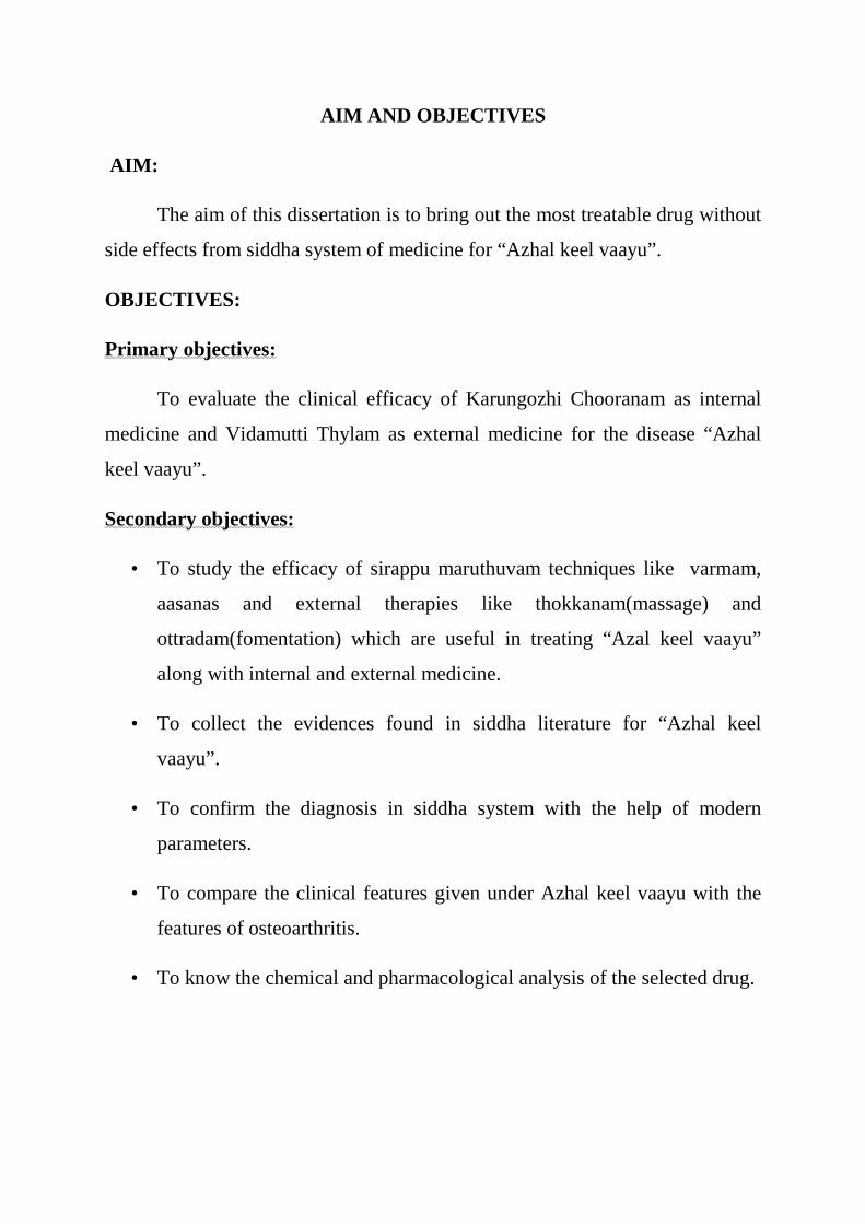
AIM AND OBJECTIVES
AIM:
The aim of this dissertation is to bring out the most treatable drug without
side effects from siddha system of medicine for “Azhal keel vaayu”.
OBJECTIVES:
Primary objectives:
To evaluate the clinical efficacy of Karungozhi Chooranam as internal
medicine and Vidamutti Thylam as external medicine for the disease “Azhal
keel vaayu”.
Secondary objectives:
• To study the efficacy of sirappu maruthuvam techniques like varmam,
aasanas and external therapies like thokkanam(massage) and
ottradam(fomentation) which are useful in treating “Azal keel vaayu”
along with internal and external medicine.
• To collect the evidences found in siddha literature for “Azhal keel
vaayu”.
• To confirm the diagnosis in siddha system with the help of modern
parameters.
• To compare the clinical features given under Azhal keel vaayu with the
features of osteoarthritis.
• To know the chemical and pharmacological analysis of the selected drug.

SIDDHA ASPECT
“jkpo; kz;lyike;Je; jhtpa Qhdk;
ckpo;tJ Nghy Tyfe; jphpthh;
mtpO kdKnkk; khjp awpTe;
jkpo;kz; lyike;Je; jj;Jt khNk”
- jpUke;jpuk;
gd;dPuhapuk; Mz;Lfl;F Kd; Njhd;wp rpj;jhe;j NguwptpaYf;Fj;
Jizahf Njhd;wp rpj;j kUj;Jtk; mg;Nguwptpiy tiue;j rpj;jh;fNs
tioue;J> vk;kf;fSk; mjidf; fw;wwpUe;J ePLop thor; nra;jnud;gJ
kpifahfh.
“tPu kUe;njd;Wk; tpz;Nzhh; kUe;njd;Wk;
ehhp kUe;njd;W ee;jp aUs; nra;jhd;
Mjp kUe;njd; wwpth GfyplQ;
Nrhjp kUe;jpJ nrhy;ynthz; zhjNj”
jkpo;ehl;by; Njhd;wpa nkhop> Nguwptpay;> kUj;Jtk; Mfpait
rptdhy; nra;ag;gl;lit vdf; $wp mij rptd; ee;jpf;Ff; fw;gpj;jhd;
vdTk;> ee;jp jpU%yh;f;Fk;> jpU%yh; kw;w rpj;jh;f;Fk; fw;gpj;jhnud xU
rhuhh; $Wth;. ,t;thW ngwg;gl;l rpj;j kUj;Jthpd; ,ay;ghf mfj;jpah;
gpd;tUkhW $Wfpwhh;.
“cj;jk Fzq;f Ss;Nshd;
cah;ngUq; fPh;j;jp As;Nshd;.
Gj;jpah yha;e;J nrhy;Nthd;.
nghUs;fisaha ty;Nyhd;

rj;jpa thh;j;ij As;Nshd;
jUk rpe;jidNa As;Nshd;
Fj;jpu thh;j;ij Ngrhf;
Fzd; ghpfhhp ahNk”
-mfj;jpah;.
kUj;JtDf;F %d;W fz;fs; vd;gij fPo;f;fz;l mbahy; mwpayhk;.
'%d;W fz;Zs;NshhpUtuhk;
md;d tHah nudpy;
Md;w NyhfkPd; wspg;gtd;
Mfpa tuDk;
Njhd;w kf;flk; Jahpidg;
Nghf;fpL Nthdhk;
vd;w khkUj; JtDNk
vd tpak;GtNu
kUj;jtdpd; 3 fz;fs; vit vd;gij
“kUe;J Rj;jp Fz epfz;L fz;fs; %d;whk;”
1. Purification of medicine
2. Diagnosis of disease and character of medicine
3. The different nomenclature of medicine
“mz;lj;jpy; cs;sNj gpz;lk;
gpz;lj;jpy; cs;sNj mz;lk;"
-rl;lKdp Qhdk;

In this sentence says that “Man is considered as the microcosm; Universe is
considered as the macrocosm”. It shows that Human body is the replication of
universe.
Universe made up of 5 elements i.e earth air, water, ether and fire.
Human body also made up of this panchaboothas.
“Ik;g+jk; gj;jhf;fp”
This line says that one of the fire elements combined with the other four
elements in different proportions to form the human body. These are the basic
reason for “Urirthathu” This Uyirthathu divided into their thodas known as
vatham, Pittham and kabam. Any deviation (or) derangement occur in this there
Uyrithathu may produce “Disease”.
KEEL VAYU.
Other Names
According to siddha maruthuvam text book keel vayu mentioned as santhu vali,
Mutthu vali, Meha soolai, Mudakku vayu and ama vatham.
• Vitiated vatha produces disease in keel (Joints) called as keel vayu.
• Pain in muttu (Joint) called as muttuvalli.
• Inability to use (mudakku) joints called as muddakku vayu,
• Improper digestion of food followed by increased kabam produces vadha
disease called as “Amavatham”
• This disease followed by meha noi called as “mehasoolai”.
According to Agathiyar Gunavagadam keelavayu mentioned as:
“jhdhd fPy;thj Nuhfk; Ngiu
rhw;WfpNwd; ePawpa tpgukhf

khdhd tha;T Nuhfk; thjNuhfk;
kfj;jhd Klf;Ftha;T Klf;F thjk;
Njdhd re;jpf rpNyl;Lk Nuhfk;
njspthd iffhypy; gpbg;G Nuhfk;
Cdhd urthjk; R+iyf;fl;L
cj;jkNd re;jpthjk; thj R+iyahNk”
-mfj;jpah; Fzthflk;.
IYAL (DEFINITION)
Keel vayu in one of vatha disease which is characterised by swelling,
pain, stiffness of the joints, difficulty to flex and extend the joint. This disease
associated with fever when increased vatha is along with Kabam.
“tspA ikAe; jd;dpiy nfl;L
typAld; tPf;fr; RuKk; fha;e;J
Kl;Lf NlhWk; KLf;fpNa nehe;J
Kl;Lf ld;dpd; ePUk; Rue;J
jhq;nfhzh typAld; nehe;jpL kk;Nk”
-rghgjp ifNaL.
AZHAZ KEEL VAYU:
When it is Vatha vitiated, diet and habits which stimulates Pittha it will
produce “Azhal Keel Vayu”.

NOI VARUM VAZHI: (AETIOLOGY)
1. Causes of ‘Keel Vayu” from Sabapathy Kaiyedu is as follows:
“ tspjU fha; fpoq;F
tiutpyh japyy; Nfhio
Kspjaph; Nghd;kpFf;F
Kiwapyh Tz;b Nfhly;
FspHjU tspapw; Nwfq;
Fdpg;Gw Tyty; ngz;bh;
fspj;jU Kaf;fk; ngw;Nwhh;
fbnray; fUtpahkhy;”.
Diet and health which gives rise to Vatha dhosa (i.e) excessive intake of
potato like roots and banana, excessive intake of cold substances like curd,
exposure to cold, staying in hill station which increase Kabam causes this
disease. Further this disease is followed by mega noi and may be hereditary.
2. According to “Yugi Muni” Aetiology is defined as follows:
“ vd;dNt thjk; jhndz;gjhFk;
,fj;jpNj kdpjh;fSf; nfa;AkhW
gpd;dNt nghd;jiia NrhuQ;nra;J
nghpNahh;fs; gpuhkziu J~zpj;Jk;
td;dNjtw; nrhj;jpr; NrhuQ; nra;J
khjhgpjhFUit kwe;j Ngh;f;Fk;
fd;dNt Ntjj;ij epe;ij nra;jhy;
fhaj;jpw; fye;jpLNk thje;jhNd”

“jhnddw; frg;NghL Jth;g;Giwg;G
rhjfkha; neQ;Rf;fpDQ; rikj;j tz;zk;
Mndd;w thwpdJ nghrpj;jyhYk;
Mfhaj; NjwyJ Fbj;jyhYk;
ghndd;w gfYhwf;f kpuh tpopg;G
gl;bdpNa kftpWj;jy; ghunka;jy;
Njndd;w nkhopahh;Nkw; rpe;ijahjy;
rPf;fpukha; thjkJ nrdpf;Fe;jhNd”
- A+fpitj;jpa rpe;jhkzp
Intake of foods which are rich in taste like bitter, astringent and chilly,
intake of old cooked foods, drinking of rain water, day time sleeping,
awakening at night, starvation, excessive weight lifting and sexual
perversion may produce Vadha disease.
3. In "Theraiyar Vagadam” the cause of Vadha disease is mentioned as
follows.
“nta;apypy; elf;ifahYk; kpfj;jz;zPh; Fbf;ifahYk;
nra;apio kfspdiur; Nrh;e;jDgtpf;ifahYk;
igaNd cz;ikahYk; ghfw;fha; jpz;ifahYk;
ijaNy thjNuhfk; rdpf;F nkd;wwpe;J nfhs;Ns”.
- Njiuah; thflk;.
Walking on hot weather, excessive intake of water, over sexual
indulgence, intake of bitter gourd may produce Vadha disease.
4. Another concept from “Pararaja Sekaram” denotes the causative factor of
Vadha disease as follows.
“fhzNt kpfTz;lhYk; fUJgl;bdp tpl;lhYk;
khdid ahh;fz; Nkhfkwf;fpD kpFe;jpl;lhYk;
Mzt kyq;flk;ik aq;qNd tplhjyhYk;

thDjd; jley; yhNsthjq; Nfhgpf;Fk; fhNz”
Over eating, undue starving, sexual perversion, Anavamalam will produce
vadha disease.
“ghhpdpw; gag;gl;lhYk; gyUld; Nfhgpj;jhYk;
fhundf; fUfpNahbf; fOkuj; Juj;jpdhYk;
Vh;ngW jdJ neQ;rpd; kpfj;Jf;f kile;jpl;lhYk;
ghhpa fw;wpdhYk; glhpDk; thjq;fhZk;”
Fear, anxiety, tension, stress will lead to Vadha disease.
5. In Agathiyar Kanma Varalaru – 300, Vadha disease is to be considered as one
among the Kanma noi,
“E}nyd;w thjk; te;j tifjh NdJ
Ez;ikaha; fd;kj;jpd; tifiaf; NfS
fhypNy Njhd;wpa fLg;Ng NjJ
iffhypy; Klf;fpaJ tPf;fNkJ
NfhypNy gLf;fpd;w tpUl;rkhd
Foe;ij kue;jid ntl;ly; Nky;Njhy;rPty;
ehspNy rPtnre;Jfhy; Kwpj;jy;
ey;y nfhk;Gjio Kwpj;jy; eypj;jy;jhNd"
- mfj;jpah; fd;k tuyhW – 300

6. Vadha disease followed by Meha noi and Ama Vatham
7. Food Variations:
Diet plays a vital role in preserving the human body. The food is formed
on the basis of 6 tastes.
“GspJth; tpQ;Rq;fwp ahw;G+hp f;Fk; thjk;”
- fz;Zrhkpak;.
Sour and astringent taste causes an increase in Vadham.
8. Environmental change:
The environmental factors (Thinai) may also pave the way for the
development of disease.
Neithal: People of this land suffer from Vadha diseases. Further it leads to
increased body mass, enlargement of liver and flatulence.
Palai: The people of this land suffer from disease of 3 thoshas.
9. Seasonal Variations:
Due to seasonal variations changes in thridhosa occur which leads to disease.
“thjth;j;jd fhyNkNjh ntd;dpy;
kUTfpd;w Mdp fw;fl khjk;
Mjidg; grpNahL fhh;j;jpif jd;dpy;
mlUNk kw;w khjq;fs; jd;dpy;
NghfNt rkpf;fpd;w fhykhFk;"
Vadha disease most common in the months from Aani to Karthikai.

In Muthuvenil Kalam (Aani and Aadi) Vadham gets aggrevated.
In Kaar Kalam (Avani and Puratasi) – Vadham in increased state.
NOI ENN (Classification)
Keelvayu is Classified into 10 types
Valikeel Vayu
Azhalkeel Vayu
Iya Keel Vayu
Vali azhal Keel Vayu
Vali iya keel Vayu
Azhal vali keel Vayu
Azhal iya keel Vayu
Iya vali Keel Vayu
Iya azhal Keel Vayu
Mukkutra keel Vayu.
SIDDHA ASPECT
“PATHOLOGY OF KEEL VAYU”.
“tspA ikAe; jd;dpiy nfl;L” – rghgjpifNaL.
I. VITATED DHOSHA OF VADHA:
“gpzpapDw; gj;jpiag; NgRtd; gpzpKjy;
thj gpj; jq;fg kd; ke;jphp je;jphp
tPjkh Alyuz; nka;k;Gu tuR nra;
Kiw nrAkhjyhd; ……..”
- Njiuah; fhg;gpak;.

According to Siddha aspect, Vatham is the initiator of all the activities of our
body. So Theran said Vatham as "Arasan" in the above lines.
"Azhal Keel Vayu" is described in Sarabentthier Kaiyedu Nool. It is the most
common Vadha disease. Incresed Vatha may produce pain, stiffness, deformity
which is dealed on below siddha books.
1. “Fwpnadf; iffhy; Fsr;R tpyhr; re;J
gwpnad nehe;Jlw; gr;irg; Gz;zhFk;.
-jpU%yh;.
2. “nrhy;yNt thjkJ kPwpw; whdhy;
Nrhh;tile;J thAtpdhy; Njfnkq;Fk;
nky;yNy iffhy;f srjp Az;lhk;
nka;Klq;Fk; epkpunthz;zh jpkpUz;lhFk;”
- mfj;jpah;
3. “fhNzg;gh thj kPwpy;
fhy;iffs; nghUj;jp NehFk;”
- fhtpaj;jpd; ehb.
4. “Nktpa thjQ; nra;Aq;
Fzq;jid tpUk;gpf; NfS
jhtpa tapW ke;jQ;
re;Jfhy; nghUe;J Nehthk;”
- ,uj;jpdr; RUf;fk; ehb.
Increased Vadha produces 6 characteristic features.
“thjq; fLik twl;rpAld; neha;ik
rPjQ; rydk; rpjwZT – VjKl
dpf;Fzj;Njh Lw;Nw apaf;fe; jUkstpw;
wf;f ghpfhue; jh”.
- fz;Zrhkpak;.

1. fbdk;
2. twl;rp
3. ,NyR
4. Fsph;r;rp
5. mirjy;
6. mZj;Jtk;.
We could take two characteristic features from above song.
1. twl;rp – Dryness.
The fluid around the joint is dried. It is compared to modern pathology,
which says that some protective factors which protect the cartilage from
aging process. Normal cartilage has 2 main components.
1. Extracellular Matrix
2. Aggrecan contains glucosaminoglycans chain
of chondroitin sulphate and keratin sulphate that is capable of
retaining water.
During aging, quantitative and qualitative changes in aggrecan reduce
the capacity of the molecules to retain water. thus aged cartilage
contains less water.
2. mirjy; - Movements.
In Azha keel Vayu, increased vadha produces restricted movements. This
is mentioned in sabapathy kaiyedu as follows.
“ Kl;Lf NlhWk; KLf;fpNa nehe;J”

VITIATED DHOSHA OF KABAM:
“ tspA ikAe; jd;dpiy nfl;L…..”
- rghgjp ifNaL.
When vadha is increased along with vitiated kabam dhosham produces
pain, stiffness, swelling and restricted movement.
Nature of Iyam:
“jplkPA nkd;gpizg;Gj; jpz;ikAw;w ahg;Gk;
mlNyh; tOtOg;Gk; Mf;iff; fplh;f;F
ntUthg; nghWikAk; NkNyhd fhg;ghk;
ngUikj;jh ikankdg; NgR"
- kUj;Jt ghujk;
In this song nature of Iyam was described. They are:
1. Steadiness (Nilaitthal)
2. Lubrication (Neippu)
3. Structure of joints
4. Patience (Poraiyudaimai)
One of the 5 types of Iyam santhigam act as a lubricator for mobilization of
joints.
“ %l;Lf ld;dpd; ePUk; Rue;J”

This line denotes increased Iyam leads to swelling of joints
Increased Vadha Increased Pittham Increased Iyam
Pain Fever Swelling
Stiffness
Restricted Movement
Diet and habits which increase the Pittham along with vitiated Vadha
dhosam may produce this disease.
CLINICAL FEATURES :
“gpj;jf;fPy; tha;T jd;dhw;
gpwq;FfPd; %l;L tPq;fpr;
rpj;jh;nra; kUe;J tj;JQ;
rPh;glhj; jd;ikj; jhfpj;
jj;JW fha;r;ry; fz;L
rhyNt jidjhd; je;Nj
nkj;jW rpfpr;ir jd;dhy;
nkd;Nky; ePq;F kg;gh”
Dietary Variation
&
Habits
- rghgjp ifNaL.
• Swelling of the joint
• Fever
• Restricted movement
• Swelling of the joints will increased day by day. Increased Pittham act as
synovid theid between the joint space which dries it. It make sound like
“Kaluk, Kaluk” when the movement of the joint. Sometimes it may cause
inable to move the joints.
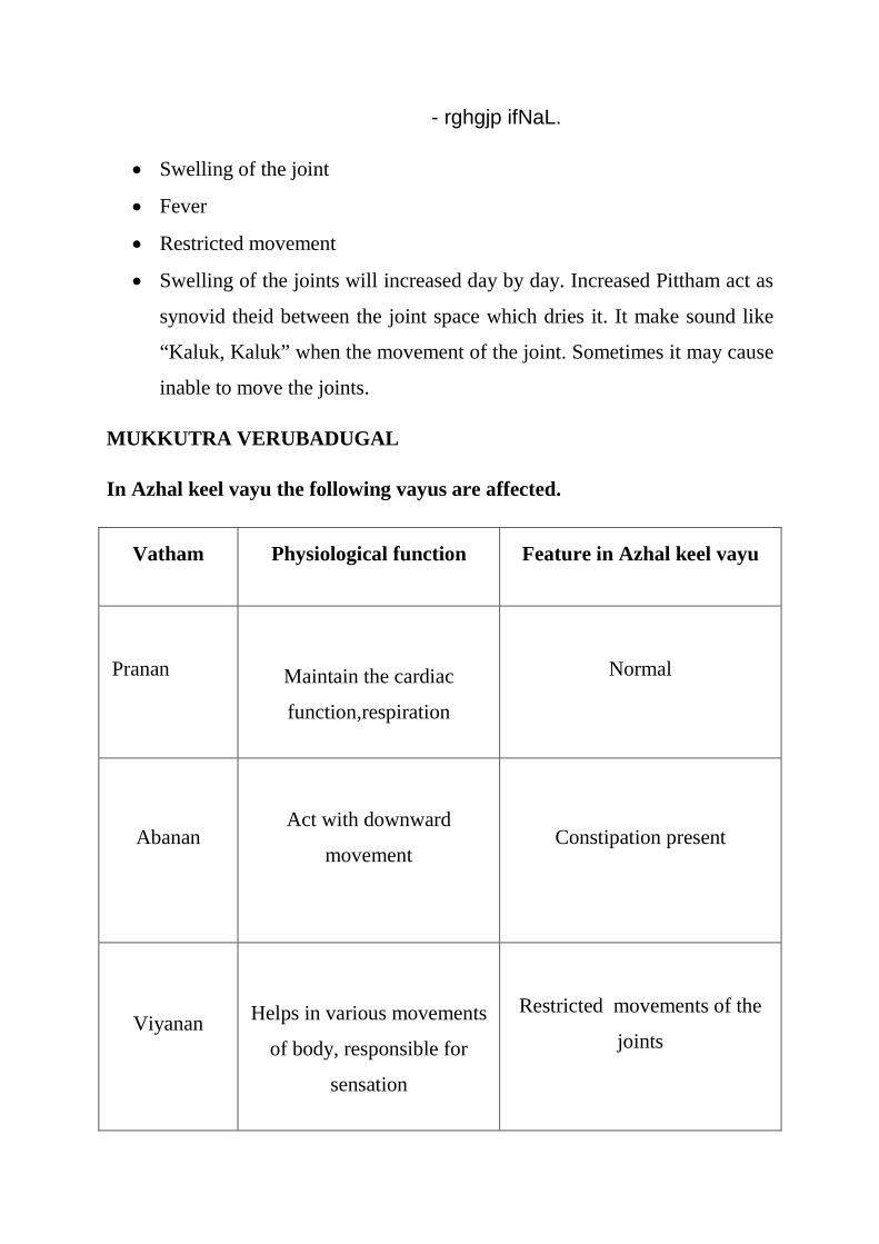
MUKKUTRA VERUBADUGAL
In Azhal keel vayu the following vayus are affected.
Vatham Physiological function Feature in Azhal keel vayu
Pranan
Maintain the cardiac
function,respiration
Normal
Abanan Act with downward
movement Constipation present
Viyanan
Helps in various movements
of body, responsible for
sensation
Restricted movements of the
joints

Samanan Regulates all other vayus Affected
Nagan
Responsible for intelligence
Helps in opening and closing
of eyes
Normal
Koorman
Responsible for lacrimation.
Helps in visualization of all
things of world
In aged patients acuity of vision
is diminished.
Kirukaran
Produce cough and
sneeze,helps in digestion
Normal
Thevathathan
Responsible for laziness,
rotation of eye balls
Sleeplessness due to pain
Thananjeyan
It leaves from the body by
blowing up the cranium only
on the 3rd day after death.
_

Pittham
Pittham is a force of heat, God to all disease, mother to dasha vayukkal,
assistant to boothas and responsible for fever.
Anar pitham Digests all the ingested
particles Affected
Ranjaga pitham Increase the blood and gives
colours to blood Affected
Saathaga pitham Makes the work to complete
what mind thinks to do
Affected.
restricted movement
Aalosaga pitham Responsible for vision of
eyes
Affected in old age
people
Prasaga pitham Gives colours to skin Not affected.
Kabam
It is classified into five types
Avalampagam Controls other 4 types of kabam Affected
Kilethagam Moistness the food Affected
Pothagam
Tharpagam
Helps to know the taste
Gives cooling to the eyes
Normal
Normal
Santhigam Gives lubrication of joint Affected

Udal Thathukkal
There are seven udal thathukkal in human body.
Saaram Strengthens the body and mind Affected
Senneer Preserves brightness, boldness
power and knowledge Affected
Oon Gives structure and shape to
body. Responsible for movement
Early stage – not
affected.
Later stage – Affected
Kozhuppu Lubricate the joints Affected
Enbu Responsible for joint movement Affected
Moolai It is present in the bones and
gives strength Affected
Sukkilam or
Suronitham Meant for reproduction Normal
DIFFERENTIAL DIAGNOSIS :
1) AGATHIYAR AYUL VETHAM-1200
“Koq;fhy; tPq;fpf; fLj;J nehe;J Nkhjpj;jpuz;L Ntjidaha;
toq;fhnjd;d eilfl;L tUj;jkpfTe; jhDKz;lha;”
-mfj;jpah; MAs;Ntjk; 1200 ghly; 296.
2) THERAIYAR VAGADAM
“Koq;fhy; thjq;fdj;J tPf;fkhfp Krpahky;
fLj;J elf;nfhl;lhnjd;W”
-Njiuah;thfk; ghly; 217.

3) PARA RASA SEKARAM –VATHA ROGA NITHANA CHIKITCHAI
“jpuz;bL Koq;fhy; tPq;fpr; Nru nehe;Jise;J Fj;jpg;
Guz;bl klf;fp kpz;bg; NghjNt eilnfhlhJ
kUz;LNty; fizkhdk;G thnsd kpspUq;fz;zha;
Kuz;lU Koq;fhy; jd;dpd; nkhope;JL thjkhNk”
-guuhrNrfuk; ghly; 182
4) NARI VATHAM :
“ele;jpbd; Koq;fhy; tPq;fp eLf;nfd Fj;jp thq;fpj;
Jlh;e;Jld; gpbj;J kpz;bj; Jbj;JldLf;fKz;lhk;
mlh;e;jpLk; tapw;W Nehth ajpfkha;j; jpuz;NlaWk;
glh;e;jpL ehpthjj;jpd; Fznkdg; gfh;e;jhud;Nw”
- guuhr Nrfuk; thjNuhfk; ghly; 209.
5) NARITHTHALAI VATHAM :
%h;f;fkh Kad;W Koq;fhy;jhd; tPq;fp Kjph;e;J
uj;jKe;jpuz;L Kaw;rpahfp
epf;fkha; epd;wpl nthzkw; jhZk; epkph;e;jpLfpy;
re;Jjhd; Klf;nfhzhky;
jPh;f;fkha; Jz;bj;J kpfr;rpf;nfd;W nrOik
ehpj;jiyNghy kpfNt tPq;fp
ehh;f;fkha; ehbANk glglf;Fk; ehpj;jiyapd;

thjnkd;Nw etpyyhNk”
-A+fpitj;jpa rpe;jhkzp800 ghly; 263.
6) UTHIRA VATHA SURONITHAM :
“itfpjkha;f; fizf;fhY Koq;fhy; jhDk;
kw;fhlQ;re;J GwtbAk; tPq;fpr;
nra;fpjkhQ; rpWtpuy;fs; kpfTk; nehe;J rpe;ij
jLkhwpNarypg;Gz;lhFk;
igfpjkhk; jLkhwpNa rypg;Gz;lhFk; ghuhkha; cw;gtpj;J moYz;lhFk;
ca;fpjkhk; mrdJ jhDk; Ntz;lh cjputhjr; RNuhzpjj;jp
Dzh;r;rpahNk”
-A+fp itj;jpa rpe;jhkzp 800 ghly;:319
7) PAYITHIYA VATHA SURONITHAM :
“czh;r;rpaha;r; RNuhzpje;jhd; kpfntJk;gp Cf;fkha;j; Njnkq;Fk;
kpfNtnehe;J
Kzh;r;rpah Koq;fhy;fs; Koq;ifnahf;f Kidahd rpWtpuy;fs;
fd;dk;new;wp
jzh;r;rpaha;r; re;J rUthq;fnkq;Fk; jhl;bfkha;f; File;J RuKKz;lhk;
gzh;r;rpaha; ghz;lJ Nghd;NkdpahFk; gapj;jpathj RNuhzpjj;jpd;
gz;GjhNd”
- A+fp itj;jpa rpe;jhkzp 800 ghly;:320

8) MEGA VATHAM :
“Klf;fpNa Koq;fhy; tPq;fp Kjph;ry Nkhjha; tPOk;
jlf;fpNa eilnfhlhJ je;JNghw; rpaYKz;lhk;
tPlf;fay; tLkhdk;G Ntnyd kpspUq;fz;zha;
klf;fpa NkfthjQ; nra;Fzk; tFf;fyhNk”
- guuhrNrfuk; thjNuhfk; ghly;-197.
9) SURONITHA SILETPAM :
“cz;ikaha; Koq;fhypy; kpff;File;J cise;JNk KJNfhL
tpyhg;gf;fq;fs;
fz;ikaha; Koq;iffs; Koq;ifr;re;J fbdkha; tPq;fpNa
Filr;rYz;lhk;
jz;ikaha; rspf;fl;Lj; jhfkhFk; rhjfkha; ,UkpNa %r;Rz;lhFk;
jpz;ikaha; ehtuz;L rpj;jpg;ghFk; nrakhd RNuhzpj rpNyl;ge;jhNd”
- A+fp itj;jpa rpe;jhkzp ghly; 415.
10) MUZHAANKALAL VATHAM :
“ jpuz;bL Koq;fhy; tPq;fpr; Nru nehe;Jise;J Fj;jpa;
Guz;bl klf;fp kpz;bg; NghjNt eilnfhlhJ
kUz;LNty; fizkhdk;G thnsd kpspUq;fz;zha;
Kuz;lU Koq;fhy; jd;dpd; nkhope;JU thjkhNk”.
- guuhrNrfuk; ghly;-182.

PINIYARI MURAIMAI(DIAGNOSIS)
Piniyari muraimai is the methodology of diagnosing the disease in siddha
system.
Envagai therugal:
'ehb ];ghprk; eh epwk; nkhop tpop
kyk; %j;jpukpit kUj;Jt uhAjk;"
• Naadi(Pulse)
• Sparisam (sense of touch)
• Naa(Tongue)
• Niram(Colour)
• Mozhi(Speech)
• Vizhi(Eyes)
• Malam(Stool)
• Moothiram(Urine)
'nka;f;Fwp epwe;njhzp tpop ehtpUkyk; iff;Fwp"
1.Naadi (Pulse)
This is a unique diagnostic method in siddha system of medicine. It is
responsible for existence of life. It is felt one inch below the wrist on the
radial side by palpatcing with the top of the index finger, middle finger and
ring finger which denotes vatham, pitham and kabam.
Suitable places for pulse reading:
'jhJ KiwNfs; jdpFjpr; re;njhL

XJW fhkpa Ke;jp neL khu;G
fhJ neL%f;Ff; fz;lk; fuk; GUtk;
NghJU cr;rp Gfo; gj;Jk; ghu;j;jpNl"
2.Sparisam(Sense of Touch):
The abnormal increased sparisam is clinically called as inflammatory
changes.
Increased sparisam-Mithaveppam(warmth) felt on affected joints in
Azhal keel vaayu.
3.Naa (Tongue):
Vadha disease Dark in colour
Pitha disease Yellow in colour
Kaba disease White colour
In Azhal keel vaayu dark & dried tongue may be seen.
4.Niram (Colour):
The colour of the affected part and general colour of the body may be
altered depends upon the severity of the disease.
In Azhal keel vaayu the affected to in red in colour.
5.Mozhi (Speech):
Speech will be affected in Vadha disease because of Piranan, Uthanan,
Kirugaran and Devathathan are disturbed.
In Azhal keel vaayu decreased tone of speech because of the severity of
disease.

6.Vizhi (Eyes):
Normaly vizhi affected in old age. In Azhal keel vaayu most commonly
affected in elderly people.
7.Malam (Stools):
Constripation common is Vadha disease. In Azhal keel vaayu malam may
be affected.
8.Moothiram (Urine):
The waste materials are executed through urine from the body.
In Azhal keel vaayu there will no specific change in neerkuri.
NEERKURI-NEIKURI:
In siddha system of medicine changes of urine is studied under two.
Peculiar headings. They are”Neerkuri and Neikuri”:
'te;j ePu;f;fup vil kzk; vQ;rnyd;
iwe;jpaYit aiwFJ KiwNa"
- Njuu; ePu;Fwp - nea;Fwp
Neikuri:
• mwntd ePz;bd/Nj thjk;
• MopNghw; gutpd; m/Nj gpj;jk;
• Kj;njhj;J epw;fpd; nkhoptnjd fgk;
- Njuu; ePu;Fwp - nea;Fwp

This procedure is an important one in siddha system of medicine to find out the
diagnosis as well as in prognosis aspect of the disease. So disease in man do not
originate itself. It is developed from the alteration of three dhoses.
LINE OF TREATMENT
In Siddha system the main aim of the treatment is to cure Udar pini (due
to Mukkutram) and Manapini (due to changes in Mukkunam). Treatment is not
only for perfect healing but also for the prevention and rejuvenation.
Line of Treatment is as follows:
1. Prevention
2. Treatment
Prevention :
The prevention methods for Azhal keel vaayu are as follows:
1. Control the body weight.
2. Modify the nature of work.
eg. Avoid prolonged standing and long distance walking.
3. Avoid excess intake of high coloric foods.
Treatment :
The aim of Neekkam is based on
To bring the deranged dhosams to normal equilibrium state.
First the deranged dhosams has to be brought to its normal state by giving
Virechanam or Vamanam or Nasiyam.

1. Purgative :
'tpNurdj;jhy; thje; jhOk;"
In Azhal keel vaayu, vatha kuttram is deranged. So a purgative vellai
ennai - 15 ml with hot water in early morning in empty stomach on the first day
is given.
2. Internal Medicine :
Karungozhi Chooranam - 1.5gm / daily with hot water.
3. External Medicine :
Vidamutti thylam - External application.
4. Complementary Therapies :
There are enormous complementary therapies followed in Siddha system
of medicine such as kattu, pattru, Nasiyam, Attai Vidal, Thokkanam, Ottradam,
Varmam, Asanam, Vedhu etc.
COMPLEMENTARY THERAPIES:
I Thokkanam (Massage)
II Ottradam (Fomentation
III Kattu (Bandage)
IV Varmam
V Aasanam
I Thokkanam :
Massage is the manipulating of superficial and deeper layers of muscle
and connective tissue using various techniques, to enhance function, aid in the

healing process, decrease muscle reflex activity, inhibits motor neuron activity
and promotes relaxation and well being massage can be applied with the hands,
fingers, elbows, knees, forearm and feet.
II Ottradam (fomentation)
Its is application of the hot or cold substances applied through packages
In siddha literature lot of ottradam describe 2 types of otttradam applied in my
study
I.Hot Formentation by medicated pouches
Dried leaf of nottri (ritex negundo) Thaluthalai Manjanathi (Moinda
tinctoria ) erukku (calotropis gigantean) which is stuffed in a cleaned doth it is
tied tightly.
II.Hot Formentation by lemon
In this method lemon is cutted hafly Each pieck of lemon tied by a cloth
and it is dipped in the hot trial drug and applied on the affected joints
III Kattu (Bandage)
The fresh Aadathodai ( Justicia adathoda ) leaves are made into pieces
and it is mixed with external medicine This Kept in a piece of cotton and
wrapped

THOKKANAM

FOMENTATION BY LEMON
FOMENTATION BY MEDICATED POUCHES

IV Varmam:
Varmam points to be manipulated for osteoarthrosis are as follows
1. Mootu varmam : Centre part of posterior apsect of both knee
joints. Mild pressure is applied using tips of middle three fingers.
2. Kuthiraimuga varmam
Location – Tibial tuberosity
Pressure is applied for three time using bulb of thumb.
3. Mootu Suzharchi
This method stimulates varmam points around knee joint by a
circulatory gripping massage around patella using thumb and index
finger.
4. Santhuvarmam :
Location : on either side of the mootu varmam
5. Sirattai varmam :
Location : on the patella bone.
6. Mozhi poruthu varmam:
Location : Posterior surface of the knee joint
7. Asaivu thiru kannu varmam:
Location : In centre of anterior surface, 2 finger breadth
sidewards to the knee joint.
8. Pathaippu Varmam:
Location : 6 finger breadth lateral to the patella.

V. Asanam :
Yogasanas are the well known procedures followed by our great siddhars
for both preventing the body from degeneration and for curing and regenerate
the body from ailments. Yogasana are followed as one of the important
complementary therapy in the line of treatment for many diseases which are
practiced worldwide now-a-days.
Padmasanam :
1. Fold the left leg in the knee and place the left foot against the right
thigh.
2. Similarly fold the right leg in the knee and place the right feet on
the left thigh.
3. Place the hands on the respective knees with case and sit erect.
Ukatasanam :
Stretch your arms, lengthen the spine and bend your knees and move your
trunk forward at 45 degrees.

It usually strengthens the muscles of the legs particularly the quadriceps
femoris.
Vajrasanam :
Fold both the knees, keep the joint with each other and sit on the pit
formed by the heels, keep the spine, neck and head straight.
This pose increases the flexibility of the knee joint and reduces the
stiffness of the knee joint.
Garudasanam :
1. From Ukatasanam shift your weight on the left leg.
2. Bend the right leg, lifting the foot from the floor and cross your
right thigh over your left.
3. Take the right foot around the left calf.

4. Bring the arms out in front.
5. Cross the left arm over the right and bring the palms to touch.
6. Lift the elbows while keeping the shoulders sliding down the back.
7. Hold 5-10 breaths.
8. Repeat on the other side.
Strengthens legs, improves balance and strengthens the shoulder.

MODERN ASPECT
ANATOMY OF THE KNEE JOINT
-Synovial joint
-Modified hinge joint
-Compound joint
Articulation :
It is a compound joint that includes two condylar joints between the
femur and the tibia and a sellar joint between the patella and the femur.
The lateral and medial articular surface of the femur and tibia are
asymmetrical. The distal surface of the medial condyle of the femur is narrow
and more curved than that of the lateral condyle.
The lateral tibial articular surface is almost circular the medial is oval
with a longer anteroposterior and these differences are reflected in the shape of
the menisci. The articular surface of the patella is divided by vertical ridge into
a large lateral and a small medial surface, the latter is further subdivided by a
vertical ridge into two smaller areas.
LIGAMENTS:
1. FIBROUS CAPSULE
The fibrous capsule is very thin and is deficient anteriorly, where
it is replaced by the quadriceps femoris, the patella and the ligamentum
patellae. The capsular ligament is weak. it is strengthened anteriorly by
the medial and lateral patellar retinacula which are extensions from the
vastus medialis and lateralis. Laterally by the iliotibial tract, medially by

expansions from the tendons of the sartorius and semimembranosus and
posteriorly by the oblique popliteal ligament
2. LIGAMENTUM PATELLAE
It is attached above to the margin and rough posterior surface of
the apex of patella and below to the smooth upper part of the tibial
tuberosity.
3. TIBIAL COLLATERAL LIGAMENT
It is a flat, triangular band attached above to the medial femoral
epicondyle, just distal to the adductor tubercle and attached below to the
upper part of the medial surface of the tibia.
4. FIBULAR COLLATERAL LIGAMENT:
The ligament is strong and cord- like. It is attached proximally to the
lateral epicondyle below the attachment of the lateral head of gastrocnemius and
above that of the tendon of popliteus. Its distal attachment is to head of the
fibula overlapped by the tendon of biceps femoris.
5. OBLIQUE POPLITEAL LIGAMENT:
It is an expansion from the tendon of semimembranosus that blends with
the capsule at the back of the joint and ascends laterally to the intercondylar
fossa and lateral femoral condyle.
6. ARCUATE POPLITEAL LIGAMENT:
It is a Y shaped posterior expansion from the short lateral ligament. It
extends backwards from the head of the fibula, arches over the tendon of the
popliteus and is attached to the posterior border of the intercondylar area of the
tibia.

7-8 CRUCIATE LIGAMENTS:
Anterior and posterior cruciate ligaments connecting tibia to femur.
These are very thick and strong fibrous bands which act as direct bonds of union
between tibia and femur to maintain anteroposterior stability of knee joint.
9.10 MENISCI:
a) Medical menisci:-
Semicircle and is broader posteriorly. It is firmly attached to the
capsule and tibial collateral ligament.
b) Lateral menisci:
It is about four-fifths of circle and is of uniform width. Its anterior
horn is attached in front of the intercondylar eminence of the tibia. The
posterior horn is attached behind the intercondylar eminence, in front of the
posterior horn of medial meniscus.
11. TRANSVERSE LIGAMENT:
It connects the anterior ends of the medial and lateral menisci.
SYNOVIAL MEMBRANE:
It is the most extensive in the body but the amount of synovial fluid
in a normal joint is only 0.5ml. a mere capillary film. The synovial membrane
of the knee joint lines the capsule, except posteriorly where it is reflected
forwards by the cruciate ligaments, forming a common covering for both
ligaments.
Attachments :
In front, it is absent from the patella. Above the patella, it is
prolonged upwards for 5 cm or more as the suprapatellar bursa. Below the

patella, it covers the deep surface of the infrapatellar pad of fat, which separates
it from the ligamentum patellae. A median fold, the infrapatellar synovial fold,
extends backwards from the pad of fat to the intracondylar fossa of the femur.
An alar fold diverges on each side from the medial fold to reach the lateral
edges of the patella.
Bursae around the knee:
Anterior:
• Subcutaneous prepatellar bursa
• Subcutaneous infrapatellar bursa
• Deep infrapatellar bursa
• Suprapatellar bursa.
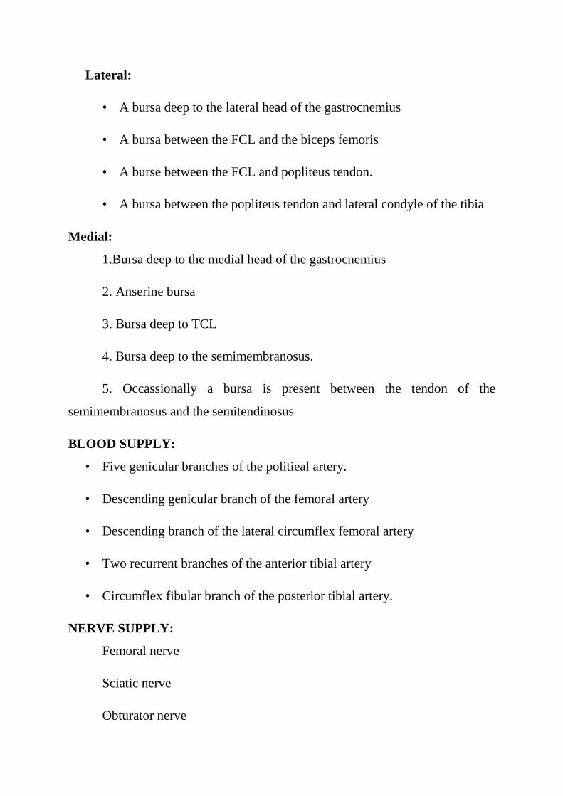
Lateral:
• A bursa deep to the lateral head of the gastrocnemius
• A bursa between the FCL and the biceps femoris
• A burse between the FCL and popliteus tendon.
• A bursa between the popliteus tendon and lateral condyle of the tibia
Medial:
1.Bursa deep to the medial head of the gastrocnemius
2. Anserine bursa
3. Bursa deep to TCL
4. Bursa deep to the semimembranosus.
5. Occassionally a bursa is present between the tendon of the
semimembranosus and the semitendinosus
BLOOD SUPPLY:
• Five genicular branches of the politieal artery.
• Descending genicular branch of the femoral artery
• Descending branch of the lateral circumflex femoral artery
• Two recurrent branches of the anterior tibial artery
• Circumflex fibular branch of the posterior tibial artery.
NERVE SUPPLY:
Femoral nerve
Sciatic nerve
Obturator nerve

ARTHRITIS OF KNEE
Osteoarthritis(Osteo arthritis) is the most common form of knee arthritis.
Osteo arthritis is usually a slowly progressive degenerative disease in which the
joint cartilage gradually wears away. It most often affects middle-aged older
people.
EPIDEMIOLOGY AND RISK FACTORS
Osteo arthritis is the most common joint disease of humans. Among the
elderly, knee Osteo arthritis is the leading cause of chronic disability in
developed countries. Radiographic evidence of knee Osteo arthritis and
especially symptomatic knee Osteo arthritis, is more common in women than in
men.
Age is the most powerful risk factor for Osteo arthritis. In a radiographic
survey of women less than 45 years old, only 2% had Osteo arthritis; between
the ages of 45 to 64 years, however the prevalence was 30% and for those older
than 65 years it was 68%. In males, the figures were similar but somewhat
lower in the older age groups.
Major trauma and repetitive joint use are also important risk factors for
Osteo arthritis. Anterior cruciate ligament insufficiency or meniscus damage
and meniscetomy may lead to knee Osteo arthritis. Although damage to the
articular cartilage may occur at the time of injury or subsequently, with use of
the affected joint, even normal cartilage will degenerate if the joint is unstable.
TYPES OF Osteo arthritis
Osteoarthritis can be broadly grouped as follows:
*Primary osteoarthritis when there is no previous pathology.

*Secondary osteoarthritis when it is secondary to some previous
pathology.
Primary Osteoarthritis
Primary osteoarthritis is due to the wear and tear changes occurring in old
age in which weight bearing joints like the hips and knees are more commonly
affected. It is uncommon in non-weight bearing joints like the shoulder and
elbow.Obesity is a predisposing factor.
Osteoarthritis is a progressive process affecting the articular cartilage of
aging joints. It is characterised by focal degeneration of the articular cartilage.
As the articular cartilage is cyclically loaded during movements of joints, it
undergoes fatigue failure leading to fragmentation of the surface and fibrillation.
In the later stage, the cartilage gets completely eroded, exposing the
sclerosed(eburnated) bone and subchondral cysts are also formed.
The bone undergoes reactive hypertrophy forming peripheral osteophytes.
The synovial membrane undergoes hyperemia and reactive inflammatory
thickening. As there is no destructive pathology, the joint dose not get
ankylosed.
Secondary Osteoarthritis
Secondary osteoarthritis is due to an abnormal wear and tear in a joint,
caused by mechanical incongruity of the articular surfaces. This incongruity
may be the result of a preceding fracture involving the articular surface or
partial destruction or deformity due to a previous disease.

Aetiology
The causes of osteoarthritis include the following:
• Endocrine: People with diabetes may prone to osteoarthritis. Other
endocrine problems also may promote development, including
acromegaly, hypothyroidism, hyperparathyroidism and obesity.
• Post traumatic: Traumatic causes can be further divided into
macrotrauma. An example of macrotrauma is an injury to the joint such
as bone break, causing the bones to line up improperly, lose stability or
damage cartilage. Microtrauma may occur chronically. An example of
this would be repetitive movements or the overuse noted in several
occupations.
• Inflammatory joint diseases: This category would include infected joints,
chronic gout and rheumatoid disease.
• Metabolic: Diseases causing errors of metabolism may cause
osteoarthritis. Examples include Paget’s disease and Wilson disease.
• Congenital or Developmental: Abnormal anatomy such as unequal leg
length may be a cause of osteoarthritis.
• Genetic: A genetic defect may promote breakdown of the protective
architecture of cartilage. Examples include collagen disturbances such as
Ehlers-Danlos syndrome.
• Neuropathic: Diseases such as diabetes can cause nerve problems.
• Other: Nutritional problems may cause osteoarthritis. Other diseases such
as hemophilia and sickle cell are further examples.

PATHOGENESIS
The main load on articular cartilage- the major target tissue Osteo
arthritis is produced by contraction of the muscles that stabilize or move the
joint.
Although cartilage is an excellent shock absorber in terms of its bulk properties,
at most sites it is only 1 to 2 mm thick- too thin to serve as the sole shock-
absorbing structure in the joint.Additional protective mechanisms are provided
by subchondral bone and periarticular muscles.
Articular cartilage serves two essential functions within the joint, both of
which are mechanical. First, it provides a remarkably smooth bearing surface,
so that the bones glide effortlessly over each other with joint movement. With
synovial fluid as lubricant, the coefficient of friction for cartilage rubbed against
cartilage even under physiologic loading, is 15 times lower than that of two ice
cubes passed across each other. Second, articular cartilage prevents the
concentration of stresses, so the bones do not shatter when the joint is loaded.
Osteo arthritis develops in either of two settings:
• The biomaterial properties of the articular cartilage and subchondral bone
are normal, but excessive loading of the joint causes the tissues to fail, or
• The applied load is reasonable, but the material properties of the cartilage
or bone are inferior.
Although articular cartilage is highly resistant to wear under conditions of
repeated oscillation,repetitive impact loading soon leads to joint failure. This
fact accounts for the high prevalence of Osteo arthritis at specific sites related to
vocational or avocational overloading. In general, the earliest changes occur at
the sites in the joint that are subject to the greatest compressive loads.
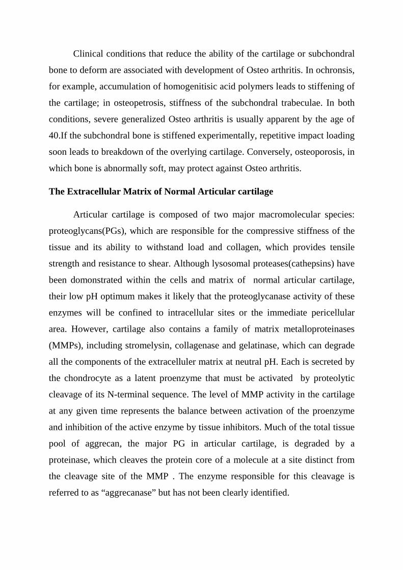
Clinical conditions that reduce the ability of the cartilage or subchondral
bone to deform are associated with development of Osteo arthritis. In ochronsis,
for example, accumulation of homogenitisic acid polymers leads to stiffening of
the cartilage; in osteopetrosis, stiffness of the subchondral trabeculae. In both
conditions, severe generalized Osteo arthritis is usually apparent by the age of
40.If the subchondral bone is stiffened experimentally, repetitive impact loading
soon leads to breakdown of the overlying cartilage. Conversely, osteoporosis, in
which bone is abnormally soft, may protect against Osteo arthritis.
The Extracellular Matrix of Normal Articular cartilage
Articular cartilage is composed of two major macromolecular species:
proteoglycans(PGs), which are responsible for the compressive stiffness of the
tissue and its ability to withstand load and collagen, which provides tensile
strength and resistance to shear. Although lysosomal proteases(cathepsins) have
been domonstrated within the cells and matrix of normal articular cartilage,
their low pH optimum makes it likely that the proteoglycanase activity of these
enzymes will be confined to intracellular sites or the immediate pericellular
area. However, cartilage also contains a family of matrix metalloproteinases
(MMPs), including stromelysin, collagenase and gelatinase, which can degrade
all the components of the extracelluler matrix at neutral pH. Each is secreted by
the chondrocyte as a latent proenzyme that must be activated by proteolytic
cleavage of its N-terminal sequence. The level of MMP activity in the cartilage
at any given time represents the balance between activation of the proenzyme
and inhibition of the active enzyme by tissue inhibitors. Much of the total tissue
pool of aggrecan, the major PG in articular cartilage, is degraded by a
proteinase, which cleaves the protein core of a molecule at a site distinct from
the cleavage site of the MMP . The enzyme responsible for this cleavage is
referred to as “aggrecanase” but has not been clearly identified.

The turnover of normal cartilage is effected through a degradative
cascade, for which many investigators consider the drivingforce appears to be
interleukin(IL) 1, a cytokine produced by mononuclear cells (including synovial
lining cells) and synthesized by chondrocytes. IL-1 stimulates the synthesis and
secretion of the latent MMPs and of tissue plasminogen activator. Plasminogen,
the substrate for the latter enzyme, may be synthesized by the chondrocyte or
may enter the cartilage from the synovial fluid. Both plasminogen and
stromelysin may play a role in activation of the latent MMPs. In addition to its
catabolic effect on cartilage, IL-1, at concentrations even lower than those
needed to stimulate cartilage degradation, suppresses PG synthesis by the
chondrocyte, inhibiting matrix repair.
The balance of the system lies with at least two inhibitors, tissue inhibitor
of metalloproteinase(TIMP) and plasminogen activator inhibitor-1 (PAI-1),
which are synthesized by the chondrocyte and limit the degradative activity of
MMPs and plasminogen activator, respectively. If TIMP or PAI-1 is destroyed
or is present in concentrations that are insufficient relative to those of active
enzymes, stromelysin and plasmin are free to act on matrix substrates.
Stromelysin can degrade the protein core of the PG and activate latent
collagense. Conversion of latent stromelysin to an active, highly destructive
protease by plasmin provides a second mechanism for matrix degradation.
Polypeptide mediators, e.g., insulin-like growth factor-1(IGF-1) and
transforming growth factor (TGF-β), stimulate biosynthesis of PGs.They
regualate matrix metabolism in normal cartilage and may play a role in matrix
repair in Osteo arthritis. Notably, these growth - modulate catabolic as well as
anabolic pathways of chondrocyte metabolism; by down-regulating chondrocyte
receptors for IL-1, they may decrease PG degradation.

In addition to its responsiveness to cytokines and a variety of biologic
mediators, chondrocyte metabolism in normal cartilage can be modulated
directly by mechanical loading. Whereas static loading and prolonged cyclic
loading inhibit synthesis of PGs and protein, loads of relatively brief duration
may stimulate matrix biosynthesis.
Pathophysioloy of Cartilage Changes in Osteo arthritis
The primary changes in Osteo arthritis begin in the cartilage. A change in
the arrangement and size of the collagen fibers is apparent. Biochemical data are
consistent with presense of a defect in the collagen network of the matrix,
perhaps due to disruption of the “glue” that binds adjacent fibers.This is among
the earliest matrix changes observed and appears to be irreversible.
Although“wear” may be a factor in the loss of cartilage, strong evidence
supports the concept that lysosomal enzymes and MMPs account for much of
the loss of cartilage matrix in Osteo arthritis. Whether their synthesis and
secretion or stimulated by IL-1 or by other factors(e.g., mechanical stimuli),
MMPs, plasmin, and cathepsins all appear to be involved in the break down of
articular cartilage in Osteo arthritis. TIMP and PAI-1 may work to stabilize the
system, at least temporarily, while growth factors, such as IGF-1, TGF-β, and
basic fibroblast growth factor, are implicated in repair processes they may heal
the lesion or, at least, stabilize the process. A stoichiometric imbalance exists
between the levels of active enzyme and the level of TIMP, which may be only
modestly increased.
The possible role of nitric oxide (NO) in articular cartilage damage in
Osteo arthritis, since NO has been shown to stimulate synthesis of MMPs by
chrondrocytes. Chondrocytes are a major source of NO, synthesis of which is
stimulated by IL-1 and tumor necrosis factor and by shear stresses on the tissue.

In an experimental model of Osteo arthritis, treatment with a selective inhibitor
of inducible NO synthase reduced the severity of cartilage damage.
The chondrocytes in Osteo arthritis cartilage undergo active cell divison
and very active metabolically, producing increased quantities of DNA,RNA
collagen, PG and noncollagenous proteins. Prior to cartilage loss and PG
depletion, this marked biosynthetic activity may lead to an increase in PG
concentration, which may associated with thickening of the cartilage and stage
of homeostasis referred to as “compensated” Osteo arthritis. These mechanisms
may maintain the joint in a reasonably functional state for years. The repair
tissue, however, often does not hold up as well under mechanical stresses as
normal hyaline cartilge end eventually, at least in some cases, the rate of PG
synthesis falls off and “end-stage” Osteo arthritis develops, with full-thickness
loss of cartilage.
CHANGES OF NORMAL CARTILAGE TO AGING CARTILAGE:
In normal articular cartilage aggrecan contains numerous GAG which is
capable of retaining water.
Several structural and biochemical changes involving the non collagenous
component of the matrix occur during aging. These changes alter biochemical
properties of the cartilage that are essential for the distribution of forces in the
weight bearing zone.
Glycosaminoglycans are modified qualitatively; they become shorters as
the cartilage ages. The concentration of kertain sulphate increase during aging.
These quantitative and qualitative changes in proteologlycans reduce the
capacity of the molecules to retain water. Thus aging cartilage contains less
water, which alters the biochemical properties of the cartilage. Fissures that
develop aging are due to stress fractures of the collgen network.

CLINICAL FEATURES
The joint pain of Osteo arthritis is often described as a deep ache and is
localized to the involved joint. Typically, the pain of Osteo arthritis is
aggrevated by joint use and relieved by rest, but as the disease progresses, it
may become persistent. Nocturnal pain , interfering with sleep, is seen
particularly in advanced Osteo arthritis of the hip and may be enervating.
Stiffness of the involved joint upon arising in the morning or after a period of
inactivity (e.g.,a night’s sleep ,an automobile ride) may be prominent but
usually lasts less than 20 min. Systemic manifestations are not a feature of
primary Osteo arthritis.
Because articular cartilage is aneural, the joint pain in Osteo arthritis must
arise from other structures . In some cases it may be due to stretching of nerve
endings in the periosteum covering osteophytes; in others, to microfractures in
subchondral bone or from medullary hypertension caused by distortion of blood
flow by thickened subchrondral trabecule. Joint instability, leading to stretching
of the joint capsule and muscle spasm may also be sources of pain.
In some patients with Osteo arthritis, joint pain may be due to synovitis.
In advanced Osteo arthritis, histologic evidence of synovial inflammation may
be as marked as that in the synovium of a patient with rhumatoid arthritis.
Synovitis in Osteo arthritis may be due to phagocytosis of shards of cartilage
and bone from the abraded joint surface (wear particles), to release from the
cartilage of soluble matrix macromolecules, or to crystals of calcium
pyrophosphate or hydroxyapatite. In other cases, immune complexes, containing
antigens derived from cartilage matrix, may be sequestered in collagenous tissue
of the joint, leading to low grade chronic synovitis. In contrast, in the earlier
stages of Osteo arthritis, even in the patient with chronic joint pain, synovial
inflammation may be absent, suggesting that the joint pain is due to one of the
other factors mentioned above.
Physical examination of the Osteo arthritis joint may reveal localized
tenderness and bony or soft tissue swelling. Bony crepitus (the sensation of
bone rubbing against bone, evoked by joint movement) is characteristic.
Synovial effusions, if present, are usually not large. Palpation may reveal some
warmth over the joint. Periarticular muscle atrophy may be due to disuse or
reflex inhibition of muscle contraction. In the advanced stages of Osteo arthritis,
there may be gross deformity, bony hypertrophy, subluxation, and marked loss
of joint motion. In many patients the disease stablizes; in some, regression of
joint pain and even of radiographic changes occurs.
Although the diagnosis of Osteo arthritis is often straightforward because
of the high prevalence of radiographic changes of Osteo arthritis in
asymptomatic individuals, it is important to ensure that joint pain in a patient
with radiographic evidence of Osteo arthritis is not due to some other cause,
such as soft tissue rheumatism (e.g., anserine bursitis at the knee, trochanteric
bursitis at the hip), radiculopathy, referral of pain from another joint (e.g., 25%
of patients with hip disease have pain referred to the knee), entrapment
neuropathy, vascular disease(claudication), or some other type of arthritis (e.g.,
crystal-induced synovitis, septic arthritis). It is usually not difficult to
differentiate Osteo arthritis from a systemic rheumatic diseases, joint
involvement is rhumatroid arthritis, because, in the latter diseases, joint
involvement is usually symmetric and polyarticular, with arthritis in wrists and
metacarpophalangeal joints (which are generally not involved in Osteo
arthritis),and there are also constitutional features such as prolonged morning
stiffness, fatigue, weight loss or fever.

Causes of joint pain in patients with Osteo arthritis
Source mechanism
Synovium inflammation
Subchondral bone medullary hypertension,
microfractures
Osteophyte Stretching of periosteal nerve
endings
Ligaments stretch
Capsule Inflammation, distention
Muscle spasm
Diagnosis
Imaging
• X-rays:
Approximately one-third of people with osteoarthritis on X-rays have
symptoms such as pain or swelling. X-rays can show narrowing of the space
between the joint (articular surface), osteophytes, cyst formation, and hardening
of the underlying bone. Scoring systems have been used to assess the extent of
the bony changes on X-rays. Separate scoring systems for the different joints
have been studied and found to be predictive of disease status. An important
finding from these studies was that the presence of osteoarthritis of the hands
was a predictive sign of deterioration of the knee joint.

• MRI :
This study is complex, noninvasive imaging technique that unlike X-
rays. X-rays provide information mainly bones. However, MRI is capable of
visualizing all structures within the joint.
• CT scan :
This study may be used to image a joint. CT scanning mainly provides
information on the bony structures of the joint but in greater detail than plain X-
rays.
• Joint Fluid Analysis :
Fluid may be extracted from the knee with a needle and syringe when
the diagnosis is uncertain or if an infection is suspected.
Blood Tests :
No currently accepted blood test marker for this disease exists. Blood
tests may be drawn in cases in which infection is suspected.
Osteoarthritis Differential Diagnosis
• Rhumatoid arthritis
• Spondyloarthropathy
• Chondrocalcinosis
• Joint trauma
• Metabolic Bone Disorders
• Hypermobility syndromes
• Neuropathic diseases
The following should also be considered in the differential diagnosis:
• Crystal deposition disease
• Pseudogout
• Inflammatory arthritis
• Seronegative spondyloarthropathies
• Infected joint
• Underlying mechanical pain
• Reactive arthritis
Differential Diagnoses :
• Abdominal Aortic Aneurysm Imaging
• Ankylosing Spondylitis
• Avascular Necrosis
• Calcium Pyrophosphate Deposition Disease
• Imaging in Neuropathic Arthropathy(Charcot joint)
• Lyme Disease
• Patellofemoral Arthritis
• Patellofemoral Syndrome
• Prepatellar Bursitis
• Psoriatic Arthritis
• Rhinosporidiosis

Osteoarthritis Treatment:
Self-Care at Home
Lifestyle changes may delay or limit osteoarthritis symptoms. These are
common home remedies:
Weight loss: One study suggested that, for women, weight loss may reduce
the risk for osteoarthritis in the knee.
Exercise: Regular exercise may help to strengthen the muscles and
potentially stimulate cartilage growth. Avoid high-impact sports. The
following types of exercise are recommended:
Range of motion,
Strengthening
Aerobic
Diet: While there is no specific osteoarthritis diet, supplements of
antioxidant vitamins C and E may provide some protection. Vitamin D and
calcium are recommended daily. Dose of calcium is 1000-1200 mg per day.
The current guideline for vitamin D is 400 IU per day.
Heat: Hot soaks and warm wax(paraffin)application may relieve pain.
Orthoses: These assistive devices, such as or neck braces and knee braces,
are used to improve function of movable parts of the body or to support,
align, prevent, or correct deformities. Splints or braces help with joint
alignment and weight redistribution. Other examples include walkers,
crutches or canes and orthopedic footwear.
Over-The-Counter(OTC)medications
Acetaminophen(tylenol) is the first drug recommended for osteoarthritis.

Nonsteroidal anti-inflammatory drugs (NSAIDs) are commonly used for
arthritis pain.These include aspirin, ibuprofen(Motrin or Advil),
naproxen(Aleve), and ketoprofen (Orudis).
Newer OTC preparations include chondrotin and glucosamine sulfate, which
are natural substances found in the joint fluid. Chondroitin is thought to
promote an increase in the making of the building blocks of
cartilage(collagen and proteoglycans) as well as having an anti-inflammatory
effect. Glucosamine may also stimulate production of the building blocks of
cartilage as well as being an anti-inflammatry agent. Glucosamine was found
to increase blood sugar in animal studies, so people with diabetes should
consult their doctor first.
Arthritis self-help course: The Arthritis Foundation offers an educational
program on the causes and treatment of arthritis. Exercise, nutrition,
relaxation and pain management programs are covered as well as ways to
communicate with your doctor. Completion of the program reduced pain by
20% and doctor visits by 40%.
Medical Treatment: The overall goal of treatment is early elimination of
risk factors, early diagnosis and surveillance of the disease and appropriate
treatment of pain. It’s also important to help people regain their mobility.
Treatment These goals may be reached through a logical approach to care
including the overlapping of treatment that does not involve medications and
treatment with medication and possibly surgical management.
Treatment that does not involve medications includes education, physical
and occupational therapy, weight reduction, exercise and assistive
devices(orthoses).

Surgical Treatment
Joint replacement surgery should be reserved for patient with advanced
Osteo arthritis in whom aggressive medical management has failed. In such
cases total joint arthroplasty may be remarkable effective in relieving pain
and increasing mobility.
Osteotomy, which is surgically more conservative, can eliminate
concentration of peak dynamic loading and may provided effective pain
relief in patients with knee Osteo arthritis. It is of greatest benefit when the
disease is only moderately advanced. Arthroscopic removal of loose
cartilage fragments can prevent locking and relieve pain.
Chondroplasty has also had some popularity as treatment of Osteo
arthritis, but well-controlled studies of its efficacy are lacking , the
fibrocartilage that resurfaces the abraded bone is inferior to normal hyaline
cartilage in its ability to withstand mechanical loads. In patients who had
undergone tibial osteotomy for medial compartment knee Osteo
arthritis,knee pain and function were not related to the extent of cartilage
regeneration 2 years later.
Autologous chondrocyte transplantation and attempts at cartilage repair
using mesenchymal stem cells and autologous osteochondral plugs are
currently being used experimentally for repair of focal chondral defects, but
have not proved to be effective in treatment of Osteo arthritis.
Management of Osteoarthirits
Simple changes around the home and daily activities cause dramatic
improvement in the symptomatology of osteoarthritis. The following are some
of the measures:

• Use of higher chair which require less effort to get in and get out should
be considered
• Changes to be made in the bathroom :
• Use of Western toilets and avoding the Indian type
• To fit the bath aids to facilitate easy getting in getting out of a bath.
• To fit railings next to the toilet and bath to facilitate ease of
movement.
• Patients are advise to climb the stairs leading the good leg taking one stair
at a time and to descend the stairs leading with the bad leg, again taking
one stair at a time
• To reduce the force acting across the injured joint patients advised to use
a walking stick which acts as a third limb. The stick should be held in the
hand opposite to the affected hip or knee. Initially it should be used
around the home. The top of the stick should come up to the wrist when
the patient stands and the tip should be provided with a firm rubber to
avoid slipping. A walking stick, by providing a third limb through which
forces can be transmitted, enables the reduction of force can be
transmitted, enables the reduction of force across the injured joint from
peak values of 5 to 1.5 times the body weight.
• Footwear with hard soles and high heels should be avoided.
• Cars with raised platforms and seats which facilitate easy getting in and
getting out should be used.
• If the patients are overweight, reduction in the weight helps to reduce the
load on the joints.

EXERCISE FOR KNEE PAIN

• General:
• Keep as upright as possible as this helps to put equal weight on
both the legs.
• Avoid sitting on a low or soft chair
• Avoid curling up in bed.
• To stretch the front of the thigh and hip, lie on the stomach at least
once a day for five minutes to thirty minutes.
• To use a walking stick when walking inside or outside the house
• To avoid uneven and rough ground or surfaces while walking.
• To wear comfortable footwears.
• Avoid squatting on the ground.
Aims of the exercises in osteoarthritis knee
• To increase the range of movements
• To increase stability and shock absorption
• To prevent deformity
• To improve posture.
• To reduce pain and stiffness.
Rules of the exercises
• Build up the exercises gradually
• Avoid rough ground while exercising
• To take warm baths before starting the exercises.

• To perform the exercises 20 times each twice a day and later four
times a day.
Types of Exercises in osteoarthritis of knee
Exercises lying on the Back
• Pelvic tilt: Tighten the thigh and relax
• Pelvic lift :Bend both the knees up, push on the feet and lift, old for a
count of five and relax
• Leg stretch: Push one leg along the floor as through you are trying to
make it longer than the other. Hold for a count of five and then repeat
with the other leg.
• Alternate leg raising: Keeping the knees straight, lift alternate legs six
inches from the ground
Exercises Lying on your side, with the Painful Hip up
• Side leg raising: Keep the top leg straight and lift it up as high as
possible, hold for a count of five and relax.
• Knee and hip flexion: Bend hip and knee of the top leg forwards,
and hold for a count of five. Then straighten the leg and stretch
backwards as fas as it will go, hold for a count of five, then relax
Exercises in Sitting Posture
• Knees together, feet apart: Keep the knees together and move the feet
apart, hold for a count of five then relax.
• Feet together, knees apart :Keep the ankles together and move the kness
apart, then relax

Exercises in standing Posture
• Standing leg swing: Hold into a table or chair with one hand, swing one
leg forward and backward. Try to get the backwards swing as wide as
possible.
• Standing side leg swing: Hold on to a chair with both hands. Swing bad
out as far as it will go and then in. The outward swing is the hardest part
and the leg should be allowed to fall back under muscular control.

MATERIALS AND METHODS
To study on clinical evaluation of the disease “AZHL KEEL VAYU”
with the drug KARUNGOZHI CHOORANAM (INTERNAL) and
VIDAMUTTI THYLAM (EXTERNAL) was carried out in Postgraduate
Sirappu Maruthuvam, Government Siddha Medical College, Palayamkottai. 20
patients of both male and female were selected for the studies and admitted in In
patient ward for among 20 IP patients, 15 IP patients will be given massage and
varmam treatment along with internal medicine and remaining 5 IP patients will
be given massage fomentation without internal medicine.
Another 20 patients are treated with trial drug in the outpatient ward.
SELECTION OF PATIENTS:
INCLUSION CRITERIA:
• Age – 40 – 65 Years
• Sex - Both male and female
• Patients having symptoms of arthritis of both knee joints, swelling,
stiffness, crepitation, restricted movements of both knee joints.
• Patients who are willing to undergo radiological investigation and give
blood for laboratory investigation.
• Patient willing to sign the informed consent stating that he/she will
consciously stick to the treatment during 48 days but can opt out of the
trial of his / her own conscious discretion.

EXCLUSION CRITERIA:
• Cardiac disease
• Diabetes mellitus
• Hypertension
• Rheumatoid arthritis
• Pregnancy and lactation
• History of trauma
• Tuberculosis
• Use of narcotic drugs
• Neurological disorder
• Patients with any other serious illness.
STUDY OF CLINICAL DIAGNOSIS:
A case sheet is prepared on the basis of siddha and modern method to
diagnose the disease and individual case sheet is maintained for each patient.
SIDDHA DIAGNOSTIC TOOLS:
• Poriyal arithal
• Pulanal arithal
• Vinathal
• Mukkutram
• Ezhu udal thathugal
• Envagai Thervu
• Thinaigal
• Paruva kalangal
• Thega nilai

LABORATORY INVESTIGATIONS:
Blood Urine
TC Albumin
DC Sugar
ESR Deposit
Hb
Aso titre
Radiological investigations:
X- ray of the knee joints (AP and Lat view) Improvement assessed by
following assessments.
Administration of trial medicine
The trial drug was prepared carefully according to the siddha literature
and given to 40 patients three times a day.
The Biochemical analysis was performed in Biochemical laboratory.
• The pharmacological analysis of trial drug for its analgesic, Acute anti-
inflammatory, chronic Anti-inflammatory was performed in
pharmacological laboratory.
• Observation of patients with signs and symptoms of the disease and their
prognosis were noted.
• Patients also advised to given hot water fomentation, some exercise for
better prognosis.

ASSESSMENT OF PROGNOSIS:
1. Clinical Assessement:
• Pain and swelling in both knee joints
• Stiffness in both knee joint
• Crepitation in joint line, medial condyle
• Tenderness in joint line, medial condyle of knee joint
• Warmth
• Periarticular atrophy
• Restricted movements of both the knee joints.
2. Radiological Assessment:
X-ray of the both knee (AP view and lateral view)

RESULTS AND OBSERVATION
Results of the study were observed with respect to the following criteria.
1. Sex Distribution
2. Age Distribution
3. Kaalam (Life span)
4. Paruva Kaalam (seasonal changes)
5. Thinai ( 5 land types)
6. Duration of illness
7. Occupational status
8. Diet
9. Onset of Disease
10. Socio economic status
11. Clinical features
12. Radiological findings
13. Disturbances in Vatham
14. Disturbance in Pitham
15. Disturbance in Kapham
16. Udal thathukkal
17. Envagai thervugal
18. Yakkai illakanam
19. Assessment of the Effect of therapy

1. Age Distribution:
S.No Age No.of Cases
Percentage
1. 41-45 4 10%
2. 46-50 5 12.5%
3. 51-55 4 10%
4. 56-60 12 30%
5. 61-65 7 17.5%
6. 66-70 7 17.5%
7. 71-75 1 2.5%
Most of the cases were above the age group of 55.
10%
12.50%
10%
30%
17.50% 17.50%
2.50%
0%
5%
10%
15%
20%
25%
30%
35%
41-45 46-50 51-55 56-60 61-65 66-70 71-75
Series 1
Series 2
Column1

2. Sex Distribution:
S.No Among Sex
No.of Cases
Percentage
1. Male 22 55%
2. Female 18 45%
.
Among 40 cases 22(55%) were males and 18(45%) were females.
55%
45%Male
Female

3. Kaalam:
Kaalam No.of Cases Percentage
1. Vadham (upto 33 yrs to 66 yrs)
- -
2. Pittham (33 yrs to 66 yrs)
32 80%
3. Kabam (above 66 yrs)
8 20%
Most of the cases 80% were in Pittha Kaalam and the rest were reported in Kaba Kaalam.
0%
80%
20%
0%
10%
20%
30%
40%
50%
60%
70%
80%
90%
Vadham Pittham Kabam

4. Paruva Kaalam:
S.No Paruvakaalam No.of Cases Percentage
1. Kaar (Aaavani, Purattasi)
(Aug 16 – Oct 15)
18 45 %
2. Koothir (Iypasi, Karthigai)
(Oct 16 – Dec 15)
7 17.5%
3. Munpani (Margali, thai)
(Dec 16 – Feb 15)
- -
4. Pinpani (Masi, Pankuni)
(Feb 16 – April15)
- -
5. Elavenil (Chithirai, Vaikasi)
(Apr 16 – June 15)
- -
6. Muthuvenil (Aani, Aadi)
(June 16 – Aug 15)
15 37.5%
The maximum incidence of Azhal Keelvayu was during the Kaar
Kaalam,Muthuvenil Kaalam and Koothir Kaalam.
0%
5%
10%
15%
20%
25%
30%
35%
40%
45%
Kaar Kaalam
Koothir Kaalam
Munpani Kaalam
Pinpani Kaalam
Elavenil Kaalam
Muthuvenil Kaalam
45%
17.50%
0% 0% 0%
37.50%
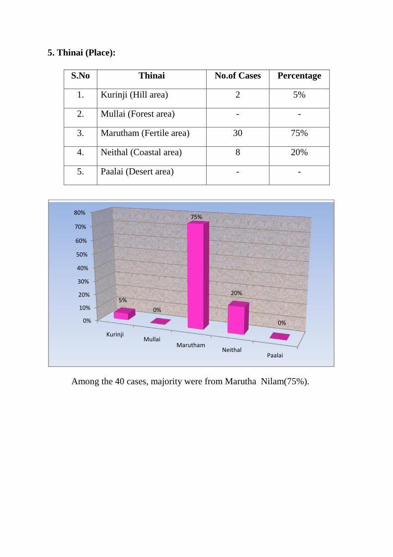
5. Thinai (Place):
S.No Thinai No.of Cases Percentage
1. Kurinji (Hill area) 2 5%
2. Mullai (Forest area) - -
3. Marutham (Fertile area) 30 75%
4. Neithal (Coastal area) 8 20%
5. Paalai (Desert area) - -
Among the 40 cases, majority were from Marutha Nilam(75%).
0%
10%
20%
30%
40%
50%
60%
70%
80%
KurinjiMullai
MaruthamNeithal
Paalai
5%
0%
75%
20%
0%

6. Duration of Illness:
S.No Duration (Months) No.of.Cases Percentage
1. 0-3 8 20%
2. 3-6 5 12.5%
3. 6-12 13 32.5%
4. 12-24 12 30%
5. 24-36 2 5%
Among the 60 cases, most of them had the duration of the illness - upto 1 year.
20%17.50%
32.50%30%
0%
5%
10%
15%
20%
25%
30%
35%
0-3 Duration (months) 3-6 Duration (months) 6-12 Duration (months)
12-24 Duration (months)

7. Occupational status:
Occupational status shows Manual labours and Ryot were more affected.
0%5%
10%15%20%25%30%35%40%45%
RyotManual Labour Teachers
Home Makers
45%
25%
5%
25%
S.No Nature of Work No.of Cases Percentage
1. Ryot 18 45%
2. Manual labour 10 25%
3. Home Makers 10 25%
4. Teachers 2 5%

8. Diet Reference:
According to this study 100% of cases were reported as Nonvegetarian.
0%
10%
20%
30%
40%
50%
60%
70%
80%
90%
100%
Non Veg Veg
100%
0%
S.No Diet Habit No. of Cases Percentage
1. Vegetarian - -
2. Non vegetarian 40 100%

9. Mode of Onset:
S.No Mode of onset No. of Cases Percentage
1. Acute - -
2. Gradual 40 100%
According to this study 100% of cases were reported gradual onset of
disease.
0%
5%
10%
15%
20%
25%
30%
35%
40%
Acute Gradual
0%
40%

10.The Socio – Economic Status:
S.No Socio – economic status No. of Cases Percentage
1. Poor 34 85%
2. Middle class 4 10%
3. Rich 2 5%
According to this study 85% of the cases were poor socio – economic status,
10% cases were from middle class families and only
5 % from rich background.
0
5
10
15
20
25
30
35
Poor Middle Class Rich
85%
10%5%

11. Clinical Features of patients:
S.No Signs and Symptoms No.of Cases Percentage
1. Pain, tenderness 40 100%
2. Swelling 22 55%
3. Morning stiffness 5 12.5%
4. Crepitations 40 100%
5. Muscle Wasting 1 2.5%
6. Deformity 4 10%
7. Constipation 6 15%
8. Loss of appetite 4 10%
9. Limited Movements 34 85%
Among the twenty cases all of them (100%) had pain, tenderness and
crepitations. 5 patients (12.5%) had morning stiffness. 15% of the patients had
constipation and 85% had painful limited movements.
100% 55% 25% 100% 2.50% 10% 15% 10% 85%0
5
10
15
20
25
30
35
40
45

12. Radiological Findings in patients
S.No Findings No.of Cases Percentage
1. Joint space narrowing 40 100%
2. Inter condylar notch prominence 5 12.5%
3. Presence of Osteophytes 22 55%
4. Subchondral Sclerosis 20 50%
5. Osteoporosis 5 12.5%
6. Deformity 5 12.5%
7. Soft tissues swelling 2 5%
Joint Space narrowed in all cases (100%), Osteophytes present in 22 cases
(55%).
0%
20%
40%
60%
80%
100%100%
12.50%
55%50%
12.50% 12.50%5%

13. Distubrances of Vatha:
Table showing the derangement of vatham:
S.No Vatham No.of Cases Percentage
1. Pranan - -
2. Abanan 32 80%
3. Viyanan 40 100%
4. Uthanan - -
5. Samanan 40 100%
6. Naagan - -
7. Koorman 2 5%
8. Kirukaran - -
9. Devethathan 35 87.5%
10. Dhananjeyan - -
• Both Viyanan ans Samanan were affected in all the 40 cases (100%)
• Abanan was affected in 32 cases (80%).
• Devethathan was affected in 35cases (87.5%)
• Koorman was affected in 2 cases (5%)
0%
10%
20%
30%
40%
50%
60%
70%
80%
90%
100%
0%
80%
100%
0%
100%
0%5%
0%
87.50%
0%

• 14.Distubrances in Pitham:
S.No Pitham No.of Cases Percentage
1. Analapitham 10 25%
2. Ranjagam 6 15%
3. Saathagam 40 100 %
4. Prasagam - -
5. Alosagam - -
Saathaga Pitham was affected in all 40 cases (100%)
Analapitham was affected in 10 cases (25%)
Ranjagam was affected in 6 cases (15%)
0% 20% 40% 60% 80% 100%
Analapitham
Ranjagam
Saathagam
Prasagam
Alosagam

15. Table showing the derangement of Kapham:
S.No Kapham No.of Cases Percentage
1. Avalambagam - -
2. Kilethagam 10 25%
3. Pothagam - -
4. Tharpagam - -
5. Santhigam 40 100%
In all the 60 cases (100%) Santhigam was affected.Kilethagam was affected in 6
cases (10%)
0%
25%
0% 0%
100%
0%
20%
40%
60%
80%
100%
120%
Avalamlagam Kilethagam Pothagam Tharapagam Santhigam

16.Table Showing the condition of Udal Kattugal:
S.No Udal Kattugal No.of Cases Percentage
1. Saaram 40 100%
2. Senneer 6 15%
3. Oon 2 5%
4. Kozhuppu 40 100%
5. Enbu 40 100%
6. Moolai 40 100%
7. Venneer/Suronitham - -
In all the cases Saaram, Kozhuppu, Enbu and Moolai were affected
(100%),Senneer was affected in 6cases (15%) and Oon was affected in 2 cases
(5%)
0%
20%
40%
60%
80%
100%
120%
140%
160%
180%
200%
100%
15%5%
100% 100% 100%
2

17.Ennvagai Thervugal:
S.No Ennvagai thervugal No. of Cases Percentage
1. Sparisam 40 100%
2. Naa 10 25%
3. Niram 10 25%
4. Mozhi - -
5. Vizhi 2 5%
6. Malam 32 80%
7. Moothiram 2 5%
8. Naadi 40 100%
Naadi - Pitha Vatham 13 cases (32.5%)
Vatha Pitham 25 cases (62.5%)
Pitham Kapham 2 cases (5%)
0%
20%
40%
60%
80%
100%
120%

18.Yakkai Ilakkanam (Physical Constitutions)
S.No Yakkai Ilakkanam No.of Cases Percentage
1. Vatha udal 2 5%
2. Pitha udal - -
3. Kapha udal 6 15%
4. Thontha udal 32 80%
Out of 40 cases were of Thontha udal. Out 32 cases, 16 pitha vatha udal
and 16 vatha pitha udal.
19. Assessment of Effect of Theraphy:
Marked Effect:
No longer any clinical manifestations
Patient could work and live normally
No recurrence after some months.
Moderate Effect:
Moderate reduction of manifestations.
Slight pain after movement
Mild Effect:
Slight reduction in the clinical manifestation
With relapse
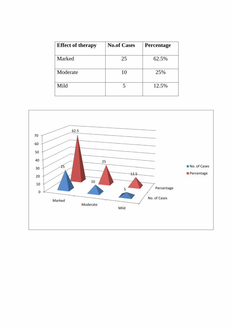
Effect of therapy No.of Cases Percentage
Marked 25 62.5%
Moderate 10 25%
Mild 5 12.5%
No. of Cases
Percentage0
10
20
30
40
50
60
70
MarkedModerate
Mild
25
10
5
62.5
25
12.5
No. of Cases
Percentage

Joint line tenderness (Knee) in Inpatients:
S.No Patient Name Age/Sex Ip.No Before
Treatment in Grades
After Treatment in
Grades
1. Subbiah 70/M 2268 III I
2. Sudaliammal 60/F 2269 III I
3. Lakshmi 75/F 2337 II Nil
4. Veerammal 65/F 2569 II I
5. Gandhimathi 58/F 2723 III I
6. Vadivel 70/M 2933 II I
7. Nainar 70/M 3060 II I
8. Muthusamy 58/M 3082 II Nil
9. Nambi 69/M 3108 III II
10. Nalla thai 65/F 3113 III I
11. Arumugam 56/M 3132 III I
12. Gomu 60/F 3187 II I
13. Muniyandi 68/M 3204 II Nil
14. Shanmugathai 69/F 3362 II Nil
15. Peter 54/M 3367 II I
16. Paul nadar 65/M 3372 II I
17. Saraswathy 60/F 3940 III I
18. Petchiammal 70/F 4352 III I
19. Michelammal 58/F 4361 III II
20. Subbiah 65/M 4441 II I

Assessment of joint tenderness
Grade I : The patient says the joint is tender
Grade II : The patient winces
Grade III : The patient winces and withdraws the affected part
Grade IV : The patient will not allow the joint to be touched.
Joint line tenderness (Knee) in Outpatients:
S.No Patient Name Age/Sex Ip.No Before
Treatment in Grades
After Treatment in
Grades
1. Vadivel 62/M 52172 III I
2. Raja 45/M 52490 II Nil
3. Santha 60/F 52539 III II
4. Velammal 42/F 56218 II Nil
5. Santhi 47/F 56576 III Nil
6. Suresh 40/M 56918 II I
7. Shanmugavel 65/M 57259 III I
8. Kannan 60/M 58001 II I
9. Nainar 60/M 58893 III I
10. Petchiammal 50/F 60777 II Nil
11. Muthulakshmi 40/F 61417 II Nil
12. Lakshmi 60/F 62802 III I
13. Subbulakshmi 57/F 65413 III I
14. Petchiammal 53/F 67171 III I
15. Saraswathy 50/F 72049 II Nil
16. Saraswathy 50/F 74170 III II

17. Thavasi Nadar 70/M 76277 III I
18. Arumugam 55/M 79076 II I
19. Madasamy 65/M 94113 III I
20. Ayishabeevi 47/F 94118 III I
Assessment of joint tenderness
Grade I : The patient says the joint is tender
Grade II : The patient winces
Grade III : The patient winces and withdraws the affected part
Grade IV : The patient will not allow the joint to be touched.

SUBBIAH, Age 60, Male
Nambi, Age 70, Male

ASSESSMENT OF SWELLING IN INPATIENT
S.NO
I.P.NO
NAME
AGE /S
EX
BEFORE TREATMENT AFTER TREATMENT
RT(CM) LT(CM) RT(CM) LT(CM)
Upper
Middle
lowe
r
upper
middle
lowe
r
Upper
middle
lowe
r
upper
Middle
Lower
1 2269
Sudali ammal
60/F
32 34.5 35 31 34 35.5 31 34 34.5 30.5 33 35
2
2569
Veer ammal
65/F
34 35.5 35 33.5 34.5 34 33.5 35 34.5 33.5 35 33.5
3
2933
Vadivel
70/M
34.5 35 35.5 35 36 35.5 34 34.5 35 35 35.5 35
4
3060
Nainaar
70/M
35 34 36 35.5 35 35.5 34.5 33 35.5 35 34.5 35
5
3108
Nambi 69/M
36 34 35 35 33.5 34 36 34 55 33.5 34 36
6
3204
Muiyandi
68/M
37 35 36 35 34 35.5 36 34 35 35 33.5 35
7
3367
Peter 54/M
35 34.5 35 34 33.5 33 34.5 34 34 34 33.5 33
8
3113
Nalla thai
65/F
36 35 34.5 35 34.5 34 35.5 34 33.5 35 34.5 34
9
3132
Arumugam
56/F
36 35 34 38 35.5 36 36 35 34 36 35 34.5
10
3187
Gomu 60/F
35 34 33.5 36 34 35.5 35 34 33.5 35 33.5 35
11 3940
Saraswathi
60/F
36 35 34.5 35 34.5 34 35.5 34 34 35 34.5 34
12 4361
Mikkel ammal
58/F
35 34.5 33 35.5 34 35 34.5 34 33 35 33.5 34.5

Among 20 cases :
Marked Effect - 8 cases
Moderate - 10 cases
Mild - 2 cases
LIST OF OUT PATIENT IN SWELLING
S.NO
O.P NO NAME AGE/SEX
Before treatment After treatment
RT LT RT LT
1 52537 Shratha 60/F 37 35 36 35 34 33 35 34.5 35 35 34 33
2 57259 Shanmuga
vel
65/M 36 35 34.5 35 34 33.5 35.5 34.5 34 34 33.5 33
3 58893 Nainaar 60/M 35 34.5 33.5 36 35 34 35 34.5 33.5 35 34 33
4 72049 Saraswathi 50/F 33.5
33 33.5 35 34.5 35 33.5 33 33.5 34 33.5 34
5 94113 MadasamY 65/M 35 34 33.5 34.5 33 34.5 34.5 33 34 34 33 34
6 94118 Ayisha beevi 47/F 36 35 35.5 37 35.5 35 35.5 34.5 35 36.5 35 34.5
7 61417 Muthu
lakshmi
40/F 35 33.5 34 35 33.5 34 34 33 33.5 35 33.5 34
8 65413 Subbu lakshmi 57/F 36 35 34.5 35 34 33.5 35.5 34 34.5 34.5 33.5 33

OP CASES - CLINICAL IMPROVEMENT
Sl. No
OP NO
NAME AGE SEX DOA DOD TREATED
DAYS RESULT
1 52172 Vadivel 62 M 20.07.12 01.09.12 42 Marked
2 52490 Raja 45 M 25.07.12 08.09.12 43 Marked
3 52539 Shantha 60 F 03.08.2 17.09.12 55 Moderate
4 56218 Velammal 42 F 08.08.12 18.10.12 40 Marked
5 56576 Shanthi 47 F 16.08.12 20.10.12 66 Moderate
6 56918 Suresh 40 M 17.08.12 17.10.12 60 Marked
7 57259 Shanmugavel 65 M 21.08.12 10.01.13 109 Marked
8 58001 Kannan 60 M 09.08.12 09.10.12 60 Moderate
9 58893 Nainaar 60 M 03.08.12 07.11.12 64 Marked
10 60777 Petchiammal 50 F 10.08.12 06.10.12 57 Marked
11 61417 Muthulakshmi 40 F 23.08.12 25.10.12 63 Marked
12 62802 Lakshmi 60 F 25.08.12 12.10.12 47 Moderate
13 65413 Subbulakshmi 57 F 28.08.12 30.12.12 63 Marked
14 67171 Petchiammal 53 F 01.09.12 30.11.12 90 Marked
15 72049 Saraswathi 50 F 05.09.12 28.12.12 83 Moderate
16 74170 Saraswathi 50 F 06.09.12 10.12.12 64 Marked
17 76777 Thavasi Nadar 70 M 11.09.12 01.01.13 53 Moderate
18 79076 Arumugam 55 M 12.09.12 10.01.13 60 Marked
19 94113 Madasamy 65 M 26.10.12 12.01.13 77 Marked
20 94118 Ayisha Beevi 47 F 14.09.12 20.10.12 36 Moderate

IP CASES - CLINICAL IMPROVEMENT
Sl. No
IP NO
NAME AGE SEX DOA DOD TREATED
DAYS RESULT
1 2268 Subbaiah 70 M 23.07.12 22.09.12 62 Moderate
2 2269 Sudaliammal 60 F 23.07.12 29.08.12 35 Mild
3 2337 Lakshmi 75 F 25.07.12 26.08.12 33 Marked
4 2569 Veerammal 65 F 28.07.12 01.09.12 36 Marked
5 2728 Kanthimathi 50 F 10.08.12 15.09.12 36 Marked
6 2933 Vadivel 70 M 15.08.12 22.09.12 38 Moderate
7 3060 Nainaar 70 M 18.08.12 30.09.12 43 Moderate
8 3082 Muthusamy 50 M 20.08.12 10.09.12 21 Marked
9 3108 Nambi 69 M 26.08.12 10.10.12 45 Mild
10 3113 Nallathai 65 M 01.09.12 15.10.12 45 Marked
11 3132 Arumugam 56 F 03.09.12 13.10.12 40 Marked
12 3187 Gomu 68 F 10.09.12 11.10.12 31 Marked
13 3204 Muniyandi 68 M 10.09.12 10.10.12 30 Marked
14 3362 Shanmugathai 69 F 20.09.12 25.10.12 35 Marked
15 3367 Peter 54 M 25.09.12 10.10.12 16 Moderate
16 3372 Paulnadar 65 M 26.09.12 19.10.12 25 Moderate
17 3940 Saraswathi 60 F 20.11.12 02.01.13 43 Mild
18 4352 Petchiammal 70 F 20.11.12 10.12.12 20 Moderate
19 4361 Mickalammal 58 F 26.11.12 12.12.12 17 Moderate
20 4441 Subbaiah 65 M 02.01.13 13.01.13 12 Mild

SELECTION OF PATIENTS:
TREATMENT OPTIONS NO OF
PATIENTS
Trial drugs only (Int & Ext medicine) 25
Trial drugs +External therapies
(Int & Ext medicine) 10
External therapy only 5
Inference:
This Clinical study includes 40 Patients, i.e. 20 from IP ward and 20 Patients from
OP ward. 10 OP patients were given both Internal and External medicines, 15 IP cases
were given Internal, External medicine alongwith external theraphy 5 IP cases, were
given only External theraphy.
0
5
10
15
20
25
NO OF PATIENTS
Trial drugs only(Int & Extmedicine)
Trial drugs+Externaltherapies (Int& Ext medicine)
Externaltheraphy only

EFFECT OF TRIAL DRUG ALONE:
Effect of therapy is assessed from the above tabulated data.
Effect of the Therapy No. of Patients Percentage
Good 7 28
Moderate 14 56
Mild 4 16
NIL 0 0
Inference:
By treating alone with trial drugs, 28 % of patients had good improvement,
56 % of patients had moderate improvement, 16% had mild improvement,
0
10
20
30
40
50
60
No. of Patients Percentage
Good
Moderate
Mild
NIL

EFFECT OF TRIAL DRUG ALONG WITH COMPLEMENTARY THERAPIES:
EFFECT OF THERAPY NO. OF PATIENTS PERCENTAGE
Good 4 40
Moderate 5 50
Mild 1 10
No 0 0
Inference:
By treating both with trial drugs and external therapies, 40 % of patients had good
improvement, 50% of patients had moderate improvement and 10 % only had mild
improvement.
0
5
10
15
20
25
30
35
40
45
50
Good Moderate Mild NIL
4 5
1 0
40
50
10
0
No. of Patients
Percentage

EFFECT OF COMPLEMENTARY THERAPY ALONE:
EFFECT OF THERAPY NO. OF PATIENTS PERCENTAGE
Good 1 20
Moderate 2 40
Mild 2 40
Nil 0 0
Inference:
By treating external therapies only, 20% of patients had good improvement,
40% of patients had moderate improvement and 40 % only had mild improvement.
0
5
10
15
20
25
30
35
40
NO. OF PATIENTS PERCENTAGE
Good
Moderate
Mild
Nil

List of In Patients of PG III – Sirappu Maruthuvum Department – Blood Analysis Karungozhi Chooranam as
internal medicine and Vidamutti Thylam as external medicine
S .No
IP .No Name Age
/Sex
TC Cells/Cumm DC Cells/Cumm Hb mgs ESR mm/hr Sugar
(mgs) Urea (mgs)
Cholesterol (mgs)
BT AT BT AT
BT AT BT AT
BT AT BT AT BT AT P L E P L E ½ hr 1hr ½
hr 1hr
1 2268 Subbaiah 70 M 8700 8900 56 41 3 55 43 2 11.2 11.4 22 44 10 20 76 78 22 20 124 124 2 2269 Sudaliammal 60 F 8900 9000 56 41 3 52 46 2 10.8 11 45 93 20 40 82 80 24 22 155 155 3 2337 Lakshmi 75 F 8700 8900 49 48 3 56 39 5 9 10 12 23 8 16 110 108 22 18 211 200 4 2569 Veerammal 65 F 8400 8600 56 39 5 56 41 3 11.5 11.8 24 56 12 24 112 110 29 24 178 175 5 2723 Kanthimathi 50 F 7500 7800 67 28 5 67 32 1 9.8 10 23 48 6 12 216 200 32 28 150 150 6 2933 Vadivel 70 M 7500 7800 60 36 4 60 38 2 11 11 16 11 3 6 79 75 33 30 177 175 7 3060 Nainaar 70 M 8200 8500 54 32 4 56 32 2 9 10 12 24 6 12 83 80 23 22 158 150 8 3082 Muthusamy 50 M 7000 7200 58 38 4 58 40 2 7 9 22 44 10 2 80 80 20 20 180 180 9 3108 Nambi 69 M 9800 9800 60 28 12 60 38 2 11 11.2 10 20 5 10 113 113 22 22 167 160 10 3113 Nallathai 65 M 8900 8700 67 30 3 65 32 3 9 10 23 48 10 20 79 79 23 23 177 177 11 3132 Arumugam 56 F 8000 8200 62 36 2 68 30 2 9.2 9.4 12 24 10 20 84 84 22 22 156 150 12 3187 Gomu 60 F 8200 8400 58 38 4 60 38 2 10 10 10 20 6 12 112 100 23 23 167 160 13 3204 Muniyandi 68 M 8400 8600 66 30 4 62 36 2 12.5 12.5 30 60 15 30 92 90 39 32 177 170 14 3362 Shanmugathai 69 F 7200 7400 68 34 4 65 34 1 9 9.2 12 24 12 24 80 80 30 20 175 170 15 3367 Peter 54 M 9000 9200 60 35 5 62 35 3 12.2 12 33 66 8 16 124 110 23 20 292 290 16 3372 PaulNadar 65 M 7900 7500 63 33 4 60 36 4 11.8 12 33 66 10 20 124 126 18 20 177 150 17 3940 Saraswathi 60 F 8100 8000 54 42 4 56 40 2 9 10 12 24 12 24 102 100 22 22 150 128 18 4352 Petchiammal 70 F 7200 7400 58 38 4 60 38 2 10.2 10.4 22 44 10 20 112 114 24 26 128 130 19 4361 Mickelammal 58 F 8000 8200 67 28 5 65 32 2 11 11 11 22 8 16 108 105 26 20 155 120 20 4441 Subbaiah 65 M 8700 8800 69 30 12 62 36 2 12.4 12 15 30 6 12 180 126 20 24 220 200

List of In Patients of PG III – Sirappu Maruthuvum Department – Blood Analysis Karungozhi Chooranam as
internal medicine and Vidamuttithylam as external medicine
S. No
IP. No
Name Age /Sex
TC Cells /Cumm
DC Cells/Cumm Hb gms% ESR mm/hr Sugar
(gms%) Urea
(gms%) Cholesterol
(gms%)
BT AT BT AT
BT AT BT AT
BT AT BT AT BT AT P L E P L E ½ hr
1hr ½ hr
1hr
1 52172 Vadivel 62/M 8900 8700 59 38 3 65 33 2 10 10.2 13 25 10 20 74 78 22 20 162 160 2. 52490 Raja 45/M 7900 7700 56 39 5 59 39 2 13.5 13.5 2 5 2 5 76 78 20 18 193 150 3. 52539 Shantha 60/F 8200 8000 62 35 3 59 38 3 10.2 10.4 40 88 10 20 73 74 22 20 270 200 4. 56218 Velammal 42/F 8100 8000 63 34 3 62 38 0 12 12 12 24 6 12 80 80 18 18 128 120 5. 56576 Shanthi 47/F 8100 8200 62 36 4 62 36 2 12 12 10 20 5 10 78 78 22 20 169 150 6. 56918 Suresh 40/M 9200 9000 69 29 2 62 34 4 14.5 14.5 3 6 3 6 98 90 20 20 213 200 7. 57259 Shanmugavel 65/M 8300 8300 54 33 13 69 29 2 12 12 10 20 5 10 79 76 18 18 156 150 8. 58001 Kannan 60/M 8100 8200 62 36 2 62 36 2 12 12 10 20 6 12 82 80 22 20 181 180 9. 5893 Nainaar 60/M 8700 8500 59 37 4 60 38 2 13 13 9 21 3 6 78 78 18 18 168 150 10. 60777 Petchiammal 50/F 8500 8700 65 33 2 68 30 2 10.5 11 4 9 2 4 108 80 20 20 150 138 11 61417 Muthulakshmi 40/F 8100 800 62 36 2 59 37 4 12 122 10 20 10 20 78 78 22 20 150 150 12. 62802 Lakshmi 60/F 8700 8700 59 37 4 49 48 3 10 10.2 30 60 15 30 83 83 16 18 162 162 13. 65413 Subbulakshmi 57/F 7000 7200 49 48 3 62 36 2 10.5 10.5 20 24 6 12 90 92 15 16 168 168 14. 67171 Petchiammal 53/F 7500 7700 54 43 3 60 38 2 12 12 10 21 5 10 112 108 25 24 182 182 15. 72049 Saraswathi 50/F 8100 8100 60 38 2 59 38 3 10 10.4 24 50 6 12 82 80 20 20 179 170 16. 71470 Saraswathi 50/F 8400 8400 59 38 3 62 38 0 9.8 10 25 55 8 16 89 86 18 16 179 170 17. 76277 Thavasi Nadar 70/M 8100 8000 62 38 0 60 38 2 10.5 10.5 20 40 10 20 92 90 18 16 186 180 18. 79076 Arumugam 55/M 7500 700 54 43 3 49 48 3 12 12 10 21 10 20 112 112 25 22 182 180 19. 94113 Madasamy 65/M 8400 8600 58 39 3 58 39 3 12.2 12.4 4 44 4 8 192 198 29 24 181 180 20. 94118 Ayisha Beevi 47/F 8300 8400 68 30 2 68 32 0 11.2 11 13 25 10 20 171 170 31 26 223 220

List of In Patinets of PG III – Sirappu Maruthuvam Department – Urine and Motion Analysis Karungozhi
Chooranam as internal medicine and vidamutti thylam as external medicine
S.No I.P.No Name Age/Sex
Urine Analysis Motion Analysis BT AT BT AT
Alb Sugar Dep Alb Sugar Dep Ova Cyst Ocualt blood
Ova Cyst Ocualt blood
1 2268 Subbaiah 70 M Nil Nil 2-5pus Nil Nil 2-3 pus Nil Nil Nil Nil Nil Nil 2 2269 Sudaliammal 60 F Nil Nil 2-3 pus Nil Nil 2-3 pus Nil Nil Nil Nil Nil Nil 3 2337 Lakshmi 75 F Nil Nil 1-2 pus Nil Nil 1-2 pus Nil Nil Nil Nil Nil Nil 4 2569 Veerammal 65 F Nil Nil 1-2 epith Nil Nil 1-2 epith Nil Nil Nil Nil Nil Nil 5 2723 Kanthimathi 50 F Nil Nil 1-2 pus Nil Nil 1-2 pus Nil Nil Nil Nil Nil Nil 6 2933 Vadivel 70 M Nil Nil 1-5 pus Nil Nil 1-5 pus Nil Nil Nil Nil Nil Nil 7 3060 Nainaar 70 M Nil Nil 1-2 epi Nil Nil 1-2 epi Nil Nil Nil Nil Nil Nil 8 3082 Muthusamy 50 M Nil Nil NAD Nil Nil NAD Nil Nil Nil Nil Nil Nil 9 3108 Nambi 69 M Nil Nil 1-2 pus Nil Nil 1-2 pus Nil Nil Nil Nil Nil Nil 10 3113 Nallathai 65 M Nil Nil 1-2 pus Nil Nil 1-2 pus Nil Nil Nil Nil Nil Nil 11 3132 Arumugam 56 F Nil Nil 1-3 pus Nil Nil 1-3 pus Nil Nil Nil Nil Nil Nil 12 3187 Gomu 60 F Nil Nil Few PW Nil Nil Few PW Nil Nil Nil Nil Nil Nil 13 3204 Muniyandi 68 M Nil Nil 1-2 pus Nil Nil 1-2 pus Nil Nil Nil Nil Nil Nil 14 3362 Shanmugathai 69 F Nil Nil 1-2 pus Nil Nil 1-2 pus Nil Nil Nil Nil Nil Nil 15 3367 Peter 54 M Nil Nil 1-2 epithyl Nil Nil 1-2 epithyl Nil Nil Nil Nil Nil Nil 16 3372 PaulNadar 65 M Nil Nil 1-3 pus Nil Nil 1-3 pus Nil Nil Nil Nil Nil Nil 17 3940 Saraswathi 60 F Nil Nil 1-2 pus Nil Nil 1-2 pus Nil Nil Nil Nil Nil Nil 18 4352 Petchiammal 70 F Nil Nil 1-2 epi Nil Nil 1-2 epi Nil Nil Nil Nil Nil Nil 19 4361 Mickelammal 58 F Nil Nil 1-2 pus Nil Nil 1-2 pus Nil Nil Nil Nil Nil Nil 20 4441 Subbaiah 65 M Nil Nil NAD Nil Nil NAD Nil Nil Nil Nil Nil Nil

List of In Patinets of PG III – Sirappu Maruthuvam Department – Urine and Motion Analysis Karungozhi
Chooranam as internal medicine and vidamutti thylam as external medicine
S.No I.P.No Name Age/Sex
Urine Analysis Motion Analysis BT AT BT AT
Alb Sugar Dep Alb Sugar Dep Ova Cyst Ocualt blood
Ova Cyst Ocualt blood
1 52172 Vadivel 62/M Nil Nil NAD Nil Nil NAD Nil Nil Nil Nil Nil Nil
2 52490 Raja 45/M Nil Nil 8-10 pus 1-2 epi Nil Nil 1-2 epi Nil Nil Nil Nil Nil Nil
3 52539 Shantha 60/F Nil Nil Fewpus epi cells
Nil Nil Fewpus epi cells
Nil Nil Nil Nil Nil Nil
4 56218 Velammal 42/F Nil Nil 2-4 pus cells Nil Nil 1-2 pus cells Nil Nil Nil Nil Nil Nil 5 56576 Shanthi 47/F Nil Nil NAD Nil Nil NAD Nil Nil Nil Nil Nil Nil 6 56918 Suresh 40/M Nil Nil 1-3 pus cells Nil Nil 1-3 pus cells Nil Nil Nil Nil Nil Nil 7 57259 Shanmugavel 65/M Nil Nil Few Puscells Nil Nil Few Puscells Nil Nil Nil Nil Nil Nil 8 58001 Kannan 60/M Nil Nil Fewpus Nil Nil Fewpus Nil Nil Nil Nil Nil Nil 9 5893 Nainaar 60/M Nil Nil Fewpus Nil Nil Fewpus Nil Nil Nil Nil Nil Nil 10 60777 Petchiammal 50/F Nil Nil 1-2 epi Nil Nil 1-2 epi Nil Nil Nil Nil Nil Nil 11 61417 Muthulakshmi 40/F Nil Nil NAD Nil Nil NAD Nil Nil Nil Nil Nil Nil 12 62802 Lakshmi 60/F Nil Nil NAD Nil Nil NAD Nil Nil Nil Nil Nil Nil 13 65413 Subbulakshmi 57/F Nil Nil 1-2 epi cells Nil Nil 1-2 epi cells Nil Nil Nil Nil Nil Nil 14 67171 Petchiammal 53/F Nil Nil NAD Nil Nil NAD Nil Nil Nil Nil Nil Nil 15 72049 Saraswathi 50/F Nil Nil NAD Nil Nil NAD Nil Nil Nil Nil Nil Nil 16 71470 Saraswathi 50/F Nil Nil 1-2 pus Nil Nil 1-2 pus Nil Nil Nil Nil Nil Nil 17 76277 Thavasi Nadar 70/M Nil Nil NAD Nil Nil NAD Nil Nil Nil Nil Nil Nil 18 79076 Arumugam 55/M Nil Nil NAD Nil Nil NAD Nil Nil Nil Nil Nil Nil 19 94113 Madasamy 65/M Nil Nil NAD Nil Nil NAD Nil Nil Nil Nil Nil Nil 20 94118 Ayisha Beevi 47/F Nil Nil 2-3 epi Nil Nil 2-3 epi Nil Nil Nil Nil Nil Nil

DISCUSSION
Azhal Keel Vayu is one of the major joint problems i.e, really the
weight bearing joints affected which makes the elder people most troublesome
especially. The disease Azhal keel vayu as explained in siddha system of
medicine has got close resemblance with that of osteoarthritis of modern system
of medicine.
The drug to treat this disease were Karungozhi Chooranam- Internally
and Vidamutty thylam Externally. This study is trial to throw light the role of
the drugs in all aspects in the management of the disease.
Fourty cases of Azhal keel vayu were diagnosed clinically on the basis
of sings and symptoms described in the text Siddha maruthuvam. The clinical
diagnosis was made confirmed by means of other siddha aspects of
examinations out of 20 cases in the out patients ward and 20 cases in the
inpatients ward – in the DEPARTMENT OF PG SIRAPPU
MARUTHUVAM.
Age distribution:
According to this study, most of the patients were above the age of 55
which was already explained by modern science that degeneration due to aging
is important cause for osteoarthritis.
Sex Distribution:
Among the Fourty cases,22(55%)were male and 18(45%)were female
patients.
Prevalence of affecting the joints:
In Azhal Keel Vayu the hallmark of involvements are articular joints,
perdominantly Knee Joint (100%).
Duration of the illness:
According to this study the duration of the illness varies fom 1month to
1yr.

Socio – economic status:
Among the 40cases selected for this study 34cases (85%)were poor
class,4cases10% middle class and only 2cases(5%)were from rich background.
Occupational status
Occupational place the important role in the aetiology of Azhal Keel
Vayu. Ryot for 15cases(40%)Manual labour account for 12(30%) cases.Home
makers 10cases(25%) and 5% of teachers were also affected by the disease.The
main cause of this disease is repeated use of joints.
Clinical Manifestation:
The major clinical symptom reported to be pain in the joints and
tenderness along joint line and crepitation (100%) in the knee joints followed by
85% had limited movement of the joints,15% of them had constipation 10% of
them had morning stiffness and 55% had swelling in knee joint.
Paruva Kaalam:
Kaar Kaalam, Koothir Kaalam and Muthuvenil Kaalam a as 45%, 37.5%,
and 17.5% shows their incidence.
Thinai:
The incidence of Azhal Keelvayu is high in people from Martutha Nilam
(70%), Neithal Nilam (20%) and Kurinchi Nilam (5%).
Generally in marutha nilam, all the three doshas are in physiological
ratio, but for these 40 patients the occupation, age and Pollution alter the
physiological ratio and cause the disease.
Diet:
I have selected all cases are 100% Non-vegetarian because my trial drug
is Karungozhi chooranam.
Almost all the patients were found of eating high calorific food like junk
foods, deep fried items which results in over weight. Out of 40 patients, 6cases
were overweight .

Disturbance in Vatham:
In the fourty cases, in all of them (100%) Viyanan and Samanan were
affected. Devathathan was affected in 35 cases (87.5%).Abanan was affected in
32 (80%) cases. Koorman was affected in 2 (5%).
Disturbance in Pittham:
According to this study, Sathagapitham was affected in all 40 cases 100%
Analapitham in 10 cases (25%) and Ranjagapitham in 6 (15%) cases.
Disturbance in Kabam:
According to this study santhigam was affected in all the cases
(100%).Kilethagam was affected in 10 (25%) cases.
Udal Kattugal:
Among the Seven Udal Kattugal, Saaram, Kozhuppu, Enbu and Moolai
were affected in 40 (100%) cases. Senneer were affected in 6(15%).
Derangement in eight parameters in our systems (Envagi thervugal):
According to this study, in Ennvagai thervugal, Naadi was affected in all
the cases (100%), Malam was affected in 32 cases(80%).Naa and Niram were
affected in 10 cases (25%) Sparism was affected in (100%) 40 cases. Vizhi was
affected in 2 cases (5%).
Laboratory investigation:
• Blood (TC, DC, ESR, Hb %, Sugar, Urea, Cholestrol)
• Urine (albumin, Sugar, deposits) were done for all 60 cases.
• Anaemia was found in 6 cases (15%) There was not much remarkable
changes in the TCand DC.
Effect of therapy:
On the basis of assessment of the curative effect of the trial drugs,
• Marked effect was recorded in 60% of cases.
• Moderate effect was observed in 30% of cases
• Mild effect was observed in 10%.

The trial drugs showed improvement in prognosis of the disease clinically
rather than radiographically.
Biochemical Analysis:
Bio Chemical analysis of the trial drug was done in the Department of
Bio-Chemistry, Government Siddha Medical College, Palayamkottai.
Karungozhi Chooranam ;contains.
• Calcium
• Sulphate
• Phosphate
• Ferrous iron
• Chloride
• Aminoacid
• Unsaturated compound
All these Bio-Chemical products are much important for bone growth and
its maintenance.
No toxic or side effects were clinically observed in any cases.
Pharmacological Analysis
Pharmacological studies done in Pharmacology Department of
Government Siddha Medical College, Palayamkottai. The results were reported
as follows.
Internal Medicine:
Karungozhi Chooranam
• Significant Analgesic action
• Significant Acute Anti –inflammatory action
• Significant Chronic Anti – inflammatory action
External Medicine:
Vidamutty thylam has significant Analgesic action and significant Acute Anti
inflammatory action.

SUMMARY
Fourty cases of Azhal Keel Vayu, was clinically diagnosed initially.
Out of them twenty cases were admitted in the in-patient PG Sirappu
Maurthuvam Ward, Govt. Siddha Medical College Hospital, Palayamkottai
were observed for clinical diagnosis, lab diagnosis and treatment by the trial
medicines. . Among them twenty cases treated in out patients ward in PG
Sirappu Maruthuvam.
• Clinical diagnosis of Azhal keel vayu was done on the basis of clinical
features described in Sabapathi Manuscript, and siddha maruthuvam text.
• Before starting the treatment, careful detailed history was taken out and
recorded for the 60 selected cases only.
• The various Siddha aspects of examination of the disease were carried out
and data were recorded in the proforma.
• Laboratory diagnosis of Azhal Keel Vayu was done by modern methods of
examination in the Govt. Siddha Medical College Hospital, Palayamkottai.
Confirm the diagnosis in siddha syestem with the help of modern parameter
given to all cases .
• The trial medicine choosen for both internal and external treatment and the
management of Azhal Keelvayu.
• Karungozhi Chooranam normal dose 500mg thrice a day with hot
water.
• Vidamutti Thylam(Externally).
• The observation made during the clinical study shows that the main drug
Guggulu chooranam (Internally) is clinically effective. It has significant
Analgesic action and Significant of Acute and Chronic Anti
Inflammatory Action.
• The action of Vidamutti thylam (Externally) over the affected joint was
also clinically effective. It has Significant Analgesic action and
Significant Acute - Anti Inflammatory Action.

• Biochemical analysis of Karungozhi chooranam contains
calcium,sulphate,chloride,ferrous iron,phosphate,unsaturated compound and
amino acid which are most important for bone growth.
• Efficacy of complementary therapies are analysed
• The drugs were found to be free from adverse effects.

CONCLUSION
Vadha diseases dealt by many of the siddhars like Yugi, Agathiyar,
Sarabenthirar etc… Azhal keel vaayu described in sabapathy kaiyedu below the
topic of keel vaayu.
Keel vaayu is most common problem in elderly people. in siddha
literature said that the major problem of “Narai, Thirai, Moopu”. In moopu there
are many problems will be arised. Degenarative changes of joints occur in this
stage, Deformity also occur.
For this reason I have chosen this disease for my dissertation. I
have selected medicine “Karungozhi Chooranam” from of “Agathiyar 2000”.
Indication of this trial drug for vadha disease such as muttu vali, Udhira vadha
suronitham. Ingredient of trial drug contains anti vadha properties. This trial
drug undergone for the pharmacological study which shows analgesic and anti
inflammatory peoperties . Fourty patients selected for this study were treated
with Karungozhi Chooranam (Internally) 500 mg thrice a day with hot
water and Vida mutti thylam (Externally).
Clinical result shows improvement as following,
Marked effect in 25 cases - 62.5%
Moderate effect in 10 cases - 25%
Mild effect in 5 cases - 12.5 %
It was noted that the internal drug Karungozhi Choornam was free
from adverse side effects i.e. no cases were reported either nausea, vomiting,
gastric irritation, other GI disturbance and allergic manifestations.
External application Vida mutti thylam was not irritant i.e no cases were
reported itching or eruption whenever applied.

ANNEXURE I
PROPERTIES OF THE TRIAL DRUG
INTERNAL MEDICINE – KARUNGOZHI CHOORANAM
“Nfhopf; fhypwF jiyAk; Nghf;fp fjpf;fKJ fJ
gpse;JFly; thq;fp Nghl;Lj;
fhh;Ngh fhprpfUQ; rPufk; ew;rPufQ; rhjpf;fha;
rhjpg;gj; jphpapNdyk;
ngUq;fhaQ; Rf;fpDld; kpsF jpg;gpyp Nahkk; Nguhd
ytq;fg;gl;il trk;Gs;sp
fhj Nguuj;ij Gd;KUq;if Nth;nrq;fj; jhhpj;Njh.
rq;FkapybNth; FUe;jd; NtNu
NtW rhpepd;w ajpd;ghjp gwq;fpg;gl;il
kpfj;J}s; gz;zp
fhahl;L ePh;gpire;J nfhz;L ,ijf;Nfhop
jdpyilj;J apUff; fl;b
fhj jpUFfs;sp eWf;fpnahU ghidapl;L ePh;tpl;L
Nky;NtUfl;b cld;Nky; itj;J
Mtp Nghfhky; Nkd;%b jPnahpj;J nte;J jpwe;J thq;Nf
fdpa NfhopajndYk;G Nghl;L kUe;JlNd
rijfha;e;J tr;ufhak;gz;zp

mjDlNd ntUfbg; gpukhzq; nfhs;S gj;jpaK
Kg;G Gspghy; Nahuhfh
rptU #iyfpue;jp jPUk;re;J typtpg;GUjp cjputhA
fhd thjKjy; #iyNeha;jPUk; nghWj;j
Ff;Flhjpajp GfYNthNk
INGREDIENTS
Karungozhi - 1 in number
Karunjeerakam - 35gm
Nareerakam - 35gm
Chathipathiri - 35gm
Perungayam - 35gm
Milagu - 35gm
Omam - 35gm
Vasambu - 35gm
Perarattai - 35gm
Senkathari Pattai - 35gm
Nayuruvi Ver - 35gm
Karbokarisi - 35gm
Sathikkai - 35gm
Elakkai - 35gm
Chukku - 35gm

Thippili - 35gm
Lavanga Pattai - 35gm
Poondu - 35gm
Punmurugai ver - 35gm
Sankan ver - 35gm
Paranki pattai - 332.5gm
METHOD OF PREPARATION:
All raw drugs are mixed with goat urine. These are kept inside the
cleaned cloth along with karungozhi and tied tightly. Take a mud pot and till it
with water then add partially crushed thirugukalli samuloom. The mouth of the
pot now closed by a cleaned cloth. The karungozhi along with drug is placed
over the cloth and then it is boiled and streamed. Then it is dried and powdered.
Dose : 1.5gm
Adjuvant : Hot water
Pathiyam :
Salt, tarmarind, milk, butter milk should be avoided
Medicinal Uses : Santhuvali, vippurathi, uthiravayu, kiranthi.
PROPERTIES OF THE INGREDIENTS: 1. fUq;Nfhop
Suvai : Inippu
Thanmai : Veppam
Pirivu : Kaarppu
Part used : Flesh

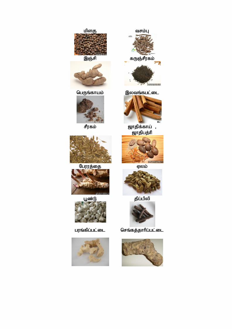
kpsF trk;G
,Q;rp fUQ;rPufk;
ngUq;fhak; ,ytq;fgl;il
rPufk; [hjpf;fha; >
[hjpgj;hp
Nguuj;ij Vyk;
g+z;L jpg;gpyp
guq;fpg;gl;il nrq;fj;jhhpg;gl;il

ACTIONS
Stimulant
Aphrodisiac
CONSTITUENTS
Karungozhi contains many kinds of amino acids (18 kinds of amino acid
including the 8 essential amino acids for human body). Vitamin B1, B2, B6,
B12, C and E, niasin, protein, fat, calcium phosphorus, iron, nicotinic acid
etc.
Laboratory tests show that Karungozhi contains certain hormones, blue
pigments of amino acids, which are required by the human body. These factors
can raise blood cells and haemoglobin. Abundance clinical experience has
indicated that Karungozhi has a particular effectiveness in treating neurodebility
(a condition of nervous debility suppred to be dependant upon impairment in the
functions of the spiral cord), osteomalasia (a condition marked by softening of
the bones), lot of hormonal problems like women's distress, sterility habitual
abortion, blood leucorrhoea, metorrhegia and sickness after giving birth to off
spring and also aids in curing pulmonary problems. TB, heart disease.
The eggs of Karungozhi have an ideal nutritive especially for old people
and high BP victims (since the cholesterol content in lower and free amino acids
are higher than that of other kinds of birds).
General properties
“Fl;lq; fbfpUkp Nfhuthjf; $l;lk;
kl;blhr; #iyaW khjuNr – Jl;l
fpue;jpnahL Gz;typNghq; NfSl Yf;F
kUe;J fUq; NfhopA+d; it”

,;e;j ghlypd;gb Nfhu thjk; NghFk; vd;gjdhy; fUq;Nfhop> thj
Neha;fis Nghf;Ftjpy; rpwe;jJ vd mwpayhk;.
2. fUQ;rPufk;
Botanical Name : Nigella sativa
Family : Ranunculaceae
Suvai : Kaippu
Thanmai : Veppam
Pirivu : Karppu
Part usd : Seed
ACTIONS :
Carminative
Diuretic
Emmenagogue
Galactogogue
Anthelmintic
Stomachic
Parasiticide
Emollient
CONSTITUENTS
The seed contains,
Fixed oil – 37.5%

Volatile oil – 1.5%
And albumin mucilage, metamarbin, melanthin,helleborin and
Arabic acid
Prolein – 26.7%, Oil 22.6%, ash 4.86% and total carbohydrate 40.0.
The major unsaturated fatty acids were linokic acid (50.3 – 49.2%) , oleic acid
(25.0 – 23.7%) and the main saturatted fatty acid is palmitic acid (17.2 –
18.4%), myristic, myristoleic, palmitoleic, margaric, margaroleic, stearic,
linolenic, arachidic, eicosenoic and lignoceric acids were also detected and
major components of which are thymoquinone, nigellone and nigllidine.
ew;rPufk;
Botanical Name : Cuminum cymimum
Family : Umbelliferae
Suvai : Karppu, Inippu
Thanmai : Thatpam
Pirivu : Inippu
Part used : Seed
ACTIONS :
Carminative
Stimulant
Stomachic
Astringent

CONSTITUENTS
α – pinene, 1-8 cineole and linaloor isolatedfrom the volatile (latif et al.,
2007) Two sesquiter penoid glucosides, cuminoside A and b and z-c methyl-D-
erythritol 1-O-β-D- glucopyranoside, z-c methyl-D-erythritol 3-O-β-D-
glucopyranoside, z-c methyl-D-erythritol 4-O-β-D- glucopyranoside and alkyl
glycosides have been isolated from the seed.
4. rhjpgj;hp
Botanical Name : Myristica fragrans
Family : Myrtacece
Suvai : Thuvarppu,Karppu
Thanmai : Veppam
Pirivu : Karppu
ACTIONS :
Stimulant
Carminative
Narcotic
Aromatic
Aphrodisiac
Tonic
ngUq;fhak;
Botanical Name : Ferula asafoetid
Family : umbelliferae

Suvai : Kaippu, karakarappu
Thanmai : Veppam
Pirivu : Karppu
Part used : Oleogumresin
ACTIONS
Stimulant
Carminative
Antispasmodic
Expectorant
Laxative
Anthelmintic
Diuretic
Aphrodisae
Emmenagogue
CONSTITUENTS
The oleo-gum resins taste and smell are like surphur due to the presence
of sulphur compounds like disulphides, symmetric tri and tetrasulphides,
Glucuronic acid. Galactose, arabinosed, rhamnose, unbelliferone, farnesiferoles
A,B, and C ferulic acid, coumarine derivaties foetidin and kamolonol have been
isolated from oleo gum resin

kpsF
Botanical Name : Piper nigrum
Family : Piper acede
Suvai : Kaippu,karppu
Thanmai : Veppam
Pirivu : Karppu
Part used : Seed, climber
ACTIONS :
Acrid
Carminative
Antiperiodic
Rubefacient
Stimulant
Resolvent
Antivatha
Antidote
CONSTITUENTS
Pyrrolidine alkamide, isopiperolein B, retrofractamide A,
Pipercide, Pipercha barride D, Pellitorin, dehdropipernonaline, ZE,4E, 8Z
isobutyl – eicosatrienamide, pellitorine, trachyone, pergumidiene and
isopiperolein B have been isolated from fruit. Essential oil contained
monoterpene hydro carbons, sabinene, β – pinene, limonene, furthermore

terpinene, β – pinene, myrcene, 3-carene, borneol, carvone, carvacrol,1,8-
cineol, linalool, sesqui – terpenes, β – caryophyllene, humulene, β – bisabolone,
aryophyllene oxide ketone, phnylether, eugend, mristicin and safrole,
bisalkaloids, dipiperamides D and E
Black pepper oil can be used to help in the treatment of pain relief,
rheumatism, increase circulation, exhaustion and muscular ache.
Xkk;
Botanical Name : Trachyspermum roxburghianum
Suvai : karppu
Thanmai : Veppam
Pirivu : Karppu
Part used : Seed
ACTIONS :
Stomachic
Antispasmodic
Carminative
Antiseptic
Stimulant
Tonic
Sialogogue

CONSTITUENTS
The essential oil of the seed contained terpinene, p – cymene, α –
pinene, thymol and carvacrol. The seed has steroptis, cumene, thymine,
aminoacids, threonine, calcium, iron, starch, tannins and dietary fibre. The seed
contain an essential oil containing about 50% thymol which is strong germicide,
antispasmodic and fungicide. Thymol is also used in toothpaste and perfumery
trk;G
Botanical Name : Acorus calamus
Suvai : karppu
Thanmai : Veppam
Pirivu : Karppu
Part used : Root
ACTIONS :
Stimulant
Stomachic
Antispasmodic
Carminative
Nauseant
Emetic
Disinfectant

Nguuj;ij:
Botanical Name : Alpina galanga
Family : Zingiberaceae
Suvai : karppu
Thanmai : Veppam
Pirivu : Karppu
Part used : Root
ACTIONS :
Expectorant
Febrifuge
Stomachic
CONSTITUENTS
1’S- 1autoxychavicol acetate, P – hydroxycinnamaldehyde and methane
were isolated from the rhizome and to possess various biological activies such
as antitumour. The essential oil contained monoterpenes, monoterpene alcohols,
esters, sesquiterpenes, methyleugenol, eugenol, acetate, chavicol and chavicol
acetate were identified.
nrq;fj;jhhpg; gl;il:
Botanical Name : Capparis Sepiaraia
Family : Capparidace
Suvai : Thuvarppu
Thanmai : Thatpam

Part used : Bark.
ACTIONS :
Stomachic
Tonic
CONSTITUENTS:
Capparine, cappariline and capparinine were isolated from roots.
ehAUtp
Botanical Name : Achyranthes aspera
Family : Amaranthaceae
Suvai : Kaippu,Thuvarppu,Karppu
Thanmai : Veppam
Pirivu : Karppu
Part used : Root
ACTIONS :
Astringent
Diuretic
Alterative
Antiperiodic

CONSTITUENTS:
Aliphatic dihydroxy ketone, characterized as 36,47 dihydroxy henpenta
contan – 4-one, tritria contanol, achyrathine pantatriacontan, 6-pentatria
contanone, hexatria contane and triacontane
Nguuj;ij
Botanical Name : Alpinia galangal
Family : Zinigiberaceae.
Suvai : Karppu
Thanmai : Veppam
Pirivu : Karppu
Part used : Root
ACTIONS
Expectorant
Febrifuge
Stomachic
CONSTITUENTS:
1’S-1 acetoxychavicol acetate, P-hydroxycinnamadehyde and nethane
were isolated from the rhizome and to possess various biological activities such
as antitumour. The essential oil contained monoterpenes, monoterpene alcohols,
esters, sesquiterpenes, methyleugenol, eugenol acetate, chaviol and chavical
acetate were identified.

fhh;Nghfhprp
Botanical Name : Psoralia corylifolia
Family : Fabaceae
Suvai : Kaippu
Thanmai : Veppam
ACTIONS:
Laxative
Stimulant
CONSTITUENTS:
Psoralidin, bakuchicin, psoralin, angelicin, Psoracorylifols A-E,
Prenylflavonoids, corylifols A and C were isolated from the seeds and they
showed significant antibacterial activities against a number of Gram(-) and
Gram(+) bacteria. Isobavachalwne neobavaisoflavone, isopsoralen and
psoralen were obtained from the seed.
Vyf;fha;
Botanical Name : Elatteria Cardamomum
Suvai : Karppu
Thanmai : Veppam
Pirivu : Karppu
Part used : Seed

ACTIONS:
Stimulant
Carminativc
Stomachic
Rf;F
Botanical Name : Zingiber officinale
Family : Zingiberaceae
Suvai : Karppu
Thanmai : Veppam
Pirivu : Karppu
Part used : Rhizome (dried)
ACTIONS:
Stimulant
Stomachic
Carminative.
CONSTITUENTS:
Ginger is known to contain a number of potentially bioactive
substances mainly gingerols shogabls, sesquiterpenes, B-bisabliene,
Zingiberene, Zingiberol, Curcumene, monoterpenes, geraial and neral

jpg;Gyp
Botanical Name : Piper longum
Family : Piperaceae.
Suvai : Inippu
Thanmai : Veppam
Pirivu : Inippu
Part used : Seed.
ACTIONS:
Astringent
Expectorant
Laxativc
Tonic.
CONSTITUENTS:
Guineensine, pipernonaline, pellitorine, piperine, piperanine,
piperidine alkaloids pipermonaline, piperundecalidine brachyamide A,
brachyamide B and brachystine and piperlonguminine were isolated from fruit.
Piperlongumine and piperlingumine and sesamin isolated from fruits.
,ytq;fg;gl;il
Botanical Name : Cinnamomum Verum
Suvai : Karppu, Inippu
Thanmai : Thatpam
Pi rivu : Inippu

Part used : Bark
ACTIONS:
Stimulant
Carminative
Aphrodisiac
G+z;L
Botainical Name : Allium Sativum
Family : Liliaceae
Suvai : Karppu
Thanmai : Veppam
Pirivu : Karppu
Part used : Tuber
ACTIONS:
Carminative
Stomachic
Tonic
Alterative
Stimiulant
Expectorart
Diurctic
Anthelmintic

CONSTITUENTS:
Sulphus compounds including allicin, premethanethiosulfonate
Dithun derivatives dially sulphide, diallyl trisulfide, proto-eruboside B,
Spirostanol, sativoside B1 analogue, eruboside B, rutin, quercetin 3 glucoside,
quercitrin myricetin, luteolin, quercetin, apigenin, kaempferol and isorhamnetin
Gd;KUq;if
Botanical Name : Moringla tinctoria
Family : Rubianceae
Part used : Root.
gwq;fpg;gl;il:
Botaical Name: Smilax china
Family :Liliaceae
Suvai : Inippu
Thanmai : Thatpam
Pirivu : Inippu
Part used : Tuber
ACTIONS:
Alterative
Antisyphilitic
Aphrodisiac
Depurative

CONSTITOENTS:
Six major stilbenes and flavonoids namelytaxifolin-3-0-glycoside,
piceid, oxyresveratrd, engeletin, resevertrol and scirpusin A have been isolated
from root tubers saponins, smilaxin, prosapogemin A, dioscin, gracilin,
dioscin, pseudoprotodioscin, methugracillin and methyl protodioscin have been
reported from root.
rq;fd;Nth;
Botanical Name : Azima tetracantha
Suvai : Kaippu
Thanmai : Veppam
Pirivu : Kaippu
Part used : Root
ACTIONS
Diuretic
Stimulant
Astringent
Tonic
Antiperiodic
Expectorant
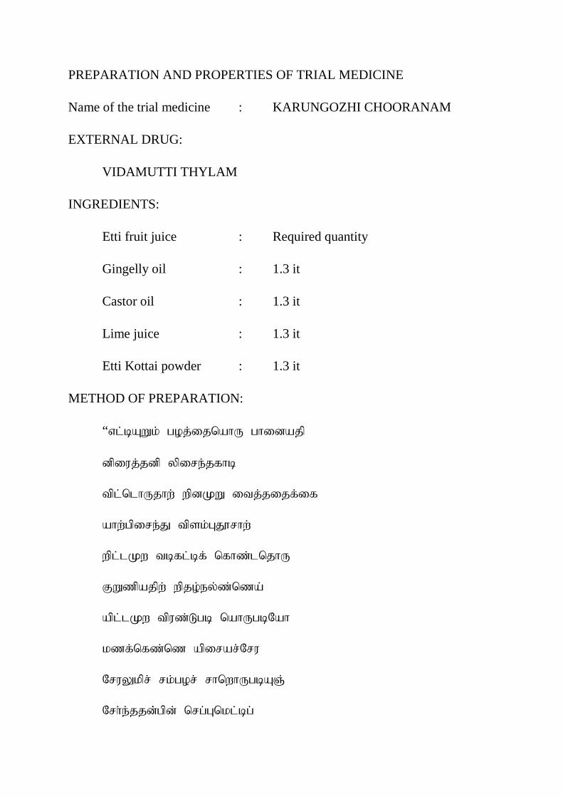
PREPARATION AND PROPERTIES OF TRIAL MEDICINE
Name of the trial medicine : KARUNGOZHI CHOORANAM
EXTERNAL DRUG:
VIDAMUTTI THYLAM
INGREDIENTS:
Etti fruit juice : Required quantity
Gingelly oil : 1.3 it
Castor oil : 1.3 it
Lime juice : 1.3 it
Etti Kottai powder : 1.3 it
METHOD OF PREPARATION:
“vl;bAWk; goj;ijnahU ghidajp
dpiuj;jdp ypire;jfhb
tpl;nlhUjhw; wpdKW itj;jijf;if
ahw;gpire;J tpsk;GJ}rhw;
wpl;lKw tbfl;bf; nfhz;lnjhU
FWzpajpw; wpjo;ey;z;nza;
apl;lKw tpuz;Lgb nahUgbNah
kzf;nfz;nz apirar;Nru
NruYkpr; rk;gor; rhnwhUgbAQ;
Nrh;e;jjd;;gpd; nrg;Gnkl;bg;

ghuKWq; nfhl;ilaJk; gbapbj;Jg;
Nghl;nlhpj;Jg; gjj;jpw; fha;r;rpr;
rhuKw ypbj;Jf; nfh spdjd;Nghpy;
tplKl;bj; ijykhFk;
ghUyNfhh;f; fpijj; njhl;Lj; jltpby;yh
jq;fnsy;yhk; gwf;Fk;ghNu
METHOD OF PREPARATION:
Etti Fruits are soaled in kaddi (Vinegar) for 4 days. Now it is crushed to get
juice. Then it is added with gingelly oil, castor oil and lime juice and powdered
form of etti kottai.Then it is heated till the drugs tum to red. Then filter the oil
and use it for external purposes. Three following books also describe and deal
with the same condition.

ANNEXTURE - II
BIO - CHEMICAL ANALYSIS OF
KARUNGOZHI CHOORANAM
Preparation of the extract:
5gms of KARUNGOZHI CHOORANAM was weighed accurately
and placed in a 250ml clean beaker. Then 50 ml distilled water is added to it
and dissolved well. Then it was boiled well for about 10 minutes. It was cooled
and filtered in a 100 ml volumetric flask and then it is made up to 100ml with
distilled water. This fluid is taken for analysis.
Qualitative analysis
S.No. Experiment Observation Inference
1. Test for calcium 2ml of the above prepared extract is taken in a clean test tube. To this add 2 ml of 4% ammonium oxalate solution.
A white precipitate is formed.
Indicates the presence of calcium.
2. Test for sulphate: 2ml of the extract is added to 5% barium chloride solution.
A white precipitate is formed.
Indicates the presence of sulphate.
3. Test for chloride The extract is treated with silver nitrate solution.
A white precipitate is formed.
Indicates the presence of chloride.
4. Test for carbonate The substance is treated with concentrated HCl.
No brisk effervescence is formed.
Absence of carbonate.

5. Test for Starch The extract is added with potassium ferro cyanide.
No blue colour is formed.
Absence of starch.
6. Test for iron Ferric The extract is treated with concentrated glacial acetic acid and potassium ferro cyanide.
No blue colour is formed.
Absence of ferric iron.
7. Test for iron Ferrous: The extract is treated with concentrated nitric acid and ammonium thio cyanate.
Blood red colour is formed.
Indicates the presence of ferrous iron.
8. Test for phosphate The extract is treated with ammonium molybdate and concentrated nitric acid.
Yellow precipitate is formed.
Indicates the presence of phosphate.
9. Test for albumin The extract is treated with Esbach’s reagent.
No yellow precipitate is formed.
Absence of Albumin.
10. Test for Tannic acid The extract is treated with ferric chloride reagent.
No Blue black precipitate is formed
Absence of Tannic acid
11. Test for unsaturation Potassium permanganate solution is added to the extract.
It gets decolorized.
Indicates the presence of unsaturated compound.
12. Test for the reducing sugar 5ml of benedict’s qualitative solution is taken in a test tube and allowed to boil for 2 mts and added 8-10 drops of the extract and again boil it for 2 mts.
No Colour change occurs.
Absence of reducing sugar

13. Test for amino acid: One or two drops of the extract are placed on a filter paper and dried it well. After drying, 1% ninhydrin is sprayed over the same and dried it well.
Violet colour is formed.
Indicates the presence of Amino acid.
14. Test for zinc: The extract is treated with potassium ferrocyanide
No white precipitate is formed
Absence of zinc
Result:
The trial drug KARUNGOZHI CHOORANAM contains,
1. calcium
2. sulphate
3. chloride
4. ferrous iron
5. phosphate
6. unsaturated compound
7. amino acid.
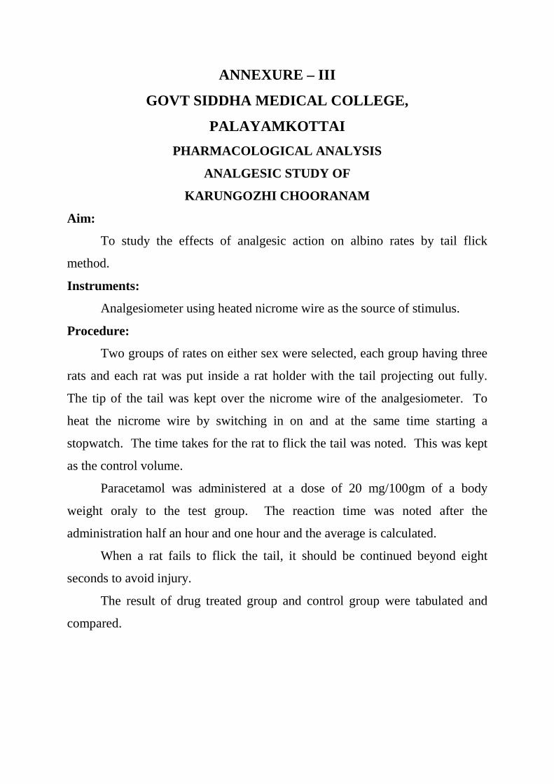
ANNEXURE – III
GOVT SIDDHA MEDICAL COLLEGE,
PALAYAMKOTTAI PHARMACOLOGICAL ANALYSIS
ANALGESIC STUDY OF
KARUNGOZHI CHOORANAM
Aim:
To study the effects of analgesic action on albino rates by tail flick
method.
Instruments:
Analgesiometer using heated nicrome wire as the source of stimulus.
Procedure:
Two groups of rates on either sex were selected, each group having three
rats and each rat was put inside a rat holder with the tail projecting out fully.
The tip of the tail was kept over the nicrome wire of the analgesiometer. To
heat the nicrome wire by switching in on and at the same time starting a
stopwatch. The time takes for the rat to flick the tail was noted. This was kept
as the control volume.
Paracetamol was administered at a dose of 20 mg/100gm of a body
weight oraly to the test group. The reaction time was noted after the
administration half an hour and one hour and the average is calculated.
When a rat fails to flick the tail, it should be continued beyond eight
seconds to avoid injury.
The result of drug treated group and control group were tabulated and
compared.

STUDY OF ANALGESIC EFFECT USING THE DRUG
KARUNGOZHI CHOORNAM
Group
Dose /
100mg Body
weight
Initial
reading
(in Sec)
After ½
hour
(in Sec)
After 1 ½
hour
(in Sec)
Mean
Difference
Control Water 2 ml
2
2 2
2
Standard Paracetomol
20 mg
2.5
4.5 6.5 6.5
Test drug
Karungozhi
chooranam
100mg
2.5
4.0
5.8
5.8
Inference:
The trial drug has significant analgesic action.
ACUTE ANTI – INFLAMMATORY ACTIVITY IN RATS
BY HIND – PAW METHOD
Aim:
To demonstrate the acute anti-inflammatory activity of Karungozhi
Chooranam in albino rats by Hind-paw method.
Procedure:
Nine Albino rats weighing 100-150gm were taken and divided into three
groups and each group consisting three rats.
First group was kept as control land received water. Second group
received lbuprofen at a dose of 20mg/100gm – body weight. Third group
animals received Karungozhi Choornam suspension at a dose of 200mg/ 100
gm – body weight.

Before administration of drugs, the Hind-paw upto the tibio-tarsal
junction in mercury plethysmograph. Soon after the measurement at the drugs
were administered. One hour after the administration of drugs a sub –
cutaneous injection of 0.1ml of 1%/W/N of carrageenin in water was made into
planter surface of both the Hind-paw of each rat.
Three hours after carrageenin injection, the Hind-paw volume was
measured once again. Difference between the initial and final value were noted
and compared.
This method is more suitable for studying anti-inflammatory activity in
acute inflammation.
EFFECTS OF KARUNGOZHI CHOORANAM IN
ACUTE ANTI – INFLAMMATORY ACTIVITIES
Group Dose volume
orally
Initial
reading
Final
reading
Mean
difference
Percentage
Inflammation
Percentage
Inhibition
Control Water 2 ml 0.55 1.4 0.85 100 -
Standard Ibuprofen
20mg/100gm 0.55 0.75 0.20 23.5 76.5
Test drug
Karungozhi
chooranam 100mg
/ 100gm
0.57 0.89 0.32 37.6 62.4
Result:
The drug Karungozhi Chooranam has Significant acute anti-
inflammatory action.
CHRONIC ANTI-INFLAMMATORY STUDY
BY COTTON – PELLETS GRANULOMA METHOD Drug:
Karungozhi Chooranam

Aim:
To study the chronic ani-inflammatory activity of the drug in albino rats
by cotton pellets implantation (granuloma) method.
Procedures:
Cotton pellets each weighing long was prepared and sterilized in an
autoclave for about one hour under 15 lbs atmosphere pressure. Nine Albino
rats weighing between 100-200gm were selected and were divided into 3
groups. Each rat was anaesthetized with ether and cotton pellets were implanted
subcutaneously in the groin, two in each side.
From the day of implantation, one group of animals received
Karungozhi Chooranam at a dose of 200mg/100gm of body weight.
On the eighth day the rats were sacrificed and the pellets were removed
weighed. Then they were put in an incubator at 600C-800C and then weighed.
The concordant weight was noted for all groups and compared.
THE EFFECT OF KARUNGOZHI CHOORANAM IN CHRONIC
ANTI – INFLAMMATORY STUDY
Group Dose given
orally
Pellet
Weight
Pellet Weight of
the Granuloma of
drugs
Percentage
inflammation
Percentage
inhibition
Control Water 1ml 10mg 250 mg 100 -
Standard
Ibuprofen
20mg/100gm
body weight
10mg 55mg 22 78
Test
drug
100mg/100gm
body weight 10 mg 98mg 40 60
Inference:
The drug Karungozhi Chooranam shows Significant chronic
anti – inflammatory action.
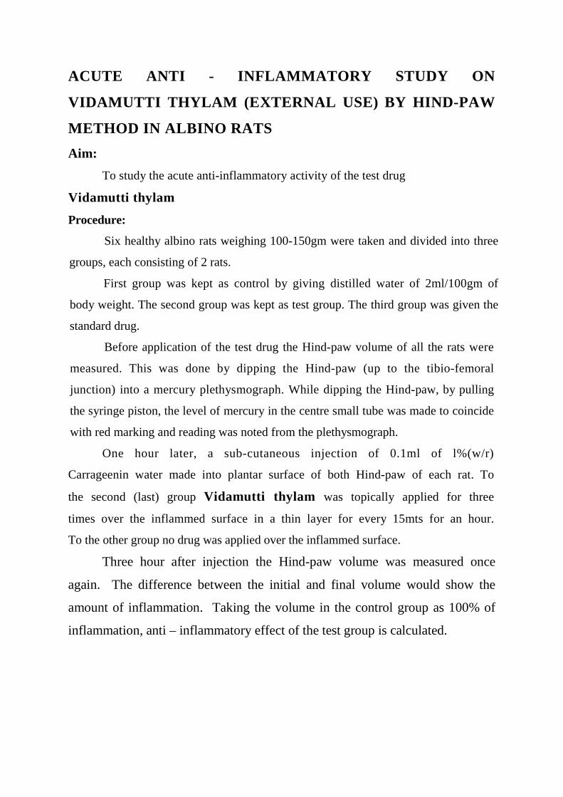
ACUTE ANTI - INFLAMMATORY STUDY ON
VIDAMUTTI THYLAM (EXTERNAL USE) BY HIND-PAW
METHOD IN ALBINO RATS Aim: To study the acute anti-inflammatory activity of the test drug
Vidamutti thylam
Procedure:
Six healthy albino rats weighing 100-150gm were taken and divided into three
groups, each consisting of 2 rats.
First group was kept as control by giving distilled water of 2ml/100gm of
body weight. The second group was kept as test group. The third group was given the
standard drug.
Before application of the test drug the Hind-paw volume of all the rats were
measured. This was done by dipping the Hind-paw (up to the tibio-femoral
junction) into a mercury plethysmograph. While dipping the Hind-paw, by pulling
the syringe piston, the level of mercury in the centre small tube was made to coincide
with red marking and reading was noted from the plethysmograph.
One hour later, a sub-cutaneous injection of 0.1ml of l%(w/r)
Carrageenin water made into plantar surface of both Hind-paw of each rat. To
the second (last) group Vidamutti thylam was topically applied for three
times over the inflammed surface in a thin layer for every 15mts for an hour.
To the other group no drug was applied over the inflammed surface.
Three hour after injection the Hind-paw volume was measured once
again. The difference between the initial and final volume would show the
amount of inflammation. Taking the volume in the control group as 100% of
inflammation, anti – inflammatory effect of the test group is calculated.

EFFECT OF VIDAMUTTI THYLAM
Group Drugs
Dose
100
gm of
body
weight
Initial
value
Final
value Difference
Percentage
Inflammation
Percentage
Inhibition
Control Water 2ml 0.55 1.4 0.85 100 -
Standard Ibuprofen 20mg 0.55 0.75 0.20 29.4 70.6
Test
drug Vidamutti Thylam Ext 0.525 0.8 0.275 40.44 59.56
Inference:
The test drug has significant anti-inflammatory action externally.
ANALGESIC STUDY OF VIDAMUTTI THYLAM
Aim:
To study the effects of analgesic action on albino rates by tail flick
method.
Instruments:
Analgesiometer using heated nicrome wire as the source of stimulus.
Procedure:
Two groups of rates on either sex were selected, each group having three
rats and each rat was put inside a rat holder with the tail projecting out fully.
The tip of the tail was kept over the nicrome wire of the analgesiometer. To
heat the nicrome wire by switching in on and at the same time starting a
stopwatch. The time takes for the rat to flick the tail was noted. This was kept
as the control volume.
Paracetamol was administered at a dose of 20 mg/100gm of a body
weight oraly to the test group. The reaction time was noted after the
administration half an hour and one hour and the average is calculated.

When a rat fails to flick the tail, it should be continued beyond eight
seconds to avoid injury.
The result of drug treated group and control group were tabulated and
compared.
STUDY OF ANALGESIC EFFECT USING THE DRUG
VIDAMUTTI THYLAM
Group Dose / 100mg
Body weight
Initial
reading (in
Sec)
After ½
hour
(in Sec)
After 1
½ hour
(in Sec)
Mean
Difference
Control Water 2 ml 2 2 2
2
Standard Paracetomol
20 mg
2.5
4.5 6.5 6.5
Test Drug
2ml 2
3
5
5
Inference:
The trial drug has significant analgesic action.

ANNEXURE – IV
ASSESSMENT FORMS
FORM I - SCREENING FORM
FORM II - CONSENT FORM
FORM III - CASE PROFORMA
FORM IV - LABORATORY INVESTIGATIONS
FORM V - CLINICAL ASSESSMENT
FORM VI - PATIENT WITHDRAWAL FORM
FORM
VII
- DRUG COMPLIANCE FORM

GOVERNMENT SIDDHA MEDICAL COLLEGE & HOSPITAL
PALAYAMKOTTAI.
POST- GRADUATE DEPARTMENT OF SIRAPPU MARUTHUVAM
AN OPEN CLINICAL TRIAL OF KARUNGOZHI CHOORANAM &
VIDAMUTTI THYLAM FOR AZHAL KEEL VAAYU
(OSTEOARTHRITIS)
FORM I –SCREENING FORM
1. OP/ IP No: 2. BED No:
3. Sl. No: 4. NAME:
5. AGE: 6. GENDER:
7. OCCUPATION: 8. SOCIAL STATUS
9. DATE OF ADMISSION: 10. DATE OF DISCHARGE:
11. POSTAL ADDRESS:

I. INCLUSION CRITERIA:
1. Sex: Both Male and Female.
2. Pain and swelling present in knee joints.
3. Crepitations present in knee joints.
4. Early morning stiffness.
5. Tenderness
II. EXCLUSION CRITERIA:
1. Diabetes Mellitus
2. Hypertension
3. Cardiac diseases
4. Pregnancy and Lactation
5. Patients with any other serious illness
6. Peptic ulcer
7. Severe trauma
8. Any other systemic diseases
III. WITHDRAWAL CRITERIA:
1. Development of any adverse reaction (ADR
2. Occurrence of any other systemic illness.

GOVERNMENT SIDDHA MEDICAL COLLEGE AND HOSPITAL
PALAYAMKOTTAI.
POST- GRADUATE DEPARTMENT OF SIRAPPU MARUTHUVAM
AN OPEN CLINICAL TRIAL OF KARUNGOZHI CHOORANAM &
VIDAMUTTI THYLAM FOR AZHAL KEEL VAAYU
(OSTEOARTHRITIS)
Form: II CONSENT FORM
CERTIFICATE BY INVESTIGATOR
I certify that I have disclosed all the details about the study in the terms readily
understood by the patient.
Signature…………………. Date……
Name……………………
CONSENT BY PATIENT
I have been informed to my satisfaction, by the attending physician, the
purpose of the clinical trial, and the nature of drug treatment and follow-up
including the laboratory investigations to be performed to monitor and
safeguard my body functions.
I am aware of my right to opt out of the trial at any time during the course
of the trial without having to give the reasons for doing so.

I, exercising my free power of choice, hereby give my consent to be
included as a subject in the clinical trial of ‘KARUNGOZHI CHOORANAM
(Internal drug & VIDAMUTTI THYLAM (External drug)’ for the treatment of
‘Azhal keel vaayu’ (osteoarthritis).
Signature…………………. Date……………….
Name…………………
murpdH rpj;j kUj;Jtf; fy;Y}hp kw;Wk; kUj;Jtkid>ghisaq;Nfhl;il
gl;lNkw;gbg;G rpwg;GkUj;Jtj;Jiw
'fUq;Nfhop #uzk;" kw;Wk; 'tplKl;b ijyk;" ,tw;wpd;
gupfupg;Gj;jpwidf; fz;lwpAk; kUj;Jt Ma;T
Xg;Gjy; gbtk;
Ma;thsuhy; rhd;wspf;fg;gl;lJ
ehd; ,e;j Ma;itf; Fwpj;j midj;J tpguq;fisAk; Nehahspf;F
GupAk; tifapy; vLj;Jiuj;Njd; vd cWjpaspf;fpNwd;.
Njjp: ifnahg;gk;:
,lk;: ngau;:

Nehahspapd; xg;Gjy;
vd;dplk; ,e;j kUj;Jt Ma;tpd; fhuzj;ijAk; kUe;jpd; jd;ik
kw;Wk; kUj;Jt topKiwiag; gw;wpAk; njhlu;e;J vdJ cly;
,af;fj;ij fz;fhzp;f;fTk>; mjidg; ghJfhf;fTk; gad;gLk; kUj;Jt
Ma;Tf;$l gupNrhjidfs; gw;wpAk; jpUg;jp mspf;Fk; tifapy; Ma;T
kUj;Jtuhy; tpsf;fpf; $wg;gl;lJ.
ehd; ,e;j kUj;Jt Ma;tpd; NghJ fhuzk; vJTk; $whky;
vg;nghOJ Ntz;LkhdhYk; ,e;j Ma;tpypUe;J vd;id tpLtpj;Jf;
nfhs;Sk; cupikia njupe;jpUf;fpd;Nwd;.
ehd; vd;Dila Rje;jpukhfj; Nju;T nra;Ak; cupikiaf; nfhz;L
moy; fPy; thA vd;Dk; Neha;f;fhd ‘fUq;Nfhop #uzk; ” kw;Wk; ‘
tplKl;b ijyk; “ Mfpatw;wpd; gupfupg;Gj; jpwidf; fz;lwpAk;
kUj;Jt Ma;tpw;F vd;id cl;gLj;j xg;Gjy; mspf;fpNwd;.
Njjp: ifnahg;gk;:
,lk;: ngau;:
Njjp: rhl;rpf;fhuh; ifnahg;gk;:
,lk;: ngau;

GOVERNMENT SIDDHA MEDICAL COLLEGE AND HOSPITAL
PALAYAMKOTTAI.
POST- GRADUATE DEPARTMENT OF SIRAPPU MARUTHUVAM
AN OPEN CLINICAL TRIAL OF KARUNGOZHI CHOORANAM &
VIDAMUTTI THYLAM FOR AZHAL KEEL VAAYU
(OSTEOARTHRITIS)
FORM III – CASE PROFORMA
1. OP/ IP No: 2. BED No: 3. Sl. No:
4. NAME: 5. AGE: 6. GENDER:
7. OCCUPATION: 8. SOCIAL STATUS
9. DATE OF ADMISSION: 10. DATE OF DISCHARGE:
11. POSTAL ADDRESS:
12. COMPLAINTS & DURATION:
13. HISTORY OF PRESENT ILLNESS:
Lecturer HOD

14. PAST HISTORY:
15. FAMILY HISTORY:
16. MENSTRUAL HISTORY (If applicable):
17. HABITS: Yes No
1. Smoker
2. Alcoholic
3. Betel nut chewer
4. Non-Veg /Vegetarian
5. Drug addiction
18. GENERAL EXAMINATION:
1. Body weight [Kg] :
2. Height [cm] :
3. Body Temperature [oF] :
4. Blood Pressure (mmHg) :
5. Pulse Rate /min. :
6. Heart Rate /min. :
7. Respiratory Rate /min. :

Yes No
8. Pallor :
9. Jaundice :
10. Clubbing :
11. Cyanosis :
12. Pedal Oedema :
13. Lymphadenopathy :
14. Jugular venous pulsation:
19. CLINICAL EXAMINATION OF KNEE JOINT:
I. INSPECTION Present Absent
1. Swelling ……………………………
2. Muscle wasting ……………………………
3. Deformity ……………………………
II. PALPATION: Present Absent
1. Tenderness …………………………..
2. Swelling ……………………………
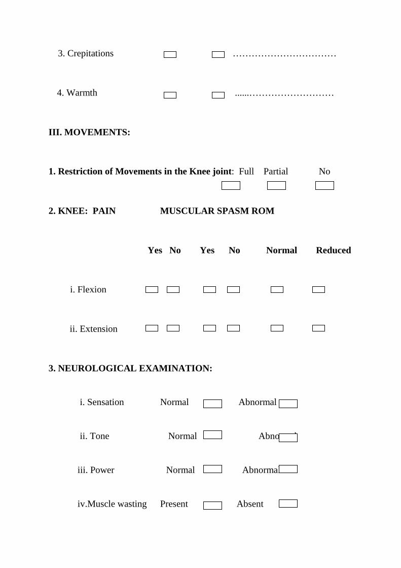
3. Crepitations ……………………………
4. Warmth ......………………………
III. MOVEMENTS:
1. Restriction of Movements in the Knee joint: Full Partial No
2. KNEE: PAIN MUSCULAR SPASM ROM
Yes No Yes No Normal Reduced
i. Flexion
ii. Extension
3. NEUROLOGICAL EXAMINATION:
i. Sensation Normal Abnormal
ii. Tone Normal Abnormal
iii. Power Normal Abnormal
iv.Muscle wasting Present Absent

4. REFLEXES: Normal Exaggerated
i. Knee jerk
ii. Ankle jerk
20. CLINICAL ASSESSMENT:
I. PAIN:
A. Pain in the knee joints: No Mild Moderate Severe
i. Onset Sudden Gradual
ii. Nature: Local Diffuse Others
B. Nature of pain Shooting Burning Others
YES NO
C. Pain during movements
II. Morning stiffness

III.Tenderness
III. Swelling
IV. Restricted joint movements
21. EXAMINATION OF OTHER SYSTEMS:
Normal Abnormal
1. CVS
2. RS
3. CNS
4. ABDOMEN
5. GENITO-URINARY

SIDDHA ASPECTS
1. NILAM:
1. Kurinji 2. Mullai 3. Marutham 4. Neithal 5. Paalai
2 . KAALAM:
1. Kaar Kaalam 2. Koothir Kaalam 3. Munpani Kaalam
4. Pinpani Kaalam 5. Ilavenir Kaalam 6. Muduvenir Kaalam
3. YAAKKAI:
1. Vatham 2. Pitham 3. Kabam
4. Vathapitham 5. Pithavatham 6. Kabavatham
7. Vathakabam 8. Pithakabam 9. Kabapitham
4. GUNAM:
1. Sathuvam 2. Rasatham 3. Thamasam

5. IYMPORIGAL: Normal Affected
1. Mei ..…………………………………..
2. Vaai ……………………………………...
3. Kan ………………………………………
4. Mookku ………………………………………
5. Sevi ………………………………………
6. KANMENDHIRIUM / KANMAVIDAYAM:
Normal Affected
1. Kai …………………………………………...
2. Kaal …………………………………………...
3. Vaai …………………………………………..
4. Eruvaai …………………………………………..
5. Karuvaai …………………………………………..

7. UYIR THATHUKKAL:
I. VATHAM: Normal Affected
1. Piraanan …………………………………………
2. Abaanan ………………………………………….
3. Viyaanan ………………………………………….
4. Uthaanan ……………………………………
5. Samaanan ……………………………………
6. Naagan ……………………………………….
7. Koorman …………………………………………...
8. Kirukaran …………………………………………...
9. Devathathan …………………………………………
10. Dhananjeyan …………………………………………

II. PITHAM : Normal Affected
1. Analam ……………………………………….
2. Ranjagam ………………………………………….
3. Saathagam ………………………………………..
4. Aalosagam ……………………………………….
5. Prasagam ……………………………………..
III. KABAM: Normal Affected
1. Avalambagam ……………………………………………
2. Kilethagam ……………………………………………
3. Pothagam ……………………………………………
4. Tharpagam ……………………………….…………...
5. Santhigam ……………………………………………
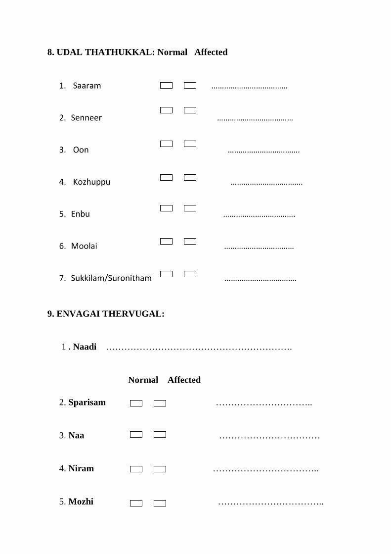
8. UDAL THATHUKKAL: Normal Affected
1. Saaram ………………………………
2. Senneer ………………………………
3. Oon …………………………….
4. Kozhuppu …………………………….
5. Enbu …………………………….
6. Moolai ……………………………
7. Sukkilam/Suronitham …………………………….
9. ENVAGAI THERVUGAL:
1 . Naadi …………………………………………………….
Normal Affected
2. Sparisam …………………………..
3. Naa ……………………………
4. Niram ……………………………..
5. Mozhi ……………………………..
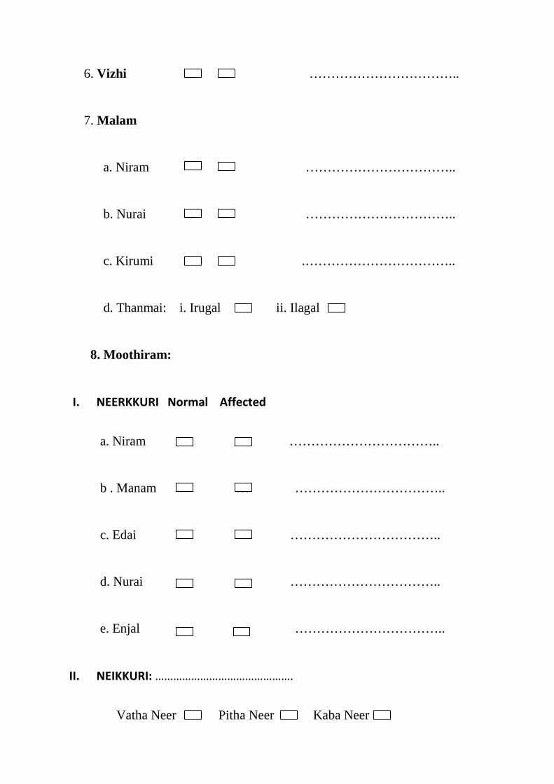
6. Vizhi ……………………………..
7. Malam
a. Niram ……………………………..
b. Nurai ……………………………..
c. Kirumi .……………………………..
d. Thanmai: i. Irugal ii. Ilagal
8. Moothiram:
I. NEERKKURI Normal Affected
a. Niram ……………………………..
b . Manam … ……………………………..
c. Edai ……………………………..
d. Nurai ……………………………..
e. Enjal ……………………………..
II. NEIKKURI: ……………………………………….
Vatha Neer Pitha Neer Kaba Neer

GOVERNMENT SIDDHA MEDICAL COLLEGE AND HOSPITAL
PALAYAMKOTTAI.
POST- GRADUATE DEPARTMENT OF SIRAPPU MARUTHUVAM
AN OPEN CLINICAL TRIAL OF KARUNGOZHI CHOORANAM &
VIDAMUTTI THYLAM FOR AZHAL KEEL VAAYU
(OSTEOARTHRITIS)
Form IV - LABORATORY INVESTIGATIONS
1. OP/ IP No: 2. BED No: 3. Sl. No:
4. NAME: 5. AGE: 6. GENDER:
7. OCCUPATION: 8. SOCIAL STATUS
9. DATE OF ENROLMENT: 10. DATE OF DISCHARGE:
11. POSTAL ADDRESS:
Date:
Lecturer HOD

I. BLOOD:
1. TC : (Cells/Cumm)
2. DC (%): N L M E
3. ESR (mm) : ½ hr 1 hr
4. Hb:
5. Total RBC:
6. Blood Sugar: a) Fasting b) Post prandial
7. Kidney function tests:
Blood urea: Serum creatinine:
8. Lipid profile:
HDL: LDL: VLDL:
Total Cholesterol : TGL:
9. Liver Function tests:
SGOT: SGPT: Alk. Phosphatase:
Albumin: Globulin: Total Protein:
Serum Bilirubin: Total Direct Indirect :

II. URINE:
1. Albumin :
2. Sugar :
3. Epithelial cells :
4. Pus cells :
5. Red blood cells :
6. Casts/Crystals :
III. MOTION:
1. Ova :
2. Cyst :
3. Occult blood :
4. Pus cells :
IV. X-RAY:

GOVERNMENT SIDDHA MEDICAL COLLEGE AND HOSPITAL
PALAYAMKOTTAI.
POST- GRADUATE DEPARTMENT OF SIRAPPU MARUTHUVAM
AN OPEN CLINICAL TRIAL OF KARUNGOZHI CHOORANAM &
VIDAMUTTI THYLAM FOR AZHAL KEEL VAAYU
(OSTEOARTHRITIS)
FORM V – CLINICAL ASSESSMENT
1. OP/ IP No: 2. BED No: 3. Sl. No:
4. NAME: 5. AGE: 6. GENDER:
7. OCCUPATION: 8. SOCIAL STATUS
9. DATE OF ADMISSION: 10. DATE OF DISCHARGE:
11. POSTAL ADDRESS:
Lecturer HOD

CLINICAL EXAMINATION OF KNEE JOINT:
I. INSPECTION: Present Absent
1. Swelling ……………………………
2. Muscle wasting ……………………………
3. Deformity ……………………………
II. PALPATION: Present Absent
1. Tenderness ………………………
2. Swelling …………………………
3. Crepitations ………………………………
4. Warmth
………………………………

III. MOVEMENTS:
1. Restriction of Movements in the Knee joint: Full Partial No
2. KNEE: PAIN MUSCULAR SPASM ROM
Yes No Yes No Normal Reduced
i. Flexion
ii. Extension
3. NEUROLOGICAL EXAMINATION:
i. Sensation: Normal Abnormal …………...
ii. Tone Normal Abnormal …………..
iii. Power Normal Abnormal ..…………
iv. Muscle wasting: Present Absent ………….
4. REFLEXES: Normal Exaggerated
i. Knee jerk
ii. Ankle jerk

20. CLINICAL ASSESSMENT:
I. PAIN:
A. Pain in the knee joints: No Mild Moderate Severe
i. Onset Sudden Gradual
ii. Nature: Local Diffuse Others
B. Nature of pain Shooting Burning Others
Yes No
C. Pain during movements
II. Morning stiffness
III.Tenderness
III. Swelling
IV. Restricted joint movements

GOVERNMENT SIDDHA MEDICAL COLLEGE AND HOSPITAL
PALAYAMKOTTAI.
POST- GRADUATE DEPARTMENT OF SIRAPPU MARUTHUVAM
AN OPEN CLINICAL TRIAL OF KARUNGOZHI CHOORANAM &
VIDAMUTTI THYLAM FOR AZHAL KEEL VAAYU
(OSTEOARTHRITIS)
FORM - VI PATIENT WITHDRAWAL FORM
1. OP / IP No ………… 2. S.No. ……………… 3.Date: ……………
4. Name ………………….. 5. Age …………… 6. Gender …………
7. Postal address:
------------------------------------------------------------------------------------------------
Complaints and Duration:
Irregular treatment:
Other causes:

GOVERNMENT SIDDHA MEDICAL COLLEGE AND HOSPITAL
PALAYAMKOTTAI.
POST- GRADUATE DEPARTMENT OF SIRAPPU MARUTHUVAM
AN OPEN CLINICAL TRIAL OF KARUNGOZHI CHOORANAM &
VIDAMUTTI THYLAM FOR AZHAL KEEL VAAYU
(OSTEOARTHRITIS)
FORM VII - DRUG COMPLIANCE FORM
Name of the Drug: KARUNGOZHI CHOORANAM
Drugs issued: ……………(mgs/Grams)
Drugs returned: ……………(mgs/Grams)
S.NO DATE
DRUG TAKEN TIME
MORNING/TI
ME
AFTERNOON/T
IME
NIGHT/TI
ME
Day 1
Day 2
Day 3
Day 4
..

Date:
Station:
Signature of the Investigator:
Signature of the Lecturer: Signature of the HOD
..
Up to day
48

BIBLIOGRAPHY
1. T.V.S. Sambasivam Pillai
2. Siddha Text book - Pothu Maruthuvam
3. Agathiyar 2000
4. Sarabenthirar Vadha Kaviyam
5. Sabapathy Kaiyedu
6. Sathaga Naadi
7. Agathiyar Aul Vedham
8. Yugi Vaithiya Chinthamani
9. Yugi Vaithiya Kaaviyam
10. Pararasa Sekaram
11. Thirumoolar Thirumanthiram
12. Harrison's Internal Medicine
13. Thinamum Yoga
14. Varma Maruthuvam - Kannan Rajaram
15. Theraiyar Kappiyam
16. Maruthuva Bharatham
17. Kannusamiyam
18. Text book of Orthopedics - Maheswari
19. Robin's Pathology
20. Orthopedic and Traumatology - Natarajan
21. Text book of Physiology - Guyton