Award Number: W81XWH-10-1-0052 - dtic.mil · Award Number: W81XWH-10-1-0052 . ... opinions and/or...
17
AD_________________ Award Number: W81XWH-10-1-0052 TITLE: PRINCIPAL INVESTIGATOR: Michael Dellinger CONTRACTING ORGANIZATION: UT Southwestern Medical Center Dallas, TX 75390 REPORT DATE: February 2013 TYPE OF REPORT: Annual Summary PREPARED FOR: U.S. Army Medical Research and Materiel Command Fort Detrick, Maryland 21702-5012 DISTRIBUTION STATEMENT: Approved for Public Release; Distribution Unlimited The views, opinions and/or findings contained in this report are those of the author(s) and should not be construed as an official Department of the Army position, policy or decision unless so designated by other documentation.
Transcript of Award Number: W81XWH-10-1-0052 - dtic.mil · Award Number: W81XWH-10-1-0052 . ... opinions and/or...

AD_________________
Award Number: W81XWH-10-1-0052 TITLE:
PRINCIPAL INVESTIGATOR: Michael Dellinger CONTRACTING ORGANIZATION: UT Southwestern Medical Center
Dallas, TX 75390
REPORT DATE: February 2013 TYPE OF REPORT: Annual Summary PREPARED FOR: U.S. Army Medical Research and Materiel Command Fort Detrick, Maryland 21702-5012 DISTRIBUTION STATEMENT: Approved for Public Release; Distribution Unlimited The views, opinions and/or findings contained in this report are those of the author(s) and should not be construed as an official Department of the Army position, policy or decision unless so designated by other documentation.

REPORT DOCUMENTATION PAGE Form Approved
OMB No. 0704-0188 Public reporting burden for this collection of information is estimated to average 1 hour per response, including the time for reviewing instructions, searching existing data sources, gathering and maintaining the data needed, and completing and reviewing this collection of information. Send comments regarding this burden estimate or any other aspect of this collection of information, including suggestions for reducing this burden to Department of Defense, Washington Headquarters Services, Directorate for Information Operations and Reports (0704-0188), 1215 Jefferson Davis Highway, Suite 1204, Arlington, VA 22202-4302. Respondents should be aware that notwithstanding any other provision of law, no person shall be subject to any penalty for failing to comply with a collection of information if it does not display a currently valid OMB control number. PLEASE DO NOT RETURN YOUR FORM TO THE ABOVE ADDRESS.
02-08-2013 1. REPORT DATE
Annual Summary 2. REPORT TYPE
15January2010–14January2013 3. DATES COVERED
4. TITLE AND SUBTITLE Delineating the Effect a Novel Anti-VEGF-A Therapy Has on the Lymphatic System of Immunocompetent Tumor-Bearing Mice.
5a. CONTRACT NUMBER
W81XWH-10-1-0052 5b. GRANT NUMBER
5c. PROGRAM ELEMENT NUMBER
6. AUTHOR(S) Michael Dellinger
5d. PROJECT NUMBER
5e. TASK NUMBER
E-Mail: [email protected]
5f. WORK UNIT NUMBER
7. PERFORMING ORGANIZATION NAME(S) AND ADDRESS(ES)
8. PERFORMING ORGANIZATION REPORT
UT Southwestern Medical Center NUMBER
Dallas, TX 75390
9. SPONSORING / MONITORING AGENCY NAME(S) AND ADDRESS(ES) 10. SPONSOR/MONITOR’S ACRONYM(S)
U.S. Army Medical Research and Materiel Command
Fort Detrick, Maryland 21702-5012
11. SPONSOR/MONITOR’S REPORT
NUMBER(S)
Approved for Public Release; Distribution Unlimited 12. DISTRIBUTION / AVAILABILITY STATEMENT
13. SUPPLEMENTARY NOTES
14. ABSTRACT Despite intense research efforts, cancer remains the second leading cause of death in the United States. Mortality is seldom caused by primary tumors, but rather by the effect of metastases on distant organs. The lymphatic system serves as a common route of metastasis for many cancers of epithelial origin. There is growing evidence that lymphangiogenesis, the sprouting of new lymphatics from pre-existing lymphatics, facilitates the dissemination of cancer. Interestingly, vascular endothelial growth factor receptor-2 (VEGFR2) signaling has been shown to stimulate lymphangiogenesis in adult mice. However, the role VEGFR2 serves in the development of the lymphatic system has not been defined. Here we use the Cre-lox system to show that the proper development of the lymphatic vasculature requires VEGFR2 expression by lymphatic endothelium. This newly identified function of VEGFR2 further defines the molecular pathways controlling the development of the lymphatic vasculature and sheds light on how therapeutic agents targeting VEGFR2 can inhibit tumor lymphangiogenesis. VEGF-A, VEGFR2, lymphangiogenesis, tumor lymphangiogenesis 15. SUBJECT TERMS
16. SECURITY CLASSIFICATION OF: 17. LIMITATION
OF ABSTRACT 18. NUMBER OF PAGES
19a. NAME OF RESPONSIBLE PERSON USAMRMC
U a. REPORT b. ABSTRACT
U U c. THIS PAGE
UU 17
19b. TELEPHONE NUMBER (include area code)

Table of Contents
Page Introduction…………………………………………………………….………..….. 4 Body………………………………………………………………………………….. 4 Key Research Accomplishments………………………………………….…….. 6 Reportable Outcomes……………………………………………………………… 7 Conclusion…………………………………………………………………………… 7 References……………………………………………………………………………. 8 Appendices…………………………………………………………………………… 9

Dellinger 4
INTRODUCTION:
Despite intense research efforts, cancer remains the second leading cause of death in the United States. Mortality is seldom caused by primary tumors, but rather by the effect of metastases on distant organs. The lymphatic system serves as a common route of metastasis for many cancers of epithelial origin. There is growing evidence that lymphangiogenesis, the sprouting of new lymphatics from pre-existing lymphatics, facilitates the dissemination of cancer. Interestingly, the growth factor VEGF-A stimulates lymphangiogenesis; however, the underlying mechanisms have not been fully defined. To achieve a better understanding of VEGF-A’s function in biology, our lab helped develop a novel anti-VEGF-A antibody (r84) that specifically blocks mouse and human VEGF-A activation of VEGFR2, but not VEGFR1. During the course of this fellowship I have used r84 to delineate the cellular and molecular mechanisms underlying VEGF-A-induced lymphangiogenesis and to characterize the effect of anti-VEGF-A therapy on the lymphogenous spread of breast cancer (Dellinger and Brekken, 2011). Additionally, I have used the Cre-Lox system to conditionally inactivate VEGFR2 in lymphatic endothelial cells to identify the direct role VEGFR2 serves in promoting lymphangiogenesis.
BODY:
Conditional inactivation of Vegfr2 in lymphatic endothelium causes lymphatic hypoplasia Lyve-1Cre mice were recently developed to conditionally delete floxed (flanking loxP) DNA sequences in LECs [1]. However, the removal of floxed DNA sequences in collecting lymphatic vessels and valves was not previously analyzed. To further characterize the pattern of Cre-mediated recombination in Lyve-1Cre mice, we crossed this strain with the mT/mG reporter strain. In mT/mG mice, Cre recombinase induces the excision of a membrane-targeted tdTomato (mT) cassette and expression of a membrane-targeted GFP cassette (mG) [2]. Ear skin whole-mounts from Lyve-1wt/Cre;mT/mG mice displayed strong GFP expression in macrophages as well as lymphatic capillaries and collecting lymphatic vessels and valves (Figure 1A,B). This result indicates that the Lyve-1Cre mouse can be used to effectively excise floxed sequences in LECs located in the different branches of the lymphatic tree. To investigate the function of Vegfr2 in the development of the lymphatic system, Lyve-1Cre mice were bred with Vegfr2 floxed mice (Vegfr2flox). Immunofluorescence staining of ear skin from adult mice revealed that all Lyve-1-positive dermal lymphatic vessels lacked Vegfr2 in Lyve-1wt/Cre;Vegfr2flox/flox mice (Figure 1C-H). Vegfr2 expression by dermal blood vessels was unaffected in Lyve-1wt/Cre;Vegfr2flox/flox mice (Figure 1C-H). Whole-mount immunofluorescence staining of ear skin for Lyve-1 showed a highly branched network of lymphatic capillaries in Vegfr2flox/flox mice (Figure 2A). Conversely, Lyve-1wt/Cre;Vegfr2flox/flox mice exhibited a hypoplastic network of lymphatic capillaries (Figure 2B). Quantitative analysis showed that there were significantly fewer lymphatic branch points in the ear skin of Lyve-1wt/Cre;Vegfr2flox/flox mice (6.042 ± 0.198, n = 6) than Vegr2flox/flox mice (11.29 ± 0.940, n = 6) (Figure 2C). However, lymphatic vessel diameter was not slightly different between Lyve-1wt/Cre;Vegfr2flox/flox mice (52.65 μm ± 1.303, n = 6) and Vegfr2flox/flox mice (49.05 μm ± 1.675, n = 6) (Figure 2D). Vegfr2 is not required for the maturation of collecting lymphatic vessels Lymphatic capillaries and collecting lymphatic vessels are differentially covered by SMCs. Lyve-1-positive lymphatic capillaries are free of SMCs whereas Lyve-1-down-regulated

Dellinger 5
collecting vessels are covered by SMCs. Interestingly, defects in the patterning of the lymphatic vasculature have been associated with the mislocalization of SMCs on Lyve-1-positive lymphatic vessels [3-5]. To determine whether the hypoplastic lymphatic network in Lyve-1wt/Cre;Vegfr2flox/flox mice was covered by SMCs, we stained ear skin from adult mice for Lyve-1 and smooth muscle actin (SMA). This revealed that the Lyve-1-positive lymphatic networks in Vegfr2flox/flox and Lyve-1wt/Cre;Vegfr2flox/flox mice were not surrounded by SMCs (Figure 3A,B; n = 3 for each genotype). However, SMCs were properly associated with Lyve-1-down-regulated vessels in both strains of mice (data not shown). Next we evaluated whether Vegfr2 is required for formation of lymphatic valves. Whole-mount immunofluorescence staining of adult ear skin for Lyve-1 and CD31 showed that Lyve-1-positive CD31-positive initial lymphatic vessels transitioned into Lyve-1-negative CD31-positive collecting lymphatic vessels in Vegfr2flox/flox and Lyve-1wt/Cre;Vegfr2flox/flox mice. Importantly, CD31-positive collecting lymphatic vessels in Vegfr2flox/flox and Lyve-1wt/Cre;Vegfr2flox/flox mice possessed normal appearing lymphatic valves consisting of a bulbous region containing intraluminal leaflets (Figure 3C,D; n = 4 for each genotype). The CD31 staining also revealed that the patterning of the blood vasculature was normal in Lyve-1wt/Cre;Vegfr2flox/flox mice (Figure 3E,F; n = 4 for each genotype). Lymphatic function is normal in mice lacking Vegfr2 in lymphatic endothelium The dramatic reduction in lymphatic vessel density led us to assess lymphatic function in Lyve-1wt/Cre;Vegfr2flox/flox mice. Evans blue dye (EBD) was injected into the hind paws of Vegfr2flox/flox and Lyve-1wt/Cre;Vegfr2flox/flox mice and was rapidly transported to the popliteal and iliac lymph nodes in both strains of mice (Figure 4; n = 6 of each genotype). EBD dye did not reflux into the mesenteric lymph nodes of Lyve-1wt/Cre;Vegfr2flox/flox mice, indicating that lymphatic valves functioned properly in these mice. Additionally, Lyve-1wt/Cre;Vegfr2flox/flox mice did not exhibit lymphedema, chylous ascites or chylothorax. Lyve-1wt/Cre;Vegfr2flox/flox embryos display reduced viability and blood vessel defects While expanding our mouse colony we found that the viability of Lyve-1wt/Cre;Vegfr2flox/flox mice was dramatically reduced. Only 12.8% of the expected frequency of Lyve-1wt/Cre;Vegfr2flox/flox mice were present at weaning (Table 1 and Figure 5A). Therefore we performed a time course experiment to determine when Lyve-1wt/Cre;Vegfr2flox/flox mice begin to die. We found that Lyve-1wt/Cre;Vegfr2flox/flox embryos were present at their expected frequencies at E12.5 and E14.5, but were only found at 40.6% and 34.5% of their expected frequencies at E16.5 and E18.5, respectively (Table 1 and Figure 5A). To determine whether impaired development of the lymphatic system was responsible for the lethal phenotype of Lyve-1wt/Cre;Vegfr2flox/flox mice, we evaluated whether the jugular lymph sacs had formed properly in these embryos. This analysis was performed because mice that fail to form jugular lymph sacs develop severe edema and die during embryogenesis [6, 7]. At E14.5, the jugular lymph sacs were present and of normal size in Lyve-1wt/Cre;Vegfr2flox/flox embryos (Figure 5B,C). At E16.5, the density of podoplanin positive dermal lymphatic vessels was not significantly different between Vegfr2flox/flox (2.81 ± 0.157; n = 6) and Lyve-1wt/Cre;Vegfr2flox/flox embryos (2.79 ± 0.513; n = 6; Figure 5D-F). Importantly, immunofluorescence staining of E14.5 and E16.5 embryos revealed that a majority of the LECs in Lyve-1wt/Cre;Vegfr2flox/flox embryos did not express Vegfr2 (Data not shown). Furthermore, none of the mutant embryos exhibited edema at any of the time points analyzed (Figure 5G-I). Therefore, lymphatic defects do not appear to be responsible for the demise of Lyve-1wt/Cre;Vegfr2flox/flox embryos.

Dellinger 6
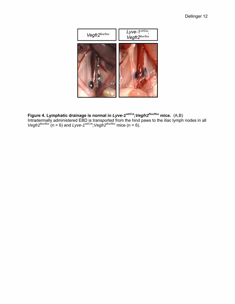
We next turned our attention to other tissues that express Lyve-1 to identify changes that could be responsible for the lethal phenotype of Lyve-1wt/Cre;Vegfr2flox/flox embryos. Lyve-1 has recently been reported to be expressed by BECs in the yolk sac and liver [8]. Indeed, crosses with mT/mG reporter mice demonstrated that Lyve-1wt/Cre mice display Cre recombinase activity in BECs in these tissues (data not shown). To determine whether the vasculature was altered in these tissues, we performed immunohistochemistry with markers of BECs. Whole-mount immunofluorescence staining for CD31 revealed that there was a 61.3% decrease in the number of branch points in yolk sacs isolated from E12.5 Lyve-1wt/Cre;Vegfr2flox/flox embryos (41.06 ± 3.662, n = 4) compared to Vegfr2flox/flox embryos (106.3 ± 3.157, n = 3) (Figure 6A-C). Additionally, immunohistochemistry of E14.5 livers for endomucin showed that there were significantly fewer blood vessels in Lyve-1wt/Cre;Vegfr2flox/flox embryos (27.00 ± 4.263, n = 4) than Vegfr2flox/flox embryos (45.88 ± 2.850, n = 4) (Figure 6D-F). Vegfr2 is not expressed by macrophages and inactivation of Vegfr2 in the myeloid lineage does not affect lymphatic development Crosses with mT/mG reporter mice revealed that Lyve-1Cre mice exhibit Cre recombinase activity in macrophages as well as lymphatic vessels (Figure 1A,B). Therefore, several experiments were performed to rule out the possibility that the lymphatic phenotype of Lyve-1wt/Cre;Vegfr2flox/flox mice was due to the inactivation of Vegfr2 in macrophages. First, we explored the expression of Vegfr2 by macrophages. Whole-mount immunofluorescence staining showed that Vegfr2 was not expressed by macrophages in the ear skin of Vegfr2wt/GFP mice (Figure 7). Next, the LysMCre strain was used to conditionally delete target sequences in the myeloid lineage. LysMCre mice were bred with mT/mG reporter mice to characterize the expression pattern of Cre recombinase and to trace the fate of cells of the myeloid lineage. All LysMwt/Cre;mT/mG mice displayed strong GFP expression by macrophages in the ear skin (Figure 8). GFP did not co-localize with Vegfr3 in the ear skin of LysMwt/Cre;mT/mG mice (Figure 8; n = 5 mice). This finding indicates that cells genetically marked by LysMCre do not differentiate into LECs during normal murine development and is in agreement with another report using the LysMCre line [9]. LysMCre mice were then crossed with Vegfr2flox mice to determine whether deleting Vegfr2 in myeloid cells affects the development of the lymphatic vasculature. Whole-mount immunofluorescence staining of ear skin for Lyve-1 revealed that the density of lymphatic vessels was not significantly different between Vegfr2flox/flox (8.550 ± 0.278, n = 5 mice) and LysMwt/Cre;Vegfr2flox/flox (9.150 ± 0.5895, n = 5 mice) littermates (Figure 9A-D). Furthermore, the diameter of lymphatic vessels was not significantly different between Vegfr2flox/flox (52.98 μm ± 1.328, n = 5 mice) and LysMwt/Cre;Vegfr2flox/flox (50.05 μm ± 2.031, n = 4 mice) littermates (Figure 9A-D). EBD was also effectively transported from injected hind paws to popliteal and iliac lymph nodes in all Vegfr2flox/flox and LysMwt/Cre;Vegfr2flox/flox mice (data not shown). Together, these data reveal that Vegfr2 is not required in the myeloid lineage for the proper development of the lymphatic system. This demonstrates that the lymphatic phenotype of Lyve-1wt/Cre;Vegfr2flox/flox mice is due to the ablation of Vegfr2 in LECs, not macrophages.
KEY RESEARCH ACCOMPLISHMENTS:
• Demonstrated that the Lyve-1Cre mouse can be used to delete floxed sequences in initial and collecting lymphatic vessels.
• Showed for the first time that Vegfr2 expression by lymphatic endothelium is required for the proper development of the lymphatic vasculature.

Dellinger 7
• Specifically, I have demonstrated that Vegfr2 is required for the expansion of the developing lymphatic network, not the enlargement of lymphatic vessels. These findings go against the current dogma regarding Vegfr2’s function in lymphangiogenesis.
REPORTABLE OUTCOMES:
Accepted Manuscripts
1. Witte MH, Dellinger MT, Papendieck CM, Boccardo F. 2012. Overlapping biomarkers, pathways, processes and syndromes in lymphatic development, growth and neoplasia. Clinical and Experimental Metastasis. (In Press).
Manuscript In Preparation
1. Dellinger MT, Hirashima M, Brekken RA. VEGFR2 directly regulates the development of the lymphatic vasculature. (Manuscript in preparation).
Poster Presentations
1. Dellinger MT and Brekken RA. Phosphorylation of Akt and ERK1/2 is required for VEGF-A/VEGFR2-induced proliferation and migration of lymphatic endothelium. Gordon Research Conference: Molecular Mechanisms in Lymphatic Function & Disease (March 4-9, 2012; Ventura, CA).
2. Dellinger MT*, Hirashima M, Brekken RA. VEGFR2 directly regulates the
development of the lymphatic vasculature. NAVBO Developmental Vascular Biology Workshop V (October 14-18, 2012; Pacific Grove, CA).
*This presentation won an award.
CONCLUSIONS:
VEGFR2 is widely recognized as an essential gene driving developmental and tumor angiogenesis. The present study demonstrates for the first time that VEGFR2 also directly promotes the development of the lymphatic vasculature. We show that the density, but not diameter, of lymphatic vessels is dramatically reduced in Lyve-1wt/Cre;Vegfr2flox/flox mice. Additionally, we demonstrate that lymphatic vessels in Lyve-1wt/Cre;Vegfr2flox/flox mice properly mature into collecting vessels. These findings indicate that VEGFR2 is required for the expansion of the lymphatic vessel network but not the maturation of the lymphatic system. This newly identified function of VEGFR2 further defines the molecular pathways controlling the development of the lymphatic vasculature and sheds light on how therapeutic agents targeting VEGFR2 can inhibit tumor lymphangiogenesis.

Dellinger 8
REFERENCES:
1. Pham, T.H., et al., Lymphatic endothelial cell sphingosine kinase activity is required for lymphocyte egress and lymphatic patterning. J Exp Med, 2010. 207(1): p. 17-27.
2. Muzumdar, M.D., et al., A global double-fluorescent Cre reporter mouse. Genesis, 2007. 45(9): p. 593-605.
3. Petrova, T.V., et al., Defective valves and abnormal mural cell recruitment underlie lymphatic vascular failure in lymphedema distichiasis. Nat Med, 2004. 10(9): p. 974-81.
4. Makinen, T., et al., PDZ interaction site in ephrinB2 is required for the remodeling of lymphatic vasculature. Genes Dev, 2005. 19(3): p. 397-410.
5. Dellinger, M., et al., Defective remodeling and maturation of the lymphatic vasculature in Angiopoietin-2 deficient mice. Dev Biol, 2008. 319(2): p. 309-20.
6. Wigle, J.T. and G. Oliver, Prox1 function is required for the development of the murine lymphatic system. Cell, 1999. 98(6): p. 769-78.
7. Karkkainen, M.J., et al., Vascular endothelial growth factor C is required for sprouting of the first lymphatic vessels from embryonic veins. Nat Immunol, 2004. 5(1): p. 74-80.
8. Gordon, E.J., N.W. Gale, and N.L. Harvey, Expression of the hyaluronan receptor LYVE-1 is not restricted to the lymphatic vasculature; LYVE-1 is also expressed on embryonic blood vessels. Dev Dyn, 2008. 237(7): p. 1901-9.
9. Gordon, E.J., et al., Macrophages define dermal lymphatic vessel calibre during development by regulating lymphatic endothelial cell proliferation. Development, 2010. 137(22): p. 3899-910.

Dellinger 9
APPENDICES:
Figure 1. Cre recombinase-mediated deletion of floxed sequences in dermal lymphatic vessels. (A,B) Whole-mount preparations of ear skin from mT/mG and Lyve-1wt/Cre;mT/mG mice. GFP is not expressed in the skin of mT/mG mice (A) but is expressed by lymphatic vessels and macrophages in the skin of Lyve-1wt/Cre;mT/mG mice (B). (C-H) Representative images of transverse sections of ear skin from adult Vegfr2flox/flox mice and Lyve-1wt/Cre;Vegfr2flox/flox mice stained with antibodies against Vegfr2 and Lyve-1. Vegfr2 (C) is expressed by Lyve-1 positive (D) lymphatic vessels in Vegfr2flox/flox mice (E, arrow). In contrast, Vegfr2 (F) expression is lost by Lyve-1 positive (G) lymphatic vessels in Lyve-1wt/Cre;Vegfr2flox/flox mice (H, arrow).

Dellinger 10
Figure 2. Lyve-1wt/Cre;Vegfr2flox/flox mice exhibit a deficiency of dermal lymphatic vessels. (A,B) Whole-mount immunofluorescence staining of ear skin from Vegfr2flox/flox and Lyve-1wt/Cre;Vegfr2flox/flox mice for Lyve-1 . (C) Quantitative analysis showing that there are significantly fewer lymphatic branch points in the ear skin of Lyve-1wt/Cre;Vegfr2flox/flox mice (6.042 ± 0.198, n = 6) than Vegr2flox/flox mice (11.29 ± 0.940, n = 6). (D) Lymphatic vessel diameter is not significantly different between Lyve-1wt/Cre;Vegfr2flox/flox (52.65 μm ± 1.303, n = 6) and Vegfr2flox/flox mice (49.05 μm ± 1.675, n = 6). *** indicates P ˂ 0.001.
Dellinger 11
Figure 3. Lymphatic maturation and blood vessel patterning are normal in Lyve-1wt/Cre;Vegfr2flox/flox mice. Whole-mount immunofluorescence staining of ear skin from adult Vegfr2flox/flox and Lyve-1wt/Cre;Vegfr2flox/flox mice for Lyve-1 (A,B), smooth muscle actin (A,B), and CD31 (C-F). (A,B) Lyve-1 positive lymphatic vessels are not covered by SMA positive cells in Vegfr2flox/flox or Lyve-1wt/Cre;Vegfr2flox/flox mice. (C,D) CD31 expressing lymphatic valves are present in the collecting vessels of Vegfr2flox/flox and Lyve-1wt/Cre;Vegfr2flox/flox mice. (E,F) The patterning of CD31 labeled blood vessels is similar for Vegfr2flox/flox and Lyve-1wt/Cre;Vegfr2flox/flox mice.
Dellinger 12
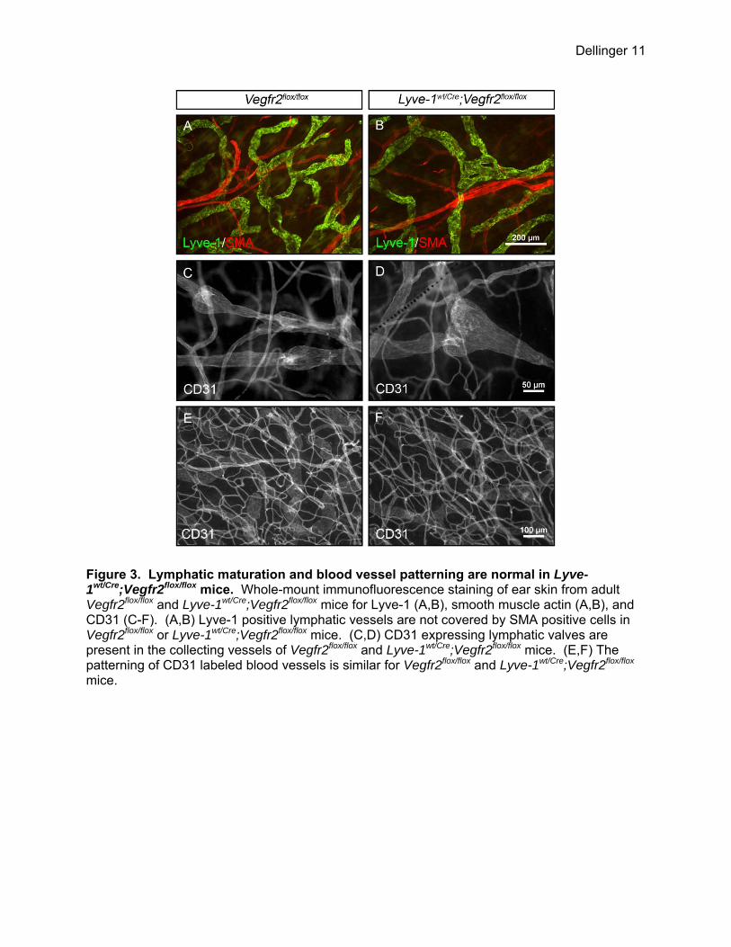
Figure 4. Lymphatic drainage is normal in Lyve-1wt/Cre;Vegfr2flox/flox mice. (A,B) Intradermally administered EBD is transported from the hind paws to the iliac lymph nodes in all Vegfr2flox/flox (n = 6) and Lyve-1wt/Cre;Vegfr2flox/flox mice (n = 6).

Dellinger 13
Figure 5. Lyve1wt/Cre;Vegfr2flox/flox embryos do not exhibit lymphatic defects. (A) Graph showing the percentage of viable Lyve-1wt/Cre;Vegfr2flox/flox mice at different developmental stages. (B,C) Immunofluorescence staining for Lyve-1 showing jugular lymph sacs in E14.5 Vegfr2flox/flox and Lyve-1wt/Cre;Vegfr2flox/flox embryos. (D) The density of dermal lymphatic vessels is not significantly different between E16.5 Vegfr2flox/flox and Lyve-1wt/Cre;Vegfr2flox/flox embryos. (E,F) Representative images showing podoplanin positive dermal lymphatic vessels (arrows) in E16.5 Vegfr2flox/flox and Lyve-1wt/Cre;Vegfr2flox/flox embryos. (G-J) Images of non-edematous E14.5 (G,H), E16.5 (I) and E18.5 (J) Vegfr2flox/flox embryos and Lyve-1wt/Cre;Vegfr2flox/flox embryos.
Dellinger 14
Figure 6. Lyve-1wt/Cre;Vegfr2flox/flox embryos display reduced blood vessel density in the yolk sac and liver. (A,B) Whole-mount immunofluorescence staining of yolk sacs from E12.5 Vegfr2flox/flox and Lyve-1wt/Cre;Vegfr2flox/flox embryos for CD31. (C) There are significantly fewer blood vessel branch points in yolk sacs from Lyve-1wt/Cre;Vegfr2flox/flox embryos (41.06 ± 3.662, n = 4 embryos) than Vegfr2flox/flox embryos (106.3 ± 3.157, n = 3 embryos). (D-F) Immunohistochemical staining of E14.5 livers for endocumin showing significantly fewer blood vessels in Lyve-1wt/Cre;Vegfr2flox/flox embryos (27.00 ± 4.263, n = 4 embryos) than in Vegfr2flox/flox embryos (45.88 ± 2.850, n = 4 embryos). ** indicates P ˂ 0.01; *** indicates P ˂ 0.001.

Dellinger 15
Figure 7. Macrophages do not express Vegfr2. (A-C) Whole-mount immunofluorescence staining showing a GFP (Vegfr2)-negative-Lyve-1-positive macrophage in the ear skin of a Vegfr2wt/GFP mouse.

Dellinger 16
Figure 8. LysMCre is not expressed by lymphatic endothelial cells. (A,B) GFP is not expressed in ear skin from mT/mG mice. (C) GFP expression by macrophages is shown in a whole-mount preparation of ear skin from an adult LysMwt/Cre;mT/mG mouse. (D) GFP (green) does not co-localize with VEGFR3 (red) in the ear skin of a LysMwt/Cre;mT/mG mouse.

Dellinger 17
Figure 9. Deleting Vegfr2 in the myeloid lineage does not affect the development of the lymphatic system. (A,B) Whole-mount immunofluorescence staining of ear skin from Vegfr2flox/flox and LysMwt/Cre;Vegfr2flox/flox mice for Lyve-1 . (C) Quantitative analysis showing that the number of lymphatic branch points in the ear skin of Vegfr2flox/flox (8.550 ± 0.278, n = 5 mice) and LysMwt/Cre;Vegfr2flox/flox mice (9.150 ± 0.5895, n = 5 mice) are not significantly different from one another. (D) Furthermore, lymphatic vessel diameter is not significantly different between Vegfr2flox/flox (52.98 μm ± 1.328, n = 5 mice) and LysMwt/Cre;Vegfr2flox/flox mice (50.05 μm ± 2.031, n = 4 mice).