Auxin Carriers Localization Drives Auxin Accumulation in
9
Auxin Carriers Localization Drives Auxin Accumulation in Plant Cells Infected by Frankia in Casuarina glauca Actinorhizal Nodules 1[W] Francine Perrine-Walker 2 , Patrick Doumas 2 , Mikael Lucas 2 , Virginie Vaissayre, Nicholas J. Beauchemin, Leah R. Band, Je ´rome Chopard, Amandine Crabos, Genevie `ve Conejero, Benjamin Pe ´ret, John R. King, Jean-Luc Verdeil, Vale ´rie Hocher, Claudine Franche, Malcolm J. Bennett, Louis S. Tisa, and Laurent Laplaze* UMR DIAPC, Institut de Recherche pour le De ´veloppement, 34394 Montpellier cedex 5, France (F.P.-W., P.D., V.V., A.C., B.P., V.H., C.F., L.L.); Centre for Plant Integrative Biology, University of Nottingham, Loughborough LE12 5RD, United Kingdom (M.L., L.R.B., B.P., J.R.K., M.J.B.); Department of Cellular, Molecular and Biomedical Sciences, University of New Hampshire, Durham, New Hampshire 03824–2617 (N.J.B., L.S.T.); UMR DAP, Institut National de Recherche en Informatique et Automatique, 34392 Montpellier cedex 5, France (J.C.); and Plate-forme d’Histocytologie et d’Imagerie cellulaire Ve ´ge ´tale, Centre International de Recherche en Agronomie pour le De ´veloppement, 34392 Montpellier cedex 5, France (G.C., J.-L.V.) Actinorhizal symbioses are mutualistic interactions between plants and the soil bacteria Frankia that lead to the formation of nitrogen-fixing root nodules. Little is known about the signaling mechanisms controlling the different steps of the establishment of the symbiosis. The plant hormone auxin has been suggested to play a role. Here we report that auxin accumulates within Frankia-infected cells in actinorhizal nodules of Casuarina glauca. Using a combination of computational modeling and experimental approaches, we establish that this localized auxin accumulation is driven by the cell-specific expression of auxin transporters and by Frankia auxin biosynthesis in planta. Our results indicate that the plant actively restricts auxin accumulation to Frankia-infected cells during the symbiotic interaction. Actinorhizal symbioses are mutualistic associations between plants belonging to eight angiosperm families collectively called actinorhizal plants and the soil actinomycete Frankia. These interactions culminate with the formation of a new root organ, the actino- rhizal nodule, where Frankia is hosted and fixes atmo- spheric nitrogen (Benson and Silvester, 1993). During intracellular infection (e.g. in Casuarina glauca or Alnus glutinosa), diffusible signals are emitted by Frankia at early stages of the interaction leading to root hair deformation. The chemical nature of these signals re- mains unknown but biochemical and genetic studies suggest that they are different from rhizobial Nod factors (Ce ´re ´monie et al., 1998; Normand et al., 2007). Frankia then infects some of the deformed root hairs through intracellular infection threads. A limited number of cell divisions are induced in the cortex close to the infection site leading to the formation of the prenodule. Frankia infects some prenodule cells and starts fixing nitrogen while new cell divisions occur in the pericycle close to a xylem pole forming a nod- ule primordium (Pawlowski and Bisseling, 1996). The nodule primordium subsequently grows and become infected with intracellular Frankia hyphae coming from the prenodule. Actinorhizal nodules have a cen- tral vasculature and the cortical symbiotic tissues containing infected and uninfected cells. Unlike le- gume nodules, actinorhizal nodules are structurally and developmentally related to lateral roots (Pawlowski and Bisseling, 1996). Little is known about the signals exchanged between the two partners during the establishment of actino- rhizal symbioses. The phytohormone auxin controls many developmental processes and has also been in- volved in plant-microbe interactions (Robert-Seilaniantz et al., 2007; Mathesius, 2008; Kazan and Manners, 2009). Recently, we studied the role of auxin influx activity during actinorhizal symbioses. Inhibition of auxin in- flux using the competitive inhibitor naphtoxyacetic acid perturbs actinorhizal nodule formation in C. glauca (Pe ´ret et al., 2007). Two genes encoding putative auxin influx carriers from C. glauca were cloned and charac- terized. One of these genes named CgAUX1 was shown to encode a functional auxin influx carrier by comple- mentation of the Arabidopsis (Arabidopsis thaliana) aux1 1 This work was supported by the Institut de Recherche pour le De ´veloppement, the Agropolis Foundation (grant no. 07024), Hatch grant NH530, and the Agence Nationale de la Recherche (grant no. ANR–08–JCJC–0070–01). L.L. is supported by the Re ´gion Languedoc- Roussillon (grant “Chercheur d’Avenir”). 2 These authors contributed equally to the article. * Corresponding author; e-mail [email protected]. The author responsible for distribution of materials integral to the findings presented in this article in accordance with the policy described in the Instructions for Authors (www.plantphysiol.org) is: Laurent Laplaze ([email protected]). [W] The online version of this article contains Web-only data. www.plantphysiol.org/cgi/doi/10.1104/pp.110.163394 1372 Plant Physiology Ò , November 2010, Vol. 154, pp. 1372–1380, www.plantphysiol.org Ó 2010 American Society of Plant Biologists Downloaded from https://academic.oup.com/plphys/article/154/3/1372/6111467 by guest on 18 January 2022
Transcript of Auxin Carriers Localization Drives Auxin Accumulation in

Auxin Carriers Localization Drives Auxin Accumulationin Plant Cells Infected by Frankia in Casuarina glaucaActinorhizal Nodules1[W]
Francine Perrine-Walker2, Patrick Doumas2, Mikael Lucas2, Virginie Vaissayre, Nicholas J. Beauchemin,Leah R. Band, Jerome Chopard, Amandine Crabos, Genevieve Conejero, Benjamin Peret, John R. King,Jean-Luc Verdeil, Valerie Hocher, Claudine Franche, Malcolm J. Bennett, Louis S. Tisa, and Laurent Laplaze*
UMR DIAPC, Institut de Recherche pour le Developpement, 34394 Montpellier cedex 5, France (F.P.-W., P.D.,V.V., A.C., B.P., V.H., C.F., L.L.); Centre for Plant Integrative Biology, University of Nottingham,Loughborough LE12 5RD, United Kingdom (M.L., L.R.B., B.P., J.R.K., M.J.B.); Department of Cellular,Molecular and Biomedical Sciences, University of New Hampshire, Durham, New Hampshire 03824–2617(N.J.B., L.S.T.); UMR DAP, Institut National de Recherche en Informatique et Automatique, 34392 Montpelliercedex 5, France (J.C.); and Plate-forme d’Histocytologie et d’Imagerie cellulaire Vegetale, Centre Internationalde Recherche en Agronomie pour le Developpement, 34392 Montpellier cedex 5, France (G.C., J.-L.V.)
Actinorhizal symbioses are mutualistic interactions between plants and the soil bacteria Frankia that lead to the formation ofnitrogen-fixing root nodules. Little is known about the signaling mechanisms controlling the different steps of theestablishment of the symbiosis. The plant hormone auxin has been suggested to play a role. Here we report that auxinaccumulates within Frankia-infected cells in actinorhizal nodules of Casuarina glauca. Using a combination of computationalmodeling and experimental approaches, we establish that this localized auxin accumulation is driven by the cell-specificexpression of auxin transporters and by Frankia auxin biosynthesis in planta. Our results indicate that the plant activelyrestricts auxin accumulation to Frankia-infected cells during the symbiotic interaction.
Actinorhizal symbioses are mutualistic associationsbetween plants belonging to eight angiosperm familiescollectively called actinorhizal plants and the soilactinomycete Frankia. These interactions culminatewith the formation of a new root organ, the actino-rhizal nodule, where Frankia is hosted and fixes atmo-spheric nitrogen (Benson and Silvester, 1993). Duringintracellular infection (e.g. in Casuarina glauca or Alnusglutinosa), diffusible signals are emitted by Frankia atearly stages of the interaction leading to root hairdeformation. The chemical nature of these signals re-mains unknown but biochemical and genetic studiessuggest that they are different from rhizobial Nodfactors (Ceremonie et al., 1998; Normand et al., 2007).Frankia then infects some of the deformed root hairsthrough intracellular infection threads. A limited
number of cell divisions are induced in the cortex closeto the infection site leading to the formation of theprenodule. Frankia infects some prenodule cells andstarts fixing nitrogen while new cell divisions occurin the pericycle close to a xylem pole forming a nod-ule primordium (Pawlowski and Bisseling, 1996). Thenodule primordium subsequently grows and becomeinfected with intracellular Frankia hyphae comingfrom the prenodule. Actinorhizal nodules have a cen-tral vasculature and the cortical symbiotic tissuescontaining infected and uninfected cells. Unlike le-gume nodules, actinorhizal nodules are structurallyand developmentally related to lateral roots (Pawlowskiand Bisseling, 1996).
Little is known about the signals exchanged betweenthe two partners during the establishment of actino-rhizal symbioses. The phytohormone auxin controlsmany developmental processes and has also been in-volved in plant-microbe interactions (Robert-Seilaniantzet al., 2007; Mathesius, 2008; Kazan andManners, 2009).Recently, we studied the role of auxin influx activityduring actinorhizal symbioses. Inhibition of auxin in-flux using the competitive inhibitor naphtoxyacetic acidperturbs actinorhizal nodule formation in C. glauca(Peret et al., 2007). Two genes encoding putative auxininflux carriers from C. glauca were cloned and charac-terized. One of these genes named CgAUX1was shownto encode a functional auxin influx carrier by comple-mentation of the Arabidopsis (Arabidopsis thaliana) aux1
1 This work was supported by the Institut de Recherche pour leDeveloppement, the Agropolis Foundation (grant no. 07024), Hatchgrant NH530, and the Agence Nationale de la Recherche (grant no.ANR–08–JCJC–0070–01). L.L. is supported by the Region Languedoc-Roussillon (grant “Chercheur d’Avenir”).
2 These authors contributed equally to the article.* Corresponding author; e-mail [email protected] author responsible for distribution of materials integral to the
findings presented in this article in accordance with the policydescribed in the Instructions for Authors (www.plantphysiol.org) is:Laurent Laplaze ([email protected]).
[W] The online version of this article contains Web-only data.www.plantphysiol.org/cgi/doi/10.1104/pp.110.163394
1372 Plant Physiology�, November 2010, Vol. 154, pp. 1372–1380, www.plantphysiol.org � 2010 American Society of Plant Biologists
Dow
nloaded from https://academ
ic.oup.com/plphys/article/154/3/1372/6111467 by guest on 18 January 2022

mutant. Interestingly, CgAUX1 is expressed in Frankia-infected cells during actinorhizal nodule formation(Peret et al., 2007). These results together with previousdata showing auxin production by Frankia suggest a rolefor auxin in infected cells during the symbiotic interac-tion (Mathesius, 2008; Peret et al., 2008; Grunewaldet al., 2009).The aim of this work was to further explore the
involvement of auxin in the C. glauca-Frankia actino-rhizal symbiosis. Using a combination of computationalmodeling and molecular and cell biology approaches,we establish that the cell-specific expression of auxintransporters leads to localized auxin accumulation inFrankia-infected cells in C. glauca nodules.
RESULTS
Auxin Accumulates in Frankia-Infected Cells inActinorhizal Nodules of C. glauca
Previous reports suggested that the plant hormoneauxin might be involved in the establishment ofactinorhizal symbioses (Hammad et al., 2003; Peretet al., 2007). To study the involvement of auxin in theC. glauca-Frankia symbiotic interaction, we used liquidchromatography mass spectrometry (LC-MS) to quan-tify three major forms of auxin: indole-3-acetic acid(IAA), phenylacetic acid (PAA), and indole-3-butyricacid (IBA) in roots of plant that were not inoculated byFrankia (control), in root portions of inoculated plantsthat did not bear nodules, and in nodules 3 weeks af-ter inoculation. In four independent experiments, thelevels of all three auxins were significantly increasedin roots of inoculated plants compared to controlplants (Fig. 1A). Moreover, a dramatic increase inPAA content was observed in nodules compared toroots (Fig. 1A). These auxin quantification resultsshow that IAA and PAA are the two major formsof auxins present in C. glauca nodules and that thesymbiotic interaction with Frankia leads to increasedPAA accumulation in plant tissues.To localize the sites of auxin accumulation in actino-
rhizal nodules, we generated transgenic C. glauca and
Allocasuarina verticillata plants containing the molecu-lar markers for auxin perception ProGH3-GUS (Hagenet al., 1991), ProDR5-GUS (Ulmasov et al., 1997), orProIAA2-GUS (Swarup et al., 2001). GH3 is an auxin-responsive gene from soybean (Glycine max) and theProGH3-GUS marker has been successfully used tostudy changes in auxin distribution during legumenodule formation (Mathesius et al., 1998). DR5 is asynthetic auxin-responsive promoter derived fromGH3 (Ulmasov et al., 1997), and IAA2 is an auxin-induced Arabidopsis gene. Both ProDR5-GUS andProIAA2-GUS are commonly used as markers to analyzechanges in auxin perception in Arabidopsis. In trans-genic Casuarinaceae, those markers exhibited veryweak basal levels of expression, and were not orvery weakly induced by exogenous auxin (Supple-mental Fig. S1). No expression was found in nodules.These results indicate that these molecular markerscannot be used in Casuarinaceae trees. Therefore, animmunolocalization approach was used to investigatethe distribution of IAA and PAA within C. glaucanodules. For Arabidopsis, IAA immunolocalizationwas shown to reflect total auxin distribution (conju-gated and free IAA), while ProDR5-GUS reveals sites offree auxin perception (Aloni et al., 2003; de Reuilleet al., 2006). In C. glauca nodules, a specific monoclonalIAA antibody binds strongly to Frankia-infected cells(Fig. 1B). Similarly, a polyclonal PAA antibody re-vealed high levels of PAA accumulation in these cells(Fig. 1C). No significant hybridization was found withthe IAA or PAA antibody in uninfected cells in thenodule cortex. Some binding was detected in vasculartissues with the IAA antibody. No signal was detectedunder the following control conditions: hybridizationwith only the secondary antibody (Fig. 1D), pretreat-ment with ethanol to remove auxin, or auxin satura-tion of the antibody. These results indicate that bothIAA and PAA accumulate in Frankia-infected cells ofC. glauca nodules. This conclusion is supported by therecent finding that EuNOD-ARP1, a gene from theactinorhizal plant Eleagnus umbellata shown to beauxin inducible in leaves, is expressed in Frankia-infected cells in nodules (Kim et al., 2007).
Figure 1. Auxin accumulates in Frankia-infected cells in C. glauca nodules. A, Auxin content measured by LC-MS in C. glaucaroots of noninoculated plants (control), roots, and nodules harvested 3 weeks after inoculation by Frankia. Significance wastested by an ANOVA test (P , 0.01). FW, Fresh weight. B, Immunolocalization of IAA. The signal is found in cells infectedby Frankia. C, Immunolocalization of PAA. A very strong signal is detected in cells infected by Frankia. D, Controlimmunolocalization. The section was hybridized only with the secondary antibody. No signal is detected in infected cells.Autofluorescence of the cell wall of infected cells can be detected. Scale bars: 100 mm (B and D), 50 mm (C).
Auxin Transport during Symbiotic Infection
Plant Physiol. Vol. 154, 2010 1373
Dow
nloaded from https://academ
ic.oup.com/plphys/article/154/3/1372/6111467 by guest on 18 January 2022

Distribution of Auxin Carriers Predicts Auxin
Accumulation in Frankia-Infected Cells ofC. glauca Nodules
We previously found that the CgAUX1 gene thatencodes an auxin influx carrier functionally equivalentto Arabidopsis AtAUX1 is expressed in the vasculartissues and in Frankia-infected cells in C. glauca nod-ules (Peret et al., 2007). To test whether this CgAUX1expression might be sufficient to explain the pattern ofauxin accumulation, we used a computational mod-eling approach similar to the ones that have beenapplied successfully to study developmental pro-cesses such as phyllotaxis (de Reuille et al., 2006;Chickarmane et al., 2010). Auxin fluxes and accumu-lation patterns in a tissue can be inferred from thecellular localization of transporters.
We generated cellular models for auxin transportoccurring inC. glauca nodule symbiotic tissues. First, thecortical tissue of C. glauca nodules was observed to havea very specific geometry and topology (SupplementalFig. S2). For instance, infected cells (easily detectedusing Frankia autofluorescence or 4#,6-diamino-phenylindole staining) were bigger (5–10 times) andmore connected, i.e. were in contact with more cells,than noninfected cells (mean connectivity, i.e. numberof contact with other cells, was 6.99 and 4.99, respec-tively). Because of the infection mechanism, infectedcells tend to be organized in files. Thus, confocalimages of C. glauca nodules were digitized and usedto generate our virtual tissues to keep the specificgeometry and topology since fluxes are known to bedependent on those properties. Three in silico modelsof cortical tissues were generated from confocal imagesof three different nodules (Fig. 2A; Supplemental Fig.S2). Two different cell types corresponding to infectedand uninfected cells were defined in the virtual tissues.
After the addition of AUX1-like auxin influx carrieractivity to the infected cells in the in silico model, thedynamic of auxin distribution was tested in the threevirtual tissues. Simulations were conducted with eitherauxin initially homogeneously distributed in the tissueor with auxin coming from the outside of the tissue(as would be the case if auxin was provided by thevascular stream). In all our simulations, the addition ofAUX1 activity in infected cells was not sufficient togenerate a significant auxin accumulation in infectedcells (Fig. 2B; Supplemental Fig. S3). This result sug-gests that other components are needed to generate aspecific auxin accumulation in Frankia-infected cells.
A search of a C. glauca EST database (Hocher et al.,2006), currently containing around 35,000 ESTs, forother auxin transport proteins that might be active innodules identified one EST corresponding to an auxinefflux carrier of the PIN family (EST CG-R02f_005_F13homologous to PIN1 and named CgPIN1 thereafter)and 20 ESTs corresponding to the previously describedputative auxin influx carriers CgAUX1 and CgLAX3(Peret et al., 2007). Expression analyses showed thatCgPIN1 and CgLAX3 were down-regulated 2.5 and
more than 16 times, respectively, in nodules comparedto uninfected roots (Fig. 2C). CgAUX1 expression wasnot significantly different in nodules compared touninfected roots (Fig. 2C). In conclusion, we identifiedgenes encoding an auxin influx carrier (CgAUX1) anda putative auxin efflux carrier (CgPIN1) expressed inC. glauca nodules.
To determine the localization of PIN1-like proteins inC. glauca nodules, we performed immunolocalizationexperiments using polyclonal anti-AtPIN1 antibodiesthat have been previously shown to detect PIN1-likeproteins in Arabidopsis and maize (Zea mays; Carraroet al., 2006). We found a strong signal occurring inthe membrane of uninfected cortical cells surround-ing Frankia-infected cells in C. glauca nodules (Fig. 2,D–G). This signal was strongest in the apical region ofthe nodule close to the nodule meristem (Fig. 2D) andappeared all around the periphery of cells, suggestingthat auxin transport was not polarized (Fig. 2, F andH). Some signal was also found in the vasculature closeto the meristem. Thus in C. glauca nodules, noninfectedcortical cells expressed a gene encoding a PIN1-likeauxin efflux carrier, while infected cells expressed agene encoding an auxin influx carrier (CgAUX1). Whilewe cannot rule out that other transporters might beinvolved in auxin transport in the cortical tissue, noother ESTcorresponding to an auxin carrier was foundin our database that contains 35,000 C. glauca ESTs(including 15,000 ESTs from nodule).
The effects of these auxin influx and efflux carrieractivities in infected and uninfected cells, respectively,were tested in our virtual symbiotic tissues modelsystem.We found in our three in silico models that thiscell-specific localization of auxin transporters wassufficient to cause a rapid auxin accumulation specif-ically in Frankia-infected cells (Fig. 2I; SupplementalFig. S4) as was observed in our auxin immunolocali-zation experiments. This prediction was very robustand held for a wide range of parameter values (Sup-plemental Fig. S5).
Frankia Is a Potential Source of Auxin Production duringthe Symbiotic Association
In our models, if auxin was supplied from outsidethe tissue it accumulated in those infected cells thatwere close to the auxin source (Supplemental Fig. S4).This prediction is in disagreement with our immuno-localization results that showed strong signal in all ofthe infected cells irrespective of their position in thesymbiotic tissues. This inconsistency suggests thatauxin comes from within symbiotic tissues rather thanfrom an external source.
Auxin production has been demonstrated for sev-eral different Frankia strains, but not strain CcI3(Wheeler et al., 1984; Berry et al., 1989; Hammadet al., 2003). IAA, IBA, and PAA production by theC. glauca-infective Frankia strain CcI3 was quantifiedby LC-MS analysis of the supernatant from in vitrocultures. Although Frankia CcI3 cultivated in BAP
Perrine-Walker et al.
1374 Plant Physiol. Vol. 154, 2010
Dow
nloaded from https://academ
ic.oup.com/plphys/article/154/3/1372/6111467 by guest on 18 January 2022
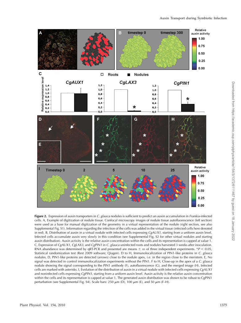
Figure 2. Expression of auxin transporters in C. glauca nodules is sufficient to predict an auxin accumulation in Frankia-infectedcells. A, Example of digitization of nodule tissue. Confocal microscopy images of nodule tissue autofluorescence (left section)were used as a base for manual digitization of the geometry in a virtual representation of the nodule (right section, see alsoSupplemental Fig. S1). Information regarding the infection of the cells was added in the virtual tissue (infected cells here denotedin red). B, Distribution of auxin in a virtual nodule with infected cells expressing CgAUX1, starting from a uniform auxin level.Infected cells accumulate auxin very slowly in this condition (see Supplemental Fig. S2 for other virtual nodules and startingauxin distribution). Auxin activity is the relative auxin concentration within the cells and its representation is capped at value 1.C, Expression of CgAUX1, CgLAX3, and CgPIN1 in C. glauca uninfected roots and nodules harvested 3 weeks after inoculation.RNA abundance was determined by qRT-PCR and presented are means 6 SD of three independent experiments. *P , 0.05,Statistical randomization test (Rest 2009 software; Qiagen). D to H, Immunolocalization of PIN1-like proteins in C. glaucanodules. D, PIN1-like proteins are detected (arrows) close to the nodule apex, i.e. in the region close to the meristem. E, Nosignal was detected in control immunolocalization experiments without the PIN1. F to H, Close-up in the apex of a C. glaucanodule showing the signal corresponding to the PIN1 antibody (F), autofluorescence (G), and the merged image (H). Infectedcells are marked with asterisks. I, Evolution of the distribution of auxin in a virtual nodule with infected cells expressing CgAUX1and noninfected cells expressing CgPIN1, starting from a uniform auxin level. Auxin activity is the relative auxin concentrationwithin the cells and its representation is capped at value 1. The generated auxin distribution was shown to be robust to CgPIN1perturbation (see Supplemental Fig. S4). Scale bars: 250 mm (D), 100 mm (E), and 50 mm (F–H).
Auxin Transport during Symbiotic Infection
Plant Physiol. Vol. 154, 2010 1375
Dow
nloaded from https://academ
ic.oup.com/plphys/article/154/3/1372/6111467 by guest on 18 January 2022

medium produced all three auxin types, IAA and PAAwere the dominant forms of auxin and IBAwas minorform (,2% of the pool; Fig. 3A). Frankia CcI3 growth inBAP medium without nitrogen source caused a sig-nificant increase in IAA and PAA production (Fig. 3A).A .3-fold increase in PAA production was detectedunder these conditions. These results indicate thatauxin production by Frankia CcI3 in vitro is dependenton the nitrogen status and that PAA may be the majorform biosynthesized.
The availability of several Frankia genome databasesincluding Frankia CcI3 (Normand et al., 2007) alloweda genome-mining approach to identify genes poten-tially involved in auxin biosynthesis and transport. Allof the Frankia genomes contained predicted genes forcomplete indole-3-pyruvate and phenyl pyruvatepathways for IAA and PAA biosynthesis, respectively(Fig. 3B). Since many beneficial plant-associated bac-teria preferentially use the indole-3-pyruvate pathwayfor IAA biosynthesis (Spaepen et al., 2007), this resultis not surprising. Frankia CcI3 orthologs were identi-fied for the three key enzymes in the indole-3-pyruvateand the phenyl pyruvate pathways. Interestingly, mostof these genes were found by quantitative reversetranscription (qRT)-PCR to be overexpressed whenbacteria were grown in BAPmediumwithout nitrogensource (Fig. 3, B and C), i.e. under conditions thatcaused a significant increase in IAA and PAA produc-tion. A gene encoding a putative microbial auxinefflux carrier (Francci3_1249) was also identifiedfrom the automatic annotation of Frankia CcI3 and
orthologs were found in all Frankia genomes. RT-PCRexperiments showed that these genes were expressedin Frankia grown in vitro and in C. glauca nodules (Fig.4), suggesting that Frankia IAA and PAA biosyntheticpathways are active in planta.
Auxin biosynthesis by Frankia was included in ourin silico model as a source of auxin. This modificationled to a rapid accumulation of auxin in all infectedplant cells (Fig. 5). If CgPIN1 activity is removed inthis new model, auxin accumulation occurs both ininfected cells and in neighboring uninfected cells (Fig.5). On the other hand, we did not observe any effectafter removing CgAUX1 activity in the model. Thisresult suggests that CgPIN1 is necessary to restrictauxin accumulation to plant cells infected by Frankia.
DISCUSSION
Two nitrogen-fixing root nodule symbioses betweenplants and bacteria have been described, actinorhizalsymbioses, formed by members of the Fagales, Ro-sales, and Cucurbitales with Frankia bacteria, and theinteraction of legumes with rhizobia. Both symbio-ses involve intracellular accommodation of bacteriawithin host cells. While symbiotic signaling mecha-nisms have been widely studied in the legume-rhizobia symbiosis (Oldroyd et al., 2009) very little isknown about signaling mechanisms involved in plant-bacteria recognition in actinorhizal symbioses. Previ-ous studies have suggested a role for auxin in the
Figure 3. Auxin production by Frankia. A,Auxin content measured by LC-MS in thesupernatant of in vitro Frankia CcI3 cul-tures. Bacteria were grown in BAP me-dium with (+N) or without (2N) nitrogensources. Data are means 6 SD. Signifi-cance was tested by an ANOVA test. * P,0.01 compared with noninoculated. B toC, Putative IAA (B) and PAA (C) biosyn-thetic pathways identified in Frankia CcI3using genome data mining. Frankia CcI3orthologs were identified for the three keyenzymes in the indole-3-pyruvate and thephenyl pyruvate pathways (see Supple-mental Table S1). Expression of the corre-sponding genes in Frankia CcI3 bacteriagrown in BAP medium with or without anitrogen source was analyzed by qRT-PCR. Fold changes values represent themean ratios of expression changes inBAP2N relative to BAP+N medium intwo independent biological replicates.
Perrine-Walker et al.
1376 Plant Physiol. Vol. 154, 2010
Dow
nloaded from https://academ
ic.oup.com/plphys/article/154/3/1372/6111467 by guest on 18 January 2022

establishment of actinorhizal symbioses (Hammadet al., 2003; Peret et al., 2007, 2008). Here, we reportthat auxin accumulates in cortical cells infected byFrankia in C. glauca nodules. This result is in agreementwith the expression of the auxin-responsive EuNOD-ARP1 gene in Frankia-infected cells in E. umbellataactinorhizal nodules (Kim et al., 2007) and with theexpression of an auxin-responsive AUX/IAA gene inFrankia-infected cells in C. glauca nodules (V. Vaissayre,A. Crabos, V. Hocher, A. Champion, and L. Laplaze,unpublished data). These results together further in-dicate that auxin-responsive genes are activated ininfected cells and therefore that at least part of theauxin we detected using immunolocalization is active.Results from a combination of experimental biology
and computational modeling approaches indicate thatthis specific accumulation of auxin in Frankia-infectedcell is driven by auxin import into infected cells byexpression of AUX1 in these cells, and auxin exportfrom uninfected cells by expression of a PIN1-like genein those cells. Our data also suggest that Frankia pro-duces auxins (both IAA and PAA) in planta, but ourdata do not allow us to conclude that Frankia is the onlysource of auxin in nodules. Although Frankia mutantstrains defective in auxin biosynthesis would be veryhelpful to clarify this point, techniques to generatestable transgenic or mutant Frankia strains have not yetbeen established (Lavire and Cournoyer, 2003).
However, it is clear that the arrangement of auxintransporters limits the auxin response to those cellsthat are infected by Frankia and prevents the diffusionof this very active biomolecule to other nodule tissuesor parts of the plant. This localization raises thequestion of the role of auxin in infected cells and wepropose three potential functions. First, auxin accu-mulation in infected cells could drive cell growth andhelp explain why infected cells are hypertrophied.Another potential role for auxin is cell wall remodelingprocesses that occur during infection. The infectionthreads are surrounded by the plant cell membraneand a new cell wall-like structure composed mainlyof pectin derivatives (Lalonde and Knowles, 1975).Auxin is known to regulate genes involved in cell wallremodeling, pectin biosynthesis, and methylation(Lerouxel et al., 2006; Swarup et al., 2008). Thus, auxinperception in infected plant cells might be necessaryfor the growth of the infection threads. Finally, auxincould be involved in preventing, controlling, or limit-ing the plant defense mechanisms. Some bacterialpathogens such as Pseudomonas syringae modulateauxin responses and/or production to colonize theirhost (Chen et al., 2007; Robert-Seilaniantz et al., 2007;Kazan and Manners, 2009). A similar process mightoccur in C. glauca cells infected by Frankia.
A role for auxin in infected cellsmight not be a generalfeature of nitrogen-fixation endosymbioses. While therole of auxin in legume nodule development is nowwelldocumented (Mathesius, 2008), little is known about itsrole in the infection process. Although legume infectionby rhizobia strain deficient in IAA production or over-producing auxin causes changes in nodule developmentand nitrogen fixation (Pii et al., 2007; Camerini et al.,
Figure 4. Expression of Frankia genes in the putative IAA and PAApathways. Expression of the corresponding genes was tested in cultureand in plant tissues using RT-PCR. All of the Frankia CcI3 putative IAAand PAA biosynthetic genes were expressed in culture and in planta,but were not expressed in plant root tissues. Furthermore, the twohousekeeping genes and the auxin efflux carrier were expressed underthese conditions.
Figure 5. Simulation of auxin distribution in virtual tissues with auxinsynthesis by Frankia. We considered an additional intracellular com-partment in infected cell in which Frankia would release auxin.Simulation showed that it would lead to auxin accumulation specifi-cally in infected cells provided that CgPIN1 was expressed in thenoninfected cell. Auxin activity is the relative auxin concentrationwithin the cells and its representation is capped at value 1. Theequations that govern diffusive and active transport are discussed indetail in Supplemental Text S1 (model notes).
Auxin Transport during Symbiotic Infection
Plant Physiol. Vol. 154, 2010 1377
Dow
nloaded from https://academ
ic.oup.com/plphys/article/154/3/1372/6111467 by guest on 18 January 2022

2008), the molecular markers of auxin perception ProDR5-GUS and ProGH3-GUS are not expressed in infected cellsof legume nodules (Mathesius et al., 1998; Pacios-Braset al., 2003) and no data on auxin immunolocalizationare available. It thus remains to be shownwhether auxinaccumulation is a common feature of endosymbioticinfection of plant cells by nitrogen-fixing bacteria.
MATERIALS AND METHODS
Plant and Bacterial Material
Casuarina glauca seeds were provided by Carter Seeds and grown as
described in Peret et al. (2007). Transgenic C. glauca and Allocasuarina glauca
plants were produced and analyzed as described (Peret et al., 2007). Frankia
strain CcI3 was grown and used to inoculate C. glauca, as described (Peret
et al., 2007).
Auxin Quantification
Frozen samples were ground in liquid nitrogen and auxins were extracted
twice for 2 h at 4�C in the dark in 80% (v/v) methanol added with 1%
butylated hydroxytoluene. For auxin quantification, 100 pmol of [2H6]IAA and
100 pmol of [13C]PAA (Euriso-Top) were added to the samples. After centri-
fugation (13,000g, 20 min, 4�C), the supernatants were collected and passed
through a C18 cartridge (Waters). Afterward, auxins were purified using a
DEAE-Sephadex A25 cartridge (formic acid conditions; Amersham Pharma-
cia) coupled to C18 cartridge as described in Prinsen et al. (2000). Auxins were
directly eluted from the C18 column with ethyl ether. Mass analysis was
conducted using a Quattro LC with an ESI Z-spray interface (MicroMass),
MassLynx software, an Alliance 2695 RP-HPLC system (Waters), and aWaters
2487 UV detector set at 280 nm. An Xterra C18 column (100 3 2 mm, 5 mm,
Waters) was used with a mix comprising solvent A water with 15 mM formic
acid and solvent B methanol with a gradient profile (starting with 60:40, A/B,
v/v, for 2 min; linear gradient up to 0:100, A/B over 15 min; a washing step
0:100, A/B for 5 min, and final equilibration at 60:40, A/B for 5 min) at a 0.3
mL/min flow rate. Chromatograms were analyzed using the Masslynx
software (Waters), and the IAA, PAA, and IBA concentrations were calculated
according to the principle of isotope dilution (Prinsen et al., 1998). Samples
were quantified in single reaction monitoring mode based on the area of major
MS signals ([M-H+]).
Auxin Immunolocalization
For IAA and PAA immunolocalization, fresh nodules sections of 50 to
60 mM were obtained with a vibratome (MicroM HM650V) and were fixed
overnight, in fixative solution (4% w/v 1-ethyl-3-[3-dimethylaminopropyl]-
carbodiimide hydrochloride [EDAC, Sigma], 4% w/v paraformaldehyde
[Fluka] in phosphate-buffered saline [10 mM pH 7.4]) at 4�C. Sections were
incubated overnight in 2% blocking buffer solution (Roche) at 4�C followed by
overnight incubation with primary antibody, anti-IAA mouse monoclonal
hybridoma antibody (Agdia) at a concentration of 0.05 mg/mL, or anti-PAA
rabbit polyclonal antibody (Abcam) at 1:200 dilution. Sections were then
rinsed (three times 15min) in phosphate-buffered saline (10mM pH 7.4). Alexa
Fluor 488 goat anti-mouse IgG1 (g1) antibody (2 mg/mL; Invitrogen) for IAA
or Alexa Fluor 488 goat anti-rabbit IgG antibody (2 mg/mL; Invitrogen) for
PAAwas used as a secondary antibody at 1:250 dilution at room temperature
during 1 h in the dark. After rinsing (five times for 10 min each), sections were
mounted in Mowiol (Calbiochem) and examined using a confocal microscope
510 META Zeiss (Laser Argon 488, BP 505–530 and LP 560 nm) with LCI Neo-
Neofluar 253/0.8 1 mm Korr DIC27 and EC Plan-Neofluar 103/0.3 objec-
tives. For the ethanol controls, sections were pretreated with ethanol for 1 h at
each concentration (70% v/v and 80% v/v in phosphate-buffered saline 10 mM
pH 7.4) prior to fixation to extract IAA or PAA from the nodule sections. To
verify the binding specificity of the primary anti-IAA mouse monoclonal
antibody was mixed with 10 times its concentration of IAA overnight. This
was to allow the binding of the free IAA to the primary antibody. The solution
was applied to the fixed sections, i.e. the bound primary antibody would not
be able to bind to the antigen on the sections. The same was done to test the
primary anti-PAA rabbit polyclonal antibody against PAA. Another control
treatment was also done whereby the secondary antibody was used alone.
Nodules from 24 and 16 plants from four independent inoculation experi-
ments were used for IAA and PAA immunolocalization, respectively.
PIN1 Immunolocalization
Fresh nodules sections of 50 to 60 mm were obtained with a vibratome
(MicroM HM650V) and were fixed for 1 h in fixative solution (4% w/v
paraformaldehyde [Fluka] in phosphate-buffered saline 10 mM pH 7.4) under
vacuum at room temperature. Sections were incubated overnight in 2%
blocking buffer solution (Roche) at 4�C followed by overnight incubation with
primary antibody, anti-PIN1 goat polyclonal antibody (200 mg/mL; Santa
Cruz Biotechnology) at 1:200 dilution. Sections were rinsed three times (15
min) in phosphate-buffered saline 10 mM pH 7.4. Alexa Fluor 488 F(ab’)2
fragment of rabbit anti-goat IgG (H + L) antibody (2 mg/mL; Invitrogen) was
used as a secondary antibody at 1:250 dilution at room temperature during 1 h
in the dark. After rinsing, sections weremounted inMowiol (Calbiochem) and
examined using a Zeiss confocal microscope 510 META (Laser Argon 488, BP
505–530 and LP 560 nm) with LCI Neo-Neofluar 253/0.8 1 mm Korr DIC27
and EC Plan-Neofluar 103/0.3 objectives. For the control treatment, the
primary antibody was not used at all.
Gene Expression Analyses
C. glauca gene expression analyses were performed by qRT-PCR using
specific primers (Supplemental Table S2) as described (Gherbi et al., 2008).
Reactions were preformed in triplicates and the comparative threshold-cycle
method was used to quantify gene expression. The results were standardized
with CgUBI expression levels (Gherbi et al., 2008).
Total RNA was extracted from Frankia as described previously (Niemann
and Tisa, 2008). To study Frankia gene expression in planta, total RNA was
extracted using the RNeasy plant mini kit (Qiagen GmbH). RNAwas quantified
with a NanoDrop (ThermoFisher). Four-hundred nanograms of total RNAwere
reverse transcribed using SuperScript III reverse transcriptase (Invitrogen) and
random hexamers primers. The RT-PCR was preformed using AmpliTaq Gold
360 master mix (Applied Biosystems) and 100 ng of cDNA as a template. qPCR
was performed using SYBR Green PCR master mix (Applied Biosystems) and
specific primer sets (Supplemental Table S2). Parameters for the Applied
Biosystems 7300 were as follows: (1) one cycle 95�C 10 min, (2) 40 cycles 95�Cfor 15 s and 60�C for 30 s, ended by one cycle at 95�C for 15 s, 60�C for 30 s, and
95�C for 15 s run. Reactions were preformed in triplicates and the comparative
threshold-cycle method was used to quantify gene expression. The results were
standardized with rpsA expression levels. For RT-PCR, the thermocycler pa-
rameters were as follows: (1) initial denaturation at 95�C 5 min, (2) 35 cycles of
denaturation at 95�C for 30 s, primer annealing at 55�C for 30 s, and primer
extension at 72�C for 30 s, and (3) a final extension step at 72�C 10 min.
Amplicons were resolved by gel electrophoresis.
Modeling
Tissues were manually digitized from confocal images to standard vector
graphic files (SVG) and automatically converted for use in the OpenAlea
modeling software platform (Pradal et al., 2008). The models take into account
cells and apoplastic compartments for computing the auxin transport (see
Supplemental Text S1 for additional details). The model parameters (mem-
brane permeability and active transports, auxin dissociation constant, cell and
apoplastic pH, cell membrane potential) were defined in accordance with
values from previously published studies (see Supplemental Text S1). Com-
putational modeling was based on IAA transport parameters as they were the
only auxin transport parameters that are well documented. However, our
model is an abstraction of the physiology of the nodule, and as such is not tied
to an auxin in particular. Similar results would be obtained for PAA assuming
that IAA and PAA transport physiology are similar.
Bioinformatics Studies
The FASTA amino acid sequences of the three Frankia genomes (CcI3,
National Center for Biotechnology Information [NCBI] RefSeq: NC_007777;
ACN14a, NCBI RefSeq: NC_00827; EAN1pec, NCBI RefSeq: NC_009921
and from the three Frankia draft genomes [EuI1c, http://genome.ornl.gov/
microbial/fran_eui1c/; EUN1f, http://genome.ornl.gov/microbial/fran_eun1f/;
Perrine-Walker et al.
1378 Plant Physiol. Vol. 154, 2010
Dow
nloaded from https://academ
ic.oup.com/plphys/article/154/3/1372/6111467 by guest on 18 January 2022

the uncultured Frankia symbiont of Datisca glomerata http://genome.ornl.
gov/microbial/fran_sym/]) were obtained from GenBank or the Department
of Energy Joint Genome Institute Genome Portal site (http://genome.jgi-psf.
org/). Functionally analyzed genes for indole acetic acid and phenyl acetic
acid biosynthesis pathways were identified by the use of Kyoto Encyclopedia
of Genes and Genomes (Kanehisa and Goto, 2000) and published literature
(Patten and Glick, 1996; Spaepen et al., 2007). Proteins representing different
biosynthesis pathways were used (Supplemental Table S1). BLASTP analyses
were performed against each Frankia genome database using each of the above
protein sequences as a query sequence for the representative IAA and PAA
biosynthesis pathways.
Sequence data from this article can be found in the GenBank/EMBL data
libraries under accession numbers FQ375841 (CgPIN1) and NC_007777.1
(Frankia sp. CcI3 genome).
Supplemental Data
The following materials are available in the online version of this article.
Supplemental Figure S1. Expression pattern of the ProIAA2-GUSmolecular
markers for auxin perception in C. glauca.
Supplemental Figure S2. Topology and digitization of symbiotic tissues.
Supplemental Figure S3. Simulation of auxin distribution in virtual
tissues with infected cells expressing CgAUX1.
Supplemental Figure S4. Simulation of auxin distribution in virtual
tissues with infected cells expressing CgAUX1 and noninfected cells
expressing CgPIN1.
Supplemental Figure S5. Robustness of auxin distribution against CgPIN1
perturbation.
Supplemental Table S1. Genome data mining results.
Supplemental Table S2. Primers used for gene expression analyses.
Supplemental Text S1. Model description.
ACKNOWLEDGMENTS
We thank Pr. K. Ljung (Swedish University of Agricultural Sciences,
Umea, Sweden) for providing us with [2H6]IAA, Pr. T. Guilfoyle (University
of Missouri) for the ProDR5-GUS and ProGH3-GUS constructs, and Dr. R.
Swarup (University of Nottingham, UK) for the ProIAA2-GUS construct. We
are grateful to Dr. D. Bogusz (Institut de Recherche pour le Developpement,
France), Dr. A. Champion (Institut de Recherche pour le Developpement,
France), Dr. S. Guyomarc’h (Universite Montpellier 2, France), and Dr. C.
Godin (Institut National de Recherche en Informatique et Automatique,
France) for helpful discussions and critical reading of this manuscript.
Received July 26, 2010; accepted September 4, 2010; published September 8,
2010.
LITERATURE CITED
Aloni R, Schwalm K, Langhans M, Ullrich CI (2003) Gradual shifts in sites
of free-auxin production during leaf-primordium development and
their role in vascular differentiation and leaf morphogenesis in Arabi-
dopsis. Planta 216: 841–853
Benson DR, Silvester WB (1993) Biology of Frankia strains, actinomycete
symbionts of actinorhizal plants. Microbiol Rev 57: 293–319
Berry AM, Kahn RKS, Booth MC (1989) Identification of indole com-
pounds secreted by Frankia HFPAri3 in defined culture-medium. Plant
Soil 118: 205–209
Camerini S, Senatore B, Lonardo E, Imperlini E, Bianco C, Moschetti G,
Rotino GL, Campion B, Defez R (2008) Introduction of a novel pathway
for IAA biosynthesis to rhizobia alters vetch root nodule development.
Arch Microbiol 190: 67–77
Carraro N, Forestan C, Canova S, Traas J, Varotto S (2006) ZmPIN1a
and ZmPIN1b encode two novel putative candidates for polar auxin
transport and plant architecture determination of maize. Plant Physiol
142: 254–264
Ceremonie H, Cournoyer B, Maillet F, Normand P, Fernandez MP (1998)
Genetic complementation of rhizobial nod mutants with Frankia DNA:
artifact or reality? Mol Gen Genet 260: 115–119
Chen Z, Agnew JL, Cohen JD, He P, Shan L, Sheen J, Kunkel BN (2007)
Pseudomonas syringae type III effector AvrRpt2 alters Arabidopsis thaliana
auxin physiology. Proc Natl Acad Sci USA 104: 20131–20136
Chickarmane V, Roeder AH, Tarr PT, Cunha A, Tobin C, Meyerowitz EM
(2010) Computational morphodynamics: a modeling framework to
understand plant growth. Annu Rev Plant Biol 61: 65–87
de Reuille PB, Bohn-Courseau I, Ljung K, Morin H, Carraro N, Godin C,
Traas J (2006) Computer simulations reveal properties of the cell-cell
signaling network at the shoot apex in Arabidopsis. Proc Natl Acad Sci
USA 103: 1627–1632
Gherbi H, Markmann K, Svistoonoff S, Estevan J, Autran D, Giczey G,
Auguy F, Peret B, Laplaze L, Franche C, et al (2008) SymRK defines a
common genetic basis for plant root endosymbioses with arbuscular
mycorrhiza fungi, rhizobia, and Frankia bacteria. Proc Natl Acad Sci
USA 105: 4928–4932
Grunewald W, van Noorden G, Van Isterdael G, Beeckman T, Gheysen G,
Mathesius U (2009) Manipulation of auxin transport in plant roots during
Rhizobium symbiosis and nematode parasitism. Plant Cell 21: 2553–2562
Hagen G, Martin G, Li Y, Guilfoyle TJ (1991) Auxin-induced expression of
the soybean GH3 promoter in transgenic tobacco plants. Plant Mol Biol
17: 567–579
Hammad Y, Nalin R, Marechal J, Fiasson K, Pepin R, Berry AM, Normand
P,Domenach A-M (2003) A possible role for phenyl acetic acid (PAA) on
Alnus glutinosa nodulation by Frankia. Plant Soil 254: 193–205
Hocher V, Auguy F, Argout X, Laplaze L, Franche C, Bogusz D (2006)
Expressed sequence-tag analysis in Casuarina glauca actinorhizal nodule
and root. New Phytol 169: 681–688
Kanehisa M, Goto S (2000) KEGG: kyoto encyclopedia of genes and
genomes. Nucleic Acids Res 28: 27–30
Kazan K, Manners JM (2009) Linking development to defense: auxin in
plant-pathogen interactions. Trends Plant Sci 14: 373–382
Kim HB, Lee H, Oh CJ, Lee NH, An CS (2007) Expression of EuNOD-ARP1
encoding auxin-repressed protein homolog is upregulated by auxin and
localized to the fixation zone in root nodules of Elaeagnus umbellata. Mol
Cells 23: 115–121
Lalonde M, Knowles R (1975) Ultrastructure, composition, and biogenesis
of encapsulation material surrounding endophyte in Alnus crispa var
Mollis root nodules. Can J Bot 53: 1951–1971
Lavire C, Cournoyer B (2003) Progress on the genetics of the N-2-fixing
actinorhizal symbiont Frankia. Plant Soil 254: 125–137
Lerouxel O, Cavalier DM, Liepman AH, Keegstra K (2006) Biosynthesis of
plant cell wall polysaccharides—a complex process. Curr Opin Plant
Biol 9: 621–630
Mathesius U (2008) Auxin: at the root of nodule development? Funct Plant
Biol 35: 651–668
Mathesius U, Schlaman HRM, Spaink HP, Of Sautter C, Rolfe BG,
Djordjevic MA (1998) Auxin transport inhibition precedes root nodule
formation in white clover roots and is regulated by flavonoids and
derivatives of chitin oligosaccharides. Plant J 14: 23–34
Niemann J, Tisa LS (2008) Nitric oxide and oxygen regulate trun-
cated hemoglobin gene expression in Frankia strain CcI3. J Bacteriol 190:
7864–7867
Normand P, Lapierre P, Tisa LS, Gogarten JP, Alloisio N, Bagnarol E, Bassi
CA, Berry AM, Bickhart DM, Choisne N, et al (2007) Genome charac-
teristics of facultatively symbiotic Frankia sp. strains reflect host range
and host plant biogeography. Genome Res 17: 7–15
Oldroyd GE, Harrison MJ, Paszkowski U (2009) Reprogramming plant
cells for endosymbiosis. Science 324: 753–754
Pacios-Bras C, Schlaman HR, Boot K, Admiraal P, Langerak JM, Stougaard J,
Spaink HP (2003) Auxin distribution in Lotus japonicus during root nodule
development. Plant Mol Biol 52: 1169–1180
Patten CL, Glick BR (1996) Bacterial biosynthesis of indole-3-acetic acid.
Can J Microbiol 42: 207–220
Pawlowski K, Bisseling T (1996) Rhizobial and actinorhizal symbioses:
what are the shared features? Plant Cell 8: 1899–1913
Peret B, Svistoonoff S, Lahouze B, Auguy F, Santi C, Doumas P, Laplaze L
(2008) A role for auxin during actinorhizal symbioses formation? Plant
Signal Behav 3: 34–35
Auxin Transport during Symbiotic Infection
Plant Physiol. Vol. 154, 2010 1379
Dow
nloaded from https://academ
ic.oup.com/plphys/article/154/3/1372/6111467 by guest on 18 January 2022

Peret B, Swarup R, Jansen L, Devos G, Auguy F, Collin M, Santi C,
Hocher V, Franche C, Bogusz D, et al (2007) Auxin influx activity is
associated with Frankia infection during actinorhizal nodule formation
in Casuarina glauca. Plant Physiol 144: 1852–1862
Pii Y, Crimi M, Cremonese G, Spena A, Pandolfini T (2007) Auxin and
nitric oxide control indeterminate nodule formation. BMC Plant Biol 7: 21
Pradal C, Dufour-Kowalski S, Boudon F, Fournier C, Godin C (2008)
OpenAlea: a visual programming and component-based software plat-
form for plant modelling. Funct Plant Biol 35: 751–760
Prinsen E, Van Dongen W, Esmans EL, Van Onckelen HA (1998) Micro
and capillary liquid chromatography tandem mass spectrometry: a new
dimension in phytohormone research. J Chromatogr A 826: 25–37
Prinsen E, Van Laer S, Oden S, Van Onckelen H (2000) Auxin analysis.
Methods Mol Biol 141: 49–65
Robert-Seilaniantz A, Navarro L, Bari R, Jones JD (2007) Pathological
hormone imbalances. Curr Opin Plant Biol 10: 372–379
Spaepen S, Vanderleyden J, Remans R (2007) Indole-3-acetic acid in
microbial and microorganism-plant signaling. FEMS Microbiol Rev 31:
425–448
Swarup K, Benkova E, Swarup R, Casimiro I, Peret B, Yang Y, Parry
G, Nielsen E, De Smet I, Vanneste S, et al (2008) The auxin influx
carrier LAX3 promotes lateral root emergence. Nat Cell Biol 10:
946–954
Swarup R, Friml J, Marchant A, Ljung K, Sandberg G, Palme K, Bennett
M (2001) Localization of the auxin permease AUX1 suggests two
functionally distinct hormone transport pathways operate in the
Arabidopsis root apex. Genes Dev 15: 2648–2653
Ulmasov T, Murfett J, Hagen G, Guilfoyle TJ (1997) Aux/IAA proteins
repress expression of reporter genes containing natural and highly
active synthetic auxin response elements. Plant Cell 9: 1963–1971
Wheeler CT, Crozier A, Sandberg G (1984) The biosynthesis of indole-3-
acetic-acid by Frankia. Plant Soil 78: 99–104
Perrine-Walker et al.
1380 Plant Physiol. Vol. 154, 2010
Dow
nloaded from https://academ
ic.oup.com/plphys/article/154/3/1372/6111467 by guest on 18 January 2022