Autotaxin through Lysophosphatidic Acid Stimulates ... · Autotaxin through Lysophosphatidic Acid...
12
of August 21, 2018. This information is current as Transendothelial Migration of Naive T Cells Stimulates Polarization, Motility, and Autotaxin through Lysophosphatidic Acid and Steven D. Rosen Yafeng Zhang, Yi-Chun Maria Chen, Matthew F. Krummel http://www.jimmunol.org/content/189/8/3914 doi: 10.4049/jimmunol.1201604 September 2012; 2012; 189:3914-3924; Prepublished online 7 J Immunol Material Supplementary 4.DC1 http://www.jimmunol.org/content/suppl/2012/09/07/jimmunol.120160 References http://www.jimmunol.org/content/189/8/3914.full#ref-list-1 , 28 of which you can access for free at: cites 58 articles This article average * 4 weeks from acceptance to publication Fast Publication! • Every submission reviewed by practicing scientists No Triage! • from submission to initial decision Rapid Reviews! 30 days* • Submit online. ? The JI Why Subscription http://jimmunol.org/subscription is online at: The Journal of Immunology Information about subscribing to Permissions http://www.aai.org/About/Publications/JI/copyright.html Submit copyright permission requests at: Email Alerts http://jimmunol.org/alerts Receive free email-alerts when new articles cite this article. Sign up at: Print ISSN: 0022-1767 Online ISSN: 1550-6606. Immunologists, Inc. All rights reserved. Copyright © 2012 by The American Association of 1451 Rockville Pike, Suite 650, Rockville, MD 20852 The American Association of Immunologists, Inc., is published twice each month by The Journal of Immunology by guest on August 21, 2018 http://www.jimmunol.org/ Downloaded from by guest on August 21, 2018 http://www.jimmunol.org/ Downloaded from
Transcript of Autotaxin through Lysophosphatidic Acid Stimulates ... · Autotaxin through Lysophosphatidic Acid...

of August 21, 2018.This information is current as
Transendothelial Migration of Naive T CellsStimulates Polarization, Motility, and Autotaxin through Lysophosphatidic Acid
and Steven D. RosenYafeng Zhang, Yi-Chun Maria Chen, Matthew F. Krummel
http://www.jimmunol.org/content/189/8/3914doi: 10.4049/jimmunol.1201604September 2012;
2012; 189:3914-3924; Prepublished online 7J Immunol
MaterialSupplementary
4.DC1http://www.jimmunol.org/content/suppl/2012/09/07/jimmunol.120160
Referenceshttp://www.jimmunol.org/content/189/8/3914.full#ref-list-1
, 28 of which you can access for free at: cites 58 articlesThis article
average*
4 weeks from acceptance to publicationFast Publication! •
Every submission reviewed by practicing scientistsNo Triage! •
from submission to initial decisionRapid Reviews! 30 days* •
Submit online. ?The JIWhy
Subscriptionhttp://jimmunol.org/subscription
is online at: The Journal of ImmunologyInformation about subscribing to
Permissionshttp://www.aai.org/About/Publications/JI/copyright.htmlSubmit copyright permission requests at:
Email Alertshttp://jimmunol.org/alertsReceive free email-alerts when new articles cite this article. Sign up at:
Print ISSN: 0022-1767 Online ISSN: 1550-6606. Immunologists, Inc. All rights reserved.Copyright © 2012 by The American Association of1451 Rockville Pike, Suite 650, Rockville, MD 20852The American Association of Immunologists, Inc.,
is published twice each month byThe Journal of Immunology
by guest on August 21, 2018
http://ww
w.jim
munol.org/
Dow
nloaded from
by guest on August 21, 2018
http://ww
w.jim
munol.org/
Dow
nloaded from

The Journal of Immunology
Autotaxin through Lysophosphatidic Acid StimulatesPolarization, Motility, and Transendothelial Migration ofNaive T Cells
Yafeng Zhang,*,† Yi-Chun Maria Chen,†,‡ Matthew F. Krummel,†,‡ and Steven D. Rosen*,†
Blood-borne lymphocytes home to lymph nodes by interacting with and crossing high endothelial venules (HEVs). The transendo-
thelial migration (TEM) step is poorly understood. Autotaxin (ATX) is an ectoenzyme that catalyzes the conversion of lysophos-
phatidylcholine (LPC) to lysophosphatidic acid (LPA), a bioactive lipid and a close relative of sphingosine 1-phosphate. HEVs
produce and secrete ATX into the blood. A prior study implicated ATX in the overall homing process, but the step in which it
functions and its mechanism of action have not been defined. In this article, we show that HA130, an inhibitor of the enzymatic
activity of ATX, slows T cell migration across lymph node HEVs in vivo. Ex vivo, ATX plus LPC or LPA itself induces the polar-
ization of mouse naive T cells and stimulates their motility on an ICAM-1 substratum. Under physiologic shear conditions in a flow
chamber, LPA or ATX/LPC strongly enhances TEM of integrin-arrested T cells across an endothelial monolayer. HA130 blunts the
TEM-promoting activity of ATX, paralleling its in vivo effects. T cells possess Mn+2-activatable receptors for ATX, which are
localized at the leading edge of polarized cells. ATX must bind to these receptors to elicit a maximal TEM response, providing a
mechanism to focus the action of LPA onto arrested lymphocytes in flowing blood. Our results indicate that LPA produced via ATX
facilitates T cell entry into lymph nodes by stimulating TEM, substantiating an additional step in the homing cascade. This entry
role for LPA complements the efflux function of sphingosine 1-phosphate. The Journal of Immunology, 2012, 189: 3914–3924.
Lymphocyte migration (homing) from the blood into sec-ondary lymphoid organs (SLOs) is an essential stepin lymphocyte recirculation, the process by which the
repertoire of naive lymphocytes rapidly cycles through SLOs,thereby enabling contact between sequestered Ags and rarecognate lymphocytes (1–3). For all SLOs except spleen, the portalof entry for blood-borne lymphocytes is high endothelial venules(HEVs) (1, 4, 5). These vessels are functionally specialized tocapture lymphocytes from the flowing blood and to support theirmigration into SLOs. As is generally the case for leukocyte–en-dothelial cell (EC) interactions (6), naive T cell recruitment acrossHEVs occurs in several sequential steps: rolling of lymphocytesalong the endothelium, arrest on the endothelium, intraluminalcrawling, and finally transendothelial migration (TEM) into theSLO (2, 4, 5). In peripheral lymph node (PLN) HEVs, the firststep is mediated by transient interactions between L-selectinon lymphocytes and a complex of mucins on HEVs (7). The sec-
ond step is due to arrest chemokines, such as CCL21, which areimmobilized apically on HEVs (2, 5, 8). Signaling through CCR7,CCL21 activates aLb2 on lymphocytes, which increases theintegrin’s affinity for ICAM-1/ICAM-2 on HEVs, leading to therapid arrest of the rolling cells (8–10). Some of the lymphocytescrawl intraluminally for several minutes before undergoing TEM,whereas the remainder undergo TEM without migration (11).TEM occurs within 2.5 min for T cells (11). Shear stress providedby blood flow is required for both the integrin-mediated arrest andTEM steps (12, 13).Previously, gene profiling of purified HEV-ECs unexpectedly
revealed a very high expression of autotaxin (ATX) transcripts (14).ATX was initially discovered as a secreted protein from A2058melanoma cells, which enhances their own motility (15). ATX isan ∼110-kDa protein with two N-terminal somatomedin B-likedomains, a phosphodiesterase domain, and a C-terminal nuclease-like domain (16, 17). ATX was later shown to be a lysophospholi-pase D, which catalyzes the conversion of lysophosphatidylcholine(LPC) to lysophosphatidic acid (LPA) (18). As an extracellularlysophospholipid, LPA engages six GPCRs (termed LPA1–6) andevokes diverse growth factor-like responses (motility, proliferation,survival, and differentiation) in multiple cell types (19, 20). LPA isnow known to be responsible for the motility-promoting action ofATX on A2058 cells, as well as on other cancer and normal cells(21). ATX performs essential functions in vasculogenesis andneural tube formation during embryonic development (22, 23). Inthe adult, ATX is present in the blood and is responsible for themaintenance of LPA in plasma (22, 23). In mice, the normal levelof LPA is 200–400 nM (24), and in humans it is 80–90 nM (25).Pathologic roles for ATX are indicated in cancer and cardiovasculardisease (26, 27). In the context of immune function, ATX is over-expressed in synovial fibroblasts in rheumatoid arthritis and hasbeen implicated in the pathogenic process (28). LPA acting throughLPA2 inhibits dendritic cell activation and dampens allergic airwayinflammation (29).
*Department of Anatomy, University of California San Francisco, San Francisco, CA94143; †Program in Immunology, University of California San Francisco, San Fran-cisco, CA 94143; and ‡Department of Pathology, University of California San Fran-cisco, San Francisco, CA 94143
Received for publication June 11, 2012. Accepted for publication August 14, 2012.
This work was supported by grants from the National Institutes of Health (R01-GM57411 and R01-GM23547 to S.D.R. and AI52116 to M.F.K.). Y.Z. was supportedby a postdoctoral fellowship from the American Heart Association (A115033).
Address correspondence and reprint requests to Dr. Steven Rosen, University ofCalifornia San Francisco, 513 Parnassus Avenue, Box 0452, San Francisco, CA94143. E-mail address: [email protected]
The online version of this article contains supplemental material.
Abbreviations used in this article: ATX, autotaxin; b-ATX, biotin linked to autotaxin;EC, endothelial cell; HEV, high endothelial venule; LN, lymph node; LPA, lysophos-phatidic acid; LPAR, lysophosphatidic acid receptor; LPC, lysophosphatidylcholine;MLN, mesenteric lymph node; PLN, peripheral lymph node; PTX, pertussis toxin;SLO, secondary lymphoid organ; S1P, sphingosine 1-phosphate; TEM, transendothe-lial migration.
Copyright� 2012 by TheAmericanAssociation of Immunologists, Inc. 0022-1767/12/$16.00
www.jimmunol.org/cgi/doi/10.4049/jimmunol.1201604
by guest on August 21, 2018
http://ww
w.jim
munol.org/
Dow
nloaded from

The discovery of abundant ATX transcripts in HEV-ECs promptedtwo studies, which confirmed that ATX protein is expressed inHEVs of SLOs (30, 31). We further found that ATX is secretedapically by HEV-ECs; ATX can bind to receptors on chemokine-activated T cells; LPA is chemokinetic for T cells; and injectionof a catalytically inactive form of ATX (T210A) partially inhibitshoming of T cells into SLOs (30). These findings led to a para-crine model of ATX function in homing (30), whereby ATX is se-creted into the lumens of HEVs and binds to proximally arrestedT cells. The bound ATX uses the abundant LPC in the plasma(∼200 mM) to produce LPA, which promotes T cell entry into thelymphoid organ.This speculative model has awaited further in vivo validation
and a mechanistic understanding of how ATX and its enzymaticproduct LPA influence lymphocyte migration upon and across anendothelial substratum under physiologic shear stress conditions.The present study addresses these issues.
Materials and MethodsReagents
Mouse ICAM-1–Fc (796-IC-050), CCL21 (457-6C-025), and TNF-a (210-TA) were from R&D Systems (Minneapolis, MN). The Abs used wereanti-CD44 (IM7; BD Biosciences, San Jose, CA), anti-CD3e (45-2C11;BD), anti-B220 (RA3-6B2; BD), anti-autotaxin AF5255; R&D Systems),anti-CD49d (PS/2; Serotec, Raleigh, NC), and anti-CD43 (eBioR2/60;eBiosciences, San Diego, CA). Stock solutions of LPA (18:1 Oleolyl-LPA; L7260, Sigma-Aldrich, St. Louis, MO) were made in methanol (10mM) and stored at 280˚C. Dilutions into aqueous buffers were preparedjust before use, with methanol serving as the carrier control. We used 18:1LPA for our studies because it occurs naturally in blood (24) and is one ofthe enzymatic products of ATX (32). Fatty-acid free BSA (A8806), L-a-lysophosphatidylcholine (L4129), pertussis toxin (PTX), and Y-27632were from Sigma-Aldrich. BrP-LPA was from Echelon (Logan, UT). Thebiotinylation-labeling kit (704-0030) came from Novus (St. Charles, MO).Cy2-streptavidin (016-220-084) and Cy3–anti-rat IgG (712-166-150) werefrom Jackson ImmunoResearch (West Grove, PA).
Mice
All mouse protocols were approved by the University of California SanFrancisco Committee for Animal Research. C57BL/6 female mice (6–8 wk;Charles River Labs, Wilmington, MA) were used for homing assays. OTII,Ub-GFP, Itgb2 null, and Itgb3 null mice were from The Jackson Laboratory.OTII-GFP mice were obtained by crossing OTII mice with Ub-GFP mice.
Cells
CD3+ T cells were purified from mechanically dispersed PLNs using theEasySep T cell Enrichment Kit (StemCell Tech, Vancouver, BC, Canada).For motility assays, CD4+ T cells were purified from PLNs of OTIIUb-GFP mice using the EasySep Mouse CD4+ T Cell Enrichment Kit(StemCell Tech). Purified cells were resuspended in RPMI 1640 plus 10%charcoal-dextran–treated FBS and incubated at 37˚C for ∼1 h before im-aging. At least 90% of the CD3+ cells and $95% of the CD4+ T cells werenaive, as defined by the criterion of CD44lo. For most of the experiments,these populations are referred to as naive T cells. TK1 cells were main-tained in RPMI 1640 with 10% FBS, 100 U/ml penicillin, 100 mg/mlstreptomycin, and 25 mM 2-ME.
T cell homing
Short-term homing of T cells was carried out as described (30). CFSE-labeled CD3+ T cells (20 3 106 in 100 ml), with or without HA130 (2nmol/g recipient mouse), in DMSO were injected i.v. into mice. HA130or DMSO was reinjected at 7 and 12 min. At 15 min, SLOs were cryo-stat processed for immunohistochemistry to highlight HEVs (MECA-79staining for lymph nodes and CD31 staining for Peyer’s patches). Thenumber of fluorescent lymphocytes outside HEVs (in the lymphoid organparenchyma) and within HEVs (both in the lumens and walls) werecounted. Two mice were processed at a time (one HA130 and one control).Six nonconsecutive sections from two mesenteric lymph nodes (MLNs)and three PLNs of each mouse were evaluated. Ratios were determined foreach section, and a mean ratio was computed for each mouse. Means fromthree mice/group were combined to yield overall means 6 SDs.
ATX/T210A preparation
The recombinant proteins were prepared as described previously (30).
Uropod assay
Eight-well chamber slides (154534, Lab-Tek; Thermo, Rochester, NY) werecoated with 3 mg/ml ICAM-1–Fc and CCL21 overnight. The slides werethen blocked with 3% fatty acid-free BSA for 1 h. TK1 lymphoma cellswere cultured with 5% charcoal-dextran–treated FBS overnight; CD3+
T cells were cultured with 10% charcoal-dextran–treated FBS for 2 h.Cells (0.5–1 3 106) were added to the chamber (200 ml) containing HBSSbuffer with 0.2% fatty acid-free BSA. LPA or ATX/LPC were added, andcells were allowed to settle at 37˚C. Attached cells were fixed with 4%formaldehyde in PBS. After washing and blocking with BSA, uropods werevisualized by staining with anti-CD44 or CD43 in combination with sec-ondary Abs. For Gai or ROCK inhibition, cells were cultured for 2 h withPTX (200 ng/ml) or Y27632 (10 mM), respectively. For ATX-inhibitionexperiments, cells were incubated with HA130 (0.3 mM) or BrP-LPA(10 mM) for 30 min prior to the addition of ATX.
Cell-motility assays
Custom crawling chambers were assembled. Dividers for six chambers weremade from polydimethylsiloxane (SYLGARD 184 Silicone Elastomer Kit,10:1 mix; Midland, MI) formed in a printed mold. Each divider was cut outusing a scalpel, placed on top of a glass slide (48311-703; VWR, Batavia,IL), and covered with a No.1 cover glass (48393-059; VWR) to create thechambers. For assays, the chambers were freshly coated with 3 mg/mlICAM-1/Fc, with or without 400 ng/ml CCL21, at 4˚C overnight. Thechambers were washed in PBS, blocked with PBS plus 0.5% fatty acid-freeBSA, and kept in the blocking buffer until use. A total of 53 104 OTII Ub-GFP CD4+ T cells was resuspended in 100 ml block and seeded into thechamber. For LPA treatment, 1 mM LPA was added to the cells immedi-ately before imaging. For ATX/LPC treatment, cells were incubated with 1mg/ml ATX for 5 min, followed by 10 mM LPC cells immediately beforeimaging. Imaging was done with a Zeiss Axiovert 200M microscope witha Plan-Neofluar 203 objective fitted with dual excitation and emissionfilter wheels and a Photometrics CoolSNAP HQ camera. MetaMorphsoftware (Universal Imaging; Molecular Devices, Sunnyvale, CA) wasused for image acquisition and microscopic control. Images were collectedin the GFP channel every 15 s for 15 min. Imaris software was used forimage analysis and tracking (Bitplane, South Windsor, CT).
ATX staining
To analyze ATX receptors, cells were incubated with a preformed complexof biotin linked to ATX (b-ATX) (10 mg/ml) and PE-conjugated strepta-vidin (2 mg/ml) in HBSS buffer (with or without 0.5 mM Mn+2) at roomtemperature. T cells were gated by CD3 staining. PS/2 mAb (10 mg/ml)was used to block a4 integrin.
To analyze b-ATX distribution by immunofluorescence, TK1 cells onan ICAM-1 substratum were exposed to 0.1 mM LPA. Following fixationwith 4% formaldehyde, washing, and blocking, the cells were sequentiallyreacted with a complex of b-ATX/Cy2-streptavidin and anti-CD44 with aCy3-conjugated secondary Ab.
Transmigration under shear flow
BEnd.3 ECs were grown to confluence in a 0.2% gelatin-coated BioFlux 48-well flow chamber plate (Fluxion Biosciences, South San Francisco, CA).Monolayers were stimulated for 16 h with TNF-a (500 U/ml). CCL21 (1mg/ml with 0.2% fatty acid-free BSA) was overlaid on the monolayer for5 min, followed by washing. T cells in HBSS with 0.2% fatty acid-freeBSAwere perfused for 2 min over the monolayer at 0.5 dyne/cm2, and theflow was stopped for 5 min. The flow rate was then increased to 1 dyne/cm2 for an additional 30 min. Images were recorded at one frame/10 s byvideo recorder. The total number of arrested cells was determined after 10min of flow. During the 30 min of flow, lymphocytes fell into three cate-gories: cells that moved less than two diameters (static), cells that crawledmore than two cell diameters without detaching/transmigrating (crawling),and cells that became dark under phase contrast (undergoing TEM).HA130 (0.3 mM final) or BrP-LPA (10 mM final) was added in someexperiments. For competition with T210A, the chamber contained a 10-fold excess of T210A relative to the indicated amount of ATX.
Statistical analysis
The unpaired Student t test was used to determine the statistical signifi-cance of pair-wise comparisons after satisfying t test criteria (33) of
The Journal of Immunology 3915
by guest on August 21, 2018
http://ww
w.jim
munol.org/
Dow
nloaded from

equivalent variances and Gaussian distributions (approximately equalmean and median values). For comparisons of three or more groups, one-way ANOVA with the Tukey posttest was used.
ResultsHA130 inhibits homing of T cells
HA130 was recently identified as a potent small-molecule inhibitorof ATX (34). The inhibitor acts to reduce both the turnover numberof ATX and its affinity for LPC. We used a modified lymphocyte-homing assay to accommodate the short half-life of HA130 inblood (∼3 min) (34). We injected CFSE-labeled T cells togetherwith HA130 i.v. into mice and then reinjected the drug at 7 and 12min. After 15 min, SLOs were sectioned and stained to revealHEVs. CFSE+ T cells were classified as either outside HEVs (inthe lymphoid organ parenchyma) or within HEVs (luminal or inHEV walls). The ratio of outside HEVs to inside HEVs was usedas an index of T cell migration across HEVs. In Peyer’s patches,very few cells migrated into the parenchyma in 15 min, so theaction of HA130 could not be evaluated. However, for PLNs andMLNs, many cells had migrated into the parenchyma (Fig. 1A).HA130 decreased the outside HEV/inside HEV ratio by 3–4-foldcompared with vehicle treatment (Fig. 1B, p, 0.01 for both PLNsand MLNs). This result is consistent with HA130 retarding themigration of T cells across LN HEVs.
LPA and ATX/LPC polarize naive T cells
LPA induces chemokinesis of T cells in Transwell assays (30). Wesought to investigate the cellular mechanisms by which LPA wasexerting these effects. Stam et al. (35) reported that LPA inducedthe formation of pseudopods in a lymphoma cell line. To extendthese findings, we examined the effects of LPA, as well as ATX,on T cells. Although T cells in the blood are round, T cells thathave entered a tissue exhibit an amoeboid “hand mirror” mor-phology, which is characterized by a broad leading edge, a cellbody with the nucleus, and a uropod at the tail (9, 36, 37). We firstasked whether LPA at concentrations previously shown to be ac-tive in Transwell assays (30) could induce the polarization of TK1cells, a mouse CD8+ T cell line (38). We quantified this responseby monitoring the accumulation of CD44 in uropods (37). TK1cells were mixed with LPA at varying concentrations and allowedto settle on an ICAM-1 substratum. With no added LPA, 10% ofthe cells were polarized by 7 min (Fig. 2A, 2B). At 0.1 mM, thepolarization was 48% and reached 78% at 10 mM. LPA at 1 mMinduced maximal polarization within 5 min, with no detectabledecrease after 30 min, indicating the absence of desensitizationover this period (Fig. 2C).We next investigated primary CD3+ T cells, which were isolated
by negative selection from LNs. At least 90% of these cells werenaive defined by the criterion of CD44lo (data not shown), and we
FIGURE 1. HA130 impedes T cell entry into LNs.
(A) HEVs in PLN and MLN sections were stained with
MECA-79. Representative images of CFSE-labeled
T cells in control and HA130-treated mice after i.v.
injection of CFSE-labeled T cells. Scale bar, 50 mM.
Green arrow indicates CFSE-labeled T cell, and red
arrow indicates MECA-79–stained HEV. (B) Bar graphs
for PLN and MLN showing mean ratios 6 SDs of
CFSE-labeled cells outside HEV/inside HEV. Two
mice were processed/experiment, with six nonconsec-
utive sections of two MLNs and three PLNs evaluated
for each mouse. The data shown represent the pooled
results from three experiments (three mice of each
group). **p , 0.01, versus DMSO control.
3916 AUTOTAXIN AND LYSOPHOSPHATIDIC ACID IN NAIVE T CELL HOMING
by guest on August 21, 2018
http://ww
w.jim
munol.org/
Dow
nloaded from

refer to this population as naive. LPA also induced polarization ofthese cells (Fig. 2D), with uropods identified by CD43 staining(37). LPA at 10 mM induced polarization to a maximum of 32%.Alon and coworkers (39) reported that immobilized CCL21 causesrapid polarization and motility of T cells on an adhesive sub-stratum. Therefore, we asked what effect LPA would have onT cells that were simultaneously exposed to CCL21, which wascoimmobilized with ICAM-1. CCL21 alone (200 ng/ml) inducedpolarization of naive T cells to 21% (Fig. 2D). Adding 10 mMLPA increased the level of polarization to 58%. A 2-fold higherlevel of CCL21 did not produce further augmentation (data notshown). Thus, soluble LPA and immobilized CCL21 act additivelyto promote uropod formation in naive T cells.Because ATX catalyzes the production of LPA, we wanted to
determine whether ATX could also induce the polarization ofT cells. When we added ATX (5 mg/ml) to TK1 cells on an ICAM-1 substratum, we observed uropod formation in the presence ofLPC but not in its absence (Fig. 3A). LPC at 10 mM was moreeffective than at 1 mM. The IC50 for ATX in this assay was ∼0.2mg/ml. Notably, the response to ATX/LPC was very rapid. After30 s of exposure to 1 mg/ml of ATX, the polarization of TK1 cellsdoubled to 34% relative to the baseline level (Fig. 3B). By 10 min,the level of polarization reached ∼75%, which was largely sus-tained for another 20 min.To verify the importance of the enzymatic activity of ATX, we
tested an enzymatically inactive form (T210A). There was nouropod-inducing activity by T210A at 5 mg/ml in the presenceof LPC (1 mM) (Fig. 3A). We also tested two small-moleculeinhibitors of ATX: HA130 (see above) and BrP-LPA (40). Thelatter compound is a bromophosphonate analog of LPA, which isa low micromolar inhibitor (IC50 ∼ 0.1 mM) of ATX and antag-onizes several of the LPA receptors (LPARs) (40). HA130 at 0.3mM and BrP-LPA at 10 mM completely ablated the activity ofATX on TK1 uropod formation (Fig. 3C). However, neither hadany effect on LPA-induced uropods. Thus, we conclude that BrP-LPA was able to block ATX-induced uropods by inhibiting ATXrather than LPA antagonism, which is consistent with the fact thatBrP-LPA does not block all LPARs (40). Pretreatment of TK1
cells with 200 ng/ml PTX did not diminish ATX-induced polari-zation, indicating that the Gai subfamily of G proteins was notinvolved in this response (Fig. 3D). Y27632 is a pharmacologicinhibitor of ROCK, a kinase that regulates myosin contractility(11). ROCK is an effector of RhoA, which is downstream ofGa12/13 G proteins (41). Incubation of TK1 cells with Y27632(10 mM) completely blocked the induction of uropods by ATX(Fig. 3D).We next examined primary naive CD3+ T cells and found that
ATX in combination with LPC also induced their polarization(Fig. 3E). As with LPA, the action of ATX on uropod formationwas additive to that of immobilized CCL21. Without LPC, ATXdid not augment polarization. ATX, as well as LPA, also inducedthe polarization of primary human T cells and neutrophils fromperipheral blood (Fig. 3F). Neutrophil polarization by LPA wasreported previously (42).
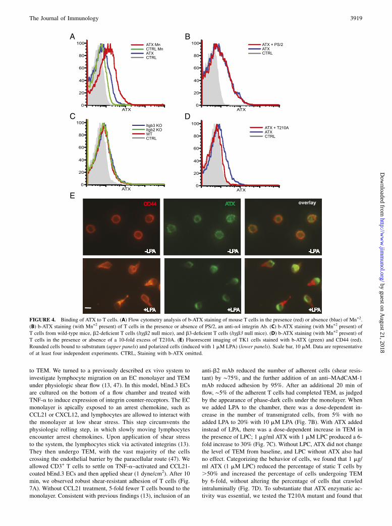
ATX receptors on T cells
Our previous work showed that human T cells can bind to plastic-immobilized ATX in an a4b1-dependent manner (30). We foundthat CCL21 stimulation of T cells or the addition of Mn+2 (whichglobally activate integrins) greatly augments a4b1-dependentbinding, suggesting that ATX binding requires the active confor-mation of a4b1 (30). Similarly, activated b1 and b3 integrins onplatelets are required to bind ATX (43). Although Mn+2 increasesthe binding of mouse T cells to immobilized ATX, we were unableto implicate the involvement of any particular integrin (30). Tofurther study ATX receptors on lymphocytes, we linked biotin toATX (b-ATX) to generate a soluble probe. Consistent with pre-vious observations (30), naive CD3+ T cells showed increased b-ATX staining in the presence of Mn+2 (Fig. 4A), as did CD4+ andCD8+ subsets of T cells as well as B cells (Supplemental Fig. 1). Aneutralizing Ab to a4b1 (PS/2) did not diminish staining ofT cells (Fig. 4B), and T cells from b2- and b3-null mice did notshow reduced staining compared with wild-type T cells (Fig. 4C).b-ATX staining of T cells was inhibited .90% by the additionof a 10-fold excess of T210A, verifying that the receptors weresaturable (Fig. 4D). Staining of rounded TK1 cells with b-ATX
FIGURE 2. LPA induces the rapid polarization of
T cells. (A) TK1 lymphoma cells were allowed to
settle on ICAM-1 substratum in the presence of the
indicated concentration of LPA. After 7 min, the
percentage of cells that became polarized was de-
termined as defined by CD44 accumulation in uro-
pods. (B) CD44 staining of TK1 cells after a 7-min
exposure to 10 mM LPA (right panel) or buffer
alone (left panel). Scale bar, 10 mM. (C) Time
course of TK1 polarization response to 0.1 mM
LPA. (D) Naive mouse T cells were allowed to settle
on substratum of ICAM-1 or ICAM-1 with coim-
mobilized CCL21 (200 ng/ml input). After 7 min,
polarization was determined based on accumulation
of CD43 into uropods. CD43 was used instead of
CD44 because of the low expression of the latter on
naive T cells. In (A), (C), and (D), means and SDs
are shown and are based on three replicate wells.
Data are representative of three independent experi-
ments. **p , 0.01, uropod formation with CCL21
versus without CCL21.
The Journal of Immunology 3917
by guest on August 21, 2018
http://ww
w.jim
munol.org/
Dow
nloaded from

revealed a patchy distribution around the cells. Upon LPA-inducedpolarization of TK1 cells, b-ATX staining was greatly enriched atthe leading edge of cells opposite from the CD44-stained uropod(Fig. 4E).
LPA and ATX/LPC induce motility of naive T cells
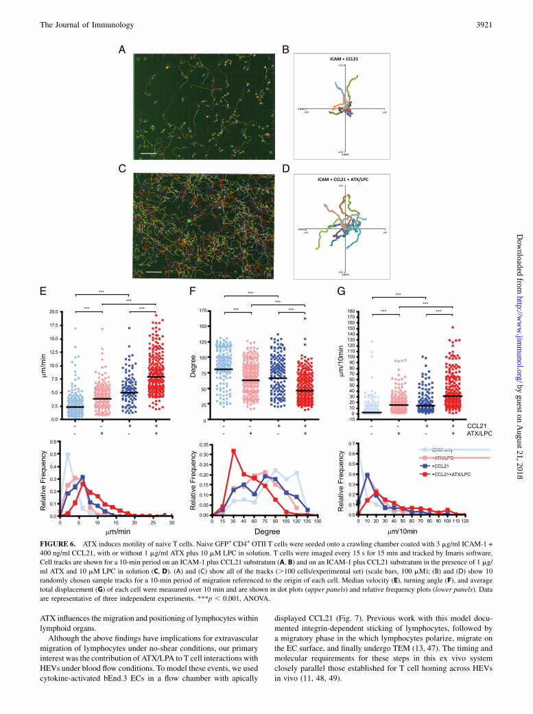
Because a polarized morphology is a requirement for active leu-kocyte migration (9, 36, 37), we performed motility assays. In thisstudy, we used CD4+ T cells, of which $95% were naive. Usinga custom chamber for visualizing two-dimensional cell behavior,we found that 1 mM LPA augmented CD4+ T cell migration ona substratum of coimmobilized ICAM-1 and CCL21 (Fig. 5). LPAinduced a 26% increase in median velocity, from 4.72 to 5.96 mm/min (p , 0.0001, Fig. 5A). Furthermore, the LPA-treated cellsexhibited a marked shift to smaller median turning angles (81.3versus 51.1˚, p , 0.0001) (Fig. 5B), indicating a greater tendencyto move in a straight line. These effects persisted for $60 minfollowing exposure to LPA (data not shown).We next investigated ATX in the migration chamber (Fig. 6).
Consistent with the previous report (39), immobilized CCL21
promoted naive T cell migration on ICAM-1, as measured byincreased velocity (2.43 versus 5.49 mm/min, p , 0.001) (Fig.6E), decreased turning angles (90.2 versus 76.8˚, p , 0.001) (Fig.6F), and increased net displacement (2.73 versus 13.9 mm/10 min,p , 0.001) (Fig. 6G). Similarly, ATX/LPC produced comparableeffects on the velocity (2.43 versus 4.59 mm/min, p , 0.001) (Fig.6E), turning angle (90.2 versus 68.5˚, p , 0.001) (Fig. 6F), andnet displacement (2.73 versus 15.2 mm/10 min, p , 0.001) (Fig.6G) of T cells compared with the no-stimulant condition. Finally,the effects of both treatments together on migration were additive.The median velocity of 8.05 mm/min (with 25% above 11.05 mm/min) in the presence of both stimulants (Fig. 6E) is comparable tomedian velocities of 5.7–15.1 mm/min observed for interstitialT cell migration within LNs (44–46). Our findings establish thatnaive T cell polarization induced by LPA or ATX/LPC is indeedtranslated into motility.
LPA and ATX/LPC promote TEM of T cells under shear stress
Arrested lymphocytes on HEVs experience the shear stress of bloodflow as they crawl on the luminal aspects of the endothelium prior
FIGURE 3. ATX in the presence of LPC induces polarization of T cells. (A) TK1 cells were allowed to settle on ICAM-1 substratum in the presence of
the indicated concentration of ATX, with (1 or 10 mM) or without LPC in solution . N, response to 5 mg/ml of T210A with 10 mM LPC. After 7 min, the
percentage of cells that became polarized was determined by CD44 accumulation in uropods. For all ATX concentrations, with the exception of 5 mg/ml: 10
mM LPC . 1 mM . 0 LPC; p , 0.01, ANOVA. For 5 mg/ml ATX: 1 and 10 mM LPC. 0 LPC; p, 0.001, ANOVA. (B) Time course of TK1 polarization
response to 1 mg/ml ATX and 1 mM LPC in solution. ■, response to 1 mM LPC alone. (C) TK1 polarization response to ATX (5 mg/ml) plus LPC (1 mM) or
to LPA (1 mM) in the presence of either BrP-LPA (10 mM) or HA130 (0.3 mM). Uropod response was measured after 7 min. ***p , 0.001, ATX/LPC
versus ATX/LPC + HA130 or versus ATX/LPC + BrP-LPA. (D) TK1 polarization response to ATX (5 mg/ml) plus LPC (1 mM) when cells were treated
with PTX (200 ng/ml) or Y27632 (10 mM). ***p , 0.001, ATX/LPC versus ATX/LPC + Y27632. (E) Naive T cell polarization response (CD43 uropod
assay) to the indicated concentration of ATX in solution with 1 mM LPC present. The substratum consisted of ICAM-1 with or without coimmobilized
CCL21 (200 ng/ml). **p , 0.01, uropod formation with CCL21 versus without CCL21. (F) Human blood T cells (left panel) and neutrophils (right panel)
were allowed to settle on ICAM-1–coated wells in the presence of LPA or ATX (1 mg/ml) with 1 mM LPC. Uropod formation was determined by staining
for the accumulation of CD44. In all panels, means and SDs are shown and are based on three replicate wells. Data are representative of three independent
experiments, with the exception of (F), for which experiments were performed twice. CTRL, No additions.
3918 AUTOTAXIN AND LYSOPHOSPHATIDIC ACID IN NAIVE T CELL HOMING
by guest on August 21, 2018
http://ww
w.jim
munol.org/
Dow
nloaded from
to TEM. We turned to a previously described ex vivo system toinvestigate lymphocyte migration on an EC monolayer and TEMunder physiologic shear flow (13, 47). In this model, bEnd.3 ECsare cultured on the bottom of a flow chamber and treated withTNF-a to induce expression of integrin counter-receptors. The ECmonolayer is apically exposed to an arrest chemokine, such asCCL21 or CXCL12, and lymphocytes are allowed to interact withthe monolayer at low shear stress. This step circumvents thephysiologic rolling step, in which slowly moving lymphocytesencounter arrest chemokines. Upon application of shear stressto the system, the lymphocytes stick via activated integrins (13).They then undergo TEM, with the vast majority of the cellscrossing the endothelial barrier by the paracellular route (47). Weallowed CD3+ T cells to settle on TNF-a–activated and CCL21-coated bEnd.3 ECs and then applied shear (1 dyne/cm2). After 10min, we observed robust shear-resistant adhesion of T cells (Fig.7A). Without CCL21 treatment, 5-fold fewer T cells bound to themonolayer. Consistent with previous findings (13), inclusion of an
anti-b2 mAb reduced the number of adherent cells (shear resis-tant) by ∼75%, and the further addition of an anti–MAdCAM-1mAb reduced adhesion by 95%. After an additional 20 min offlow, ∼5% of the adherent T cells had completed TEM, as judgedby the appearance of phase-dark cells under the monolayer. Whenwe added LPA to the chamber, there was a dose-dependent in-crease in the number of transmigrated cells, from 5% with noadded LPA to 20% with 10 mM LPA (Fig. 7B). With ATX addedinstead of LPA, there was a dose-dependent increase in TEM inthe presence of LPC; 1 mg/ml ATX with 1 mM LPC produced a 6-fold increase to 30% (Fig. 7C). Without LPC, ATX did not changethe level of TEM from baseline, and LPC without ATX also hadno effect. Categorizing the behavior of cells, we found that 1 mg/ml ATX (1 mM LPC) reduced the percentage of static T cells by.50% and increased the percentage of cells undergoing TEMby 6-fold, without altering the percentage of cells that crawledintraluminally (Fig. 7D). To substantiate that ATX enzymatic ac-tivity was essential, we tested the T210A mutant and found that
FIGURE 4. Binding of ATX to T cells. (A) Flow cytometry analysis of b-ATX staining of mouse T cells in the presence (red) or absence (blue) of Mn+2.
(B) b-ATX staining (with Mn+2 present) of T cells in the presence or absence of PS/2, an anti-a4 integrin Ab. (C) b-ATX staining (with Mn+2 present) of
T cells from wild-type mice, b2-deficient T cells (ltgb2 null mice), and b3-deficient T cells (ltgb3 null mice). (D) b-ATX staining (with Mn+2 present) of
T cells in the presence or absence of a 10-fold excess of T210A. (E) Fluorescent imaging of TK1 cells stained with b-ATX (green) and CD44 (red).
Rounded cells bound to substratum (upper panels) and polarized cells (induced with 1 mM LPA) (lower panels). Scale bar, 10 mM. Data are representative
of at least four independent experiments. CTRL, Staining with b-ATX omitted.
The Journal of Immunology 3919
by guest on August 21, 2018
http://ww
w.jim
munol.org/
Dow
nloaded from

it was inactive at 5 mg/ml (Fig. 7E). Furthermore, both HA130and BrP-LPA abolished the enhancing effect of ATX on TEM(Fig. 7E). However, BrP-LPA had no effect on LPA-inducedTEM (data not shown). To confirm that naive cells within theCD3+ T cell population were responding in this assay, we fur-ther purified the cells by negative selection for CD44. ATX/LPCinduced the same extent of TEM by this enriched population($99% naive) as that observed for parental cells (90% naive)(Supplemental Fig. 2).The bEnd.3 system provided the opportunity to model the
previous in vivo observation that i.v. injection of T210A markedlyreduced T cell homing (30). Our interpretation was that the in-active ATX displaced endogenous ATX from T cells and pre-vented the local production of motility-enhancing LPA in thevicinity of the cells. Because T210A competes the binding of ATXto T cells (Fig. 4D), inclusion of an excess of T210A relative toATX in the flow chamber allowed us to determine whether theactive enzyme had to bind to T cells to stimulate TEM. We testedtwo concentrations of ATX with or without a 10-fold excess ofT210A (Fig. 7F). At 5 mg/ml of ATX, 28% of the arrested T cellscompleted TEM during the 30-min period of flow. The inclusionof 50 mg/ml of T210A reduced TEM to 16%. ATX at 0.5 mg/mlstimulated 12% TEM. Inclusion of 5 mg/ml of T210A reducedTEM to the background level. These results indicate that bindingof ATX to T cells is required for optimal TEM-promoting activity.
DiscussionOur previous study demonstrated the activity of LPA on primarymouse and human T cells in a Transwell assay (30). Because
lymphocytes normally migrate in contact with other cells (e.g.,endothelium) or extracellular matrix, the Transwell assay was notinformative about whether LPA could induce motility responses inT cells on a biologically relevant substratum. In the present in-vestigation, we found that LPA promoted the transformation ofT cells on an ICAM-1 substratum from a rounded shape to a po-larized morphology with a well-defined leading edge and uropod(Fig. 2). The distinctive hand mirror morphology is a prerequisitefor active cell migration of leukocytes (36, 37). Consistent withthis, we verified that LPA induced motility of naive T cells on anICAM-1 substratum (Fig. 5). Surface-bound CCL21 also promotedmotility of these cells, as previously reported (39), and soluble LPAand immobilized CCL21 functioned additively.It was critical to determine whether ATX could serve as a source
of LPA in these assays. Indeed, ATX/LPC added to TK1 cells ef-ficiently induced their polarization (Fig. 3). PTX treatment had noeffect on this response, in contrast to its abrogation of CCL21-induced polarization and motility of lymphocytes (39). ATX/LPCwas also active on naive T cells, inducing both their polarizationand motility, and these cells also responded additively to CCL21and ATX/LPC (Figs. 3, 6). Several studies reported that the randommigration of T cells within LNs strongly depends on Gai signaling(44, 45). Contributing to this motility are CCR7 and its ligands(CCL21 and CCL19) (44–46). Interestingly, a component of T cellmotility remains after complete PTX inhibition of Gai signaling,implicating a chemokine-independent mechanism (44, 45). Nota-bly, ATX transcripts and protein are detected within the parenchymaof LNs, in addition to their very high expression in HEVs (30). Atopic for further study is whether LPA produced by extravascular
FIGURE 5. LPA induces motility of naive T cells. Naive GFP+ CD4+ OTII T cells were seeded, with or without 1 mM LPA, onto a chamber coated with
ICAM-1 and CCL21. Median velocity (upper panels) and turning angle (lower panels) of each cell were measured and are shown in dot plots and relative
frequency plots. Data are representative of three independent experiments. ***p , 0.001.
3920 AUTOTAXIN AND LYSOPHOSPHATIDIC ACID IN NAIVE T CELL HOMING
by guest on August 21, 2018
http://ww
w.jim
munol.org/
Dow
nloaded from
ATX influences the migration and positioning of lymphocytes withinlymphoid organs.Although the above findings have implications for extravascular
migration of lymphocytes under no-shear conditions, our primaryinterest was the contribution of ATX/LPA to T cell interactions withHEVs under blood flow conditions. To model these events, we usedcytokine-activated bEnd.3 ECs in a flow chamber with apically
displayed CCL21 (Fig. 7). Previous work with this model docu-mented integrin-dependent sticking of lymphocytes, followed bya migratory phase in the which lymphocytes polarize, migrate onthe EC surface, and finally undergo TEM (13, 47). The timing andmolecular requirements for these steps in this ex vivo systemclosely parallel those established for T cell homing across HEVsin vivo (11, 48, 49).
FIGURE 6. ATX induces motility of naive T cells. Naive GFP+ CD4+ OTII T cells were seeded onto a crawling chamber coated with 3 mg/ml ICAM-1 +
400 ng/ml CCL21, with or without 1 mg/ml ATX plus 10 mM LPC in solution. T cells were imaged every 15 s for 15 min and tracked by Imaris software.
Cell tracks are shown for a 10-min period on an ICAM-1 plus CCL21 substratum (A, B) and on an ICAM-1 plus CCL21 substratum in the presence of 1 mg/
ml ATX and 10 mM LPC in solution (C, D). (A) and (C) show all of the tracks (.100 cells/experimental set) (scale bars, 100 mM); (B) and (D) show 10
randomly chosen sample tracks for a 10-min period of migration referenced to the origin of each cell. Median velocity (E), turning angle (F), and average
total displacement (G) of each cell were measured over 10 min and are shown in dot plots (upper panels) and relative frequency plots (lower panels). Data
are representative of three independent experiments. ***p , 0.001, ANOVA.
The Journal of Immunology 3921
by guest on August 21, 2018
http://ww
w.jim
munol.org/
Dow
nloaded from
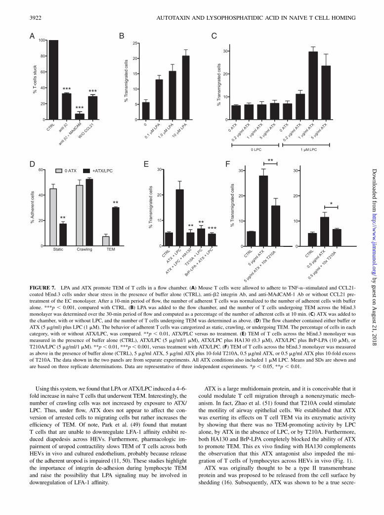
Using this system, we found that LPA or ATX/LPC induced a 4–6-fold increase in naive T cells that underwent TEM. Interestingly, thenumber of crawling cells was not increased by exposure to ATX/LPC. Thus, under flow, ATX does not appear to affect the con-version of arrested cells to migrating cells but rather increases theefficiency of TEM. Of note, Park et al. (49) found that mutantT cells that are unable to downregulate LFA-1 affinity exhibit re-duced diapedesis across HEVs. Furthermore, pharmacologic im-pairment of uropod contractility slows TEM of T cells across bothHEVs in vivo and cultured endothelium, probably because releaseof the adherent uropod is impaired (11, 50). These studies highlightthe importance of integrin de-adhesion during lymphocyte TEMand raise the possibility that LPA signaling may be involved indownregulation of LFA-1 affinity.
ATX is a large multidomain protein, and it is conceivable that itcould modulate T cell migration through a nonenzymatic mech-anism. In fact, Zhao et al. (51) found that T210A could stimulatethe motility of airway epithelial cells. We established that ATXwas exerting its effects on T cell TEM via its enzymatic activityby showing that there was no TEM-promoting activity by LPCalone, by ATX in the absence of LPC, or by T210A. Furthermore,both HA130 and BrP-LPA completely blocked the ability of ATXto promote TEM. This ex vivo finding with HA130 complementsthe observation that this ATX antagonist also impeded the mi-gration of T cells of lymphocytes across HEVs in vivo (Fig. 1).ATX was originally thought to be a type II transmembrane
protein and was proposed to be released from the cell surface byshedding (16). Subsequently, ATX was shown to be a true secre-
FIGURE 7. LPA and ATX promote TEM of T cells in a flow chamber. (A) Mouse T cells were allowed to adhere to TNF-a–stimulated and CCL21-
coated bEnd.3 cells under shear stress in the presence of buffer alone (CTRL), anti-b2 integrin Ab, and anti-MAdCAM-1 Ab or without CCL21 pre-
treatment of the EC monolayer. After a 10-min period of flow, the number of adherent T cells was normalized to the number of adherent cells with buffer
alone. ***p , 0.001, compared with CTRL. (B) LPA was added to the flow chamber, and the number of T cells undergoing TEM across the bEnd.3
monolayer was determined over the 30-min period of flow and computed as a percentage of the number of adherent cells at 10 min. (C) ATX was added to
the chamber, with or without LPC, and the number of T cells undergoing TEM was determined as above. (D) The flow chamber contained either buffer or
ATX (5 mg/ml) plus LPC (1 mM). The behavior of adherent T cells was categorized as static, crawling, or undergoing TEM. The percentage of cells in each
category, with or without ATX/LPC, was compared. **p , 0.01, ATX/PLC versus no treatment. (E) TEM of T cells across the bEnd.3 monolayer was
measured in the presence of buffer alone (CTRL), ATX/LPC (5 mg/ml/1 mM), ATX/LPC plus HA130 (0.3 mM), ATX/LPC plus BrP-LPA (10 mM), or
T210A/LPC (5 mg/ml/1 mM). **p , 0.01, ***p , 0.001, versus treatment with ATX/LPC. (F) TEM of T cells across the bEnd.3 monolayer was measured
as above in the presence of buffer alone (CTRL), 5 mg/ml ATX, 5 mg/ml ATX plus 10-fold T210A, 0.5 mg/ml ATX, or 0.5 mg/ml ATX plus 10-fold excess
of T210A. The data shown in the two panels are from separate experiments. All ATX conditions also included 1 mM LPC. Means and SDs are shown and
are based on three replicate determinations. Data are representative of three independent experiments. *p , 0.05, **p , 0.01.
3922 AUTOTAXIN AND LYSOPHOSPHATIDIC ACID IN NAIVE T CELL HOMING
by guest on August 21, 2018
http://ww
w.jim
munol.org/
Dow
nloaded from
tory protein, which is processed at its N terminus by the removalof a signal sequence and furin-mediated cleavage (17). We used b-ATX to verify the presence of ATX receptors on mouse T cells(Fig. 4). Although ATX binding was enhanced by Mn+2, we couldnot directly implicate the involvement of particular integrins, aswas reported for human T cells (a4b1) (30) and platelets (b1 andb3) (43).Nonetheless, we were still able to support the functional rele-
vance of ATX binding to T cells in the bEnd.3 system. Thus, wefound that ATX-stimulated TEMwas markedly reduced when ATXwas added in the presence of a 10-fold excess of T210A, a conditionthat reduced the level of ATX associated with the surface of T cellsbut did not change the amount of active enzyme in the flow chamber(Fig. 7). This ex vivo result rationalizes and complements theprevious in vivo demonstration that i.v. administration of T210Ainhibits lymphocyte homing (30).Two mutually nonexclusive mechanisms can be envisioned for
how ATX binding to T cells potentiates its TEM-promoting ac-tivity. First, the crystal structure of ATX reveals a hydrophobicchannel, which is thought to serve as conduit for the passage ofLPA from the active site of the enzyme to an exit site on the surfaceof the enzyme (52, 53). Because the exit site is predicted to be at theinterface of ATX with its cellular binding partner, the hydrophobicchannel could be the basis for a shuttling mechanism to transferLPA close to its signal-transduction receptors on the partner cell(52, 53). Notably, we detected ATX binding at the leading edgeof lymphocytes where many chemoreceptors are localized (37).An additional refinement to enhance signaling would be an actualphysical association between ATX and LPAR. In fact, lung epi-thelial cells, which migrate in response to ATX, exhibit a cellsurface complex of ATX with LPA1 and b4 integrin (51). Theexistence of an analogous complex on lymphocytes would bepredicted to facilitate shuttling of LPA to its LPARs so as tocounteract the dissipative action of blood flow on focally gener-ated LPA and to sequester LPA from the action of lipid phosphatephosphatases in the blood. A second possible mechanism forpotentiation is that the interaction of ATX with its receptors onT cells activates the enzyme, as appears to occur when ATX bindsto activated platelets (43).Although the current study establishes an important role for the
ATX–LPA signaling axis in T cell migration, the relevant LPARsremain to be identified. Of the six LPARs, four (LPA2, LPA4,LPA5, and LPA6) are expressed at the transcript level in pop-ulations of mouse T cells, as determined by mining of the Im-munological Genome Project database (http://www.immgen.org).Functions other than cell migration are served by lymphocyteLPARs (e.g., in cytokine secretion, cell proliferation, and cellsurvival) (54). TK1 cells express transcripts for LPA2, LPA5, andLPA6 (data not shown). Because Y27632 prevented the polari-zation response of this cell type to ATX/LPC (Fig. 3D), the Ga12/13–Rho axis is implicated in the signaling pathway (41). All threeof the LPARs present in TK1 cells can couple to this class of Gproteins (19, 55). Importantly, our findings that 10 mM BrP-LPAdid not inhibit either LPA-induced polarization or LPA-inducedTEM argues against the involvement of LPA1–4 in these responses,because BrP-LPA antagonizes these receptors with submicromolarKi’s (56). Interestingly, BrP-LPA is a weak agonist of LPA5 (40).Thus, LPA5 and LPA6 (not yet investigated with BrP-LPA) are ofinterest with respect to their potential roles in the polarization andTEM of T cells. Clearly, the endothelium is not a passive partnerduring TEM, and locally produced LPA may also signal throughLPARs on ECs during the process (31).Sphingosine 1-phosphate (S1P) and LPA are closely related
lysophospholipids, both having a three-carbon scaffold, a phos-
phate head group, and a single fatty acid chain (20). Like LPA, S1Psignals through multiple GPCRs (20). Notably, S1P regulates theefflux of lymphocytes from LNs into lymph (57, 58). The presentstudy substantiates a role for LPA in the entry of blood-bornelymphocytes into LNs and, thus, expands the functions of extra-cellular lysophospholipids in leukocyte trafficking.
AcknowledgmentsWe thank Drs. Harald Albers, Huib Ovaa, and Wouter Moolenaar of the
Netherlands Cancer Institute for generously providing HA130 prior to its
commercial availability. Preliminary experiments on LPA-induced polariza-
tion of lymphocytes were conducted by Hidenobu Kanda and Yuka Morita.
DisclosuresThe authors have no financial conflicts of interest.
References1. Gowans, J. L., and E. J. Knight. 1964. The route of recirculation of lymphocytes
in the rat. Proc. R. Soc. Lond. B Biol. Sci. 159: 257–282.2. Butcher, E. C., and L. J. Picker. 1996. Lymphocyte homing and homeostasis.
Science 272: 60–66.3. Ruddle, N. H., and E. M. Akirav. 2009. Secondary lymphoid organs: responding
to genetic and environmental cues in ontogeny and the immune response. J.Immunol. 183: 2205–2212.
4. Girard, J.-P., and T. A. Springer. 1995. High endothelial venules (HEVs): spe-cialized endothelium for lymphocyte migration. Immunol. Today 16: 449–457.
5. von Andrian, U. H., and T. R. Mempel. 2003. Homing and cellular traffic inlymph nodes. Nat. Rev. Immunol. 3: 867–878.
6. Ley, K., C. Laudanna, M. I. Cybulsky, and S. Nourshargh. 2007. Getting to thesite of inflammation: the leukocyte adhesion cascade updated. Nat. Rev. Immu-nol. 7: 678–689.
7. Rosen, S. D. 2004. Ligands for L-selectin: homing, inflammation, and beyond.Annu. Rev. Immunol. 22: 129–156.
8. Alon, R., and Z. Shulman. 2011. Chemokine triggered integrin activation andactin remodeling events guiding lymphocyte migration across vascular barriers.Exp. Cell Res. 317: 632–641.
9. Thelen, M., and J. V. Stein. 2008. How chemokines invite leukocytes to dance.Nat. Immunol. 9: 953–959.
10. Hogg, N., I. Patzak, and F. Willenbrock. 2011. The insider’s guide to leukocyteintegrin signalling and function. Nat. Rev. Immunol. 11: 416–426.
11. Soriano, S. F., M. Hons, K. Schumann, V. Kumar, T. J. Dennier, R. Lyck,M. Sixt, and J. V. Stein. 2011. In vivo analysis of uropod function duringphysiological T cell trafficking. J. Immunol. 187: 2356–2364.
12. Cinamon, G., V. Shinder, and R. Alon. 2001. Shear forces promote lymphocytemigration across vascular endothelium bearing apical chemokines. Nat. Immu-nol. 2: 515–522.
13. Shulman, Z., R. Pasvolsky, E. Woolf, V. Grabovsky, S. W. Feigelson, N. Erez,Y. Fukui, and R. Alon. 2006. DOCK2 regulates chemokine-triggeredlateral lymphocyte motility but not transendothelial migration. Blood 108:2150–2158.
14. Palmeri, D., F. R. Zuo, S. D. Rosen, and S. Hemmerich. 2004. Differential geneexpression profile of human tonsil high endothelial cells: implicationsfor lymphocyte trafficking. J. Leukoc. Biol. 75: 910–927.
15. Stracke, M. L., H. C. Krutzsch, E. J. Unsworth, A. Arestad, V. Cioce,E. Schiffmann, and L. A. Liotta. 1992. Identification, purification, and partialsequence analysis of autotaxin, a novel motility-stimulating protein. J. Biol.Chem. 267: 2524–2529.
16. Murata, J., H. Y. Lee, T. Clair, H. C. Krutzsch, A. A. Arestad, M. E. Sobel,L. A. Liotta, and M. L. Stracke. 1994. cDNA cloning of the human tumormotility-stimulating protein, autotaxin, reveals a homology with phosphodies-terases. J. Biol. Chem. 269: 30479–30484.
17. Jansen, S., C. Stefan, J. W. Creemers, E. Waelkens, A. Van Eynde, W. Stalmans,and M. Bollen. 2005. Proteolytic maturation and activation of autotaxin (NPP2),a secreted metastasis-enhancing lysophospholipase D. J. Cell Sci. 118: 3081–3089.
18. Umezu-Goto, M., Y. Kishi, A. Taira, K. Hama, N. Dohmae, K. Takio, T. Yamori,G. B. Mills, K. Inoue, J. Aoki, and H. Arai. 2002. Autotaxin has lysophospho-lipase D activity leading to tumor cell growth and motility by lysophosphatidicacid production. J. Cell Biol. 158: 227–233.
19. Choi, J. W., D. R. Herr, K. Noguchi, Y. C. Yung, C.-W. Lee, T. Mutoh, M.-E. Lin,S. T. Teo, K. E. Park, A. N. Mosley, and J. Chun. 2010. LPA receptors: subtypesand biological actions. Annu. Rev. Pharmacol. Toxicol. 50: 157–186.
20. Blaho, V. A., and T. Hla. 2011. Regulation of mammalian physiology, devel-opment, and disease by the sphingosine 1-phosphate and lysophosphatidic acidreceptors. Chem. Rev. 111: 6299–6320.
21. Okudaira, S., H. Yukiura, and J. Aoki. 2010. Biological roles of lysophosphatidicacid signaling through its production by autotaxin. Biochimie 92: 698–706.
22. Tanaka, M., S. Okudaira, Y. Kishi, R. Ohkawa, S. Iseki, M. Ota, S. Noji,Y. Yatomi, J. Aoki, and H. Arai. 2006. Autotaxin stabilizes blood vessels and isrequired for embryonic vasculature by producing lysophosphatidic acid. J. Biol.Chem. 281: 25822–25830.
The Journal of Immunology 3923
by guest on August 21, 2018
http://ww
w.jim
munol.org/
Dow
nloaded from
23. van Meeteren, L. A., P. Ruurs, C. Stortelers, P. Bouwman, M. A. van Rooijen,J. P. Pradere, T. R. Pettit, M. J. Wakelam, J. S. Saulnier-Blache, C. L. Mummery,et al. 2006. Autotaxin, a secreted lysophospholipase D, is essential for bloodvessel formation during development. Mol. Cell. Biol. 26: 5015–5022.
24. Tomsig, J. L., A. H. Snyder, E. V. Berdyshev, A. Skobeleva, C. Mataya,V. Natarajan, D. N. Brindley, and K. R. Lynch. 2009. Lipid phosphate phos-phohydrolase type 1 (LPP1) degrades extracellular lysophosphatidic acid in vivo.Biochem. J. 419: 611–618.
25. Kishimoto, T., T. Matsuoka, S. Imamura, and K. Mizuno. 2003. A novel col-orimetric assay for the determination of lysophosphatidic acid in plasma using anenzymatic cycling method. Clin. Chim. Acta 333: 59–67.
26. Smyth, S. S., H. Y. Cheng, S. Miriyala, M. Panchatcharam, and A. J. Morris.2008. Roles of lysophosphatidic acid in cardiovascular physiology and disease.Biochim. Biophys. Acta 1781: 563–570.
27. Houben, A. J., and W. H. Moolenaar. 2011. Autotaxin and LPA receptor sig-naling in cancer. Cancer Metastasis Rev. 30: 557–565.
28. Nikitopoulou, I., N. Oikonomou, E. Karouzakis, I. Sevastou, N. Nikolaidou-Katsaridou, Z. Zhao, V. Mersinias, M. Armaka, Y. Xu, M. Masu, et al. 2012.Autotaxin expression from synovial fibroblasts is essential for the pathogenesisof modeled arthritis. J. Exp. Med. 209: 925–933.
29. Emo, J., N. Meednu, T. J. Chapman, F. Rezaee, M. Balys, T. Randall,T. Rangasamy, and S. N. Georas. 2012. Lpa2 is a negative regulator of bothdendritic cell activation and murine models of allergic lung inflammation. J.Immunol. 188: 3784–3790.
30. Kanda, H., R. Newton, R. Klein, Y. Morita, M. D. Gunn, and S. D. Rosen. 2008.Autotaxin, an ectoenzyme that produces lysophosphatidic acid, promotes theentry of lymphocytes into secondary lymphoid organs. Nat. Immunol. 9: 415–423.
31. Nakasaki, T., T. Tanaka, S. Okudaira, M. Hirosawa, E. Umemoto, K. Otani,S. Jin, Z. Bai, H. Hayasaka, Y. Fukui, et al. 2008. Involvement of the lyso-phosphatidic acid-generating enzyme autotaxin in lymphocyte-endothelial cellinteractions. Am. J. Pathol. 173: 1566–1576.
32. Wijesinghe, D. S., E. K. Mayton, J. A. Mietla, A. Mukherjee, J. Wu, X. Fang,and C. E. Chalfant. 2011. Characterization of lysophosphatidic acid subspeciesproduced by autotaxin using a modified HPLC ESI-MS/MS method. Anal.Methods 3: 2822–2828.
33. Lamb, T. J., A. L. Graham, and A. Petrie. 2008. T testing the immune system.Immunity 28: 288–292.
34. Albers, H. M., A. Dong, L. A. van Meeteren, D. A. Egan, M. Sunkara, E. W. vanTilburg, K. Schuurman, O. van Tellingen, A. J. Morris, S. S. Smyth, et al. 2010.Boronic acid-based inhibitor of autotaxin reveals rapid turnover of LPA in thecirculation. Proc. Natl. Acad. Sci. USA 107: 7257–7262.
35. Stam, J. C., F. Michiels, R. A. van der Kammen, W. H. Moolenaar, andJ. G. Collard. 1998. Invasion of T-lymphoma cells: cooperation between Rhofamily GTPases and lysophospholipid receptor signaling. EMBO J. 17: 4066–4074.
36. Krummel, M. F., and I. Macara. 2006. Maintenance and modulation of T cellpolarity. Nat. Immunol. 7: 1143–1149.
37. Sanchez-Madrid, F., and J. M. Serrador. 2009. Bringing up the rear: defining theroles of the uropod. Nat. Rev. Mol. Cell Biol. 10: 353–359.
38. Evans, S. S., W. C. Wang, M. D. Bain, R. Burd, J. R. Ostberg, and E. A. Repasky.2001. Fever-range hyperthermia dynamically regulates lymphocyte delivery tohigh endothelial venules. Blood 97: 2727–2733.
39. Woolf, E., I. Grigorova, A. Sagiv, V. Grabovsky, S. W. Feigelson, Z. Shulman,T. Hartmann, M. Sixt, J. G. Cyster, and R. Alon. 2007. Lymph node chemokinespromote sustained T lymphocyte motility without triggering stable integrin ad-hesiveness in the absence of shear forces. Nat. Immunol. 8: 1076–1085.
40. Zhang, H., X. Xu, J. Gajewiak, R. Tsukahara, Y. Fujiwara, J. Liu, J. I. Fells,D. Perygin, A. L. Parrill, G. Tigyi, and G. D. Prestwich. 2009. Dual activitylysophosphatidic acid receptor pan-antagonist/autotaxin inhibitor reduces breast
cancer cell migration in vitro and causes tumor regression in vivo. Cancer Res.69: 5441–5449.
41. Tybulewicz, V. L., and R. B. Henderson. 2009. Rho family GTPases and theirregulators in lymphocytes. Nat. Rev. Immunol. 9: 630–644.
42. Chettibi, S., A. J. Lawrence, R. D. Stevenson, and J. D. Young. 1994. Effect oflysophosphatidic acid on motility, polarisation and metabolic burst of humanneutrophils. FEMS Immunol. Med. Microbiol. 8: 271–281.
43. Fulkerson, Z., T. Wu, M. Sunkara, C. V. Kooi, A. J. Morris, and S. S. Smyth.2011. Binding of autotaxin to integrins localizes lysophosphatidic acid produc-tion to platelets and mammalian cells. J. Biol. Chem. 286: 34654–34663.
44. Huang, J. H., L. I. Cardenas-Navia, C. C. Caldwell, T. J. Plumb, C. G. Radu,P. N. Rocha, T. Wilder, J. S. Bromberg, B. N. Cronstein, M. Sitkovsky, et al.2007. Requirements for T lymphocyte migration in explanted lymph nodes. J.Immunol. 178: 7747–7755.
45. Okada, T., and J. G. Cyster. 2007. CC chemokine receptor 7 contributes to Gi-dependent T cell motility in the lymph node. J. Immunol. 178: 2973–2978.
46. Worbs, T., T. R. Mempel, J. Bolter, U. H. von Andrian, and R. Forster. 2007.CCR7 ligands stimulate the intranodal motility of T lymphocytes in vivo. J. Exp.Med. 204: 489–495.
47. Gerard, A., R. A. van der Kammen, H. Janssen, S. I. Ellenbroek, andJ. G. Collard. 2009. The Rac activator Tiam1 controls efficient T-cell traffickingand route of transendothelial migration. Blood 113: 6138–6147.
48. Boscacci, R. T., F. Pfeiffer, K. Gollmer, A. I. Sevilla, A. M. Martin, S. F. Soriano,D. Natale, S. Henrickson, U. H. von Andrian, Y. Fukui, et al. 2010. Compre-hensive analysis of lymph node stroma-expressed Ig superfamily membersreveals redundant and nonredundant roles for ICAM-1, ICAM-2, and VCAM-1in lymphocyte homing. Blood 116: 915–925.
49. Park, E. J., A. Peixoto, Y. Imai, A. Goodarzi, G. Cheng, C. V. Carman, U. H. vonAndrian, and M. Shimaoka. 2010. Distinct roles for LFA-1 affinity regulationduring T-cell adhesion, diapedesis, and interstitial migration in lymph nodes.Blood 115: 1572–1581.
50. Heasman, S. J., L. M. Carlin, S. Cox, T. Ng, and A. J. Ridley. 2010. CoordinatedRhoA signaling at the leading edge and uropod is required for T cell trans-endothelial migration. J. Cell Biol. 190: 553–563.
51. Zhao, J., D. He, E. Berdyshev, M. Zhong, R. Salgia, A. J. Morris, S. S. Smyth,V. Natarajan, and Y. Zhao. 2011. Autotaxin induces lung epithelial cell migrationthrough lysoPLD activity-dependent and -independent pathways. Biochem. J.439: 45–55.
52. Hausmann, J., S. Kamtekar, E. Christodoulou, J. E. Day, T. Wu, Z. Fulkerson,H. M. Albers, L. A. van Meeteren, A. J. Houben, L. van Zeijl, et al. 2011.Structural basis of substrate discrimination and integrin binding by autotaxin.Nat. Struct. Mol. Biol. 18: 198–204.
53. Nishimasu, H., S. Okudaira, K. Hama, E. Mihara, N. Dohmae, A. Inoue,R. Ishitani, J. Takagi, J. Aoki, and O. Nureki. 2011. Crystal structure of autotaxinand insight into GPCR activation by lipid mediators. Nat. Struct. Mol. Biol. 18:205–212.
54. Goetzl, E. J., and H. Rosen. 2004. Regulation of immunity by lysosphingolipidsand their G protein-coupled receptors. J. Clin. Invest. 114: 1531–1537.
55. Yanagida, K., K. Masago, H. Nakanishi, Y. Kihara, F. Hamano, Y. Tajima, R. Taguchi,T. Shimizu, and S. Ishii. 2009. Identification and characterization of a novel lyso-phosphatidic acid receptor, p2y5/LPA6. J. Biol. Chem. 284: 17731–17741.
56. Jiang, G., Y. Xu, Y. Fujiwara, T. Tsukahara, R. Tsukahara, J. Gajewiak, G. Tigyi,and G. D. Prestwich. 2007. Alpha-substituted phosphonate analogues of lyso-phosphatidic acid (LPA) selectively inhibit production and action of LPA.ChemMedChem 2: 679–690.
57. Rosen, H., M. G. Sanna, S. M. Cahalan, and P. J. Gonzalez-Cabrera. 2007.Tipping the gatekeeper: S1P regulation of endothelial barrier function. TrendsImmunol. 28: 102–107.
58. Schwab, S. R., and J. G. Cyster. 2007. Finding a way out: lymphocyte egressfrom lymphoid organs. Nat. Immunol. 8: 1295–1301.
3924 AUTOTAXIN AND LYSOPHOSPHATIDIC ACID IN NAIVE T CELL HOMING
by guest on August 21, 2018
http://ww
w.jim
munol.org/
Dow
nloaded from