Autophagy genes protect against Salmonella typhimurium infection
6
Autophagy genes protect against Salmonella typhimurium infection and mediate insulin signaling-regulated pathogen resistance Kailiang Jia a , Collin Thomas a,1 , Muhammad Akbar a , Qihua Sun a , Beverley Adams-Huet b , Christopher Gilpin c , and Beth Levine a,d,e,2 a Department of Internal Medicine, b Department of Biostatistics and Clinical Sciences, c Department of Cell Biology, and d Department of Microbiology, e The Howard Hughes Medical Institute,University of Texas Southwestern Medical Center, Dallas, TX 75390 Edited by Kathryn V. Anderson, The Sloan-Kettering Institute, New York, NY, and approved July 7, 2009 (received for review January 1, 2009) A conserved insulin-like pathway modulates both aging and patho- gen resistance in Caenorhabditis elegans. However, the specific in- nate effector functions that mediate this pathogen resistance are largely unknown. Autophagy, a lysosomal degradation pathway, plays a role in controlling intracellular bacterial pathogen infections in cultured cells, but less is known about its role at the organismal level. We examined the effects of autophagy gene inactivation on Salmonella enterica Serovar Typhimurium (Salmonella typhimurium) infection in 2 model organisms, Caenorhabditis elegans and Dictyo- stelium discoideum. In both organisms, genetic inactivation of the autophagy pathway increases bacterial intracellular replication, de- creases animal lifespan, and results in apoptotic-independent death. In C. elegans, genetic knockdown of autophagy genes abrogates pathogen resistance conferred by a loss-of-function mutation, daf- 2(e1370), in the insulin-like tyrosine kinase receptor or by over- expression of the DAF-16 FOXO transcription factor. Thus, autophagy genes play an essential role in host defense in vivo against an intracellular bacterial pathogen and mediate pathogen resistance in long-lived mutant nematodes. I mmune functions decline with age (1), but the mechanisms underlying immunosenescence are not well understood. Because a conserved insulin-like signaling pathway controls both aging and pathogen resistance in Caenorhabditis elegans (2, 3), the identifica- tion of cellular innate immune functions regulated by this pathway may help elucidate the basis of age-related declines in immunity. One candidate target is autophagy, a lysosomal degradation pathway that decreases with age, is implicated in the degradation of intracellular bacteria, and is required for the lifespan extension of nematodes with mutation in the daf-2 insulin-like signaling pathway (4–7). The autophagy pathway is mediated by evolutionarily con- served genes (called atg genes) and, in vitro, targets both extracellular bacteria that invade intracellularly and intracellular bacterial pathogens for lysosomal degradation (4). Autophagy limits the intracellular growth of Listeria monocytogenes in Drosophila (8), suggesting a role for autophagy in defense against intracellular bacteria in vivo. Besides promoting the direct degradation of intracellular pathogens, autophagy has other functions in immunity (4), including the delivery of microbial genetic material or peptides to endosomes or MHC Class II loading compartments, respectively, for activation of innate or adaptive immunity. Furthermore, a polymorphism in the atg gene, ATG16L1, is linked to genetic susceptibility to the inf lam- matory bowel disorder, Crohn’s disease, which has led to the speculation that mutations in the autophagy pathway may alter the normal gut response to enteric bacterial pathogens (9). Indeed, loss of Atg16l1 in mouse models recapitulates certain aspects of the pathology of human Crohn’s disease, and the Crohn’s disease-associated ATG16L1 variant may have impaired antibacterial autophagy function in a human gut epithelial cell line (10). The unicellular organism Dictyostelium discoideum and the mul- ticellular organism C. elegans are useful models for studying inter- actions between hosts and pathogens (11, 12), including the gram- negative bacterium Salmonella typhimurium. Salmonella is a pathogen that causes human food-borne illness worldwide, infects both mammalian intestinal epithelial cells and macrophages, and has an increased propensity to cause invasive, extra-intestinal disease in the elderly (1, 13). In C. elegans, S. typhimurium estab- lishes persistent infection in the intestinal lumen but does not replicate inside intestinal epithelial cells (14, 15), suggesting that host defense mechanisms to combat Salmonella infection may be more successful in nematode than in mammalian cells. In Dictyostelium, a soil amoeba that is highly susceptible to invasion by bacterial patho- gens that commonly infect human macrophages (12), Salmonella is efficiently taken up but fails to multiply intracellularly. Therefore, the identification of factors that govern susceptibility to Salmonella infection in the nematode and soil amoeba may be relevant to understanding Salmonella host-pathogen interactions in mamma- lian intestinal epithelial cells and macrophages, respectively. Long-lived mutant nematodes display resistance to Salmonella infection (2, 16). In C. elegans, the daf-2 insulin-like signaling pathway is a well-characterized regulator of pathogen resistance, longevity, and autophagy (2, 3, 5, 16). Nematodes with loss-of- function mutations in the insulin-like tyrosine kinase receptor daf-2 have extended lifespan and pathogen-resistant phenotypes (3, 16), including resistance to infection with S. typhimurium. The major target of the DAF-2 pathway is DAF-16, a forkhead transcription factor required for both longevity and pathogen resistance in daf-2 mutants (16). Moreoever, DAF-2 negatively regulates autophagy, and lifespan extension in daf-2 mutants requires atg genes (5–7). These findings, coupled with the role of autophagy in pathogen degradation, led us to postulate that autophagy may represent a mechanism of innate immunity against Salmonella infection in vivo that is required for the pathogen resistance of long-lived mutant nematodes. To evaluate the role of autophagy in pathogen resistance, we used feeding RNAi to inactivate several C. elegans atg genes, including bec-1 and lgg-1, in S. typhimurium-infected wild-type, and long-lived daf-2-mutant and DAF-16 over-expressing nematodes. We also examined S. typhimurium infection in Dictyostelium with null mutations in three atg genes, ATG1, ATG6, and ATG7. Our results Author contributions: K.J., C.T., and B.L. designed research; K.J., C.T., M.A., and Q.S. performed research; K.J. contributed new reagents/analytic tools; K.J., C.T., B.A.-H., C.G., and B.L. analyzed data; and K.J. and B.L. wrote the paper. The authors declare no conflict of interest. This article is a PNAS Direct Submission. Freely available online through the PNAS open access option. 1 Present Address: Center for Advanced Studies in Math and Natural Sciences, Collin College, 2800 East Spring Creek Parkway, Plano, TX 75074. 2 To whom correspondence should be addressed. E-mail: [email protected]. This article contains supporting information online at www.pnas.org/cgi/content/full/ 0813319106/DCSupplemental. 14564 –14569 PNAS August 25, 2009 vol. 106 no. 34 www.pnas.orgcgidoi10.1073pnas.0813319106
Transcript of Autophagy genes protect against Salmonella typhimurium infection

Autophagy genes protect against Salmonellatyphimurium infection and mediate insulinsignaling-regulated pathogen resistanceKailiang Jiaa, Collin Thomasa,1, Muhammad Akbara, Qihua Suna, Beverley Adams-Huetb, Christopher Gilpinc,and Beth Levinea,d,e,2
aDepartment of Internal Medicine, bDepartment of Biostatistics and Clinical Sciences, cDepartment of Cell Biology, and dDepartment of Microbiology, eTheHoward Hughes Medical Institute,University of Texas Southwestern Medical Center, Dallas, TX 75390
Edited by Kathryn V. Anderson, The Sloan-Kettering Institute, New York, NY, and approved July 7, 2009 (received for review January 1, 2009)
A conserved insulin-like pathway modulates both aging and patho-gen resistance in Caenorhabditis elegans. However, the specific in-nate effector functions that mediate this pathogen resistance arelargely unknown. Autophagy, a lysosomal degradation pathway,plays a role in controlling intracellular bacterial pathogen infectionsin cultured cells, but less is known about its role at the organismallevel. We examined the effects of autophagy gene inactivation onSalmonella enterica Serovar Typhimurium (Salmonella typhimurium)infection in 2 model organisms, Caenorhabditis elegans and Dictyo-stelium discoideum. In both organisms, genetic inactivation of theautophagy pathway increases bacterial intracellular replication, de-creases animal lifespan, and results in apoptotic-independent death.In C. elegans, genetic knockdown of autophagy genes abrogatespathogen resistance conferred by a loss-of-function mutation, daf-2(e1370), in the insulin-like tyrosine kinase receptor or by over-expression of the DAF-16 FOXO transcription factor. Thus, autophagygenes play an essential role in host defense in vivo against anintracellular bacterial pathogen and mediate pathogen resistance inlong-lived mutant nematodes.
Immune functions decline with age (1), but the mechanismsunderlying immunosenescence are not well understood. Because
a conserved insulin-like signaling pathway controls both aging andpathogen resistance in Caenorhabditis elegans (2, 3), the identifica-tion of cellular innate immune functions regulated by this pathwaymay help elucidate the basis of age-related declines in immunity. Onecandidate target is autophagy, a lysosomal degradation pathway thatdecreases with age, is implicated in the degradation of intracellularbacteria, and is required for the lifespan extension of nematodes withmutation in the daf-2 insulin-like signaling pathway (4–7).
The autophagy pathway is mediated by evolutionarily con-served genes (called atg genes) and, in vitro, targets bothextracellular bacteria that invade intracellularly and intracellularbacterial pathogens for lysosomal degradation (4). Autophagylimits the intracellular growth of Listeria monocytogenes inDrosophila (8), suggesting a role for autophagy in defense againstintracellular bacteria in vivo. Besides promoting the directdegradation of intracellular pathogens, autophagy has otherfunctions in immunity (4), including the delivery of microbialgenetic material or peptides to endosomes or MHC Class IIloading compartments, respectively, for activation of innate oradaptive immunity. Furthermore, a polymorphism in the atggene, ATG16L1, is linked to genetic susceptibility to the inflam-matory bowel disorder, Crohn’s disease, which has led to thespeculation that mutations in the autophagy pathway may alterthe normal gut response to enteric bacterial pathogens (9).Indeed, loss of Atg16l1 in mouse models recapitulates certainaspects of the pathology of human Crohn’s disease, and theCrohn’s disease-associated ATG16L1 variant may have impairedantibacterial autophagy function in a human gut epithelial cellline (10).
The unicellular organism Dictyostelium discoideum and the mul-ticellular organism C. elegans are useful models for studying inter-
actions between hosts and pathogens (11, 12), including the gram-negative bacterium Salmonella typhimurium. Salmonella is apathogen that causes human food-borne illness worldwide, infectsboth mammalian intestinal epithelial cells and macrophages, andhas an increased propensity to cause invasive, extra-intestinaldisease in the elderly (1, 13). In C. elegans, S. typhimurium estab-lishes persistent infection in the intestinal lumen but does notreplicate inside intestinal epithelial cells (14, 15), suggesting thathost defense mechanisms to combat Salmonella infection may bemore successful in nematode than in mammalian cells. In Dictyostelium,a soil amoeba that is highly susceptible to invasion by bacterial patho-gens that commonly infect human macrophages (12), Salmonella isefficiently taken up but fails to multiply intracellularly. Therefore, theidentification of factors that govern susceptibility to Salmonellainfection in the nematode and soil amoeba may be relevant tounderstanding Salmonella host-pathogen interactions in mamma-lian intestinal epithelial cells and macrophages, respectively.
Long-lived mutant nematodes display resistance to Salmonellainfection (2, 16). In C. elegans, the daf-2 insulin-like signalingpathway is a well-characterized regulator of pathogen resistance,longevity, and autophagy (2, 3, 5, 16). Nematodes with loss-of-function mutations in the insulin-like tyrosine kinase receptor daf-2have extended lifespan and pathogen-resistant phenotypes (3, 16),including resistance to infection with S. typhimurium. The majortarget of the DAF-2 pathway is DAF-16, a forkhead transcriptionfactor required for both longevity and pathogen resistance in daf-2mutants (16). Moreoever, DAF-2 negatively regulates autophagy,and lifespan extension in daf-2 mutants requires atg genes (5–7).These findings, coupled with the role of autophagy in pathogendegradation, led us to postulate that autophagy may represent amechanism of innate immunity against Salmonella infection in vivothat is required for the pathogen resistance of long-lived mutantnematodes.
To evaluate the role of autophagy in pathogen resistance, weused feeding RNAi to inactivate several C. elegans atg genes,including bec-1 and lgg-1, in S. typhimurium-infected wild-type, andlong-lived daf-2-mutant and DAF-16 over-expressing nematodes.We also examined S. typhimurium infection in Dictyostelium with nullmutations in three atg genes, ATG1, ATG6, and ATG7. Our results
Author contributions: K.J., C.T., and B.L. designed research; K.J., C.T., M.A., and Q.S.performed research; K.J. contributed new reagents/analytic tools; K.J., C.T., B.A.-H., C.G.,and B.L. analyzed data; and K.J. and B.L. wrote the paper.
The authors declare no conflict of interest.
This article is a PNAS Direct Submission.
Freely available online through the PNAS open access option.
1Present Address: Center for Advanced Studies in Math and Natural Sciences, Collin College,2800 East Spring Creek Parkway, Plano, TX 75074.
2To whom correspondence should be addressed. E-mail: [email protected].
This article contains supporting information online at www.pnas.org/cgi/content/full/0813319106/DCSupplemental.
14564–14569 � PNAS � August 25, 2009 � vol. 106 � no. 34 www.pnas.org�cgi�doi�10.1073�pnas.0813319106

indicate thatatggenesplayaconservedrole inantibacterialhostdefensein vivo and mediate pathogen resistance in long-lived mutant animals.
ResultsAutophagy Mediates Host Defense Against Salmonella in C. elegans. Toevaluate whether atg genes function in host defense against Salmo-nella infection in C. elegans, we used feeding RNAi to silence twopreviously characterized C. elegans atg genes, bec-1 and lgg-1 (5),which are orthologs of yeast ATG6 and ATG8 and function inautophagic vesicle nucleation and autophagic vesicle expansion,respectively. The survival of Salmonella-infected animals treatedwith either bec-1 or lgg-1 RNAi was significantly shortened ascompared to Salmonella-infected control vector-treated animals(Fig. 1 A and B, Table S1)] . Although a null mutation in bec-1 orhigh-dose atg gene RNAi injection is embryonically lethal (5, 17),feeding RNAi against bec-1 or lgg-1 did not shorten the lifespan ofN2 (wild-type) animals exclusively fed nonpathogenic Escherichiacoli (Fig. 1 A and B). We confirmed that feeding RNAi decreasedthe level of atg gene RNA by RT-PCR, and that bec-1 feeding-RNAi treatment exerted previously described effects of atg geneinhibition [e.g., abnormal dauer development in daf-2(e1370) mu-tants and autophagy inhibition in worms that transgenically expressthe autophagy marker GFP::LGG-1] (data not shown). We alsoknocked down the expression of atg-7 (involved in autophagicvesicle expansion) in N2 worms using RNAi. Similar to bec-1 andlgg-1 RNAi, the atg-7-RNAi animals were more susceptible toSalmonella infection than control animals (Fig. S1A, Table S2).Thus, inactivation of three different atg genes increased nematodesusceptibility to lethal Salmonella infection. This is unlikely toreflect nonspecific or off-target effects of atg gene-RNAi treatmentbecause no alterations in Salmonella susceptibility were observed innematodes treated with feeding RNAi against two irrelevant con-trol genes, unc-22 (required for normal muscle function) or him-3(required for meiotic chromosome segregation) (Fig. S1 B and C,Table S2). Furthermore, neither bec-1-RNAi treatment [similar toa previous report by Hansen et al (7)] nor Salmonella infectionaltered pharyngeal pumping rates (Fig. S2), suggesting that theincreased pathogen susceptibility in atg gene RNAi-treated animalsis not because of alterations in food (bacterial) uptake.
BEC-1 interacts with CED-9 (a Bcl-2 homolog) and disruptionof bec-1 triggers apoptosis during embryo development (17). There-fore, we studied apoptosis-deficient nematodes with a loss-of-function mutation in the C. elegans caspase, ced-3(n717) to examinewhether apoptosis is involved in the increased susceptibility ofautophagy-deficient worms to Salmonella infection. Salmonellainfection decreased the lifespan of wild-type worms more than thatof ced-3(n717)-mutant animals (Fig. 1C and Table S1), indicatingthat the ced-3 mutation protects against Salmonella infection. [Thebasis for the difference between this finding and those of Aballay et al.(18) is unclear, but might reflect subtle differences in genetic strains].Importantly, bec-1 RNAi significantly decreased the lifespan of Salmo-nella-infected ced-3-mutant animals (Fig. 1D and Table S1); theseanimals had similar mortality as bec-1 RNAi-treated Salmonella-infected wild-type animals (Fig. 1A). Therefore, the mechanism ofincreased Salmonella sensitivity because of atg gene inactivationdoes not involve caspase-dependent apoptosis.
bec-1 Restricts Bacterial Replication and Cytopathology in C. elegansIntestinal Epithelial Cells. To examine the mechanism by which atggenes protect against Salmonella infection, we performed EManalyses of control vector- and bec-1-RNAi-treated animals (Fig. 1E–J). Immediately after a 2-day Salmonella ingestion period, feworganisms were observed in the intestinal lumen and some bacteriawere observed inside the intestinal epithelial cells (Fig. 1 E and F).In the control group, intracellular bacteria were found primarilyinside early intact autophagosomes or autolysosomes (Fig. 1E anddata not shown), but rarely inside cytoplasmic Salmonella-containing vacuoles (SCVs). In contrast, in bec-1-RNAi animals,
the cytoplasm contained intact SCVs (Fig. 1F). At day two afterSalmonella ingestion, the number of organisms in the intestinallumen increased in both groups, but more dramatically in thebec-1-RNAi animals (Fig. 1 G and H). Moreover, in control animals
A B
C D
E F
G H
I J
K L
Fig. 1. Atg genes mediate host defense against Salmonella in C. elegans. (A andB) Survival curves of wild-type (N2) animals treated with either control vector orindicated atg gene RNAi-feeding plasmids following a 2-day exposure to S.typhimurium or normal food (i.e., nonpathogenic Escherichia coli) at 20 °C (see SIMaterialsandMethods fordetails). (CandD) Survival curvesofwild-type (N2)andced-3(n717)-mutant animals following a 2-day exposure to S. typhimurium ornormal food without atg gene RNAi treatment (C) or with control vector or bec-1RNAi treatment (D) at 20 °C. For (A) to (D), see Table S1 for statistical details. (E–J)Representative EMs of Salmonella-infected control N2 animals and bec-1-RNAianimals at day 0 (E and F), 2 (G and H), and 4 (I and J) after a 2-day Salmonellaingestion period. (F, G, I, and J) Arrowheads denote intraluminal bacteria. (E)White arrow denotes bacterial-containing early autophagosome. (G) Asteriskdenotes autolysosome with partially degraded bacterial debris. (F–H and J) Blackarrows denote SCVs inside intestinal epithelial cells. IE, intestinal epithelial cells;IL, intestinal lumen; MV, microvilli. (Scale bars, 1 �m.) (K and L) Growth curves ofS. typhimurium in N2 animals treated with either control vector or bec-1-RNAifeeding plasmids in the absence (K) or presence (L) of 100 �g/ml gentamicin. For(K)and(L), values representmean�SEMfortriplicatesamplesof�10animalspertreatment group per sample. Similar results were observed in two independentexperiments.
Jia et al. PNAS � August 25, 2009 � vol. 106 � no. 34 � 14565
MIC
ROBI
OLO
GY

the intestinal epithelial cells contained rare, visible intact bacteriaand numerous autolysosomes containing bacterial debris (Fig. 1G),whereas in bec-1-RNAi animals the intestinal epithelial cells con-tained numerous intact bacteria (which were similar in size andmorphology to those found in the intestinal lumen) and very fewautophagosomes or autolyososmes (Fig. 1H). At this stage, theepithelial cells in both groups had intact basement membranes andmicrovilli, although increased cytoplasmic vacuolization was ob-served in bec-1-RNAi animals. However, at day 4 after Salmonellaingestion, in the bec-1-RNAi group the intestinal epithelial cellswere completely destroyed in most animals, and there were massivesheets of bacteria extending from the intestinal lumen to the bodywall muscle (Fig. 1J). In contrast, the epithelial cells remainedlargely intact in the control animals (Fig. 1I), indicating that the atggene bec-1 successfully protects the intestinal epithelium cells fromoverwhelming bacterial infection and cellular destruction.
These EM analyses suggest that Salmonella invades the intestinalepithelial cells in both control and bec-1-RNAi animals; however,in the former group the bacteria are efficiently degraded by theautophagy pathway, whereas in the latter group the intracellularbacterial population expands, leading to extensive cytoplasmicdestruction and premature death of the organism. To confirm thatSalmonella replicates intracellularly in bec-1-RNAi animals, wecompared bacterial growth curves in animals cultured in thepresence or absence of gentamicin, an antibiotic that inhibitsextracellular but not intracellular Salmonella replication. In theabsence of gentamicin, bacterial colony counts increased graduallyover time in both control and bec-1-RNAi animals, with a largermagnitude of increase in the bec-1-RNAi animals (P � 0.0001, t-test)(Fig. 1K). In the presence of gentamicin, no increase was observed inbacterial colony counts in control animals (Fig. 1L). In contrast, inbec-1-RNAi animals, the Salmonella growth curve paralleled thatobserved in the absence of gentamicin, indicating that Salmonellareplicates intracellularly in autophagy-deficient nematodes.
Atg Genes Restrict Intracellular Bacterial Replication and Host CellularDestruction in Dictyostelium. To further confirm that atg genes restrictintracellular Salmonella multiplication independently of bacterialinvasion (which is difficult to assess in vivo in a multicellularorganism), we used a unicellular model host organism, D. discoi-deum. Unlike many bacteria that replicate intracellularly in mam-malian macrophages, Salmonella is rapidly degraded after inter-nalization and is nonpathogenic in Dictyostelium (12). We foundthat GFP-labeled Salmonella invaded wild-type Dictyostelium andpreviously described Dictyostelium mutants (19) lacking the atggenes, ATG1 (a serine/threonine kinase involved in autophagyinduction), ATG6 (the ortholog of bec-1), and ATG7 with similarkinetics and to a similar degree (Fig. 2A). In Dictyostelium thattransgenically express a fluorescent autophagy marker protein,GFP-Atg8 (19), CFP-labeled Salmonella colocalized with GFP-Atg8 by 2-h postinfection (p.i.) (Fig. 2B), indicating that internal-ized bacteria are efficiently targeted to autophagosomes. Similarly,EM analysis revealed the degradation of Salmonella inside autoly-sosomes in wild-type Dictyostelium (Fig. 2C). In contrast, in atg1-,atg6-, and atg7-mutant Dicytostelium, no bacteria were observedinside autophagosomes or autolysosomes (Fig. 2C). Instead, thebacteria were observed exclusively in intact SCVs containing singlebacteria or in larger vacuoles that contained multiple bacteria.Thus, the autophagic machinery is not necessary for bacterialinvasion or formation of SCVs, but is required for the fusion ofSCVs with lysosomal compartments. By 24-h p.i., Salmonella wererarely visible in wild-type Dictyostelium and the cytoplasm appearednormal, whereas atg1-, atg6-, and atg7-mutant Dictyostelium exhibitedsevere cytopathology, including extensive cytoplasmic vacuolizationand disruption of plasma membrane integrity (data not shown).
These findings are consistent with autophagic degradation ofintracellular Salmonella in wild-type Dicytostelium and intracellularmultiplication of Salmonella with resulting cytopathology in
autophagy-deficient amoebae. To confirm this, we measured Sal-monella replication (Fig. 2D) and cell survival (Fig. 2E) in thepresence of gentamicin. In wild-type Dictyostelium, there was nobacterial growth (Fig. 2D), and the Dictyostelium remained healthywith a long-term survival curve that parallels that of amoebaegrown in axenic media (Fig. 2E and data not shown). In contrast,in atg1-, atg6-, and atg7-mutant Dictyostelium, bacterial colonycounts increased over time (Fig. 2D), indicating intracellular mul-tiplication, and the Dictyostelium died within 24- to 72-h p.i. (Fig.2E). Because Dictyostelium lack caspases or other essential com-ponents of the apoptotic machinery (20), this Salmonella-induceddeath of autophagy-deficient amoebae, like that of autophagy-deficient nematodes, does not involve caspase-dependent apopto-sis. Taken together, these findings demonstrate that the autophagicmachinery restricts intracellular bacterial multiplication and cyto-pathology in two model organisms, C. elegans and Dictyostelium.
Autophagy Deficiency Abrogates Pathogen Resistance of daf-2-MutantAdults. To evaluate whether autophagy is an effector mechanism ofenhanced Salmonella resistance in long-lived C. elegans mutants, wefirst examined whether autophagy is induced in adult nematodeswith a loss-of-function mutation in daf-2 (2). Using a previouslydescribed assay that measures autophagosomes in the seam cell, atype of specialized hypodermal cell that has detectable basal- andstimulus-induced autophagy (5), we observed a higher number of
Fig. 2. Atg genes restrict intracellular Salmonella replication and pathogenicityin D. discoideum. (A) Internalization and vacuolar localization of GFP-Salmonellain both wild-type (WT) and atg1 mutant Dictyostelium at 2-h postinfection (p.i.).Similar resultswereobserved inatg6�andatg7�Dictyostelium (datanot shown).(B) Colocalization of CFP-Salmonella and a transgenic autophagosomal marker,GFP-Atg8, at 2-h p.i. in wild-type Dictyostelium. (C) Representative EMs 12-h p.i.of wild-type Dictyostelium showing an intact SCV (black arrow) and an autoly-sosome (white arrow) containing a partially degraded Salmonella bacterieum(arrowhead), and of atg1� and atg6� Dictyostelium that lack autolyosomes buthave SCVs (black arrows) that contain multiple organisms, indicative of activeintracellular bacterial multiplication. (Scale bars, 1 �m.) (D) Growth of intracel-lular Salmonella in wild-type and atg gene-mutant Dictyostelium strains. (E)Survival of Dictyostelium infected in (D). For (D) and (E), results represent mean �SEMfor triplicate samplesandsimilar resultswereobserved inthree independentexperiments.
14566 � www.pnas.org�cgi�doi�10.1073�pnas.0813319106 Jia et al.
autophagosomes in daf-2(e1370)-mutant adults than in N2 wild-type worms (P � 0.0001, t-test), which was suppressed by bec-1-RNAi treatment (Fig. 3 A and B). Thus, the DAF-2 signalingpathway negatively regulates autophagy in the adult worm, consis-tent with the report by Hansen et al. (7).
Next, we examined whether atg genes are required for thepathogen-resistance phenotype of daf-2(e1370)-mutant adults. Be-cause atg genes have previously been shown to be required for thelifespan extension of daf-2 mutants (5, 6), we sought to avoidconfounding effects of autophagy on lifespan regulation that occurindependently of daf-2-mediated pathogen resistance. To do this,we used a low dose of the RNAi inducer isopropyl-�-D-thiogalactopyranoside (IPTG) (1 nM) that was titrated to identifya concentration at which bec-1 RNAi and lgg-1 RNAi did not havesignificant effects on the lifespans of uninfected daf-2(e1370)-mutant animals (Fig. 3 C and D and Table S1). Similar to previousreports with other bacterial pathogens (3), the control daf-2(e1370)-mutant animals survive longer than wild-type animals when in-fected with S. typhimurium (Fig. 3 compared to Fig. 1 and Table S1).Bec-1 and lgg-1 RNAi significantly shortened the lifespan of Sal-monella-infected daf-2(e1370) animals at doses that only minimallyshortened the lifespan of E. coli-fed control daf-2(1370) animals(Fig. 3 C and D, Table S1). Indeed, the mean lifespan of bec-1-RNAi or lgg-1-RNAi Salmonella-infected daf-2 animals was similarto that of control Salmonella-infected wild-type N2 animals (Fig. 1A and B, Table S1), indicating that atg gene knockdown is sufficient
to completely abrogate pathogen resistance conferred by a muta-tion in the insulin-like signaling pathway.
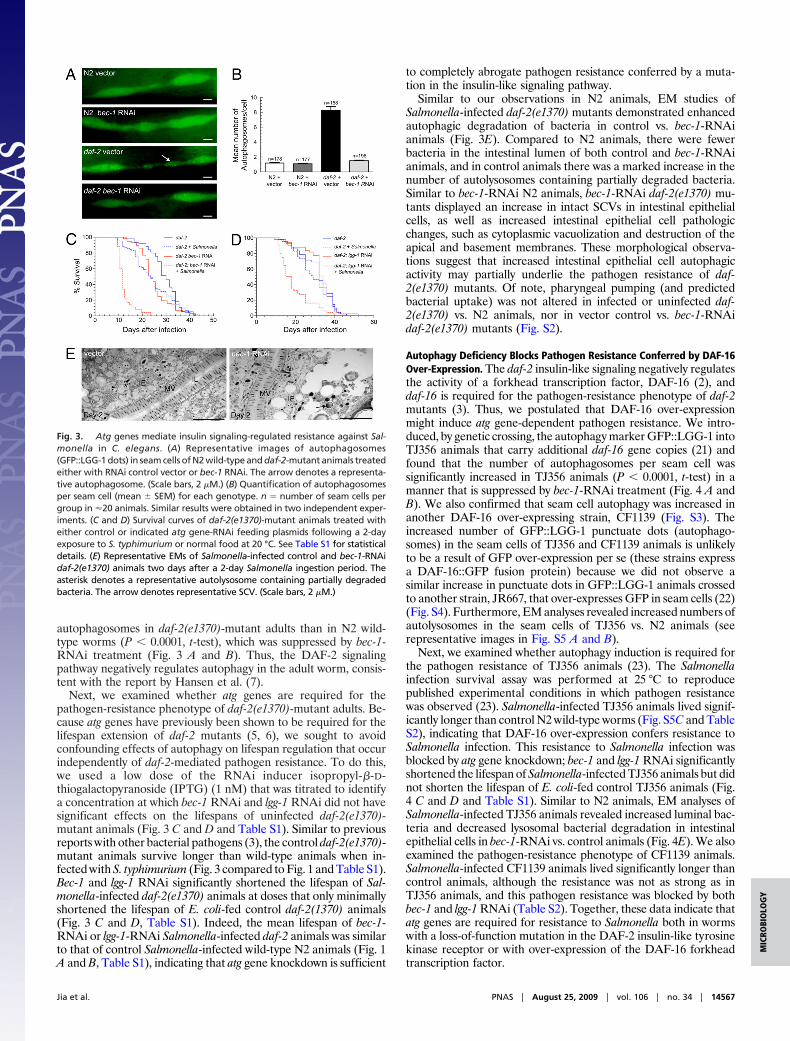
Similar to our observations in N2 animals, EM studies ofSalmonella-infected daf-2(e1370) mutants demonstrated enhancedautophagic degradation of bacteria in control vs. bec-1-RNAianimals (Fig. 3E). Compared to N2 animals, there were fewerbacteria in the intestinal lumen of both control and bec-1-RNAianimals, and in control animals there was a marked increase in thenumber of autolysosomes containing partially degraded bacteria.Similar to bec-1-RNAi N2 animals, bec-1-RNAi daf-2(e1370) mu-tants displayed an increase in intact SCVs in intestinal epithelialcells, as well as increased intestinal epithelial cell pathologicchanges, such as cytoplasmic vacuolization and destruction of theapical and basement membranes. These morphological observa-tions suggest that increased intestinal epithelial cell autophagicactivity may partially underlie the pathogen resistance of daf-2(e1370) mutants. Of note, pharyngeal pumping (and predictedbacterial uptake) was not altered in infected or uninfected daf-2(e1370) vs. N2 animals, nor in vector control vs. bec-1-RNAidaf-2(e1370) mutants (Fig. S2).
Autophagy Deficiency Blocks Pathogen Resistance Conferred by DAF-16Over-Expression. The daf-2 insulin-like signaling negatively regulatesthe activity of a forkhead transcription factor, DAF-16 (2), anddaf-16 is required for the pathogen-resistance phenotype of daf-2mutants (3). Thus, we postulated that DAF-16 over-expressionmight induce atg gene-dependent pathogen resistance. We intro-duced, by genetic crossing, the autophagy marker GFP::LGG-1 intoTJ356 animals that carry additional daf-16 gene copies (21) andfound that the number of autophagosomes per seam cell wassignificantly increased in TJ356 animals (P � 0.0001, t-test) in amanner that is suppressed by bec-1-RNAi treatment (Fig. 4 A andB). We also confirmed that seam cell autophagy was increased inanother DAF-16 over-expressing strain, CF1139 (Fig. S3). Theincreased number of GFP::LGG-1 punctuate dots (autophago-somes) in the seam cells of TJ356 and CF1139 animals is unlikelyto be a result of GFP over-expression per se (these strains expressa DAF-16::GFP fusion protein) because we did not observe asimilar increase in punctuate dots in GFP::LGG-1 animals crossedto another strain, JR667, that over-expresses GFP in seam cells (22)(Fig. S4). Furthermore, EM analyses revealed increased numbers ofautolysosomes in the seam cells of TJ356 vs. N2 animals (seerepresentative images in Fig. S5 A and B).
Next, we examined whether autophagy induction is required forthe pathogen resistance of TJ356 animals (23). The Salmonellainfection survival assay was performed at 25 °C to reproducepublished experimental conditions in which pathogen resistancewas observed (23). Salmonella-infected TJ356 animals lived signif-icantly longer than control N2 wild-type worms (Fig. S5C and TableS2), indicating that DAF-16 over-expression confers resistance toSalmonella infection. This resistance to Salmonella infection wasblocked by atg gene knockdown; bec-1 and lgg-1 RNAi significantlyshortened the lifespan of Salmonella-infected TJ356 animals but didnot shorten the lifespan of E. coli-fed control TJ356 animals (Fig.4 C and D and Table S1). Similar to N2 animals, EM analyses ofSalmonella-infected TJ356 animals revealed increased luminal bac-teria and decreased lysosomal bacterial degradation in intestinalepithelial cells in bec-1-RNAi vs. control animals (Fig. 4E). We alsoexamined the pathogen-resistance phenotype of CF1139 animals.Salmonella-infected CF1139 animals lived significantly longer thancontrol animals, although the resistance was not as strong as inTJ356 animals, and this pathogen resistance was blocked by bothbec-1 and lgg-1 RNAi (Table S2). Together, these data indicate thatatg genes are required for resistance to Salmonella both in wormswith a loss-of-function mutation in the DAF-2 insulin-like tyrosinekinase receptor or with over-expression of the DAF-16 forkheadtranscription factor.
Fig. 3. Atg genes mediate insulin signaling-regulated resistance against Sal-monella in C. elegans. (A) Representative images of autophagosomes(GFP::LGG-1 dots) in seam cells of N2 wild-type and daf-2-mutant animals treatedeither with RNAi control vector or bec-1 RNAi. The arrow denotes a representa-tive autophagosome. (Scale bars, 2 �M.) (B) Quantification of autophagosomesper seam cell (mean � SEM) for each genotype. n � number of seam cells pergroup in �20 animals. Similar results were obtained in two independent exper-iments. (C and D) Survival curves of daf-2(e1370)-mutant animals treated witheither control or indicated atg gene-RNAi feeding plasmids following a 2-dayexposure to S. typhimurium or normal food at 20 °C. See Table S1 for statisticaldetails. (E) Representative EMs of Salmonella-infected control and bec-1-RNAidaf-2(e1370) animals two days after a 2-day Salmonella ingestion period. Theasterisk denotes a representative autolysosome containing partially degradedbacteria. The arrow denotes representative SCV. (Scale bars, 2 �M.)
Jia et al. PNAS � August 25, 2009 � vol. 106 � no. 34 � 14567
MIC
ROBI
OLO
GY

DiscussionOur findings demonstrate that atg genes play an evolutionarilyconserved role in host defense against the mammalian intracellularbacterial pathogen, S. typhimurium. In C. elegans and Dictyostelium,the genetic knockdown or knockout of essential atg genes dramat-ically alters the fate of the invading bacterium and of the hostorganism. The invading bacterium, rather than being targeted forlysosomal degradation, establishes an intracellular replicative niche,which leads to cellular destruction and premature organismaldeath. Thus, in these two model organisms, the presence or absenceof atg genes is sufficient to determine whether Salmonella is asuccessful intracellular pathogen.
In mammals, Salmonella is phagocytosed by macrophages andcan invade nonphagocytic cells through the activity of the type-IIIsecretion system (24). Salmonella replicates intracellularly within aspecialized compartment, the SCV, and to be a successful intra-
cellular pathogen, Salmonella must avoid fusion of the SCV with thelysosome (24). Our results indicate that in both Dictyostelium andC. elegans, Salmonella invades intracellularly, resides in SCVs, andis targeted to lysosomes for degradation in wild-type but notautophagy-deficient organisms. Therefore, we propose that theautophagic machinery is used by these organisms to deliver Sal-monella to the lysosome. Autophagy likely plays a similar role inmammalian cells, as previous in vitro studies have shown thatautophagy inactivation enhances Salmonella replication in macro-phages (25). Perhaps a major difference in Salmonella infection inmammalian cells (where it replicates intracellularly) versus that inlower eukaryotic organisms (where it cannot replicate intracellu-larly) may relate to the ability of the bacterium to partially antag-onize the host autophagic machinery. This could explain whygenetic inactivation of the autophagic machinery in lower eu-karyotes (such as C. elegans or Dictyostelium) mimics Salmonellapathogenesis in mammalian cells, in that the bacteria can replicateinside and destroy intestinal epithelial cells and phagocytic cells.
Our findings clearly indicate that atg genes function in a cell-autonomous manner, in intestinal epithelial cells in the nematodeor in the unicellular organism, Dictyostelium, to limit intracellularbacterial replication and cellular destruction. In the presence ofgentamicin that kills extracellular bacteria, Salmonella only multi-plies successfully and only causes cytopathology when atg genes areinactivated. However, in C. elegans we cannot exclude multifactorialbases for increased bacterial multiplication or for increased animalmortality in autophagy-deficient organisms. We observed a defectin intestinal epithelial cell bacterial lysosomal degradation in atggene RNAi-treated animals, but increased bacterial replicationmight also result from enhanced epithelial cell bacterial uptake orinvasion or impaired intestinal epithelial cell secretion of antimi-crobial peptides. Indeed, in autophagy-deficient organisms, weobserved an increase in luminal bacteria, and in daf-2-mutantanimals (which have higher levels of basal autophagy), a decreasein luminal bacteria. Recently, mice defective in intestinal expressionof the atg genes, Atg16L1 or Atg5, were found to have defects in thesecretory function of the Paneth cell, a key cell type involved in gutantimicrobial peptide secretion (10). Interestingly, several genesencoding antibacterial lysozymes were induced in daf-2 mutants,including 2 intestinally expressed genes, lys-7 and lys-8 (26). Fur-thermore, genes encoding C. elegans antimicrobial peptides, abf-2and spp-1, are induced during Salmonella infection and required tolimit Salmonella infection in the intestinal lumen (27). Thus, it ispossible that, in addition to restricting intracellular bacterial mul-tiplication, autophagy is also involved in the production and secre-tion of antimicrobial peptides that limit intraluminal replication.
The mechanisms by which atg gene inactivation increases sus-ceptibility to lethal Salmonella infection are not completely under-stood. Ultrastructural analyses of both Salmonella-infected nema-todes and slime molds at later time points after infection revealvirtually complete cellular effacement by bacteria. Thus, one spec-ulation is that the high bacterial burden or harmful productssecreted by the bacteria leads to cytotoxicity. Although Salmonellahas been reported to cause apoptotic death of intestinal epithelialcells (28), we did not observe a role for apoptosis in the increasedlethality of Salmonella-infected autophagy-deficient nematodes.Another possibility is that, in addition to its role in decreasing theintracellular bacterial burden, autophagy, as a prosurvival mecha-nism, functions to ‘‘buffer’’ the stress imposed by intracellularbacterial infection.
Our findings not only identify an important role for atg genes inantibacterial host defense in vivo, they also identify atg genes asmediators of the pathogen-resistance phenotype of long-lived mu-tant nematodes. Pathogen resistance in daf-2-mutant animals re-quires at least two essential atg genes: bec-1 and lgg-1. Thus,autophagy is likely a specific innate effector that mediates not onlylifespan extension (5–7), but also pathogen resistance in nematodeswith mutation in the insulin-like signaling pathway. This connection
Fig. 4. Atg genes are required for DAF-16-mediated resistance to Salmonellainfection. (A) Representative pictures of autophagosomes in seam cells of N2wild-type and TJ356 animals treated either with RNAi control vector or bec-1RNAi. TJ356 is heterozygous for both gfp::lgg-1 and daf-16::gfp. The oval-shapedgreen staining near the seam cells in TJ356 animals represent nuclei of intestinalcells that express DAF-16::GFP. The arrow denotes a representative autophago-some. (Scale bars, 2 �M.) (B) Quantification of autophagosomes per seam cell(mean � SEM) for each genotype. n � number of seam cells per group in �20animals. Similar resultswereobtained intwoindependentexperiments. (CandD)Survival curves of TJ356 animals treated with either control or indicated atg geneRNAi-feeding plasmids following exposure to S. typhimurium or normal food at25 °C. n � number of animals per treatment group. See Table S1 for statisticaldetails. (E) Representative EMs of Salmonella-infected control and bec-1-RNAiTJ356 animals two days after a 2-day Salmonella ingestion period. The asteriskdenotes a representative autolysosome containing partially degraded bacteria.Arrowheads represent intestinal luminal bacteria and the arrow denotes repre-sentative SCV-containing bacteria. (Scale bars, 2 �M.)
14568 � www.pnas.org�cgi�doi�10.1073�pnas.0813319106 Jia et al.

between autophagy-mediated lifespan extension and pathogenresistance may be evolutionarily conserved. Perhaps age-relateddecreases in autophagy that occur in mammals may be mechanis-tically linked to age-related increases in susceptibility to certaininfectious diseases, including those caused by intracellular bacterialpathogens.
One apparent discrepancy between our findings and a previousreport (7) relates to the role of DAF-16 in autophagy regulation. Inthe present study, we found that two different DAF-16 over-expressing strains had increased levels of autophagy. However,Hansen et al. (7) found that a daf-16 null mutation did not blockautophagy induction in daf-2 mutants. One potential explanationfor this discrepancy is that another unidentified protein mayfunction redundantly with DAF-16 in autophagy regulation. Thismight explain why a daf-16 null mutation has no effect on autoph-agy, whereas DAF-16 over-expression induces autophagy. Furtherstudies are required to more clearly delineate the role of DAF-16,and other potential transcription factors downstream of DAF-2, inmediating autophagy and pathogen resistance in C. elegans. How-ever, of note, forkhead transcription factors seem to play anevolutionarily conserved role in autophagy induction; FOXO over-expression induces autophagy in Drosophila (29) and Foxo3 regu-lates autophagy in mouse muscle cells (30, 31).
In conclusion, our data demonstrate that autophagy is a criticalhost-defense mechanism that limits intracellular infection with thebacterial pathogen S. typhimurium. Based upon our findings innematode intestinal epithelial cells and in Dictyostelium, we spec-ulate that the autophagic machinery may play a conserved role inprotecting mammalian epithelial cells and phagocytes from bacte-rial attack. Furthermore, our data demonstrate a critical role forautophagy in mediating insulin-like signaling-regulated pathogenresistance in long-lived mutant nematodes. Thus, human atg genepolymorphisms (e.g., the Crohn’s ATG16L variant) or age-relatedchanges that reduce autophagy may contribute to impaired intes-tinal immunity to bacterial pathogens.
Materials and MethodsC. elegans, Dictyostelium, and Salmonella Strains. Wild-type strains were the C.elegans Bristol strain N2, the D. discoideum strain DH1, and Salmonella entericaSerovar Typhimurium ATCC14028s (S. typhimurium). All mutant strains havebeen previously published and are described in the SI Materials and Methods.
Strains TJ356 and CF1139 were out-crossed with our laboratory strain of N2before Salmonella infection experiments. See details in the SI Materials andMethods.
RNAi Methods. C. elegans feeding-RNAi experiments were performed as de-scribed (32), with the exception that a lower dose (1 nM instead of 1 mM) of IPTGwas used to induce expression of bec-1 RNAi in daf-2(e1370)-mutant animals andDAF-16 over-expression strains (TJ356 and CF1139) to avoid decreases in lifespanin uninfected animals. The construction of feeding-RNAi plasmids is described inthe SI Materials and Methods.
Salmonella Infection and Survival Studies. All C. elegans RNAi treatment, Salmo-nella infection, and lifespan experiments were performed at 20 °C unless other-wise indicated, as described in detail in the SI Materials and Methods. All survivalexperiments were repeated at least two times, and the results shown representan analysis of the combined data for the total number of animals per experimen-tal group in all experiments. The results of all individual experiments were similarto that of the combined data. The mean lifespans, total number of worms pergroup, total censored animals, and survival statistical analyses are listed in TablesS1 and S2. See the SI Materials and Methods for details of statistical methods.Salmonella infection and survival studies in Dictyostelium were performed asdescribed in the SI Materials and Methods.
Measurement of Bacterial Growth. See the SI Materials and Methods for furtherdetails of bacterial growth measurement.
Microscopic Analyses. For transmission EM of C. elegans, �100 adult nematodesper experimental group were collected and processed as previously described (5).Transmission EM analysis of Dictyostelium was performed as previously described(33). Light microscopic analyses of fluorescent nematodes and amoebae wereperformed using a Zeiss Axioplan2 Imaging microscope.
Autophagy Induction Analysis. N2- and daf-2(e1370)-transgenic strains carryingthe gfp::lgg-1 autophagy marker were described previously (5) and similar meth-ods were used to measure seam cell autophagy in young adults (i.e., within 12 hbeyond the L4 stage). The generation of TJ356 and CF1139 animals carrying thegfp::lgg-1 autophagy marker is described in the SI Materials and Methods.
ACKNOWLEDGMENTS. We thank Alejandro Aballay, Leon Avery, Scott Cameron,Simon Daefler, and Richard Kessin for providing reagents, Tom Januszewski forassistance with electron microscopy, and the Caenorhabditis Genetics Center for C.elegans strains used in this study. This work was supported by National Institutes ofHealth Grant RO1 AI051367 (to B.L.), National Institutes of Health Clinical Transla-tional Science Award Grant UL1 RRO24982 (to B.A.-H.), and an Ellison MedicalFoundation Senior Scholars Award in Infectious Diseases (to B.L.) and a EllisonMedical Foundation New Scholars Award in Aging (to K.J.).
1. Gavazzi G, Krause KH (2002) Ageing and infection. Lancet Infect Dis 2:659–666.2. Kenyon C (2005) The plasticity of aging: Insights from long-lived mutants. Cell 120:449–460.3. Garsin D, et al. (2003) Long-lived C. elegans daf-2 mutants are resistant to bacterial
pathogens. Science 300:1921.4. Levine B, Deretic V (2007) Unveiling the roles of autophagy in innate and adaptive
immunity. Nat Rev Immunol 7:767–777.5. Melendez A, et al. (2003) Autophagy genes are essential for dauer development and
lifespan extension in C. elegans. Science 301:1387–1391.6. Hars ES, et al. (2007) Autophagy regulates ageing in C. elegans. Autophagy 3:93–95.7. Hansen M, et al. (2008) A role for autophagy in the extension of lifespan by dietary
restriction in C. elegans. PLoS Genet 4:e24.8. Yano T, et al. (2008) Autophagic control of listeria through intracellular innate immune
recognition in Drosophila. Nat Immunol 9:908–916.9. MasseyDC,ParkesM(2007)Genome-wideassociationscanninghighlights twoautophagy
genes, ATG16L1 and IRGM, as being significantly associated with Crohn’s disease Auto-phagy 3:649–651.
10. DereticV,MasterS,SinghS(2008)Autophagygivesanodandawinktothe inflammasomeand Paneth cells in Crohn’s disease. Dev Cell 15:641–642.
11. Millet AC, Ewbank JJ (2004) Immunity in Caenorhabditis elegans. Curr Opin Immunol 16:4–9.12. Skriwan C, et al. (2002) Various bacterial pathogens and symbionts infect the amoeba
Dictyostelium discoideum. Int J Med Microbiol 291:615–624.13. VoetschAC,etal. (2004)FoodNetestimateof theburdenof illness causedbynontyphoidal
Salmonella infections in the United States. Clin Infect Dis 38(Suppl 3):S127–S134.14. Aballay A, Yorgey P, Ausubel FM (2000) Salmonella typhimurium proliferates and establishes
a persistent infection in the intestine of Caenorhabditis elegans. Curr Biol 10:1539–1542.15. Labrousse A, Chauvet S, Couillault C, Kurz CL, Ewbank JJ (2000) Caenorhabditis elegans is
a model host for Salmonella typhimurium. Curr Biol 10:1543–1545.16. Kurz CL, Tan MW (2004)Regulation of aging and innate immunity in C. elegans. Aging Cell
3:185–193.17. Takacs-Vellai K, et al. (2005) Inactivation of the autophagy gene bec-1 triggers apoptotic
cell death in C. elegans. Curr Biol 15:1513–1517.18. Aballay A, Ausubel FM (2001) Programmed cell death mediated by ced-3 and ced-4
protects Caenorhabditis elegans from Salmonella typhimurium-mediated killing. ProcNatl Acad Sci USA 98(5):2735–2739.
19. Otto GP, et al. (2004) Macroautophagy is dispensable for intracellular replication ofLegionella pneumophila in Dictyostelium discoideum. Mol Microbiol 51:63–72.
20. Golstein P, Aubry L, Levraud JP (2003) Cell-death alternative model organisms: Why andwhich? Nat Rev Mol Cell Biol 4:798–807.
21. Henderson ST, Johnson TE (2001) daf-16 integrates developmental and environmentalinputs to mediate aging in the nematode Caenorhabditis elegans. Curr Biol 11:1975–1980.
22. Terns RM, Kroll-Conner P, Zhu J, Chung S, Rothman JH (1997) A deficiency screen forzygotic loci required for establishment and patterning of the epidermis in Caenorhabditiselegans. Genetics 146:185–206.
23. Singh V, Aballay A (2006) Heat-shock transcription factor (HSF)-1 pathway required forCaenorhabditis elegans immunity. Proc Natl Acad Sci USA 103:13092–13097.
24. FinlayBB,Brumell JH(2000)Salmonella interactionswithhostcells: invitroto invivo.PhilosTrans R Soc Lond B Biol Sci 355:623–631.
25. Birmingham CL, Smith AC, Bakowski MA, Yoshimori T, Brumell JH (2006) Autophagycontrols Salmonella infection in response to damage to the Salmonella-containing vacu-ole. J Biol Chem 281:11374–11383.
26. Murphy CT, et al. (2003) Genes that act downstream of DAF-16 to influence the lifespanof Caenorhabditis elegans. Nature 424:277–283.
27. Alegado RA, Tan MW (2008) Resistance to antimicrobial peptides contributes topersistence of Salmonella typhimurium in the C. elegans intestine. Cell Microbiol10:1259–1273.
28. Guiney DG (2005) The role of host cell death in Salmonella infections. Curr Top MicrobiolImmunol 289:131–150.
29. Juhasz G, et al. (2007) Gene expression profiling identifies FKBP39 as an inhibitor ofautophagy in larval Drosophila fat body. Cell Death Differ 14:1181–1190.
30. Zhao J, et al. (2007) FoxO3 coordinately activates protein degradation by theautophagic/lysosomal and proteasomal pathways in atrophying muscle cells. CellMetab 6:472– 483.
31. MammucariC,etal. (2007)FoxO3controlsautophagy inskeletalmuscle invivo.CellMetab6:458–471.
32. Kamath RS, Martinez-Campos M, Zipperlen P, Fraser AG, Ahringer J (2001) Effectiveness ofspecific RNA-mediated interference through ingested double-stranded RNA in Caeno-rhabditis elegans. Genome Biol, 10.1186/gb-2000-2-1-research0002.
33. Solomon JM, Rupper A, Cardelli JA, Isberg RR (2000) Intracellular growth of Legionellapneumophila in Dictyostelium discoideum, a system for genetic analysis of host-pathogeninteractions. Infect Immun 68:2939–2947.
Jia et al. PNAS � August 25, 2009 � vol. 106 � no. 34 � 14569
MIC
ROBI
OLO
GY